


Abstract
Objective: The objective of this study is to evaluate the ability of silver oxide nanoparticle (Ag2ONP)-functionalized high-efficiency particulate air (HEPA) filters and colloidal Ag2ONP suspensions to inhibit biofilm formation by major respiratory pathogens causing infections at operating rooms. Background: Respiratory infections caused by bacterial pathogens such as Streptococcus pneumoniae, Pseudomonas aeruginosa and Staphylococcus species are often associated with the formation of biofilms, which confer increased resistance to antibiotics and host immune responses. Effective strategies to prevent biofilm formation on biological surfaces and in air filtration systems are urgently needed in clinical settings. Methods: The biofilm-forming ability of each bacterial strain was assessed by crystal violet microplate assay, viable count or confocal microscopy after prior incubation of the culture medium with Ag2ONP-coated HEPA filter material or colloidal Ag2ONP suspension. Results: Both silver-functionalized filters and silver nanoparticle suspensions significantly inhibited biofilm formation by S. pneumoniae and P. aeruginosa, with near-complete suppression observed. In the case of S. aureus and S. epidermidis, the silver nanoparticle suspension showed partial inhibition of biofilm development. Conclusions: Ag2ONP-functionalized HEPA filters and colloidal Ag2ONP suspensions effectively prevent biofilm formation by major respiratory pathogens, for both Gram-negative and Gram-positive bacteria. These materials show promise for integration with air filtration and surface coating systems to reduce microbial load and transmission in healthcare environments such as operating room facilities.
1. Introduction
Airborne transmission of pathogenic microorganisms represents a major public health concern, contributing to diseases such as pneumonia, meningitis, and asthma. Susceptibility varies according to age, occupational exposure, environmental factors, and individual health status [1]. Indoor air filtration via heating, ventilation, and air-conditioning (HVAC) systems is widely used to mitigate exposure to airborne allergens and pathogens [2,3,4]. High-efficiency particulate air (HEPA) filters are deployed in operating rooms and biological laboratories for their superior particle- and microorganism-removal capacity [5]. The protective efficacy of these systems relies on proper installation, regular inspection, and timely filter replacement. Humidity and organic matter accumulation can facilitate microbial adhesion and biofilm formation on ducts and filters, creating persistent reservoirs of pathogens. Poorly maintained HEPA systems may paradoxically contribute to the airborne spread of clinically significant microorganisms and can be a major cause of infections among patients and staff [6,7].
Pathogens such as Pseudomonas aeruginosa, Staphylococcus aureus, or Streptococcus pneumoniae can form biofilms on these filters. As they are damp or poorly ventilated surfaces, the air may be loaded with organic particles such as dust, or there may be an accumulation of debris in these ducts [8,9]. All these aspects enhance the formation of biofilms by these pathogens, increasing the risk of suffering from hospital-associated infections, which are of great concern as these microorganisms growing as biofilms are more resistant to antimicrobial treatment and hospital-grade disinfectant agents used in operating rooms [10].
Beyond treatment, preventive strategies aimed at limiting biofilm establishment are critical, particularly in high-risk clinical environments such as operating rooms and on implantable medical devices, where persistent biofilms can readily develop [11,12]. To mitigate microbial persistence, antimicrobial surface modification strategies have gained considerable attention. Silver and silver oxide nanoparticles (Ag2ONPs) exhibit unique physicochemical properties that underpin their broad-spectrum antibacterial activity and position them as promising candidates to address the growing challenge of antimicrobial resistance. Their antimicrobial efficacy is largely associated with the sustained release of Ag+ ions, which promote multiple bactericidal mechanisms, including the disruption of membrane integrity, increased permeability, the generation of reactive oxygen species (ROS), the inhibition of protein synthesis, and interference with DNA replication [13]. Accumulating evidence supports the effectiveness of Ag2ONPs against a wide range of clinically relevant pathogens, including multidrug-resistant and antibiotic-resistant strains [14,15].
This study aimed to characterize the antibacterial capacity of colloidal Ag2ONPs suspensions and HEPA filters functionalized with these Ag2ONPs to prevent the growth and biofilm formation of clinically relevant pathogens.
2. Results
2.1. Antimicrobial Effect of Ag2ONP Suspensions Against Biofilms of Different Species
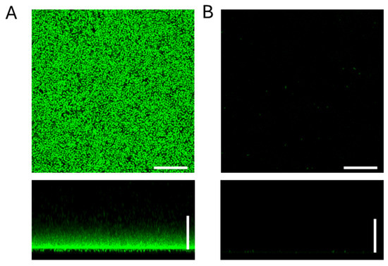
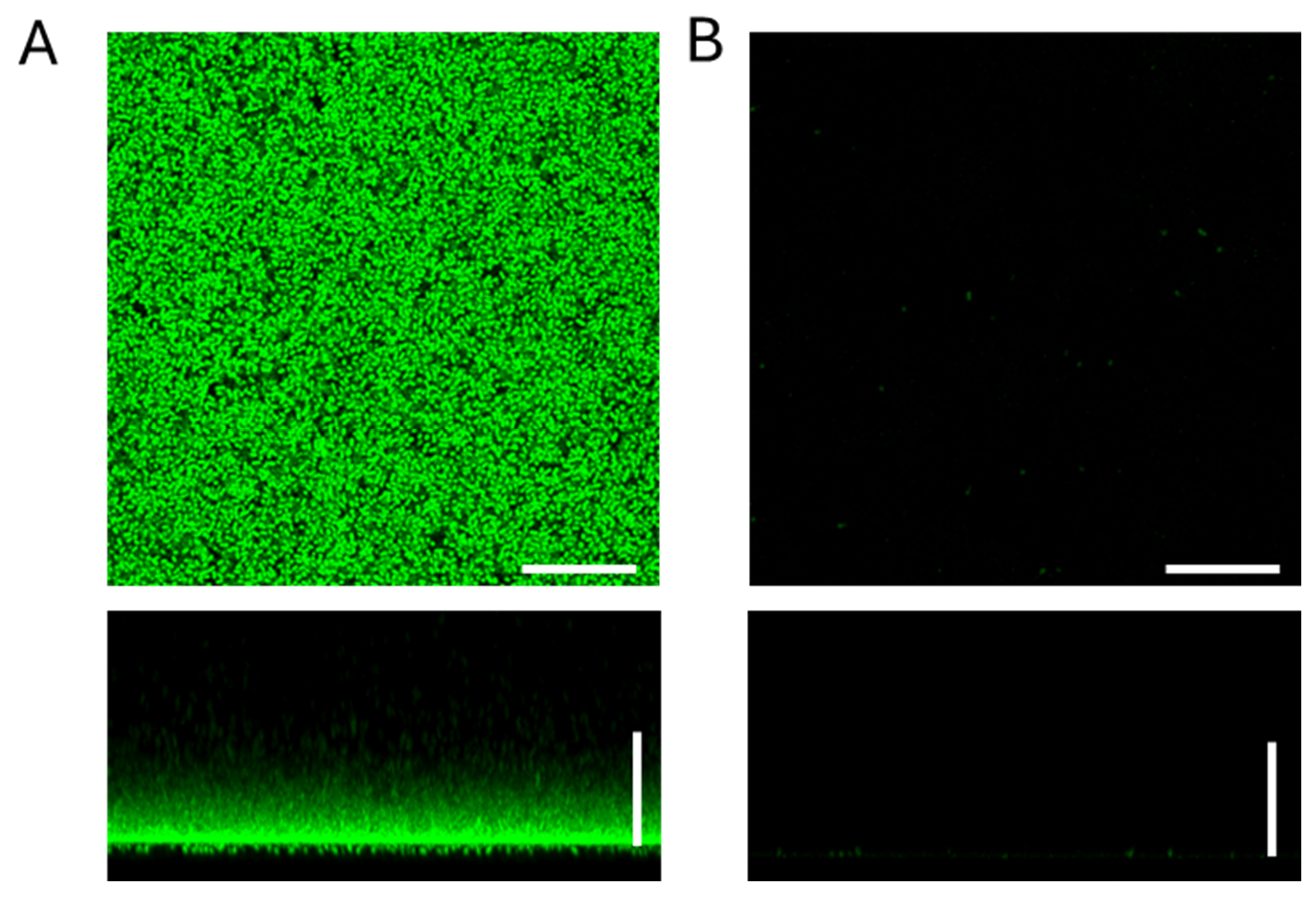
The first step was to determine the antibacterial effect of the Ag2ONP suspension against the biofilms of four different bacteria. Hence, we tested the 0.1 M Ag2ONP suspension evaluating its antimicrobial activity by measuring viable bacterial counts (Table 1) whereas images of biofilm degradation were observed by confocal microscopy (Figure 1). In the case of S. pneumoniae, we included two different strains, observing a complete inhibition of biofilm viability compared to the control group lacking Ag2ONPs (Table 1).
Table 1.
Inhibition of biofilm of different strains with 0.1 M Ag2ONP suspension.
Figure 1.
CLSM images of biofilm inhibition of S. pneumoniae strain R6 with 0.1 M Ag2ONs suspension. The biofilm cells were stained with the BacLight LIVE/DEAD viability kit, with viable cells appearing green (Syto9) and non-viable cells appearing red (IP). (A) X–Y projections (images captured at 0.5 μm intervals) and X–Z projections (images captured at 5.0 μm intervals) of strain R6 in C + Y medium (control). (B) X–Y projections (images captured at 0.5 μm intervals) and X–Z projections (images captured at 5.0 μm intervals) of strain R6 with 0.1 M AgNP suspension. The bars indicate 25 μm.
This strong activity was confirmed by confocal microscopy studies as the bacterial structure of the pneumococcal biofilm was completely destroyed in the presence of the Ag2ONPs (Figure 1). The same antimicrobial effect in terms of viable bacterial counts was observed for the P. aeruginosa strain with a fully inhibition of the biofilm in the presence of Ag2ONPs (Table 1). In the case of methicillin-resistant S. aureus (MRSA) and Staphylococcus epidermidis strains, we also observed a marked reduction between 3 and 5 Log-units (1.77 × 10⁹ vs. 6.06 × 10⁵ for MRSA and 1.78 × 10¹¹ vs. 1.58 × 10⁶ for S. epidermidis), although we did not achieve a full inhibition as we obtained for S. pneumoniae and P. aeruginosa (Table 1).
2.2. Ag2ONP-Functionalized HEPA Filters Effectively Inhibit Biofilm Formation by Streptococcus pneumoniae and Pseudomonas aeruginosa
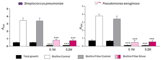
The antimicrobial activity of Ag2ONP-functionalized HEPA filters (Figure 2) was evaluated using S. pneumoniae and P. aeruginosa selected as representative Gram-positive and Gram-negative respiratory pathogens associated with biofilm formation. Two silver concentrations (0.1 and 0.2 M) were tested. Silver functionalization markedly affected bacterial growth. Complete growth inhibition was observed for both pathogens, even at the lower concentration (0.1 M) (Figure 2).
Figure 2.
Inhibition of S. pneumoniae strain YNM4 (left) and P. aeruginosa strain ATCC27853 (right) biofilm with Ag2ONPs-functionalized HEPA filters. C + Y or LB bacterial culture medium was previously incubated at 37 °C under agitation for 24 h with each of the functionalized filters. The strain was inoculated in each of these media, incubated for 5 h at 34 °C or 24 h at 37 °C. The black bars indicate total growth (adherent (biofilm) plus non-adherent (planktonic) cells). The biofilm was processed by staining with crystal violet for 15 min, washed 3 times with distilled water, solubilized with ethanol and then measured for absorbance at 595 or 600 nm. Medium with filter functionalized with 0.2 M silver (Filter Ag 0.2 M; Dark Pink), 0.1 M silver (Filter Ag 0.1 M; Light Pink), unfunctionalized filter (Filter Control; Grey) and medium without filter (Control; White). Assays were performed in triplicates, including at least three independent experiments. Standard deviation bars are shown, and asterisks mark results that are statistically significant (two-tailed Student’s t test: ***, p < 0.001).
The impact of HEPA filters on biofilm formation was also assessed. Control filters lacking silver functionalization showed no effect on bacterial growth or biofilm development. In contrast, Ag2ONP-functionalized HEPA filters resulted marked reduced biofilm formation for both S. pneumoniae and P. aeruginosa (Figure 2).
3. Discussion
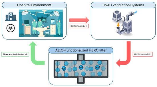
The treatment of bacterial biofilms remains a major clinical challenge due to their intrinsic tolerance to antimicrobial agents and their enhanced capacity to evade host immune responses compared with planktonic cells [16]. Conventional antibiotics, when used alone, are often ineffective against established biofilms and may contribute to therapeutic failure. Consequently, alternative antibiofilm strategies have been explored, including enzibiotics, antioxidant or nanoparticles–antibiotic combinations, and nanoparticle-based approaches [17,18]. For example, AgNPs combined with antibiotics have demonstrated efficacy against bacterial biofilms in chronic rhinosinusitis models [19] and to inhibit the growth of the different bacterial pathogens [20,21]. Previous studies suggest that combining physical filtration with active biocidal properties represents a promising strategy to minimize biofilm formation on inert surfaces. For example, chlorhexidine-coated air filters and nanocellulose filters incorporating silver and MXene particles have demonstrated broad-spectrum antimicrobial activity against bacteria, fungi, and viruses [22,23]. Similarly, AgNP biocomposites embedded in microfibrillated cellulose have shown bactericidal activity against both susceptible and resistant strains, offering environmentally adaptable options for surface coatings and medical devices [24]. In this context, HEPA filters functionalized with metal oxide agents have been reported to inhibit the growth of S. pneumoniae and P. aeruginosa, highlighting their potential role in reducing microbial burden in controlled environments [25]. Therefore, the implementation of silver oxide nanoparticle-based filters in HVAC systems to improve air quality in operating rooms and hospital environments is feasible (Figure 3), as this system would be capable to prevent biofilm formation by clinically relevant pathogens.
Figure 3.
Diagram showing how silver oxide nanoparticle-based filters in HVAC can help improve air quality in the hospital environment. Created by BioRender.com.
The present study demonstrates that Ag2ONP suspensions and Ag2ONP-functionalized HEPA filters effectively prevent biofilm formation, particularly in S. pneumoniae and P. aeruginosa. The Ag2ONP suspension achieved complete inhibition of biofilm development in both species, whereas functionalized filters produced a marked but comparatively lower inhibitory effect. This difference may be attributed to the higher immediate bioavailability of Ag+ ions in suspension, facilitating direct and sustained interaction with bacterial cells. In contrast, silver immobilized on filter surfaces likely provides a slower or more localized ion release. While this controlled release may be advantageous in airflow systems such as hospital operating rooms, it may exhibit reduced activity against mature or structurally complex biofilms [26].
The Ag2ONP suspension also inhibited biofilm formation by MRSA and S. epidermidis, although to a lesser extent than observed for S. pneumoniae and P. aeruginosa. These differences may be explained by variations in biofilm matrix composition and architecture. Biofilms formed by Staphylococcus spp. are characterized by dense intercellular polysaccharide networks with limited pore size, which may restrict nanoparticle diffusion [27,28]. In contrast, the water-channel architecture of P. aeruginosa biofilms [29] and the comparatively hydrated, eDNA- and protein-rich matrix of S. pneumoniae facilitates greater nanoparticle penetration [30].
A key limitation of this study is the use of a static in vitro biofilm model that evaluates the activity of medium conditioned by contact with the filters, rather than their performance under dynamic airflow conditions, thereby highlighting a gap between the in vitro model and real-world air filtration applications. Although this approach enables controlled assessment of antimicrobial activity, it does not fully replicate dynamic clinical conditions, such as continuous airflow, fluctuations in humidity, or interactions with host tissues and polymicrobial communities [31,32,33]. Future studies should focus on optimizing filter functionalization to achieve controlled and sustained silver ion release, mechanical stress, or disposal processes could potentially lead to particle or ion release. Therefore, while our results demonstrate promising antibacterial and antibiofilm performance, further studies are needed to evaluate long-term stability, potential Ag+ release, and environmental impact under realistic operating conditions [34]. Validation under dynamic conditions, such as airflow chambers or simulated hospital environments, will be essential to determine real-world applicability. Additionally, integrating synergistic agents—including antimicrobial peptides, biofilm-dispersing enzymes, or nanoparticles containing complementary metals—may enhance antibiofilm efficacy while reducing the silver concentration required, thereby minimizing potential toxicological concerns and production costs [35].
Overall, our findings support the potential of Ag2ONP-functionalized HEPA filters as a preventive strategy to reduce bacterial colonization and biofilm formation in settings where air quality and surface sterility are critical, including hospitals, operating rooms, and medical ventilation systems. The strong antibiofilm activity observed with Ag2ONP suspensions, even against resistant strains such as MRSA, further highlights their potential as complementary antimicrobial agents, although their application beyond air filtration systems requires further investigation.
4. Materials and Methods
4.1. Strains, Media and Culture Conditions
For this study we used five strains of Gram-positive bacteria and one Gram-negative bacterial strain (Table 2). Among the five Gram-positive strains, three of them were S. pneumoniae, and the other two were S. aureus and S. epidermidis.
Table 2.
Strains used in this study with their description.
For broth cultures, the pneumococcal strains were grown in CpH8 medium supplemented with 0.08% yeast extract (C + Y medium), Staphylococcal strains were grown in Tryptic Soy Broth medium supplemented with glucose (0.4%) and yeast extract (0.3%) (TSBGY medium) and the P. aeruginosa strain was grown in Luria–Bertani broth (LB medium). Growth was controlled by measuring the optical density at 550 nm for S. pneumoniae, S. aureus and S. epidermidis strains, whereas for P. aeruginosa we measured at 600 nm. All strains were preserved in their specific medium and stored in frozen aliquots at −80 °C.
4.2. Filter Functionalization, Characterization and Preparation of AgNP Suspensions
Commercially available H13 (EN 1822) HEPA filter (Venfilter, Barcelona, Spain) and 0.1 M and 0.2 M colloidal suspensions of Ag2 particles, were prepared, functionalized, and characterized as previously described [25]. Once the particles were collected through vacuum filtration, they were washed twice with dH2O and then dried at 150 °C for 1 h in vacuum to evaporate any residual water and convert any resulting hydroxide into the oxide form. Ag2ONPs were produced by chemical reduction of silver nitrate (AgNO3) with potassium hydroxide (KOH), and 0.1 M and 0.2 M colloidal suspensions in DI H2O were prepared to a final volume of 25 mL. Glass Fibre HEPA filter media was cut and removed in a clean environment, cleaned with a nitrogen (N2) stream and positioned over filter paper with tape. The spraying process was carried out with a mini-handheld spray gun (Sagola 474, Vitoria-Gasteiz, Spain), at a normal position with respect to the filter media, distanced at approximately 15 cm height from surface. Each colloidal suspension was sprayed onto the filter for 10 s per area, with two repetitions. After coating deposition, the filter dried in air at room temperature and then was cut into 2 × 2 cm2 squares for further characterization. Sample morphology was characterized by scanning electron microscopy (Apreo 2, ThermoFischer Scientific, Waltham, MA, USA) at low vacuum and an applied voltage of 5–10 kV. Chemical composition was determined by energy dispersive spectroscopy (EDS, UltimMax 40 from Oxford Instruments (Oxford, UK), using AZtec Software for acquisition and data processing).
Prior to biological testing, all samples were sterilized using low-temperature (<40 °C) ethylene oxide in an ANPROLENE AN75 sterilizer (Andersen Sterilizers, Haw River, NC, USA), with a gas dwell time of approximately 12 h, relative humidity of ~50%, using EO cartridges containing 87% ethylene oxide as the active ingredient.
4.3. Biofilm Inhibition Tests
Filter extracts were prepared by incubating the different filters (functionalized with Ag2ONPs at 0.1 M and 0.2 M, as well as non-functionalized control filters) in culture media (LB, TSBGY, or C + Y) for 24 h at 37 °C under shaking conditions. After incubation, the resulting media, containing soluble components released from the filters, were collected and used as filter extracts for subsequent experiments. The strains were incubated at 37 °C in the corresponding medium (LB, TSBGY or C + Y) to an optical density of 0.5–0.6 at 550 nm for S. pneumoniae, S. aureus, and S. epidermidis strains and to an optical density of 1 at 600 nm for P. aeruginosa strain. The culture was then diluted to an optical density corresponding to 4.5 × 10⁶ Colony-Forming Unit (CFU)/mL in fresh medium with filter extract (previously incubated with both the functionalized filters or non-functionalized filter) and in fresh medium without filters as control. Then, 200 μL per well was added to a 96-well polystyrene microtiter plate (Costar 3595; Corning, Corning, NY, USA), and plates were incubated 5 h at 34 °C for pneumococcal strains and 24 h at 37 °C for all other strains. After the incubation, the biofilm was processed by staining with crystal violet as previously described [36,37]. Briefly, total growth (adhered and not adhered cells) was measured (595 nm by S. pneumoniae/600 nm by P. aeruginosa) using the BioTek Epoch 2 reader (Winooski, VT, USA) and then CV (1%) was added for 15 min, washed three times with distilled water, solubilized with ethanol and then measured the biofilm biomass at 595 nm (A595) or 600 nm (A600).
In addition, the Ag2ONP suspensions with which the filters were functionalized were used to perform biofilm inhibition tests in which cell viability was determined. For this purpose, the 0.2 M Ag2ONP suspension was incubated with the corresponding culture medium as previously described in volume–volume ratio at 37 °C for 24 h with shaking. Thus, the silver concentration would be 0.1 M. A non-preincubated culture medium was used as a control. The supernatant of this suspension or non-preincubated culture medium was then used for the biofilm inhibition test. After the corresponding incubations of the biofilm, the supernatant was removed, 100 µL per well of PBS was added, the biofilm was homogenized, and serial dilutions were made and subsequently plated on blood agar, Tryptic Soy Agar, or LB plates.
4.4. Confocal Laser Scanning Microscopy
For CLSM assays, S. pneumoniae strain R6 was used. A culture of the strain grown in its corresponding medium to an optical density of 0.5–0.6 at 550 nm and diluted on one side in the same medium as the control and on the other side in the medium pre-incubated with the silver oxide nanoparticles to have 4.5 × 10⁶ CFU/mL was inoculated in 200 µL on a plate (µ-Slide 4 Well Glass Bottom, Ibidi, Gräfelfing, Germany). They were incubated at 37 °C for 24 h. After the incubation period, the sample was washed with water and stained for 15 min at room temperature with the BacLight LIVE/DEAD viability kit, with viable cells appearing green (Syto9) and non-viable cells appearing red (IP). We observed the samples with the Leica spectral SP5 confocal microscope at 1024 × 1024 700 Hz; the images were analyzed using the LAS AF software.
4.5. Statistical Analysis
Data presented represent results obtained from at least three repeated independent experiments, representing at least three replicates. Statistical analyses were performed using GraphPad InStat v. 8.0 (GraphPad Software, La Jolla, CA, USA). For comparison of two groups, we used the two-tailed Student’s t-test. Differences were considered statistically significant at *, p < 0.001.
Author Contributions
Conceptualization, J.Y., J.S., M.E.-R. and M.D.; Methodology, B.F.G.-R. and M.L.; Formal Analysis, M.L.; Writing—Original Draft Preparation, M.L.; Writing—Review and Editing, M.L., M.D., J.S., M.E.-R., B.F.G.-R. and J.Y.; Supervision, M.D., J.S. and J.Y.; Project Administration, M.E.-R., J.Y. and M.D.; Funding Acquisition, J.Y. and M.D. All authors have read and agreed to the published version of the manuscript.
Funding
This research was funded by the Spanish Ministry of Science, Innovation and Universities (grant PID2024-161570OB-I00) and by Instituto de Salud Carlos III (grant PI24CIII/00045). Additionally, this study was supported by the project MAMAP, funded by the REACT-EU call from the Comunidad de Madrid and the European Regional Development Fund, and by the project (MAD2D-CM)-IMDEA Materials funded by Comunidad de Madrid, by the Recovery, Transformation and Resilience Plan and by NextGenerationEU. M. Echeverry-Rendón acknowledges the support of the Comunidad de Madrid through the “Talento César Nombela” Project (2023-T1/TEC-29099).
Institutional Review Board Statement
Not applicable.
Informed Consent Statement
Not applicable.
Data Availability Statement
All epidemiological and experimental data requests should be submitted to J.S. (jsempere@isciii.es) or M.D. (miriam.domenech@isciii.es). Requests will be assessed for scientific rigour before being granted, and a data-sharing agreement might be required.
Conflicts of Interest
All authors declare no conflicts of interest.
Abbreviations
The following abbreviations are used in this manuscript:
| AgNPs | Silver Nanoparticles |
| ATCC | American Type Culture Collection |
| CECT | Spanish Collection of Standard Crops |
| CFU | Colony-Forming Unit |
| CLSM | Confocal laser scanning microscopy |
| C + Y | CpH8 medium supplemented with 0.08% yeast extract |
| HEPA | High-Efficiency Particulate Air |
| LB | Luria–Bertani broth |
| MRSA | Methicillin-resistant Staphylococcus aureus |
| TSBGY | Tryptic Soy Broth medium supplemented with glucose (0.4%) and yeast extract (0.3%) |
References
- Al-Shaarani, A.A.Q.A.; Pecoraro, L. A review of pathogenic airborne fungi and bacteria: Unveiling occurrence, sources, and profound human health implication. Front. Microbiol. 2024, 15, 1428415. [Google Scholar] [CrossRef] [Scilit] [PubMed]
- Bolashikov, Z.D.; Melikov, A.K. Methods for air cleaning and protection of building occupants from airborne pathogens. Build. Environ. 2009, 44, 1378–1385. [Google Scholar] [CrossRef] [Scilit] [PubMed]
- Gherasim, A.; de Blay, F. Does Air Filtration Work for Cat Allergen Exposure? Curr. Allergy Asthma Rep. 2020, 20, 18. [Google Scholar] [CrossRef] [Scilit] [PubMed]
- Vijayan, V.; Paramesh, H.; Salvi, S.; Dalal, A.K. Enhancing indoor air quality—The air filter advantage. Lung India 2015, 32, 473. [Google Scholar] [CrossRef] [Scilit]
- Mittal, H.; Parks, S.R.; Pottage, T.; Walker, J.T.; Bennett, A.M. Survival of Microorganisms on HEPA Filters. Appl. Biosaf. 2011, 16, 163–166. [Google Scholar] [CrossRef] [Scilit]
- Lee, S.-T.; Liang, C.-C.; Chien, T.-Y.; Wu, F.-J.; Fan, K.-C.; Wan, G.-H. Effect of ventilation rate on air cleanliness and energy consumption in operation rooms at rest. Environ. Monit. Assess. 2018, 190, 178. [Google Scholar] [CrossRef] [Scilit]
- Wu, H.T.; Li, Q.S.; Dai, R.C.; Liu, S.; Wu, L.; Mao, W.; Ji, C.H. Effects of air-conditioning systems in the public areas of hospitals: A scoping review. Epidemiol. Infect. 2021, 149, e201. [Google Scholar] [CrossRef] [Scilit]
- Patra, S.; Saha, S.; Singh, R.; Tomar, N.; Gulati, P. Biofilm battleground: Unveiling the hidden challenges, current approaches and future perspectives in combating biofilm associated bacterial infections. Microb. Pathog. 2025, 198, 107155. [Google Scholar] [CrossRef] [Scilit]
- Ortiz, G.; Yagüe, G.; Segovia, M.; Catalán, V. A Study of Air Microbe Levels in Different Areas of a Hospital. Curr. Microbiol. 2009, 59, 53–58. [Google Scholar] [CrossRef] [Scilit]
- Abdelhamid, A.G.; Yousef, A.E. Combating Bacterial Biofilms: Current and Emerging Antibiofilm Strategies for Treating Persistent Infections. Antibiotics 2023, 12, 1005. [Google Scholar] [CrossRef] [Scilit]
- Falde, E.J.; Yohe, S.T.; Colson, Y.L.; Grinstaff, M.W. Superhydrophobic materials for biomedical applications. Biomaterials 2016, 104, 87–103. [Google Scholar] [CrossRef] [Scilit] [PubMed]
- Paul, S.; Rao, L.; Stein, L.H.; Salemi, A.; Mitra, S. Development of a Carbon Nanotube-Enhanced FAS Bilayer Amphiphobic Coating for Biological Fluids. Nanomaterials 2023, 13, 3138. [Google Scholar] [CrossRef] [Scilit] [PubMed]
- Karataş, H.; Eker, F.; Akdaşçi, E.; Bechelany, M.; Karav, S. Silver Nanoparticles in Antibacterial Research: Mechanisms, Applications, and Emerging Perspectives. Int. J. Mol. Sci. 2026, 27, 927. [Google Scholar] [CrossRef] [Scilit] [PubMed]
- Xie, N. Synthesis and antibacterial effects of silver nanoparticles (AgNPs) against multi-drug resistant bacteria. Biomed. Mater. Eng. 2024, 35, 451–463. [Google Scholar] [CrossRef] [Scilit]
- de Lacerda Coriolano, D.; de Souza, J.B.; Bueno, E.V.; dos Santos Medeiros, S.M.d.F.R.; Cavalcanti, I.D.L.; Cavalcanti, I.M.F. Antibacterial and antibiofilm potential of silver nanoparticles against antibiotic-sensitive and multidrug-resistant Pseudomonas aeruginosa strains. Braz. J. Microbiol. 2021, 52, 267–278. [Google Scholar] [CrossRef] [Scilit]
- Domenech, M.; Ramos-Sevillano, E.; García, E.; Moscoso, M.; Yuste, J. Biofilm formation avoids complement immunity and phagocytosis of Streptococcus pneumoniae. Infect. Immun. 2013, 81, 2606–2615. [Google Scholar] [CrossRef] [Scilit]
- Domenech, M.; García, E.; Moscoso, M. In Vitro Destruction of Streptococcus pneumoniae Biofilms with Bacterial and Phage Peptidoglycan Hydrolases. Antimicrob. Agents Chemother. 2011, 55, 4144–4148. [Google Scholar] [CrossRef] [Scilit]
- Vázquez, R.; García, P. Synergy Between Two Chimeric Lysins to Kill Streptococcus pneumoniae. Front. Microbiol. 2019, 10, 1251. [Google Scholar] [CrossRef] [Scilit]
- Feizi, S.; Cooksley, C.M.; Nepal, R.; Psaltis, A.J.; Wormald, P.-J.; Vreugde, S. Silver nanoparticles as a bioadjuvant of antibiotics against biofilm-mediated infections with methicillin-resistant Staphylococcus aureus and Pseudomonas aeruginosa in chronic rhinosinusitis patients. Pathology 2022, 54, 453–459. [Google Scholar] [CrossRef] [Scilit]
- Khatoon, N.; Alam, H.; Khan, A.; Raza, K.; Sardar, M. Ampicillin Silver Nanoformulations against Multidrug resistant bacteria. Sci. Rep. 2019, 9, 6848. [Google Scholar] [CrossRef] [Scilit]
- Halawani, E.M.; Hassan, A.M.; Gad El-Rab, S.M. Nanoformulation of Biogenic Cefotaxime-Conjugated-Silver Nanoparticles for Enhanced Antibacterial Efficacy Against Multidrug-Resistant Bacteria and Anticancer Studies. Int. J. Nanomed. 2020, 15, 1889–1901. [Google Scholar] [CrossRef] [Scilit]
- Qin, X.; Xiong, Y.; Xuan, S.; Kang, J.; Wang, D.; Wang, H.; Wang, L.; Wu, Z. Nanocellulose-reinforced air filter with gradient hierarchical structure for highly effective and reuseable antibacterial air filtration. J. Membr. Sci. 2024, 693, 122340. [Google Scholar] [CrossRef] [Scilit]
- Watson, R.; Oldfield, M.; Bryant, J.A.; Riordan, L.; Hill, H.J.; Watts, J.A.; Alexander, M.R.; Cox, M.J.; Stamataki, Z.; Scurr, D.J.; et al. Efficacy of antimicrobial and anti-viral coated air filters to prevent the spread of airborne pathogens. Sci. Rep. 2022, 12, 2803. [Google Scholar] [CrossRef] [Scilit] [PubMed]
- Garza-Cervantes, J.A.; Mendiola-Garza, G.; de Melo, E.M.; Dugmore, T.I.J.; Matharu, A.S.; Morones-Ramirez, J.R. Antimicrobial activity of a silver-microfibrillated cellulose biocomposite against susceptible and resistant bacteria. Sci. Rep. 2020, 10, 7281. [Google Scholar] [CrossRef] [Scilit] [PubMed]
- Ribeiro, B.; Vázquez-López, A.; Vazquez-Pufleau, M.; Llamosí, M.; Sempere, J.; Yuste, J.; Domenech, M.; Wang, D.Y.; Vilatela, J.J.; Llorca, J.; et al. Control of microbial agents by functionalization of commercial air filters with metal oxide particles. Mater. Chem. Phys. 2024, 313, 128684. [Google Scholar] [CrossRef] [Scilit]
- Gallus, I.J.; Boyraz, E.; Maryška, J. Antimicrobial Properties of Nanofiber Membrane and Commercial Micromembrane by Modification with Diethylenetriamine (DETA) and Attachment of Silver Nanoparticles. J. Nanomater. 2023, 2023, 8927774. [Google Scholar] [CrossRef] [Scilit]
- Stewart, P.S. Diffusion in Biofilms. J. Bacteriol. 2003, 185, 1485–1491. [Google Scholar] [CrossRef] [Scilit]
- Feng, Q.L.; Wu, J.; Chen, G.Q.; Cui, F.Z.; Kim, T.N.; Kim, J.O. A mechanistic study of the antibacterial effect of silver ions on Escherichia coli and Staphylococcus aureus. J. Biomed. Mater. Res. 2000, 52, 662–668. [Google Scholar] [CrossRef] [Scilit]
- Karavolos, M.H.; Winzer, K.; Williams, P.; Khan, C.M.A. Pathogen espionage: Multiple bacterial adrenergic sensors eavesdrop on host communication systems. Mol. Microbiol. 2013, 87, 455–465. [Google Scholar] [CrossRef] [Scilit]
- Bibbs, R.K.; Harris, R.D.; Peoples, V.A.; Barnett, C.; Singh, S.R.; Dennis, V.A.; Coats, M.T. Silver polyvinyl pyrrolidone nanoparticles exhibit a capsular polysaccharide influenced bactericidal effect against Streptococcus pneumoniae. Front. Microbiol. 2014, 5, 665. [Google Scholar] [CrossRef] [Scilit]
- Bjarnsholt, T.; Alhede, M.; Alhede, M.; Eickhardt-Sørensen, S.R.; Moser, C.; Kühl, M.; Jensen, P.Ø.; Høiby, N. The in vivo biofilm. Trends Microbiol. 2013, 21, 466–474. [Google Scholar] [CrossRef] [Scilit]
- Malysheva, A.; Ivask, A.; Doolette, C.L.; Voelcker, N.H.; Lombi, E. Cellular binding, uptake and biotransformation of silver nanoparticles in human T lymphocytes. Nat. Nanotechnol. 2021, 16, 926–932. [Google Scholar] [CrossRef] [Scilit]
- Gao, X.; Li, R.; Yourick, J.J.; Sprando, R.L. Transcriptomic and proteomic responses of silver nanoparticles in hepatocyte-like cells derived from human induced pluripotent stem cells. Toxicol. In Vitro 2022, 79, 105274. [Google Scholar] [CrossRef] [Scilit]
- Li, F.; Tan, J.; Yang, Q.; He, M.; Yu, R.; Liu, C.; Zhou, X. Multi-Endpoint Toxicity Tests and Effect-Targeting Risk Assessment of Surface Water and Pollution Sources in a Typical Rural Area in the Yellow River Basin, China. Chemosensors 2022, 10, 502. [Google Scholar] [CrossRef] [Scilit]
- Naskar, A.; Kim, K. Nanomaterials as Delivery Vehicles and Components of New Strategies to Combat Bacterial Infections: Advantages and Limitations. Microorganisms 2019, 7, 356. [Google Scholar] [CrossRef] [Scilit]
- Sempere, J.; Llamosí, M.; Román, F.; Lago, D.; González-Camacho, F.; Pérez-García, C.; Yuste, J. Clearance of mixed biofilms of Streptococcus pneumoniae and methicillin-susceptible/resistant Staphylococcus aureus by antioxidants N-acetyl-l-cysteine and cysteamine. Sci. Rep. 2022, 12, 6668. [Google Scholar] [CrossRef] [Scilit]
- Llamosí, M.; Sempere, J.; Coronel, P.; Gimeno, M.; Yuste, J.; Domenech, M. Combination of Cefditoren and N-acetyl-l-Cysteine Shows a Synergistic Effect against Multidrug-Resistant Streptococcus pneumoniae Biofilms. Microbiol. Spectr. 2022, 10, e03415-22. [Google Scholar] [CrossRef] [Scilit] [PubMed]



Disclaimer/Publisher’s Note: The statements, opinions and data contained in all publications are solely those of the individual author(s) and contributor(s) and not of MDPI and/or the editor(s). MDPI and/or the editor(s) disclaim responsibility for any injury to people or property resulting from any ideas, methods, instructions or products referred to in the content. |
© 2026 by the authors. Licensee MDPI, Basel, Switzerland. This article is an open access article distributed under the terms and conditions of the Creative Commons Attribution (CC BY) license.