


Abstract
DNA hydrogels are an emerging class of biomaterials with programmability, biodegradability, biocompatibility, and dynamic responsiveness, enabling precise regulation of osteoblast and mesenchymal stem cell (MSC) proliferation and differentiation, activation of key signaling pathways, and promotion of angiogenesis and bone matrix mineralization. In contrast, conventional bone repair materials exhibit limitations including poor mechanical strength, uncontrollable degradation, and inadequate matching with native bone properties, restricting their application in osteoporotic defect repair. Current osteoporotic defect therapies, mainly anti-resorptive and anabolic agents, remain insufficient for many patients. Here, we propose pure and hybrid DNA hydrogels as novel therapeutic platforms to restore the dynamic balance between bone resorption and formation, thereby enhancing osteogenesis and facilitating bone regeneration and remodeling under osteoporotic conditions. Although challenges such as high production cost and long-term safety persist, integration with advanced technologies (e.g., 3D printing and gene editing) may provide theoretical support for further investigation of personalized and intelligent therapeutic strategies at the pre-clinical research stage, offering new insights into osteoporotic bone defects and bone tissue regeneration.
1. Introduction
Bone is a critical and dynamic tissue in the human body that responds to mechanical stress and protects internal organs, while playing essential roles in bone metabolism and hematopoiesis. Its primary cellular components include osteoblasts, osteoclasts, bone lining cells, and osteocytes, which function coordinately to maintain the structural integrity of the skeletal framework [1,2]. However, bone tissue damage is highly prevalent in clinical settings. Various conditions, including fractures, osteoporotic bone defects, bone tumors (BTs), and congenital disorders, can lead to skeletal impairment [3,4,5]. It has been reported that millions of patients worldwide receive treatment annually for bone-related disorders, and this number continues to rise each year [6,7]. The healing process of osteoporotic bone defects is highly complex, involving inflammatory responses and cellular proliferation and differentiation that occur in an interconnected and overlapping manner [8,9,10]. During the inflammatory phase, the body initiates an immune response to injury and removes necrotic tissue [11]. During the proliferative and differentiation phase, osteoblasts (OBs) and mesenchymal stem cells (MSCs), among others, play pivotal roles in osteogenesis [12,13]. Osteoporosis is a chronic metabolic bone disease characterized by an imbalance between osteoclast-mediated bone resorption and osteoblast-driven bone formation [14]. Within the pathological microenvironment, osteoporosis is primarily manifested by excessive activation and increased formation of osteoclasts, suppression of osteogenic activity, and concomitant impairment of angiogenesis [15].
Traditional bone repair strategies primarily include autologous bone grafting and allogeneic bone grafting [16,17]. Autologous bone grafting is regarded as the gold standard for the treatment of bone defects due to its excellent osteoinductive, osteoconductive, and osteogenic properties, and it serves as a benchmark for evaluating the performance of other bone grafts and substitutes [18]. Osteoinduction refers to the ability to induce MSCs to differentiate into OBs, thereby promoting new bone formation [19,20]. Osteoconduction provides a structural scaffold that supports the ingrowth of new bone tissue [21]. Osteogenicity denotes the capacity of OBs to secrete various bioactive factors that precisely regulate bone formation and remodeling [22]. However, autologous bone grafting has notable limitations, including limited donor bone availability and restricted harvest volume. Repeated harvesting procedures may increase patient discomfort and surgical trauma, leading to complications such as donor-site pain, infection, hematoma, and nerve injury [23,24]. These complications not only prolong the recovery period but may also result in partial functional impairment at the donor site, thereby negatively affecting patients’ quality of life [25]. Although allogeneic bone grafting alleviates the issue of limited donor supply, it faces the challenge of immune rejection [26,27]. In addition, allogeneic grafts carry risks of disease transmission and bacterial infection, including potential transmission of hepatitis viruses and human immunodeficiency virus (HIV) [28,29]. Given the risks and challenges associated with current bone grafting approaches, there is an urgent need to develop more effective and safer strategies for bone regeneration [30,31].
DNA is a natural, non-toxic, and biodegradable biopolymer. Owing to its excellent biocompatibility, bioresorbability, hydrophilicity, and structural anisotropy, it has emerged as an ideal building block for hydrogel fabrication [32,33]. Previous studies have reported that DNA extracted from pathogens and endogenous fungal mycelia typically consists of fragments larger than 10 kb, exhibiting high stability, high extraction purity, and no residual toxic reagents [34]. Its long-chain structure satisfies the template length requirements for DNA hydrogel formation, while the intrinsic nucleotide composition supports crosslinking modifications. Through physical or chemical crosslinking, DNA can form a three-dimensional hydrogel network that meets both the structural and functional demands of hydrogel-based materials, thereby serving as a feasible construction unit for hydrogel engineering. As an emerging biomaterial, DNA hydrogels have attracted increasing attention in the field of osteoporotic bone defects in recent years and can be broadly classified into pure DNA hydrogels and hybrid DNA hydrogels based on their compositional characteristics [35,36].
Pure DNA hydrogels refer to three-dimensional network structures formed exclusively by DNA strands through complementary base pairing, without the need for additional crosslinkers or synthetic polymers [37,38]. As hydrogels are entirely composed of DNA chains, they can establish stable architectures via hydrogen bonding, physical entanglement, or enzyme-mediated reactions [39]. Owing to their structural stimuli responsiveness and biodegradability, pure DNA hydrogels have emerged as important materials in biomedical applications. These characteristics enable pure DNA hydrogels to closely mimic the structure and function of the natural extracellular matrix (ECM), thereby providing an optimal microenvironment for cell adhesion, proliferation, and differentiation, effectively promoting bone tissue regeneration and repair [40,41]. Moreover, pure DNA hydrogels can serve as carriers for drugs, growth factors, and cells, allowing precise regulation of the bone regeneration process and offering novel strategies for the treatment of osteoporotic bone defects.
Hybrid DNA hydrogels are formed by physically or chemically assembling biomass-derived DNA with other polymers [42,43]. Hybrid DNA hydrogels have demonstrated positive effects on enhancing mechanical strength, improving in vivo stability, and increasing cost-effectiveness [38]. The unique structure of hybrid DNA hydrogels endows them with numerous superior properties, including programmability, excellent stability and flexibility, good biocompatibility and biodegradability, controllable stimuli responsiveness, ease of synthesis, and efficient modifiability [39,43].
Currently, several challenges remain for the application of DNA hydrogels in osteoporotic bone defects, such as the requirement for expensive equipment and specialized techniques, difficulties in ensuring product consistency and stability, and limitations in fully replicating the complex mechanical properties of native bone tissue. However, integrating DNA hydrogels with interdisciplinary technologies, such as gene editing, 3D printing, biosensing, and nanotechnology, can greatly overcome these challenges. Owing to their excellent performance, DNA hydrogels exhibit unique advantages in osteoporotic bone defects and microenvironmental regulation. In-depth investigation of DNA hydrogel applications in osteoporotic bone defects not only facilitates the advancement of bone regenerative medicine and improves therapeutic outcomes for patients but also provides essential theoretical and technical support for related disciplines, including biomaterials science and tissue engineering. Furthermore, DNA hydrogels display prominent research value in the field of regenerative medicine based on current in vitro and small animal results, while relevant human clinical verification is still absent. To visually present the application logic and core value of DNA hydrogels in bone-related diseases, an overview is provided in a graphical abstract format, as shown in Figure 1.
Figure 1.
DNA Hydrogels: Properties, mechanisms and applications. This circular overview summarizes diverse bone disease scenarios applicable to DNA hydrogels (including skull defects, bone tumors, osteoporosis, bone infection, fractures, osteoarthritis, diabetic alveolar bone injury) and core functional mechanisms covering osteogenic differentiation, angiogenesis and immunomodulation, as well as forward-looking directions such as 3D printing, gene editing, tissue engineering and medical imaging translation.
2. Properties and Advantages of DNA Hydrogel
The fundamental building block of pure DNA hydrogels is the DNA strand, which can be designed into various modules and patterns based on complementary base-pairing principles (A–T and G–C specific pairing) to achieve self-assembly and form complex, stable three-dimensional network structures [37,44]. Gao and colleagues reported that increasing the length of DNA strands or the proportion of base pairs can enhance the crosslinking density of the hydrogel, thereby improving its mechanical strength [39].
The fundamental building blocks of hybrid DNA hydrogels are hydrophilic polymer chains, which are crosslinked with DNA strands through physical or chemical methods [36]. Amino groups in DNA can react with aldehyde-bearing polymers to form Schiff base linkages, enabling the design of pH-responsive DNA hydrogels [45]. Meanwhile, the negatively charged phosphate groups on the surface of DNA molecules can interact with positively charged compounds, such as chitosan quaternary ammonium salts (Macklin Biochemical Co., Ltd., Shanghai, China), thereby endowing the hydrogel with unique functional properties. Due to the tunable and programmable nature of their structure and properties, pure DNA hydrogels can be engineered with specific functional modifications as needed, resulting in more stable architectures and fully leveraging the advantages of the polymer. These characteristics endow DNA hydrogels with a range of superior properties, offering broad potential for applications in bone tissue regeneration.
Through the integration of DNA with other polymers, hybrid DNA hydrogels can achieve synergistic effects, often described as “1 + 1 > 2.” Owing to the outstanding properties of pure DNA hydrogels and hybrid DNA hydrogels, they provide a platform for biomedical applications such as osteoporotic bone defects.
2.1. Properties of DNA Hydrogels
Programmability is a prominent feature that distinguishes hybrid DNA hydrogels from conventional hydrogels [36]. Programmability refers to the rational design of DNA sequences according to specific application requirements, allowing precise adjustment of the structure and functions of DNA hydrogels [46]. In targeted therapy, the incorporation of specific nucleic acid aptamer sequences enables the hydrogel to selectively recognize and bind target cells or biomolecules, thus constructing target-encoded DNA hydrogels for precise therapeutic interventions [47]. In the field of cancer therapy, Zhang and colleagues designed injectable DNA hydrogels loaded with anticancer drugs, which could selectively target tumor cells and significantly enhance therapeutic efficacy [48]. Regarding osteoporotic bone defects, programmable DNA nanostructures and components can precisely regulate the mineralization of calcium phosphate, effectively guide bone tissue formation, and accelerate bone remodeling [35,49]. The sequence programmability of DNA hydrogels also enables specific capture and delivery of complex bioactive payloads through precise molecular design, facilitating the construction of smarter therapeutic systems. GS/sEV@DNAgel developed by Xing et al. leverages the programmable properties of DNA hydrogels. DNA aptamers that specifically recognize CD63 (Cluster of Differentiation 63), a surface marker on small extracellular vesicles (sEVs), are incorporated into its network structure, achieving high-affinity and specific binding between aptamers and antigens [50].
DNA hydrogels exhibit stimuli-responsiveness to factors such as temperature, pH, ionic strength, and biomolecules [51]. These responsive behaviors enable the hydrogels to undergo conformational changes in response to environmental variations, dynamically modulating their macroscopic properties and thereby facilitating controlled drug release and regulation of cellular behavior [44,52]. Upon temperature changes, certain thermoresponsive DNA hydrogels undergo reversible phase transitions at their critical solution temperature, allowing precise control over drug release rates [53,54]. When the microenvironmental pH fluctuates at the lesion sites of osteoporotic bone defects, pH-responsive DNA hydrogels respond accordingly, promoting drug release and enhancing therapeutic efficacy [55].
DNA is a natural molecule within living organisms; therefore, DNA hydrogels exhibit excellent biocompatibility and generally do not elicit severe immune responses [56]. Nucleotides, as the building blocks for synthetic DNA, participate in cellular metabolic activities and promote the proliferation and differentiation of OBs [57,58]. In vitro studies have shown that OBs cultured on DNA hydrogels demonstrate high cell viability and proliferation capacity [35,59]. In vivo experiments similarly indicate that implanted DNA hydrogels exhibit favorable biocompatibility, providing strong support for tissue regeneration [60]. Collectively, both in vitro and in vivo studies demonstrate that DNA hydrogels can support normal cellular metabolism and function, show negligible cytotoxicity, and exert beneficial effects during osteogenesis. DNA hydrogels undergo safe biodegradation, yielding small molecules such as nucleotides that can be metabolized and absorbed by the body, thereby avoiding potential risks associated with long-term implantation [61,62]. Moreover, the self-healing properties and injectability of DNA hydrogels make them suitable for minimally invasive therapies, offering a multifunctional and customizable approach to bone remodeling. In terms of physical properties, DNA hydrogels enable cascade regulation of the immunity–angiogenesis–osteogenesis axis via tunable pore sizes: they simultaneously induce the polarization of macrophages toward the anti-inflammatory M2 phenotype and facilitate the proliferation of vascular endothelial cells. By modulating crosslinking density, DNA hydrogels can be tailored to possess an elastic modulus matching that of osteoporotic bone, thereby mitigating the stress shielding effect. DNA hydrogels self-assemble into three-dimensional porous architectures that closely recapitulate the structure of the native extracellular matrix (ECM). Benefiting from this structural similarity, they are capable of recapitulating the physical and chemical cues delivered by the native ECM to cells. Their hierarchical hydrophilic pores and dynamic network structure, which mimic those of ECM, establish efficient transport pathways between cells and the matrix for nutrients, ions and growth factors, ultimately boosting osteogenic differentiation [63]. Future work should focus on optimizing degradation kinetics and long-term safety to facilitate clinical translation. Based on the key properties of DNA hydrogels, Figure 2 provides a visual overview of the correspondence between their characteristics and potential application scenarios.
Figure 2.
Key properties and applications of DNA hydrogels.
The diagram systematically exhibits four intrinsic advantages of DNA hydrogels, including programmability, stimuli-responsiveness, biodegradability and functionalized modification. These characteristics support gene-editing manipulation, lesion microenvironment-responsive targeted delivery, eco-friendly raw material extraction and in vivo degradability, as well as structural optimization to facilitate angiogenesis and osteogenic regeneration for osteoporotic bone defect therapy.
2.2. Advantages of DNA Hydrogels Compared to Traditional Materials
Conventional biomaterials used for bone repair primarily include natural and synthetic biodegradable polymers, ceramics (including bioactive glass), metallic materials, and polymeric hydrogels [64]. Among these, titanium, a commonly used metal in orthopedic applications, exhibits excellent mechanical properties; however, its implantation may lead to the release of titanium ions, potentially exacerbating inflammation in surrounding tissues and interfering with normal cellular functions [65,66]. Ceramic materials, such as hydroxyapatite, are among the most widely applied biomaterials for bone repair. While they possess certain osteoconductive and bioresorbable properties, they suffer from poor toughness, limited resorbability, and restricted bioactivity [67,68]. Existing polymeric hydrogels include natural polymers (natural polysaccharides and proteins), synthetic polymers (artificially chemically synthesized), and composite polymers [69]. Natural polymeric hydrogels with wide raw material sources, superior biocompatibility and low immunogenicity lack sufficient mechanical strength for weight-bearing bone defects. Synthetic hydrogels possess customizable mechanical and degradable properties but bear potential cytotoxic risks over long-term implantation. Composite hydrogels combine the bioactivity of natural polymers and robust mechanical performance of synthetic polymers, while the asynchrony between degradation and drug release and complicated multi-component manufacturing remain major drawbacks. Compared with traditional bone regenerative materials, DNA hydrogels combine the advantages of both DNA and hydrogel systems, demonstrating superior potential in bone tissue regeneration. In terms of biocompatibility, DNA hydrogels exhibit favorable bioactivity, further enhancing cell–material interactions and promoting bone tissue regeneration [70]. Studies have shown that DNA hydrogels can provide a favorable microenvironment for cells by reducing immune responses and simultaneously promoting OB proliferation and differentiation [71]. Traditional bone repair materials still face challenges in precise drug delivery and controlled release. Hybrid DNA hydrogels, by integrating their multifunctional structures with intrinsic biological activity, can overcome these limitations. Through the design of specific DNA sequences, they enable efficient drug loading and controllable release, thereby addressing shortcomings of conventional materials [49,72].
In terms of stimuli-responsiveness, DNA hydrogels can sense changes in the microenvironment at osteoporotic bone defect sites, such as alterations in pH or increases in specific biomolecule concentrations, enabling precise drug release and thereby significantly enhancing therapeutic efficacy [73]. Regarding programmability, hybrid DNA hydrogels utilize their programmable three-dimensional network structures to achieve precise encapsulation of growth factors, which are subsequently released in vivo to sustainably promote bone tissue regeneration [74]. Compared with traditional materials, DNA hydrogels loaded with growth factors can maintain effective concentrations for a longer duration while precisely regulating OB proliferation and differentiation, thereby accelerating the repair of bone defects [75,76]. Traditional materials generally exhibit fixed properties and functions after formation, whereas DNA hydrogels can achieve structural and functional diversity through simple modifications of DNA sequences to meet various clinical needs [77]. Owing to their flexible programmability, DNA hydrogels can be tailored to different anatomical sites and severities of osteoporotic bone defects, with tunable mechanical properties and degradation rates, thereby improving the precision and efficacy of bone defect repair observed in in vitro and small animal experimental models [78,79]. For load-bearing sites, such as the femur, where higher mechanical strength is required, DNA hydrogels with elevated mechanical properties and slower degradation rates should be designed to provide stable support during bone regeneration [77,80]. Conversely, for non-load-bearing small defects, such as those in the cranial bone, lower mechanical strength is sufficient, while faster repair and integration with surrounding tissues are prioritized. In these cases, DNA hydrogels with lower mechanical strength and faster degradation rates are more appropriate. Furthermore, the drug release profiles of DNA hydrogels can be precisely engineered based on factors such as local inflammation levels and growth factor requirements at the lesion site [78].
DNA hydrogels exhibit significant overall advantages over traditional bone repair materials in terms of mechanical adaptability and degradation safety. Regarding mechanical performance, conventional metallic materials often display mechanical properties that do not match those of native bone tissue, which can induce stress shielding effects and negatively affect normal bone regeneration and remodeling [81]. For example, the elastic modulus of titanium alloys is much higher than that of human bone, reducing the mechanical load on bone, increasing bone resorption, and impairing bone healing [82]. Ceramic materials, on the other hand, are highly brittle and prone to fracture under substantial external forces [83]. Natural polymer-based hydrogels generally suffer from inferior mechanical properties. Their overall elastic modulus, tensile strength and compressive strength are far lower than those of human cortical bone, resulting in poor load-bearing capacity, soft texture, and susceptibility to breakage and collapse [84]. In contrast, hybrid DNA hydrogels demonstrate tunable mechanical properties; by adjusting DNA sequences and crosslinking density, they can potentially achieve favorable mechanical matching with native bone tissue [85]. Studies have incorporated specific nanomaterials or biomolecules into hydrogels to enhance mechanical performance while maintaining excellent biocompatibility and bioactivity [86]. Regarding biodegradability, some traditional repair materials face difficulties in degradation and may generate unsafe degradation products. For instance, certain polymeric materials degrade slowly in vivo, potentially leading to long-term residue and eliciting inflammatory responses [87]. In the degradation of DNA hydrogels, the products are small molecules such as nucleotides, which can be metabolized and absorbed by the body, posing no adverse effects on surrounding tissues and exhibiting high safety. Moreover, during bone remodeling, these degradation products can participate in normal metabolic processes, providing essential material support for new bone formation [88]. This feature renders DNA hydrogels particularly advantageous in osteoporotic bone defect applications, reducing potential risks and concerns for patients [58,89].
The following summarizes the performance comparison between traditional bone repair materials and DNA hydrogels, highlighting that programmability represents a unique advantage of DNA hydrogels, as shown in Table 1.
Table 1.
Performance comparison of different bone repair materials in osteoporotic bone defects.
3. DNA Hydrogels for Osteoporotic Bone Defects
Osteoporotic bone defect formation is a complex and continuous process precisely coordinated by various endogenous factors [111]. Common components include cellular constituents, growth factors, and signaling pathways, which collectively function to repair damaged bone tissue [120]. Bone remodeling is driven by multiple cell populations working synergistically within a complex network of signaling regulation [121]. At the cellular level, the main participants include OBs, osteoclasts (OCs), and macrophages.
3.1. Regulation Mechanisms of Osteoporotic Bone Defects Based on Bone Remodeling Under Multiple Cells and Growth Factors
Bone remodeling is achieved through a dynamic balance between osteoblast-mediated bone formation and osteoclast-driven bone resorption, ultimately resulting in structurally mature and functionally stable bone tissue [122]. The primary cause of osteoporotic bone defects is the disruption of this bone homeostasis.
Osteogenesis begins with the differentiation of MSCs into the osteoblastic lineage, proceeding through a pre-osteoblast (pre-OB) intermediate stage; mature osteoblasts synthesize and deposit bone matrix proteins, thereby promoting bone formation [123]. MSCs can be isolated from various tissues, among which bone marrow-derived mesenchymal stem cells (BMSCs, ScienCell Research Laboratories, Carlsbad, CA, United States, Cat. No. R7500) are one of the most widely applied due to their distinct advantages [124]. BMSCs were first identified as multipotent cells in the bone marrow, possessing the potential for multilineage differentiation, self-renewal, and immunomodulation and capable of differentiating into osteoblasts, chondrocytes, adipocytes, and other cell types, playing crucial roles in tissue engineering and regenerative medicine [125]. Studies have shown that DNA hydrogels can specifically interact with BMSCs to construct a protective microenvironment, effectively buffering shear stress and thereby delaying the pathological progression of osteoarthritis (OA). Miao et al. developed a macroporous double-network DNA hydrogel, which provides nucleic acid aptamers and nano-inducers, enhancing BMSC recruitment, promoting differentiation toward the osteogenic lineage, and accelerating bone remodeling. DNA hydrogels can promote BMSC adhesion and osteogenic differentiation, likely due to enhanced electrostatic interactions between the negatively charged glycoproteins and proteoglycans on the cell surface, which activate intracellular osteogenesis-related signaling pathways [126,127].
OBs are the functional cells responsible for bone regeneration, with a core role in synthesizing and secreting the organic bone matrix and guiding its mineralization, a process that forms the biological foundation for skeletal development and lifelong bone remodeling [128]. The osteogenic-promoting potential of DNA hydrogels may originate from their phosphate-rich backbone structure and adenine content [35]. Adenine can enhance OB differentiation under both normal and inflammatory conditions, upregulating collagen gene transcription and increasing alkaline phosphatase (ALP) activity [129]. ALP activity is an early marker of OB differentiation; its enhancement directly accelerates OB differentiation and activation, further progressing them toward maturation [130]. Studies have shown that as a three-dimensional growth scaffold, DNA hydrogels can maintain OB viability in vitro while promoting their osteogenic differentiation in vivo [35].
During bone remodeling, OCs are responsible for bone resorption, adhering to bone surfaces and degrading the bone matrix to effectively remove old or damaged bone, which is essential for maintaining bone health and homeostasis [131,132]. In a rat calvarial model, DNA hydrogels inhibited OC activity at 10 days post-surgery, protecting new bone mineralization; by 28 days post-surgery, moderate OC activity was maintained to match the needs of bone tissue remodeling, collectively supporting the dynamic balance of bone defect healing. These findings suggest that the regulatory balance between osteoblast and osteoclast activity is a fundamental determinant of bone repair capacity [133].
Immune cells play an auxiliary role in bone regeneration. Among the cells of the immune system, macrophages are one of the most extensively studied cell types, serving not only as a core component of the tissue regeneration microenvironment but also exerting regulatory effects on BMSCs [134]. Chu et al. [135] indicated that the close interaction between BMSCs and macrophages plays a key role in maintaining bone tissue homeostasis. Related studies have also confirmed that macrophages possess the potential and capability to regulate the osteogenic differentiation of BMSCs [136].
Bone formation is a multifactorial and synergistically regulated process, requiring the coordinated participation of multiple bone growth factors [137]. For instance, the synergistic effects of bone morphogenetic proteins (BMPs), vascular endothelial growth factor (VEGF), and platelet-derived growth factor (PDGF) have been validated in various animal models of osteoporotic bone defects [138]. Transforming growth factor-β (TGF-β) is a multifunctional growth factor with dual osteogenic and osteoinductive activities, which plays critical roles in multiple biological processes including cell growth, differentiation, apoptosis and immune responses [139]. As a core member of the TGF-β superfamily, BMPs are multifunctional growth factors with both osteogenic-promoting and bone-inducing functions [140,141]. Their primary role is to induce the proliferation of MSCs and other osteoprogenitor cells and to direct their differentiation into OBs, thereby initiating osteogenesis across multiple cell types [140,142]. In a cell study using DNA hydrogels loaded with BMP-2 (Bone Morphogenetic Protein-2), BMP was shown to effectively promote the proliferation and differentiation of rat MSCs, while histochemical staining indicated increased expression of ALP and osteocalcin (OCN) [143]. BMP-loaded DNA hydrogels can enhance ALP activity in OBs and significantly promote OB differentiation and bone mineralization. Bai et al. demonstrated that a novel nanocomposite system composed of tetrahedral framework nucleic acids (tFNAs) complexed with quercetin (tFNAs/Que) promoted the early expression of osteogenic markers such as Runx2 (Runt-related Transcription Factor 2) and Osterix, thereby enhancing BMSC osteogenic differentiation [144].
Given the synergistic coupling between osteogenesis and angiogenesis, angiogenesis-related cytokines (PDGF, VEGF) play an indispensable regulatory role in the bone regeneration process [145]. VEGF acts as a critical coordinator in the growth plate, regulating chondrocyte apoptosis, hypertrophic chondrocyte function, extracellular matrix remodeling, angiogenesis, and bone formation. In DNA hydrogels loaded with VEGF, precise release and synergistic effects play key roles in the promotion of angiogenesis [76]. Based on the above, the core regulatory mechanisms of DNA hydrogel-mediated bone regeneration and the interactions among various factors are illustrated in Figure 3.
Figure 3.
Schematic diagram of the mechanism by which DNA hydrogels regulate bone regeneration.
After implantation into osteoporotic bone defects, DNA hydrogels exert synergistic regulatory effects on angiogenesis, osteogenic differentiation, immune microenvironment remodeling and extracellular matrix mineralization via multiple signaling pathways. It relieves excessive inflammation and oxidative stress, balances the activity of osteoblasts and osteoclasts, rectifies the disturbed bone metabolism under osteoporotic pathological conditions, and ultimately repairs bone lesions and reconstructs normal bone tissue structure.
3.2. Regulation Mechanisms of Key Signaling Pathways in Bone Regeneration Under Osteoporotic Bone Defects
During bone regeneration and remodeling, multiple signaling pathways act in a coordinated manner to finely regulate the proliferation and differentiation of OBs and to precisely control the synthesis and mineralization of the bone matrix they mediate [146]. This review summarizes that DNA hydrogels, via controlled delivery of drugs and osteogenic growth factors, achieve targeted regulation of canonical osteogenic signaling pathways including intracellular Wnt/β-catenin, BMP, and PI3K/Akt/mTOR, thereby modulating bone remodeling processes.
The Wnt/β-catenin and BMP signaling pathways are two critical pathways regulating bone formation. Wnt signaling includes the canonical β-catenin-dependent pathway and non-canonical β-catenin-independent pathways [147]. Wnt proteins are secreted and, upon binding to their corresponding receptors, cause β-catenin to accumulate in the cytoplasm and translocate to the nucleus, where it interacts with transcription factors to form a complex that activates the transcription and expression of osteogenic genes—this constitutes the canonical Wnt pathway [148,149]. Non-canonical Wnt5a can directly participate in osteoblast differentiation and can also indirectly enhance Wnt/β-catenin signaling by promoting Lrp5/6 expression through autocrine secretion by OBs [150]. The Wnt/β-catenin pathway is essential for skeletal development, tissue formation, and the maintenance of growth plate and articular cartilage function [151]. Activation of this pathway induces the expression of transcription factors, promoting osteoblast differentiation [150]. Pre-clinical studies in transgenic mice and human studies have shown that abnormal Wnt/β-catenin signaling is a core factor in osteoarthritic disease, acting directly on bone tissue [152]. Ben et al. demonstrated that inhibition of Wnt/β-catenin signaling reduces bone mass, whereas activation promotes bone formation [153]. Moreover, the Wnt/β-catenin pathway plays a key role in regulating angiogenesis [154]. Shen J. et al. [155] reported that the angiogenic factor EGFL6 stimulates vascular formation and osteogenic differentiation in vitro through activation of Wnt/β-catenin signaling, enhancing BMSC osteogenic potential and promoting angiogenesis to accelerate bone regeneration. Zhao et al. [156] found that macrophages, via MSR1-mediated activation of the PI3K/AKT/GSK3β/β-catenin pathway, promote BMSC osteogenic differentiation under co-culture conditions. In a dual-network DNA hydrogel system, nucleic acid aptamers are utilized to achieve targeted recruitment of endogenous bone marrow mesenchymal stem cells (BMSCs). This biomaterial can sustainably release Wnt signaling molecules at the injury site, activate the intracellular osteogenic Wnt/β-catenin signaling axis in BMSCs, drive the directional osteogenic differentiation of stem cells, and ultimately facilitate robust new bone regeneration within the defect area [157]. There are two key inhibitors for the Wnt/β-catenin signaling pathway: sclerostin and DKK-1 [158]. Among them, sclerostin is a glycoprotein associated with bone metabolism regulation that is highly specifically secreted by osteocytes. It binds to LRP5/6 and LRP4, thereby inhibiting the intracellular Wnt/β-catenin signaling pathway in osteocytes [159,160]. DKK-1 belongs to the cysteine-rich protein family. It antagonizes the canonical Wnt pathway by interacting with LRP5/6 and Frizzled and exerts crucial regulatory effects on new-bone formation in bone-related diseases [161].
As a central regulatory pathway in bone regeneration, the Wnt signaling pathway exhibits distinct but interconnected mechanisms in its canonical and non-canonical routes. Figure 4 illustrates the specific signal transduction processes and molecular mechanisms.
Figure 4.
Regulatory mechanism of the Wnt signaling pathway.
This figure illustrates three major branches of Wnt signaling cascades, including the canonical Wnt/β-catenin pathway, non-canonical Wnt/Ca2+ pathway and non-canonical Wnt/planar cell polarity (PCP) pathway. It displays the whole signal transduction process from membrane receptor activation and intracellular cascade transmission to nuclear translocation of downstream transcription factors for target gene transcription.
Currently, in vertebrate skeletal biology, the BMP superfamily signaling pathway has been the most extensively studied [162]. Depending on whether it is Smad-dependent, BMP signaling can be classified into canonical and non-canonical pathways [140]. The BMP signaling pathway begins when BMP molecules specifically bind to type I and type II serine/threonine kinase receptors. This leads to phosphorylation of the glycine–serine (GS) domain in the type I receptor, activating receptor kinase activity. The activated receptor can then transmit signals to the nucleus through two mechanisms: Smad phosphorylation or MAPK (mitogen-activated protein kinase) activation, thereby regulating the expression of osteogenesis-related genes and promoting osteoblast differentiation [163,164]. The non-canonical pathways include MAPK-Smad2/3 and ERK-MAPK cascades induced via BMP-mediated MAPK signaling [165]. Studies have shown that BMP-loaded DNA hydrogels implanted at osteoporotic sites can sustainably release BMP, activate BMP signaling, upregulate osteoblast markers such as ALP and OCN, enhance bone matrix synthesis and mineralization, and drive MSC differentiation toward osteoblasts, thereby accelerating bone tissue remodeling [166,167]. Acting as a porous scaffold, DNA hydrogel delivers BMP to protect the growth factor and realize sustained release. It further activates the BMP/Smad signaling pathway, upregulates osteogenic genes such as Runx2, ALP and OCN, stimulates mesenchymal stem cell proliferation and differentiation, and accelerates bone defect repair [157]. During osteogenesis, although the WNT and BMP signaling pathways possess independent signaling systems and can individually regulate osteogenesis via their respective ligands and receptors, as well as intracellular and nuclear signaling factors, they do not function in complete isolation and exert mutual regulatory effects [168]. Studies have revealed that the osteo-inducer Ganoderma-A (GD-A) can markedly upregulate the expression of multiple osteoblastic differentiation markers. It mainly induces the differentiation of human amniotic mesenchymal stem cells into osteoblasts by mediating the crosstalk between the Wnt/β-catenin and BMP/SMAD signaling pathways [169].
The Notch signaling pathway regulates somite segmentation, patterning, and the differentiation of osteoprogenitor chondrocytes, collectively promoting normal axial skeletal development [170]. This pathway serves as a key cell fate determinant during prenatal skeletal development and continues to play a role in maintaining adult tissue homeostasis [171]. Notch ligands interact with receptors via their extracellular domains, triggering a proteolytic cascade that cleaves and releases the Notch intracellular domain (NIC) from the cell membrane [172]. Once released, the NIC translocates to the nucleus with the aid of co-activators (such as transcription factors) and activates downstream target gene expression through both canonical and non-canonical mechanisms [173,174]. Studies have reported that Jagged1, a critical ligand in the Notch pathway, promotes osteogenic differentiation of human periodontal ligament cells by activating Notch signaling [175]. Additionally, activation of the Notch pathway can inhibit β-catenin expression, indicating crosstalk between Notch and Wnt/β-catenin signaling [176]. Specifically, they co-regulate progenitor-cell proliferation and osteogenic differentiation. Notch1 promoter methylation attenuates Notch pathway activity, de-repressing Wnt signaling to boost osteogenic differentiation, while Wnt signaling promotes early osteoblast formation yet suppresses terminal cellular differentiation [177].
The PI3K/Akt/mTOR signaling pathway is a key intracellular signal transduction pathway that plays an important role in bone metabolism and remodeling, widely participating in the regulation of OBs, OCs, and bone marrow mesenchymal stem cells (BMSCs) proliferation and differentiation [178]. Activation of the PI3K/Akt/mTOR pathway begins when membrane-bound class I PI3K is specifically recruited and activated by upstream signals mediated through G-protein-coupled receptors (GPCRs) or receptor tyrosine kinases (RTKs) [179,180]. Activated class I PI3K catalyzes the phosphorylation of phosphatidylinositol 4,5-bisphosphate (PIP2), generating the second messenger phosphatidylinositol 3,4,5-trisphosphate (PIP3) [181]. PIP3 then acts as a second messenger to recruit signaling molecules such as phosphoinositide-dependent kinase 1 (PDK1) to the cell membrane, leading to phosphorylation and activation of Akt [182]. Activated Akt directly stimulates mTORC1, which is a major downstream effector regulating cell growth, metabolic activity, and protein synthesis [183]. Additionally, sustained Akt activation can inhibit apoptosis and promote cell growth, proliferation, and DNA repair [184]. Studies have shown that multifunctional DNA hydrogels encapsulating exosomes can significantly enhance alveolar bone defect repair, possibly through proteins enriched in ASC-EVs activating the PI3K-Akt pathway, while miRNA-150-5p carried by these exosomes further enhances osteogenesis via regulation of this pathway [185]. Studies have demonstrated that an engineered exosome co-modified with CD47 protein and 5HT1D antibody can be fabricated using a nanosecond-pulsed microfluidic system [186]. Such engineering modification endows exosomes with the capability to resist phagocytosis by macrophages and evade immune clearance. These findings suggest that combining DNA hydrogels with the above-mentioned engineered exosomes holds great promise as a novel strategy to address the bottleneck of hampered drug delivery within the pathological microenvironment of osteoporosis.
In dual-signal axis regulation of bone remodeling, functionalized hydrogels with black phosphorus nanosheets have been applied [187]. On one hand, VEGF activates the VEGFR2/PI3K/AKT pathway, promoting endothelial cell migration and lumen maturation via Notch-Dll4, establishing functional vascular networks. On the other hand, phosphate ions released from black phosphorus nanosheet degradation activate the FGF23/Klotho-FGFR1c pathway, upregulating the BMP-2/Smad1/5/8–Runx2 cascade in MSCs, while the ROS/p38 MAPK pathway enhances Osterix transcriptional activity, driving temporal expression of osteogenic markers and directing bone matrix mineralization.
This multi-target, multi-pathway synergy reshapes the microenvironmental interplay between angiogenesis and osteogenesis, overcoming the limitations of traditional materials that rely on single-action pathways. Each signaling pathway plays a core regulatory role in osteogenesis, and activation triggers a cascade of downstream effects. Direct targeting of these pathways allows for precise intervention and optimization of the bone formation process [188].
DNA hydrogels can regulate these key pathways in multiple dimensions. Their molecular mechanisms are systematically summarized in Table 2.
Table 2.
Key signaling pathways activated by DNA hydrogel.
3.3. DNA Hydrogels Activate Corresponding Signaling Pathways via Multiple Mechanisms
Both pure DNA hydrogels and hybrid DNA hydrogels can, through molecular loading or surface modification, enhance tissue interactions, achieve slow in vivo release, and simultaneously activate relevant signaling pathways, conferring multifunctional therapeutic properties [190,191]. For example, in osteoporotic bone defects, pro-inflammatory factors such as TNF-α, IL-1β, and ROS significantly increase, while osteogenesis-related factors such as Runx2 and BMP-2 decrease. DNA hydrogels can be loaded with growth factors like BMP-2 and VEGF, activating signaling pathways to restore factor levels and promote bone remodeling.
Another strategy exploits the microenvironment-responsive properties of DNA hydrogels: when the hydrogel senses specific microenvironmental changes at the osteoporotic bone defect site, its structure transforms, releasing signaling pathway activators on demand to achieve targeted pathway activation [60]. Currently, various intelligent responsive hydrogels have been developed, including those responding to endogenous biological signals (enzymes, pH, temperature, ROS) and exogenous physical stimuli (magnetic, electrical, light), allowing them to function according to physiological characteristics or external environmental cues [192].
Hybrid DNA hydrogels use a double-network system, integrating the advantages of multiple components, combining balanced mechanical strength with good bioactivity. Zhou et al. [193] designed a double-network DNA-SF hydrogel that remodels the mechanical microenvironment for cartilage repair by regulating matrix stiffness: the DNA network induces SF (Simatech Biomaterials Co., Ltd., Suzhou, China) to form β-sheets, creating a mechanical gradient; this synergistically activates Wnt/β-catenin and TGF-β/Smad pathways, upregulates cartilage marker genes, directs BMSCs toward chondrogenic differentiation, and ultimately achieves both structural and functional cartilage repair.
Furthermore, DNA hydrogels may indirectly activate signaling pathways through interactions with cell surface receptors. Specific ligands on the hydrogel surface can bind to cell surface receptors, triggering intracellular signaling cascades and ultimately activating Wnt/β-catenin or BMP pathways to promote bone regeneration [194]. Studies have shown that surface modification of DNA hydrogels with bioactive molecules (e.g., collagen, integrins) enhances interactions with surrounding tissues [195,196]. Hao et al. designed a biomaterial using integrin-targeting ligands LLP2A and LXW7 (targeting bone formation-related integrin α4β1 and vascularization-related integrin αvβ3, respectively) to regulate endogenous cell adhesion and promote vascularized bone regeneration. In vitro experiments demonstrated that the material improved adhesion of MSCs, OBs, and endothelial cells via the two integrins; in adult rat cranial and fetal sheep spinal osteoporotic bone defect models, it enhanced bone and vessel formation, highlighting the role of integrins in cell adhesion and bone regeneration [189]. Cellular and animal experiments show that the dual-integrin-targeting strategy of this biomaterial synergistically modulates cell adhesion and achieves coupled angiogenesis–osteogenesis. Nevertheless, the interaction between the two ligands is unclear. The fetal-sheep-bone model cannot fully recapitulate clinical pathological osteoporotic bone defects, and data from mature large-animal models are still needed. Another study [197] developed RGD-modified silk fibroin–DNA double-network hydrogel microspheres (RSD-MSs), which synergistically regulate the integrin α5β1–FAK–PI3K/Akt mechanotransduction pathway through biomimetic ECM topology and dynamic DNA–β-sheet double-network structures. The RGD peptide specifically enhances cell adhesion and activates the SOX9 transcription factor-mediated chondrogenic program. This system simulates the natural cartilage microenvironment, promotes BMSCs to differentiate into functional chondrocytes, upregulates GAG synthase expression, and achieves regeneration of hyaline cartilage-specific ECM. This hydrogel, through physical–chemical–biological multidimensional regulation, remodels the cartilage homeostatic microenvironment and provides an innovative material platform for organ-on-chip-level cartilage organoid construction.
4. Clinical Applications of DNA Hydrogels
Beyond various osteoporotic bone defects and bone pathologies, DNA hydrogels show promising potential in multiple pre-clinical scenarios related to bone regeneration and remodeling, with repair strategies validated in experimental models for different types of bone defects and pathogenic causes.
Osteoporotic bone defects are fundamentally caused by systemic bone metabolism disorders, leading to localized pathological damage. These processes are regulated by multiple factors, including hormone levels, cytokines, and nutrient metabolism, and are closely related to deterioration of bone microarchitecture and cellular dysfunction. Traditional bone grafting faces limitations due to donor scarcity and immune rejection, making functional regeneration difficult [198]. Han et al. [60] developed a thermosensitive DNA hydrogel capable of self-assembly at 37 °C via nucleic acid nanotechnology. Its porous structure was loaded with Apt02-functionalized tetrahedral framework nucleic acids (tFNA) to form a multimodal repair system. Apt02 mimics VEGF activity, targeting endothelial cell VEGFR-1/2 receptors, inhibiting cytokine–receptor interaction pathways, and promoting angiogenesis. Meanwhile, tFNA modulates key molecules in the Hippo signaling pathway within BMSCs, driving osteogenic differentiation and matrix mineralization. Craniofacial bone defects often result from imbalanced bone microenvironment homeostasis, leading to dysregulation of the angiogenesis–osteogenesis coupled signaling network and impaired bone regeneration. In this composite DNA hydrogel system, Apt02 only mimics the biological activity of VEGF rather than native VEGF protein. Theoretically, its pro-angiogenic efficacy is inferior to that of directly loaded native VEGF, and its in vivo vascular-inducing effect remains to be further verified. Furthermore, the effectiveness of this study has not been validated in large-animal bone defect models, and there is still a practical gap toward clinical translation. Yang et al. [199] constructed a dual-nanostructured DNA dynamic hydrogel. Using an amyloid fiber–clay nanosheet hybrid network to mimic ECM topology, it achieves hierarchical regulation of biological signals. QK peptides activate the VEGF/VWF pathway to promote angiogenesis, while clay-released Si4+ and Mg2+ ions upregulate Runx2 expression via Wnt/β-catenin and BMP/Smad signaling, promoting stem cell osteogenic differentiation and reconstructing an angiogenesis–osteogenesis coupled microenvironment, enabling precise spatiotemporal control. Featuring an ECM-biomimetic structure, this hybrid DNA hydrogel acts as a physical scaffold to support adhesion, spreading and migration of BMSCs and endothelial cells, facilitating their colonization and infiltration while mimicking the physical microenvironment of native bone growth. However, limitations remain: in vivo degradation of QK peptide compromises the stability of its pro-angiogenic effect, and ion release from clay is difficult to precisely control.
Secondary osteoporosis includes bone loss caused by metabolic disorders, diabetes-induced imbalance in osteogenesis–osteoclastogenesis, and joint damage from rheumatoid arthritis leading to OA. Systemic diseases or local lesions, such as diabetes or bone trauma, often result in secondary bone defects, with repair challenged by “pathological microenvironment disorder, osteogenesis–osteoclast imbalance, and insufficient vascular regeneration.” DNA hydrogels, with their superior properties, can modulate the bone regeneration microenvironment, integrate multimodal therapeutic functions, and provide novel biomaterial-based repair strategies for refractory bone defects at the pre-clinical research stage.
OP arises from an imbalance in bone remodeling, where osteoclast-mediated bone resorption exceeds osteoblast-mediated bone formation. Traditional hydrogels mainly exert effects through drug delivery. In contrast, DNA hydrogels can achieve bidirectional regulation of both cell types via their structure or sequence, simultaneously inhibiting osteoclast activity and promoting osteoblast differentiation, while recruiting endogenous stem cells. A study [200] reported a GelMA (EFL Biotechnology, Suzhou, China) hydrogel integrated with CH6 aptamer-functionalized tetrahedral DNA nanostructures (TDNs) for mandibular bone regeneration in osteoporotic bone defects, demonstrating efficacy in complex bone lesions. In summary, osteoporotic bone regeneration imbalance originates from disrupted interactions among osteoblasts, osteoclasts, and immune cells, which weakens cellular activity and microenvironmental support. DNA hydrogels can effectively ameliorate this pathological state, promoting coordinated recovery of bone regeneration and remodeling.
Diabetes-induced impairment of bone regeneration is primarily caused by a chronic inflammatory microenvironment triggered by hyperglycemia, leading to abnormal overexpression of matrix metalloproteinase-9 (MMP-9), which activates oxidative stress cascades and disrupts bone homeostasis. This pathological process inhibits the BMP2/Runx2 osteogenic signaling pathway and VEGF-mediated angiogenesis, while simultaneously activating the NF-κB pathway driven by pro-inflammatory factors such as TNF-α and IL-6, thereby creating a vicious cycle in the bone repair microenvironment. Jing et al. [185] developed a smart responsive PEG/DNA hydrogel, using an MMP-9-specific DNA aptamer as a molecular switch, which triggers hydrogel degradation at the peak of inflammation and precisely releases stem cell apical papilla-derived exosomes (SCAP-Exo, Cyagen Biosciences, Santa Clara, CA, USA). These exosomes, enriched with miRNA-126-5p and miRNA-150-5p, respectively target the MAPK/ERK and Wnt/β-catenin signaling axes and synergistically activate the PI3K-Akt-mTOR pathway, achieving dual repair by promoting both endothelial cell angiogenesis and osteoblast differentiation. This composite hydrogel is a polymer-native DNA hybrid system. The polymer matrix improves the mechanical properties and structural stability of the hydrogel, while DNA facilitates the modular construction of MMP-9-responsive aptamer switches to realize inflammation-microenvironment-driven controlled exosome release. Their hybridization synergistically combines mechanical performance and responsive programmability, showing great potential as a prospective carrier for exosome delivery and bone repair. This approach mitigates the vascularization–mineralization coupling defects under diabetic pathological conditions, offering a promising pre-clinical therapeutic paradigm for metabolic bone defect regeneration. Existing studies have demonstrated that the development of osteoporotic bone defects is strongly correlated with the disruption of systemic hormonal homeostasis. Reduced estrogen levels upregulate RANKL expression in osteoclast precursors and downregulate OPG secretion, disrupting the coupling balance between osteoblasts and osteoclasts, which ultimately leads to markedly enhanced bone resorption relative to bone formation. Excess glucocorticoids, by contrast, suppress Wnt protein synthesis and block the BMP signaling pathway, driving mesenchymal stem cells (MSCs) to preferentially differentiate into adipocytes and impairing their osteogenic differentiation potential [201].
In future pre-clinical studies, DNA hydrogels could be combined with gene-editing technologies and rationally designed to precisely modulate cellular gene expression, further improving the efficiency of bone tissue regeneration. By performing precise imaging of the patient’s lesion site, 3D printing technology can be used to fabricate DNA hydrogel scaffolds with specific shapes, structures, and properties according to individual patient requirements, achieving precise therapy [202]. After integration with 3D-printed polycaprolactone scaffolds, the system exhibits both mechanical support and synergistic bioactivity, achieving vascular–osteogenic coupled repair. Transcriptomic sequencing has verified its molecular targeting mechanisms, providing a precise nucleic acid-based nanotherapy strategy for bone regeneration [203].
In recent years, DNA hydrogels have attracted widespread attention in the field of bone tissue engineering. To address key issues such as impaired bone regeneration and metabolic imbalance under osteoporotic bone defects and other pathological conditions, researchers have employed functionalized loading strategies to promote osteogenesis, exert anti-inflammatory effects, and enhance angiogenesis. The specific clinical application strategies, experimental models, and outcomes are summarized in Table 3.
Table 3.
Clinical application strategies of DNA hydrogels in bone regeneration.
5. Limitations and Future Development of DNA Hydrogels in Bone Tissue Regeneration
Currently, various DNA hydrogels have been developed, constructed through different DNA nanostructure assembly strategies or combined with novel functional materials and elements via multiple crosslinking strategies [191]. As advanced DNA assemblies, DNA hydrogels retain the material characteristics of traditional hydrogels while integrating DNA’s unique biological functions, showing broad application potential in cutting-edge fields such as biosensing, tissue engineering, disease therapy, and protein engineering [208].
Traditional hydrogels usually struggle to mimic the multi-stage regulation process of bone healing and are often unable to fully induce osteogenic activity in practical applications, significantly limiting their guidance capability in bone tissue regeneration [43]. Hybrid DNA hydrogels based on synthetic biology possess excellent features such as programmability, biocompatibility, biodegradability, and stimuli-responsiveness due to the unique structure of DNA. One study [209] proposed a novel bioconversion approach driven by acidic DNA, directly using polymer chains from biomass DNA derived from fungi, plants, animals, and other biological resources to prepare DNA hydrogels. This overcomes the traditional reliance on precise sequencing and molecular modules, broadens raw material sources, and reduces preparation costs, providing a solid theoretical basis for applying easily scalable biological resources, such as Ganoderma (lingzhi) fungi, in bone regeneration materials. This approach is expected to solve the challenge of large-scale DNA hydrogel production and lays a foundation for its industrial and clinical application. In recent years, DNA hydrogels have emerged as promising biomaterials for promoting bone regeneration, showing enormous potential in the field of bone tissue engineering. Current research increasingly focuses on biomimetic strategies, aiming to replicate the natural structure and biochemical microenvironment of bone tissue.
The clinical prospects of DNA hydrogels are broad, but many challenges remain. At present, studies on the efficacy and mechanism of DNA hydrogels for osteoporotic bone defect repair remain at the pre-clinical stage. Most experiments are performed in rodent calvarial and alveolar bone defect models, while only a few studies adopt fetal-sheep-bone models. Long-term in vivo safety and efficacy evaluations in large animals (rabbit, goat, pig) are still absent, and no clinical trial data are available. Owing to prominent inter-species differences between small animals and humans in bone metabolism cycles, bone microarchitecture and immune response, as well as osteogenic–osteoclastic balance, the pro-osteogenic, anti-inflammatory and controlled-release effects observed in animal experiments cannot be directly translated into clinical outcomes and do not equal real clinical efficacy.
Current therapies for osteoporotic bone defects mainly include bioactive bone-tissue-engineered scaffolds, degradable metallic scaffolds, and cell- and gene-based therapies. The current preparation of DNA hydrogels is costly and complex, requiring expensive equipment and specialized techniques, making large-scale production difficult [210,211]. In terms of raw materials, synthetic DNA strands or DNA origami have been conventionally adopted, while the research trend is shifting toward naturally derived genomic DNA. Such genomic DNA can be extracted from salmon sperm, chicken blood and other biological waste streams, which delivers a more sustainable and cost-effective approach [212]. However, this material carries risks of pathogen contamination and immune rejection.
From a technical perspective, the fabrication process is governed by multiple parameters including temperature, pH value and ionic strength. Minor fluctuations in these parameters exert noticeable impacts on the mechanical properties and biological activity of hydrogels. As a result, consistent product quality across batches cannot be guaranteed during large-scale manufacturing.
Furthermore, the mechanical properties of DNA hydrogels need better alignment with those of natural bone tissue to meet the requirements of repairing bone defects in different locations, especially since clinical biomaterials should mimic the repair and tensile capabilities of human tissues [213]. Currently, although strategies such as modifying DNA sequences and incorporating nanomaterials have partially improved the mechanical properties of hydrogels, it remains difficult to fully replicate the complex mechanical characteristics of natural bone tissue. Meanwhile, DNA derived from biomass sources such as Ganoderma fungi may offer advantages over synthetic DNA in terms of lower cost and wider availability. However, the purity, integrity, and sequence information of Ganoderma DNA remain uncertain, which may affect the performance and functionality of the resulting hydrogel.
In addition, the long-term interactions between DNA hydrogels and the immune system require further investigation to rule out potential immunological risks. Although DNA hydrogels demonstrate considerable potential in promoting bone regeneration and constructing bone organoids, translating them from laboratory research to clinical application still requires a lengthy process [214]. Currently, treatments for osteoporotic bone defects (anti-resorptive drugs and anabolic agents) still face limitations in efficacy and notable long-term side effects, indicating a need for optimized clinical strategies [215]. Overcoming these obstacles is crucial for advancing the practical application of DNA hydrogels in regenerative medicine, enabling them to become feasible clinical solutions for bone repair and organoid construction. Future research should focus on addressing the challenges faced by DNA hydrogels across design, application, clinical translation, and commercialization processes. Although current materials and technologies provide important support for bone regeneration, they have not yet met the performance and safety standards required for broad clinical application. To advance this field, deep integration of materials science, biology, and engineering is necessary to overcome the critical limitations of existing bone substitute materials. In the future, integrating DNA hydrogels with cutting-edge technologies such as gene editing and 3D printing will promote personalized and intelligent approaches in bone regenerative medicine, offering new avenues for complex bone defect repair and bone organoid construction. DNA hydrogels are expected to broaden their applications in bone-disease-related research and complex bone tissue engineering, providing promising candidate therapeutic strategies for pre-clinical studies of bone defects.
6. Conclusions
DNA hydrogels, owing to their unique programmability, biodegradability, biocompatibility, and dynamic responsiveness, may exhibit favorable pre-clinical application potential for bone regeneration and remodeling. By regulating the adhesion, proliferation, and differentiation of osteoblasts and MSCs, DNA hydrogels activate key signaling pathways, remodel the bone repair microenvironment, and achieve coordinated control of vascularization and mineralization. Compared to traditional bone repair materials, DNA hydrogels demonstrate notable advantages in drug-controlled release, mechanical adaptability, and immune modulation.
The primary causes of osteoporotic bone defects are excessive bone resorption, limited bone formation, and reduced angiogenesis. As a scaffold for bone tissue engineering, DNA hydrogels can provide a favorable microenvironment for osteoblast growth, help maintain the balance between bone resorption and formation, and show promise for ameliorating pathological conditions in experimental models of osteoporotic bone defects.
Despite continuous advances in the design strategies of DNA hydrogels, many limitations and challenges remain, such as their application in 3D printing and gene-based technologies. Future research is expected to leverage the precise regulation of functional DNA components to achieve more efficient bone vascularization and regeneration, thereby advancing pre-clinical research progress toward clinical translation in regenerative medicine.
Author Contributions
Conceptualization, J.C., H.J. and X.Z.; methodology, J.C. and H.J.; investigation, D.L. and X.J.; resources, H.W. and Y.J.; writing—original draft preparation, X.Z.; writing—review and editing, J.C. and H.J.; visualization, J.C. and X.G.; supervision, X.J. and X.G.; project administration, H.W. and Y.J.; funding acquisition, H.W., D.L. and Y.J. All authors have read and agreed to the published version of the manuscript.
Funding
This research was funded by the Department of Science and Technology of Jilin Province (No. 20230401075YY); Scientific and Technological Research Program of Jilin Provincial Department of Education, Task (No. JJKH20241080CY); Changchun Municipal Bureau of Science and Technology (No. 25ZSLX26).
Institutional Review Board Statement
Not applicable.
Informed Consent Statement
Not applicable.
Data Availability Statement
No new data were created or analyzed in this study. Data sharing is. applicable to this article.
Acknowledgments
During the preparation of this manuscript, the authors used BioRender, (Web version https://app.biorender.com/) for the purposes of creating figures. The authors have reviewed and edited the output and take full responsibility for the content of this publication.
Conflicts of Interest
The authors declare no conflicts of interest.
Abbreviations
The following abbreviations are used in this manuscript:
| MSC(s) | Mesenchymal Stem Cell(s) |
| BMSCs | Bone Marrow Mesenchymal Stem Cells |
| OP | Osteoporosis |
| BT | Bone Tumors |
| ECM | Extracellular Matrix |
| OC | Osteoclast |
| OA | Osteoarthritis |
| ALP | Alkaline Phosphatase |
| BMP | Bone Morphogenetic Protein |
| HIV | Human Immunodeficiency Virus |
| VEGF | Vascular Endothelial Growth Factor |
| PDGF | Platelet-Derived Growth Factor |
| TGF-α/β | Transforming Growth Factor-α/β |
| OCN | Osteocalcin |
| tFNAs | tetrahedral Framework Nucleic Acids |
| pre-OB | pre-Osteoblast |
| GS | Glycine–Serine |
| MAPK | Mitogen-Activated Protein Kinase |
| NIC | Notch Intracellular domain |
| GPCRs | G-Protein-Coupled Receptors |
| RTKs | Receptor Tyrosine Kinases |
| PDK1 | Phosphoinositide-Dependent Kinase 1 |
| TDN | Tetrahedral DNA Nanostructures |
| M1/2 | M1/2 macrophages |
| mTOR | Mammalian Target of Rapamycin |
| Runx2 | Runt-related Transcription Factor 2 |
| PI3K | Phosphoinositide 3-Kinase |
| Akt | Protein Kinase B |
| NF-κB | Nuclear Factor Kappa-B |
| IL-6 | Interleukin-6 |
| IL-1β | Interleukin-1β |
| LRP5/6 | Low-density Lipoprotein Receptor-related Protein 5/6 |
| CK1 | Casein Kinase 1 |
| GSK3 | Glycogen Synthase Kinase 3 |
| AXin | Axis Inhibition Protein |
| APC | Adenomatous Polyposis Coli |
| DVL | Dishevelled |
| β-catenin | Beta-catenin |
| PYGO | Pygopus |
| LGS | Legless |
| PKG | Protein Kinase G |
| TCF/LEF | T-Cell Factor/Lymphoid Enhancer Factor |
| cGMP | (Cyclic) Guanosine Monophosphate |
| PLC | Phospholipase C |
| IP3 | Inositol 1,4,5-Trisphosphate |
| HSCs | Hematopoietic Stem Cells |
| RANKL | Receptor Activator of Nuclear Factor κB Ligand |
| OPG | Osteoprotegerin |
| SASP | Senescence-Associated Secretory Phenotype |
| ROS | Reactive Oxygen Species |
References
- Yudoh, K.; Sugishita, Y.; Suzuki-Takahashi, Y. Bone Development and Regeneration 2.0. Int. J. Mol. Sci. 2023, 24, 8761. [Google Scholar] [CrossRef] [Scilit] [PubMed]
- Ranganathan, S.; Balagangadharan, K.; Selvamurugan, N. Chitosan and gelatin-based electrospun fibers for bone tissue engineering. Int. J. Biol. Macromol. 2019, 133, 354–364. [Google Scholar] [CrossRef] [Scilit] [PubMed]
- Weber, B.; Lackner, I.; Knecht, D.; Braun, C.K.; Gebhard, F.; Huber-Lang, M.; Hildebrand, F.; Horst, K.; Pape, H.-C.; Ignatius, A.; et al. Systemic and Cardiac Alterations After Long Bone Fracture. Shock 2020, 54, 761–773. [Google Scholar] [CrossRef] [Scilit] [PubMed]
- Lee, H.; Wang, A.; Cheng, R.; Moran, J.; Al-Dasuqi, K.; Irshaid, L.; Maloney, E.; Porrino, J. Update of pediatric bone tumors—Notochordal tumors, chondrogenic tumors, and vascular tumors of the bone. Skelet. Radiol. 2022, 52, 1101–1117. [Google Scholar] [CrossRef] [Scilit] [PubMed]
- Masi, L.; Ferrari, S.; Javaid, M.K.; Papapoulos, S.; Pierroz, D.D.; Brandi, M.L. IOF Skeletal Rare Diseases Working Group. Bone fragility in patients affected by congenital diseases non skeletal in origin. Orphanet J. Rare Dis. 2021, 16, 11. [Google Scholar] [CrossRef] [Scilit] [PubMed]
- Wang, Y.-H.; Zhao, C.-Z.; Wang, R.-Y.; Du, Q.-X.; Liu, J.-Y.; Pan, J. The crosstalk between macrophages and bone marrow mesenchymal stem cells in bone healing. Stem Cell Res. Ther. 2022, 13, 511. [Google Scholar] [CrossRef] [Scilit] [PubMed]
- Devi, G.V.Y.; Nagendra, A.H.; Shenoy, P.S.; Chatterjee, K.; Venkatesan, J. Fucoidan-Incorporated Composite Scaffold Stimulates Osteogenic Differentiation of Mesenchymal Stem Cells for Bone Tissue Engineering. Mar. Drugs 2022, 20, 589. [Google Scholar] [CrossRef] [Scilit] [PubMed]
- Hu, K.; Olsen, B.R. Osteoblast-derived VEGF regulates osteoblast differentiation and bone formation during bone repair. J. Clin. Investig. 2016, 126, 509–526. [Google Scholar] [CrossRef] [Scilit] [PubMed]
- Levy, S.; Feduska, J.M.; Sawant, A.; Gilbert, S.R.; Hensel, J.A.; Ponnazhagan, S. Immature myeloid cells are critical for enhancing bone fracture healing through angiogenic cascade. Bone 2016, 93, 113–124. [Google Scholar] [CrossRef] [Scilit] [PubMed]
- Yang, J.; Xiao, L.; Zhang, L.; Luo, G.; Ma, Y.; Wang, X.; Zhang, Y. Platelets: A Potential Factor that Offers Strategies for Promoting Bone Regeneration. Tissue Eng. Part B Rev. 2024, 30, 631–643. [Google Scholar] [CrossRef] [Scilit] [PubMed]
- Maruyama, M.; Rhee, C.; Utsunomiya, T.; Zhang, N.; Ueno, M.; Yao, Z.; Goodman, S.B. Modulation of the Inflammatory Response and Bone Healing. Front. Endocrinol. 2020, 11, 386. [Google Scholar] [CrossRef] [Scilit] [PubMed]
- Noronha-Matos, J.B.; Correia-de-Sá, P. Mesenchymal Stem Cells Ageing: Targeting the “Purinome” to Promote Osteogenic Differentiation and Bone Repair. J. Cell. Physiol. 2016, 231, 1852–1861. [Google Scholar] [CrossRef] [Scilit] [PubMed]
- Xu, X.; Jiang, H.; Li, X.; Wu, P.; Liu, J.; Wang, T.; Zhou, X.; Xiong, J.; Li, W. Bioinformatics analysis on the differentiation of bone mesenchymal stem cells into osteoblasts and adipocytes. Mol. Med. Rep. 2017, 15, 1571–1576. [Google Scholar] [CrossRef] [Scilit] [PubMed]
- Gu, H.-Y.; Liu, N.; Lin, F.-X.; Yin, J. Nrf2 signaling pathway: Focus on oxidative stress in osteoporosis. Osteoporos. Int. 2025, 36, 1837–1854. [Google Scholar] [CrossRef] [Scilit] [PubMed]
- Jiang, J.; Cai, C.; Li, S.; Xiong, Y.; Liu, C.; Shu, Z.; Yang, J.; Zheng, M.; Tang, T.; Liu, W.; et al. Triple-effect strategy with taxifolin-whitlockite nanoparticles embedded hydrogel for osteoporotic bone defect repair and bone homeostasis modulation. Chem. Eng. J. 2025, 515, 163133. [Google Scholar] [CrossRef] [Scilit]
- Richter, D.L.; Tanksley, J.A.; Miller, M.D. Osteochondral Autograft Transplantation. Sports Med. Arthrosc. Rev. 2016, 24, 74–78. [Google Scholar] [CrossRef] [Scilit] [PubMed]
- Shimozono, Y.; Hurley, E.T.; Nguyen, J.T.; Deyer, T.W.; Kennedy, J.G. Allograft Compared with Autograft in Osteochondral Transplantation for the Treatment of Osteochondral Lesions of the Talus. J. Bone Jt. Surg. 2018, 100, 1838–1844. [Google Scholar] [CrossRef] [Scilit] [PubMed]
- Wang, W.; Yeung, K.W.K. Bone grafts and biomaterials substitutes for bone defect repair: A review. Bioact. Mater. 2017, 2, 224–247. [Google Scholar] [CrossRef] [Scilit] [PubMed]
- Liu, S.; Hu, C.; Ren, Z. Bone Tissue Engineering: Scaffolds with Osteoinductivity for Bone Regeneration. BioMed Res. Int. 2017, 2017, 1038476. [Google Scholar] [CrossRef] [Scilit] [PubMed]
- Buser, Z.; Brodke, D.S.; Youssef, J.A.; Meisel, H.-J.; Myhre, S.L.; Hashimoto, R.; Park, J.-B.; Tim Yoon, S.; Wang, J.C. Synthetic bone graft versus autograft or allograft for spinal fusion: A systematic review. J. Neurosurg. Spine 2016, 25, 509–516. [Google Scholar] [CrossRef] [Scilit] [PubMed]
- Zhang, M.; Li, X.; Lin, H. Magnetic casein/CaCO3/Fe3O4 microspheres stimulate osteogenic differentiation. APL Mater. 2024, 12, 091101. [Google Scholar] [CrossRef] [Scilit]
- Yang, C.; Ji, J.; Lv, Y.; Li, Z.; Luo, D. Application of Piezoelectric Material and Devices in Bone Regeneration. Nanomaterials 2022, 12, 4386. [Google Scholar] [CrossRef] [Scilit] [PubMed]
- Tuchman, A.; Brodke, D.S.; Youssef, J.A.; Meisel, H.-J.; Dettori, J.R.; Park, J.-B.; Yoon, S.T.; Wang, J.C. Iliac Crest Bone Graft versus Local Autograft or Allograft for Lumbar Spinal Fusion: A Systematic Review. Glob. Spine J. 2016, 6, 592–606. [Google Scholar] [CrossRef] [Scilit] [PubMed]
- Zhang, J.; Liu, W.; Schnitzler, V.; Tancret, F.; Bouler, J.-M. Calcium phosphate cements for bone substitution: Chemistry, handling and mechanical properties. Acta Biomater. 2013, 10, 1035–1049. [Google Scholar] [CrossRef] [Scilit] [PubMed]
- Ferreira, C.; Vuurberg, G.; Oliveira, J.M.; Espregueira-Mendes, J.; Pereira, H.; Reis, R.L.; Ripoll, P.L. Good clinical outcome after osteochondral autologous transplantation surgery for osteochondral lesions of the talus but at the cost of a high rate of complications: A systematic review. J. ISAKOS 2016, 1, 184–191. [Google Scholar] [CrossRef] [Scilit]
- Hsieh, J.-L.; Shen, P.-C.; Wu, P.-T.; Jou, I.M.; Wu, C.-L.; Shiau, A.-L.; Wang, C.-R.; Chong, H.-E.; Chuang, S.-H.; Peng, J.-S.; et al. Knockdown of toll-like receptor 4 signaling pathways ameliorate bone graft rejection in a mouse model of allograft transplantation. Sci. Rep. 2017, 7, 46050. [Google Scholar] [CrossRef] [Scilit] [PubMed]
- Beare, J.E.; Fleissig, Y.; Kelm, N.Q.; Reed, R.M.; LeBlanc, A.J.; Hoying, J.B.; Kaufman, C.L. Mimicking Clinical Rejection Patterns in a Rat Osteomyocutaneous Flap Model of Vascularized Composite Allotransplantation. J. Surg. Res. 2023, 295, 28–40. [Google Scholar] [CrossRef] [Scilit] [PubMed]
- Cook, S.D.; Salkeld, S.L.; Prewett, A.B. Simian immunodeficiency virus (human HIV-II) transmission in allograft bone procedures. Spine 1995, 20, 1338–1342. [Google Scholar] [CrossRef]
- Kandeel, A.A.-M. Chronic locked posterior gleno-humeral dislocation: Technical note on fibular grafting for restoration of humeral head sphericity. J. Orthop. Surg. Res. 2021, 16, 683. [Google Scholar] [CrossRef] [Scilit] [PubMed]
- de Carvalho, A.B.G.; Rahimnejad, M.; Oliveira, R.L.M.S.; Sikder, P.; Saavedra, G.S.F.A.; Bhaduri, S.B.; Gawlitta, D.; Malda, J.; Kaigler, D.; Trichês, E.S.; et al. Personalized bioceramic grafts for craniomaxillofacial bone regeneration. Int. J. Oral Sci. 2024, 16, 62. [Google Scholar] [CrossRef] [Scilit] [PubMed]
- Bhumiratana, S.; Vunjak-Novakovic, G. Concise review: Personalized human bone grafts for reconstructing head and face. Stem Cells Transl. Med. 2011, 1, 64–69. [Google Scholar] [CrossRef] [Scilit] [PubMed]
- Hivare, P.; Gangrade, A.; Swarup, G.; Bhavsar, K.; Singh, A.; Gupta, R.; Thareja, P.; Gupta, S.; Bhatia, D. Peptide functionalized DNA hydrogel enhances neuroblastoma cell growth and differentiation. Nanoscale 2022, 14, 8611–8620. [Google Scholar] [CrossRef] [Scilit] [PubMed]
- Kim, J.; Choi, Y.-S.; Park, G.; Kim, M.; Myung, J.S.; Choi, W.J.; Park, S.M.; Yoon, D.K. On-Demand Aligned DNA Hydrogel Via Light Scanning. ACS Nano 2023, 17, 22778–22787. [Google Scholar] [CrossRef] [Scilit] [PubMed]
- Keriö, S.; Terhonen, E.; LeBoldus, J. Safe DNA-extraction Protocol Suitable for Studying Tree-fungus Interactions. Bio-Protoc. 2020, 10, e3634. [Google Scholar] [CrossRef] [Scilit] [PubMed]
- Athanasiadou, D.; Meshry, N.; Monteiro, N.G.; Ervolino-Silva, A.C.; Chan, R.L.; McCulloch, C.A.; Okamoto, R.; Carneiro, K.M.M. DNA hydrogels for bone regeneration. Proc. Natl. Acad. Sci. USA 2023, 120, e2220565120. [Google Scholar] [CrossRef] [Scilit] [PubMed]
- Wu, R.; Li, W.; Yang, P.; Shen, N.; Yang, A.; Liu, X.; Ju, Y.; Lei, L.; Fang, B. DNA hydrogels and their derivatives in biomedical engineering applications. J. Nanobiotechnol. 2024, 22, 518. [Google Scholar] [CrossRef] [Scilit] [PubMed]
- Bush, J.; Hu, C.-H.; Veneziano, R. Mechanical Properties of DNA Hydrogels: Towards Highly Programmable Biomaterials. Appl. Sci. 2021, 11, 1885. [Google Scholar] [CrossRef] [Scilit]
- Lee, M.; Lee, M.; Kim, S.; Park, N. Stimuli-Responsive DNA Hydrogel Design Strategies for Biomedical Applications. Biosensors 2025, 15, 355. [Google Scholar] [CrossRef] [Scilit] [PubMed]
- Gao, M.; Gawel, K.; Stokke, B.T. Toehold of dsDNA exchange affects the hydrogel swelling kinetics of a polymer–dsDNA hybrid hydrogel. Soft Matter 2010, 7, 1741–1746. [Google Scholar] [CrossRef] [Scilit]
- Li, G.; Gao, F.; Yang, D.; Lin, L.; Yu, W.; Tang, J.; Yang, R.; Jin, M.; Gu, Y.; Wang, P.; et al. ECM-mimicking composite hydrogel for accelerated vascularized bone regeneration. Bioact. Mater. 2024, 42, 241–256. [Google Scholar] [CrossRef] [Scilit] [PubMed]
- Wei, Y.; Wang, K.; Luo, S.; Li, F.; Zuo, X.; Fan, C.; Li, Q. Programmable DNA Hydrogels as Artificial Extracellular Matrix. Small 2022, 18, 2107640. [Google Scholar] [CrossRef] [Scilit] [PubMed]
- Ye, R.; Zhu, Z.; Gu, T.; Cao, D.; Jiang, K.; Dai, Q.; Xing, K.; Jiang, Y.; Zhou, S.; Cai, P.; et al. Neutrophil extracellular traps-inspired DNA hydrogel for wound hemostatic adjuvant. Nat. Commun. 2024, 15, 5557. [Google Scholar] [CrossRef] [Scilit] [PubMed]
- Bai, L.; Li, M.; Su, J. 432A perspective on light-based bioprinting of DNA hydrogels for advanced bone regeneration: Implication for bone organoids. Int. J. Bioprinting 2023, 9, 688. [Google Scholar] [CrossRef] [Scilit] [PubMed]
- Chen, P.; Yu, C.; Chen, J.; Xu, L.; Liu, H. DNA-based supramolecular hydrogels: From construction strategies to biomedical applications. Chin. Chem. Lett. 2023, 34, 108627. [Google Scholar] [CrossRef] [Scilit]
- Lee, S.R.; Ong, C.Y.J.; Wong, J.Y.; Ke, Y.; Lim, J.Y.C.; Dong, Z.; Long, Y.; Hu, Y. Programming the Assembly of Oligo-Adenine with Coralyne into a pH-Responsive DNA Hydrogel. ACS Appl. Mater. Interfaces 2024, 16, 15394–15404. [Google Scholar] [CrossRef] [Scilit] [PubMed]
- Yang, S.; Li, S.; Yao, C.; Yang, D. Controlled assembly of exogenous DNA-based materials in living cells. Fundam. Res. 2024, 6, 2189–2201. [Google Scholar] [CrossRef] [Scilit] [PubMed]
- Chang, Y.; Zheng, W.; Duan, M.; Su, T.; Wang, Z.; Wu, S.; Duan, N. Construction of Aptamer-Functionalized DNA Hydrogels for Effective Inhibition of Shiga Toxin II Toxicity. J. Agric. Food Chem. 2024, 72, 3533–23543. [Google Scholar] [CrossRef] [Scilit] [PubMed]
- Zhang, J.; Guo, Y.; Pan, G.; Wang, P.; Li, Y.; Zhu, X.; Zhang, C. Injectable Drug-Conjugated DNA Hydrogel for Local Chemotherapy to Prevent Tumor Recurrence. ACS Appl. Mater. Interfaces 2020, 12, 21441–21449. [Google Scholar] [CrossRef] [Scilit] [PubMed]
- Shi, R.; Zhan, H.; Jiang, S.; Lin, K.; Yuan, C. DNA-Based Hydrogels for Musculoskeletal Reconstruction: Harnessing Dynamic Programmability and Multimodal Therapeutic Integration. Adv. Sci. 2025, 12, e11099. [Google Scholar] [CrossRef] [Scilit] [PubMed]
- Xing, J.; Li, S.; Wang, Y.; Jia, X.; Lin, R.; Liu, X.; Wang, D.; Cui, N.; Ji, P.; Chen, J.; et al. Versatile DNA Hydrogel-Mediated Delivery of Ginsenoside-Encapsulated Small Extracellular Vesicles to Boost Diabetic Wound Repair. Adv. Sci. 2026, 13, e22920. [Google Scholar] [CrossRef] [Scilit] [PubMed]
- Wang, Z.; Chen, R.; Yang, S.; Li, S.; Gao, Z. Design and application of stimuli-responsive DNA hydrogels: A review. Mater. Today Bio 2022, 16, 100430. [Google Scholar] [CrossRef] [Scilit] [PubMed]
- Wang, C.; Zhang, J. Recent Advances in Stimuli-Responsive DNA-Based Hydrogels. ACS Appl. Bio Mater. 2022, 5, 1934–1953. [Google Scholar] [CrossRef] [Scilit] [PubMed]
- Uzumcu, A.T.; Guney, O.; Okay, O. Nanocomposite DNA hydrogels with temperature sensitivity. Polymer 2016, 100, 169–178. [Google Scholar] [CrossRef] [Scilit]
- Xiong, X.; Wu, C.; Zhou, C.; Zhu, G.; Chen, Z.; Tan, W. Responsive DNA-Based Hydrogels and Their Applications. Macromol. Rapid Commun. 2013, 34, 1271–1283. [Google Scholar] [CrossRef] [Scilit] [PubMed]
- Lee, S.H.; Lee, C.K.; Shin, S.R.; Kim, S.I.; So, I.; Kim, S.J. The Peculiar Response of DNA Hydrogel Fibers to a Salt and pH Stimulus. Macromol. Rapid Commun. 2009, 30, 430–434. [Google Scholar] [CrossRef] [Scilit] [PubMed]
- Wang, S.-J.; Zhang, J.; Zhang, J.-Z.; Ning, R.-N.; Li, C.-C.; Xu, X.; Jiang, M.; Qiu, W.-W. Synthesis and Biological Evaluation of Heterocyclic Ring-Fused 20(S)-Protopanaxadiol Derivatives as Potent Antiosteoporosis Agents. J. Med. Chem. 2023, 66, 11965–11984. [Google Scholar] [CrossRef] [Scilit] [PubMed]
- Lee, H.; Hwa, S.; Cho, S.; Kim, J.H.; Song, H.J.; Ko, Y.; Park, J.B. Impact of Polydeoxyribonucleotides on the Morphology, Viability, and Osteogenic Differentiation of Gingiva-Derived Stem Cell Spheroids. Medicina 2024, 60, 1610. [Google Scholar] [CrossRef] [Scilit] [PubMed]
- Wu, X.; Hu, Y.; Sheng, S.; Yang, H.; Li, Z.; Han, Q.; Zhang, Q.; Su, J. DNA-based hydrogels for bone regeneration: A promising tool for bone organoids. Mater. Today Bio 2025, 31, 101502. [Google Scholar] [CrossRef] [Scilit] [PubMed]
- Peng, L.; Li, W.; Peng, G.; Wei, D.; Gou, L.; Zhou, Y.; Zhou, Y.; Chen, X.; Wu, L.; Zhang, W.; et al. Antibacterial and DNA-Based Hydrogels In Situ Block TNF-α to Promote Diabetic Alveolar Bone Rebuilding. Macromol. Rapid Commun. 2023, 45, 2300559. [Google Scholar] [CrossRef] [Scilit] [PubMed]
- Han, Y.; Wu, Y.; Wang, F.; Li, G.; Wang, J.; Wu, X.; Deng, A.; Ren, X.; Wang, X.; Gao, J.; et al. Heterogeneous DNA hydrogel loaded with Apt02 modified tetrahedral framework nucleic acid accelerated critical-size bone defect repair. Bioact. Mater. 2024, 35, 1–16. [Google Scholar] [CrossRef] [Scilit] [PubMed]
- Tanaka, S.; Wakabayashi, K.; Fukushima, K.; Yukami, S.; Maezawa, R.; Takeda, Y.; Tatsumi, K.; Ohya, Y.; Kuzuya, A. Intelligent, Biodegradable, and Self-Healing Hydrogels Utilizing DNA Quadruplexes. Chem.-Asian J. 2017, 12, 2388–2392. [Google Scholar] [CrossRef] [Scilit] [PubMed]
- Wang, Z.; Li, W.; Gou, L.; Zhou, Y.; Peng, G.; Zhang, J.; Liu, J.; Li, R.; Ni, H.; Zhang, W.; et al. Biodegradable and Antioxidant DNA Hydrogel as a Cytokine Delivery System for Diabetic Wound Healing. Adv. Healthc. Mater. 2022, 11, 2200782. [Google Scholar] [CrossRef] [Scilit] [PubMed]
- Qiu, M.; Wang, J.; Pang, X.; Liu, D.; Dong, Y. Preparation strategies and biomedical applications of DNA hydrogels. Chem. Sci. 2026, 17, 3875–3893. [Google Scholar] [CrossRef] [Scilit] [PubMed]
- García-Gareta, E.; Coathup, M.J.; Blunn, G.W. Osteoinduction of bone grafting materials for bone repair and regeneration. Bone 2015, 81, 112–121. [Google Scholar] [CrossRef] [Scilit] [PubMed]
- Eger, M.; Hiram-Bab, S.; Liron, T.; Sterer, N.; Carmi, Y.; Kohavi, D.; Gabet, Y. Mechanism and Prevention of Titanium Particle-Induced Inflammation and Osteolysis. Front. Immunol. 2019, 9, 2963. [Google Scholar] [CrossRef] [Scilit] [PubMed]
- Rodrigues, D.C.; Urban, R.M.; Jacobs, J.J.; Gilbert, J.L. In vivo severe corrosion and hydrogen embrittlement of retrieved modular body titanium alloy hip-implants. J. Biomed. Mater. Res. Part B Appl. Biomater. 2009, 88B, 206–219. [Google Scholar] [CrossRef] [Scilit] [PubMed]
- Lopez, C.D.; Diaz-Siso, J.R.; Witek, L.; Bekisz, J.M.; Gil, L.F.; Cronstein, B.N.; Flores, R.L.; Torroni, A.; Rodriguez, E.D.; Coelho, P.G. Dipyridamole Augments Three-Dimensionally Printed Bioactive Ceramic Scaffolds to Regenerate Craniofacial Bone. Plast. Reconstr. Surg. 2019, 32, 1177–1181. [Google Scholar] [CrossRef] [Scilit] [PubMed]
- Elgali, I.; Omar, O.; Dahlin, C.; Thomsen, P. Guided bone regeneration: Materials and biological mechanisms revisited. Eur. J. Oral Sci. 2017, 125, 315–337. [Google Scholar] [CrossRef] [Scilit] [PubMed]
- Gong, Y.; Bu, Y.; Li, Y.; Hao, D.; He, B.; Kong, L.; Huang, W.; Gao, X.; Zhang, B.; Qu, Z.; et al. Hydrogel-based delivery system applied in the local anti-osteoporotic bone defects. Front. Bioeng. Biotechnol. 2022, 10, 1058300. [Google Scholar] [CrossRef] [Scilit] [PubMed]
- Xue, X.; Hu, Y.; Deng, Y.; Su, J. Recent Advances in Design of Functional Biocompatible Hydrogels for Bone Tissue Engineering. Adv. Funct. Mater. 2021, 31, 2009432. [Google Scholar] [CrossRef] [Scilit]
- Cai, L.; Mao, J.; Wang, H.; Chen, G.; Xu, X.; Yuan, Q.; Chen, W. Application of DNA-based hydrogels as drug delivery system for immunomodulatory therapy. J. Drug Deliv. Sci. Technol. 2023, 86, 104677. [Google Scholar] [CrossRef] [Scilit]
- Kang, H.; Liu, H.; Zhang, X.; Yan, J.; Zhu, Z.; Peng, L.; Yang, H.; Kim, Y.; Tan, W. Photoresponsive DNA-Cross-Linked Hydrogels for Controllable Release and Cancer Therapy. Langmuir 2010, 27, 399–408. [Google Scholar] [CrossRef] [Scilit] [PubMed]
- Abu Owida, H.; Alnaimat, F. Recent Progress in Stimuli-Responsive Hydrogels Application for Bone Regeneration. Adv. Polym. Technol. 2023, 2023, 2934169. [Google Scholar] [CrossRef] [Scilit]
- Lou, J.; Meyer, C.; Chen, A.; Weitz, D.A.; Mooney, D.J. Immobilization of BMP-2 in porous hydrogels to spatially regulate osteogenesis. J. Control. Release 2025, 379, 944–950. [Google Scholar] [CrossRef] [Scilit] [PubMed]
- Wang, Y.; Lei, Y.; Wang, N.; Zhang, J.; Cui, W.; Luo, X. Increased physiological osteochondral repair via space-specific sequestrating endogenous BMP-2 founctional hydrogel. Chem. Eng. J. 2024, 501, 157687. [Google Scholar] [CrossRef] [Scilit]
- Liu, S.; Liu, Y.; Zhou, L.; Li, C.; Zhang, M.; Zhang, F.; Ding, Z.; Wen, Y.; Zhang, P. XT-type DNA hydrogels loaded with VEGF and NGF promote peripheral nerve regeneration via a biphasic release profile. Biomater. Sci. 2021, 9, 8221–8234. [Google Scholar] [CrossRef] [Scilit] [PubMed]
- Li, Y.; Chen, R.; Zhou, B.; Dong, Y.; Liu, D. Rational Design of DNA Hydrogels Based on Molecular Dynamics of Polymers. Adv. Mater. 2023, 36, 2307129. [Google Scholar] [CrossRef] [Scilit] [PubMed]
- Xi, L.; Shang, Y.; Wang, Z.; Wang, J.; Wu, Q.; Shen, Y.; Ding, Y. Programmable DNA hydrogels for biosensing and point-of-care test. Coord. Chem. Rev. 2024, 518, 216084. [Google Scholar] [CrossRef] [Scilit]
- Jeon, K.; Lee, C.; Lee, J.Y.; Kim, D.-N. DNA Hydrogels with Programmable Condensation, Expansion, and Degradation for Molecular Carriers. ACS Appl. Mater. Interfaces 2024, 16, 24162–24171. [Google Scholar] [CrossRef] [Scilit] [PubMed]
- Wang, Y.; Xue, Y.; Wang, J.; Zhu, Y.; Zhu, Y.; Zhang, X.; Liao, J.; Li, X.; Wu, X.; Qin, Y.-X.; et al. A Composite Hydrogel with High Mechanical Strength, Fluorescence, and Degradable Behavior for Bone Tissue Engineering. Polymers 2019, 11, 1112. [Google Scholar] [CrossRef] [Scilit] [PubMed]
- Fan, L.; Chen, S.; Yang, M.; Liu, Y.; Liu, J. Metallic Materials for Bone Repair. Adv. Healthc. Mater. 2023, 13, 2302132. [Google Scholar] [CrossRef] [Scilit] [PubMed]
- Chaudhuri, O.; Gu, L.; Klumpers, D.; Darnell, M.; Bencherif, S.A.; Weaver, J.C.; Huebsch, N.; Lee, H.-p.; Lippens, E.; Duda, G.N.; et al. Hydrogels with tunable stress relaxation regulate stem cell fate and activity. Nat. Mater. 2015, 15, 326–334. [Google Scholar] [CrossRef] [Scilit] [PubMed]
- Nayak, A.K.; Maity, M.; Barik, H.; Behera, S.S.; Dhara, A.K.; Hasnain, M.S. Bioceramic materials in bone-implantable drug delivery systems: A review. J. Drug Deliv. Sci. Technol. 2024, 95, 105524. [Google Scholar] [CrossRef] [Scilit]
- Vasilyev, A.V.; Kuznetsova, V.S.; Bukharova, T.B.; Grigoriev, T.E.; Zagoskin, Y.; Korolenkova, M.V.; Zorina, O.A.; Chvalun, S.N.; Goldshtein, D.V.; Kulakov, A.A. Development prospects of curable osteoplastic materials in dentistry and maxillofacial surgery. Heliyon 2020, 6, e04686. [Google Scholar] [CrossRef] [Scilit] [PubMed]
- Liu, H.; Cao, T.; Xu, Y.; Dong, Y.; Liu, D. Tuning the Mechanical Properties of a DNA Hydrogel in Three Phases Based on ATP Aptamer. Int. J. Mol. Sci. 2018, 19, 1633. [Google Scholar] [CrossRef] [Scilit] [PubMed]
- Qiao, L.; Zhao, Y.; Zhang, M.; Tao, Y.; Xiao, Y.; Zhang, N.; Zhang, Y.; Zhu, Y. Preparation Strategies, Functional Regulation, and Applications of Multifunctional Nanomaterials-Based DNA Hydrogels. Small Methods 2023, 8, 2301261. [Google Scholar] [CrossRef] [Scilit] [PubMed]
- Liao, L.; Peng, C.; Li, S.; Lu, Z.; Fan, Z. Evaluation of bioresorbable polymers as potential stent material—In vivo degradation behavior and histocompatibility. J. Appl. Polym. Sci. 2016, 134, 44355. [Google Scholar] [CrossRef] [Scilit]
- Hwang, H.S.; Lee, C.-S. Exosome-Integrated Hydrogels for Bone Tissue Engineering. Gels 2024, 10, 762. [Google Scholar] [CrossRef] [Scilit] [PubMed]
- Wang, W.; Liu, S.; Chen, B.; Yan, X.; Li, S.; Ma, X.; Yu, X. DNA-Inspired Adhesive Hydrogels Based on the Biodegradable Polyphosphoesters Tackified by a Nucleobase. Biomacromolecules 2019, 20, 3672–3683. [Google Scholar] [CrossRef] [Scilit] [PubMed]
- Mo, F.; Jiang, K.; Zhao, D.; Wang, Y.; Song, J.; Tan, W. DNA hydrogel-based gene editing and drug delivery systems. Adv. Drug Deliv. Rev. 2020, 168, 79–98. [Google Scholar] [CrossRef] [Scilit] [PubMed]
- Sicilia, G.; Grainger-Boultby, C.; Francini, N.; Magnusson, J.P.; Saeed, A.O.; Fernández-Trillo, F.; Spain, S.G.; Alexander, C. Programmable polymer-DNA hydrogels with dual input and multiscale responses. Biomater. Sci. 2013, 2, 203–211. [Google Scholar] [CrossRef] [Scilit] [PubMed]
- Alizadeh, P.; Chermehini, N.Y.; Baharampour, A.; Ghanavati, P.; Babadi, A.J. DNA hydrogels for glioblastoma: Programmable, tumor-responsive nanocarriers for precision therapy and theranostics. Cancer Nanotechnol. 2025, 17, 1. [Google Scholar] [CrossRef] [Scilit]
- Chen, M.; Wang, Y.; Zhang, J.; Peng, Y.; Li, S.; Han, D.; Ren, S.; Qin, K.; Li, S.; Gao, Z. Stimuli-responsive DNA-based hydrogels for biosensing applications. J. Nanobiotechnol. 2022, 20, 40. [Google Scholar] [CrossRef] [Scilit] [PubMed]
- Zhao, D.-W.; Fan, X.-C.; Zhao, Y.-X.; Zhao, W.; Zhang, Y.-Q.; Zhang, R.-H.; Cheng, L. Biocompatible Nano-Hydroxyapatites Regulate Macrophage Polarization. Materials 2022, 15, 6986. [Google Scholar] [CrossRef] [Scilit] [PubMed]
- Kolmas, J.; Krukowski, S.; Laskus, A.; Jurkitewicz, M. Synthetic hydroxyapatite in pharmaceutical applications. Ceram. Int. 2016, 42, 2472–2487. [Google Scholar] [CrossRef] [Scilit]
- Chen, X.; Zhang, B.; Gong, Y.; Zhou, P.; Li, H. Mechanical properties of nanodiamond-reinforced hydroxyapatite composite coatings deposited by suspension plasma spraying. Appl. Surf. Sci. 2018, 439, 60–65. [Google Scholar] [CrossRef] [Scilit]
- Zhang, Z.; Guan, Y.; Han, J.; Li, M.; Shi, M.; Deng, H. Regional Features of MuSK Antibody-Positive Myasthenia Gravis in Northeast China. Front. Neurol. 2020, 11, 516211. [Google Scholar] [CrossRef] [Scilit] [PubMed]
- Gangolli, R.; Pushalkar, S.; Beutel, B.G.; Danna, N.; Duarte, S.; Ricci, J.L.; Fleisher, K.; Saxena, D.; Coelho, P.G.; Witek, L.; et al. Calcium Sulfate Disks for Sustained-Release of Amoxicillin and Moxifloxacin for the Treatment of Osteomyelitis. Materials 2024, 17, 4086. [Google Scholar] [CrossRef] [Scilit] [PubMed]
- Lu, J.; Yu, H.; Chen, C. Biological properties of calcium phosphate biomaterials for bone repair: A review. RSC Adv. 2018, 8, 2015–2033. [Google Scholar] [CrossRef] [Scilit] [PubMed]
- Shendage, S.S.; Kamble, G.; Chavan, R.; Jadav, N.R.; Doong, R.-a.; Chang, J.-Y.; Ghule, A.V. Bioactive Glass for Bone Tissue Regeneration: Focusing on the Key Biological Properties. ACS Biomater. Sci. Eng. 2025, 12, 71–89. [Google Scholar] [CrossRef] [Scilit] [PubMed]
- Ramanathan, S.; Lin, Y.-C.; Thirumurugan, S.; Hu, C.-C.; Duann, Y.-F.; Chung, R.-J. Poly(methyl methacrylate) in Orthopedics: Strategies, Challenges, and Prospects in Bone Tissue Engineering. Polymers 2024, 16, 367. [Google Scholar] [CrossRef] [Scilit] [PubMed]
- Keane Tahmaseb, G.C.; Keane, A.M.; Foppiani, J.A.; Myckatyn, T.M. An Update on Implant-Associated Malignancies and Their Biocompatibility. Int. J. Mol. Sci. 2024, 25, 4653. [Google Scholar] [CrossRef] [Scilit] [PubMed]
- Carek, A.; Slokar Benić, L.; Bubalo, V. Metal Ions Release from Welded Co—Cr Dental Alloys. Materials 2023, 16, 3398. [Google Scholar] [CrossRef] [Scilit] [PubMed]
- Brochu, B.M.; Sturm, S.R.; Kawase De Queiroz Goncalves, J.A.; Mirsky, N.A.; Sandino, A.I.; Panthaki, K.Z.; Panthaki, K.Z.; Nayak, V.V.; Daunert, S.; Witek, L.; et al. Advances in Bioceramics for Bone Regeneration: A Narrative Review. Biomimetics 2024, 9, 690. [Google Scholar] [CrossRef] [Scilit] [PubMed]
- Lai, Y.-L.; Lai, S.-B.; Yen, S.-K. Paclitaxel/hydroxyapatite composite coatings on titanium alloy for biomedical applications. Mater. Sci. Eng. C 2017, 79, 622–628. [Google Scholar] [CrossRef] [Scilit] [PubMed]
- Yang, J.; Liu, C.; Sun, H.; Liu, Y.; Liu, Z.; Zhang, D.; Zhao, G.; Wang, Q.; Yang, D. The progress in titanium alloys used as biomedical implants: From the view of reactive oxygen species. Front. Bioeng. Biotechnol. 2022, 10, 1092916. [Google Scholar] [CrossRef] [Scilit] [PubMed]
- Tang, L.; Chen, H.; Zhu, X.; Zubair, M.; Sun, T.; Yang, L.; Lu, X.; Song, Z. Enhancing Mechanical and Biodegradation Properties of Zn-0.5Fe Alloys Through Rotary Forging. Materials 2025, 18, 722. [Google Scholar] [CrossRef] [Scilit] [PubMed]
- Amini, Z.; Lari, R. A systematic review of decellularized allograft and xenograft–derived scaffolds in bone tissue regeneration. Tissue Cell 2021, 69, 101494. [Google Scholar] [CrossRef] [Scilit] [PubMed]
- Miron, R.J. Optimized bone grafting. Periodontol. 2000 2023, 94, 143–160. [Google Scholar] [CrossRef] [Scilit] [PubMed]
- Tuchman, A.; Brodke, D.S.; Youssef, J.A.; Meisel, H.-J.; Dettori, J.R.; Park, J.-B.; Yoon, S.T.; Wang, J.C. Autograft versus Allograft for Cervical Spinal Fusion. Glob. Spine J. 2017, 7, 59–70. [Google Scholar] [CrossRef] [Scilit] [PubMed]
- Łuczak, J.W.; Palusińska, M.; Matak, D.; Pietrzak, D.; Nakielski, P.; Lewicki, S.; Grodzik, M.; Szymański, Ł. The Future of Bone Repair: Emerging Technologies and Biomaterials in Bone Regeneration. Int. J. Mol. Sci. 2024, 25, 12766. [Google Scholar] [CrossRef] [Scilit] [PubMed]
- Sayin, B.; Çaliş, S.; Atılla, B.; Marangoz, S.; Hincal, A.A. Implantation of vancomycin microspheres in blend with human/rabbit bone grafts to infected bone defects. J. Microencapsul. 2006, 23, 553–566. [Google Scholar] [CrossRef] [Scilit] [PubMed]
- Ku, J.-K.; Kim, J.-Y.; Huh, J.-K. Immediate Implants with Early Loading Accompanying Autogenous Bone Grafting in a Maxilla with Periodontal Destruction: A Case Report. Appl. Sci. 2021, 11, 7560. [Google Scholar] [CrossRef] [Scilit]
- Taşdemir, U.; Kirtay, M.; Keleş, A.; Çil, N.; Abban, G.; Dodurga, Y. Autogenous Tooth Bone Graft and Simvastatin Combination Effect on Bone Healing. J. Craniofacial Surg. 2020, 31, 2350–2354. [Google Scholar] [CrossRef] [Scilit] [PubMed]
- Pape, H.C.; Evans, A.; Kobbe, P. Autologous bone graft: Properties and techniques. J. Orthop. Trauma 2010, 24, S36–S40. [Google Scholar] [CrossRef] [Scilit] [PubMed]
- Takeuchi, A.; Tsuchiya, H.; Setsu, N.; Gokita, T.; Tome, Y.; Asano, N.; Minami, Y.; Kawashima, H.; Fukushima, S.; Takenaka, S.; et al. What Are the Complications, Function, and Survival of Tumor-devitalized Autografts Used in Patients with Limb-sparing Surgery for Bone and Soft Tissue Tumors? A Japanese Musculoskeletal Oncology Group Multi-institutional Study. Clin. Orthop. Relat. Res. 2023, 481, 2110–2124. [Google Scholar] [CrossRef] [Scilit] [PubMed]
- El Sayed, M.M. Production of Polymer Hydrogel Composites and Their Applications. J. Polym. Environ. 2023, 31, 2855–2879. [Google Scholar] [CrossRef] [Scilit]
- Raupov, I.; Nosenko, T.; Grigoreva, V.; Zazulya, V.; Sukhoroslov, G.; Shkodkin, V. Investigation of the Gelation Process of a Polymer Composition Based on an Acrylic Polymer. Gels 2026, 12, 204. [Google Scholar] [CrossRef] [Scilit] [PubMed]
- Tomczykowa, M.; Plonska-Brzezinska, M.E. Conducting Polymers, Hydrogels and Their Composites: Preparation, Properties and Bioapplications. Polymers 2019, 11, 350. [Google Scholar] [CrossRef] [Scilit] [PubMed]
- Dang, Y.; Zhang, Y.; Luo, G.; Li, D.; Ma, Y.; Xiao, Y.; Xiao, L.; Wang, X. The decisive early phase of biomaterial-induced bone regeneration. Appl. Mater. Today 2024, 38, 102236. [Google Scholar] [CrossRef] [Scilit]
- Cai, B.; Lin, D.; Li, Y.; Wang, L.; Xie, J.; Dai, T.; Liu, F.; Tang, M.; Tian, L.; Yuan, Y.; et al. N2-Polarized Neutrophils Guide Bone Mesenchymal Stem Cell Recruitment and Initiate Bone Regeneration: A Missing Piece of the Bone Regeneration Puzzle. Adv. Sci. 2021, 8, 2100584. [Google Scholar] [CrossRef] [Scilit] [PubMed]
- Wang, L.; You, X.; Zhang, L.; Zhang, C.; Zou, W. Mechanical regulation of bone remodeling. Bone Res. 2022, 10, 16. [Google Scholar] [CrossRef] [Scilit] [PubMed]
- Chaturvedi, R.; Singha, P.K.; Dey, S. Water soluble bioactives of nacre mediate antioxidant activity and osteoblast differentiation. PLoS ONE 2013, 8, e84584. [Google Scholar] [CrossRef] [Scilit] [PubMed]
- Miao, C.; Lei, M.; Hu, W.; Han, S.; Wang, Q. A brief review: The therapeutic potential of bone marrow mesenchymal stem cells in myocardial infarction. Stem Cell Res. Ther. 2017, 8, 242. [Google Scholar] [CrossRef] [Scilit] [PubMed]
- Wang, J.; Liu, S.; Li, J.; Zhao, S.; Yi, Z. Roles for miRNAs in osteogenic differentiation of bone marrow mesenchymal stem cells. Stem Cell Res. Ther. 2019, 10, 197. [Google Scholar] [CrossRef] [Scilit] [PubMed]
- Bugga, P.; Mrksich, M. Sequential Photoactivation of Self-Assembled Monolayers to Direct Cell Adhesion and Migration. Langmuir 2019, 35, 5937–5943. [Google Scholar] [CrossRef] [Scilit] [PubMed]
- Gačanin, J.; Synatschke, C.V.; Weil, T. Biomedical Applications of DNA-Based Hydrogels. Adv. Funct. Mater. 2019, 30, 1906253. [Google Scholar] [CrossRef] [Scilit]
- Henry, J.P.; Bordoni, B. Histology, Osteoblasts. In StatPearls; StatPearls Publishing LLC: Treasure Island, FL, USA, 2023. [Google Scholar]
- Chen, Y.P.; Chu, Y.L.; Tsuang, Y.H.; Wu, Y.; Kuo, C.Y.; Kuo, Y.J. Anti-Inflammatory Effects of Adenine Enhance Osteogenesis in the Osteoblast-Like MG-63 Cells. Life 2020, 10, 116. [Google Scholar] [CrossRef] [Scilit] [PubMed]
- Lecoeur, L.; Ouhayoun, J.P. In vitro induction of osteogenic differentiation from non-osteogenic mesenchymal cells. Biomaterials 1997, 18, 989–993. [Google Scholar] [CrossRef] [Scilit] [PubMed]
- Si, Y.; Dong, S.; Li, M.; Gu, J.; Luo, M.; Wang, X.; Wang, Z.; Li, X.; Zhang, C. Curcumin-encapsulated exosomes in bisphosphonate-modified hydrogel microspheres promote bone repair through macrophage polarization and DNA damage mitigation. Mater. Today. Bio 2025, 32, 101874. [Google Scholar] [CrossRef] [Scilit] [PubMed]
- Chim, S.M.; Tickner, J.; Chow, S.T.; Kuek, V.; Guo, B.; Zhang, G.; Rosen, V.; Erber, W.; Xu, J. Angiogenic factors in bone local environment. Cytokine Growth Factor Rev. 2013, 24, 297–310. [Google Scholar] [CrossRef] [Scilit] [PubMed]
- Lee, W.-C.; Guntur, A.R.; Long, F.; Rosen, C.J. Energy Metabolism of the Osteoblast: Implications for Osteoporosis. Endocr. Rev. 2017, 38, 255–266. [Google Scholar] [CrossRef] [Scilit] [PubMed]
- Sorkin, M.; Huber, A.K.; Hwang, C.; Carson, W.F.t.; Menon, R.; Li, J.; Vasquez, K.; Pagani, C.; Patel, N.; Li, S.; et al. Regulation of heterotopic ossification by monocytes in a mouse model of aberrant wound healing. Nat. Commun. 2020, 11, 722. [Google Scholar] [CrossRef] [Scilit] [PubMed]
- Wan, X.; Zhang, W.; Dai, L.; Chen, L. The Role of Extracellular Vesicles in Bone Regeneration and Associated Bone Diseases. Curr. Issues Mol. Biol. 2024, 46, 9269–9285. [Google Scholar] [CrossRef] [Scilit] [PubMed]
- Cai, W.; Mao, S.; Wang, Y.; Gao, B.; Zhao, J.; Li, Y.; Chen, Y.; Zhang, D.; Yang, J.; Yang, G. An Engineered Hierarchical Hydrogel with Immune Responsiveness and Targeted Mitochondrial Transfer to Augmented Bone Regeneration. Adv. Sci. 2024, 11, 2406287. [Google Scholar] [CrossRef] [Scilit] [PubMed]
- Marx, R.E. Bone and bone graft healing. Oral Maxillofac. Surg. Clin. N. Am. 2007, 19, 455–466. [Google Scholar] [CrossRef] [Scilit] [PubMed]
- Hu, K.; Olsen, B.R. The roles of vascular endothelial growth factor in bone repair and regeneration. Bone 2016, 91, 30–38. [Google Scholar] [CrossRef] [Scilit] [PubMed]
- Mohammad, K.S.; Bu Izran, F.H. TGF-β at the Crossroads: Orchestrating the Bone Metastatic Microenvironment and Shaping Therapeutic Frontiers. Front. Biosci. 2025, 30, 43850. [Google Scholar] [CrossRef] [Scilit] [PubMed]
- Katagiri, T.; Watabe, T. Bone Morphogenetic Proteins. Cold Spring Harb. Perspect. Biol. 2016, 8, a021899. [Google Scholar] [CrossRef] [Scilit] [PubMed]
- Mueller, T.D.; Nickel, J. Promiscuity and specificity in BMP receptor activation. FEBS Lett. 2012, 586, 1846–1859. [Google Scholar] [CrossRef] [Scilit] [PubMed]
- Dimitriou, R.; Jones, E.; McGonagle, D.; Giannoudis, P.V. Bone regeneration: Current concepts and future directions. BMC Med. 2011, 9, 66. [Google Scholar] [CrossRef] [Scilit] [PubMed]
- Liang, P.; Zheng, J.; Zhang, Z.; Hou, Y.; Wang, J.; Zhang, C.; Quan, C. Bioactive 3D scaffolds self-assembled from phosphorylated mimicking peptide amphiphiles to enhance osteogenesis. J. Biomater. Sci. Polym. Ed. 2019, 30, 34–48. [Google Scholar] [CrossRef] [Scilit] [PubMed]
- Bai, L.; Feng, M.; Zhang, Q.; Cai, Z.; Li, Q.; Li, Y.; Ma, C.; Xiao, J.; Lin, Y. Synergistic Osteogenic and Antiapoptotic Framework Nucleic Acid Complexes Prevent Diabetic Osteoporosis. Adv. Funct. Mater. 2024, 34, 2314789. [Google Scholar] [CrossRef] [Scilit]
- Miao, Y.; Lu, T.; Cui, S.; Xu, Z.; Liu, X.; Zhang, Y. Engineering natural DNA matrices with halloysite nanotubes to fabricate injectable therapeutic hydrogels for bone regeneration. J. Orthop. Transl. 2024, 49, 218–229. [Google Scholar] [CrossRef] [Scilit] [PubMed]
- Guasto, A.; Cormier-Daire, V. Signaling Pathways in Bone Development and Their Related Skeletal Dysplasia. Int. J. Mol. Sci. 2021, 22, 4321. [Google Scholar] [CrossRef] [Scilit] [PubMed]
- Willert, K.; Nusse, R. Wnt proteins. Cold Spring Harb. Perspect. Biol. 2012, 4, a007864. [Google Scholar] [CrossRef] [Scilit] [PubMed]
- Molenaar, M.; van de Wetering, M.; Oosterwegel, M.; Peterson-Maduro, J.; Godsave, S.; Korinek, V.; Roose, J.; Destrée, O.; Clevers, H. XTcf-3 transcription factor mediates beta-catenin-induced axis formation in Xenopus embryos. Cell 1996, 86, 391–399. [Google Scholar] [CrossRef] [Scilit] [PubMed]
- Niehrs, C. The complex world of WNT receptor signalling. Nat. Rev. Mol. Cell Biol. 2012, 13, 767–779. [Google Scholar] [CrossRef] [Scilit] [PubMed]
- Maeda, K.; Kobayashi, Y.; Koide, M.; Uehara, S.; Okamoto, M.; Ishihara, A.; Kayama, T.; Saito, M.; Marumo, K. The Regulation of Bone Metabolism and Disorders by Wnt Signaling. Int. J. Mol. Sci. 2019, 20, 5525. [Google Scholar] [CrossRef] [Scilit] [PubMed]
- Yuasa, T.; Kondo, N.; Yasuhara, R.; Shimono, K.; Mackem, S.; Pacifici, M.; Iwamoto, M.; Enomoto-Iwamoto, M. Transient activation of Wnt/β-catenin signaling induces abnormal growth plate closure and articular cartilage thickening in postnatal mice. Am. J. Pathol. 2009, 175, 1993–2003. [Google Scholar] [CrossRef] [Scilit] [PubMed]
- Zhou, Y.; Wang, T.; Hamilton, J.L.; Chen, D. Wnt/β-catenin Signaling in Osteoarthritis and in Other Forms of Arthritis. Curr. Rheumatol. Rep. 2017, 19, 53. [Google Scholar] [CrossRef] [Scilit] [PubMed]
- Ben-Ghedalia-Peled, N.; Vago, R. Wnt Signaling in the Development of Bone Metastasis. Cells 2022, 11, 3934. [Google Scholar] [CrossRef] [Scilit] [PubMed]
- Daneman, R.; Agalliu, D.; Zhou, L.; Kuhnert, F.; Kuo, C.J.; Barres, B.A. Wnt/beta-catenin signaling is required for CNS, but not non-CNS, angiogenesis. Proc. Natl. Acad. Sci. USA 2009, 106, 641–646. [Google Scholar] [CrossRef] [Scilit] [PubMed]
- Shen, J.; Sun, Y.; Liu, X.; Zhu, Y.; Bao, B.; Gao, T.; Chai, Y.; Xu, J.; Zheng, X. EGFL6 regulates angiogenesis and osteogenesis in distraction osteogenesis via Wnt/β-catenin signaling. Stem Cell Res. Ther. 2021, 12, 415. [Google Scholar] [CrossRef] [Scilit] [PubMed]
- Zhao, S.-J.; Kong, F.-Q.; Jie, J.; Li, Q.; Liu, H.; Xu, A.-D.; Yang, Y.-Q.; Jiang, B.; Wang, D.-D.; Zhou, Z.-Q.; et al. Macrophage MSR1 promotes BMSC osteogenic differentiation and M2-like polarization by activating PI3K/AKT/GSK3β/β-catenin pathway. Theranostics 2020, 10, 17–35. [Google Scholar] [CrossRef] [Scilit] [PubMed]
- Miao, Y.; Liu, X.; Luo, J.; Yang, Q.; Chen, Y.; Wang, Y. Double-Network DNA Macroporous Hydrogel Enables Aptamer-Directed Cell Recruitment to Accelerate Bone Healing. Adv. Sci. 2024, 11, e2303637. [Google Scholar] [CrossRef] [Scilit] [PubMed]
- Ivanova, M.M.; Dao, J.; Kasaci, N.; Friedman, A.; Noll, L.; Goker-Alpan, O. Wnt signaling pathway inhibitors, sclerostin and DKK-1, correlate with pain and bone pathology in patients with Gaucher disease. Front. Endocrinol. 2022, 13, 1029130. [Google Scholar] [CrossRef] [Scilit] [PubMed]
- Marini, F.; Giusti, F.; Palmini, G.; Brandi, M.L. Role of Wnt signaling and sclerostin in bone and as therapeutic targets in skeletal disorders. Osteoporos. Int. 2023, 34, 213–238. [Google Scholar] [CrossRef] [Scilit] [PubMed]
- Sun, Z.W.; Zhu, Q.L.; Gong, J.P.; Sui, Y.H.; Liu, X.Y. Alendronate nanoparticles promote fracture healing by inhibiting LRP4 to activate the Wnt/β-catenin pathway. Eur. Cells Mater. 2025, 51, 103–119. [Google Scholar] [CrossRef] [Scilit]
- Li, L.; Mao, J.; Sun, L.; Liu, W.; Wu, D. Second cysteine-rich domain of Dickkopf-2 activates canonical Wnt signaling pathway via LRP-6 independently of dishevelled. J. Biol. Chem. 2002, 277, 5977–5981. [Google Scholar] [CrossRef] [Scilit] [PubMed]
- Majidinia, M.; Sadeghpour, A.; Yousefi, B. The roles of signaling pathways in bone repair and regeneration. J. Cell. Physiol. 2017, 233, 2937–2948. [Google Scholar] [CrossRef] [Scilit] [PubMed]
- Chen, G.; Deng, C.; Li, Y.P. TGF-β and BMP signaling in osteoblast differentiation and bone formation. Int. J. Biol. Sci. 2012, 8, 272–288. [Google Scholar] [CrossRef] [Scilit] [PubMed]
- Li, X.; Cao, X.U. BMP Signaling and Skeletogenesis. Ann. N. Y. Acad. Sci. 2006, 1068, 26–40. [Google Scholar] [CrossRef] [Scilit] [PubMed]
- Bal, Z.; Kushioka, J.; Kodama, J.; Kaito, T.; Yoshikawa, H.; Korkusuz, P.; Korkusuz, F. BMP and TGFß Use and Release in Bone Regeneration. Turk. J. Med. Sci. 2020, 50, 1707–1722. [Google Scholar] [CrossRef] [Scilit] [PubMed]
- Maisani, M.; Sindhu, K.R.; Fenelon, M.; Siadous, R.; Rey, S.; Mantovani, D.; Chassande, O. Prolonged delivery of BMP-2 by a non-polymer hydrogel for bone defect regeneration. Drug Deliv. Transl. Res. 2017, 8, 178–190. [Google Scholar] [CrossRef] [Scilit] [PubMed]
- Kim, D.N.; Joung, Y.H.; Darvin, P.; Kang, D.Y.; Sp, N.; Byun, H.J.; Cho, K.H.; Park, K.D.; Lee, H.K.; Yang, Y.M. Methylsulfonylmethane enhances BMP-2-induced osteoblast differentiation in mesenchymal stem cells. Mol. Med. Rep. 2016, 14, 460–466. [Google Scholar] [CrossRef] [Scilit] [PubMed]
- Arya, P.N.; Saranya, I.; Selvamurugan, N. Crosstalk between Wnt and bone morphogenetic protein signaling during osteogenic differentiation. World J. Stem Cells 2024, 16, 102–113. [Google Scholar] [CrossRef] [Scilit] [PubMed]
- Wang, Y.Q.; Wang, N.X.; Luo, Y.; Yu, C.Y.; Xiao, J.H. Ganoderal A effectively induces osteogenic differentiation of human amniotic mesenchymal stem cells via cross-talk between Wnt/β-catenin and BMP/SMAD signaling pathways. Biomed. Pharmacother. 2020, 123, 109807. [Google Scholar] [CrossRef] [Scilit] [PubMed]
- Mead, T.J.; Yutzey, K.E. Notch signaling and the developing skeleton. In Notch Signaling in Embryology and Cancer; Advances in Experimental Medicine and Biology; Springer: New York, NY, USA, 2012. [Google Scholar] [CrossRef] [Scilit] [PubMed]
- Bai, S.; Kopan, R.; Zou, W.; Hilton, M.J.; Ong, C.-T.; Long, F.; Ross, F.P.; Teitelbaum, S.L. NOTCH1 regulates osteoclastogenesis directly in osteoclast precursors and indirectly via osteoblast lineage cells. J. Biol. Chem. 2008, 283, 6509–6518. [Google Scholar] [CrossRef] [Scilit] [PubMed]
- Yamada, T.; Yamazaki, H.; Yamane, T.; Yoshino, M.; Okuyama, H.; Tsuneto, M.; Kurino, T.; Hayashi, S.-I.; Sakano, S. Regulation of osteoclast development by Notch signaling directed to osteoclast precursors and through stromal cells. Blood 2003, 101, 2227–2234. [Google Scholar] [CrossRef] [Scilit] [PubMed]
- Sfeir, N.; Kajdan, M.; Jalaguier, S.; Bonnet, S.; Teyssier, C.; Pyrdziak, S.; Yuan, R.; Bousquet, E.; Maraver, A.; Bernex, F.; et al. RIP140 regulates transcription factor HES1 oscillatory expression and mitogenic activity in colon cancer cells. Mol. Oncol. 2024, 18, 1510–1530. [Google Scholar] [CrossRef] [Scilit] [PubMed]
- Dong, Y.; Jesse, A.M.; Kohn, A.; Gunnell, L.M.; Honjo, T.; Zuscik, M.J.; O’Keefe, R.J.; Hilton, M.J. RBPjkappa-dependent Notch signaling regulates mesenchymal progenitor cell proliferation and differentiation during skeletal development. Development 2010, 137, 1461–1471. [Google Scholar] [CrossRef] [Scilit] [PubMed]
- Manokawinchoke, J.; Sumrejkanchanakij, P.; Boonprakong, L.; Pavasant, P.; Egusa, H.; Osathanon, T. NOTCH2 participates in Jagged1-induced osteogenic differentiation in human periodontal ligament cells. Sci. Rep. 2020, 10, 13329. [Google Scholar] [CrossRef] [Scilit] [PubMed]
- Feng, Y.; Wan, P.; Yin, L.; Lou, X. The inhibition of MicroRNA-139-5p promoted osteoporosis of bone marrow-derived mesenchymal stem cell by targeting Wnt/beta-catenin signaling pathway by NOTCH1. J. Microbiol. Biotechnol. 2019, 30, 448–458. [Google Scholar] [CrossRef] [Scilit] [PubMed]
- Zhou, Y.; Li, J.; Zhou, K.; Liao, X.; Zhou, X.; Shen, K. The methylation of Notch1 promoter mediates the osteogenesis differentiation in human aortic valve interstitial cells through Wnt/β-catenin signaling. J. Cell. Physiol. 2019, 234, 20366–20376. [Google Scholar] [CrossRef] [Scilit] [PubMed]
- Liu, C.; Zhang, J.; Ye, Z.; Luo, J.; Peng, B.; Wang, Z. Research on the role and mechanism of the PI3K/Akt/mTOR signalling pathway in osteoporosis. Front. Endocrinol. 2025, 16, 1541714. [Google Scholar] [CrossRef] [Scilit] [PubMed]
- Vivanco, I.; Sawyers, C.L. The phosphatidylinositol 3-Kinase AKT pathway in human cancer. Nat. Rev. Cancer 2002, 2, 489–501. [Google Scholar] [CrossRef] [Scilit] [PubMed]
- Engelman, J.A.; Luo, J.; Cantley, L.C. The evolution of phosphatidylinositol 3-kinases as regulators of growth and metabolism. Nat. Rev. Genet. 2006, 7, 606–619. [Google Scholar] [CrossRef] [Scilit] [PubMed]
- Chalhoub, N.; Baker, S.J. PTEN and the PI3-kinase pathway in cancer. Annu. Rev. Pathol. Mech. Dis. 2009, 4, 127–150. [Google Scholar] [CrossRef] [Scilit] [PubMed]
- Manning, B.D.; Toker, A. AKT/PKB Signaling: Navigating the Network. Cell 2017, 169, 381–405. [Google Scholar] [CrossRef] [Scilit] [PubMed]
- Laplante, M.; Sabatini, D.M. mTOR signaling at a glance. J. Cell Sci. 2009, 122, 3589–3594. [Google Scholar] [CrossRef] [Scilit] [PubMed]
- Tsuji, T.; Sapinoso, L.M.; Tran, T.; Gaffney, B.; Wong, L.; Sankar, S.; Raymon, H.K.; Mortensen, D.S.; Xu, S. CC-115, a dual inhibitor of mTOR Kinase and DNA-PK, blocks DNA damage repair pathways and selectively inhibits ATM-deficient cell growth in vitro. Oncotarget 2017, 8, 74688–74702. [Google Scholar] [CrossRef] [Scilit] [PubMed]
- Jing, X.; Wang, S.; Tang, H.; Li, D.; Zhou, F.; Xin, L.; He, Q.; Hu, S.; Zhang, T.; Chen, T.; et al. Dynamically Bioresponsive DNA Hydrogel Incorporated with Dual-Functional Stem Cells from Apical Papilla-Derived Exosomes Promotes Diabetic Bone Regeneration. ACS Appl. Mater. Interfaces 2022, 14, 6082–16099. [Google Scholar] [CrossRef] [Scilit] [PubMed]
- Zhang, H.; Xing, J.; Sun, M.; Cui, Y.; Li, Y.; Hu, S.; Wang, X.; Jiang, H.; Zhang, Y.; Jiang, X.; et al. Engineered exosomes for targeted microRNA delivery to reverse liver fibrosis. Biomaterials 2026, 324, 123510. [Google Scholar] [CrossRef] [Scilit] [PubMed]
- Miao, Y.; Chen, Y.; Luo, J.; Liu, X.; Yang, Q.; Shi, X.; Wang, Y. Black phosphorus nanosheets-enabled DNA hydrogel integrating 3D-printed scaffold for promoting vascularized bone regeneration. Bioact. Mater. 2022, 21, 97–109. [Google Scholar] [CrossRef] [Scilit] [PubMed]
- Oryan, A.; Afzali, S.A.; Maffulli, N. Manipulation of signaling pathways in bone tissue engineering and regenerative medicine: Current knowledge, novel strategies, and future directions. Injury 2024, 55, 111976. [Google Scholar] [CrossRef] [Scilit] [PubMed]
- Hao, D.; Liu, R.; Fernandez, T.G.; Pivetti, C.; Jackson, J.E.; Kulubya, E.S.; Jiang, H.-J.; Ju, H.-Y.; Liu, W.-L.; Panitch, A.; et al. A bioactive material with dual integrin-targeting ligands regulates specific endogenous cell adhesion and promotes vascularized bone regeneration in adult and fetal bone defects. Bioact. Mater. 2022, 20, 179–193. [Google Scholar] [CrossRef] [Scilit] [PubMed]
- Zhu, L.; Liu, Y.; Wang, A.; Zhu, Z.; Li, Y.; Zhu, C.; Che, Z.; Liu, T.; Liu, H.; Huang, L. Application of BMP in Bone Tissue Engineering. Front. Bioeng. Biotechnol. 2022, 10, 810880. [Google Scholar] [CrossRef] [Scilit] [PubMed]
- Morya, V.; Walia, S.; Mandal, B.B.; Ghoroi, C.; Bhatia, D. Functional DNA Based Hydrogels: Development, Properties and Biological Applications. ACS Biomater. Sci. Eng. 2020, 6, 6021–6035. [Google Scholar] [CrossRef] [Scilit] [PubMed]
- Ni, X.; Xing, X.; Deng, Y.; Li, Z. Applications of Stimuli-Responsive Hydrogels in Bone and Cartilage Regeneration. Pharmaceutics 2023, 15, 982. [Google Scholar] [CrossRef] [Scilit] [PubMed]
- Pazhamannil, R.V.; Alkhedher, M. Advances in additive manufacturing for bone tissue engineering: Materials, design strategies, and applications. Biomed. Mater. 2024, 20, 012002. [Google Scholar] [CrossRef] [Scilit] [PubMed]
- Wang, Y.; Xiong, Y.; Shi, K.; Effah, C.Y.; Song, L.; He, L.; Liu, J. DNA nanostructures for exploring cell–cell communication. Chem. Soc. Rev. 2024, 53, 4020–4044. [Google Scholar] [CrossRef] [Scilit] [PubMed]
- Ma, Y.; Duan, X.; Huang, J. DNA Hydrogels as Functional Materials and Their Biomedical Applications. Adv. Funct. Mater. 2023, 34, 2309070. [Google Scholar] [CrossRef] [Scilit]
- Lattuada, E.; Leo, M.; Caprara, D.; Salvatori, L.; Stoppacciaro, A.; Sciortino, F.; Filetici, P. DNA-GEL, Novel Nanomaterial for Biomedical Applications and Delivery of Bioactive Molecules. Front. Pharmacol. 2020, 11, 1345. [Google Scholar] [CrossRef] [Scilit] [PubMed]
- Shen, C.; Wang, J.; Li, G.; Hao, S.; Wu, Y.; Song, P.; Han, Y.; Li, M.; Wang, G.; Xu, K.; et al. Boosting cartilage repair with silk fibroin-DNA hydrogel-based cartilage organoid precursor. Bioact. Mater. 2024, 35, 429–444. [Google Scholar] [CrossRef] [Scilit] [PubMed]
- Manolagas, S.C. Birth and death of bone cells: Basic regulatory mechanisms and implications for the pathogenesis and treatment of osteoporosis. Endocr. Rev. 2000, 21, 115–137. [Google Scholar] [CrossRef] [Scilit] [PubMed]
- Yang, Q.; Miao, Y.; Luo, J.; Chen, Y.; Wang, Y. Amyloid Fibril and Clay Nanosheet Dual-Nanoengineered DNA Dynamic Hydrogel for Vascularized Bone Regeneration. ACS Nano 2023, 17, 17131–17147. [Google Scholar] [CrossRef] [Scilit] [PubMed]
- Hong, S.; Cui, Y.; He, D.; Wu, H.; Jiang, W.; Cao, J.; Wang, X. GelMA Hydrogels Integrated With aptamer CH6-Functionalized Tetrahedral DNA Nanostructures for Osteoporotic Mandibular Regeneration. Macromol. Biosci. 2025, 25, 2400471. [Google Scholar] [CrossRef] [Scilit] [PubMed]
- Cheng, C.H.; Chen, L.R.; Chen, K.H. Osteoporosis Due to Hormone Imbalance: An Overview of the Effects of Estrogen Deficiency and Glucocorticoid Overuse on Bone Turnover. Int. J. Mol. Sci. 2022, 23, 1376. [Google Scholar] [CrossRef] [Scilit] [PubMed]
- Chen, Z.; Zhang, H.; Huang, J.; Weng, W.; Geng, Z.; Li, M.; Su, J. DNA-encoded dynamic hydrogels for 3D bioprinted cartilage organoids. Mater. Today Bio 2025, 31, 101509. [Google Scholar] [CrossRef] [Scilit] [PubMed]
- Wang, B.; Feng, C.; Liu, Y.; Mi, F.; Dong, J. Recent advances in biofunctional guided bone regeneration materials for repairing defective alveolar and maxillofacial bone: A review. Jpn. Dent. Sci. Rev. 2022, 58, 233–248. [Google Scholar] [CrossRef] [Scilit] [PubMed]
- Li, W.; Wang, C.; Wang, Z.; Gou, L.; Zhou, Y.; Peng, G.; Zhu, M.; Zhang, J.; Li, R.; Ni, H.; et al. Physically Cross-Linked DNA Hydrogel-Based Sustained Cytokine Delivery for In Situ Diabetic Alveolar Bone Rebuilding. ACS Appl. Mater. Interfaces 2022, 14, 5173–25182. [Google Scholar] [CrossRef] [Scilit] [PubMed]
- Zhu, M.; Zhang, H.; Zhou, Q.; Sheng, S.; Gao, Q.; Geng, Z.; Chen, X.; Lai, Y.; Jing, Y.; Xu, K.; et al. Dynamic GelMA/DNA Dual-Network Hydrogels Promote Woven Bone Organoid Formation and Enhance Bone Regeneration. Adv. Mater. 2025, 37, 2501254. [Google Scholar] [CrossRef] [Scilit] [PubMed]
- Cui, J.; Wu, D.; Sun, Q.; Yang, X.; Wang, D.; Zhuang, M.; Zhang, Y.; Gan, M.; Luo, D. A PEGDA/DNA Hybrid Hydrogel for Cell-Free Protein Synthesis. Front. Chem. 2020, 8, 28. [Google Scholar] [CrossRef] [Scilit] [PubMed]
- Wu, X.; Wang, F.; Li, R.; Yu, B.; Qi, T.; Li, Y.; Chen, X.; Wang, J.; Geng, Z.; Song, P.; et al. Enzyme-Programmable DNA-PEG Hydrogel Spatiotemporally Regulates Bone Regeneration Microenvironment. Adv. Mater. 2025, 38, e14461. [Google Scholar] [CrossRef] [Scilit] [PubMed]
- Wang, Y.; Zhang, Y.; Zhang, Q.; Li, X.; Yan, Q.; Zhu, Y. Mechanical properties modulation and biological applications of DNA hydrogels. Adv. Sens. Energy Mater. 2024, 3, 100113. [Google Scholar] [CrossRef] [Scilit]
- Sarma, S.; Thakur, N.; Varshney, N.; Jha, H.C.; Sarma, T.K. Chromatin inspired bio-condensation between biomass DNA and guanosine monophosphate produces all-nucleic hydrogel as a hydrotropic drug carrier. Commun. Chem. 2024, 7, 261. [Google Scholar] [CrossRef] [Scilit] [PubMed]
- Qi, H.; Ghodousi, M.; Du, Y.; Grun, C.; Bae, H.; Yin, P.; Khademhosseini, A. DNA-directed self-assembly of shape-controlled hydrogels. Nat. Commun. 2013, 4, 2275. [Google Scholar] [CrossRef] [Scilit] [PubMed]
- Jian, X.; Feng, X.; Luo, Y.; Li, F.; Tan, J.; Yin, Y.; Liu, Y. Development, Preparation and Biomedical Applications of DNA-based Hydrogels. Front. Bioeng. Biotechnol. 2021, 9, 661409. [Google Scholar] [CrossRef] [Scilit] [PubMed]
- Kokkiligadda, S.; Ampasala, S.K.; Um, S.H. DNA-based hydrogels: A promising material for future energy storage applications. Nanoscale Horiz. 2026, 11, 451–468. [Google Scholar] [CrossRef] [Scilit] [PubMed]
- Li, G.; Huang, K.; Deng, J.; Guo, M.; Cai, M.; Zhang, Y.; Guo, C.F. Highly Conducting and Stretchable Double-Network Hydrogel for Soft Bioelectronics. Adv. Mater. 2022, 34, 2200261. [Google Scholar] [CrossRef] [Scilit] [PubMed]
- Zhao, Y.; Zheng, Z.; Yu, C.-Y.; Wei, H. Engineered cyclodextrin-based supramolecular hydrogels for biomedical applications. J. Mater. Chem. B 2023, 12, 39–63. [Google Scholar] [CrossRef] [Scilit] [PubMed]
- Elahmer, N.R.; Wong, S.K.; Mohamed, N.; Alias, E.; Chin, K.-Y.; Muhammad, N. Mechanistic Insights and Therapeutic Strategies in Osteoporosis: A Comprehensive Review. Biomedicines 2024, 12, 1635. [Google Scholar] [CrossRef] [Scilit] [PubMed]
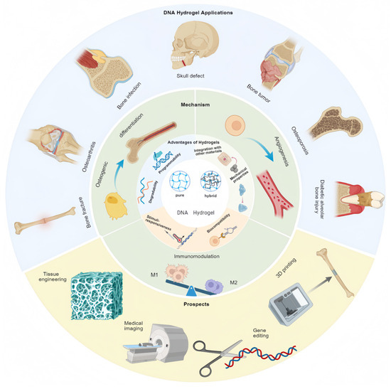
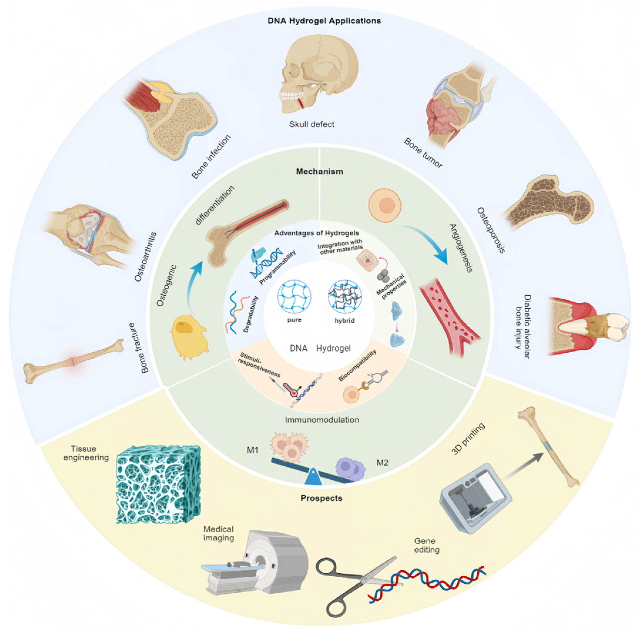
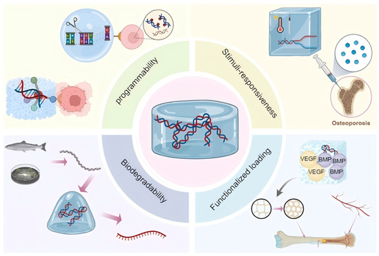
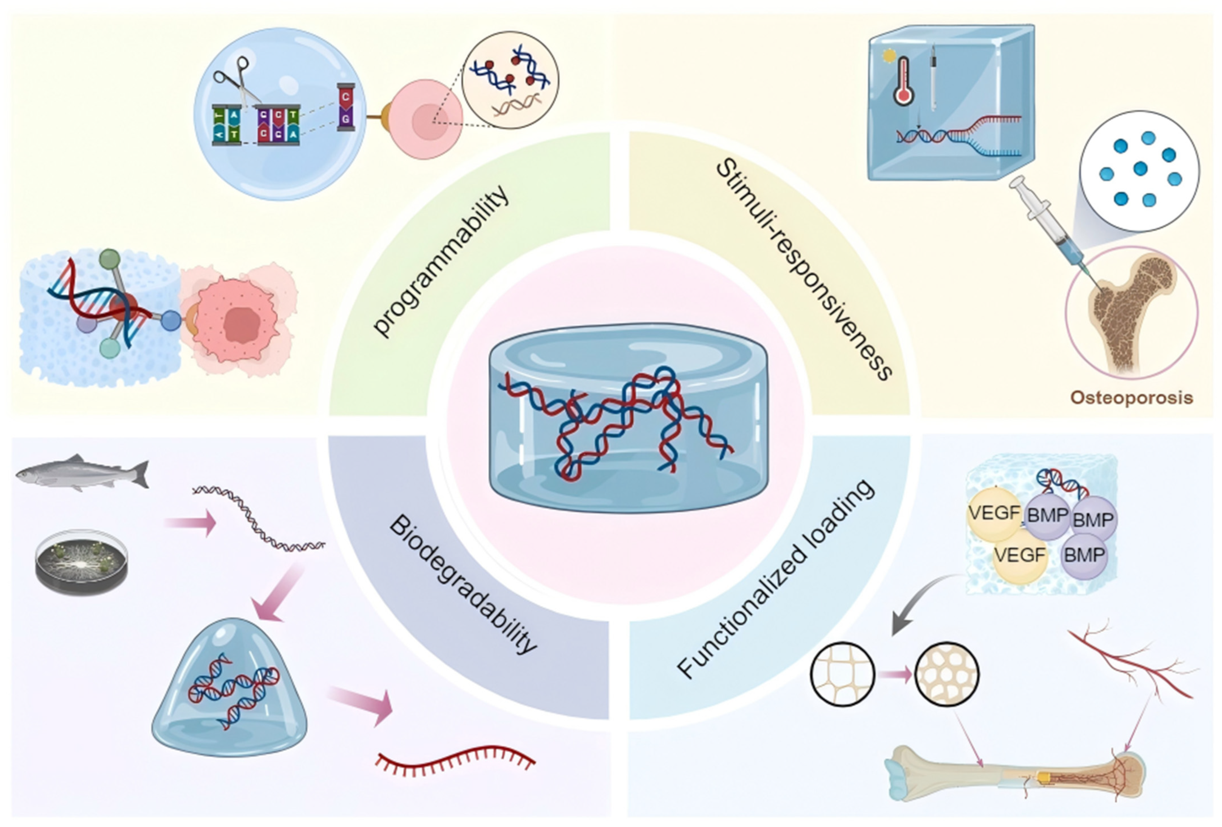
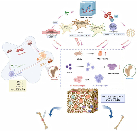
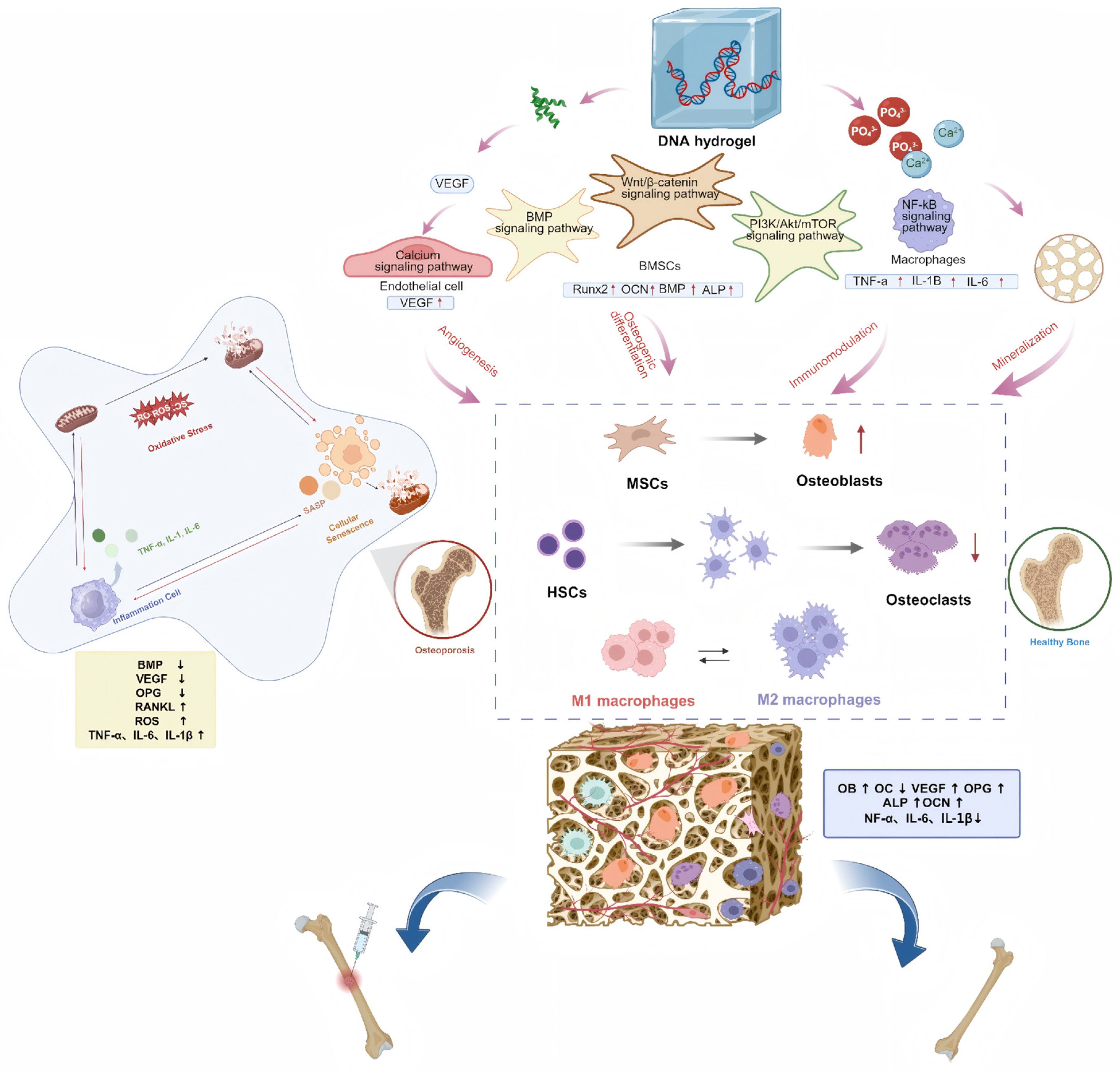
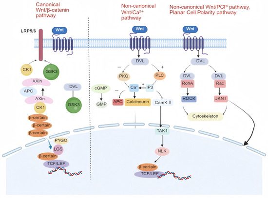
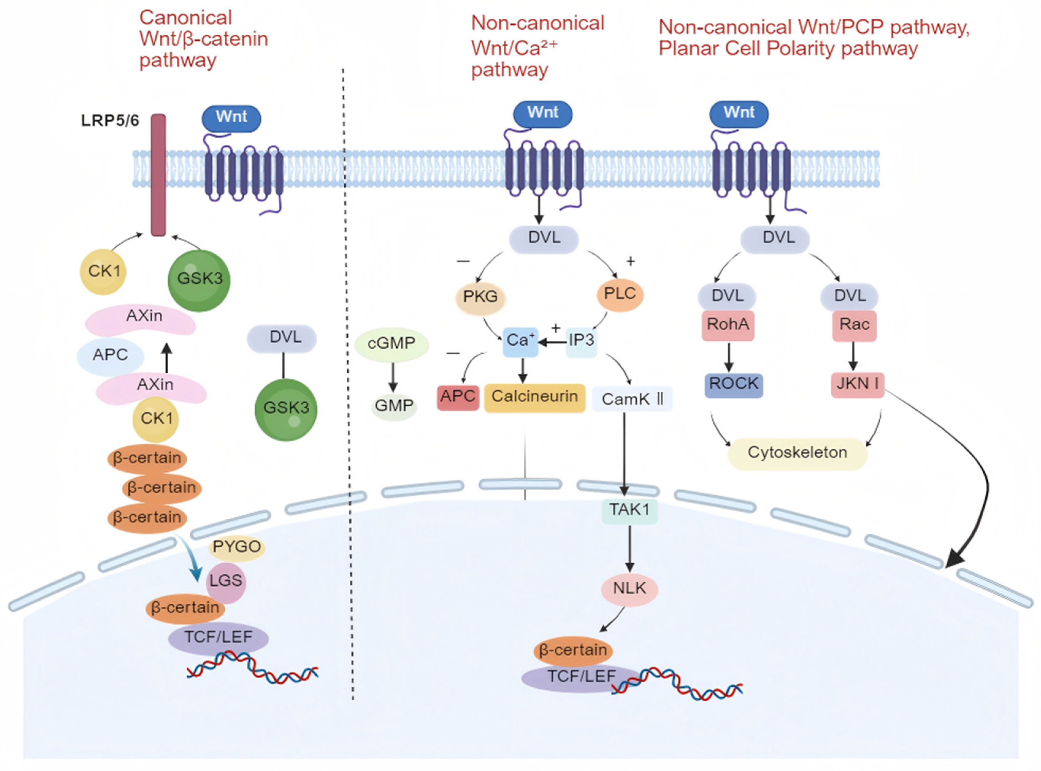

Disclaimer/Publisher’s Note: The statements, opinions and data contained in all publications are solely those of the individual author(s) and contributor(s) and not of MDPI and/or the editor(s). MDPI and/or the editor(s) disclaim responsibility for any injury to people or property resulting from any ideas, methods, instructions or products referred to in the content. |
© 2026 by the authors. Licensee MDPI, Basel, Switzerland. This article is an open access article distributed under the terms and conditions of the Creative Commons Attribution (CC BY) license.