


Abstract
Implant-associated infections (IAIs) caused by Staphylococcus aureus are a major cause of orthopedic implant failure, driven by biofilm formation and chronic inflammation. Macrophages regulate bacterial clearance and tissue repair through polarization into pro-inflammatory (M1) and anti-inflammatory (M2) phenotypes. This systematic review evaluated preclinical in vivo studies according to PICO criteria (Population: IAI animal models; Intervention: functionalized biomaterials; Comparator: non-functionalized controls; Outcome: bacterial burden reduction associated with macrophage reprogramming). A PRISMA-guided search of PubMed, Scopus, Web of Science, and Embase (January 2015–December 2025) identified 23 eligible studies, assessed using SYRCLE’s risk-of-bias tool. Due to heterogeneity, a narrative synthesis was performed without meta-analysis. Effect measures included bacterial CFU reduction, macrophage polarization markers, cytokine expression, and histological outcomes. In 87.5% of studies, macrophage modulation was associated with reduced bacterial load, biofilm disruption, and improved peri-implant tissue repair and bone integration. These findings support immunomodulatory biomaterials as promising strategies to manage IAIs through combined antibacterial and immune-regulatory mechanisms. However, further validation in larger animal models is required, particularly for nanomaterials. The protocol of this systematic review was registered on the Open Science Framework.
1. Introduction
In the orthopedic field, the use of devices (e.g., prostheses, nails, cages, screws) to treat pathological conditions affecting musculoskeletal tissues represents a standard and consolidated clinical practice from which patients generally benefit [1,2]. Despite generally favorable outcomes, material-related risks and adverse events may lead to significant clinical and psychosocial consequences for patients [3].
The most serious complication is the susceptibility of prosthetic implants to bacteria colonization, which may evolve into implant-associated infections (IAIs). IAIs affect approximately 1.5–2.5% of primary total hip and knee arthroplasty [4,5], and are associated with considerable morbidity, chronic pain and functional impairment, and in the worst cases, the need for complex revision procedures [6]. Revision surgery further increases the risk of reinfection, with IAI incidence reaching up to 20% [7], imposing a significant burden both for patients and healthcare systems as implant numbers continue to rise [8,9,10].
In the IAI field, extensive research focuses on the mechanisms leading to bacterial adhesion, colonization and subsequent biofilm formation on devices’ surfaces [11]. Gram-positive bacteria predominate, with Staphylococcus aureus representing one of the main causative agents. Bacterial attachment is mediated by microbial adhesive components that recognize host matrix proteins such as fibronectin, fibrinogen, and vitronectin, initiating rapid surface colonization and biofilm development through stages of attachment, maturation, and dispersal of planktonic cells [12]. Biofilms confer high tolerance to antimicrobial agents by limiting drug penetration and shielding bacteria from immune responses [13]. In addition, biofilm-forming pathogens such as S. aureus employ multiple immune evasion strategies, including toxin production, inhibition of opsonization and phagocytosis, interference with complement activation, and intracellular survival within macrophages, all contributing to infection persistence [14,15].
In this dynamic microecosystem located at the implant–host interface, innate and adaptive immune responses play crucial roles [16].
Among innate immune cells, tissue-resident macrophages are abundant and rapidly activated responders to implanted materials that critically influence implant outcomes. They clear debris and pathogens, regulate the foreign body response, and support tissue integration, but in the presence of infection they may also sustain inflammation and impair tissue repair [14]. Upon reaching the site of implant, macrophages exhibit remarkable functional plasticity, adapting their phenotype to local microenvironmental cues through a process known as polarization [17]. This phenomenon is commonly described by the M1 pro-inflammatory phenotype, induced by interferon-gamma (IFN-γ) and lipopolysaccharide (LPS), and characterized by microbicidal activity, reactive oxygen species (ROS) and pro-inflammatory cytokine production, such as tumor necrosis factor-alpha (TNF-α), interleukin-1 beta (IL-1β) and interleukin-6 (IL-6). On the other hand, the M2 anti-inflammatory phenotype is usually driven by interleukin-4 (IL-4) and interleukin-13 (IL-13), and it is associated with immune regulation, tissue remodeling and production of interleukin-10 (IL-10) and transforming growth factor-beta (TGF-β) [18]. Although simplified, this framework is valuable for understanding macrophage behavior in IAIs.
During the early stages, planktonic bacteria activate macrophages via Toll-like receptor 2 (TLR2), driving an M1 pro-inflammatory response aimed at bacterial clearance [14]. As a biofilm matures, a shift towards the M2 anti-inflammatory phenotype is promoted by immune evasion and S. aureus-derived factors, supporting bacterial persistence and chronic infection establishment [19,20,21].
Dysregulated macrophage polarization at the implant site may lead to pathological outcomes: excessive M1 activation promotes osteoclastogenesis [22] and peri-implant bone resorption, contributing to osteolysis and implant failure [23], whereas sustained M2 response may create a permissive niche for chronic bacterial persistence [24]. Together, these alterations identify macrophage polarization as a critical pathological node in IAI.
Current treatment strategies for IAI rely mainly on systemic antibiotic therapy combined with surgical debridement or implant removal in the worst cases. However, the biofilm microenvironment markedly limits the efficacy of conventional antibiotics, by hindering drug penetration and decreasing bacterial susceptibility [25]. Consequently, complete eradication of biofilm-associated infections remains difficult to achieve with systemic approaches alone [26].
This therapeutic gap highlights the need for alternative strategies capable of both preventing bacterial adhesion [27] and modulating the host immune response through macrophage polarization [28]. Therefore, this review aims to evaluate in vivo biomaterial-based approaches designed to modulate macrophage polarization as a novel strategy for the prevention and treatment of S. aureus implant-associated infections.
2. Materials and Methods
2.1. Search Strategy
This literature review was performed through a systematic search across four electronic databases (PubMed®, Scopus®, Web of Science® and Embase®: www.pubmed.gov, www.scopus.com, www.webofscience.com and www.embase.com), according to the PRISMA guidelines (Supplementary Table S1) [29]. The review protocol was registered on the Open Science Framework (OSF: https://osf.io/ur9q5; accessed on 13 July 2026)). The search strategy included following keywords combined with booleans: “Staphylococcus Aureus” OR “S. Aureus” AND “implant-associated infection” OR “periprosthetic infection” OR “peri-implant infection” OR “IAI” OR “peri-prosthetic joint infection” OR “PJI” AND “macrophage.” The full search strategies for each database are reported in Table 1. Screening and analysis of the retrieved records were carried out in a blinded and independent manner by two reviewers (G.C. and MA.S.). A web-based tool for narrative and systematic reviews, Rayyan [30], was used for the detection and removal of duplicates and the determination of the studies included or excluded based on titles and abstracts.
Table 1.
Full search strategies for database queries.
The inclusion criteria comprised all in vivo preclinical studies investigating whether a new generation of biomaterials tested in the context of implant-associated infection (IAI) sustained by S. aureus can also affect macrophage repolarization. Additional criteria included publication in English within the timeframe of 1 January 2015 to 31 December 2025. Exclusions criteria consisted of papers written in languages other than English, reviews, abstracts, clinical studies, editorials, comments, publisher’s notes, white papers, and letters. Studies lacking the assessment of S. aureus exposure or an implant-associated infection model were also excluded. The reference list of the included studies was manually screened in order to identify further relevant papers not captured by the electronic database search. Discrepancies between reviewers were resolved through discussion and, when needed, with the involvement of a third reviewer (G.G.).
2.2. Data Extraction
In accordance with PRISMA suggestions, a well-defined PICO question was formulated to facilitate accurate data extraction from included studies: Population (P): animal models of IAI caused by S. aureus, Intervention (I): functionalized biomaterials or approach in which macrophage polarization at the implant site was investigated, Comparator (C): non-functionalized implants, or control groups receiving implants without functionalization, Outcome (O): reduction in bacterial burden associated with macrophage reprogramming. Starting from the study focus, G.C. and MI.S. systematically extracted the key characteristics from the selected studies. The collected data from in vivo studies were: the type, size and characteristics of the tested material/approach; specifics of the animal model used for implant-associated infection (IAI), including species, strain, sex, age, number of animals, and experimental groups; the implantation site for the IAI model; the microbiological investigations to evaluate the bacterial load and biofilm assessment; the tests used for macrophage reprogramming assessment; the established experimental times; the main findings; and, if reported, the signaling pathways analyzed.
A third author (MA.S.) checked the extracted data for accuracy and completeness. Any disagreements were resolved through discussion and, when necessary, with the consultation of another reviewer (G.G.). Extracted data are reported in Table 2.
Table 2.
Summary of the in vivo preclinical studies.
2.3. Risk-of-Bias Assessment
The SYRCLE tool [53], based on a 10-domain checklist including “Selection,” “Performance,” “Detection,” “Attrition,” “Reporting” and other bias, was used to assess the risk of bias of the in vivo studies. The risk of bias was classified as low, high, or unclear, depending on whether the domains were clearly reported, not reported, or reported ambiguously. Two authors (G.C. and MA.S.) conducted the assessment independently, and disagreements were resolved through discussion or by consulting a third reviewer (MI.S.).
2.4. Data Synthesis
Results for each outcome were reported using the original data from the included studies, including the CFU counts, percentage of bacterial load reduction, and qualitative or semi-quantitative microbiological, histological and macrophage polarization assessments. Due to heterogeneity in the study design, biomaterials, and outcome measures, a quantitative meta-analysis was not performed, and the results were synthesized descriptively. No sensitivity analyses or assessment of the certainty of the evidence were conducted. No assessment of risk of bias due to missing results in the synthesis was performed.
3. Results
3.1. Search Strategy
The review process began with a comprehensive literature search using the predefined search strings. This search identified 61 articles from PubMed (www.pubmed.gov), 33 from Web of Science (www.webofscience.com), 74 from Scopus (www.scopus.com) and 871 from Embase (www.embase.com).
All retrieved references were imported into Rayyan, a publicly available software tool for systematic review management, to remove duplicates [30]. This step led to the exclusion of 141 duplicate records, leaving 898 articles for screening.
During the initial title and abstract screening, 121 records were excluded: 56 reviews, 4 abstracts, 50 clinical studies, 4 editorials, 4 comments, 1 publisher’s note, 1 white paper and 1 letter. The remaining 777 articles underwent full-text screening, which resulted in the exclusion of 754 non-inherent studies, as illustrated in the PRISMA flow chart (Figure 1). Following this selection process, 23 in vivo studies met the eligibility criteria and were included in the review (Figure 1): 11 from PubMed, 1 from Web of Science, 6 from Scopus and 5 from Embase. Manual screening of the reference lists of all included studies did not yield additional eligible records.
Figure 1.
Search workflow. Search strategy according to the PRISMA guidelines [29].
3.2. Risk-of-Bias Assessment
The risk-of-bias assessment revealed that most domains were assessed as “high risk,” as illustrated in Figure 2a,b. Domain 1, “sequence generation,” was at high risk in over half of the studies (60.9%) due to a lack of reporting on the randomization process for animal allocation. Similarly, domain 3, “allocation concealment,” was rated high risk in the majority of papers (82.6%), as the characteristics used for group assignment were not fully described, and no study clearly explained how allocation was performed. Domains 4, “random housing,” and 6, “random outcome assessment,” showed a high risk of bias in 95.7% of studies, as 22 out of 23 studies did not specify how animals were housed within the facility after group assignment, nor whether experimental samples were analyzed in random order. Domains 5 (“blinding” as performance bias) and 7 (“blinding” as detection bias) were high risk in all studies (100%), because investigators were aware of group assignments and assessors were not blinded during outcome evaluation. Finally, Domain 8, “incomplete outcome data,” was rated as unclear risk in 47.8% of studies, due to discrepancies between the number of animals reported in the methods and those reported in the results.
Figure 2.
In vivo risk of bias results. Overall risk of bias (a) and assessment for each in vivo study (b) by applying SYRCLE’s RoB tool [31]. Studies are presented in the same order as in Table 2 (functionalized PEEK implants, functionalized titanium implants, and nanomaterials) [20,32,33,34,35,36,37,38,39,40,41,42,43,44,45,46,47,48,49,50,51,52,53]. Each domain (D; D1—sequence generation, D2—baseline characteristics, D3—allocation concealment, D4—random housing, D5—blinding of caregivers, D6—random outcome assessment, D7—blinding of outcome assessment, D8—incomplete outcome data, D9—selective outcome reporting and D10—other bias) is reported as low risk (green), high risk (red) and unclear risk (yellow) of bias.
3.3. In Vivo Preclinical Studies
The in vivo studies were grouped according to the type of material investigated and the treatment performed to confer antibacterial properties: 2 studies out of 23 (8.7%) developed functionalized polyetheretherketone (PEEK) implants [31,32], 14 papers out of 23 (60.9%) investigated functionalized titanium implants [33,34,35,36,37,38,39,40,41,42,43,44,45,46], while 7 out of 23 (30.4%) adopted nanomaterials [20,47,48,49,50,51,52] for the treatment of implant-associated infections.
3.3.1. Functionalized PEEK Implants
In the study by Wang et al. [31], a PEEK implant was modified with a polydopamine coating, followed by the deposition of strontium (Sr) ions and the antimicrobial peptide PMAP-36 (PEEK-PDA-Sr/AMP). In Li et al.’s study [32], a PEEK implant was firstly sulfonated to produce SPEEK. This sulfonated PEEK was then functionalized with glucose-gated nanocoating, which contained glucose oxidase (GOx) and manganese carbonyl nanocrystals (MnCO), developing a near-infrared (NIR)-responsive system. In both studies, a male Sprague–Dawley rat was selected as the animal model, in which femoral and tibial defects were surgically created and injected with an S. Aureus suspension to induce IAI. The bacterial suspension concentration was only reported in the Wang et al. study, which used a bacterial inoculum of 104 CFU/mL. Macrophage reprogramming was assessed using multiple techniques, including immunohistochemistry (IHC) focused on the expression of M1 and M2 markers (cluster of differentiation, CD86 and CD206) in the peri-implant tissue, immunofluorescence, enzyme-linked immunosorbent assay (ELISA) and real-time quantitative polymerase chain reaction (RT-qPCR) to evaluate the expression of surface and intracellular markers of M1 and M2 macrophages (inducible nitric oxide synthase, iNOS, and CD206), as well as the expression of pro-inflammatory cytokines (IL-6 and TNF-α). Both studies showed an increase in CD206 expression, an M2 marker, following treatment, as well as a reduction in the intracellular M1 marker iNOS and the pro-inflammatory cytokines secreted by them. Macrophage reprogramming after these treatments was supported by inhibition of the pro-inflammatory nuclear factor kappa-light-chain-enhancer of activated B cells (NF-kB) pathway, mediated by the release of Sr ions in Wang’s study and by hyperglycemia in Li’s study, by activating glucose oxidase, which promotes the release of carbon monoxide (Figure 3). These changes in macrophage phenotype impacted the infection outcomes. In Wang’s study, the treatment with PEEK-PDA-Sr/AMP completely cleared the implantation site from bacteria, as revealed by the absence of bacterial colonies in peri-implant tissue sections stained with Giemsa. The antimicrobial peptide activity combined with Sr-mediated immunomodulation demonstrated that the functionalized PEEK implant can effectively control the infection. Similarly, SPEEK-GOx/MnCO treatment combined with photodynamic therapy (PDT) significantly reduced bacterial load and viability in Li’s approach. Under NIR, the decomposition of MnCO was accelerated, promoting the increased local release of carbon monoxide (CO), which acted as a gas therapeutic agent contributing to the antibacterial activity. This effect was associated with macrophage metabolic reprogramming toward the anti-inflammatory but bactericidal state.
Figure 3.
Modulation of macrophage polarization by functionalized implants. Implant surface modifications modulated intracellular pathways, either inhibiting pro-inflammatory pathways (e.g., NF-κB and MAPK), resulting in decreased (↓) expression of M1 markers (e.g., CD86 and iNOS) and pro-inflammatory cytokines (e.g., TNF-α, IL-6 and IL-1β), or promoting signaling pathways associated with M2 polarization and tissue repair (e.g., PI3K/AKT and related downstream mediators like PPARγ), leading to increased (↑) expression of M2 markers (e.g., CD206, ARG1, IL-4, IL-10, VEGF and TGFβ).
However, despite the promising infection-related outcomes, no quantitative microbiological data confirming bacterial clearance, such as colony forming unit (CFU) count, were provided. In Wang’s study, only the initial inoculum concentration was reported, whereas Li’s study did not specify the bacterial load used to test the biomaterial.
Nevertheless, histological observations on peri-implant tissue sections stained with Masson’s trichrome suggested that functionalized PEEK implants promoted peri-implant matrix deposition and new bone formation, in line with immunohistochemistry results of osteogenic markers, runt-related transcription factor 2 (RUNX2), osteocalcin (OCN) and Collagen Type I Alpha 1 Chain (COL1A1).
3.3.2. Functionalized Titanium Implants
Titanium-based substrates have been functionalized by adopting different strategies. One approach involved the deposition of piezoelectric materials such as barium titanate (BaTiO3) [37,43] and polyvinylidene fluoride (PVDF) [44], which respond to ultrasound and promote macrophage reprogramming through ROS generation or direct interaction with macrophages. Surface modifications also included interfacial functionalization based on nanoparticles (NPs) such as polydopamine (PDA) [46] and iron (III)–tannic acid complex (FeIIITA) NPs [42] conjugated to antibacterial agents and responsive to NIR, which produced photothermal effects for bacteria killing. Other emerging approaches included hybrid functionalization, in which pure titanium was coated with a composite system combining inorganic components such as metallic ions or a metal oxide like zinc oxide (ZnO) [36] with organic materials such as amyloid lactoferrin and poly-γ-glutamic acid (γ-PGA) [34,41], or with hybrid coatings, including gelatin methacryloyl (GelMa) hydrogel [35,39], which served as delivery platforms for bioactive molecules [40] and antibacterial drugs [33,45]. Finally, Li et al. investigated a bioengineered strategy, including nanostructured layers linked to peptide NPs carrying a DNA plasmid coding for macrophage Chimeric Antigen Receptor (CAR) and allowing for specific recognition of S. Aureus [38]. Rats were used in the majority of studies (12 out of 14, 85.7%) [33,34,35,36,37,40,41,42,43,44,45,46], most of which were male, while the remaining studies employed male mice (2 out of 14, 14.3%) [38,39] to establish an IAI model. Titanium-based implants were predominantly placed in the femoral or tibial intramedullary canal or implanted subcutaneously, reflecting their intended orthopedic applications. In contrast, in the study by Chen et al. [36], the material was implanted in the maxilla to simulate dental application. To induce infection, selected strains of S. aureus or Methicillin-Resistant S. Aureus (MRSA) were introduced directly into the created bone defect in 8 of 14 studies (57.1%) [33,34,35,38,39,41,42,45], whereas in 3 studies materials were pre-exposed to bacterial suspension prior to the implantation (21.4%) [36,37,40], or implanted after pre-formed biofilm on the titanium surface (3 out of 14, 21.4%) [43,44,46]. Bacterial concentrations ranged from 1 × 10⁴ to 1 × 10⁸ CFU/mL.
Macrophage reprogramming was assessed by multiple techniques, including surface marker analysis by flow cytometry, as well as gene and protein expression and tissue-level evaluation through RT-qPCR, ELISA, immunohistochemistry, and immunofluorescence. Markers characteristic of M1 macrophages (CD86, CD80, iNOS) and M2 macrophages (arginase 1, Arg-1, and CD206), as well as pro-inflammatory cytokines (TNF-α, IL-6 and IL-1β) and anti-inflammatory cytokines (IL-10, and TGF-β), were predominantly analyzed in peri-implant tissues at experimental times ranging widely from 4 h to 8 weeks post IAI establishment.
Overall, 64.3% of the included studies (9 out of 14) [33,34,35,36,39,40,41,44,46] reported a shift toward an anti-inflammatory macrophage phenotype following material-based treatments, as evidenced by increased expression of M2-associated markers such as CD206 and Arg-1, together with a concomitant reduction in M1 markers and pro-inflammatory cytokines. This immunomodulatory effect induced by treatments was generally associated with a significant reduction in bacterial CFUs, and fewer histologically detectable bacteria revealed with hematoxylin & eosin (H&E), Giemsa and/or Gram staining. These findings were further supported by pathway analyses, which revealed that M2 polarization was promoted through multiple mechanisms, including (i) inhibition of inflammatory signaling pathways involved in M1 differentiation, such as NF-κB [33,40,46]; (ii) material-driven increases in ROS; (iii) release of immunomodulatory factors, including IL-4 [39], known to favor M2 polarization; (iv) release of bioactive ions such as silver ions (Ag+) to switch macrophages to the M2 type [41]; and (v) the generation of self-stimulated electric fields capable of regulating macrophages toward an anti-inflammatory phenotype [44]. Notably, in the studies by Wang [34], Guan [35], and Chen [36], the signaling pathways underlying macrophage polarization were not specifically investigated. By contrast, 35.7% of the studies (5 out of 14) [37,38,42,43,45] revealed a predominantly pro-inflammatory response by macrophages, characterized by elevated expression of M1-associated surface and intracellular markers (CD86 and iNOS) and increased production of key pro-inflammatory cytokines (TNF-α and IL-1β). The generation of a robust pro-inflammatory response by functionalized titanium implants facilitated bacterial clearance, with significantly decreased CFU counts or bacteria having been eradicated; suppressed biofilm, as evidenced by Scanning Electron Microscopy (SEM); and a lower inflammatory reaction surrounding the IAI site, as shown by histological analyses.
Mechanistic analyses indicated that M1 polarization was promoted by increased production of reactive oxygen and nitrogen species (ROS and NO) [37], activation of pro-inflammatory signaling cascades such as phosphoinositide 3-kinase–protein kinase B (PI3K–AKT), mitogen-activated protein kinase (MAPK) [43], interleukin-12 Receptor–Signal Transducer and Activator of Transcription 4 (IL12R–STAT4) and Interferon-gamma–Signal Transducer and Activator of Transcription 1 (IFN-γ–STAT1), with the latter being driven by IL-12 delivery [45], as depicted in Figure 3. Other factors include the engagement of TLRs by surface-functionalizing molecules [42], and the expression of specific macrophage receptors that boost the bactericidal activity against S. aureus [38]. M2 polarization, particularly in studies involving titanium implants, was associated with enhanced peri-implant matrix deposition and bone formation. This was supported by micro-CT analyses showing improvements in bone parameters (trabecular number, Tb.N, trabecular thickness, Tb.Th, bone mineral density, BMD, bone volume–total volume ratio, BV/TV), as well as by histological stainings such as Masson’s trichrome, which highlighted new collagen fiber deposition and osteoid tissue formation. These findings were further confirmed by immunohistochemistry and immunofluorescence analyses of osteogenic markers, including RUNX2, alkaline phosphatase (ALP), OCN and osteopontin (OPN) in peri-implant tissue, which reflect active osteogenesis and bone formation.
3.3.3. Nanomaterials
Several studies have investigated nanomaterials as therapeutic platforms for IAI treatment. In particular, four studies employed physically activated nanomaterials, with three out of four investigating NIR-responsive nanostructures consisting of organic materials, hybrid materials or organic–inorganic metal–organic frameworks (MOFs) to exploit the photothermal effect to penetrate the biofilm and stimulate ROS production through copper ion (Cu+) release. One study proposed a white light-responsive nanoplatform, based on organic–inorganic materials, in which a photocatalyst was used to boost ROS production and enhance immune response against MRSA [48]. Other approaches included the use of intrinsically active MOF [49], represented by organic nanoparticles delivering a metabolic inhibitor for macrophage polarization modulation [20], and inorganic metal and non-metal nanosheets stabilized by polyvinylpyrrolidone (PVP) [52]. Finally, controlling the delivery of drugs such as rosiglitazone for peri-implant microenvironment modulation represents another exploited strategy [50].
All studies used mice (7 out of 7, 100%), principally male, in which the materials were placed either subcutaneously (5 out of 7, 71.4%) [47,48,50,51,52] or in surgically created femoral defects (2 out of 7, 28.6%) [20,49]. Implant-associated infection was induced by pre-exposure of the implants to S. aureus or MRSA suspensions [20,50,51], implantation of pre-formed biofilms [47], or direct injection of bacterial suspensions into the defect site after implantation [48,49,52], with reported concentration ranging from 103 to 107 CFU/mL, although this information was not consistently specified across all studies. In most studies, nanomaterial-based treatments were administered as single injections at the implantation site between 1 and 3 days after IAI induction in the animal model.
Macrophage reprogramming was evaluated using methods similar to those applied for titanium-functionalized implants. Surface and intracellular markers for M1-type (CD86, iNOS) and M2-type (CD206, Arg-1) macrophages were analyzed. Pro-inflammatory (TNF-α, IL-6, IL-1β, hypoxia-inducible factor-1 (HIF-1α), IL-12 and triggering receptor expressed on myeloid cells 1 (Trem1)) and anti-inflammatory (IL-10, Vascular and Endothelial growth factor (VEGF) Trem2) factors were evaluated. These parameters were assessed at the gene and protein levels, as well as at the tissue level, using flow cytometry, RT-Qpcr, ELISA and immunofluorescence. This occurred within a time frame ranging from 3 days to 2 weeks after surgery.
Except for Chen et al. [50], all works reported a significant shift in macrophage populations toward a pro-inflammatory phenotype, sustained by increased ROS production, pathogen-associated molecular patterns (PAMPs) recognition and metabolic reprogramming toward a glycolytic profile (Figure 4). This was correlated with bacterial killing and biofilm disruption, as observed through CFU counts, SEM analyses and histological stainings, evidencing their antibacterial effects mediated by immunomodulation. Jiang’s group observed an early increase in M1 type during infection, followed by a later transition toward M2 phenotype, promoting a reduction in bacterial burden, as assessed by the measurement of infection area size, CFU count and biofilm observation by SEM, and providing support to tissue repair and healing [48]. On the other hand, the approach proposed by Chen promoted M2 polarization through rosiglitazone-mediated activation of PPAR-γ signaling, which inhibited Nf-kB activity, the main molecular master orchestrating M1 polarization and inflammatory cytokine production. However, the study did not provide clear in vivo evidence of complete bacterial eradication mediated by M2 type activated by the treatment. Nevertheless, preliminary in vitro experiments demonstrated that indocyanine green + rosiglitazone (ICG + RSG) NPs were able to restore phagocytic activity of macrophages against pathogens, destroying biofilm and inhibiting bacterial growth by employing photothermal and photodynamic therapies [50].
Figure 4.
Modulation of macrophage polarization by nanomaterials. Various nanomaterials activated pro-inflammatory signaling cascades (e.g., NF-kB, ROS) or inhibited anti-inflammatory pathways (e.g., PI3K/AKT and related downstream mediators, OxPhos), leading to upregulation (↑) of M1-associated markers and mediators (CD86, iNOS, TNF-α, HIF-1α, IL-6, IL-12, IL-1β) and downregulation (↓) of M2-linked ones (CD206, Arg1, Trem2, IL-10, VEGF). Collectively, nanomaterials shifted the macrophage phenotype balance toward M1 polarization.
4. Discussion
In the current scientific landscape, research on IAI has mainly focused on the development of antibacterial materials aimed at preventing, reducing and/or counteracting bacterial adhesion and growth onto material surfaces. Increasing attention is now being paid to the comprehension of the role exerted by the immune system as well, which is known to be actively involved in infection events. Consequently, the next-generation materials are not only designed to exert antimicrobial action but are also engineered to interact with and stimulate the immune cell response, especially macrophages [54], which are key regulators of inflammation, tissue regeneration and bacterial clearance [55,56]. In this context, understanding how materials could influence macrophage behavior and polarization is crucial for the transition from strategies to develop purely antibacterial materials to ones with immunomodulatory activity. In line with this transition, the aim of the present review was to identify and describe this new class of biomaterials in which the combination of antibacterial and immunomodulatory functions—particularly in terms of macrophage involvement—is employed to counteract, contain or prevent IAI in a preclinical in vivo setting.
A considerable heterogeneity has emerged in terms of types of materials used, functionalization strategy employed, and immune activation mechanisms, although the analysis of macrophage reprogramming results revealed that this is consistently associated with positive infection outcomes.
This interplay between antibacterial action and macrophage reprogramming is particularly evident in NIR responsive platforms. NIR responsive photothermal and photodynamic systems physically help kill bacteria by disrupting the integrity of cell membranes and organelles while modulating macrophage polarization toward anti-inflammatory yet bactericidal phenotype [7,17,21,22,25]. This created an osteoimmune environment favorable for the recruitment of bone marrow mesenchymal stem cells (BMSCs), extracellular matrix deposition and mineralization, and new bone formation [57,58,59]. Finally, as reported in the literature and in the study by Yu Y. et al., studies have shown that, compared to implantation with the material alone, stimulation with NIR drastically reduced the bacterial load. This was probably due to the material’s synergistic immunomodulatory and antibacterial action. At the molecular level, the ROS increase induced by the photodynamic effect of NIR led to a metabolic reprogramming in macrophages around the implant by regulating key signaling pathways, including Nf-kB, PI3K-Akt and MAPK [60,61,62,63].
This metabolic reprogramming is closely linked to immunometabolic adaptations that define macrophage functional phenotypes. Macrophage polarization is closely linked to metabolic reprogramming, which is a central feature of “immunometabolism.” During inflammatory activation, classically activated M1 macrophages primarily rely on glycolysis to meet their immediate energetic demands, a process driven in part by hypoxia-inducible factor-1α (HIF-1α), which promotes the expression of glycolytic enzymes and glucose transporters. Glycolytic activation is also linked to the pentose phosphate pathway, which produces the NADPH necessary for the generation of reactive oxygen species (ROS) via NADPH oxidase, thereby facilitating the killing of pathogens. By contrast, M2 macrophages are associated with tissue repair and the resolution of inflammation, relying predominantly on oxidative phosphorylation and fatty acid β-oxidation. Unlike the rapid, glycolysis-driven M1 response that characterizes the early inflammatory phase, M2 activation occurs during the resolution phase, when the immune response shifts progressively from clearing pathogens to restoring tissue. This longer-lasting functional state requires sustained ATP production, which is efficiently supported by oxidative metabolic pathways [64]. This metabolic profile enables prolonged cellular activity with reduced ROS generation, favoring anti-inflammatory and tissue-remodeling functions and reflecting adaptation to a resolving microenvironment. Within this metabolic framework, intracellular signaling pathways further refine macrophage polarization outcomes [65,66].
Notably, PI3K-Akt pathway is reported to play a context- and stage-dependent role in macrophage activation and polarization [67]. Although it generally promotes M2 polarization, different Akt isoforms (Akt1, Akt2, Akt3) finely determine the macrophage polarization fate [68]. During the early stages of inflammation, PI3K-Akt signaling contributes to fine-tuning rather than abolishing M1 responses, thereby preserving macrophage antimicrobial and bactericidal functions while preventing excessive activation through modulation of TLR/NF-kB signaling. In the later phase, this pathway contributes to the transition to the M2 phenotype and sustains anti-inflammatory programs [69].
Similarly, ultrasound-activated piezoelectric coatings have been shown to modulate macrophage polarization [37,43], accelerate bacterial killing and support tissue damage healing and implant integration in vitro and in IAI models [70,71]. Therefore, as with NIR-responsive biomaterials, ultrasound-activated piezoelectric platforms exert their effects through a combination of physical antibacterial actions and immune modulation. Even the release of metallic ions such as silver (Ag), copper (Cu), zinc (Zn) and strontium (Sr) from coated implants or nanomaterials could contribute to infection control through macrophage reprogramming, enhancing ROS generation [47,51] or regulating key signaling pathways in macrophages [31,33,41]. Together, these findings suggested that both externally triggered physical activation strategies and ion-releasing coatings could be effective approaches to enhance antimicrobial activity and support osteoimmune regulation, as consistently reported in the literature [72,73,74].
Interestingly, while titanium and PEEK implants displayed polarization toward either M1 or M2 macrophages depending on the specific functionalization strategy, nanomaterials predominantly promoted M1-type polarization. This divergence may reflect different therapeutic goals. M1-oriented responses are primarily aimed to enhance bacterial eradication through improvement of their bactericidal activity, whereas M2-skewing is more often associated with promoting tissue repair and implant integration after bacterial damage to tissues [75,76,77,78].
This difference can be attributed to the size of the materials. Materials within the phagocytosable range, such as most of investigated nanomaterials in the included studies, sustained inflammatory activation of macrophages compatible with pro-inflammatory M1 phenotype, while larger implants like titanium and PEEK were not phagocytosable and promoted tissue healing response associated more with M2 type [79,80,81]. For example, Cu nanodots embedded in mesoporous silicon nanoparticles (Motor@CP) [47], bovine serum albumin and manganese dioxide nanoclusters (BMCV) [48], and bismuth-based metal–organic frameworks (Bi-MOFs) [49], with sizes in the nanoscale range (114–808 nm), promoted a significant increase in the M1/M2 macrophage ratio. This effect was evidenced by the upregulation of M1 markers (CD86 and iNOS) and pro-inflammatory cytokines such as IL-6 and TNF-α. Conversely, larger biomaterials such as titanium and PEEK implants, functionalized with bioactive molecules (e.g., lactoferrin, antimicrobial peptide and quercetin) [31,34,40], nanoenzymatic systems such as glucose oxidase [32], or inorganic nanofilms [36], and characterized by millimetric dimensions, supported macrophage polarization toward the M2 phenotype. This was associated with increased expression of M2 markers (CD206 and ARG1) and anti-inflammatory mediators, including IL-10 and TGF-β.
Despite these differences, the included studies consistently reported favorable infection control outcomes, as with reduction in bacterial load and biofilm thickness, suggesting that multiple polarization approaches may converge toward effective antibacterial performances. Together, these findings suggest that modulation of macrophage behavior, orchestrated by material composition, surface and/or dimension properties, and exogenous stimulation strategies, can synergistically enhance both antimicrobial activity and tissue repair.
The results of macrophage-mediated infection control and tissue repair proposed by the included studies are promising, but several limitations should be acknowledged. All included studies used only rodent models that were predominantly male, excluding the use of large animal models like pigs [82], which more closely replicate human anatomy, immune responses [83], biomechanical conditions, and clinical implant dimensions [84,85]. The literature clearly documents that innate immunity is subject to sex-based differences: male rodents tend toward more pro-inflammatory responses, while females generally show a more regulatory profile, more efficient bacterial killing alongside better-regulated cytokine responses, and greater M2-like polarization capacity [86,87,88]. Within the included studies, only three employed female animal IAI models: Yamada et al. [20] and Chen C. et al. [50] both used mice for testing nanomaterials, while Xu K. [42] et al. investigated a functionalized titanium implant on a rat model. Despite using the same sex of animal model, these studies reported different outcomes in terms of polarization. Yamada et al. and Xu K. et al. observed a promotion towards the M1 phenotype, whereas Chen C. et al. observed M2 polarization. These results suggest that the type of material used and the experimental design are the main factors influencing polarization direction, rather than the sex of the animal itself. However, the predominant use of male animals in the remaining studies may introduce a systematic bias, so sex-stratified preclinical studies are required.
Furthermore, the assessment of biocompatibility and antimicrobial efficacy may be influenced by rodents’ rapid healing and lower immunological complexity [81,83,89,90]. These factors collectively limit the direct extrapolation of these preclinical findings to human clinical settings.
Moreover, heterogeneity in material types, functionalization strategies, infection induction protocols, implantation sites and quantitative, semi-quantitative or qualitative presentation of outcomes complicated the direct comparison among studies, precluding the performance of a metanalysis and limiting the synthesis to a narrative approach. Notably, eight of the twenty-four studies used subcutaneous implantation models to evaluate osteoimmunomodulatory and antibacterial materials, particularly for nanomaterials, [37,39,43,47,48,50,51,52]. While useful for preliminary biocompatibility assessment, subcutaneous implants do not replicate the bone microenvironment, which differs in immune cellular composition, vascularization and extracellular matrix and does not allow for osseointegration assessment [91,92,93]. The discussed differences should be considered when interpreting results from bone implants and subcutaneous models, especially in terms of macrophage behavior. Additionally, only the study of Chen B. et al. [36] investigated a maxillary implant site, and the reported results are not directly comparable to those obtained in bone implant models, given the distinct composition of the oral microflora, which comprises a highly diverse microbial community responsible for specific oral pathological conditions such as periodontitis and peri-implantitis, and which varies considerably across the different niches of the oral cavity, as well as the unique local immune response, shaped by continuous antigenic stimulation and specialized mucosal immunity mechanisms [94,95,96,97].
Although this review focused on immune modulation and infection control using next-gen biomaterials, it is the ultimate goal of biomaterials intended for orthopedic applications to achieve effective osseointegration. The investigated functionalized materials enhanced bone formation and peri-implant matrix deposition compared with unmodified controls such as pure PEEK or pure titanium [31,32,33,34,35,36,38,40,41,42,44,45,46]. This suggests that, beyond their antibacterial activity, these materials can support new bone deposition and stable integration, mitigating local inflammation through macrophage polarization toward the M2 phenotype and promoting tissue repair.
Nevertheless, methodological limitations should be considered. Most studies showed a high or unclear risk of bias, particularly regarding randomization procedures, allocation concealment, housing conditions, and blinding during surgical procedures and outcome assessment. These factors may have influenced the robustness and reproducibility of the reported results and could have contributed to an overestimation of treatment efficacy. Beyond methodological concerns, safety assessments were variably reported. While the most of studies (10/16, 62.5%) reported no histopathological abnormalities in major organs (liver, kidney, lungs, spleen and heart) or biochemical parameter alterations on routine blood tests, demonstrating overall biosafety, nanomaterials were less consistently evaluated [51,52]. As highlighted by Codispoti et al. [98], the safety of nanomaterials remains a critical aspect, despite their promising antimicrobial potential, due to the risk of unintended immune activation or chronic inflammatory responses [99,100,101,102]. In addition, toxicological investigations are required to exclude their accumulation in clearance organs such as the liver, spleen and kidney [103,104]. Thus, further studies would be valuable to better define the balance between biological safety, long-term tissue effects and efficacy in view of potential clinical translation.
Finally, it is clear that the immune response to IAIs cannot be attributed to the activity of a single cellular population, as considered in the present review. The immune system is highly complex, featuring dynamic and constantly evolving interactions among different cell types (such as macrophages, T and B lymphocytes, and neutrophils) and soluble mediators (cytokines, chemokines). This multifactorial network drives several scenarios, from chronic inflammation to implant integration or rejection, complicating the identification of therapeutic targets. Within this network, targeted approaches focusing on key pathways—such as macrophage TLR signaling or NF-κB-mediated inflammation—provide a rational starting point for therapeutic development and the testing of immunomodulatory strategies aimed at reducing biofilm burden and improving implant tolerance. In this context, myeloid-derived suppressor cells (MDSCs), a heterogeneous myeloid-derived cell population, have also been implicated in modulating macrophage function under chronic infection conditions [105,106,107]. During IAI, MDSCs could exert their immunosuppressive role on macrophages under the influence of S. aureus biofilm, driving a shift from pro-inflammatory M1 macrophages to the anti-inflammatory and pro-fibrotic M2 subtype, thereby supporting bacterial persistence and immune evasion [19,20,108,109,110,111,112,113,114].
These considerations highlight the need to use more advanced preclinical models in the future, given the complexity of the in vivo osteoimmune environment. This is crucial in order to determine whether the antibacterial efficacy of functionalized biomaterials involves not only direct modulation of macrophage behavior, but also their influence on other immune networks, orchestrating in turn macrophage phenotype.
5. Conclusions
The evidence discussed in this systematic review indicated that the successful management of S. aureus IAI relies not only on the intrinsic antibacterial properties of biomaterials, but also on their capacity to modulate macrophage polarization state. Both M1- and M2-oriented responses proved compatible with effective infection control, depending on material properties and functionalization strategies, sometimes combined with externally triggered stimuli. Despite relevant methodological limitations, macrophage-targeted immunomodulation might represent a promising approach to simultaneously controlling infection and supporting peri-implant tissue healing and implant osseointegration.
Supplementary Materials
The following supporting information can be downloaded at: https://www.mdpi.com/article/10.3390/jfb17070342/s1. PRISMA 2020 Checklist.
Author Contributions
Conceptualization—G.C. and M.S. (Maria Sartori); Data curation—G.C., M.S. (Michael Salvatore) and M.S. (Maria Sartori); Formal analysis—G.C., M.S. (Maria Sartori) and M.S. (Michael Salvatore); Funding acquisition—G.G.; Investigation—G.C., M.S. (Michael Salvatore) and M.S. (Maria Sartori); Methodology—G.C. and M.S. (Maria Sartori); Project administration—G.G. and M.S. (Maria Sartori); Resources—G.G. and M.S. (Maria Sartori); Software—G.C. and A.L.; Supervision—G.G., L.G. and T.L.; Validation—G.C., M.S. (Michael Salvatore) and A.L.; Visualization—G.C., M.S. (Michael Salvatore), A.L., and L.G.; Writing—original draft—G.C., M.S. (Michael Salvatore), and A.L.; Writing—review and editing—M.S. (Maria Sartori), L.G., T.L., and G.G. All authors have read and agreed to the published version of the manuscript.
Funding
This research was funded by the Italian Ministry of Health × 5 × 1000 Anno 2023, Redditi 2022; 5M-2023-23687105 Project “Nuove frontiere per lo sviluppo e valutazione di tecniche di medicina rigenerativa e ricostruttive per la prevenzione e la cura delle patologie muscoloscheletriche: ortobiologia e tecniche chirurgiche ricostruttive”.
Institutional Review Board Statement
Not applicable.
Informed Consent Statement
Not applicable.
Data Availability Statement
No new data were created or analyzed in this study. Data sharing is not applicable to this article.
Acknowledgments
The authors acknowledge Fondazione Cassa di Risparmio in Bologna (Carisbo) for supporting research projects that fostered the scientific background and expertise that made this work possible.
Conflicts of Interest
The authors declare no conflicts of interest.
References
- Misir, A. Current developments in orthopaedic implant technology. J. Orthop. Surg. Res. 2025, 20, 927. [Google Scholar] [CrossRef] [Scilit] [PubMed]
- Zoccali, C.; Scoccianti, G.; Biagini, R.; Daolio, P.A.; Giardina, F.L.; Campanacci, D.A. Antibacterial hydrogel coating in joint mega-prosthesis: Results of a comparative series. Eur. J. Orthop. Surg. Traumatol. 2021, 31, 1647–1655. [Google Scholar] [CrossRef] [Scilit] [PubMed]
- Grab, J. Psychological and social aspects in orthopaedics and trauma surgery, challenges and solutions in trauma: A didactic overview. EFORT Open Rev. 2025, 10, 431–438. [Google Scholar] [CrossRef] [Scilit] [PubMed]
- Ayoade, F.; Li, D.; Mabrouk, A.; Todd, J.R. Periprosthetic Joint Infection. In StatPearls; StatPearls Publishing: Treasure Island, FL, USA, 2026. [Google Scholar]
- Günther, K.-P. B|Surgical Treatment. In The EFORT White Book: “Orthopaedics and Traumatology in Europe”; Verhaar, J.a.N., Kjærsgaard-Andersen, P., Limb, D., Günther, K.-P., Karachalios, T., Eds.; Dennis Barber Ltd.: Lowestoft, UK, 2021. [Google Scholar]
- Aftab, M.H.S.; Joseph, T.; Almeida, R.; Sikhauli, N.; Pietrzak, J.R.T. Periprosthetic Joint Infection: A Multifaceted Burden Undermining Arthroplasty Success. Orthop. Rev. 2025, 17, 138205. [Google Scholar] [CrossRef] [Scilit] [PubMed]
- Egerci, O.F.; Yapar, A.; Dogruoz, F.; Selcuk, H.; Kose, O. Preventive strategies to reduce the rate of periprosthetic infections in total joint arthroplasty; a comprehensive review. Arch. Orthop. Trauma Surg. 2024, 144, 5131–5146. [Google Scholar] [CrossRef] [Scilit] [PubMed]
- Grupkovic, J.; Ceculovic, M.; Dabetic, U.; Aleksandric, D.; Bogosavljevic, N.; Lazovic, R.; Zagorac, S. Infection in Joint Arthroplasty: Diagnosis, Prevention, and Treatment Strategies-A Comprehensive Narrative Review. Life 2025, 15, 1853. [Google Scholar] [CrossRef] [Scilit] [PubMed]
- Moreira, P.A.M.; Gontijo, T.d.C.; Colen, G.C.; Morganti, A.C.; Ulloa Gómez, F.I.; Mourão, P.A.; Mota, G.A.R.; Couto, B.R.G.M.; Rocha, P.A.d.V.; Machado, L.G.; et al. Comparison of Cost Savings of Methods of Prevention of Orthopedic Implant-Associated Infection in Arthroplasty and Closed Fracture Surgery Patients. Antibiotics 2025, 14, 900. [Google Scholar] [CrossRef] [Scilit] [PubMed]
- Widmer, A.F. New Developments in Diagnosis and Treatment of Infection in Orthopedic Implants. Clin. Infect. Dis. 2001, 33, S94–S106. [Google Scholar] [CrossRef] [Scilit] [PubMed]
- Arciola, C.R.; Campoccia, D.; Montanaro, L. Implant infections: Adhesion, biofilm formation and immune evasion. Nat. Rev. Microbiol. 2018, 16, 397–409. [Google Scholar] [CrossRef] [Scilit] [PubMed]
- Pietrocola, G.; Campoccia, D.; Motta, C.; Montanaro, L.; Arciola, C.R.; Speziale, P. Colonization and Infection of Indwelling Medical Devices by Staphylococcus aureus with an Emphasis on Orthopedic Implants. Int. J. Mol. Sci. 2022, 23, 5958. [Google Scholar] [CrossRef] [Scilit] [PubMed]
- Lister, J.L.; Horswill, A.R. Staphylococcus aureus biofilms: Recent developments in biofilm dispersal. Front. Cell. Infect. Microbiol. 2014, 4, 178. [Google Scholar] [CrossRef] [Scilit] [PubMed]
- Pidwill, G.R.; Gibson, J.F.; Cole, J.; Renshaw, S.A.; Foster, S.J. The Role of Macrophages in Staphylococcus aureus Infection. Front. Immunol. 2020, 11, 620339. [Google Scholar] [CrossRef] [Scilit] [PubMed]
- de Jong, N.W.M.; van Kessel, K.P.M.; van Strijp, J.A.G. Immune Evasion by Staphylococcus aureus. Microbiol. Spectr. 2019, 7, 10. [Google Scholar] [CrossRef] [Scilit] [PubMed]
- Maimaiti, Z.; Li, Z.; Xu, C.; Fu, J.; Hao, L.-B.; Chen, J.-Y.; Chai, W. Host Immune Regulation in Implant-Associated Infection (IAI): What Does the Current Evidence Provide Us to Prevent or Treat IAI? Bioengineering 2023, 10, 356. [Google Scholar] [CrossRef] [Scilit] [PubMed]
- Stout, R.D.; Suttles, J. Functional plasticity of macrophages: Reversible adaptation to changing microenvironments. J. Leukoc. Biol. 2004, 76, 509–513. [Google Scholar] [CrossRef] [Scilit] [PubMed]
- Arango Duque, G.; Descoteaux, A. Macrophage cytokines: Involvement in immunity and infectious diseases. Front. Immunol. 2014, 5, 491. [Google Scholar] [CrossRef] [Scilit] [PubMed]
- Thurlow, L.R.; Hanke, M.L.; Fritz, T.; Angle, A.; Aldrich, A.; Williams, S.H.; Engebretsen, I.L.; Bayles, K.W.; Horswill, A.R.; Kielian, T. Staphylococcus aureus biofilms prevent macrophage phagocytosis and attenuate inflammation in vivo. J. Immunol. 2011, 186, 6585–6596. [Google Scholar] [CrossRef] [Scilit] [PubMed]
- Yamada, K.J.; Heim, C.E.; Xi, X.; Attri, K.S.; Wang, D.; Zhang, W.; Singh, P.K.; Bronich, T.K.; Kielian, T. Monocyte metabolic reprogramming promotes pro-inflammatory activity and Staphylococcus aureus biofilm clearance. PLoS Pathog. 2020, 16, e1008354. [Google Scholar] [CrossRef] [Scilit] [PubMed]
- Wang, S.; Liu, G.; Li, Y.; Pan, Y. Metabolic Reprogramming Induces Macrophage Polarization in the Tumor Microenvironment. Front. Immunol. 2022, 13, 840029. [Google Scholar] [CrossRef] [Scilit] [PubMed]
- Wu, C.-S.; Chen, C.-Y.; Yang, C.-H.; Hsu, Y.-P.; Yu, C.-H.; Chen, Y.-H.; Chen, S.-K. The alterations of molecular repertoire of the RANKL-induced osteoclastogenesis in the M1 macrophage-derived inflammatory milieu. Sci. Rep. 2025, 15, 16137. [Google Scholar] [CrossRef] [Scilit] [PubMed]
- Cong, Y.; Wang, Y.; Yuan, T.; Zhang, Z.; Ge, J.; Meng, Q.; Li, Z.; Sun, S. Macrophages in aseptic loosening: Characteristics, functions, and mechanisms. Front. Immunol. 2023, 14, 1122057. [Google Scholar] [CrossRef] [Scilit] [PubMed]
- Lathram, W.A.; Radka, C.D. Intracellular survival of Staphylococcus aureus in macrophages during osteomyelitis. Virulence 2025, 16, 2553789. [Google Scholar] [CrossRef] [Scilit] [PubMed]
- Kreis, C.; Aschenbrenner, F.K.; Günther, D.; Tholema-Hans, N.; Koeppe, J.; Rosslenbroich, S.B.; Raschke, M.J.; Fuchs, T. Establishment of a reliable in-vivo model of implant-associated infection to investigate innovative treatment options. Sci. Rep. 2022, 12, 3979. [Google Scholar] [CrossRef] [Scilit] [PubMed]
- Daffinee, K.E.; Piehl, E.C.; Bleick, C.; LaPlante, K.L. Eradication of Staphylococcus epidermidis within Biofilms: Comparison of Systemic versus Supratherapeutic Concentrations of Antibiotics. Antimicrob. Agents Chemother. 2023, 67, e00108-23. [Google Scholar] [CrossRef] [Scilit] [PubMed]
- Bohara, S.; Suthakorn, J. Surface coating of orthopedic implant to enhance the osseointegration and reduction of bacterial colonization: A review. Biomater. Res. 2022, 26, 26. [Google Scholar] [CrossRef] [Scilit] [PubMed]
- Wu, K.Z.; Le, Z.; Myint, B.; Chan, B.; Liu, L.; Huang, H.; Sing, S.L.; Tay, A. Bioactive coating provides antimicrobial protection through immunomodulation and phage therapeutics. Mater. Today Bio 2024, 26, 101022. [Google Scholar] [CrossRef] [Scilit] [PubMed]
- Page, M.J.; McKenzie, J.E.; Bossuyt, P.M.; Boutron, I.; Hoffmann, T.C.; Mulrow, C.D.; Shamseer, L.; Tetzlaff, J.M.; Akl, E.A.; Brennan, S.E.; et al. The PRISMA 2020 statement: An updated guideline for reporting systematic reviews. BMJ 2021, 372, n71. [Google Scholar] [CrossRef] [Scilit] [PubMed]
- Ouzzani, M.; Hammady, H.; Fedorowicz, Z.; Elmagarmid, A. Rayyan-a web and mobile app for systematic reviews. Syst. Rev. 2016, 5, 210. [Google Scholar] [CrossRef] [Scilit] [PubMed]
- Wang, L.; Wang, Q.; Wang, F.; Xiong, S.; Yang, X.; Zhao, J.; Lu, X.; Zhang, Y.; Chen, P.; Qian, S.; et al. Multifunctional PEEK implants via mussel adhesion-mediated assembly for osteoimmune regulation and antibacterial properties. Front. Bioeng. Biotechnol. 2025, 13, 1624106. [Google Scholar] [CrossRef] [Scilit] [PubMed]
- Li, J.; Fan, M.; Jiao, Z.; Chan, Y.K.; Cheng, L.; Li, J.; Deng, Y.; Liang, K. Glucose-gated nanocoating endowing polyetheretherketone implants for enzymatic gas therapy to boost infectious diabetic osseointegration. Nano Today 2024, 59, 102541. [Google Scholar] [CrossRef] [Scilit]
- Tian, Z.; Guan, J.; Su, H.; Guan, L.; Yang, H.; Xia, C.; Han, P.; Lin, H.; Zhang, X.; Lan, J.; et al. pH-Responsive silk protein/ZIF-8@bacitracin composite coating on strontium-doped nanorod surface for combating implant-associated infections. Int. J. Biol. Macromol. 2025, 315, 144661. [Google Scholar] [CrossRef] [Scilit] [PubMed]
- Wang, J.; Wang, X.; Shen, X.; Cao, B.; Guan, J.; Jia, F.; Han, P.; Lin, H.; Huang, X.; Zhang, X.; et al. Mg-Co double hydroxide/lactoferrin composite coating on Ti-based orthopedic implants: A pioneering strategy for augmenting bone defect restoration. Int. J. Biol. Macromol. 2025, 308, 142489. [Google Scholar] [CrossRef] [Scilit] [PubMed]
- Guan, J.; Wang, X.; Tian, Z.; Jia, F.; Wang, J.; Xie, L.; Lan, J.; Han, P.; Lin, H.; Huang, X.; et al. Controlled-release of cinnamaldehyde from MXene/ZIF8/gelatin composite coatings: An integrated strategy to combat implant-associated infection. Colloids Surf. B Biointerfaces 2025, 251, 114615. [Google Scholar] [CrossRef] [Scilit] [PubMed]
- Chen, B.; Wang, W.; Hu, M.; Liang, Y.; Wang, N.; Li, C.; Li, Y. “Photo-Thermo-Electric” Dental Implant for Anti-Infection and Enhanced Osteoimmunomodulation. ACS Nano 2024, 18, 24968–24983. [Google Scholar] [CrossRef] [Scilit] [PubMed]
- Sun, M.; Wang, J.; Huang, X.; Hang, R.; Han, P.; Guo, J.; Yao, X.; Chu, P.K.; Zhang, X. Ultrasound-driven radical chain reaction and immunoregulation of piezoelectric-based hybrid coating for treating implant infection. Biomaterials 2024, 307, 122532. [Google Scholar] [CrossRef] [Scilit] [PubMed]
- Li, Z.; Zhang, S.; Fu, Z.; Liu, Y.; Man, Z.; Shi, C.; Tang, C.; Chen, C.; Chai, Q.; Yang, Z.; et al. Surficial nano-deposition locoregionally yielding bactericidal super CAR-macrophages expedites periprosthetic osseointegration. Sci. Adv. 2023, 9, eadg3365. [Google Scholar] [CrossRef] [Scilit] [PubMed]
- He, Y.; Li, K.; Yang, X.; Leng, J.; Xu, K.; Yuan, Z.; Lin, C.; Tao, B.; Li, X.; Hu, J.; et al. Calcium Peroxide Nanoparticles-Embedded Coatings on Anti-Inflammatory TiO2 Nanotubes for Bacteria Elimination and Inflammatory Environment Amelioration. Small 2021, 17, e2102907. [Google Scholar] [CrossRef] [Scilit] [PubMed]
- Yang, N.; Wu, T.; Li, M.; Hu, X.; Ma, R.; Jiang, W.; Su, Z.; Yang, R.; Zhu, C. Silver-quercetin-loaded honeycomb-like Ti-based interface combats infection-triggered excessive inflammation via specific bactericidal and macrophage reprogramming. Bioact. Mater. 2025, 43, 48–66. [Google Scholar] [CrossRef] [Scilit] [PubMed]
- Yu, Z.; Wang, Z.; Chen, Y.; Wang, Y.; Tang, L.; Xi, Y.; Lai, K.; Zhang, Q.; Li, S.; Xu, D.; et al. Programmed surface platform orchestrates anti-bacterial ability and time-sequential bone healing for implant-associated infection. Biomaterials 2025, 313, 122772. [Google Scholar] [CrossRef] [Scilit] [PubMed]
- Xu, K.; Li, K.; He, Y.; Li, X.; Lin, C.; Wu, J.; Liu, S.; Ding, Y.; Yang, Y.; Gou, S.; et al. Functional Titanium Substrates Synergetic Photothermal Therapy for Enhanced Antibacterial and Osteogenic Performance via Immunity Regulation. Adv. Healthc. Mater. 2023, 12, 2300494. [Google Scholar] [CrossRef] [Scilit] [PubMed]
- Li, K.; Xu, W.; Chen, Y.; Liu, X.; Shen, L.; Feng, J.; Zhao, W.; Wang, W.; Wu, J.; Ma, B.; et al. Piezoelectric Nanostructured Surface for Ultrasound-Driven Immunoregulation to Rescue Titanium Implant Infection. Adv. Funct. Mater. 2023, 33, 2214522. [Google Scholar] [CrossRef] [Scilit]
- Sun, L.; Chen, X.; Ma, K.; Chen, R.; Mao, Y.; Chao, R.; Wang, H.; Yu, B.; Wang, J.; Zhang, S. Novel Titanium Implant: A 3D Multifunction Architecture with Charge-Trapping and Piezoelectric Self-Stimulation. Adv. Healthc. Mater. 2023, 12, 2202620. [Google Scholar] [CrossRef] [Scilit] [PubMed]
- Lian, Q.; Zheng, S.; Shi, Z.; Li, K.; Chen, R.; Wang, P.; Liu, H.; Chen, Y.; Zhong, Q.; Liu, Q.; et al. Using a degradable three-layer sandwich-type coating to prevent titanium implant infection with the combined efficient bactericidal ability and fast immune remodeling property. Acta Biomater. 2022, 154, 650–666. [Google Scholar] [CrossRef] [Scilit] [PubMed]
- Yu, Y.-L.; Wu, J.-J.; Lin, C.-C.; Qin, X.; Tay, F.R.; Miao, L.; Tao, B.-L.; Jiao, Y. Elimination of methicillin-resistant Staphylococcus aureus biofilms on titanium implants via photothermally-triggered nitric oxide and immunotherapy for enhanced osseointegration. Mil. Med. Res. 2023, 10, 21. [Google Scholar] [CrossRef] [Scilit] [PubMed]
- He, L.; Pan, Q.; Li, M.; Wang, Z.; Wang, L.; Zhang, C.; Wang, Z.-H.; Shi, J.; Li, D. Amplified copper ion interference and immunomodulation using self-thermophoretic nanomotors to treat refractory implant-associated biofilm infections. Nat. Commun. 2025, 16, 9009. [Google Scholar] [CrossRef] [Scilit] [PubMed]
- Jiang, F.; Wang, J.; Ren, Z.; Hu, Y.; Wang, B.; Li, M.; Yu, J.; Tang, J.; Guo, G.; Cheng, Y.; et al. Targeted Light-Induced Immunomodulatory Strategy for Implant-Associated Infections via Reversing Biofilm-Mediated Immunosuppression. ACS Nano 2024, 18, 6990–7010. [Google Scholar] [CrossRef] [Scilit] [PubMed]
- Yang, Y.; Huang, K.; Yuan, K.; Liu, Y.; Lin, Y.; Cao, L.; Yang, G.; Hu, Y.; Lei, P.; Li, S.; et al. A novel antibacterial immune activator: Bi-MOF acts as H2S scavenger to suppress HIF-1α S-sulfhydration and alleviate implant-associated infection. Nano Today 2024, 57, 102334. [Google Scholar] [CrossRef] [Scilit]
- Chen, C.; Xiao, Q.; Xiao, L.; Feng, M.; Liu, F.; Yao, K.; Cui, Y.; Zhang, T.; Zhang, Y. Drug delivery nanoparticles for preventing implant bacterial infections based on the bacteria and immunity mechanisms. Biomater. Sci. 2024, 12, 413–424. [Google Scholar] [CrossRef] [Scilit] [PubMed]
- Su, Z.; Kong, L.; Mei, J.; Li, Q.; Qian, Z.; Ma, Y.; Chen, Y.; Ju, S.; Wang, J.; Jia, W.; et al. Enzymatic bionanocatalysts for combating peri-implant biofilm infections by specific heat-amplified chemodynamic therapy and innate immunomodulation. Drug Resist. Updat. 2023, 67, 100917. [Google Scholar] [CrossRef] [Scilit] [PubMed]
- Yang, C.; Luo, Y.; Shen, H.; Ge, M.; Tang, J.; Wang, Q.; Lin, H.; Shi, J.; Zhang, X. Inorganic nanosheets facilitate humoral immunity against medical implant infections by modulating immune co-stimulatory pathways. Nat. Commun. 2022, 13, 4866. [Google Scholar] [CrossRef] [Scilit] [PubMed]
- Hooijmans, C.R.; Rovers, M.M.; de Vries, R.B.; Leenaars, M.; Ritskes-Hoitinga, M.; Langendam, M.W. SYRCLE’s risk of bias tool for animal studies. BMC Med. Res. Methodol. 2014, 14, 43. [Google Scholar] [CrossRef] [Scilit] [PubMed]
- Hanke, M.L.; Heim, C.E.; Angle, A.; Sanderson, S.D.; Kielian, T. Targeting macrophage activation for the prevention and treatment of Staphylococcus aureus biofilm infections. J. Immunol. 2013, 190, 2159–2168. [Google Scholar] [CrossRef] [Scilit] [PubMed]
- Wynn, T.A.; Vannella, K.M. Macrophages in tissue repair, regeneration, and fibrosis. Immunity 2016, 44, 450–462. [Google Scholar] [CrossRef] [Scilit] [PubMed]
- Brancewicz, J.; Wójcik, N.; Sarnowska, Z.; Robak, J.; Król, M. The Multifaceted Role of Macrophages in Biology and Diseases. Int. J. Mol. Sci. 2025, 26, 2107. [Google Scholar] [CrossRef] [Scilit] [PubMed]
- Li, B.; Liu, F.; Ye, J.; Cai, X.; Qian, R.; Zhang, K.; Zheng, Y.; Wu, S.; Han, Y. Regulation of Macrophage Polarization Through Periodic Photo-Thermal Treatment to Facilitate Osteogenesis. Small 2022, 18, e2202691. [Google Scholar] [CrossRef] [Scilit] [PubMed]
- Wu, Y.; Jiang, S.; He, J.; Tan, J.; Zhou, J.; Zou, Z.; Wang, J.; Jiang, J.; Liu, X.; Peng, X. Near-Infrared Switch-Driven Macrophage Dynamically Reprogramming for Anti-Infection and Tissue Healing on Polyetheretherketone Implants. Adv. Sci. 2025, 12, e12592. [Google Scholar] [CrossRef] [Scilit] [PubMed]
- Zhang, Q.; Li, C.; Yin, B.; Yan, J.; Gu, Y.; Huang, Y.; Chen, J.; Lao, X.; Hao, J.; Yi, C.; et al. A biomimetic upconversion nanoreactors for near-infrared driven H2 release to inhibit tauopathy in Alzheimer’s disease therapy. Bioact. Mater. 2024, 42, 165–177. [Google Scholar] [CrossRef] [Scilit] [PubMed]
- Yuan, Z.; Tao, B.; He, Y.; Mu, C.; Liu, G.; Zhang, J.; Liao, Q.; Liu, P.; Cai, K. Remote eradication of biofilm on titanium implant via near-infrared light triggered photothermal/photodynamic therapy strategy. Biomaterials 2019, 223, 119479. [Google Scholar] [CrossRef] [Scilit] [PubMed]
- Liao, W.-T.; Hung, C.-H.; Liang, S.-S.; Yu, S.; Lu, J.-H.; Lee, C.-H.; Chai, C.-Y.; Yu, H.-S. Anti-Inflammatory Effects Induced by Near-Infrared Light Irradiation through M2 Macrophage Polarization. J. Investig. Dermatol. 2021, 141, 2056–2066.e10. [Google Scholar] [CrossRef] [Scilit] [PubMed]
- Tian, T.; Wang, Z.; Chen, L.; Xu, W.; Wu, B. Photobiomodulation activates undifferentiated macrophages and promotes M1/M2 macrophage polarization via PI3K/AKT/mTOR signaling pathway. Lasers Med. Sci. 2023, 38, 86. [Google Scholar] [CrossRef] [Scilit] [PubMed]
- Golovynska, I.; Stepanov, Y.V.; Golovynskyi, S.; Zhou, T.; Stepanova, L.I.; Garmanchuk, L.V.; Ohulchanskyy, T.Y.; Qu, J. Macrophages Modulated by Red/NIR Light: Phagocytosis, Cytokines, Mitochondrial Activity, Ca2+ Influx, Membrane Depolarization and Viability. Photochem. Photobiol. 2022, 98, 484–497. [Google Scholar] [CrossRef] [Scilit] [PubMed]
- Galván-Peña, S.; O’Neill, L.A.J. Metabolic Reprograming in Macrophage Polarization. Front. Immunol. 2014, 5, 420. [Google Scholar] [CrossRef] [Scilit] [PubMed]
- Soto-Heredero, G.; de las Heras, M.M.G.; Gabandé-Rodríguez, E.; Oller, J.; Mittelbrunn, M. Glycolysis—A key player in the inflammatory response. FEBS J. 2020, 287, 3350–3369. [Google Scholar] [CrossRef] [Scilit] [PubMed]
- Viola, A.; Munari, F.; Sánchez-Rodríguez, R.; Scolaro, T.; Castegna, A. The Metabolic Signature of Macrophage Responses. Front. Immunol. 2019, 10, 1462. [Google Scholar] [CrossRef] [Scilit] [PubMed]
- Vergadi, E.; Ieronymaki, E.; Lyroni, K.; Vaporidi, K.; Tsatsanis, C. Akt Signaling Pathway in Macrophage Activation and M1/M2 Polarization. J. Immunol. 2017, 198, 1006–1014. [Google Scholar] [CrossRef] [Scilit] [PubMed]
- Xu, F.; Kang, Y.; Zhang, H.; Piao, Z.; Yin, H.; Diao, R.; Xia, J.; Shi, L. Akt1-mediated regulation of macrophage polarization in a murine model of Staphylococcus aureus pulmonary infection. J. Infect. Dis. 2013, 208, 528–538. [Google Scholar] [CrossRef] [Scilit] [PubMed]
- Yin, C.; Cai, J.; Gou, Y.; Li, D.; Tang, H.; Wang, L.; Liu, H.; Luo, B. Dynamic changes in human THP-1-derived M1-to-M2 macrophage polarization during Thelazia callipaeda MIF induction. Front. Immunol. 2022, 13, 1078880. [Google Scholar] [CrossRef] [Scilit] [PubMed]
- Roy, S.; Wang, S.; Ullah, Z.; Hao, H.; Xu, R.; Roy, J.; Gong, T.; Hasan, I.; Jiang, W.; Li, M.; et al. Defect-Engineered Biomimetic Piezoelectric Nanocomposites With Enhanced ROS Production, Macrophage Re-polarization, and Ca2+ Channel Activation for Therapy of MRSA-Infected Wounds and Osteomyelitis. Small 2025, 21, e2411906. [Google Scholar] [CrossRef] [Scilit] [PubMed]
- Connolly, T.; Johnson, C.; Chen, A.; Kempa, K.; Hatt, D.; Naughton, M.J. Barium titanate piezoelectric nanoparticles induce M1 polarization in mouse macrophages via ultrasound in vitro. Sci. Rep. 2025, 15, 39715. [Google Scholar] [CrossRef] [Scilit] [PubMed]
- Wu, B.; Tang, Y.; Yao, K.; Luo, X.; Feng, S.; Wang, K.; Zhou, X.; Xiang, L. Ion-incorporated titanium implants for staged regulation of antibacterial activity and immunoregulation-mediated osteogenesis. Nanoscale 2024, 16, 7167–7184. [Google Scholar] [CrossRef] [Scilit] [PubMed]
- Ao, J.; Zhang, X.; You, Y.; Chen, Y.; Liu, Z.; Gao, J.; Qin, C.; Hao, L.; Zhao, J.; Jiang, R. Bioinspired Hybrid Nanostructured PEEK Implant with Enhanced Antibacterial and Anti-inflammatory Synergy. ACS Appl. Mater. Interfaces 2024, 16, 38989–39004. [Google Scholar] [CrossRef] [Scilit] [PubMed]
- Wang, T.; Bai, J.; Lu, M.; Huang, C.; Geng, D.; Chen, G.; Wang, L.; Qi, J.; Cui, W.; Deng, L. Engineering immunomodulatory and osteoinductive implant surfaces via mussel adhesion-mediated ion coordination and molecular clicking. Nat. Commun. 2022, 13, 160. [Google Scholar] [CrossRef] [Scilit] [PubMed]
- Schlundt, C.; Fischer, H.; Bucher, C.H.; Rendenbach, C.; Duda, G.N.; Schmidt-Bleek, K. The multifaceted roles of macrophages in bone regeneration: A story of polarization, activation and time. Acta Biomater. 2021, 133, 46–57. [Google Scholar] [CrossRef] [Scilit] [PubMed]
- Wang, L.; Yang, K.; Xie, X.; Wang, S.; Gan, H.; Wang, X.; Wei, H. Macrophages as Multifaceted Orchestrators of Tissue Repair: Bridging Inflammation, Regeneration, and Therapeutic Innovation. J. Inflamm. Res. 2025, 18, 8945–8959. [Google Scholar] [CrossRef] [Scilit] [PubMed]
- Zhang, Y.; Dai, Z.; Li, X.; He, A.; Zheng, J.; Ding, M.; Li, Q.; Mou, Y.; Yang, D.; Xiu, W.; et al. Emerging non-antibiotic strategies for implant-associated biofilm infections by reprogramming the dysregulated immune microenvironment. npj Biofilms Microbiomes 2026, 12, 42. [Google Scholar] [CrossRef] [Scilit] [PubMed]
- Morandini, L.; Heath, T.; Sheakley, L.S.; Avery, D.; Grabiec, M.; Friedman, M.; Martin, R.K.; Boyd, J.; Olivares-Navarrete, R. Adoptive transfer of immunomodulatory macrophages reduces the pro-inflammatory microenvironment and increases bone formation on titanium implants. Acta Biomater. 2024, 188, 432–445. [Google Scholar] [CrossRef] [Scilit] [PubMed]
- Ma, Q.-L.; Zhao, L.-Z.; Liu, R.-R.; Jin, B.-Q.; Song, W.; Wang, Y.; Zhang, Y.-S.; Chen, L.-H.; Zhang, Y.-M. Improved implant osseointegration of a nanostructured titanium surface via mediation of macrophage polarization. Biomaterials 2014, 35, 9853–9867. [Google Scholar] [CrossRef] [Scilit] [PubMed]
- Feldman, D. The Effect of Size of Materials Formed or Implanted In Vivo on the Macrophage Response and the Resultant Influence on Clinical Outcome. Materials 2021, 14, 4572. [Google Scholar] [CrossRef] [Scilit] [PubMed]
- Veiseh, O.; Doloff, J.C.; Ma, M.; Vegas, A.J.; Tam, H.H.; Bader, A.R.; Li, J.; Langan, E.; Wyckoff, J.; Loo, W.S.; et al. Size- and shape-dependent foreign body immune response to materials implanted in rodents and non-human primates. Nat. Mater. 2015, 14, 643–651. [Google Scholar] [CrossRef] [Scilit] [PubMed]
- Lunney, J.K.; Van Goor, A.; Walker, K.E.; Hailstock, T.; Franklin, J.; Dai, C. Importance of the pig as a human biomedical model. Sci. Transl. Med. 2021, 13, eabd5758. [Google Scholar] [CrossRef] [Scilit] [PubMed]
- Henriksen, N.L.; Gottlieb, H.; Bue, M.; Vittrup, S.; Jensen, L.K. In vivo models of infection: Large animals—Mini review on human-scale one-stage revision in a porcine osteomyelitis model. Injury 2024, 55, 111842. [Google Scholar] [CrossRef] [Scilit] [PubMed]
- Pabst, R. The pig as a model for immunology research. Cell Tissue Res. 2020, 380, 287–304. [Google Scholar] [CrossRef] [Scilit] [PubMed]
- Chen, F.; Schiffer, N.E.; Song, J. Animal Models of Orthopedic Implant-Associated Infections and Revisions. ACS Biomater. Sci. Eng. 2025, 11, 2052–2068. [Google Scholar] [CrossRef] [Scilit] [PubMed]
- Klein, S.L.; Flanagan, K.L. Sex differences in immune responses. Nat. Rev. Immunol. 2016, 16, 626–638. [Google Scholar] [CrossRef] [Scilit] [PubMed]
- Braga Tibaes, J.R.; Barreto Silva, M.I.; Wollin, B.; Vine, D.; Tsai, S.; Richard, C. Sex differences in systemic inflammation and immune function in diet-induced obesity rodent models: A systematic review. Obes. Rev. 2024, 25, e13665. [Google Scholar] [CrossRef] [Scilit] [PubMed]
- Scotland, R.S.; Stables, M.J.; Madalli, S.; Watson, P.; Gilroy, D.W. Sex differences in resident immune cell phenotype underlie more efficient acute inflammatory responses in female mice. Blood 2011, 118, 5918–5927. [Google Scholar] [CrossRef] [Scilit] [PubMed]
- Wancket, L.M. Animal Models for Evaluation of Bone Implants and Devices: Comparative Bone Structure and Common Model Uses. Vet. Pathol. 2015, 52, 842–850. [Google Scholar] [CrossRef] [Scilit] [PubMed]
- Jensen, L.K.; Henriksen, N.L.; Blirup, S.A.; Jensen, H.E. Guideline for Preclinical Studies of Bone Infections in Large Animals Based on a Systematic Review of 316 Non-Rodent Models. J. Bone Jt. Surg. Am. 2019, 101, 1894–1903. [Google Scholar] [CrossRef] [Scilit] [PubMed]
- Giraldo-Osorno, P.M.; Wirsig, K.; Asa’ad, F.; Omar, O.; Trobos, M.; Bernhardt, A.; Palmquist, A. Macrophage-to-osteocyte communication: Impact in a 3D in vitro implant-associated infection model. Acta Biomater. 2024, 186, 141–155. [Google Scholar] [CrossRef] [Scilit] [PubMed]
- Rahim, M.I.; Baseer, S.; Paasch, D.; Steglich, M.; Waqas, S.F.H.; Lachmann, N.; Falk, C.S.; Stiesch, M. Commensal microflora coating endows implants with biofilm-repellent, immunomodulatory and osteogenic properties. Acta Biomater. 2026, 212, 266–280. [Google Scholar] [CrossRef] [Scilit] [PubMed]
- Li, Y.; Li, X.; Guo, D.; Meng, L.; Feng, X.; Zhang, Y.; Pan, S. Immune dysregulation and macrophage polarization in peri-implantitis. Front. Bioeng. Biotechnol. 2024, 12, 1291880. [Google Scholar] [CrossRef] [Scilit] [PubMed]
- Rahnama-Hezavah, M.; Mertowska, P.; Mertowski, S.; Skiba, J.; Krawiec, K.; Łobacz, M.; Grywalska, E. How Can Imbalance in Oral Microbiota and Immune Response Lead to Dental Implant Problems? Int. J. Mol. Sci. 2023, 24, 17620. [Google Scholar] [CrossRef] [Scilit] [PubMed]
- Huang, M.; Wang, C.; Li, P.; Lu, H.; Li, A.; Xu, S. Role of immune dysregulation in peri-implantitis. Front. Immunol. 2024, 15, 1466417. [Google Scholar] [CrossRef] [Scilit] [PubMed]
- Albrektsson, T.; Tengvall, P.; Amengual, L.; Coli, P.; Kotsakis, G.A.; Cochran, D. Osteoimmune regulation underlies oral implant osseointegration and its perturbation. Front. Immunol. 2022, 13, 1056914. [Google Scholar] [CrossRef] [Scilit] [PubMed]
- Heyman, O.; Horev, Y.; Koren, N.; Barel, O.; Aizenbud, I.; Aizenbud, Y.; Brandwein, M.; Shapira, L.; Hovav, A.H.; Wilensky, A. Niche Specific Microbiota-Dependent and Independent Bone Loss around Dental Implants and Teeth. J. Dent. Res. 2020, 99, 1092–1101. [Google Scholar] [CrossRef] [Scilit] [PubMed]
- Codispoti, G.; Cavazza, L.; Carniato, M.; Bilancia, G.; Giavaresi, G.; Tschon, M. Nanomaterial-based strategies to modulate macrophage polarization in osteoarthritis: A systematic review. Biomater. Adv. 2026, 181, 214662. [Google Scholar] [CrossRef] [Scilit] [PubMed]
- Liu, J.; Liu, Z.; Pang, Y.; Zhou, H. The interaction between nanoparticles and immune system: Application in the treatment of inflammatory diseases. J. Nanobiotechnol. 2022, 20, 127. [Google Scholar] [CrossRef] [Scilit] [PubMed]
- Dobrovolskaia, M.A.; Shurin, M.; Shvedova, A.A. Current understanding of interactions between nanoparticles and the immune system. Toxicol. Appl. Pharmacol. 2016, 299, 78–89. [Google Scholar] [CrossRef] [Scilit] [PubMed]
- Pandey, R.K.; Prajapati, V.K. Molecular and immunological toxic effects of nanoparticles. Int. J. Biol. Macromol. 2018, 107, 1278–1293. [Google Scholar] [CrossRef] [Scilit] [PubMed]
- Su, Z.; Wu, X.; Yu, S.; Peng, Y.; Zhang, L.; Yang, T.; Zhong, K.; Li, Z.; Jiang, N. Engineered nanomaterials and macrophages: Toxicity mechanisms and protective strategies. Int. Immunopharmacol. 2025, 162, 115194. [Google Scholar] [CrossRef] [Scilit] [PubMed]
- Graham, U.M.; Dozier, A.K.; Oberdörster, G.; Yokel, R.A.; Molina, R.; Brain, J.D.; Pinto, J.M.; Weuve, J.; Bennett, D.A. Tissue Specific Fate of Nanomaterials by Advanced Analytical Imaging Techniques—A Review. Chem. Res. Toxicol. 2020, 33, 1145–1162. [Google Scholar] [CrossRef] [Scilit] [PubMed]
- Pallardy, M.J.; Turbica, I.; Biola-Vidamment, A. Why the Immune System Should Be Concerned by Nanomaterials? Front. Immunol. 2017, 8, 544. [Google Scholar] [CrossRef] [Scilit] [PubMed]
- Seebach, E.; Kubatzky, K.F. Chronic Implant-Related Bone Infections—Can Immune Modulation be a Therapeutic Strategy? Front. Immunol. 2019, 10, 1724. [Google Scholar] [CrossRef] [Scilit]
- Gabrilovich, D.I. Myeloid-Derived Suppressor Cells. Cancer Immunol. Res. 2017, 5, 3–8. [Google Scholar] [CrossRef] [Scilit] [PubMed]
- Gabrilovich, D.I.; Nagaraj, S. Myeloid-derived suppressor cells as regulators of the immune system. Nat. Rev. Immunol. 2009, 9, 162–174. [Google Scholar] [CrossRef] [Scilit] [PubMed]
- Bertrand, B.P.; Heim, C.E.; West, S.C.; Chaudhari, S.S.; Ali, H.; Thomas, V.C.; Kielian, T. Role of Staphylococcus aureus Formate Metabolism during Prosthetic Joint Infection. Infect. Immun. 2022, 90, e00428-22. [Google Scholar] [CrossRef] [Scilit] [PubMed]
- Gries, C.M.; Kielian, T. Staphylococcal Biofilms and Immune Polarization During Prosthetic Joint Infection. J. Am. Acad. Orthop. Surg. 2017, 25, S20–S24. [Google Scholar] [CrossRef] [Scilit] [PubMed]
- Horn, C.M.; Kielian, T. Crosstalk Between Staphylococcus aureus and Innate Immunity: Focus on Immunometabolism. Front. Immunol. 2021, 11, 621750. [Google Scholar] [CrossRef] [Scilit] [PubMed]
- Heim, C.E.; Vidlak, D.; Scherr, T.D.; Hartman, C.W.; Garvin, K.L.; Kielian, T. IL-12 promotes myeloid-derived suppressor cell recruitment and bacterial persistence during Staphylococcus aureus orthopedic implant infection. J. Immunol. 2015, 194, 3861–3872. [Google Scholar] [CrossRef] [Scilit] [PubMed]
- Heim, C.E.; Vidlak, D.; Kielian, T. Interleukin-10 production by myeloid-derived suppressor cells contributes to bacterial persistence during Staphylococcus aureus orthopedic biofilm infection. J. Leukoc. Biol. 2015, 98, 1003–1013. [Google Scholar] [CrossRef] [Scilit] [PubMed]
- Heim, C.E.; Vidlak, D.; Scherr, T.D.; Kozel, J.A.; Holzapfel, M.; Muirhead, D.E.; Kielian, T. Myeloid-derived suppressor cells (MDSCs) contribute to S. aureus orthopedic biofilm infection. J. Immunol. 2014, 192, 3778–3792. [Google Scholar] [CrossRef] [Scilit] [PubMed]
- Bosch, M.E.; Bertrand, B.P.; Heim, C.E.; Alqarzaee, A.A.; Chaudhari, S.S.; Aldrich, A.L.; Fey, P.D.; Thomas, V.C.; Kielian, T. Staphylococcus aureus ATP Synthase Promotes Biofilm Persistence by Influencing Innate Immunity. mBio 2020, 11, e01581-20. [Google Scholar] [CrossRef] [Scilit] [PubMed]
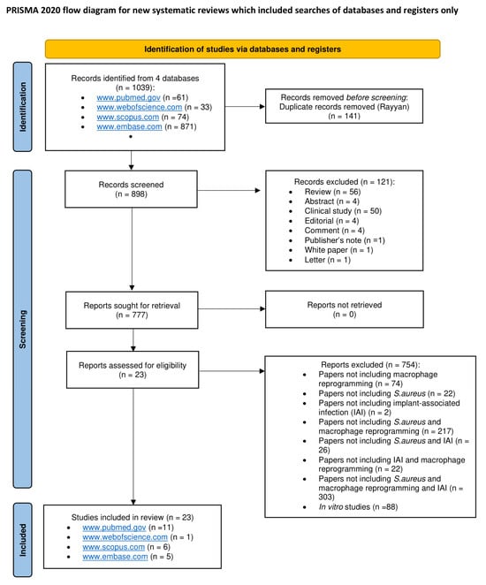
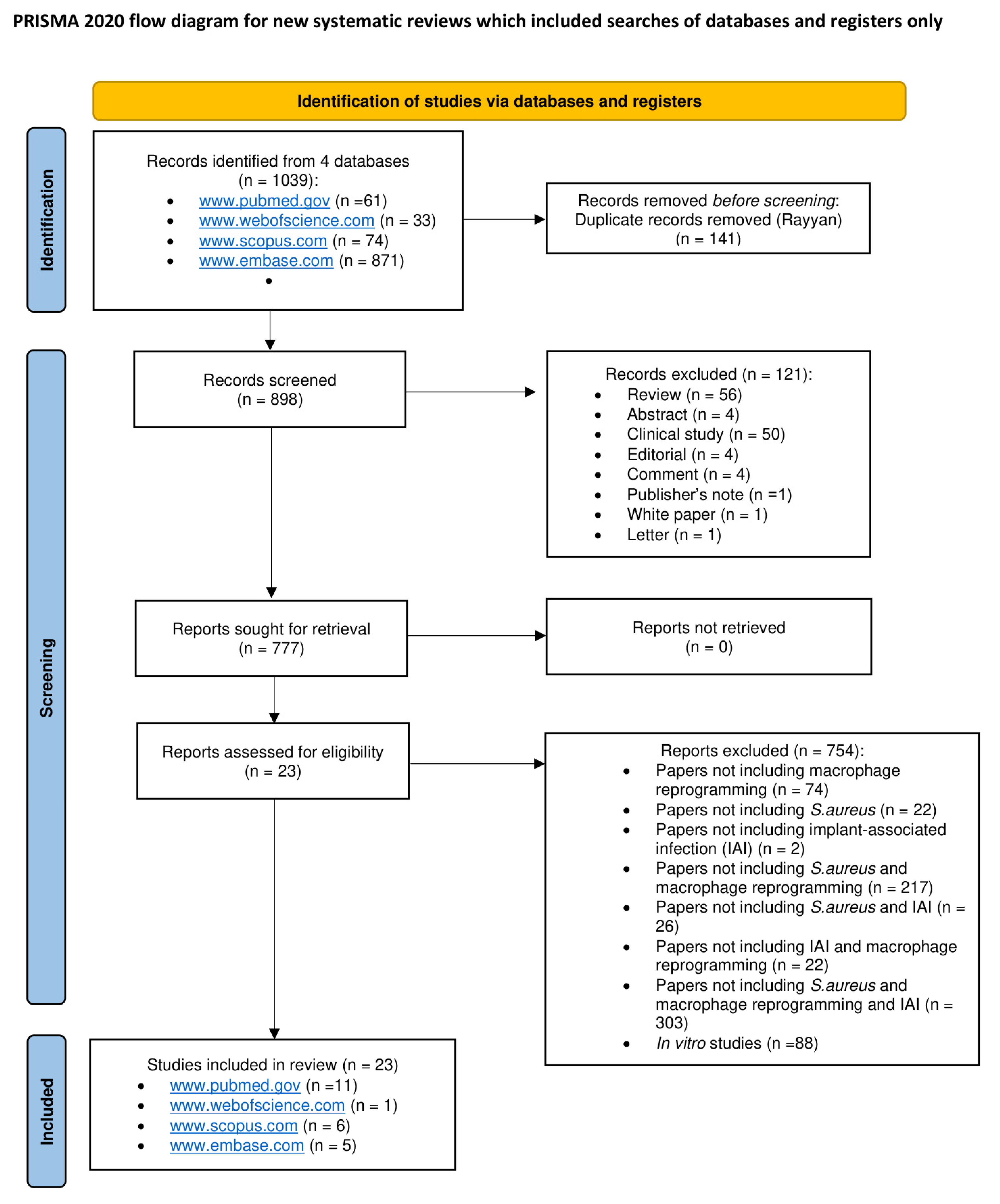
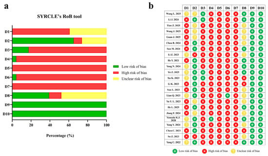
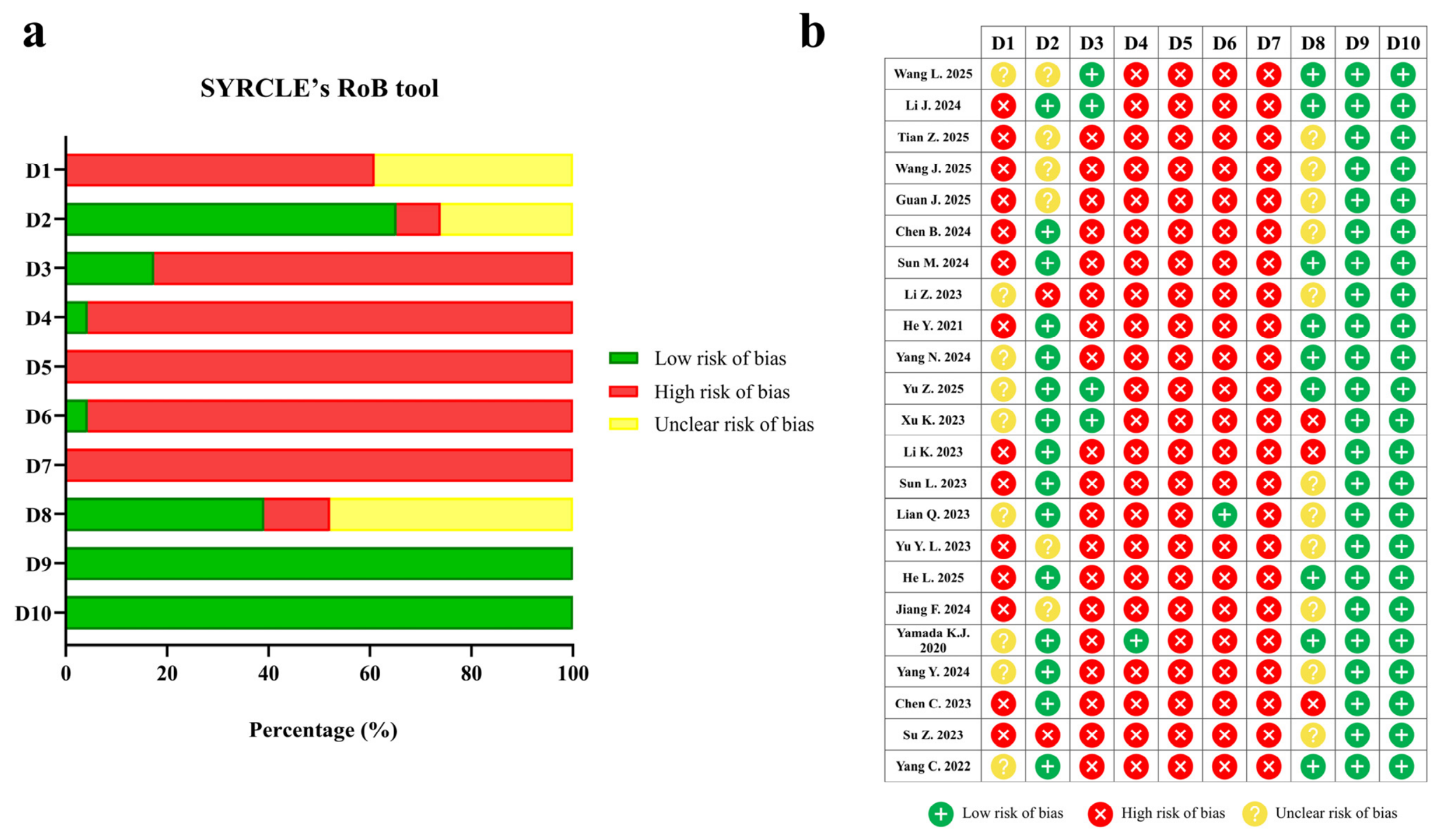
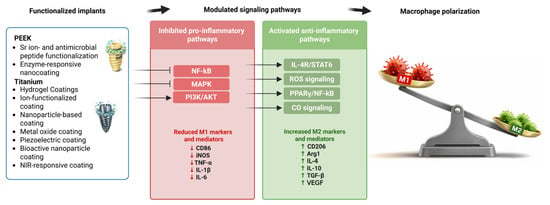
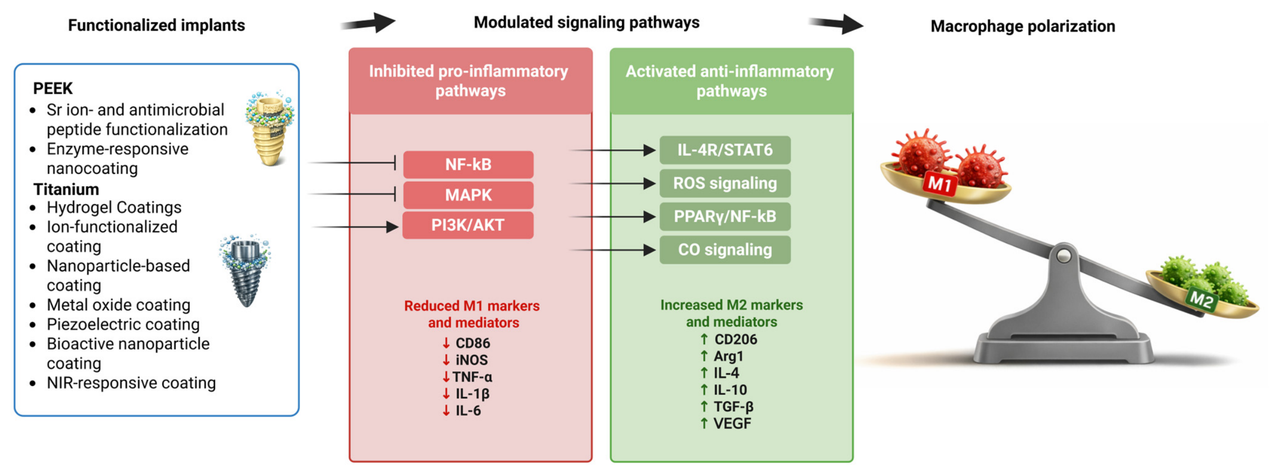
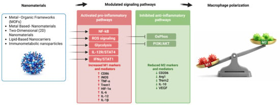
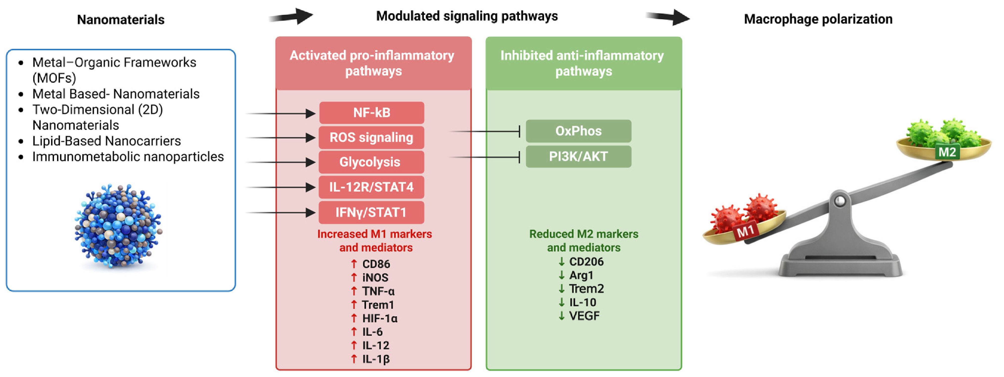

Disclaimer/Publisher’s Note: The statements, opinions and data contained in all publications are solely those of the individual author(s) and contributor(s) and not of MDPI and/or the editor(s). MDPI and/or the editor(s) disclaim responsibility for any injury to people or property resulting from any ideas, methods, instructions or products referred to in the content. |
© 2026 by the authors. Licensee MDPI, Basel, Switzerland. This article is an open access article distributed under the terms and conditions of the Creative Commons Attribution (CC BY) license.