


Abstract
Tissue engineering integrates concepts from medicine, biology, and engineering to create living constructs capable of repairing, replacing, or supporting damaged tissues. This multidisciplinary field relies on the interplay between biomaterials, cellular sources, and bioactive signaling to achieve functional tissue regeneration. This review provides a comprehensive overview of recent advances in scaffold design, highlighting natural, synthetic, and hybrid materials, as well as innovative fabrication techniques such as electrospinning, 3D bioprinting, and smart biomaterials. It discusses the role of stem cells and growth factors in directing regeneration and examines a wide range of clinical applications, including skin regeneration, cartilage repair, bone tissue engineering, dental and periodontal regeneration, nerve repair, cardiac tissue engineering, liver tissue models, and ophthalmic applications. Current challenges, such as immune responses, limited vascularization, scalability, and regulatory barriers, are addressed alongside emerging strategies aimed at improving clinical translation. By integrating diverse tissue types and engineering approaches within a unified framework, this review offers a broad yet detailed perspective on the current state and future directions of regenerative medicine.
1. Introduction
Tissue engineering (TE) brings together principles from medicine, biology, and engineering to develop living constructs capable of repairing, replacing, or supporting damaged tissues [1]. To function effectively, these engineered materials must do more than simply replicate the shape of native tissues; they need to support cellular attachment and survival, enable oxygen and nutrient delivery, and facilitate waste removal during regeneration [2]. In recent years, growing attention has been directed toward creating advanced platforms that closely mimic the structural and physicochemical characteristics of the native extracellular matrix (ECM) [2]. Achieving this level of biomimicry requires close collaboration across disciplines, with researchers from nanotechnology, biotechnology, and material science contributing to scaffold design and fabrication. Advances in nanotechnology have enabled the development of scaffolds that more precisely replicate ECM architecture at the nanoscale, enhancing cell–material interactions and biological performance [3]. Various fabrication techniques, such as solvent casting [4], freeze drying [5], self-assembly [6], template synthesis [7], phase separation [8], melt blowing [9], and electrospinning [2] have been explored to achieve scaffolds with optimal biofunctionality. As the field continues to evolve, such strategies are bringing us closer to clinically viable solutions for tissue repair and regeneration.
This review aims to summarize the latest advances in TE, focusing on the synergistic roles of biomaterials, scaffolds, cellular sources, and growth factors in promoting tissue regeneration. It explores a wide range of clinical applications, including skin regeneration, cartilage repair, bone TE, dental and periodontal regeneration, nerve repair, cardiac TE, liver tissue models, and ophthalmic applications. The review also addresses current challenges hindering clinical translation and highlights emerging technologies that may shape the future of regenerative medicine. The success of TE approaches, whether in cell-based therapies, artificial organs, or complex tissue replacements, relies on the development of multifunctional biomaterials with finely tuned surface chemistry and mechanical properties. These features must be tailored not only to support cell viability and guide tissue growth in situ, but also to match the mechanical behavior of the target tissue [10,11]. By integrating diverse tissue types and engineering strategies into a unified framework, this work provides a holistic perspective that links fundamental principles of scaffold design, cellular biology, and bioactive signaling with their translation to a wide spectrum of clinical applications. It combines recent experimental findings with advanced fabrication methods such as 3D bioprinting, hybrid scaffolds, and smart biomaterials, while also addressing translational challenges to bridge laboratory innovations with clinical practice. Rather than discussing each tissue independently, this review aims to identify shared design principles and translational barriers that shape the future of scaffold-based regenerative strategies.
2. Biomaterials and Scaffold Strategies
The design of scaffolds in TE relies heavily on the selection of appropriate biomaterials, which directly influence biocompatibility, biodegradability, mechanical integrity, and cell–material interactions. Biomaterials are generally categorized into natural, synthetic, and hybrid polymers, each offering specific benefits and limitations that make them suitable for different TE applications [12]. Natural polymers, such as collagen, gelatin, chitosan, alginate, and hyaluronic acid (HA), are derived from biological sources and closely mimic the components of the ECM. Their inherent bioactivity promotes cell adhesion and signaling, while their biodegradability and biocompatibility make them highly attractive for regenerative applications. However, natural polymers often present challenges in terms of mechanical strength, batch-to-batch variability, and thermal stability [13]. In contrast, synthetic polymers like poly (lactic acid) (PLA), poly (glycolic acid) (PGA), poly (lactic-co-glycolic acid) (PLGA), poly (ε-caprolactone) (PCL), and poly (glycerol sebacate) (PGS) offer greater control over mechanical properties, degradation rates, and structural uniformity [14,15,16].
These materials are easier to process and modify chemically, making them suitable for a wide range of applications [16]. However, synthetic scaffolds often lack the biological cues necessary for optimal cell response. To address these limitations, hybrid scaffolds have emerged as a promising strategy, combining the structural advantages of synthetic materials with the bioactivity of natural polymers [17]. Hybrid scaffolds are preferred over purely synthetic ones because they combine the mechanical strength and tunable degradation of synthetic polymers with the bioactivity and cell-friendly properties of natural materials. This synergy improves biocompatibility, supports better cell adhesion and proliferation, and enhances tissue regeneration, especially in complex environments like bone, where both structural support and biological signaling are essential [18].
Recent innovations have driven the development of smart biomaterial-engineered systems capable of responding to environmental cues such as pH, temperature, or mechanical stimuli [19]. These biomaterials can be functionalized with bioactive molecules (e.g., growth factors, peptides) and designed to support dynamic interactions with cells during tissue regeneration [20]. Notably, advancements in 3D bioprinting have revolutionized scaffold fabrication, enabling the precise deposition of cells and biomaterials (bioinks) in predefined architectures [21]. Bioprinting techniques, such as extrusion-based printing, stereolithography (SLA), and selective laser sintering (SLS), enable the construction of patient-specific implants with tunable porosity, shape, and biochemical composition. These techniques enhance the capacity to mimic native tissue microenvironments and are increasingly used in the design of complex structures for bone, cartilage, and organ regeneration [22,23].
One of the major challenges in scaffold design is achieving proper vascularization and mechanical compatibility with host tissues. Effective vascular integration is essential for nutrient and oxygen supply, especially in large or dense tissue constructs [24]. Scaffold features such as interconnected porosity, appropriate pore size, and surface topography significantly influence cellular infiltration and angiogenesis [25]. From a mechanical standpoint, scaffolds must match the stiffness and elasticity of the target tissue to provide appropriate structural support and mechanical signaling [26]. For example, bone scaffolds require higher compressive strength, while soft tissues like cardiac or neural constructs benefit from more flexible materials [27]. Advances in scaffold fabrication methods, such as electrospinning, gas foaming, and phase separation, allow for fine-tuning of these properties, enabling the creation of scaffolds that are both mechanically stable and biologically instructive [28].
3. Cellular Sources and Growth Factors
TE is fundamentally based on two biological foundations: choosing appropriate cellular sources and incorporating essential growth factors that regulate regeneration and direct tissue-specific results [29]. Among cellular sources, mesenchymal stem cells (MSCs) are the most versatile and widely used due to their ability to differentiate into multiple lineages, their immunomodulatory properties, and their relative ease of extraction from bone marrow, adipose tissue, or the umbilical cord [30]. These cells have been extensively used for bone regeneration, cartilage repair, nerve healing, and dental reconstruction [31,32,33]. Adipose-derived stem cells (ADSCs), which can be obtained with minimal invasiveness, show potential for soft tissue repair and skin applications [34]. Induced pluripotent stem cells (iPSCs) are increasingly recognized for their ability to create more complex tissue models, such as the liver [35], cardiovascular [36], and ophthalmic tissues [37], due to their pluripotency and capacity to produce patient-specific cell types. Growth factors act as biological mediators that play pivotal roles in regulating cellular behavior, promoting angiogenesis, and supporting the development of tissue-specific architecture. Integrating stem cell technologies with precise growth factor signaling is crucial for developing functional tissue replacements [38]. Ongoing research into delivery systems, such as bioactive scaffolds and controlled-release carriers, aims to refine the spatial and temporal dynamics of these biological signals, ultimately improving clinical outcomes across various TE applications [39].
4. Applications for Tissue Engineering
TE has emerged in recent years as a multidisciplinary research field with applications in skin, bone, cartilage, dentistry, nerve, cardiac, liver, and ocular regeneration. By creating optimized microenvironments, this approach accelerates tissue repair and regeneration. Although each tissue type requires its specific strategy, a common principle across all applications is achieving a balance between biocompatibility, mechanical performance, and biological signaling to ensure functional integration and long-term stability in vivo. The details of these applications are fully described in the following sections and summarized in Table 1.
Table 1.
Overview of biomaterials, growth factors, and cell sources used in tissue engineering.
4.1. Skin Tissue Engineering
Skin TE has become a vital solution for treating extensive wounds, burns, and chronic skin injuries, particularly in situations where traditional therapies such as autografts, despite being the clinical gold standard, are constrained by donor site morbidity or limited graft availability. The primary goal is to restore the skin’s multilayered structure and function through coordinated matrix remodeling and regenerative strategies [91]. A key element in the development of tissue-engineered skin substitutes is the use of scaffold matrices engineered to support three-dimensional cell growth. Designed to be biocompatible, biodegradable, and suitable as wound dressings, these scaffolds provide an optimal environment for tissue repair. Recent advances have shifted towards seeding scaffolds with specific cell populations, such as keratinocytes and fibroblasts, to further promote and refine regenerative outcomes [40]. In the context of skin regeneration, growth factors such as epidermal growth factor (EGF) and vascular endothelial growth factor (VEGF) play a pivotal role in promoting re-epithelialization and stimulating neovascularization within wound beds [92,93,94].
Scaffolds in Skin Tissue Engineering
Scaffolds are essential to skin TE, functioning as three-dimensional matrices that support cell adhesion, migration, proliferation, and differentiation while directing new tissue formation [95]. They are fabricated from a wide range of natural and synthetic biomaterials, used individually or in combination to balance biocompatibility, biodegradability, and mechanical strength. Natural biomaterials, such as collagen, cellulose, and chitosan, are protein- or polysaccharide-based and closely mimic the native ECM, making them highly biocompatible, readily degradable, and well-suited for skin cell growth. In contrast, synthetic biomaterials, including nanomaterials like polyvinylpyrrolidone (PVP), PCL, polyethylene glycol (PEG), and PLA, provide mechanical strength and structural stability [40]. Combining these materials in composite scaffolds, such as PCL–collagen matrices or silk fibroin–polyurethane (PU) blends, merges biological cues with enhanced durability. Collagen remains the most widely used natural scaffold due to its ECM-like architecture, while clinically applied acellular dermal matrices (e.g., Integra®, AlloDerm®) also leverage preserved ECM structure to support host cell infiltration and gradual tissue remodeling [40].
Notably, blood plasma, fibrinogen, and fibrin have also been highlighted as promising natural biomaterials for skin tissue engineering, demonstrating strong preclinical wound-healing potential, although their clinical translation remains limited [96]. Examples of commonly used natural and synthetic scaffolds for skin TE are summarized in Table 2.
In skin TE, several fabrication strategies have been developed, with four primary approaches: ECM-secreting cell sheets, pre-fabricated porous scaffolds made from synthetic, natural, or biodegradable materials, decellularized ECM scaffolds, and cell-laden hydrogels that provide a hydrated, supportive 3D environment [40]. Electrospun gelatin nanofiber scaffolds have shown promising potential for wound healing applications [97]. Various gelatin-based formulations for wound and burn dressings, such as gelatin–alginate sponges [98], gelatin incorporated with EGF [99], and gelatin films, have also demonstrated efficacy in the treatment of skin injuries. To further improve scaffold performance, researchers have explored the incorporation of nanoparticles, which can enhance antibacterial activity, angiogenesis, and cell proliferation (Table 3). For example, poly(3-hydroxyoctanoate)-fabricated nano-sized 45S5 Bioglass® scaffolds have been reported for use in wound dressings, as they provide a favorable environment for tissue growth, offering properties such as wettability and rough surfaces, and can accelerate blood clot formation [100]. Similarly, Chua et al. reported that composite scaffolds that integrate natural polymers such as collagen with synthetic components like elastin or glycosaminoglycans have shown enhanced vascularization, mechanical stability, and long-term integration in clinical burn treatments, underscoring the potential of combining bioactivity with tunable degradation profiles [101].
Table 2.
Overview of natural and synthetic scaffolds used in skin tissue engineering and their functional roles in wound repair.
Different wound types require different scaffold formats. Porous scaffolds, such as sponges and foams made from collagen, PCL, or PLGA, offer high surface area and interconnected pores that facilitate cell penetration and nutrient diffusion. These are typically fabricated using salt leaching or 3D printing to control porosity and architecture [40]. Fibrous scaffolds, particularly electrospun mats, closely mimic the fibrous structure of natural ECM and have been created using polymers such as chitosan, silk fibroin, and gelatin to support the growth of keratinocytes and fibroblasts [109]. Hydrogel-based scaffolds provide high water content and favorable mechanical properties, making them especially suitable for irregular wound geometries and full-thickness defects. Materials such as alginate, HA, and PEG have been used to create hydrogels capable of controlled drug and growth factor release [110]. These systems can function as injectable materials or wound dressings that conform to wound beds while delivering bioactive cues.
In addition to the scaffold architecture, cellular components play a critical role in tissue restoration. Primary human keratinocytes and fibroblasts are frequently employed to reconstruct the epidermis and dermis, respectively. More recently, MSCs and ADSCs have gained attention for their secretion of pro-regenerative cytokines and promotion of neovascularization. For example, ADSCs embedded in a fibrin scaffold enhanced wound closure and dermal regeneration in diabetic rats [40], while human umbilical cord-derived MSCs in a decellularized dermal matrix improved epithelialization and reduced scar formation [41].
Progress toward clinical translation includes bilayered scaffolds that mimic the epidermal–dermal interface, with keratinocytes on the surface and fibroblasts embedded in a collagen matrix, as well as growth factor integrated systems (e.g., microspheres or surface-immobilized ligands) that enhance vascularization and skin barrier restoration [101,119]. Despite these advances, challenges remain, including poor integration with host tissue, limited sensory recovery, and inadequate vascularization in chronic or extensive wounds. Current research is addressing these limitations through pre-vascularized scaffolds, immunomodulatory hydrogel systems, and patient-specific cell sources such as iPSCs [101,119]. Huang et al. demonstrated that bFGF-loaded collagen scaffolds seeded with bFGF-overexpressing human umbilical cord mesenchymal stromal cells significantly accelerated diabetic full-thickness wound healing through enhanced angiogenesis, collagen deposition, and re-epithelialization, primarily via activation of the HIF-1 signaling pathway [120]. Similarly, bFGF-loaded silk fibroin scaffolds enhanced dermal regeneration and neovascularization in full-thickness defects [42], and TGF-β1-modified hydrogel systems reduced inflammation while improving ECM deposition and collagen synthesis in vivo [43].
These advances highlight the progress of scaffold-based approaches; however, the next frontier lies in smart and dynamic platforms such as 4D bioprinting and Artificial intelligence (AI)-assisted scaffold design, which enable responsive, patient-specific constructs to overcome current challenges in vascularization, integration, and controlled therapeutic delivery [121].
Table 3.
Nanomaterial-based scaffolds for skin TE.
4.2. Bone Tissue Engineering
Bone tissue engineering (BTE) relies on a triad of components: progenitor cells, bioactive signaling molecules, and scaffolds that provide essential structural and biological cues for new bone formation [136]. Among these, skeletal stem cells (SSCs) have emerged as pivotal elements due to their intrinsic ability to differentiate into multiple lineages, including osteoblasts, chondrocytes, and adipocytes [137]. The concept of SSCs was first confirmed by Friedenstein and colleagues, who demonstrated that colony-forming unit-fibroblasts (CFU-Fs) from bone marrow stromal cells could generate bone in vivo. Bianco et al. proposed that SSCs reside in the perivascular niches of bone marrow and can form complete heterotopic ossicles, including bone and hematopoietic marrow, when transplanted [2,138].
4.2.1. Bioactive Molecules and Growth Factor Delivery
Different factors that promote tissue growth have been found at the skeletal damage site and have a physiologic role in healing bone fractures. Osteoinductive GFs such as platelet-derived growth factors (PDGFs), bone morphogenic proteins (BMPs), insulin-like growth factors (IGFs), transforming growth factors (TGFs-ß), and VEGFs have presented great application potentials in bone healing and osteogenesis for regulating cell behavior, including recruitment, migration, adhesion, proliferation, and differentiation [139]. The incorporation of growth factors into scaffolds further amplifies their regenerative potential. BMP-2 is one of the most widely studied osteoinductive agents and has been shown to significantly enhance stem cell recruitment, proliferation, and osteogenic differentiation when loaded into PLGA or gelatin-based scaffolds [140]. For example, gelatin-based hydrogels and poly PLGA scaffolds loaded with BMP-2 have shown increased bone mineralization and improved defect healing in various preclinical models [141]. Chai et al. developed an injectable scaffold system using photo-crosslinked GelMA hydrogel loaded with bone marrow mesenchymal stem cells (BMSCs) and bone morphogenetic protein 2 (BMP2) and demonstrated that this combination significantly enhanced osteogenic differentiation and bone defect repair in vivo [142]. Similarly, VEGF has been integrated into scaffold systems to stimulate angiogenesis, which is essential for the survival of large tissue-engineered constructs. Studies have shown that co-delivery of VEGF and BMP-2 enhances both vascularization and osteogenesis, leading to more robust and stable bone formation [143]. These combined approaches emphasize the importance of delivering both osteogenic and angiogenic signals to recapitulate the complexity of bone healing.
4.2.2. Scaffold Materials in Bone Tissue Engineering
Scaffolds used in BTE must replicate the natural ECM in terms of both structure and function. These scaffolds provide a three-dimensional environment that facilitates cell adhesion, proliferation, and differentiation [144]. A wide range of natural and synthetic polymers has been explored for this purpose. Natural polymers such as collagen, chitosan, and alginate offer excellent biocompatibility and bioactivity, but are often limited by poor mechanical strength and rapid degradation [145]. Several studies have demonstrated the efficacy of specific scaffold systems in promoting bone regeneration. Farjaminejad et al. developed novel bio-rubber scaffolds based on poly (glycerol sebacic acid) modified with succinic acid and ε-caprolactone, reinforced with nano-hydroxyapatite and nanoclay, enabling precise tuning of hydrophilicity, mechanical strength, and degradation rate to optimize performance for bone regeneration applications [15].
Wang et al. fabricated a 3D-printed PLA scaffold incorporating nano-hydroxyapatite (nHA), which significantly enhanced mineralization and vascular infiltration when tested in vivo [146]. Similarly, Diao et al. developed β-tricalcium phosphate (β-TCP) scaffolds with gradient pore sizes (ranging from 300 to 500 µm) and observed enhanced migration and differentiation of MSCs, supporting better integration with host tissue [147]. Lee et al. also discussed the impact of scaffold architecture, highlighting how mechanical stiffness, pore size, and rough surface influence osteogenic differentiation [46].
Collagen type I, particularly when functionalized with the P-15 cell-binding domain, provides a biomimetic and highly effective environment for promoting the adhesion, proliferation, and differentiation of MSCs [148]. These fibrous scaffolds not only serve as a suitable substrate for cell growth but also actively participate in osteogenesis by guiding the formation and spatial arrangement of carbonated apatite minerals. Moreover, under cellular stress conditions, the collagen matrix can stimulate MSC migration to injury sites through the release of chemotactic signals [149,150]. As shown in Figure 1, scanning electron microscopy (SEM) images illustrate the sequential stages of cell–scaffold interaction: (A) before cell seeding, the fibrous structure of collagen is clearly visible; (B) 8 h after seeding, cells adhere effectively to the collagen fibers; (C) at 24 h, ECM formation and cell ingrowth into the collagen fiber network are evident; and (D) after 48 h, complete cell–matrix–scaffold integration is achieved.
Figure 1.
Representative SEM images showing the sequential attachment and growth of human bone marrow-derived MSCs on type I collagen fibers derived from human skin. (A) Before cell seeding, (B) 8 h after seeding, cells adhere to collagen fibers; (C) 24 h after seeding, ECM formation and cell ingrowth into the collagen fiber network are observed; (D) 48 h after seeding, complete integration of cells, ECM, and the collagen scaffold is achieved. Reprinted from Ref. [44].
Nanoparticles in bone TE have introduced new dimensions to scaffold development by improving structural integrity, biological signaling, and drug delivery capabilities [151]. In a recent review, Farjaminejad et al. outlined the role of nano-hydroxyapatite in mimicking the mineral phase of bone and enhancing osteoconductivity [47].
Recent advances suggest that integrating electrical stimulation (ES) into BTE can enhance osteogenesis and angiogenesis via BMP and Wnt/β-catenin pathway activation. Piezoelectric scaffolds, such as PVDF/PVDF-TrFE, BaTiO3-reinforced polymers, and β-PCL nanofibers, generate endogenous electrical cues under mechanical loading, mimicking bone’s bioelectric environment and promoting osteoblast activity [152].
Despite substantial advancements, several challenges remain in translating these strategies into clinical applications. Variability in stem cell populations, immune responses to synthetic materials, inconsistent scaffold degradation rates, and the complexity of vascular integration continue to limit the routine use of BTE in clinical settings. Black et al. emphasized the importance of aligning scaffold design with biological requirements and preclinical validation to facilitate regulatory approval and clinical translation [153]. The need for standardization in scaffold fabrication, material testing, and cell characterization remains urgent to bridge the gap between laboratory research and patient treatment [153]. One promising approach to overcoming these challenges is the integration of AI, which can accelerate material discovery, optimize scaffold properties, and reduce experimental time and cost. For example, Mackay et al. (2021) applied a conditional generative adversarial network (GAN) to predict the behavior of Stro-1 selected human bone marrow stromal cells (HBMSCs), successfully reducing culture time by 24 h, demonstrating how AI can directly address resource-intensive steps in bone tissue engineering [154].
Overall, BTE integrates biological and material sciences to develop scaffolds that can restore bone integrity. Specific examples, such as BMP-2-loaded PLGA scaffolds, nano-HA-reinforced polymer matrices, and GO-enhanced constructs, reflect the sophistication of current approaches. With continuous refinement of scaffold composition, cellular selection, and biofunctionalization, BTE holds strong promise for becoming a routine part of regenerative orthopedic and dental therapies.
4.3. Cartilage Tissue Engineering
Cartilage TE has emerged as an effective approach for repairing damaged cartilage, particularly articular cartilage, which regenerates poorly due to its limited blood supply and nerve connections, low cell density, and slow matrix regeneration. Conventional clinical methods such as autologous chondrocyte implantation, microfracture, and osteochondral grafts often result in the development of mechanically weaker fibrocartilage and are associated with inconsistent long-term results, donor site complications, and chondrocyte dedifferentiation [51]. Cartilage TE strategies rely on the coordinated use of three essential components: chondrogenic progenitor cells such as bone marrow-derived mesenchymal stem cells (BMSCs), signaling molecules like TGF-β1 to induce chondrogenic differentiation, and biocompatible scaffolds, particularly injectable hydrogels such as GelMA, which provide structural support and promote cell viability, ECM production, and integration with host tissue [52].
MSCs, especially BMSCs, have been extensively studied as a promising cell source for cartilage regeneration due to their strong chondrogenic potential, immunomodulatory functions, and ease of harvesting from bone marrow aspirates. When stimulated with factors like TGF-β1, BMSCs can differentiate into chondrocytes and produce key cartilage ECM components such as collagen type II and aggrecan. Their ability to modulate inflammatory responses and enhance matrix synthesis supports their therapeutic potential in osteochondral repair strategies. Studies indicate that MSCs placed within scaffolds can significantly promote subchondral bone regeneration and improve cartilage integration in defect models [53]. The incorporation of chondrogenic growth factors significantly enhances the functional performance of engineered cartilage. Commonly used factors, such as TGF-β, BMPs, and IGF-1, effectively promote chondrogenic differentiation. Research by Zhao et al. demonstrated that scaffolds infused with TGF-β1 led to increased cartilage matrix deposition and reduced inflammation, while the addition of BMP-7 improved cartilage-specific gene expression and matrix development [52,155].
Scaffold Material Cartilage Tissue Engineering
Biocompatible scaffold materials play a crucial role in the survival, growth, and chondrogenic differentiation of MSCs. These scaffolds must provide optimal mechanical strength, feature interconnected porosity, and be biodegradable while mimicking the intrinsic ECM of cartilage [52]. Zhao et al. reviewed various scaffold types used in cartilage TE, including natural polymers like collagen and chitosan, synthetic polymers such as PLGA, and decellularized cartilage matrix. For instance, chitosan-collagen composite scaffolds promoted chondrocyte proliferation and improved histological structure in animal studies [52]. Furthermore, alginate and HA-based hydrogels have been shown to enhance nutrient diffusion and boost matrix production, contributing to improved neocartilage formation [156]. 3D bioprinting is an innovative technique in cartilage TE, allowing for the precise layer-by-layer placement of cells and biomaterials, thus resembling the natural cartilage structure more closely than conventional methods. Among the materials used, GelMA-based bioinks stand out for their excellent cell compatibility and adjustable mechanical properties [157]. Zheng et al. developed GelMA/silk fibroin interpenetrating network hydrogels with enhanced mechanical strength and biocompatibility, which supported BMSC chondrogenesis and promoted in vivo cartilage regeneration, highlighting their potential for functional cartilage repair [158]. Together, these findings highlight the synergistic interaction of MSCs, bioactive scaffolds, and chondrogenic signals in regenerating functional cartilage tissue. Ongoing advancements in biomaterial design and bioprinting technologies are anticipated to further enhance the clinical application of cartilage TE for treating osteoarthritis and cartilage injuries.
4.4. Dental and Periodontal Applications
TE has become a groundbreaking technique in modern dentistry, offering innovative strategies for regenerating dental pulp, periodontal structures, and alveolar bone [159]. Conventional treatments like root canal therapy, periodontal surgery, and bone grafting often fall short in restoring the biological and functional integrity of dental tissues [160]. In contrast, TE employs a triad of stem cells, signaling molecules, and scaffolds to overcome these limitations and enhance regeneration [161].
In dental pulp regeneration, scaffolding materials such as collagen, chitosan, HA, and fibrin are utilized to mimic the native ECM and support cellular organization and vascularization [162]. For instance, chitosan/gelatin scaffolds crosslinked with 0.1% glutaraldehyde and seeded with BMP-2 pretreated dental pulp stem cells have been shown to enhance osteo/odontogenic gene expression and promote in vivo mineralized bone formation, supporting their potential for alveolar/orofacial reconstruction [163]. Keswani et al. demonstrated that the clinical application of platelet-rich fibrin in immature permanent teeth led to continued root development, including increased root length and dentinal wall thickness, due to sustained growth factor release that enhances angiogenesis and tissue repair [164]. For periodontal regeneration, stem cells from sources such as the periodontal ligament, dental follicle, and gingiva can be combined with biomaterial scaffolds, such as collagen, chitosan, PLGA, and hydrogels, often supplemented with bioactive molecules like BMP-2 and PDGF to enhance osteogenesis, angiogenesis, and regeneration of the periodontal complex [165]. Mafalda et al. developed electrospun polycaprolactone/chitosan scaffolds loaded with lyophilized extracellular matrix from periodontal ligament stem cells, which maintained favorable physical properties, enhanced PDLSC proliferation, and significantly promoted osteogenic differentiation and mineralization, showing promise for alveolar bone regeneration in periodontal tissue engineering [166]. Injectable hydrogels, particularly GelMA, have gained attention for their ability to deliver stem cells and bioactive factors minimally invasively, supporting extracellular matrix production and integration with surrounding pulp tissue [167].
Advanced fabrication methods like 3D bioprinting and electrospinning have enabled the design of anatomically and functionally relevant scaffolds with aligned fibers and gradient structures. These techniques provide spatial control over cell placement and enhance mechanical strength, promoting the regeneration of both soft and hard periodontal tissues [165,168]. An example of such an approach is shown in Figure 2, depicting a 3D-bioprinted hybrid polycaprolactone–hydroxyapatite scaffold with anatomically defined architecture and microchannels to facilitate cell homing and angiogenesis, illustrating the potential of advanced scaffold fabrication for periodontal TE [168]. Cell homing strategies utilizing bioactive molecules, such as stromal cell-derived factor-1 (SDF-1) and VEGF, have gained attention as promising approaches in periodontal and pulp regeneration. When incorporated into scaffolds, these factors can recruit endogenous progenitor cells to defect sites, thereby reducing or eliminating the need for exogenous stem cell transplantation [169]. Nanotechnology has significantly advanced restorative dentistry through the development of nanoparticle-enhanced dental composites. Incorporating materials such as silica, nano-hydroxyapatite, and zirconia improves mechanical strength, aesthetics, durability, and antibacterial properties, while reducing polymerization shrinkage [170]. Clinical evidence supports their safety, biocompatibility, and potential for longer-lasting restorations, although further research is needed to address technical and economic challenges and to develop smart, multifunctional materials for personalized and regenerative care [171].
Figure 2.
Three-dimensionally bioprinted hybrid polycaprolactone–hydroxyapatite scaffolds replicating a rat mandibular incisor (A) and the human mandibular first molar (B), featuring 200 µm microstrands with interconnecting microchannels. (C,D) Human molar scaffold channels (E) were infused with stromal-derived factor-1 and BMP-7 in type I collagen before gelation. Reprinted from Ref. [168].
4.5. Nerve Regeneration
Nerve tissue engineering (NTE) has emerged as a promising strategy to overcome the limited regenerative capacity of the central and peripheral nervous systems, addressing conditions such as spinal cord injury (SCI), traumatic brain injury (TBI), degenerative diseases, and peripheral nerve injuries (Figure 3A), where conventional treatments often fall short in restoring function [172]. TE combines biomaterials, cells (e.g., iPSC, iPSC-derived neural crest stem cells, Schwann cells, and MSC), and bioactive molecules such as nerve growth factor (NGF), brain-derived neurotrophic factor (BDNF), glial cell line-derived neurotrophic factor (GDNF), FGFs, and VEGF to create a microenvironment conducive to nerve repair and regeneration (Figure 3B) [173].
Figure 3.
(A) NTE aims to restore function in conditions such as SCI, TBI, degenerative diseases, and peripheral nerve injuries, where traditional treatments fall short. By combining biomaterials, cells (e.g., iPSCs, Schwann cells, MSCs), and bioactive molecules (e.g., NGF, BDNF, GDNF, FGFs, VEGF), NTE creates a supportive microenvironment for nerve repair and regeneration. (B) Schematic representation of the NTE approach, showing cell sources, biomaterial scaffolds, and neurotrophic factors working together to form an engineered neural construct that promotes neuronal regeneration (Created with BioRender.com).
A central focus in NTE is the design of scaffolds that can mimic the native ECM, support cellular growth, and guide axonal regrowth. Natural polymers such as collagen, gelatin, and chitosan are frequently used due to their biocompatibility and ability to support Schwann cell adhesion and proliferation. For instance, chitosan-based conduits have demonstrated efficacy in promoting neurite extension and enhancing myelination [57]. Synthetic polymers like PLGA and PCL are also widely utilized for their tunable mechanical properties and degradation rates, often modified with nanoparticles or growth factors to enhance bioactivity [58].
Advanced fabrication techniques such as electrospinning and 3D bioprinting have enabled the creation of aligned nanofiber scaffolds that guide axon orientation. Incorporating electrical stimulation and conductive materials, including graphene and polypyrrole (PPy), further promotes neurite outgrowth and functional recovery. One study reported the integration of Schwann cells into electrospun PLGA scaffolds, resulting in improved axonal regrowth in a rat sciatic nerve model [58]. Moreover, recent work has emphasized the role of immune modulation in successful nerve repair. Creating scaffolds that modulate the local immune response, such as those releasing anti-inflammatory agents or promoting M2 macrophage polarization, has been shown to significantly enhance regeneration outcomes. This immunomodulatory approach, combined with scaffold topography and biochemical signaling, provides a holistic strategy for addressing both peripheral nerve injuries and central nervous system trauma [174].
Feng et al. developed a chitosan conduit filled with regulatory T cells (Tregs) and supported by a nano artificial growth factor hydrogel containing a cyclic BDNF mimetic sequence for a 6 mm sciatic nerve defect in mice. This approach preserved Treg stability and Foxp3 phenotype, enhanced angiogenesis, promoted M2 macrophage polarization, and accelerated nerve regeneration, myelination, and functional recovery, highlighting the potential of scaffold-mediated immune modulation in peripheral nerve repair [175].
Beyond scaffold-based methods, cell-free therapies using extracellular vesicles, particularly exosomes (30–150 nm), are emerging as promising tools for neural repair. Nochlabadi et al. highlighted that these vesicles, secreted by various cells, deliver bioactive molecules that promote angiogenesis, reduce inflammation, and support cell proliferation and differentiation. Their small size, biocompatibility, low immunogenicity, and ability to cross the blood–brain barrier make them attractive for nerve regeneration, with ongoing research focusing on enhancing targeting and therapeutic delivery [176].
4.6. Liver Tissue Engineering
Liver TE (LTE) has emerged as a promising approach to address the shortage of donor organs and the limitations of conventional liver transplantation, particularly for patients with advanced or terminal liver diseases such as chronic hepatitis C infection, non-alcoholic fatty liver disease, alcoholic fatty liver disease, decompensated cirrhosis, hepatocellular carcinoma, biliary atresia, and Wilson’s disease (Figure 4A). These conditions, often culminating in cirrhosis and portal hypertension, progressively destroy hepatic structure and function, leaving transplantation as the only curative option in many cases [60]. A key strategy involves the use of decellularized liver extracellular matrix (dECM), which preserves the liver’s native microarchitecture, vascular network, and biochemical composition. Perfusion-based decellularization and recellularization techniques have enabled the engineering of whole-liver grafts with preserved vascular networks, enhancing their transplantation potential. For example, dECM scaffolds derived from human liver tissue have been used as bioinks for 3D bioprinting, creating a physiologically relevant environment for stem cell differentiation and hepatic phenotype induction (Figure 4B) [61].
Figure 4.
(A) Progression of liver diseases leading to the need for transplantation. Conditions such as fatty liver disease, cirrhosis, hepatocellular carcinoma, and advanced liver cancer may ultimately require liver transplant as a treatment option (Created with BioRender.com). (B) Schematic representation of bioengineered liver development from decellularized scaffolds. Ongoing decellularization of an individual rat liver lobe is achieved through continuous detergent perfusion, producing an acellular scaffold. This scaffold is recellularized with autologous cells, including hepatocytes, endothelial cells, and cholangiocytes, which are expanded and differentiated in vitro. The seeded scaffold is then matured in a bioreactor to generate a bioengineered liver for potential transplantation into the patient. Panels A and B were designed using BioRender (Created with BioRender.com). (C) Macroscopic view of interspecies biocompatibility assessment of human liver cubic scaffolds implanted into the omentum of immunocompetent mice (n = 6). Images at weeks 1 and 3 post-implantation show that the scaffold has integrated well with surrounding host abdominal tissues, including the liver, intestine, and pancreas, with no signs of acute rejection or inflammation. (C) reprinted from Ref. [177].
Overall, LTE strategies integrate functional hepatic cells with bioactive scaffolds and signaling molecules to restore native liver architecture and recover metabolic and synthetic functions. The primary objectives include sustaining long-term hepatocyte viability, maintaining urea and albumin metabolism, and promoting vascularization for efficient oxygen and nutrient delivery [60,178]. Demonstrating interspecies biocompatibility, Mazza et al. implanted human liver cubic scaffolds into the omentum of immunocompetent mice and observed early post-implantation engraftment within host abdominal structures, without signs of acute rejection or inflammation [177].
Scaffold Materials and Bioactive Factors in LTE
Scaffolds fabricated from both synthetic and natural polymers have been extensively investigated to mimic the properties of the hepatic ECM while offering tunable degradation rates and mechanical characteristics. For instance, gelatin–alginate composite hydrogels have been shown to support HepG2 cell viability and maintain urea and albumin secretion, with scaffold microarchitecture significantly influencing hepatic function [60]. Similarly, peptide-based hydrogels fabricated via 3D bioprinting recreated hepatic lobule-like spatial arrangements and channel-like pathways for nutrient perfusion, enhancing the metabolic activity of MSCs differentiated into hepatocyte-like cells [178]. Poly (ethylene glycol)-fibrinogen (PEG-Fib) hydrogels supported hepatocyte-like cells with improved hepatic gene expression and metabolic function compared to 2D cultures [178]. Electrospun poly(L-lactide-co-ε-caprolactone)/gelatin (PLCL/Gel) scaffolds promoted cell adhesion and liver-specific function in co-culture with endothelial and hepatocyte cells [62]. Porous scaffolds and microcarriers made from alginate, chitosan, silk fibroin, and PCL have also been explored, sometimes functionalized with RGD peptides or heparin to improve cell attachment and bioactivity. In some cases, these structures were coated with liver-derived ECM or supplemented with growth factors to better replicate the native hepatic microenvironment [62,63]. Bioprinting technologies have played a significant role in creating complex liver-mimetic architectures. For example, GelMA-based bioinks containing hepatocytes and human umbilical vein endothelial cells (HUVECs) have been used to fabricate 3D liver constructs, demonstrating enhanced albumin secretion, CYP450 activity, and bile canaliculi formation over time. The inclusion of endothelial cells improved vascular-like channel formation and reduced hypoxia in larger constructs [62,63].
Combining bioprinting technologies with bioactive scaffold materials, either natural or synthetic, growth factors, and suitable hepatic cell sources can provide an effective platform for functional liver regeneration. However, challenges such as achieving long-term hepatic functionality, complete vascular integration, and protocol standardization for clinical translation remain to be addressed.
4.7. Cardiac Tissue Engineering
Cardiac TE has emerged as a promising approach to address the limited regenerative capacity of the adult heart. Cell-based strategies, particularly those utilizing human pluripotent stem cells (hPSCs), have gained increasing attention due to their ability to differentiate into cardiomyocytes (CMs) and other cardiac-relevant cell types. However, several challenges remain in achieving functional and clinically relevant cardiac constructs. A wide range of cell sources have been explored for cardiac regeneration, including MSCs, cardiac progenitor cells, and more recently, hPSC-derived cardiomyocytes (hPSC-CMs). Among these, iPSCs are especially appealing due to their self-renewal capacity and patient-specific compatibility. Advances in differentiation protocols now enable the generation of distinct subtypes of cardiomyocytes, atrial, ventricular, and pacemaker cells, with high purity through modulation of the Wnt/β-catenin pathway [64]. Despite this progress, hPSC-CMs remain phenotypically immature, resembling fetal rather than adult cardiomyocytes in terms of structure, contractile force, metabolism, and electrophysiological properties [64,65]. Immaturity limits their utility in both in vivo therapies and in vitro disease models.
To overcome immaturity, a variety of bioinspired strategies have been developed. Functional maturation has been improved through the application of electrical and mechanical stimulation, co-culture with non-myocyte cells such as fibroblasts and endothelial cells, and exposure to ECM-like environments. Cardiac TE benefits from angiogenic and cardioprotective signals provided by VEGF and hepatocyte growth factor (HGF), which together enhance vascular integration and maintain myocardial viability post-injury [179]. For instance, dynamic culture systems and engineered scaffolds have been used to replicate in vivo mechanical loading, which promotes sarcomere alignment and electrophysiological development [64,65]. Conductive biomaterials have been introduced to facilitate synchronized electrical signaling. Incorporation of gold nanorods (GNRs), carbon-based nanomaterials, and silicon nanowires into hydrogels has enhanced expression of cardiac markers (e.g., Connexin-43, cardiac troponin I), improved calcium handling, and supported synchronous contractions in engineered tissues [66].
Scaffold Materials and Bioactive Factors in Cardiac Tissue Engineering
Recent advances in cardiac TE have brought together bioprinting technologies and smart biomaterials to create more functional and physiologically relevant constructs. Among these, 3D bioprinting enables precise spatial control over the distribution of multiple cell types, such as cardiomyocytes, fibroblasts, and endothelial cells, within bioinks that mimic the anisotropic architecture of native myocardium [67]. Techniques like layer-by-layer printing and the incorporation of anisotropic conductive scaffolds have resulted in improved cell alignment, synchronous contractility, and structural fidelity in engineered cardiac tissues. Moreover, the integration of vascular-like channels and supportive stromal cells has significantly enhanced nutrient diffusion, oxygen delivery, and overall viability in thicker constructs, addressing one of the main limitations of avascular tissue models [65,67].
The selection of appropriate materials for scaffold fabrication plays a vital role in the successful functional regeneration of cardiac tissue. To date, two main categories of materials, synthetic polymers and natural or decellularized extracellular matrix (dECM), have been widely explored, each offering distinct advantages and limitations. Among synthetic polymers, materials such as PCL, polyglycerol sebacate (PGS), PU, PLA, and polyglycolic acid (PGA) have been frequently utilized. These polymers are biodegradable, possess favorable mechanical properties, and allow ease of processing. However, they are often hydrophobic and lack cell recognition sites, which compromises cell adhesion and requires further surface modification [68]. Several researchers have attempted to address these limitations. For instance, Tallawi et al. developed PGS/PCL-based composites functionalized with biological coatings such as collagen, laminin, and fibronectin, which significantly enhanced cellular proliferation, tissue alignment, and synchronous contractile behavior [69]. Likewise, Boffito and Ciardelli employed techniques such as electrospinning and bioprinting to fabricate anisotropic scaffold architectures that mimic myocardial tissue. Through chemical and biological surface modifications, including RGD peptide grafting, they improved cellular adhesion and functionality [68].
Decellularized matrices, particularly those derived from human or animal hearts, have gained increasing attention due to their preserved 3D ECM structure and native protein composition. Barbulescu et al. demonstrated that human heart-derived dECM retained vascular architecture, collagen, elastin, and essential ECM components following optimized decellularization processes. These scaffolds supported enhanced stem cell proliferation and differentiation, while also downregulating fibrotic gene expression and upregulating pro-angiogenic markers [70]. Additional studies have shown that cardiac dECM can effectively support cardiomyocytes, fibroblasts, and endothelial cells by providing a bioactive environment, although challenges remain regarding immunogenicity and standardization of decellularization protocols [70].
Roacho-Pérez et al. highlighted that synthetic scaffolds, along with appropriate cell sources and growth factors, represent one of the three essential pillars of cardiac tissue regeneration [71]. They emphasized that effective cardiac scaffolds must not only be biocompatible and biodegradable but also possess electrical conductivity, contractile properties, and the ability to replicate the anisotropic nature of myocardial tissue. In this context, materials such as PCL, gelatin, collagen, fibrin, PPy, graphene, and conductive nanoparticles have been explored, with hybrid formulations like PCL/PPy and collagen/graphene showing potential to enhance electrical conductivity and promote synchronized cardiac tissue formation in electrically responsive environments [71].
Hybrid strategies that combine synthetic polymers with biofunctionalization approaches offer scaffolds integrating the mechanical strength of synthetic materials with the biological activity of natural ECM, supporting effective cardiac regeneration. Recent preclinical and clinical studies (Table 4) highlight these approaches. Several early-phase clinical trials have explored cardiac TE strategies for heart regeneration. The PRESERVATION I (NCT01226563) [180,181,182] and AUGMENT-HF (NCT01311791) [183,184] trials tested injectable hydrogels, IK-5001 and Algisyl-LVR™, respectively, aiming to prevent adverse left ventricular remodeling in heart failure patients, but showed only minor symptomatic improvements without significant structural benefits [64]. Epicardial application of porcine-derived CorMatrix® ECM patches (NCT02887768) [185,186] and intramyocardial injection of the myocardium-specific VentriGel ECM hydrogel (NCT02305602) [187] demonstrated encouraging safety profiles, with VentriGel also showing functional improvement trends in chronic post-MI patients [64]. Cell-based approaches included transplantation of hESC-derived cardiovascular progenitors embedded in fibrin patches for severe ischemic dysfunction (NCT02057900) [188,189], which proved safe but with modest efficacy, and ongoing trials using Wharton’s jelly MSC-seeded ECM patches (NCT04011059) or engineered heart muscle from hiPSC-CMs and stromal cells (NCT04396899) aim to assess both safety and regenerative potential [64]. While most trials reported limited functional gains, they have established feasibility and safety, paving the way for optimized biomaterial-cell combinations and delivery strategies. (Data from ClinicalTrials.gov (https://clinicaltrials.gov/).)
Table 4.
Preclinical and clinical studies in cardiac TE.
4.8. Ophthalmic Applications
TE offers promising therapeutic strategies for several prevalent ocular diseases, including corneal disorders (e.g., keratoconus, limbal stem cell deficiency; ~1 in 500 prevalence), through stromal regeneration using biomaterials, stem cells, collagen cross-linking, and grafting [90,200]; age-related macular degeneration (AMD) (~10% of individuals over 65) via stem cell–derived retinal pigment epithelium (RPE) patches and engineered retinal grafts [200,201]; and diabetic retinopathy (~30% of diabetics) using similar retinal repair approaches [202]. Additional applications include glaucoma (engineered implants to regulate intraocular pressure or regenerate optic nerve fibers) [203], uveitis (localized drug-delivery systems to reduce uveal inflammation) [204], and optic nerve injury (nerve grafts or scaffolds to promote neuronal regeneration) [205]. Collectively, these strategies underscore TE’s potential to restore vision and improve the quality of life in patients with diverse ocular pathologies [90,206].
A variety of cells have been utilized in ophthalmic TE. For conjunctival scaffolds, the main types were conjunctiva mesenchymal stem cells (CJMSCs) and rabbit conjunctival epithelial cells (rCjECs) [72,207,208,209,210]. For corneal scaffolds, studies used human adipose mesenchymal stem cells (hADSCs), limbal stromal stem cells (LSSCs), keratocytes, limbal epithelial progenitor cells (LEPC), limbal mesenchymal stromal cells (LMSC), limbal melanocytes (LM), corneal epithelial cells (CEC, HCECs), corneal fibroblasts (HCFs), corneal endothelial cells (hCEC), human keratocytes (HKs), and rabbit-derived corneal epithelial and stromal cells, as well as hESC-derived corneal epithelial cells (hESC-CEC) [76,77,211]. For lacrimal gland scaffolds, reported cell types included adult rabbit lacrimal gland progenitor cells, lacrimal acinar epithelial cells from Sprague-Dawley rats, porcine lacrimal gland epithelial cells, purified rabbit lacrimal gland acinar cells, and lacrimal gland epithelial cells [86,87,88,89].
Ocular TE utilizes various biomaterials matched to target tissues. Natural materials (e.g., autologous fibrin [212], collagen–GAG composites [76], fibrin hydrogel [213,214]) support epithelialization, mimic native tissue, and enhance nutrient delivery, but may have donor source limitations or reduced durability. Synthetic polymers (e.g., PCL, PLLA, PLGA, PET, PGS [77,79,80] provide mechanical strength, biocompatibility, and guide cell differentiation, though often need surface modification and long-term testing. Biohybrid scaffolds (e.g., collagen/PLCL, GelMA-HA, hSAPN-αHA) combine natural and synthetic advantages, offering ECM mimicry, infection control, and optical improvements, but involve high costs or complex manufacturing [72,208,215,216]. Decellularized tissues (e.g., RHC cornea, lacrimal gland ECM [88,89], optic nerve DON [205]) preserve native architecture and reduce immune rejection yet face donor limitations and functional assessment requirements. A detailed description of these biomaterials, including their sources, properties, and applications, is provided in Table 5.
Table 5.
Summary of biomaterials used in ocular TE.
Two notable clinical studies have explored the application of scaffolds in ocular TE. The first trial (EudraCT no. 2006-006585-42) investigated the use of carbodiimide-crosslinked recombinant human collagen (RHC) implants as corneal scaffolds, aimed at addressing the global shortage of donor corneas [77]. Over a follow-up period of four years, these bioengineered corneas demonstrated stable integration without signs of rejection. Remarkably, recipients did not require the prolonged immunosuppressive therapy typically necessary after donor corneal transplantation. No infiltration of inflammatory dendritic cells was observed in the implant area, whereas patients who received donor corneas, despite immunosuppression, showed central corneal dendritic cell migration during rejection episodes [77]. The implanted RHC corneas also supported gradual regeneration, with nerve and stromal cell repopulation leading to microstructural characteristics similar to healthy corneas. On average, patients achieved a corrected visual acuity of 20/54 at four years, with an improvement of over five Snellen lines. Researchers suggested that using materials with greater mechanical stability could further enhance vision outcomes [77].
The second trial (EudraCT no. 2010-024290-40) involved a nanostructured fibrin–agarose corneal substitute seeded with allogeneic cells to replicate the mechanical, optical, and biological properties of the human anterior cornea [211]. This multi-center, controlled, randomized, open-label study, conducted across ten hospitals in Spain, covers both phase I and II stages. It focuses on adults with severe trophic corneal ulcers unresponsive to conventional therapies or complicated by previous ulcers. The main goal is to evaluate the safety and feasibility of the bioengineered graft, while also collecting evidence of its therapeutic benefit [211]. Safety monitoring includes tracking adverse events, implant stability, infection, and neovascularization. Follow-up assessments are scheduled over 24 months, with 27 post-implant visits at progressively longer intervals [211]. Collectively, these trials illustrate the promise of scaffold-based TE in ocular regeneration. By integrating advances in biomaterials and stem cell science, such approaches could transform ophthalmic care and provide new therapeutic options for complex corneal diseases [208].
5. Discussion
Tissue engineering has progressed remarkably over the past two decades, evolving from simple 2D scaffolds to sophisticated 3D and even 4D constructs capable of dynamic interaction with biological and physical cues [121]. A broad range of biomaterials—including natural polymers (e.g., collagen, chitosan), synthetic polymers (e.g., PLGA, PCL, PU), and their hybrids—have been explored to balance biocompatibility, mechanical strength, and controlled degradation. Smart scaffolds, responsive to stimuli such as pH, temperature, or mechanical loading, represent one of the most significant advances, while nanotechnology has enabled reinforcement with bioactive particles and incorporation of conductive or piezoelectric elements [19,223]. Among these advances, 4D bioprinting has emerged as a transformative approach by incorporating time as a functional dimension, enabling engineered constructs to undergo controlled structural or functional changes in response to environmental stimuli. Unlike conventional 3D-printed scaffolds, 4D systems utilize stimuli-responsive materials such as shape-memory polymers and smart hydrogels that can adapt to temperature, pH, hydration, or mechanical forces after implantation. This dynamic adaptability allows constructs to improve tissue integration, promote guided cell alignment, and enhance vascularization in vivo. Recent studies have demonstrated applications of 4D bioprinting in musculoskeletal, cardiac, and soft tissue regeneration, where controlled deformation and remodeling better mimic physiological processes. However, challenges remain in achieving predictable transformation kinetics, long-term mechanical stability, and regulatory standardization [224,225,226]. Together, these platforms support cell survival, differentiation, vascularization, and bioelectric signaling. The integration of growth factors, stem cells, and computational tools such as AI is further accelerating scaffold design and translation [19,223]. Despite these innovations, persistent barriers, including inadequate vascularization, immune reactions, and variability in stem cell behavior still limit consistent clinical translation [224].
5.1. Current Challenges and Limitations in Tissue Engineering
Despite substantial advances across diverse tissue applications, several recurring challenges continue to limit the consistent clinical translation of tissue engineering strategies. One of the most critical barriers is inadequate vascularization in large or complex constructions, which restricts oxygen and nutrient diffusion and compromises long-term graft survival. Immune reactions and foreign body responses further complicate host integration, particularly when synthetic or hybrid biomaterials are used. Mechanical mismatch between engineered scaffolds and native tissues may lead to structural instability or implant failure, especially in load-bearing environments such as bone and cartilage regeneration. In addition, variability in stem cell sources, differentiation potential, and degradation kinetics contribute to inconsistent therapeutic outcomes and reduced reproducibility across studies. Scalability, cost, and regulatory approval remain major translational obstacles. The absence of standardized manufacturing protocols limited large-scale production capacity, and complex regulatory pathways significantly slowed the transition from laboratory research to clinical implementation. Addressing these interconnected limitations will be essential to ensure reliable, safe, and economically viable regenerative therapies. These key challenges and limitations are summarized in Table 6.
Table 6.
Major challenges and limitations in tissue engineering.
Across different tissue types, common translational bottlenecks emerge despite biological differences. Vascularization remains the dominant limitation in metabolically active tissues such as liver and cardiac constructs, whereas mechanical mismatch is more critical in load-bearing tissues such as bone and cartilage. Immune modulation and long-term functional integration represent universal challenges. These cross-tissue parallels suggest that future progress in tissue engineering will depend less on tissue-specific solutions alone and more on platform technologies that address shared biological and regulatory constraints.
Different tissues highlight these challenges in unique ways. In skin TE, scaffold systems based on natural polymers (collagen, gelatin, chitosan) or synthetic ones (PCL, PLGA, PU) have improved vascularization, mechanical strength, and stability, especially in hybrid forms [40,102,105]. Cellular strategies using keratinocytes, fibroblasts, MSCs, and ADSCs, combined with growth factors such as EGF, VEGF, bFGF, and TGF-β1, further enhance angiogenesis and dermal remodeling [43,92,93,94,119,120]. Yet poor host integration, limited sensory recovery, and insufficient vascularization in extensive wounds remain obstacles. Pre-vascularized scaffolds, immunomodulatory hydrogels, and patient-specific iPSCs offer promising solutions [101,119]. Looking forward, automated bioreactors, robotic systems, and 3D/4D bioprinting with optimized bioinks may standardize production, while smart biomaterials and AI-assisted design could generate patient-specific constructs with pigmentation, vascularization, and appendages, reducing reliance on autografts and expanding applications to congenital naevi, vitiligo, and epidermolysis bullosa [225,226].
For bone regeneration, significant advances have been made in repairing critical-sized bone defects by combining progenitor cells, bioactive molecules, and scaffolds [227]. SSCs and GFs such as BMP-2, VEGF, PDGF, and IGF are central to osteogenesis and angiogenesis, while co-delivery strategies help recreate the complex bone-healing environment. However, clinical application of BMP-2 has been associated with dose-dependent adverse effects, including ectopic bone formation, inflammatory complications, and soft tissue swelling, which have limited its widespread and standardized clinical use [137,139]. Scaffold design now integrates biocompatibility, tunable mechanics, and bioactivity through hybrid polymers, nanoparticles (e.g., nano-hydroxyapatite, nanoclay) [15,47,151]. Incorporating conductive and piezoelectric materials enables electrical stimulation, which activates pathways such as BMP and Wnt/β-catenin to further promote osteogenesis and angiogenesis [152]. Despite these advances, translation remains limited. Scaffolds often fail mechanically, trigger immune responses, or lack vascular integration, while variability in stem cell behavior and degradation rates adds further complexity. Pre-vascularization strategies like flap prefabrication and axial vascularization improve outcomes but add surgical complexity and donor-site morbidity [227]. Moving forward, multifunctional scaffolds that combine structural strength with controlled delivery of osteogenic and angiogenic cues, immune modulation, and electrical stimulation will be key to developing clinically reliable bone tissue engineering solutions for orthopedic and dental regeneration.
Cartilage engineering highlights the difficulty of replicating the layered architecture and biomechanical demands of native tissue [228]. Natural biomaterials provide biocompatibility but insufficient strength, while synthetic ones offer mechanical reproducibility but limited biological cues. Composite scaffolds aim to bridge these gaps, but host integration, immune reactions, and stability remain challenges. Advances in cell sourcing (chondrocytes, MSCs, iPSCs) and control of environmental factors such as growth factors, oxygen, and mechanical loading have improved outcomes. Nevertheless, TGF-β–mediated chondrogenesis carries the risk of hypertrophic differentiation and endochondral ossification, which may compromise long-term cartilage stability and functional durability [52,229]. However, achieving long-term, structurally durable cartilage regeneration for disorders like osteoarthritis remains elusive [229].
Regeneration of the periodontium is particularly complex, as both hard and soft tissues must be rebuilt in a microbe-rich, mechanically stressed oral environment [230]. Guided tissue and bone regeneration membranes show potential but often degrade prematurely, fail mechanically, or become infected [231]. Hybrid strategies combining collagen or nanofiber scaffolds with growth factors, stem cells, or platelet-rich plasma have improved outcomes [163,166], yet predictable and durable restoration of tooth-supporting structures is still elusive. Beyond the periodontium, whole-tooth regeneration represents an even greater challenge. The “processed tooth” concept requires precise control of key parameters, including identification of master regulatory genes, architectural fidelity (shape, size, color), development of culture systems capable of producing vascularized and innervated constructs, in vivo enamel regeneration, and long-term functional stability against acid attack and oral disease [232]. While advances in biomaterials, stem cell biology, and biofabrication have brought this vision closer to reality, fully functional and clinically reliable dental regeneration remains an ambitious but promising frontier [171].
Cardiac TE has made encouraging progress, with constructs showing improved organization, gene expression, and electrophysiological activity. However, poor survival and engraftment of transplanted cells, immune rejection, and arrhythmogenic risks limit translation [65]. Moreover, variability among iPSC lines and differentiation protocols hampers reproducibility, and hPSC-derived cardiomyocytes remain immature compared to native tissue. Platforms such as “heart-on-a-chip” systems and large-animal models provide valuable insights, but scalability, cost, and standardization remain obstacles. Future advances will depend on integrating patient-specific iPSCs with smart biomaterials, developmental biology insights, and scalable biofabrication [64].
Neural TE presents an even more delicate challenge. The brain is incredibly soft and has a very strict immune environment. Implants often trigger microglial activation and scar formation, blocking communication and impairing function. Hydrogels offer promise by mimicking brain mechanics and delivering regenerative signals, but conductivity is essential for neural communication. To address this, scaffolds are increasingly combined with conductive materials such as graphene and PPy, which provide conductivity, hydrophilicity, and tunability [57]. Natural polymers (collagen, gelatin, chitosan) and synthetic ones (PLGA, PCL) are widely used, often fabricated via electrospinning or 3D bioprinting to guide axonal regrowth. Immunomodulatory scaffolds that promote M2 macrophage polarization or incorporate regulatory T cells with growth factor–loaded hydrogels further improve outcomes [174]. Cell-free approaches such as exosomes are also emerging, capable of reducing inflammation, delivering regenerative signals, and crossing the blood–brain barrier [176]. While these advances highlight a multifaceted strategy, most remain at the preclinical stage, and ensuring long-term safety, integration, and reproducibility will be critical for clinical translation.
Significant progress has been achieved in liver regeneration by combining functional hepatocytes with biomimetic scaffolds such as decellularized liver ECM, gelatin–alginate hydrogels, PEG–fibrinogen composites, and peptide-based bioinks [60,61]. These materials support hepatocyte viability, metabolic activity, and cytochrome P450 function [62]. Bioprinting technologies have further enabled the recreation of lobule-like structures, improving perfusion and stem cell–derived hepatocyte maturation. Functionalization with peptides, heparin, and growth factors such as VEGF and HGF enhances vascularization and metabolic activity [62]. Yet sustaining long-term hepatic function and generating stable, perfusable vascular networks remain key obstacles, alongside the need to standardize bioprinting protocols for regulatory approval. Future directions include integrating pre-vascularized scaffolds with perfusion bioreactors, adopting multicellular co-cultures, and validating these strategies in large-animal models to accelerate the translation of engineered liver grafts [61]. In addition, large-scale manufacturing of dECM-based constructs and the prevention of thrombosis within engineered vascular networks represent major translational barriers, particularly for clinically relevant graft sizes.
Research in ocular regeneration is also accelerating, with scaffold-based strategies showing potential to restore vision in diverse eye conditions [85,233]. Scaffold-based strategies are attractive for their ability to restore vision and improve patient outcomes, though long-term performance and clinical validation remain uncertain [234]. Current studies are assessing scaffold durability and therapeutic effects, while acknowledging that not all injuries or diseases may be suitable targets. The choice of scaffold design and composition is still debated, and robust clinical trials are required to establish efficacy and safety [211]. Although different scaffold types—natural, synthetic, biohybrid, and decellularized—provide unique strengths in mimicking the ECM and supporting regeneration, challenges such as immune reactions, poor integration, high manufacturing costs, and inconsistent degradation limit translation. Overcoming these barriers will be essential for scaffold-based ocular regeneration to advance into reliable therapies for patients with vision disorders [211].
5.2. Future Direction
Looking ahead, the future of tissue engineering will depend on overcoming key barriers that currently limit translation into reliable therapies. Standardization of scaffold design, cell sourcing, and preclinical testing is essential for reproducibility and regulatory approval. At the same time, the growing focus on personalized medicine emphasizes tailoring therapies through autologous stem cells, patient-specific iPSCs, and 3D-bioprinted constructs adapted to genetic and immunological profiles. Multifunctional scaffolds capable of providing structural support while delivering drugs, growth factors, or diagnostic nanoparticles may evolve into integrated therapeutic platforms. Advances in nanotechnology, bioelectronics, and computational modeling will further accelerate this progress by improving bioactivity, enabling mechanical or electrical stimulation, and predicting scaffold performance in vivo. In particular, AI and machine learning are emerging as powerful tools in tissue engineering by enabling predictive modeling of scaffold performance, biomaterial selection, and cellular responses. AI-driven algorithms can analyze large experimental datasets to optimize mechanical properties, degradation kinetics, and growth factor delivery strategies, thereby reducing trial-and-error experimentation. Deep learning approaches are also being applied to automate image-based assessment of tissue regeneration and vascularization, improving reproducibility and quantitative evaluation. Furthermore, generative design models may support the development of patient-specific scaffolds tailored to anatomical and biological parameters. Despite these promising applications, challenges related to data standardization, model interpretability, and regulatory validation must be addressed before AI-integrated tissue engineering platforms can achieve widespread clinical adoption. Ultimately, successful translation will require close collaboration among researchers, clinicians, industry, and regulators to bridge the gap between discovery and clinical practice. Together, these innovations could establish tissue engineering as a cornerstone of regenerative medicine, addressing some of the most complex and debilitating diseases of our time.
6. Conclusions
TE has evolved into a highly interdisciplinary field capable of addressing complex clinical challenges across multiple organ systems, including skin, bone, cartilage, dental, cardiac, neural, hepatic, and ocular tissues. As highlighted throughout this review, the integration of advanced biomaterials, stem cell-based strategies, nanotechnology, and biofabrication techniques such as 3D and 4D bioprinting has substantially improved scaffold functionality and biological performance. Hybrid scaffolds combining natural and synthetic polymers have emerged as particularly promising platforms, offering a balance between mechanical stability and bioactivity tailored to tissue-specific requirements.
However, despite encouraging preclinical and early clinical outcomes, several critical barriers continue to limit widespread clinical translation. Recurrent challenges such as inadequate vascularization, immune-mediated responses, mechanical mismatch, stem cell variability, scalability, and regulatory complexity remain consistent across different tissue applications. This review underscores that overcoming these limitations will require not only material innovation but also improved standardization, predictive design strategies, and integration of computational tools, including artificial intelligence, to enhance reproducibility and patient-specific optimization.
Looking forward, the future of TE will likely depend on the development of multifunctional, smart, and immunomodulatory scaffolds capable of dynamic adaptation to the host environment, combined with scalable manufacturing systems and regulatory alignment. A shift from proof-of-concept models toward standardized, clinically validated platforms will be essential. Through coordinated collaboration among researchers, clinicians, industry, and regulatory bodies, TE has the potential to transition from experimental promise to reliable therapeutic reality, fundamentally reshaping regenerative medicine.
Author Contributions
Conceptualization, R.F. and A.J.; methodology, R.F. and S.F.; software, not applicable; validation, R.F., S.F. and A.J.; formal analysis, R.F. and S.F.; investigation, R.F. and S.F.; resources, A.M. and F.G.-G.; data curation, R.F.; writing-original draft preparation, R.F. and S.F.; writing-review and editing, A.J., A.M., L.N. and F.G.-G.; visualization, R.F.; supervision, A.J.; project administration, A.J.; funding acquisition, A.J. All authors have read and agreed to the published version of the manuscript.
Funding
This research received no external funding.
Institutional Review Board Statement
Not applicable.
Informed Consent Statement
Not applicable.
Data Availability Statement
No new data were created or analyzed in this study. Data sharing is not applicable to this article.
Conflicts of Interest
The authors declare no conflicts of interest.
Abbreviations
The following abbreviations are used in this manuscript: Artificial Intelligence (AI); Adipose-Derived Stem Cell (ADSC); Brain-Derived Neurotrophic Factor (BDNF); Bone Morphogenetic Protein (BMP); Bone Marrow-Derived Stem Cell (BMSC); Ciliary Neurotrophic Factor (CNTF); Dental Pulp Stem Cell (DPSC); Extracellular Matrix (ECM); Epidermal Growth Factor (EGF); Embryonic Stem Cell (ESC); Fibroblast Growth Factor (FGF); Basic Fibroblast Growth Factor (bFGF); Glial Cell-Derived Neurotrophic Factor (GDNF); Growth Hormone (GH); Hyaluronic Acid (HA); Hepatic Progenitor Cell (HPC); Human Periodontal Ligament Stem Cell (hPDLSC); Insulin-Like Growth Factor (IGF); Induced Pluripotent Stem Cell (iPSC); Mesenchymal Stem Cell (MSC); Nerve Growth Factor (NGF); Neural Stem Cell (NSC); Neurotrophin-3 (NT3); Poly(ε-caprolactone) (PCL); Platelet-Derived Growth Factor (PDGF); Polyethylene Glycol (PEG); Polyethersulfone (PES); Poly(glycolic acid) (PGA); Poly(glycerol sebacate) (PGS); Poly(lactic acid) (PLA); Poly(lactic-co-glycolic acid) (PLGA); Polyvinylpyrrolidone (PVP); Small Intestinal Submucosa (SIS); Stereolithography (SLA); Selective Laser Sintering (SLS); Tissue Engineering (TE); Transforming Growth Factor Beta (TGF-β); Vascular Endothelial Growth Factor (VEGF).
References
- Farjaminejad, S.; Farjaminejad, R.; Hasani, M.; Shojaei, S. Innovative drug delivery systems in bone regeneration: Benefits and applications in tissue engineering. J. Bionic Eng. 2025, 22, 2286–2307. [Google Scholar] [CrossRef] [Scilit]
- Rahmati, M.; Mills, D.K.; Urbanska, A.M.; Saeb, M.R.; Venugopal, J.R.; Ramakrishna, S.; Mozafari, M. Electrospinning for Tissue Engineering Applications. Prog. Mater. Sci. 2021, 117, 100721. [Google Scholar] [CrossRef] [Scilit]
- Lemos, R.; Maia, F.R.; Reis, R.L.; Oliveira, J.M. Engineering of Extracellular Matrix-Like Biomaterials at Nano- and Macroscale toward Fabrication of Hierarchical Scaffolds for Bone Tissue Engineering. Adv. Nanobiomed Res. 2022, 2, 2100116. [Google Scholar] [CrossRef] [Scilit]
- Biscaia, S.; Branquinho, M.V.; Alvites, R.D.; Fonseca, R.; Sousa, A.C.; Pedrosa, S.S.; Caseiro, A.R.; Guedes, F.; Patrício, T.; Viana, T.; et al. 3D Printed Poly(ε-Caprolactone)/Hydroxyapatite Scaffolds for Bone Tissue Engineering: A Comparative Study on a Composite Preparation by Melt Blending or Solvent Casting Techniques and the Influence of Bioceramic Content on Scaffold Properties. Int. J. Mol. Sci. 2022, 23, 2318. [Google Scholar] [CrossRef] [Scilit]
- Fereshteh, Z. Freeze-Drying Technologies for 3D Scaffold Engineering. In Functional 3D Tissue Engineering Scaffolds; Elsevier: Amsterdam, The Netherlands, 2018; pp. 151–174. [Google Scholar]
- Zhang, S.; Xing, M.; Li, B. Biomimetic Layer-by-Layer Self-Assembly of Nanofilms, Nanocoatings, and 3D Scaffolds for Tissue Engineering. Int. J. Mol. Sci. 2018, 19, 1641. [Google Scholar] [CrossRef] [Scilit] [PubMed]
- Sowmya, B.; Hemavathi, A.B.; Panda, P.K. Poly (ε-Caprolactone)-Based Electrospun Nano-Featured Substrate for Tissue Engineering Applications: A Review. Prog. Biomater. 2021, 10, 91–117. [Google Scholar] [CrossRef] [Scilit] [PubMed]
- Zeinali, R.; del Valle, L.J.; Torras, J.; Puiggalí, J. Recent Progress on Biodegradable Tissue Engineering Scaffolds Prepared by Thermally-Induced Phase Separation (TIPS). Int. J. Mol. Sci. 2021, 22, 3504. [Google Scholar] [CrossRef] [Scilit]
- Pawar, A.; Anderson, B.; Pourdeyhimi, B.; McNulty, A.L.; Fisher, M.; Shirwaiker, R. 3D Melt Blowing of Elastollan Thermoplastic Polyurethane for Tissue Engineering Applications: A Pilot Study. Manuf. Lett. 2024, 41, 357–363. [Google Scholar] [CrossRef] [Scilit] [PubMed]
- Ashammakhi, N.; GhavamiNejad, A.; Tutar, R.; Fricker, A.; Roy, I.; Chatzistavrou, X.; Hoque Apu, E.; Nguyen, K.-L.; Ahsan, T.; Pountos, I.; et al. Highlights on Advancing Frontiers in Tissue Engineering. Tissue Eng. Part B Rev. 2022, 28, 633–664. [Google Scholar] [CrossRef] [Scilit]
- Shafiq, M.; Ali, O.; Han, S.-B.; Kim, D.-H. Mechanobiological Strategies to Enhance Stem Cell Functionality for Regenerative Medicine and Tissue Engineering. Front. Cell Dev. Biol. 2021, 9, 747398. [Google Scholar] [CrossRef] [Scilit]
- Asadi, N.; Del Bakhshayesh, A.R.; Davaran, S.; Akbarzadeh, A. Common Biocompatible Polymeric Materials for Tissue Engineering and Regenerative Medicine. Mater. Chem. Phys. 2020, 242, 122528. [Google Scholar] [CrossRef] [Scilit]
- Ebhodaghe, S.O. Natural Polymeric Scaffolds for Tissue Engineering Applications. J. Biomater. Sci. Polym. Ed. 2021, 32, 2144–2194. [Google Scholar] [CrossRef] [Scilit]
- Janoušková, O. Synthetic Polymer Scaffolds for Soft Tissue Engineering. Physiol. Res. 2018, 67, S335–S348. [Google Scholar] [CrossRef] [Scilit] [PubMed]
- Farjaminejad, S.; Shojaei, S.; Goodarzi, V.; Ali Khonakdar, H.; Abdouss, M. Tuning Properties of Bio-Rubbers and Its Nanocomposites with Addition of Succinic Acid and ε-Caprolactone Monomers to Poly(Glycerol Sebacic Acid) as Main Platform for Application in Tissue Engineering. Eur. Polym. J. 2021, 159, 110711. [Google Scholar] [CrossRef] [Scilit]
- Farjaminejad, S.; Farjaminejad, R.; Hasani, M.; Garcia-Godoy, F.; Abdouss, M.; Marya, A.; Harsoputranto, A.; Jamilian, A. Advances and Challenges in Polymer-Based Scaffolds for Bone Tissue Engineering: A Path Towards Personalized Regenerative Medicine. Polymers 2024, 16, 3303. [Google Scholar] [CrossRef] [Scilit] [PubMed]
- Wasyłeczko, M.; Sikorska, W.; Chwojnowski, A. Review of Synthetic and Hybrid Scaffolds in Cartilage Tissue Engineering. Membranes 2020, 10, 348. [Google Scholar] [CrossRef] [Scilit]
- Zou, L.; Zhang, Y.; Liu, X.; Chen, J.; Zhang, Q. Biomimetic Mineralization on Natural and Synthetic Polymers to Prepare Hybrid Scaffolds for Bone Tissue Engineering. Colloids Surf. B Biointerfaces 2019, 178, 222–229. [Google Scholar] [CrossRef] [Scilit]
- Zhang, K.; Wang, S.; Zhou, C.; Cheng, L.; Gao, X.; Xie, X.; Sun, J.; Wang, H.; Weir, M.D.; Reynolds, M.A.; et al. Advanced Smart Biomaterials and Constructs for Hard Tissue Engineering and Regeneration. Bone Res. 2018, 6, 31. [Google Scholar] [CrossRef] [Scilit]
- Khan, H.M.; Liao, X.; Sheikh, B.A.; Wang, Y.; Su, Z.; Guo, C.; Li, Z.; Zhou, C.; Cen, Y.; Kong, Q. Smart Biomaterials and Their Potential Applications in Tissue Engineering. J. Mater. Chem. B 2022, 10, 6859–6895. [Google Scholar] [CrossRef] [Scilit]
- Mirshafiei, M.; Rashedi, H.; Yazdian, F.; Rahdar, A.; Baino, F. Advancements in Tissue and Organ 3D Bioprinting: Current Techniques, Applications, and Future Perspectives. Mater. Des. 2024, 240, 112853. [Google Scholar] [CrossRef] [Scilit]
- Azari, F.; Jazaei, R. Advancements and Challenges in Soft Tissue Engineering: A Comprehensive Review of Additive Manufacturing Technologies. In Biomedical and Biotechnology Engineering; American Society of Mechanical Engineers: New York, NY, USA, 2024; Volume 4. [Google Scholar]
- Farjaminejad, R.; Farjaminejad, S.; Nucci, L.; d’Apuzzo, F.; Grassia, V.; Majidi, K.; Jamilian, A. 3D Printing Approach in Maxillofacial Surgery in Iran: An Evaluation Using the Non-Adoption, Abandonment, Scale-Up, Spread, and Sustainability (NASSS) Framework. Appl. Sci. 2024, 14, 3075. [Google Scholar] [CrossRef] [Scilit]
- Rademakers, T.; Horvath, J.M.; Blitterswijk, C.A.; LaPointe, V.L.S. Oxygen and Nutrient Delivery in Tissue Engineering: Approaches to Graft Vascularization. J. Tissue Eng. Regen. Med. 2019, 13, 1815–1829. [Google Scholar] [CrossRef] [Scilit] [PubMed]
- Mukasheva, F.; Adilova, L.; Dyussenbinov, A.; Yernaimanova, B.; Abilev, M.; Akilbekova, D. Optimizing Scaffold Pore Size for Tissue Engineering: Insights across Various Tissue Types. Front. Bioeng. Biotechnol. 2024, 12, 1444986. [Google Scholar] [CrossRef] [Scilit] [PubMed]
- Guimarães, C.F.; Gasperini, L.; Marques, A.P.; Reis, R.L. The Stiffness of Living Tissues and Its Implications for Tissue Engineering. Nat. Rev. Mater. 2020, 5, 351–370. [Google Scholar] [CrossRef] [Scilit]
- Elkhoury, K.; Morsink, M.; Sanchez-Gonzalez, L.; Kahn, C.; Tamayol, A.; Arab-Tehrany, E. Biofabrication of Natural Hydrogels for Cardiac, Neural, and Bone Tissue Engineering Applications. Bioact. Mater. 2021, 6, 3904–3923. [Google Scholar] [CrossRef] [Scilit]
- Liu, T.; Wang, Y.; Kuang, T. Oriented Porous Polymer Scaffolds in Tissue Engineering: A Comprehensive Review of Preparation Strategies and Applications. Macromol. Mater. Eng. 2024, 309, 2300246. [Google Scholar] [CrossRef] [Scilit]
- Ren, X.; Zhao, M.; Lash, B.; Martino, M.M.; Julier, Z. Growth Factor Engineering Strategies for Regenerative Medicine Applications. Front. Bioeng. Biotechnol. 2020, 7, 469. [Google Scholar] [CrossRef] [Scilit]
- Andrzejewska, A.; Lukomska, B.; Janowski, M. Concise Review: Mesenchymal Stem Cells: From Roots to Boost. Stem Cells 2019, 37, 855–864. [Google Scholar] [CrossRef] [Scilit]
- Kangari, P.; Talaei-Khozani, T.; Razeghian-Jahromi, I.; Razmkhah, M. Mesenchymal Stem Cells: Amazing Remedies for Bone and Cartilage Defects. Stem Cell Res. Ther. 2020, 11, 492. [Google Scholar] [CrossRef] [Scilit]
- Costela-Ruiz, V.J.; Melguizo-Rodríguez, L.; Bellotti, C.; Illescas-Montes, R.; Stanco, D.; Arciola, C.R.; Lucarelli, E. Different Sources of Mesenchymal Stem Cells for Tissue Regeneration: A Guide to Identifying the Most Favorable One in Orthopedics and Dentistry Applications. Int. J. Mol. Sci. 2022, 23, 6356. [Google Scholar] [CrossRef] [Scilit]
- Yi, S.; Zhang, Y.; Gu, X.; Huang, L.; Zhang, K.; Qian, T.; Gu, X. Application of Stem Cells in Peripheral Nerve Regeneration. Burn. Trauma 2020, 8, tkaa002. [Google Scholar]
- Papadopoulos, K.S.; Piperi, C.; Korkolopoulou, P. Clinical Applications of Adipose-Derived Stem Cell (ADSC) Exosomes in Tissue Regeneration. Int. J. Mol. Sci. 2024, 25, 5916. [Google Scholar] [CrossRef] [Scilit]
- Aboul-Soud, M.A.M.; Alzahrani, A.J.; Mahmoud, A. Induced Pluripotent Stem Cells (IPSCs)—Roles in Regenerative Therapies, Disease Modelling and Drug Screening. Cells 2021, 10, 2319. [Google Scholar] [CrossRef] [Scilit] [PubMed]
- Salem, T.; Frankman, Z.; Churko, J.M. Tissue Engineering Techniques for Induced Pluripotent Stem Cell Derived Three-Dimensional Cardiac Constructs. Tissue Eng. Part B Rev. 2022, 28, 891–911. [Google Scholar] [CrossRef] [Scilit] [PubMed]
- Utine, C.A.; Güven, S. Tissue Engineering and Ophthalmology. Turk. J. Ophthalmol. 2024, 54, 159–169. [Google Scholar] [CrossRef] [Scilit]
- Kwon, S.G.; Kwon, Y.W.; Lee, T.W.; Park, G.T.; Kim, J.H. Recent Advances in Stem Cell Therapeutics and Tissue Engineering Strategies. Biomater. Res. 2018, 22, 36. [Google Scholar] [CrossRef] [Scilit]
- Bolívar-Monsalve, E.J.; Alvarez, M.M.; Hosseini, S.; Espinosa-Hernandez, M.A.; Ceballos-González, C.F.; Sanchez-Dominguez, M.; Shin, S.R.; Cecen, B.; Hassan, S.; Di Maio, E.; et al. Engineering Bioactive Synthetic Polymers for Biomedical Applications: A Review with Emphasis on Tissue Engineering and Controlled Release. Mater. Adv. 2021, 2, 4447–4478. [Google Scholar] [CrossRef] [Scilit]
- Chaudhari, A.A.; Vig, K.; Baganizi, D.R.; Sahu, R.; Dixit, S.; Dennis, V.; Singh, S.R.; Pillai, S.R. Future Prospects for Scaffolding Methods and Biomaterials in Skin Tissue Engineering: A Review. Int. J. Mol. Sci. 2016, 17, 1974. [Google Scholar] [CrossRef] [Scilit]
- Jiang, S.; Zhuang, Y.; Cai, M.; Wang, X.; Lin, K. Decellularized Extracellular Matrix: A Promising Strategy for Skin Repair and Regeneration. Eng. Regen. 2023, 4, 357–374. [Google Scholar] [CrossRef] [Scilit]
- Chouhan, D.; Mandal, B.B. Silk Biomaterials in Wound Healing and Skin Regeneration Therapeutics: From Bench to Bedside. Acta Biomater. 2020, 103, 24–51. [Google Scholar] [CrossRef] [Scilit]
- Wang, Z.; Shi, H.; Silveira, P.A.; Mithieux, S.M.; Wong, W.C.; Liu, L.; Pham, N.T.H.; Hawkett, B.S.; Wang, Y.; Weiss, A.S. Tropoelastin Modulates Systemic and Local Tissue Responses to Enhance Wound Healing. Acta Biomater. 2024, 184, 54–67. [Google Scholar] [CrossRef] [Scilit] [PubMed]
- Chen, F.M.; Liu, X. Advancing Biomaterials of Human Origin for Tissue Engineering. Prog. Polym. Sci. 2016, 53, 86–168. [Google Scholar] [CrossRef] [Scilit]
- Bian, D.; Wu, Y.; Song, G. Basic Fibroblast Growth Factor Combined with Extracellular Matrix-Inspired Mimetic Systems for Effective Skin Regeneration and Wound Healing. Mater. Today Commun. 2023, 35, 105876. [Google Scholar] [CrossRef] [Scilit]
- Lee, S.S.; Du, X.; Kim, I.; Ferguson, S.J. Scaffolds for Bone-Tissue Engineering. Matter 2022, 5, 2722–2759. [Google Scholar] [CrossRef] [Scilit]
- Hatami Kaleshtari, A.; Farjaminejad, S.; Hasani, M.; Farjaminejad, R.; Foroozandeh, A.; Abdouss, M.; Hasanzadeh, M. Hydroxyapatites and Nano-Hydroxyapatites as Scaffolds in Drug Delivery towards Efficient Bone Regeneration: A Review. Carbohydr. Polym. Technol. Appl. 2025, 9, 100692. [Google Scholar] [CrossRef] [Scilit]
- Manzini, B.M.; Machado, L.M.R.; Noritomi, P.Y.; da Silva, J.V.L. Advances in Bone Tissue Engineering: A Fundamental Review. J. Biosci. 2021, 46, 17. [Google Scholar] [CrossRef] [Scilit]
- Aslam Khan, M.U.; Al-Arjan, W.S.; Binkadem, M.S.; Mehboob, H.; Haider, A.; Raza, M.A.; Abd Razak, S.I.; Hasan, A.; Amin, R. Development of Biopolymeric Hybrid Scaffold-Based on Aac/Go/Nhap/TiO2 Nanocomposite for Bone Tissue Engineering: In-Vitro Analysis. Nanomaterials 2021, 11, 1319. [Google Scholar] [CrossRef] [Scilit]
- Chen, L.; Liu, J.; Guan, M.; Zhou, T.; Duan, X.; Xiang, Z. Growth Factor and Its Polymer Scaffold-Based Delivery System for Cartilage Tissue Engineering. Int. J. Nanomed. 2020, 15, 6097–6111. [Google Scholar] [CrossRef] [Scilit]
- Yue, L.; Lim, R.; Owens, B.D. Latest Advances in Chondrocyte-Based Cartilage Repair. Biomedicines 2024, 12, 1367. [Google Scholar] [CrossRef] [Scilit]
- Zhao, X.; Hu, D.A.; Wu, D.; He, F.; Wang, H.; Huang, L.; Shi, D.; Liu, Q.; Ni, N.; Pakvasa, M.; et al. Applications of Biocompatible Scaffold Materials in Stem Cell-Based Cartilage Tissue Engineering. Front. Bioeng. Biotechnol. 2021, 9, 603444. [Google Scholar] [CrossRef] [Scilit]
- Kwon, D.G.; Kim, M.K.; Jeon, Y.S.; Nam, Y.C.; Park, J.S.; Ryu, D.J. State of the Art: The Immunomodulatory Role of MSCs for Osteoarthritis. Int. J. Mol. Sci. 2022, 23, 1618. [Google Scholar] [CrossRef] [Scilit]
- Yamada, S.; Shanbhag, S.; Mustafa, K. Scaffolds in Periodontal Regenerative Treatment. Dent. Clin. N. Am. 2022, 66, 111–130. [Google Scholar] [CrossRef] [Scilit] [PubMed]
- Requicha, J.F.; Viegas, C.A.; Hede, S.; Leonor, I.B.; Reis, R.L.; Gomes, M.E. Design and Characterization of a Biodegradable Double-Layer Scaffold Aimed at Periodontal Tissue-Engineering Applications. J. Tissue Eng. Regen. Med. 2016, 10, 392–403. [Google Scholar] [CrossRef] [Scilit] [PubMed]
- Osorio, R.; Andrés Alfonso-Rodríguez, C.; Osorio, E.; Medina-Castillo, A.L.; Alaminos, M.; Toledano-Osorio, M.; Toledano, M. Novel Potential Scaffold for Periodontal Tissue Engineering. Clin. Oral Investig. 2017, 21, 2695–2707. [Google Scholar] [CrossRef] [Scilit] [PubMed]
- Wang, E.W.; Zhang, J.; Huang, J.H. Repairing Peripheral Nerve Injury Using Tissue Engineering Techniques. Neural Regen. Res. 2015, 10, 1393–1394. [Google Scholar] [CrossRef] [Scilit]
- Jahromi, M.; Razavi, S.; Bakhtiari, A. The Advances in Nerve Tissue Engineering: From Fabrication of Nerve Conduit to in Vivo Nerve Regeneration Assays. J. Tissue Eng. Regen. Med. 2019, 13, 2077–2100. [Google Scholar] [CrossRef] [Scilit]
- Chiono, V.; Tonda-Turo, C. Trends in the Design of Nerve Guidance Channels in Peripheral Nerve Tissue Engineering. Prog. Neurobiol. 2015, 131, 87–104. [Google Scholar] [CrossRef] [Scilit]
- Nair, D.G.; Weiskirchen, R. Recent Advances in Liver Tissue Engineering as an Alternative and Complementary Approach for Liver Transplantation. Curr. Issues Mol. Biol. 2024, 46, 262–278. [Google Scholar] [CrossRef] [Scilit]
- Rodríguez-Fernández, J.; Donato, M.T.; Gallego-Ferrer, G.; Tolosa, L. Challenges and Perspectives of Liver Tissue Engineering: From Cell Therapy to Bioprinting. Int. J. Bioprint. 2024, 10, 116–140. [Google Scholar] [CrossRef] [Scilit]
- Farag, M.M. Recent Trends on Biomaterials for Tissue Regeneration Applications: Review. J. Mater. Sci. 2023, 58, 527–558. [Google Scholar] [CrossRef] [Scilit]
- Friedrich, R.P.; Cicha, I.; Alexiou, C. Iron Oxide Nanoparticles in Regenerative Medicine and Tissue Engineering. Nanomaterials 2021, 11, 2337. [Google Scholar] [CrossRef] [Scilit] [PubMed]
- Tenreiro, M.F.; Louro, A.F.; Alves, P.M.; Serra, M. Next Generation of Heart Regenerative Therapies: Progress and Promise of Cardiac Tissue Engineering. npj Regen. Med. 2021, 6, 30. [Google Scholar] [CrossRef] [Scilit] [PubMed]
- Cho, S.; Discher, D.E.; Leong, K.W.; Vunjak-Novakovic, G.; Wu, J.C. Challenges and Opportunities for the next Generation of Cardiovascular Tissue Engineering. Nat. Methods 2022, 19, 1064–1071. [Google Scholar] [CrossRef] [Scilit] [PubMed]
- Esmaeili, H.; Patino-Guerrero, A.; Hasany, M.; Ansari, M.O.; Memic, A.; Dolatshahi-Pirouz, A.; Nikkhah, M. Electroconductive Biomaterials for Cardiac Tissue Engineering. Acta Biomater. 2022, 139, 118–140. [Google Scholar] [CrossRef] [Scilit]
- Wang, Z.; Wang, L.; Li, T.; Liu, S.; Guo, B.; Huang, W.; Wu, Y. 3D Bioprinting in Cardiac Tissue Engineering. Theranostics 2021, 11, 7948–7969. [Google Scholar] [CrossRef] [Scilit]
- Boffito, M.; Sartori, S.; Ciardelli, G. Polymeric Scaffolds for Cardiac Tissue Engineering: Requirements and Fabrication Technologies. Polym. Int. 2014, 63, 2–11. [Google Scholar] [CrossRef] [Scilit]
- Tallawi, M.; Rosellini, E.; Barbani, N.; Grazia Cascone, M.; Rai, R.; Saint-Pierre, G.; Boccaccini, A.R. Strategies for the Chemical and Biological Functionalization of Scaffolds for Cardiac Tissue Engineering: A Review. J. R. Soc. Interface 2015, 12, 20150254. [Google Scholar] [CrossRef] [Scilit]
- Barbulescu, G.I.; Bojin, F.M.; Ordodi, V.L.; Goje, I.D.; Barbulescu, A.S.; Paunescu, V. Decellularized Extracellular Matrix Scaffolds for Cardiovascular Tissue Engineering: Current Techniques and Challenges. Int. J. Mol. Sci. 2022, 23, 13040. [Google Scholar] [CrossRef] [Scilit]
- Roacho-Pérez, J.A.; Garza-Treviño, E.N.; Moncada-Saucedo, N.K.; Carriquiry-Chequer, P.A.; Valencia-Gómez, L.E.; Matthews, E.R.; Gómez-Flores, V.; Simental-Mendía, M.; Delgado-Gonzalez, P.; Delgado-Gallegos, J.L.; et al. Artificial Scaffolds in Cardiac Tissue Engineering. Life 2022, 12, 1117. [Google Scholar] [CrossRef] [Scilit]
- Yan, D.; Zhang, S.; Yu, F.; Gong, D.; Lin, J.; Yao, Q.; Fu, Y. Insight into Levofloxacin Loaded Biocompatible Electrospun Scaffolds for Their Potential as Conjunctival Substitutes. Carbohydr. Polym. 2021, 269, 118341. [Google Scholar] [CrossRef] [Scilit]
- Hao, T.; Li, J.; Yao, F.; Dong, D.; Wang, Y.; Yang, B.; Wang, C. Injectable Fullerenol/Alginate Hydrogel for Suppression of Oxidative Stress Damage in Brown Adipose-Derived Stem Cells and Cardiac Repair. ACS Nano 2017, 11, 5474–5488. [Google Scholar] [CrossRef] [Scilit]
- Ganji, Y.; Li, Q.; Quabius, E.S.; Böttner, M.; Selhuber-Unkel, C.; Kasra, M. Cardiomyocyte Behavior on Biodegradable Polyurethane/Gold Nanocomposite Scaffolds under Electrical Stimulation. Mater. Sci. Eng. C 2016, 59, 10–18. [Google Scholar] [CrossRef] [Scilit]
- Jiang, C.; Wang, K.; Liu, Y.; Zhang, C.; Wang, B. Using Wet Electrospun PCL/Gelatin/CNT Yarns to Fabricate Textile-Based Scaffolds for Vascular Tissue Engineering. ACS Biomater. Sci. Eng. 2021, 7, 2627–2637. [Google Scholar] [CrossRef] [Scilit] [PubMed]
- Doillon, C.J.; Watsky, M.A.; Hakim, M.; Wang, J.; Munger, R.; Laycock, N.; Osborne, R.; Griffith, M. A Collagen-Based Scaffold for a Tissue Engineered Human Cornea: Physical and Physiological Properties. Int. J. Artif. Organs 2003, 26, 764–773. [Google Scholar] [CrossRef] [Scilit] [PubMed]
- Fagerholm, P.; Lagali, N.S.; Ong, J.A.; Merrett, K.; Jackson, W.B.; Polarek, J.W.; Suuronen, E.J.; Liu, Y.; Brunette, I.; Griffith, M. Stable Corneal Regeneration Four Years after Implantation of a Cell-Free Recombinant Human Collagen Scaffold. Biomaterials 2014, 35, 2420–2427. [Google Scholar] [CrossRef] [Scilit]
- Li, K.; Li, J.; Wang, W.; Tong, S.; Liggio, J.; Ge, M. Evaluating the Effectiveness of Joint Emission Control Policies on the Reduction of Ambient VOCs: Implications from Observation during the 2014 APEC Summit in Suburban Beijing. Atmos. Env. 2017, 164, 117–127. [Google Scholar] [CrossRef] [Scilit]
- Lavik, E.B.; Klassen, H.; Warfvinge, K.; Langer, R.; Young, M.J. Fabrication of Degradable Polymer Scaffolds to Direct the Integration and Differentiation of Retinal Progenitors. Biomaterials 2005, 26, 3187–3196. [Google Scholar] [CrossRef] [Scilit]
- Worthington, K.S.; Wiley, L.A.; Kaalberg, E.E.; Collins, M.M.; Mullins, R.F.; Stone, E.M.; Tucker, B.A. Two-Photon Polymerization for Production of Human IPSC-Derived Retinal Cell Grafts. Acta Biomater. 2017, 55, 385–395. [Google Scholar] [CrossRef] [Scilit]
- Xiang, P.; Wu, K.-C.; Zhu, Y.; Xiang, L.; Li, C.; Chen, D.-L.; Chen, F.; Xu, G.; Wang, A.; Li, M.; et al. A Novel Bruch’s Membrane-Mimetic Electrospun Substrate Scaffold for Human Retinal Pigment Epithelium Cells. Biomaterials 2014, 35, 9777–9788. [Google Scholar] [CrossRef] [Scilit]
- Kador, K.E.; Alsehli, H.S.; Zindell, A.N.; Lau, L.W.; Andreopoulos, F.M.; Watson, B.D.; Goldberg, J.L. Retinal Ganglion Cell Polarization Using Immobilized Guidance Cues on a Tissue-Engineered Scaffold. Acta Biomater. 2014, 10, 4939–4946. [Google Scholar] [CrossRef] [Scilit]
- Edin, J.; Scalet, N.; Griffith, M. AB020. 3D Scaffolds for Optic Nerve Regeneration. Ann. Eye Sci. 2018, 3, AB020. [Google Scholar] [CrossRef] [Scilit]
- Yan, L.; Zhao, B.; Liu, X.; Li, X.; Zeng, C.; Shi, H.; Xu, X.; Lin, T.; Dai, L.; Liu, Y. Aligned Nanofibers from Polypyrrole/Graphene as Electrodes for Regeneration of Optic Nerve via Electrical Stimulation. ACS Appl. Mater. Interfaces 2016, 8, 6834–6840. [Google Scholar] [CrossRef] [Scilit] [PubMed]
- Long, L.; Liu, Z.; Wang, T.; Deng, X.; Yang, K.; Li, L.; Zhao, C. Polyethersulfone Dead-end Tube as a Scaffold for Artificial Lacrimal Glands in Vitro. J. Biomed. Mater. Res. B Appl. Biomater. 2006, 78, 409–416. [Google Scholar] [CrossRef] [Scilit] [PubMed]
- Lin, H.; Sun, G.; He, H.; Botsford, B.; Li, M.; Elisseeff, J.H.; Yiu, S.C. Three-Dimensional Culture of Functional Adult Rabbit Lacrimal Gland Epithelial Cells on Decellularized Scaffold. Tissue Eng. Part A 2016, 22, 65–74. [Google Scholar] [CrossRef] [Scilit]
- Selvam, S.; Thomas, P.B.; Gukasyan, H.J.; Yu, A.S.; Stevenson, D.; Trousdale, M.D.; Mircheff, A.K.; Schechter, J.E.; Smith, R.E.; Yiu, S.C. Transepithelial Bioelectrical Properties of Rabbit Acinar Cell Monolayers on Polyester Membrane Scaffolds. Am. J. Physiol.-Cell Physiol. 2007, 293, C1412–C1419. [Google Scholar] [CrossRef] [Scilit]
- Spaniol, K.; Metzger, M.; Roth, M.; Greve, B.; Mertsch, S.; Geerling, G.; Schrader, S. Engineering of a Secretory Active Three-Dimensional Lacrimal Gland Construct on the Basis of Decellularized Lacrimal Gland Tissue. Tissue Eng. Part A 2015, 21, 2605–2617. [Google Scholar] [CrossRef] [Scilit]
- Massie, I.; Spaniol, K.; Barbian, A.; Poschmann, G.; Stühler, K.; Geerling, G.; Metzger, M.; Mertsch, S.; Schrader, S. Evaluation of Decellularized Porcine Jejunum as a Matrix for Lacrimal Gland Reconstruction In Vitro for Treatment of Dry Eye Syndrome. Investig. Opthalmology Vis. Sci. 2017, 58, 5564. [Google Scholar] [CrossRef] [Scilit]
- Gwon, A.; Gruber, L. Engineering the Crystalline Lens with a Biodegradable or Non-Degradable Scaffold. Exp. Eye Res. 2010, 91, 220–228. [Google Scholar] [CrossRef] [Scilit]
- Vaidyanathan, L. Growth Factors in Wound Healing—A Review. Biomed. Pharmacol. J. 2021, 14, 1469–1480. [Google Scholar] [CrossRef] [Scilit]
- Nour, S.; Imani, R.; Chaudhry, G.R.; Sharifi, A.M. Skin Wound Healing Assisted by Angiogenic Targeted Tissue Engineering: A Comprehensive Review of Bioengineered Approaches. J. Biomed. Mater. Res. A 2021, 109, 453–478. [Google Scholar] [CrossRef] [Scilit]
- Hosseini, M.; Shafiee, A. Engineering Bioactive Scaffolds for Skin Regeneration. Small 2021, 17, 2101384. [Google Scholar] [CrossRef] [Scilit] [PubMed]
- Nour, S.; Baheiraei, N.; Imani, R.; Khodaei, M.; Alizadeh, A.; Rabiee, N.; Moazzeni, S.M. A Review of Accelerated Wound Healing Approaches: Biomaterial-Assisted Tissue Remodeling. J. Mater. Sci. Mater. Med. 2019, 30, 120. [Google Scholar] [CrossRef] [Scilit] [PubMed]
- Singh, D.; Singh, D.; Han, S. 3D Printing of Scaffold for Cells Delivery: Advances in Skin Tissue Engineering. Polymers 2016, 8, 19. [Google Scholar] [CrossRef] [Scilit] [PubMed]
- Sierra-Sánchez, Á.; Sanabria-de la Torre, R.; Ubago-Rodríguez, A.; Quiñones-Vico, M.I.; Montero-Vílchez, T.; Sánchez-Díaz, M.; Arias-Santiago, S. Blood Plasma, Fibrinogen or Fibrin Biomaterial for the Manufacturing of Skin Tissue-Engineered Products and Other Dermatological Treatments: A Systematic Review. J. Funct. Biomater. 2025, 16, 79. [Google Scholar] [CrossRef] [Scilit]
- Norouzi, M.; Boroujeni, S.M.; Omidvarkordshouli, N.; Soleimani, M. Advances in Skin Regeneration: Application of Electrospun Scaffolds. Adv. Healthc. Mater. 2015, 4, 1114–1133. [Google Scholar] [CrossRef] [Scilit]
- Choi, Y.S.; Lee, S.B.; Hong, S.R.; Lee, Y.M.; Song, K.W.; Park, M.H. Studies on Gelatin-Based Sponges. Part III: A Comparative Study of Cross-Linked Gelatin/Alginate, Gelatin/Hyaluronate and Chitosan/Hyaluronate Sponges and Their Application as a Wound Dressing in Full-Thickness Skin Defect of Rat. J. Mater. Sci. Mater. Med. 2001, 12, 67–73. [Google Scholar] [CrossRef] [Scilit]
- Ulubayram, K. EGF Containing Gelatin-Based Wound Dressings. Biomaterials 2001, 22, 1345–1356. [Google Scholar] [CrossRef] [Scilit]
- Rai, R.; Boccaccini, A.R.; Knowles, J.C.; Locke, I.C.; Gordge, M.P.; McCormick, A.; Salih, V.; Mordon, N.; Keshavarz, T.; Roy, I.; et al. Fabrication of a Novel Poly(3-Hydroxyoctanoate)∕Nanoscale Bioactive Glass Composite Film with Potential as a Multifunctional Wound Dressing. In Proceedings of the AIP Conference Procedings, Ischia, Italy, 20–23 June 2010; pp. 126–128. [Google Scholar]
- Farjaminejad, S.; Farjaminejad, R.; Sotoudehbagha, P.; Razavi, M. Non-Invasive Medical Imaging in the Evaluation of Composite Scaffolds in Tissue Engineering: Methods, Challenges, and Future Directions. J. Compos. Sci. 2025, 9, 400. [Google Scholar] [CrossRef] [Scilit]
- Shevchenko, R.V.; James, S.L.; James, S.E. A Review of Tissue-Engineered Skin Bioconstructs Available for Skin Reconstruction. J. R. Soc. Interface 2010, 7, 229–258. [Google Scholar] [CrossRef] [Scilit]
- Still, J.; Glat, P.; Silverstein, P.; Griswold, J.; Mozingo, D. The Use of a Collagen Sponge/Living Cell Composite Material to Treat Donor Sites in Burn Patients. Burns 2003, 29, 837–841. [Google Scholar] [CrossRef] [Scilit]
- Gugerell, A.; Neumann, A.; Kober, J.; Tammaro, L.; Hoch, E.; Schnabelrauch, M.; Kamolz, L.; Kasper, C.; Keck, M. Adipose-Derived Stem Cells Cultivated on Electrospun l-Lactide/Glycolide Copolymer Fleece and Gelatin Hydrogels under Flow Conditions—Aiming Physiological Reality in Hypodermis Tissue Engineering. Burns 2015, 41, 163–171. [Google Scholar] [CrossRef] [Scilit] [PubMed]
- Hong, Y.; Chen, X.; Jing, X.; Fan, H.; Gu, Z.; Zhang, X. Fabrication and Drug Delivery of Ultrathin Mesoporous Bioactive Glass Hollow Fibers. Adv. Funct. Mater. 2010, 20, 1503–1510. [Google Scholar] [CrossRef] [Scilit]
- Tong, M.; Tuk, B.; Hekking, I.M.; Vermeij, M.; Barritault, D.; Van Neck, J.W. Stimulated Neovascularization, Inflammation Resolution and Collagen Maturation in Healing Rat Cutaneous Wounds by a Heparan Sulfate Glycosaminoglycan Mimetic, OTR4120. Wound Repair Regen. 2009, 17, 840–852. [Google Scholar] [CrossRef] [Scilit] [PubMed]
- Mogoşanu, G.D.; Grumezescu, A.M. Natural and Synthetic Polymers for Wounds and Burns Dressing. Int. J. Pharm. 2014, 463, 127–136. [Google Scholar] [CrossRef] [Scilit] [PubMed]
- Hu, M.S.; Maan, Z.N.; Wu, J.-C.; Rennert, R.C.; Hong, W.X.; Lai, T.S.; Cheung, A.T.M.; Walmsley, G.G.; Chung, M.T.; McArdle, A.; et al. Tissue Engineering and Regenerative Repair in Wound Healing. Ann. Biomed. Eng. 2014, 42, 1494–1507. [Google Scholar] [CrossRef] [Scilit]
- Jeong, K.H.; Park, D.; Lee, Y.C. Polymer-Based Hydrogel Scaffolds for Skin Tissue Engineering Applications: A Mini-Review. J. Polym. Res. 2017, 24, 112. [Google Scholar] [CrossRef] [Scilit]
- Bhardwaj, N.; Chouhan, D.; Mandal, B.B. 3D Functional Scaffolds for Skin Tissue Engineering. In Functional 3D Tissue Engineering Scaffolds: Materials, Technologies, and Applications; Elsevier: Amsterdam, The Netherlands, 2018; pp. 345–365. ISBN 9780081009802. [Google Scholar]
- Zhong, S.P.; Zhang, Y.Z.; Lim, C.T. Tissue Scaffolds for Skin Wound Healing and Dermal Reconstruction. WIREs Nanomed. Nanobiotechnol. 2010, 2, 510–525. [Google Scholar] [CrossRef] [Scilit]
- Ataç, B.; Wagner, I.; Horland, R.; Lauster, R.; Marx, U.; Tonevitsky, A.G.; Azar, R.P.; Lindner, G. Skin and Hair On-a-Chip: In Vitro Skin Models versus Ex Vivo Tissue Maintenance with Dynamic Perfusion. Lab Chip 2013, 13, 3555–3561. [Google Scholar] [CrossRef] [Scilit]
- Wang, F.; Wang, M.; She, Z.; Fan, K.; Xu, C.; Chu, B.; Chen, C.; Shi, S.; Tan, R. Collagen/Chitosan Based Two-Compartment and Bi-Functional Dermal Scaffolds for Skin Regeneration. Mater. Sci. Eng. C 2015, 52, 155–162. [Google Scholar] [CrossRef] [Scilit]
- Weyers, A.; Linhardt, R.J. Neoproteoglycans in Tissue Engineering. FEBS J. 2013, 280, 2511–2522. [Google Scholar] [CrossRef] [Scilit]
- Franco, R.A.; Nguyen, T.H.; Lee, B.-T. Preparation and Characterization of Electrospun PCL/PLGA Membranes and Chitosan/Gelatin Hydrogels for Skin Bioengineering Applications. J. Mater. Sci. Mater. Med. 2011, 22, 2207–2218. [Google Scholar] [CrossRef] [Scilit] [PubMed]
- Bragulla, H.H.; Homberger, D.G. Structure and Functions of Keratin Proteins in Simple, Stratified, Keratinized and Cornified Epithelia. J. Anat. 2009, 214, 516–559. [Google Scholar] [CrossRef] [Scilit] [PubMed]
- Dror, Y.; Ziv, T.; Makarov, V.; Wolf, H.; Admon, A.; Zussman, E. Nanofibers Made of Globular Proteins. Biomacromolecules 2008, 9, 2749–2754. [Google Scholar] [CrossRef] [Scilit] [PubMed]
- Baino, F.; Novajra, G.; Vitale-Brovarone, C. Bioceramics and Scaffolds: A Winning Combination for Tissue Engineering. Front. Bioeng. Biotechnol. 2015, 3, 202. [Google Scholar] [CrossRef] [Scilit]
- Yu, J.R.; Navarro, J.; Coburn, J.C.; Mahadik, B.; Molnar, J.; Holmes, J.H.; Nam, A.J.; Fisher, J.P. Current and Future Perspectives on Skin Tissue Engineering: Key Features of Biomedical Research, Translational Assessment, and Clinical Application. Adv. Healthc. Mater. 2019, 8, 1801471. [Google Scholar] [CrossRef] [Scilit]
- Huang, F.; Gao, T.; Feng, Y.; Xie, Y.; Tai, C.; Huang, Y.; Ling, L.; Wang, B. Bioinspired Collagen Scaffold Loaded with BFGF-Overexpressing Human Mesenchymal Stromal Cells Accelerating Diabetic Skin Wound Healing via HIF-1 Signal Pathway Regulated Neovascularization. ACS Appl. Mater. Interfaces 2024, 16, 45989–46004. [Google Scholar] [CrossRef] [Scilit]
- Damiati, L.A.; Alsudir, S.A.; Mohammed, R.Y.; Majrashi, M.A.; Albrahim, S.H.; Algethami, A.; Alghamdi, F.O.; Alamari, H.A.; Alzaydi, M.M. 4D Printing in Skin Tissue Engineering: A Revolutionary Approach to Enhance Wound Healing and Combat Infections. Bioprinting 2025, 45, e00386. [Google Scholar] [CrossRef] [Scilit]
- Nyambat, B.; Chen, C.-H.; Wong, P.-C.; Chiang, C.-W.; Satapathy, M.K.; Chuang, E.-Y. Genipin-Crosslinked Adipose Stem Cell Derived Extracellular Matrix-Nano Graphene Oxide Composite Sponge for Skin Tissue Engineering. J. Mater. Chem. B 2018, 6, 979–990. [Google Scholar] [CrossRef] [Scilit]
- Mahmoud, N.N.; Hikmat, S.; Abu Ghith, D.; Hajeer, M.; Hamadneh, L.; Qattan, D.; Khalil, E.A. Gold Nanoparticles Loaded into Polymeric Hydrogel for Wound Healing in Rats: Effect of Nanoparticles’ Shape and Surface Modification. Int. J. Pharm. 2019, 565, 174–186. [Google Scholar] [CrossRef] [Scilit]
- Xi, P.; Li, Y.; Ge, X.; Liu, D.; Miao, M. Effect of Nano-Silver Hydrogel Coating Film on Deep Partial Thickness Scald Model of Rabbit. Saudi J. Biol. Sci. 2018, 25, 797–800. [Google Scholar] [CrossRef] [Scilit]
- Palem, R.R.; Madhusudana Rao, K.; Kang, T.J. Self-Healable and Dual-Functional Guar Gum-Grafted-Polyacrylamidoglycolic Acid-Based Hydrogels with Nano-Silver for Wound Dressings. Carbohydr. Polym. 2019, 223, 115074. [Google Scholar] [CrossRef] [Scilit]
- Li, G.; Zhang, D.; Qin, S. Preparation and Performance of Antibacterial Polyvinyl Alcohol/Polyethylene Glycol/Chitosan Hydrogels Containing Silver Chloride Nanoparticles via One-Step Method. Nanomaterials 2019, 9, 972. [Google Scholar] [CrossRef] [Scilit] [PubMed]
- Almasian, A.; Najafi, F.; Eftekhari, M.; Shams Ardekani, M.R.; Sharifzadeh, M.; Khanavi, M. Preparation of Polyurethane/Pluronic F127 Nanofibers Containing Peppermint Extract Loaded Gelatin Nanoparticles for Diabetic Wounds Healing: Characterization, In Vitro, and In Vivo Studies. Evid.-Based Complement. Altern. Med. 2021, 2021, 6646702. [Google Scholar] [CrossRef] [Scilit] [PubMed]
- Sanhueza, C.; Hermosilla, J.; Bugallo-Casal, A.; Da Silva-Candal, A.; Taboada, C.; Millán, R.; Concheiro, A.; Alvarez-Lorenzo, C.; Acevedo, F. One-Step Electrospun Scaffold of Dual-Sized Gelatin/Poly-3-Hydroxybutyrate Nano/Microfibers for Skin Regeneration in Diabetic Wound. Mater. Sci. Eng. C 2021, 119, 111602. [Google Scholar] [CrossRef] [Scilit] [PubMed]
- Wang, X.; Ma, B.; Xue, J.; Wu, J.; Chang, J.; Wu, C. Defective Black Nano-Titania Thermogels for Cutaneous Tumor-Induced Therapy and Healing. Nano Lett. 2019, 19, 2138–2147. [Google Scholar] [CrossRef] [Scilit]
- Janani, I.; Lakra, R.; Kiran, M.S.; Korrapati, P.S. Selectivity and Sensitivity of Molybdenum Oxide-Polycaprolactone Nanofiber Composites on Skin Cancer: Preliminary in-Vitro and in-Vivo Implications. J. Trace Elem. Med. Biol. 2018, 49, 60–71. [Google Scholar] [CrossRef] [Scilit]
- Sadeghianmaryan, A.; Karimi, Y.; Naghieh, S.; Alizadeh Sardroud, H.; Gorji, M.; Chen, X. Electrospinning of Scaffolds from the Polycaprolactone/Polyurethane Composite with Graphene Oxide for Skin Tissue Engineering. Appl. Biochem. Biotechnol. 2020, 191, 567–578. [Google Scholar] [CrossRef] [Scilit]
- Mebert, A.M.; Alvarez, G.S.; Peroni, R.; Illoul, C.; Hélary, C.; Coradin, T.; Desimone, M.F. Collagen-Silica Nanocomposites as Dermal Dressings Preventing Infection in Vivo. Mater. Sci. Eng. C 2018, 93, 170–177. [Google Scholar] [CrossRef] [Scilit]
- Silina, E.V.; Stupin, V.A.; Suzdaltseva, Y.G.; Aliev, S.R.; Abramov, I.S.; Khokhlov, N.V. Application of Polymer Drugs with Cerium Dioxide Nanomolecules and Mesenchymal Stem Cells for the Treatment of Skin Wounds in Aged Rats. Polymers 2021, 13, 1467. [Google Scholar] [CrossRef] [Scilit]
- Zhai, M.; Xu, Y.; Zhou, B.; Jing, W. Keratin-Chitosan/n-ZnO Nanocomposite Hydrogel for Antimicrobial Treatment of Burn Wound Healing: Characterization and Biomedical Application. J. Photochem. Photobiol. B 2018, 180, 253–258. [Google Scholar] [CrossRef] [Scilit]
- Zhu, F.; Wang, C.; Yang, S.; Wang, Q.; Liang, F.; Liu, C.; Qiu, D.; Qu, X.; Hu, Z.; Yang, Z. Injectable Tissue Adhesive Composite Hydrogel with Fibroblasts for Treating Skin Defects. J. Mater. Chem. B 2017, 5, 2416–2424. [Google Scholar] [CrossRef] [Scilit] [PubMed]
- Dixon, D.T.; Gomillion, C.T. Conductive Scaffolds for Bone Tissue Engineering: Current State and Future Outlook. J. Funct. Biomater. 2021, 13, 1. [Google Scholar] [CrossRef] [Scilit]
- Li, Q.; Xu, R.; Lei, K.; Yuan, Q. Insights into Skeletal Stem Cells. Bone Res. 2022, 10, 61. [Google Scholar] [CrossRef] [Scilit] [PubMed]
- Bianco, P.; Cao, X.; Frenette, P.S.; Mao, J.J.; Robey, P.G.; Simmons, P.J.; Wang, C.-Y. The Meaning, the Sense and the Significance: Translating the Science of Mesenchymal Stem Cells into Medicine. Nat. Med. 2013, 19, 35–42. [Google Scholar] [CrossRef] [Scilit] [PubMed]
- Oliveira, É.R.; Nie, L.; Podstawczyk, D.; Allahbakhsh, A.; Ratnayake, J.; Brasil, D.L.; Shavandi, A. Advances in Growth Factor Delivery for Bone Tissue Engineering. Int. J. Mol. Sci. 2021, 22, 903. [Google Scholar] [CrossRef] [Scilit]
- Bi, Z.; Shi, X.; Liao, S.; Li, X.; Sun, C.; Liu, J. Strategies of Immobilizing BMP-2 with 3D-Printed Scaffolds to Improve Osteogenesis. Regen. Med. 2023, 18, 425–441. [Google Scholar] [CrossRef] [Scilit]
- Wu, E.; Huang, L.; Shen, Y.; Wei, Z.; Li, Y.; Wang, J.; Chen, Z. Application of Gelatin-Based Composites in Bone Tissue Engineering. Heliyon 2024, 10, e36258. [Google Scholar] [CrossRef] [Scilit]
- Chai, S.; Huang, J.; Mahmut, A.; Wang, B.; Yao, Y.; Zhang, X.; Zhuang, Z.; Xie, C.; Xu, Z.; Jiang, Q. Injectable Photo-Crosslinked Bioactive BMSCs-BMP2-GelMA Scaffolds for Bone Defect Repair. Front. Bioeng. Biotechnol. 2022, 10, 875363. [Google Scholar] [CrossRef] [Scilit]
- Lv, J.; Xiu, P.; Tan, J.; Jia, Z.; Cai, H.; Liu, Z. Enhanced Angiogenesis and Osteogenesis in Critical Bone Defects by the Controlled Release of BMP-2 and VEGF: Implantation of Electron Beam Melting-Fabricated Porous Ti6Al4V Scaffolds Incorporating Growth Factor-Doped Fibrin Glue. Biomed. Mater. 2015, 10, 035013. [Google Scholar] [CrossRef] [Scilit]
- Shi, H.; Zhou, K.; Wang, M.; Wang, N.; Song, Y.; Xiong, W.; Guo, S.; Yi, Z.; Wang, Q.; Yang, S. Integrating Physicomechanical and Biological Strategies for BTE: Biomaterials-Induced Osteogenic Differentiation of MSCs. Theranostics 2023, 13, 3245–3275. [Google Scholar] [CrossRef] [Scilit]
- Sharma, S.; Sudhakara, P.; Singh, J.; Ilyas, R.A.; Asyraf, M.R.M.; Razman, M.R. Critical Review of Biodegradable and Bioactive Polymer Composites for Bone Tissue Engineering and Drug Delivery Applications. Polymers 2021, 13, 2623. [Google Scholar] [CrossRef] [Scilit]
- Wang, W.; Zhang, B.; Zhao, L.; Li, M.; Han, Y.; Wang, L.; Zhang, Z.; Li, J.; Zhou, C.; Liu, L. Fabrication and Properties of PLA/Nano-HA Composite Scaffolds with Balanced Mechanical Properties and Biological Functions for Bone Tissue Engineering Application. Nanotechnol. Rev. 2021, 10, 1359–1373. [Google Scholar] [CrossRef] [Scilit]
- Diao, J.; Ding, H.; Huang, M.; Fu, X.; Zou, F.; Li, T.; Zhao, N.; Mao, C.; Wang, Y. Bone Defect Model Dependent Optimal Pore Sizes of 3D-Plotted Beta-Tricalcium Phosphate Scaffolds for Bone Regeneration. Small Methods 2019, 3, 1900237. [Google Scholar] [CrossRef] [Scilit]
- Rico-Llanos, G.A.; Borrego-González, S.; Moncayo-Donoso, M.; Becerra, J.; Visser, R. Collagen Type I Biomaterials as Scaffolds for Bone Tissue Engineering. Polymers 2021, 13, 599. [Google Scholar] [CrossRef] [Scilit] [PubMed]
- Kolliopoulos, V.; Polanek, M.; Wong Yan Ling, M.; Tiffany, A.; Spiller, K.L.; Harley, B.A.C. Crosstalk between Macrophages and Mesenchymal Stem Cells Shape Patterns of Osteogenesis and Immunomodulation in Mineralized Collagen Scaffolds. Bioact. Mater. 2025, 44, 34–45. [Google Scholar] [CrossRef] [Scilit] [PubMed]
- Hong, M.-H.; Lee, J.H.; Jung, H.S.; Shin, H.; Shin, H. Biomineralization of Bone Tissue: Calcium Phosphate-Based Inorganics in Collagen Fibrillar Organic Matrices. Biomater. Res. 2022, 26, 42. [Google Scholar] [CrossRef] [Scilit] [PubMed]
- Farjaminejad, S.; Farjaminejad, R.; Garcia-Godoy, F. Nanoparticles in Bone Regeneration: A Narrative Review of Current Advances and Future Directions in Tissue Engineering. J. Funct. Biomater. 2024, 15, 241. [Google Scholar] [CrossRef] [Scilit]
- Farjaminejad, S.; Dingle, A.M. The Role of Electrical Stimulation in Bone Regeneration: Mechanistic Insights and Therapeutic Advances. Bioelectron. Med. 2025, 11, 18. [Google Scholar] [CrossRef] [Scilit]
- Black, C.R.M.; Goriainov, V.; Gibbs, D.; Kanczler, J.; Tare, R.S.; Oreffo, R.O.C. Bone Tissue Engineering. Curr. Mol. Biol. Rep. 2015, 1, 132–140. [Google Scholar] [CrossRef] [Scilit]
- Mackay, B.S.; Marshall, K.; Grant-Jacob, J.A.; Kanczler, J.; Eason, R.W.; Oreffo, R.O.C.; Mills, B. The Future of Bone Regeneration: Integrating AI into Tissue Engineering. Biomed. Phys. Eng. Express 2021, 7, 052002. [Google Scholar] [CrossRef] [Scilit]
- Yang, Z.; Cao, F.; Li, H.; He, S.; Zhao, T.; Deng, H.; Li, J.; Sun, Z.; Hao, C.; Xu, J.; et al. Microenvironmentally Optimized 3D-Printed TGFβ-Functionalized Scaffolds Facilitate Endogenous Cartilage Regeneration in Sheep. Acta Biomater. 2022, 150, 181–198. [Google Scholar] [CrossRef] [Scilit] [PubMed]
- Tsanaktsidou, E.; Kammona, O.; Kiparissides, C. Recent Developments in Hyaluronic Acid-Based Hydrogels for Cartilage Tissue Engineering Applications. Polymers 2022, 14, 839. [Google Scholar] [CrossRef] [Scilit] [PubMed]
- Wu, Y.; Kennedy, P.; Bonazza, N.; Yu, Y.; Dhawan, A.; Ozbolat, I. Three-Dimensional Bioprinting of Articular Cartilage: A Systematic Review. Cartilage 2021, 12, 76–92. [Google Scholar] [CrossRef] [Scilit] [PubMed]
- Zheng, K.; Zheng, X.; Yu, M.; He, Y.; Wu, D. BMSCs-Seeded Interpenetrating Network GelMA/SF Composite Hydrogel for Articular Cartilage Repair. J. Funct. Biomater. 2023, 14, 39. [Google Scholar] [CrossRef] [Scilit]
- Farjaminejad, R.; Farjaminejad, S.; Hasani, M.; Garcia-Godoy, F.; Sayahpour, B.; Marya, A.; Jamilian, A. The role of tissue engineering in orthodontic and orthognathic treatment: A narrative review. Oral 2025, 5, 21. [Google Scholar] [CrossRef] [Scilit]
- Farjaminejad, R.; Farjaminejad, S.; Garcia-Godoy, F. Regenerative endodontic therapies: Harnessing stem cells, scaffolds, and growth factors. Polymers 2025, 17, 1475. [Google Scholar] [CrossRef] [Scilit]
- Alsuraifi, A.; Mouzan, M.M.; Ali, A.A.A.; Algzaare, A.; Aqeel, Z.; Ezzat, D.; Ayad, A. Revolutionizing Tooth Regeneration: Innovations from Stem Cells to Tissue Engineering. Regen. Eng. Transl. Med. 2025, 11, 625–650. [Google Scholar] [CrossRef] [Scilit]
- Huang, C.-C.; Narayanan, R.; Warshawsky, N.; Ravindran, S. Dual ECM Biomimetic Scaffolds for Dental Pulp Regenerative Applications. Front. Physiol. 2018, 9, 495. [Google Scholar] [CrossRef] [Scilit]
- Bakopoulou, A.; Georgopoulou, A.; Grivas, I.; Bekiari, C.; Prymak, O.; Loza, K.; Epple, M.; Papadopoulos, G.C.; Koidis, P.; Chatzinikolaidou, M. Dental Pulp Stem Cells in Chitosan/Gelatin Scaffolds for Enhanced Orofacial Bone Regeneration. Dent. Mater. 2019, 35, 310–327. [Google Scholar] [CrossRef] [Scilit]
- Keswani, D.; Pandey, R.K.; Ansari, A.; Gupta, S. Comparative Evaluation of Platelet-Rich Fibrin and Mineral Trioxide Aggregate as Pulpotomy Agents in Permanent Teeth with Incomplete Root Development: A Randomized Controlled Trial. J. Endod. 2014, 40, 599–605. [Google Scholar] [CrossRef] [Scilit]
- Chen, H.; Song, G.; Xu, T.; Meng, C.; Zhang, Y.; Xin, T.; Yu, T.; Lin, Y.; Han, B. Biomaterial Scaffolds for Periodontal Tissue Engineering. J. Funct. Biomater. 2024, 15, 233. [Google Scholar] [CrossRef] [Scilit]
- Santos, M.S.; Cordeiro, R.; Moura, C.S.; Cabral, J.M.S.; Ferreira, F.C.; Silva, J.C.; Carvalho, M.S. Bioactive Nanofibrous Scaffolds Incorporating Decellularized Cell-Derived Extracellular Matrix for Periodontal Tissue Engineering. ACS Appl. Nano Mater. 2024, 7, 4501–4517. [Google Scholar] [CrossRef] [Scilit]
- Huang, L.; Chen, X.; Yang, X.; Zhang, Y.; Qiu, X. GelMA-based Hydrogel Biomaterial Scaffold: A Versatile Platform for Regenerative Endodontics. J. Biomed. Mater. Res. B Appl. Biomater. 2024, 112, e35412. [Google Scholar] [CrossRef] [Scilit] [PubMed]
- Kim, K.; Lee, C.H.; Kim, B.K.; Mao, J.J. Anatomically Shaped Tooth and Periodontal Regeneration by Cell Homing. J. Dent. Res. 2010, 89, 842–847. [Google Scholar] [CrossRef] [Scilit] [PubMed]
- Meng, L.; Wei, Y.; Liang, Y.; Hu, Q.; Xie, H. Stem Cell Homing in Periodontal Tissue Regeneration. Front. Bioeng. Biotechnol. 2022, 10, 1017613. [Google Scholar] [CrossRef] [Scilit]
- Ghodrati, H.; Narimani, A.; Moharami, A.; Namazikhah, M.; Gerayeli, M.; Kermani, F.A.; Karimzadeh, M.; Alizadeh, S. Nanotechnology and Dental Composites: Revolutionizing Dentistry. Nanomed. Res. J. 2025, 10, 1–11. [Google Scholar] [CrossRef]
- Farjaminejad, R.; Farjaminejad, S.; Garcia-Godoy, F.; Hassani, M.; Rahimikia, A.K. The Future of Biomaterials in Dentistry: Innovations in Drug Delivery and Regenerative Medicine. J. Appl. Dent. Oral Sci. 2026, 11, 160–177. [Google Scholar] [CrossRef] [Scilit]
- Doblado, L.R.; Martínez-Ramos, C.; Pradas, M.M. Biomaterials for Neural Tissue Engineering. Front. Nanotechnol. 2021, 3, 643507. [Google Scholar] [CrossRef] [Scilit]
- Soman, S.; Vijayavenkataraman, S. Perspectives on 3D Bioprinting of Peripheral Nerve Conduits. Int. J. Mol. Sci. 2020, 21, 5792. [Google Scholar] [CrossRef] [Scilit]
- Zhao, X.; Deng, H.; Feng, Y.; Wang, Y.; Yao, X.; Ma, Y.; Zhang, L.; Jie, J.; Yang, P.; Yang, Y. Immune-Cell-Mediated Tissue Engineering Strategies for Peripheral Nerve Injury and Regeneration. J. Mater. Chem. B 2024, 12, 2217–2235. [Google Scholar] [CrossRef] [Scilit]
- Feng, Y.; Yao, X.; Ma, Y.; Jiang, Y.; Zhao, X.; Deng, H.; Dai, X.; Jie, J.; Wang, W.; Yang, Y.; et al. Adoptive Treg Therapy Using Chitosan Conduits/Self-Assembled Peptide Hydrogels for Enhanced Immunomodulation and Peripheral Nerve Regeneration. Chem. Eng. J. 2025, 506, 159812. [Google Scholar] [CrossRef] [Scilit]
- Nochalabadi, A.; Khazaei, M.; Rezakhani, L. Exosomes and Tissue Engineering: A Novel Therapeutic Strategy for Nerve Regenerative. Tissue Cell 2025, 93, 102676. [Google Scholar] [CrossRef] [Scilit] [PubMed]
- Mazza, G.; Rombouts, K.; Rennie Hall, A.; Urbani, L.; Vinh Luong, T.; Al-Akkad, W.; Longato, L.; Brown, D.; Maghsoudlou, P.; Dhillon, A.P.; et al. Decellularized Human Liver as a Natural 3D-Scaffold for Liver Bioengineering and Transplantation. Sci. Rep. 2015, 5, 13079. [Google Scholar] [CrossRef] [Scilit] [PubMed]
- Wang, J.; Huang, D.; Yu, H.; Cheng, Y.; Ren, H.; Zhao, Y. Developing Tissue Engineering Strategies for Liver Regeneration. Eng. Regen. 2022, 3, 80–91. [Google Scholar] [CrossRef] [Scilit]
- Kitsuka, T.; Takahashi, F.; Reinhardt, J.; Watanabe, T.; Ulziibayar, A.; Yimit, A.; Kelly, J.; Shinoka, T. Advances in Cardiac Tissue Engineering. Bioengineering 2022, 9, 696. [Google Scholar] [CrossRef] [Scilit] [PubMed]
- Rao, S.V.; Zeymer, U.; Douglas, P.S.; Al-Khalidi, H.; White, J.A.; Liu, J.; Levy, H.; Guetta, V.; Gibson, C.M.; Tanguay, J.-F.; et al. Bioabsorbable Intracoronary Matrix for Prevention of Ventricular Remodeling After Myocardial Infarction. J. Am. Coll. Cardiol. 2016, 68, 715–723. [Google Scholar] [CrossRef] [Scilit]
- Frey, N.; Linke, A.; Süselbeck, T.; Müller-Ehmsen, J.; Vermeersch, P.; Schoors, D.; Rosenberg, M.; Bea, F.; Tuvia, S.; Leor, J. Intracoronary Delivery of Injectable Bioabsorbable Scaffold (IK-5001) to Treat Left Ventricular Remodeling After ST-Elevation Myocardial Infarction. Circ. Cardiovasc. Interv. 2014, 7, 806–812. [Google Scholar] [CrossRef] [Scilit]
- Rao, S.V.; Zeymer, U.; Douglas, P.S.; Al-Khalidi, H.; Liu, J.; Gibson, C.M.; Harrison, R.W.; Joseph, D.S.; Heyrman, R.; Krucoff, M.W. A Randomized, Double-Blind, Placebo-Controlled Trial to Evaluate the Safety and Effectiveness of Intracoronary Application of a Novel Bioabsorbable Cardiac Matrix for the Prevention of Ventricular Remodeling after Large ST-Segment Elevation Myocardial Infarction: Rationale and Design of the PRESERVATION I Trial. Am. Heart J. 2015, 170, 929–937. [Google Scholar] [CrossRef] [Scilit]
- Anker, S.D.; Coats, A.J.S.; Cristian, G.; Dragomir, D.; Pusineri, E.; Piredda, M.; Bettari, L.; Dowling, R.; Volterrani, M.; Kirwan, B.-A.; et al. A Prospective Comparison of Alginate-Hydrogel with Standard Medical Therapy to Determine Impact on Functional Capacity and Clinical Outcomes in Patients with Advanced Heart Failure (AUGMENT-HF Trial). Eur. Heart J. 2015, 36, 2297–2309. [Google Scholar] [CrossRef] [Scilit]
- Lee, R.J.; Hinson, A.; Bauernschmitt, R.; Matschke, K.; Fang, Q.; Mann, D.L.; Dowling, R.; Schiller, N.; Sabbah, H.N. The Feasibility and Safety of Algisyl-LVRTM as a Method of Left Ventricular Augmentation in Patients with Dilated Cardiomyopathy: Initial First in Man Clinical Results. Int. J. Cardiol. 2015, 199, 18–24. [Google Scholar] [CrossRef] [Scilit]
- Mewhort, H.E.M.; Turnbull, J.D.; Satriano, A.; Chow, K.; Flewitt, J.A.; Andrei, A.-C.; Guzzardi, D.G.; Svystonyuk, D.A.; White, J.A.; Fedak, P.W.M. Epicardial Infarct Repair with Bioinductive Extracellular Matrix Promotes Vasculogenesis and Myocardial Recovery. J. Heart Lung Transplant. 2016, 35, 661–670. [Google Scholar] [CrossRef] [Scilit]
- Mewhort, H.E.M.; Turnbull, J.D.; Meijndert, H.C.; Ngu, J.M.C.; Fedak, P.W.M. Epicardial Infarct Repair with Basic Fibroblast Growth Factor–Enhanced CorMatrix-ECM Biomaterial Attenuates Postischemic Cardiac Remodeling. J. Thorac. Cardiovasc. Surg. 2014, 147, 1650–1659. [Google Scholar] [CrossRef] [Scilit]
- Traverse, J.H.; Henry, T.D.; Dib, N.; Patel, A.N.; Pepine, C.; Schaer, G.L.; DeQuach, J.A.; Kinsey, A.M.; Chamberlin, P.; Christman, K.L. First-in-Man Study of a Cardiac Extracellular Matrix Hydrogel in Early and Late Myocardial Infarction Patients. JACC Basic. Transl. Sci. 2019, 4, 659–669. [Google Scholar] [CrossRef] [Scilit] [PubMed]
- Menasché, P.; Vanneaux, V.; Hagège, A.; Bel, A.; Cholley, B.; Parouchev, A.; Cacciapuoti, I.; Al-Daccak, R.; Benhamouda, N.; Blons, H.; et al. Transplantation of Human Embryonic Stem Cell–Derived Cardiovascular Progenitors for Severe Ischemic Left Ventricular Dysfunction. J. Am. Coll. Cardiol. 2018, 71, 429–438. [Google Scholar] [CrossRef] [Scilit] [PubMed]
- Puymirat, E.; Geha, R.; Tomescot, A.; Bellamy, V.; Larghero, J.; Trinquart, L.; Bruneval, P.; Desnos, M.; Hagège, A.; Pucéat, M.; et al. Can Mesenchymal Stem Cells Induce Tolerance to Cotransplanted Human Embryonic Stem Cells? Mol. Ther. 2009, 17, 176–182. [Google Scholar] [CrossRef] [Scilit] [PubMed]
- Riegler, J.; Tiburcy, M.; Ebert, A.; Tzatzalos, E.; Raaz, U.; Abilez, O.J.; Shen, Q.; Kooreman, N.G.; Neofytou, E.; Chen, V.C.; et al. Human Engineered Heart Muscles Engraft and Survive Long Term in a Rodent Myocardial Infarction Model. Circ. Res. 2015, 117, 720–730. [Google Scholar] [CrossRef] [Scilit]
- Gaetani, R.; Feyen, D.A.M.; Verhage, V.; Slaats, R.; Messina, E.; Christman, K.L.; Giacomello, A.; Doevendans, P.A.F.M.; Sluijter, J.P.G. Epicardial Application of Cardiac Progenitor Cells in a 3D-Printed Gelatin/Hyaluronic Acid Patch Preserves Cardiac Function after Myocardial Infarction. Biomaterials 2015, 61, 339–348. [Google Scholar] [CrossRef] [Scilit]
- Weinberger, F.; Breckwoldt, K.; Pecha, S.; Kelly, A.; Geertz, B.; Starbatty, J.; Yorgan, T.; Cheng, K.-H.; Lessmann, K.; Stolen, T.; et al. Cardiac Repair in Guinea Pigs with Human Engineered Heart Tissue from Induced Pluripotent Stem Cells. Sci. Transl. Med. 2016, 8, 363ra148. [Google Scholar] [CrossRef] [Scilit]
- Mawad, D.; Mansfield, C.; Lauto, A.; Perbellini, F.; Nelson, G.W.; Tonkin, J.; Bello, S.O.; Carrad, D.J.; Micolich, A.P.; Mahat, M.M.; et al. A Conducting Polymer with Enhanced Electronic Stability Applied in Cardiac Models. Sci. Adv. 2016, 2, e1601007. [Google Scholar] [CrossRef] [Scilit]
- Shadrin, I.Y.; Allen, B.W.; Qian, Y.; Jackman, C.P.; Carlson, A.L.; Juhas, M.E.; Bursac, N. Cardiopatch Platform Enables Maturation and Scale-up of Human Pluripotent Stem Cell-Derived Engineered Heart Tissues. Nat. Commun. 2017, 8, 1825. [Google Scholar] [CrossRef] [Scilit]
- Li, J.; Minami, I.; Shiozaki, M.; Yu, L.; Yajima, S.; Miyagawa, S.; Shiba, Y.; Morone, N.; Fukushima, S.; Yoshioka, M.; et al. Human Pluripotent Stem Cell-Derived Cardiac Tissue-like Constructs for Repairing the Infarcted Myocardium. Stem Cell Rep. 2017, 9, 1546–1559. [Google Scholar] [CrossRef] [Scilit]
- Gao, L.; Gregorich, Z.R.; Zhu, W.; Mattapally, S.; Oduk, Y.; Lou, X.; Kannappan, R.; Borovjagin, A.V.; Walcott, G.P.; Pollard, A.E.; et al. Large Cardiac Muscle Patches Engineered From Human Induced-Pluripotent Stem Cell–Derived Cardiac Cells Improve Recovery From Myocardial Infarction in Swine. Circulation 2018, 137, 1712–1730. [Google Scholar] [CrossRef] [Scilit] [PubMed]
- Lin, X.; Liu, Y.; Bai, A.; Cai, H.; Bai, Y.; Jiang, W.; Yang, H.; Wang, X.; Yang, L.; Sun, N.; et al. A Viscoelastic Adhesive Epicardial Patch for Treating Myocardial Infarction. Nat. Biomed. Eng. 2019, 3, 632–643. [Google Scholar] [CrossRef] [Scilit] [PubMed]
- Pecha, S.; Yorgan, K.; Röhl, M.; Geertz, B.; Hansen, A.; Weinberger, F.; Sehner, S.; Ehmke, H.; Reichenspurner, H.; Eschenhagen, T.; et al. Human IPS Cell-Derived Engineered Heart Tissue Does Not Affect Ventricular Arrhythmias in a Guinea Pig Cryo-Injury Model. Sci. Rep. 2019, 9, 9831. [Google Scholar] [CrossRef] [Scilit] [PubMed]
- Hong, X.; Luo, A.C.; Doulamis, I.; Oh, N.; Im, G.; Lin, C.; del Nido, P.J.; Lin, R.; Melero-Martin, J.M. Photopolymerizable Hydrogel for Enhanced Intramyocardial Vascular Progenitor Cell Delivery and Post-Myocardial Infarction Healing. Adv. Healthc. Mater. 2023, 12, 2301581. [Google Scholar] [CrossRef] [Scilit]
- Boroumand, S.; Rahmani, M.; Sigaroodi, F.; Ganjoury, C.; Parandakh, A.; Bonakdar, A.; Khani, M.; Soleimani, M. The Landscape of Clinical Trials in Corneal Regeneration: A Systematic Review of Tissue Engineering Approaches in Corneal Disease. J. Biomed. Mater. Res. B Appl. Biomater. 2024, 112, e35449. [Google Scholar] [CrossRef] [Scilit]
- Zheng, C.; Wen, D.; Xu, K.; Zhang, X.; Ren, X.; Li, X. Advances in Biomaterials as a Retinal Patch for the Repair of Rhegmatogenous Retinal Detachment. Front. Bioeng. Biotechnol. 2022, 10, 997243. [Google Scholar] [CrossRef] [Scilit]
- Zhang, X.; Saaddine, J.B.; Chou, C.-F.; Cotch, M.F.; Cheng, Y.J.; Geiss, L.S.; Gregg, E.W.; Albright, A.L.; Klein, B.E.K.; Klein, R. Prevalence of Diabetic Retinopathy in the United States, 2005–2008. JAMA 2010, 304, 649–656. [Google Scholar] [CrossRef] [Scilit]
- Dhawale, K.K.; Tidake, P. A Comprehensive Review of Recent Advances in Minimally Invasive Glaucoma Surgery: Current Trends and Future Directions. Cureus 2024, 16, e65236. [Google Scholar] [CrossRef] [Scilit]
- Wu, Y.; Li, X.; Fu, X.; Huang, X.; Zhang, S.; Zhao, N.; Ma, X.; Saiding, Q.; Yang, M.; Tao, W.; et al. Innovative Nanotechnology in Drug Delivery Systems for Advanced Treatment of Posterior Segment Ocular Diseases. Adv. Sci. 2024, 11, 2403399. [Google Scholar] [CrossRef] [Scilit]
- Shaheen, N.; Khaled, M.; Seo, S.; Bezchlibnyk, Y.; Flouty, O.; Bharmauria, V. Neurostimulation in Neuro-Ophthalmology: Mechanisms and Therapeutic Potential. In Current Concepts in Neuro-Ophthalmology; IntechOpen: London, UK, 2024. [Google Scholar]
- Hsueh, Y.-J.; Chen, Y.-N.; Tsao, Y.-T.; Cheng, C.-M.; Wu, W.-C.; Chen, H.-C. The Pathomechanism, Antioxidant Biomarkers, and Treatment of Oxidative Stress-Related Eye Diseases. Int. J. Mol. Sci. 2022, 23, 1255. [Google Scholar] [CrossRef] [Scilit]
- Yao, Q.; Hu, Y.; Yu, F.; Zhang, W.; Fu, Y. A Novel Application of Electrospun Silk Fibroin/Poly(L-Lactic Acid-co-ε-Caprolactone) Scaffolds for Conjunctiva Reconstruction. RSC Adv. 2018, 8, 18372–18380. [Google Scholar] [CrossRef] [Scilit] [PubMed]
- Yao, Q.; Zhang, W.; Hu, Y.; Chen, J.; Shao, C.; Fan, X.; Fu, Y. Electrospun Collagen/Poly(L-Lactic Acid-co-ε-Caprolactone) Scaffolds for Conjunctival Tissue Engineering. Exp. Ther. Med. 2017, 14, 4141–4147. [Google Scholar] [CrossRef] [Scilit] [PubMed]
- Rahmani, A.; Nadri, S.; Kazemi, H.S.; Mortazavi, Y.; Sojoodi, M. Conductive Electrospun Scaffolds with Electrical Stimulation for Neural Differentiation of Conjunctiva Mesenchymal Stem Cells. Artif. Organs 2019, 43, 780–790. [Google Scholar] [CrossRef] [Scilit]
- Uchino, Y.; Shimmura, S.; Miyashita, H.; Taguchi, T.; Kobayashi, H.; Shimazaki, J.; Tanaka, J.; Tsubota, K. Amniotic Membrane Immobilized Poly(Vinyl Alcohol) Hybrid Polymer as an Artificial Cornea Scaffold That Supports a Stratified and Differentiated Corneal Epithelium. J. Biomed. Mater. Res. B Appl. Biomater. 2007, 81, 201–206. [Google Scholar] [CrossRef] [Scilit]
- Mousavi, Z.; Bagheri, M.; Rostaminasab, G.; Mikaeili, A.; Djalilian, A.R.; Rezakhani, L. Tissue Engineering Strategies for Ocular Regeneration; from Bench to the Bedside. Heliyon 2024, 10, e39398. [Google Scholar] [CrossRef] [Scilit]
- Safinaz, M.K.; Norzana, A.G.; Hairul Nizam, M.H.; Ropilah, A.R.; Faridah, H.A.; Chua, K.H.; Ruszymah, B.H.I.; Jemaima, C.H. The Use of Autologous Fibrin as a Scaffold for Cultivating Autologous Conjunctiva in the Treatment of Conjunctival Defect. Cell Tissue Bank. 2014, 15, 619–626. [Google Scholar] [CrossRef] [Scilit]
- Anjum, R.; Ayoubian, H.; Schmitz, F. Differential Synaptic Distribution of the Scaffold Proteins Cask and Caskin1 in the Bovine Retina. Mol. Cell. Neurosci. 2014, 62, 19–29. [Google Scholar] [CrossRef] [Scilit]
- Singh, D.; Wang, S.-B.; Xia, T.; Tainsh, L.; Ghiassi-Nejad, M.; Xu, T.; Peng, S.; Adelman, R.A.; Rizzolo, L.J. A Biodegradable Scaffold Enhances Differentiation of Embryonic Stem Cells into a Thick Sheet of Retinal Cells. Biomaterials 2018, 154, 158–168. [Google Scholar] [CrossRef] [Scilit]
- Miyashita, H.; Shimmura, S.; Kobayashi, H.; Taguchi, T.; Asano-Kato, N.; Uchino, Y.; Kato, M.; Shimazaki, J.; Tanaka, J.; Tsubota, K. Collagen-immobilized Poly (Vinyl Alcohol) as an Artificial Cornea Scaffold That Supports a Stratified Corneal Epithelium. J. Biomed. Mater. Res. B Appl. Biomater. 2006, 76, 56–63. [Google Scholar] [CrossRef] [Scilit]
- Nguyen, D.D.; Luo, L.-J.; Lai, J.-Y. Thermogels Containing Sulfated Hyaluronan as Novel Topical Therapeutics for Treatment of Ocular Surface Inflammation. Mater. Today Bio 2022, 13, 100183. [Google Scholar] [CrossRef] [Scilit]
- Xu, W.; Kong, B.; Xie, H.; Zhang, J.; Liu, W.; Liu, S.; Zhang, Y.; Yang, F.; Xiao, J.; Mi, S.; et al. PCL Scaffold Combined with Rat Tail Collagen Type I to Reduce Keratocyte Differentiation and Prevent Corneal Stroma Fibrosis after Injury. Exp. Eye Res. 2022, 217, 108936. [Google Scholar] [CrossRef] [Scilit] [PubMed]
- Gavrilova, N.A.; Borzenok, S.A.; Revishchin, A.V.; Tishchenko, O.E.; Ostrovkiy, D.S.; Bobrova, M.M.; Safonova, L.A.; Efimov, A.E.; Agapova, O.I.; Agammedov, M.B.; et al. The Effect of Biodegradable Silk Fibroin-Based Scaffolds Containing Glial Cell Line-Derived Neurotrophic Factor (GDNF) on the Corneal Regeneration Process. Int. J. Biol. Macromol. 2021, 185, 264–276. [Google Scholar] [CrossRef] [Scilit] [PubMed]
- Ghiasi, M.; Hashemi, M.; Salimi, A.; Jadidi, K.; Tavallaie, M.; Aghamollaei, H. Combination of Natural Scaffolds and Conditional Medium to Induce the Differentiation of Adipose-Derived Mesenchymal Stem Cells into Keratocyte-like Cells and Its Safety Evaluation in the Animal Cornea. Tissue Cell 2023, 82, 102117. [Google Scholar] [CrossRef] [Scilit]
- Majidnia, E.; Ahmadian, M.; Salehi, H.; Amirpour, N. Development of an Electrospun Poly(ε-Caprolactone)/Collagen-Based Human Amniotic Membrane Powder Scaffold for Culturing Retinal Pigment Epithelial Cells. Sci. Rep. 2022, 12, 6469. [Google Scholar] [CrossRef] [Scilit]
- Sun, J.-H.; Li, G.; Wu, T.-T.; Lin, Z.-J.; Zou, J.-L.; Huang, L.-J.; Xu, H.-Y.; Wang, J.-H.; Ma, Y.-H.; Zeng, Y.-S. Decellularization Optimizes the Inhibitory Microenvironment of the Optic Nerve to Support Neurite Growth. Biomaterials 2020, 258, 120289. [Google Scholar] [CrossRef] [Scilit]
- Bai, Y.-R.; Lai, B.-Q.; Han, W.-T.; Sun, J.-H.; Li, G.; Ding, Y.; Zeng, X.; Ma, Y.-H.; Zeng, Y.-S. Decellularized Optic Nerve Functional Scaffold Transplant Facilitates Directional Axon Regeneration and Remyelination in the Injured White Matter of the Rat Spinal Cord. Neural Regen. Res. 2021, 16, 2276. [Google Scholar] [CrossRef] [Scilit]
- Gharibshahian, M.; Torkashvand, M.; Bavisi, M.; Aldaghi, N.; Alizadeh, A. Recent Advances in Artificial Intelligent Strategies for Tissue Engineering and Regenerative Medicine. Ski. Res. Technol. 2024, 30, e70016. [Google Scholar] [CrossRef] [Scilit]
- Webber, M.J.; Khan, O.F.; Sydlik, S.A.; Tang, B.C.; Langer, R. A Perspective on the Clinical Translation of Scaffolds for Tissue Engineering. Ann. Biomed. Eng. 2015, 43, 641–656. [Google Scholar] [CrossRef] [Scilit]
- Dearman, B.L.; Boyce, S.T.; Greenwood, J.E. Advances in Skin Tissue Bioengineering and the Challenges of Clinical Translation. Front. Surg. 2021, 8, 640879. [Google Scholar] [CrossRef] [Scilit]
- Jorgensen, A.M.; Mahajan, N.; Atala, A.; Murphy, S.V. Advances in Skin Tissue Engineering and Regenerative Medicine. J. Burn Care Res. 2023, 44, S33–S41. [Google Scholar] [CrossRef] [Scilit]
- Tsiklin, I.L.; Shabunin, A.V.; Kolsanov, A.V.; Volova, L.T. In Vivo Bone Tissue Engineering Strategies: Advances and Prospects. Polymers 2022, 14, 3222. [Google Scholar] [CrossRef] [Scilit] [PubMed]
- Nordberg, R.C.; Bielajew, B.J.; Takahashi, T.; Dai, S.; Hu, J.C.; Athanasiou, K.A. Recent Advancements in Cartilage Tissue Engineering Innovation and Translation. Nat. Rev. Rheumatol. 2024, 20, 323–346. [Google Scholar] [CrossRef] [Scilit] [PubMed]
- Welsh, B.L.; Sikder, P. Advancements in Cartilage Tissue Engineering: A Focused Review. J. Biomed. Mater. Res. B Appl. Biomater. 2025, 113, e35520. [Google Scholar] [CrossRef] [Scilit]
- Ward, E. A Review of Tissue Engineering for Periodontal Tissue Regeneration. J. Vet. Dent. 2022, 39, 49–62. [Google Scholar] [CrossRef] [Scilit]
- Cheah, C.W.; Al-Namnam, N.M.; Lau, M.N.; Lim, G.S.; Raman, R.; Fairbairn, P.; Ngeow, W.C. Synthetic Material for Bone, Periodontal, and Dental Tissue Regeneration: Where Are We Now, and Where Are We Heading Next? Materials 2021, 14, 6123. [Google Scholar] [CrossRef] [Scilit]
- Olaru, M.; Sachelarie, L.; Calin, G. Hard Dental Tissues Regeneration—Approaches and Challenges. Materials 2021, 14, 2558. [Google Scholar] [CrossRef] [Scilit]
- Hynes, S.R.; Lavik, E.B. A Tissue-Engineered Approach towards Retinal Repair: Scaffolds for Cell Transplantation to the Subretinal Space. Graefe’s Arch. Clin. Exp. Ophthalmol. 2010, 248, 763–778. [Google Scholar] [CrossRef] [Scilit]
- Treharne, A.J.; Grossel, M.C.; Lotery, A.J.; Thomson, H.A. The Chemistry of Retinal Transplantation: The Influence of Polymer Scaffold Properties on Retinal Cell Adhesion and Control. Br. J. Ophthalmol. 2011, 95, 768–773. [Google Scholar] [CrossRef] [Scilit]
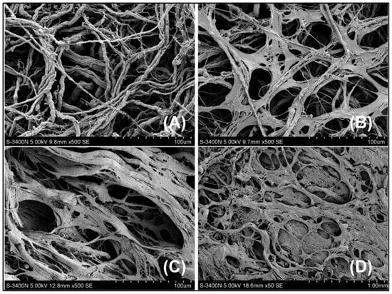
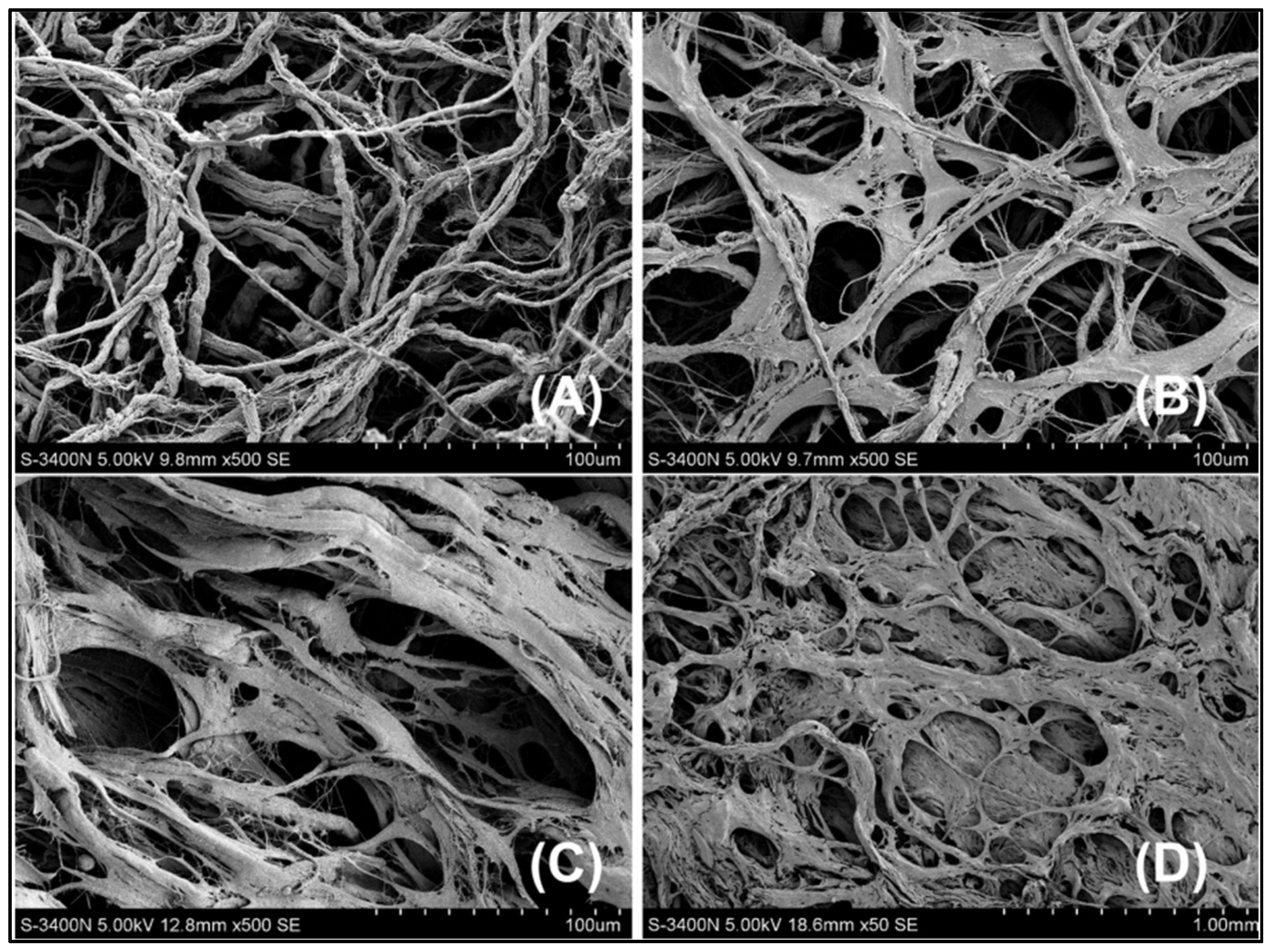
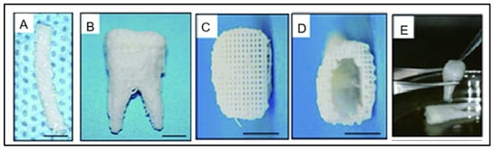
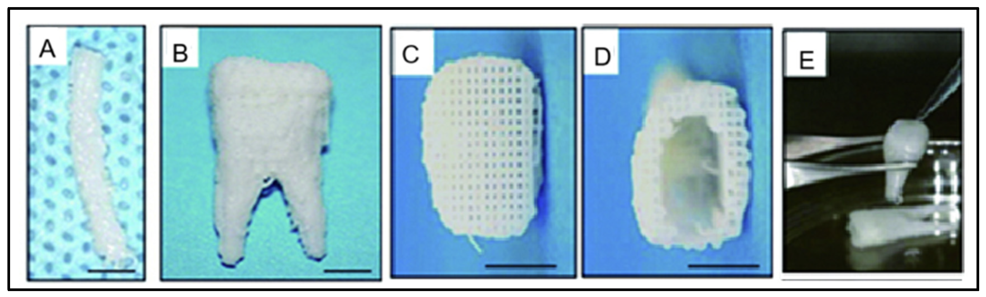
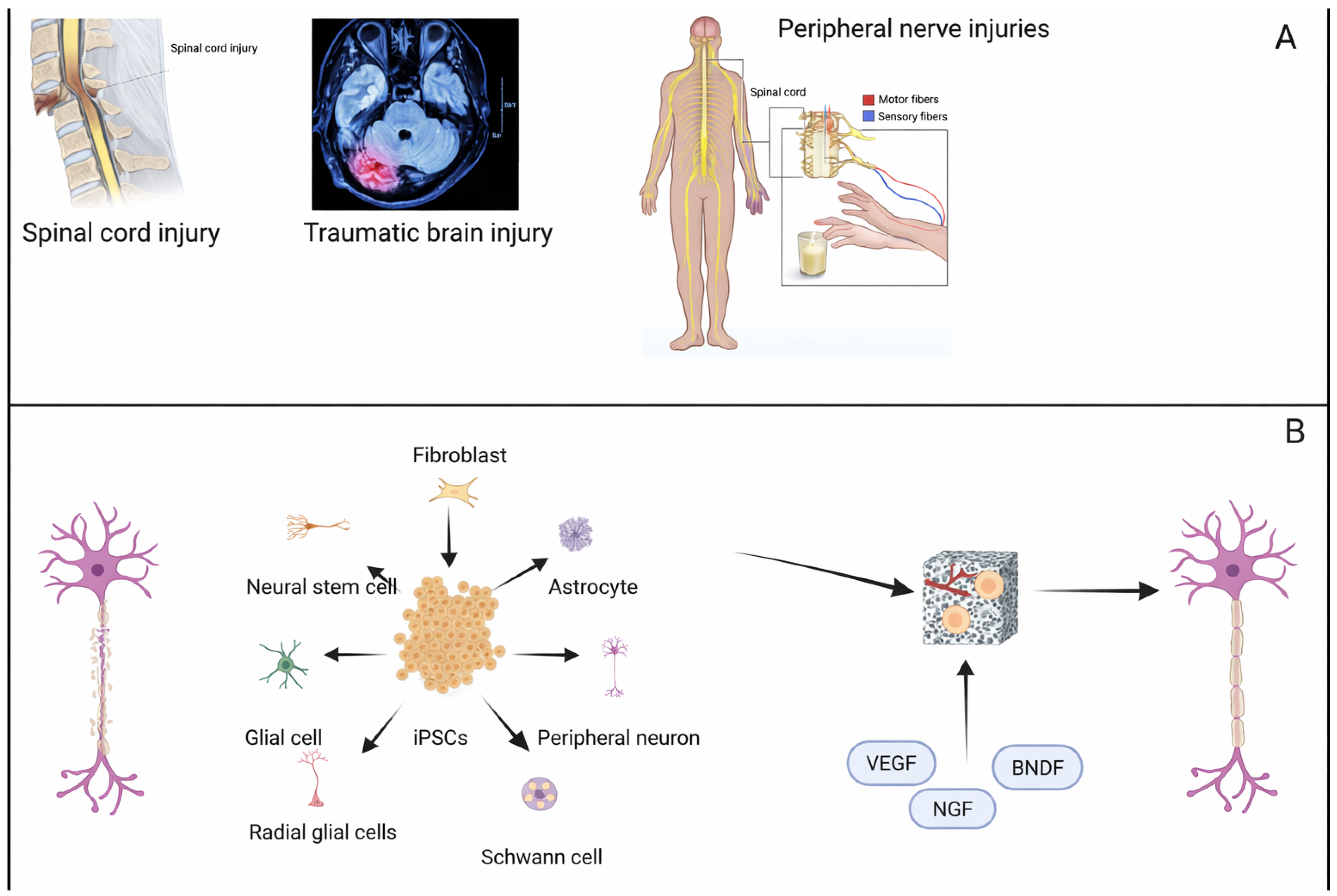
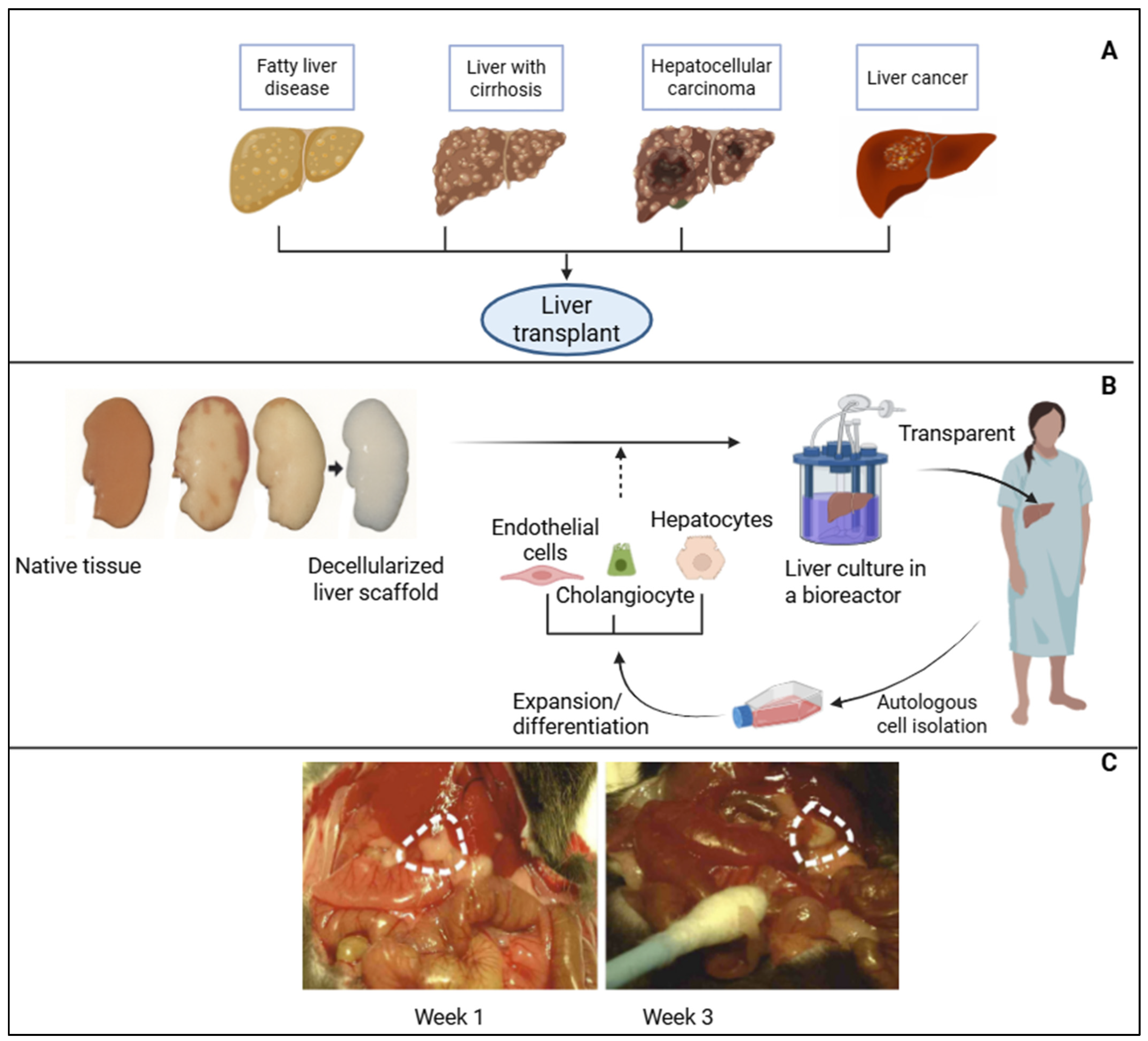

Disclaimer/Publisher’s Note: The statements, opinions and data contained in all publications are solely those of the individual author(s) and contributor(s) and not of MDPI and/or the editor(s). MDPI and/or the editor(s) disclaim responsibility for any injury to people or property resulting from any ideas, methods, instructions or products referred to in the content. |
© 2026 by the authors. Licensee MDPI, Basel, Switzerland. This article is an open access article distributed under the terms and conditions of the Creative Commons Attribution (CC BY) license.