


Abstract
Alexandrium pacificum is a typical harmful dinoflagellate in aquatic ecosystems that produces paralytic shellfish toxins (PSTs). While PSTs are well known for bioaccumulating in shellfish and posing severe food-safety risks to human consumers, their direct impacts on other aquatic species, such as shrimp, remain poorly studied. The current study investigated the damage caused by A. pacificum to the intestinal tissues of Exopalaemon carinicauda. For this purpose, adult E. carinicauda were divided into a control group and two experimental groups, with the latter involving a seven-day exposure to 0.5 × 10⁴ and 1.0 × 10⁴ A. pacificum cells/mL, respectively. Histological analyses subsequently showed obvious pathological alterations, including an irregular morphology and loosened inter-tissue connections, within the intestinal tissues of the exposure groups. Additionally, oxidative stress-related genes (Sod2, HSP70 12A, HSP90) were significantly upregulated, while cat, Gtpx and GSTM4 were significantly downregulated. The relative expression of Crustin, ALF and proPO3 decreased, whereas that of LGBP, TNFAIP-like and casp3 increased. Finally, analysis of the intestinal microbiota revealed that exposure to A. pacificum could also induce alterations in the intestinal microbial community, as reflected by a lower α-diversity, a high abundance of Proteobacteria and Vibrionaceae as well as functional predictions indicating suppressed metabolic pathways such as energy metabolism. The findings indicated that exposure to A. pacificum may induce histopathological and microbiota changes in E. carinicauda intestines, along with oxidative stress and changes in immune responses. This study reveals the negative impacts of A. pacificum on the intestinal health of E. carinicauda, which should be carefully considered and analyzed in aquaculture practices.
1. Introduction
The ridgetail white prawn (Exopalaemon carinicauda) is an important aquatic species in China, which serves as a high-quality protein source for human diets and holds significant economic value in the aquaculture industry [1]. However, over the years, continuous expansion of the farming scale [2] has led to inadequate wastewater treatment as well as excessive feeding, thus resulting in a surplus of nutrients in farming waters [3], which then become breeding grounds for Vibrio [4,5], viruses [6] and dinoflagellates [7]. The latter, in particular, represents a widely distributed group of photosynthetic single-cell eukaryotes that serve as primary producers in aquatic habitats [8]. However, their rapid proliferation can also inhibit the growth of beneficial algae [9], disrupt the balance of aquatic ecosystems [10], induce a deterioration of water quality [11], and ultimately affect the overall health of shrimp ponds [12]. Numerous studies have reported that harmful dinoflagellates can negatively impact survival rate, tissue health, and immunity of shrimp [7,13], all of which eventually led to major financial losses to the aquaculture industry.
Alexandrium spp., belonging to the phylum Dinophyta (Dinoflagellata), are widely distributed in China’s coastal waters. The primary concern associated with PSTs produced by these dinoflagellates is their bioaccumulation in filter-feeding shellfish [14], which act as vectors posing fatal food-safety risks to human consumers, whereas the toxins typically do not cause direct lethality to the bivalve hosts themselves. Previous studies have shown that exposure to Alexandrium minutum was not lethal to Crassostrea gigas, but significantly altered physiological and hematological responses, including feeding- and respiration-related variables [15]. Exposure also activated detoxification and antioxidant systems in the oyster gills [16]. For instance, Alexandrium tamarense was found to reduce the heart rate and increase mortality in early-stage Larimichthys crocea, while also inducing immune activation and calcium dysregulation [17]. Similarly, existing research indicates that exposure to Alexandrium pacificum can affect the survival and activity of Perna canaliculus immune cells [18]. A. pacificum is one of the most common toxin-producing dinoflagellates, particularly notable as PSTs persist even in its resting cysts and non-motile temporary cysts. As such, this species is of significant research importance and value. Previous studies have further shown that A. pacificum at a density of 1.0 × 10⁴ cells/mL for 72 h, can cause oxidative damage to Litopenaeus vannamei by inhibiting ATP synthesis and regulating processes such as cell proliferation and apoptosis through suppression of protein synthesis [19]. Moreover, the adverse physiological effects observed in marine organisms are not solely attributed to PSTs; other bioactive extracellular compounds (BECs) and reactive metabolites produced by A. pacificum may also contribute to cytotoxicity, mucosal damage, and inflammatory responses. Therefore, the excessive proliferation of A. pacificum can negatively impact both aquatic organism health and seafood safety.
Animal intestines harbor a large number of microorganisms, and in this context, a stable microbial community can promote host health by producing beneficial metabolites [20], whereas microbial dysbiosis may impair host health and elevate the risk of pathogen invasion [21]. Studies have also shown that the intestinal microbiota of crustaceans is not only involved in the digestion and absorption of nutrients, but is also crucial for maintaining immune homeostasis and defending against pathogen invasion [22]. Therefore, it is expected that the intestinal microbiome can influence shrimp health and is significantly correlated with the shrimp’s non-specific immunity. However, the impact of exposure to A. pacificum on E. carinicauda’s intestine remains poorly understood. It is noteworthy that, beside its economic significance within the aquaculture industry, E. carinicauda also represents a model organism for studying the effects of environmental stresses on crustacean physiology [23,24]. Therefore, the present study investigated the changes induced in the intestinal tissues of E. carinicauda following direct exposure to A. pacificum. The findings of this study are expected to provide new insights into the mechanisms through which toxin-producing dinoflagellates affect aquatic animals, thereby facilitating an assessment of the ecological risks posed by harmful algal blooms within the aquaculture industry.
2. Materials and Methods
2.1. Animal Selection and Culture
Healthy E. carinicauda were purchased from Jiaxin Aquaculture Company in Lianyungang City, Jiangsu Province, China. The experiments were conducted at the Jiangsu Key Laboratory of Marine Biotechnology, Jiangsu Ocean University, Lianyungang City, Jiangsu Province, China. The average body length was 5.1 ± 0.4 cm, and the body weight was 1.3 ± 0.2 g. Before the experiments, the shrimp were acclimated for 7 days in a 40 L rectangular glass tank filled with filtered and autoclaved natural seawater (salinity: 25 ± 1‰, representing the coastal environment of Haizhou Bay). The stocking density in each tank was 45 individuals, with a male-to-female ratio of approximately 1:1. The water temperature was maintained at 25 ± 1 °C, the pH value was kept between 8.0 and 8.2, and dissolved oxygen (DO) was maintained above 6.0 mg/L. Aeration was continuously provided under a photoperiod of 14 h of light and 10 h of darkness. The total ammonia nitrogen (TAN) and nitrite concentrations were kept below 0.10 mg/L and 0.05 mg/L, respectively. Approximately 30% to 50% of the water was replaced daily through drip feeding. The shrimp were fed twice daily (at 08:00 and 18:00) with commercial pellet feed (China Ocean Fisheries Group Co., Ltd.; approximate composition: crude protein ≥ 42.0%, crude fat ≥ 6.0%, crude fiber ≤ 5.0%, crude ash ≤ 15.0%) at a rate of 3% to 5% of their body weight. Residual feed and feces were completely suctioned out 1.5 h after feeding. Before the experiments, routine polymerase chain reaction (PCR) testing confirmed that the shrimp population was free of white spot syndrome virus (WSSV) and hepatopancreatic cytoplasmic bacteria (EHP). Only healthy individuals with intermolt stages (confirmed by crustal hardness), vigorous swimming activity, clean appendages, and clear intestinal fullness were selected for subsequent experiments.
2.2. Culture of Alexandrium pacificum
For batch cultivation, A. pacificum (ACHKT, GenBank: KM091276) was inoculated into f/2 medium without silicate (Si) in a 5 L conical flask and then incubated under the following conditions: a salinity of 25, a temperature of 20 ± 1 °C, a light intensity of 60 ± 10 μE m−2 s−1 and a light:dark cycle (L:D) of 14 h:10 h. The culture was kept in an illuminated incubator with manual shaking three times daily. Algal cultures in the exponential growth phase (days 10–12 after inoculation) were harvested and adjusted to the target experimental densities for shrimp exposure.
2.3. Experimental Design and Sample Collection
After one week of acclimation, healthy intermolt E. carinicauda were randomly allocated into three groups: a control group (CK, seawater without algae) and two algal exposure groups (AP1: 0.5 × 10⁴ cells/mL; AP2: 1.0 × 10⁴ cells/mL). These exposure levels were chosen based on previously established concentrations used to investigate sublethal and acute toxicological responses of shrimp species exposed to harmful dinoflagellates [19,25]. Each treatment had three replicate 40 L glass tanks filled with 30 L of experimental solution, with 40 shrimp per tank. Algal cultures harvested in the late exponential phase were used. The exposure lasted 7 days under identical conditions to algal batch culture: water temperature of 20 ± 1 °C, illumination of 60 ± 10 μE m−2 s−1, and a 14 h:10 h L:D photoperiod. Continuous gentle aeration was maintained (DO > 6.0 mg/L, TAN < 0.10 mg/L, NO2− < 0.01 mg/L). Every 24 h, 50% (15 L) of the tank water was siphoned out to eliminate waste, and newly prepared seawater in the CK group and seawater with concentrated algal suspension in the AP1 and AP2 groups were replenished. Cell densities were enumerated every 12 h: 1 mL water samples were fixed with 1% Lugol’s iodine solution, transferred to a 0.1 mL counting chamber, settled for 5 min, and counted under an optical microscope across three technical counts. The measured actual mean algal densities were (0.48 ± 0.04) × 104 cells/mL (AP1) and (0.96 ± 0.07) × 104 cells/mL (AP2). It should be noted that the cellular toxin quota of the A. pacificum strain was not quantified in this study; hence, the experimental design assesses the integrated ecological impact of live A. pacificum bloom exposure rather than purified toxin pharmacology. After seven days of exposure, sampling was conducted independently from each of the three replicate tanks per group to eliminate tank effects. For RNA extraction, three shrimp were randomly sampled from each replicate tank and pooled into one composite sample, yielding three biological replicates per group (a total of nine shrimp per group). Their intestinal tissues were excised and stored in 0.3 mL TRIzol reagent at −80 °C. Separately and independently, another three shrimp were sampled from each replicate tank (totaling nine independent shrimp per group) for gut microbiota analysis. The shrimp were rinsed with sterile water to clean their body surfaces, dissected under sterile conditions to collect intestinal tissues, and pooled by tank (three shrimp per replicate, three biological replicates per group). After being flash-frozen in liquid nitrogen, the samples were kept at −80 °C for gut microbiota sequencing.
2.4. Histopathological Analyses
Samples of intestinal tissues (specifically the midgut segment) from each group of E. carinicauda were collected at the end of the seven-day exposure period (three shrimp randomly sampled per replicate tank, totaling nine shrimp per group). Due to the 24 h fasting period experienced by the shrimp, the food contents in their intestinal lumen were essentially cleared. To avoid the impact of residual intestinal lumen contents or feces on the mucosal structure, before fixation, the resected midgut lumen was gently rinsed with 100 μL of sterile phosphate-buffered saline (PBS, pH 7.4) using a microsyringe. Tissues were fixed in 4% paraformaldehyde solution for 24 h. Dehydration was then performed using the following ethanol gradient: 75% (2 h), 85% (2 h), 90% (2 h) and 95% (1 h). The tissue samples were subsequently treated with xylene to facilitate paraffin infiltration, after which they were immersed in molten paraffin to ensure thorough saturation. The resulting paraffin-embedded samples were then sliced (thickness 4–6 μm) with a microtome (three non-consecutive sections per shrimp). After being mounted on glass slides and air-drying, sections were subjected to hematoxylin–eosin (H&E) staining. Histological alterations were evaluated under an optical microscope across at least three randomly selected fields per section.
2.5. Gene Expression Analysis
Total RNA was extracted from intestinal tissue samples using the UNIQ-10 Column Trizol Total RNA Extraction Kit (Sangon Biotech, Shanghai, China). RNA concentration and purity were measured using a NanoDrop 2000 spectrophotometer (Thermo Scientific, Waltham, MA, USA), with all samples exhibiting 260/280 ratios between 1.95 and 2.05 and 260/230 > 2.0. RNA integrity was confirmed by 1.2% agarose gel electrophoresis. One microgram (1.0 μg) of total RNA was reverse transcribed into cDNA using the TransScript® One-Step gDNA Removal and cDNA Synthesis SuperMix (TransGen Biotech, Beijing, China) to digest potential genomic DNA carryover.
Quantitative real-time PCR was performed on a LightCycler® 96 system (Roche, Indianapolis, IN, USA) using the PerfectStart® Green qPCR SuperMix (TransGen Biotech). Each 20 μL reaction contained 10 μL of 2× PerfectStart Green qPCR SuperMix, 0.4 μL each of forward and reverse primers, 1.0 μL of diluted cDNA template (1:5), and 8.2 μL of nuclease-free water. The thermal profile was as follows: 94 °C for 30 s, followed by 45 cycles of 94 °C for 5 s and 60 °C for 30 s. Melting curve analysis was performed from 65 °C to 97 °C to confirm primer specificity. Three independent biological replicates per treatment were assayed, each in technical triplicates, with no-template controls included. The 18S rRNA gene was used as the internal reference gene; its stability was confirmed by consistent threshold cycle values across all treatments. Relative expression levels were calculated using the 2−ΔΔCt method. All the primers used in this study were designed using Primer Premier 5.0 and are listed in Table S1.
2.6. 16S rDNA Sequencing Analysis
Intestinal microbial community profiles were analyzed by high-throughput 16S rDNA gene sequencing. A total of 9 biological samples were evaluated. Microbial genomic DNA was extracted using the E.Z.N.A.® Soil DNA Kit (Omega Bio-tek, Norcross, GA, USA) following the manufacturer’s protocol, and DNA purity and concentration were verified using a NanoDrop 2000 spectrophotometer (Thermo Scientific, USA) and 1% agarose gel electrophoresis. PCR amplification was performed under the following conditions: initial denaturation at 95 °C for 3 min, followed by 27 cycles of 95 °C for 30 s, 55 °C for 30 s, and 72 °C for 45 s, with a final extension at 72 °C for 10 min. Amplicons were purified using the AxyPrep DNA Gel Extraction Kit (Axygen Biosciences, Union City, CA, USA) and quantified with a QuantiFluor™-ST Fluorometer (Promega, Madison, WI, USA). Equimolar amounts of purified amplicons were pooled and paired-end sequenced on an Illumina NovaSeq 6000 platform (San Diego, CA, USA).
Raw demultiplexed sequencing reads were imported into QIIME2 (version 2022.2). Primer sequences were removed using the Cutadapt plugin. Sequence quality control, denoising, dereplication, paired-end read merging, and chimera identification (using the consensus model) were conducted using the DADA2 pipeline to generate amplicon sequence variants (ASVs) [26]. High-quality representative ASV sequences were taxonomically annotated against the SILVA reference database (Release 138.1) using the naive Bayes classifier with a confidence threshold of 0.70. To control for variations in sequencing depth, the ASV abundance matrix was normalized by subsampling to the minimum sequence count among all libraries prior to downstream alpha- and beta-diversity calculations.
Alpha diversity metrics (Observed ASVs, Chao1, Shannon, Simpson, and Pielou’s evenness) were calculated using QIIME2. Rarefaction curves were generated to evaluate sampling depth sufficiency. Shared and unique ASVs among groups were visualized with Venn diagrams using the R package Venn Diagram. Beta diversity differences in microbial community structures were evaluated using Principal Coordinates Analysis (PCoA) and Non-metric Multidimensional Scaling (NMDS) based on Bray–Curtis dissimilarities, with community differences statistically validated using permutational multivariate analysis of variance. Biomarker taxa with significant differential abundance among treatments were identified by Linear Discriminant Analysis Effect Size (LEfSe) using non-parametric Kruskal–Wallis tests (p < 0.05) with a logarithmic LDA score threshold >3.0. In addition, metabolic functional profiles of the communities were predicted using PICRUSt2 (Phylogenetic Investigation of Communities by Reconstruction of Unobserved States, version 2.4.1) based on the KEGG database; these profiles were interpreted specifically as predicted functional potentials.
2.7. Statistical Analysis
All results were presented as mean ± SE. Prior to parametric analyses, the data were tested for normality using the Shapiro–Wilk test and for homogeneity of variance using Levene’s test. Data satisfying these assumptions were subjected to one-way analysis of variance (ANOVA), followed by Duncan’s multiple range test (SPSS version 27.0). p-values < 0.05 were considered statistically significant. Differential abundance of predicted metabolic pathways (PICRUSt2/KEGG) between the control and A. pacificum-exposed groups was analyzed using STAMP software (version 2.1.3). Pairwise comparisons were assessed using two-sided Welch’s t-tests with Welch’s inverted 95% confidence intervals (CI) to determine effect size. Multiple hypothesis test corrections were performed using the Benjamini–Hochberg false discovery rate (FDR), with an adjusted p-value < 0.05 considered statistically significant.
3. Results
3.1. Histological Changes in the Intestines
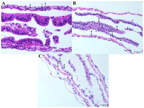
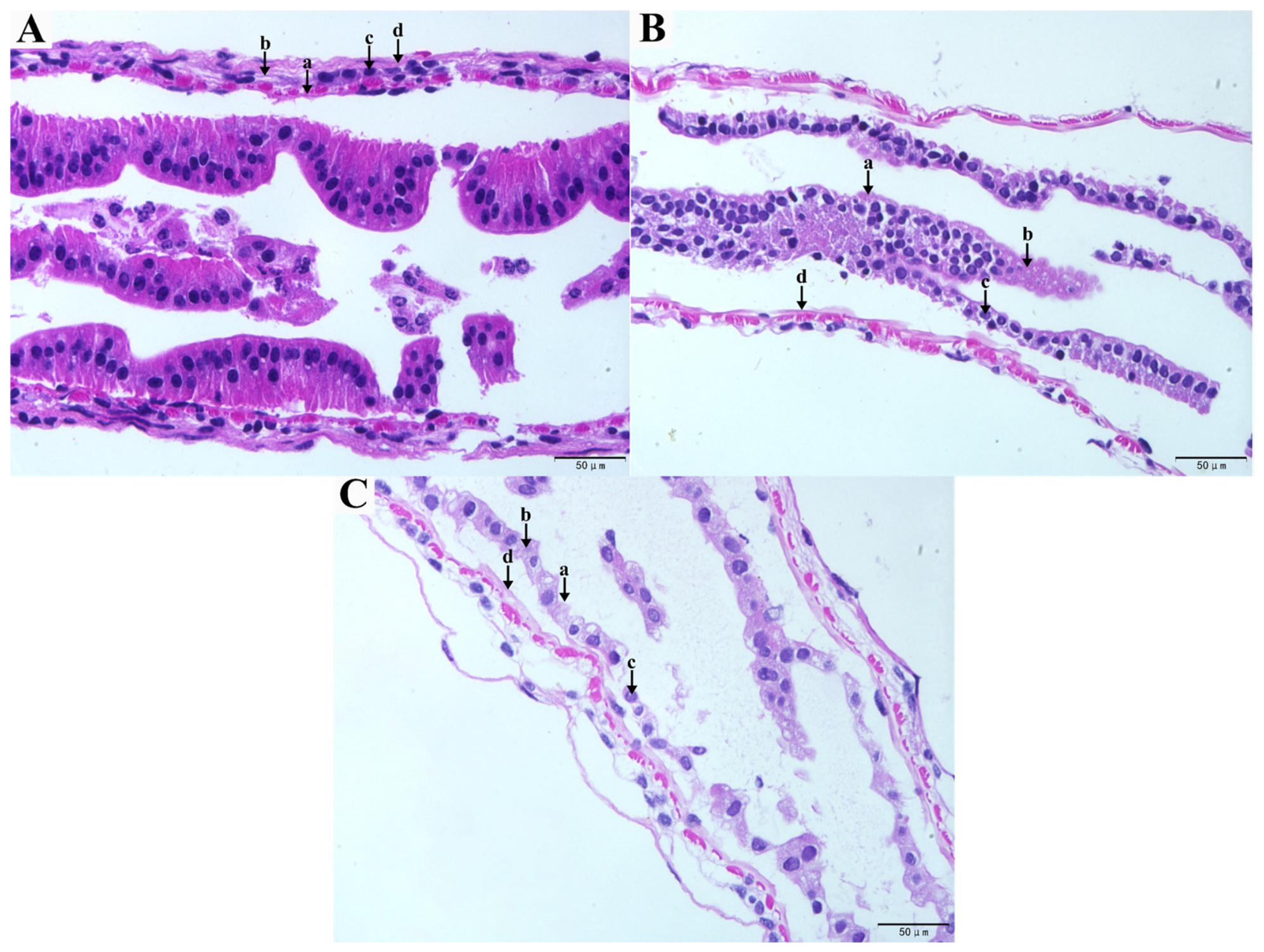
In the CK group, the intestinal tissue was intact and well-defined, with the epithelial cells arranged in a neat and compact manner (Figure 1A). However, compared to the control group, both AP1 and AP2 exhibited significant structural damage, including loose tissue connections, shedding of brush border microvilli, nuclear displacement, and splitting or disruption of the basement membrane and epithelial cells (Figure 1B,C).
Figure 1.
Histopathological changes in the intestinal tissues of E. carinicauda. (A) Control (healthy intestinal tract); (B) 0.5 × 10⁴ cells/mL group; (C) 1.0 × 10⁴ cells/mL group; a: brush edge; b: epithelial cells; c: cell nuclei; d: basement membrane.
3.2. Intestinal Antioxidant Indicators
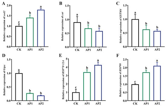
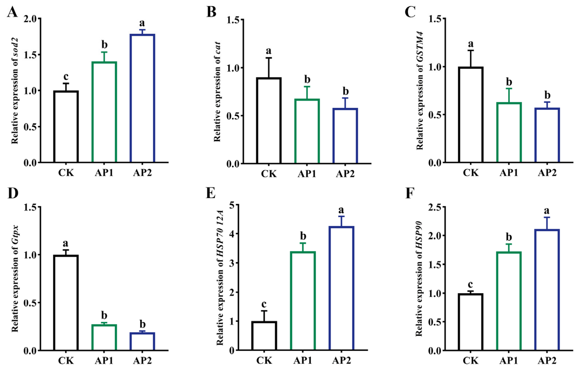
To determine E. carinicauda’s response to intestinal oxidative stress induced by exposure to A. pacificum, the expression of oxidative stress-related genes was studied. Compared with the control, cat, GSTM4 and Gtpx had significantly lower expression levels in both AP1 and AP2, while that of Sod2, HSP70 12A and HSP90 increased markedly (p < 0.05, Figure 2A–F). Specifically, relative to the control, Sod2, HSP70 12A, and HSP90 expression increased by 1.41-, 3.40-, and 1.72-fold in the AP1 group, and reached 1.79-, 4.26-, and 2.11-fold in the AP2 group, respectively. Comparing the two experimental groups further revealed that Sod2, HSP70 12A and HSP90 expression was markedly higher in the AP2 group as compared to AP1, representing further increases of 27.0%, 25.3%, and 22.7%, respectively (p < 0.05, Figure 2A,E,F). However, these two groups did not differ significantly in terms of cat, GSTM4 and Gtpx expression (p > 0.05, Figure 2B–D).
Figure 2.
Expression of oxidative stress-related genes in E. carinicauda’s intestine after exposure to A. pacificum. (A) Sod2; (B) cat; (C) GSTM4; (D) Gtpx; (E) HSP70 12A; (F) HSP90. The 18S gene was selected as the reference. Bars show the mean ± SE (n = 3). Significant differences (p < 0.05) between groups are indicated by different letters (a, b, c).
3.3. Intestinal Immune Responses
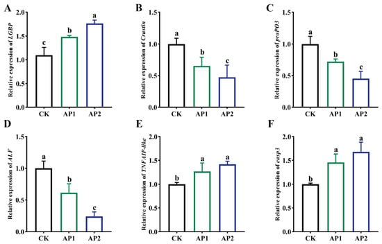
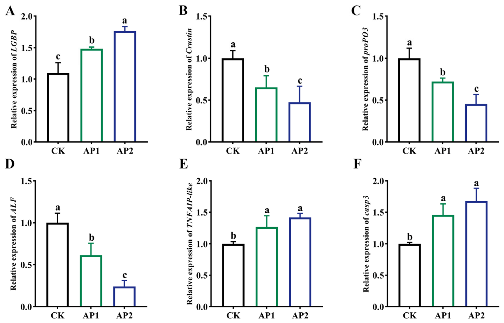
The expression of immunity-related genes was examined to investigate how exposure to A. pacificum impacted the immune status of E. carinicauda’s intestinal environment. Compared with the control, ALF, Crustin and proPO3 expression was reduced in both the AP1 and AP2 groups, with those in the latter group further exhibiting a significantly lower expression level as compared to AP1 (p < 0.05, Figure 3B–D). The expression level of LGBP, however, showed an opposite trend, with significantly higher levels in the AP1 and AP2 groups compared to the control group (p < 0.05, Figure 3A). However, the relative expression of TNFAIP-like and casp3 was not significantly different between the AP1 and AP2 groups (p > 0.05, Figure 3E,F), although their expression levels were significantly higher compared with the control group.
Figure 3.
Immunity-related gene expression in E. carinicauda’s intestine after exposure to A. pacificum. (A) LGBP; (B) Crustin; (C) proPO3; (D) ALF; (E) TNFAIP-like; (F) casp3. The 18S gene was used as the reference. Bars show the mean ± SE (n = 3). Significant differences (p < 0.05) between groups are indicated by different letters (a, b, c).
3.4. Changes in Intestinal Microbiota Composition
3.4.1. Richness and Diversity
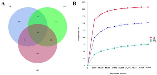
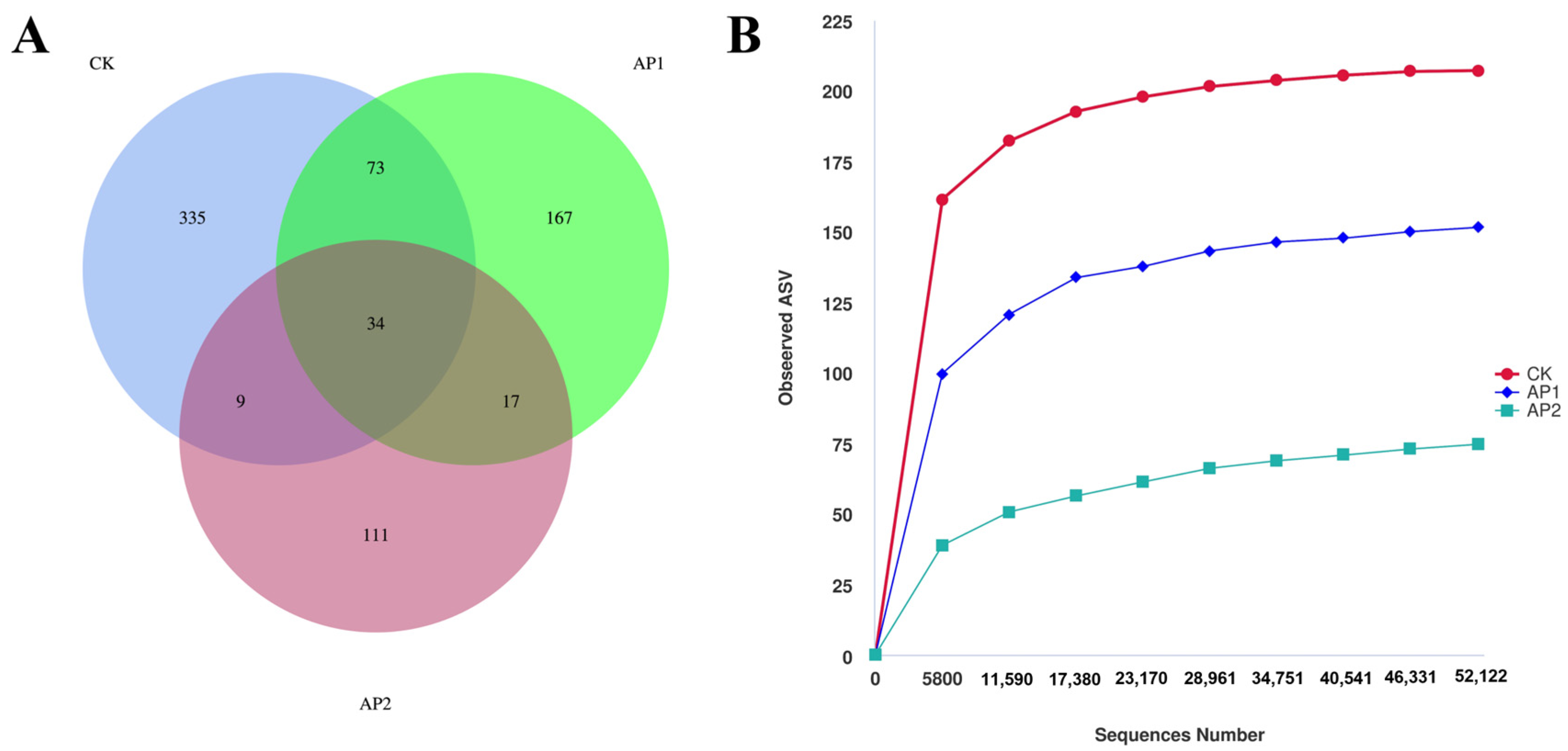
Venn diagrams were constructed based on ASV occurrence matrices (defined as presence across biological replicates within a group). Rarefaction analysis was conducted by randomly subsampling sequence reads incrementally up to maximum library depths to confirm whether sequencing saturation was achieved across all samples. Using ASV similarity clustering analysis, Venn diagram analysis was conducted to characterize the microbial community structures of the three sample groups by pooling all biological replicates within each group (Figure 4A), while the rarefaction curve indirectly reflects the species richness of the samples (Figure 4B). Overall, after pooling the replicates per treatment, the CK, AP1 and AP2 groups had 335, 167 and 111 unique ASVs, with 34 ASVs shared across all three groups. Furthermore, the coverage values of all samples were 1.00, indicating that the sequencing data were reliable, with the sequencing depth also sufficient to cover all species in the samples. As shown in Table 1, subsequent α-diversity analyses revealed that the CK group had higher Chao1 and Simpson indices compared with the AP1 and AP2 groups, although the differences between the CK and the AP1 groups were not significant. Collectively, the findings showed that exposure to A. pacificum reduced both the species diversity and richness of E. carinicauda’s intestinal microbiota. Similarly, the Shannon and Pielou_e indices of the CK group were significantly higher than those of the AP1 and AP2 groups (p < 0.05), thus indicating that exposure to A. pacificum significantly reduced the evenness of the intestinal microbiota.
Figure 4.
(A) Venn diagram showing the unique and shared ASVs among the three groups; (B) rarefaction curve of observed ASVs. Note: CK, AP1, AP2 refer to the control group, 0.5 × 10⁴ cells/mL and 1.0 × 10⁴ cells/mL groups.
Table 1.
Alpha diversity indices of intestinal microbial communities of E. carinicauda.
3.4.2. Intestinal Microbial Composition
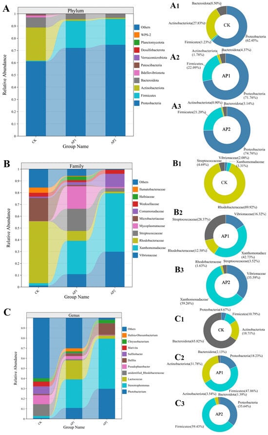
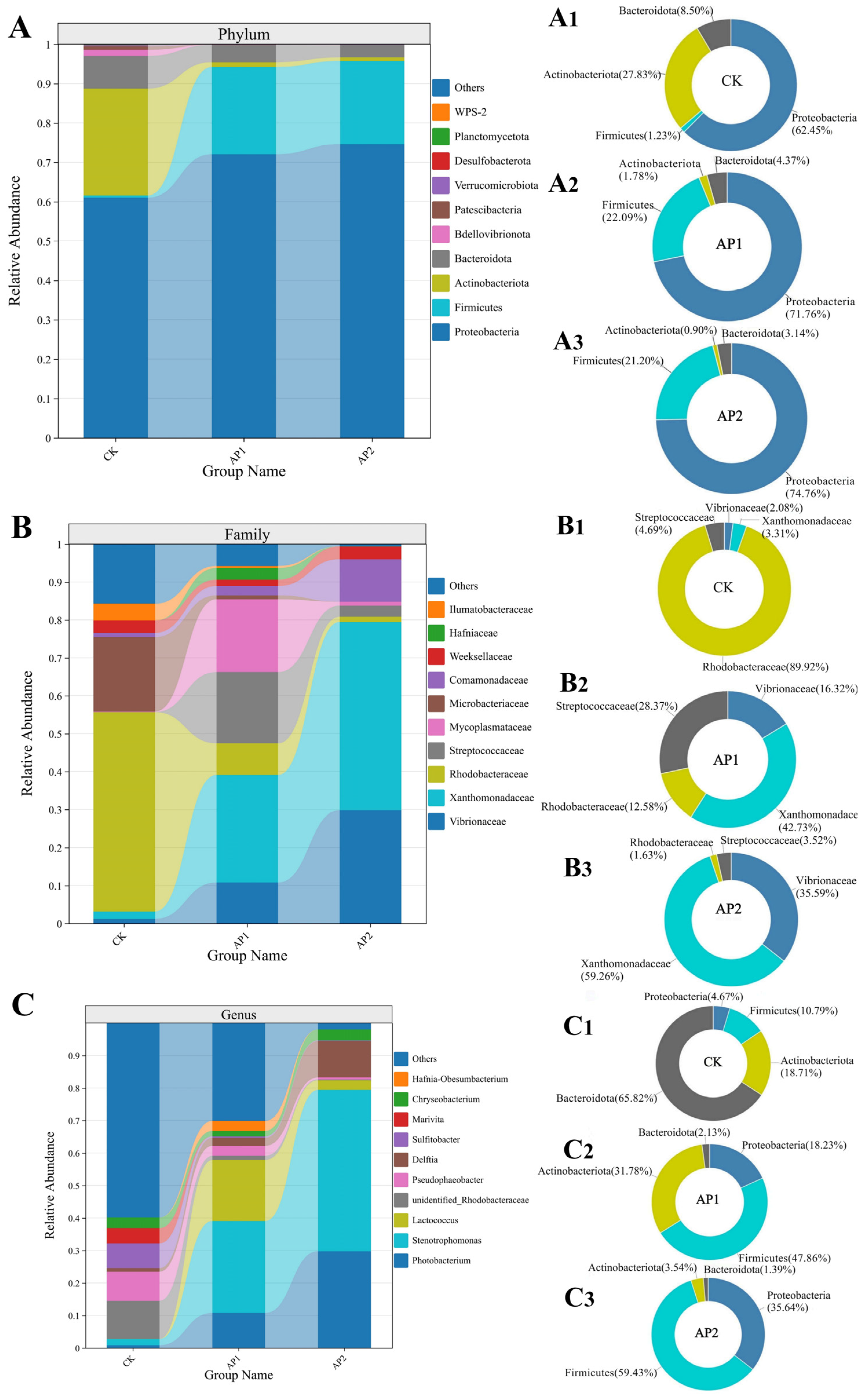
Figure 5A shows the top 10 phyla ranked in terms of their relative abundance of gut microbial communities at the phylum level; the intestinal bacterial community across all groups was predominantly composed of four phyla: Proteobacteria, Firmicutes, Actinobacteriota, and Bacteroidota. Proteobacteria was the dominant phylum in all treatments. However, the relative abundance of Firmicutes exhibited prominent variations: it accounted for only 0.5% in the CK group, but surged remarkably to dominant proportions in both exposure groups, such that the combined relative abundance of Proteobacteria and Firmicutes reached 95.2% in the AP1 group and 95.7% in the AP2 group. Concurrently, the relative abundances of Actinobacteriota and Bacteroidota, which accounted for 27.2% and 8.3% in the CK group, respectively, dropped markedly in both AP1 and AP2 groups (p < 0.05). Figure 5B presents the top 10 families ranked in terms of their relative abundance in intestinal microbial communities. Among these, Vibrionaceae, Xanthomonadaceae, and Rhodobacteraceae were predominant across the three groups. Notably, compared with the control group (1.22%), both experimental groups exhibited a significant increase in the relative abundance of Vibrionaceae, 10.80% in AP1 and 29.80% in AP2 (p < 0.05); moreover, the abundance in AP2 was significantly higher than that in AP1 (p < 0.05), indicating a dose-dependent proliferation of Vibrionaceae. Conversely, exposure to A. pacificum markedly decreased the relative abundance of Rhodobacteraceae from 52.51% (CK) to 8.33% (AP1) and 1.25% (AP2) (p < 0.05); however, the difference between AP1 and AP2 was not statistically significant (p > 0.05). Figure 5C displays the top 10 genera. In this case, the relative abundances of Photobacterium and Stenotrophomonas were significantly higher in both treatment groups compared with the control (p < 0.05). Specifically, the proportion of Photobacterium increased significantly in a concentration-dependent manner from 0.83% in CK to 10.70% in AP1 and 29.70% in AP2 (p < 0.05).
Figure 5.
Relative abundance of taxa in each group of samples at the phylum (A), family (B) and genus (C) levels. Pie charts of significantly different species in each group (A1–C3).
3.4.3. Changes in Intestinal Bacterial Populations
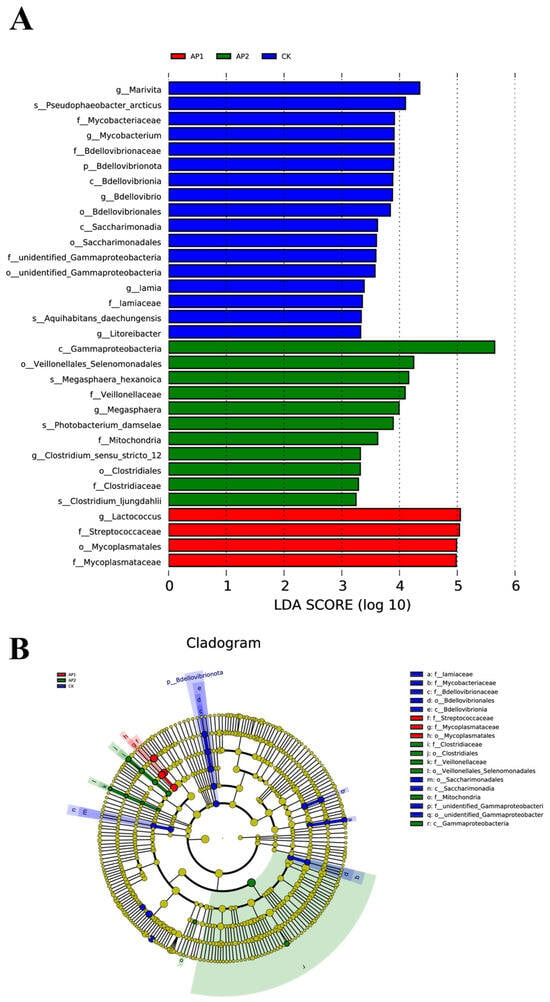
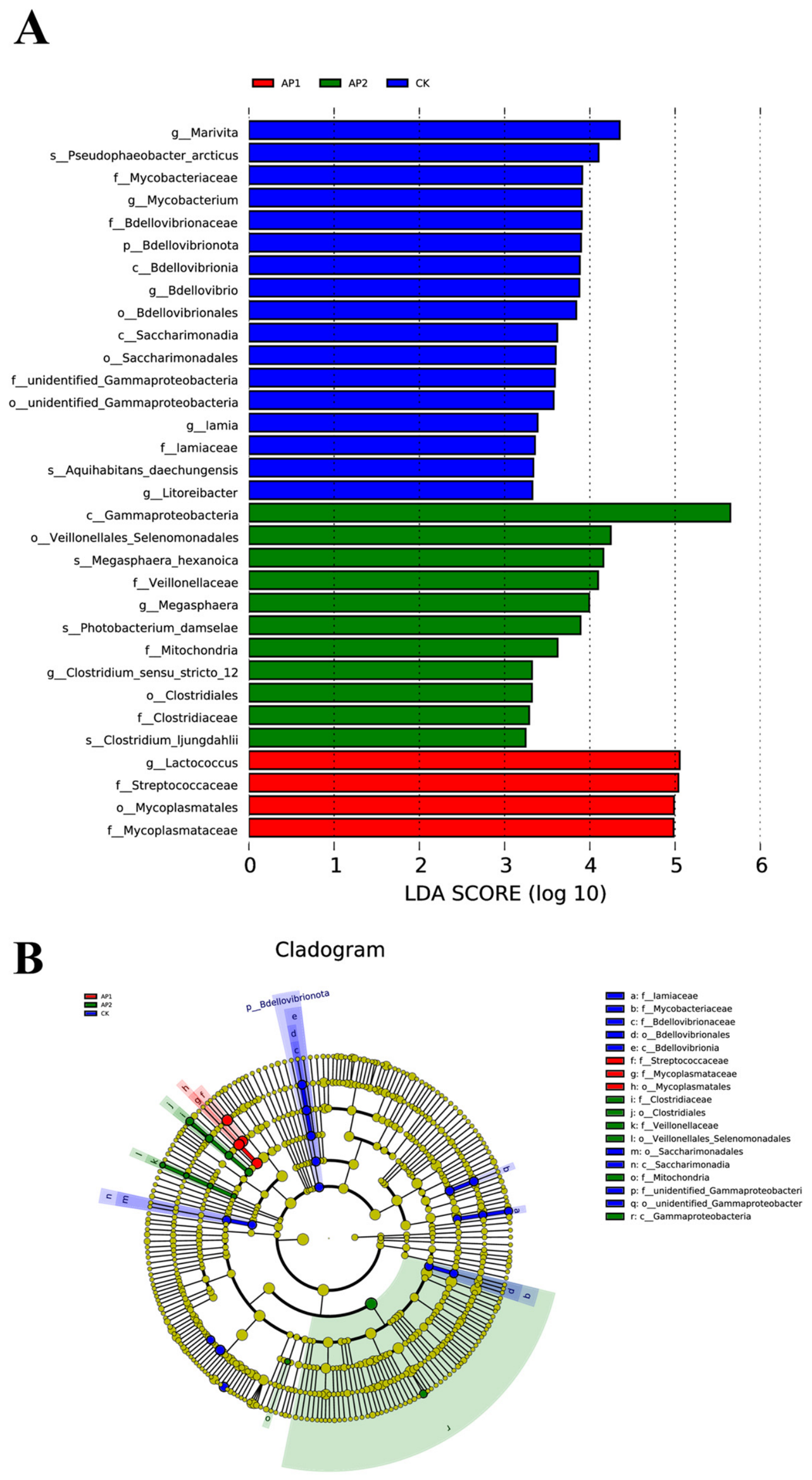
LEfSe analysis was performed to identify taxa that discriminated among the CK, AP1, and AP2 groups. Using a significance threshold of p < 0.05 and an LDA score threshold of >3.0, 32 differentially abundant biomarker taxa were identified (Figure 6). Of these taxa, 17 were enriched in the CK group, 4 were enriched in the AP1 group, and 11 were enriched in the AP2 group. In the CK group, Rhodobacteraceae-related taxa were prominent biomarkers. The AP1 group was characterized by the enrichment of Lactococcus, Streptococcaceae, Mycoplasmataceae, and Mycoplasmatales. In the AP2 group, Gammaproteobacteria-related taxa, including Photobacterium damselae, as well as Veillonellales-Selenomonadales-related taxa, were among the discriminative biomarkers.
Figure 6.
(A) LDA scores of species with significant abundance differences among groups; (B) phylogenetic cladogram showing species with significant abundance variations in each group.
3.4.4. Functional Prediction Analysis
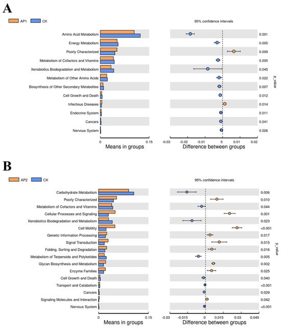
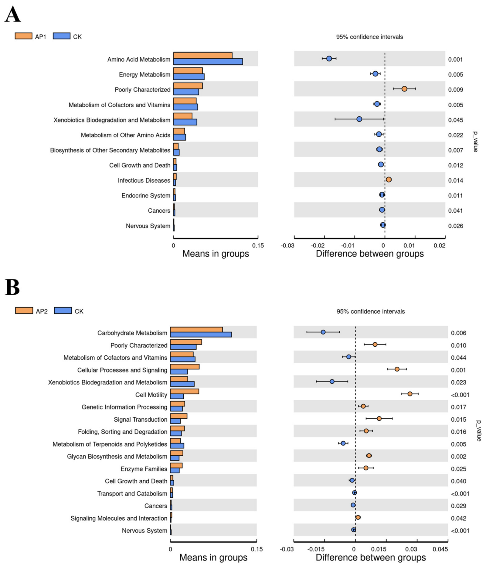
Functional prediction analysis using PICRUSt2 revealed 12 MetaCyc pathways that differed significantly between the control and the AP1 group (p < 0.05) (Figure 7A). Notably, the AP1 group exhibited blocked pathways related to amino acid metabolism, energy metabolism, metabolism of cofactors and vitamins, as well as biosynthesis of other secondary metabolites compared with the CK group. Furthermore, comparisons between the CK and AP2 groups showed that 17 MetaCyc pathways were markedly different (p < 0.05), and of these, 8 pathways, including carbohydrate metabolism, xenobiotic biodegradation and metabolism were blocked, while 9 were enriched in genetic information processing, cellular processes and signal transduction (Figure 7B).
Figure 7.
Based on PICRUSt2 analysis, significantly different metabolic pathways were identified between the control and AP1 groups (A) as well as between the control and AP2 groups (B). The p-values are based on Welch’s t-test and corrected with Benjamini–Hochberg FDR (p < 0.05).
4. Discussion
A. pacificum is a typically harmful dinoflagellate that not only causes severe environmental problems but also leads to major financial losses in the aquaculture industry [27]. Moreover, it can even threaten human health through its bioaccumulation along the food chain [13,28]. In crustaceans, the intestine is a vital organ that plays a key role in substance accumulation [29], making it highly sensitive to external environmental stressors (such as harmful algal blooms, pathogens, and toxic xenobiotics). As such, it is also highly susceptible to damage from harmful algal bloom contamination. However, the harmful effects of A. pacificum on E. carinicauda, in particular, remain poorly understood. Therefore, this study sought to investigate how exposure to A. pacificum negatively impacts the intestinal tissues of E. carinicauda through a combination of histological analysis of gut structure, assessment of immune responses and composition of intestinal microbiota.
The intestine also plays essential roles in nutrition and immunity, with its protective effects primarily relying on the non-specific immune system [22]. This system functions through tight junctions between the luminal surface cell membranes and epithelial cells [30]. Furthermore, in the case of crustaceans, due to the absence of an acquired immune system [31], the intestinal mucosal barrier also acts as a critical first line defense against intestinal antigens [31]. For instance, exposing L. vannamei to high concentrations of ammonia nitrogen was shown to induce significant intestinal damage, including shedding of the intestinal epithelial mucosa [32]. Importantly, throughout the present trial, strict daily complete water renewal ensured that background water quality was maintained within optimal ranges (TAN <0.10 mg/L, NO2− <0.02 mg/L), confirming that the observed intestinal alterations were directly driven by A. pacificum exposure rather than waterborne nitrogenous waste accumulation. In this study, following seven days of exposure to A. pacificum, the intestinal tissues of shrimp from the experimental groups exhibited an irregular morphology, enlarged intercellular spaces as well as loose tissue connections, all of which indicated that the presence of the algae compromised the integrity of the intestinal tissues in E. carinicauda and increased the risk of pathogen infection. Collectively, the findings suggested that A. pacificum can damage the structure of E. carinicauda’s intestinal tissues, thus potentially impairing the non-specific immune function and leading to adverse effects on the shrimp’s health.
Increased oxidative stress is one of the toxic effects of environmental pollution on shrimp [33]. Environmental stress has been shown to induce the production of reactive oxygen species (ROS) in organisms [34], which in turn causes oxidative damage to proteins [35]. In this study, transcriptional analysis showed an upregulation of Sod2, accompanied by the downregulation of cat, GSTM4, and Gtpx transcripts in the exposure groups. A plausible hypothesis for this transcriptional profile is that shrimp attempted to mobilize their frontline superoxide dismutase to scavenge excess superoxide anions; however, the downstream clearance enzymes were transcriptionally repressed, potentially indicating that sustained algal exposure disrupted normal antioxidant gene regulation. Although post-transcriptional events, translation, and enzyme kinetics were not directly assessed here, these coordinated transcriptional alterations suggest that A. pacificum exposure creates a severe challenge to intestinal redox balance. Similar findings were observed in the intestines of L. vannamei exposed to ammonia stress, where the expression level of SOD increased while that of GPx decreased, indicating the occurrence of oxidative stress when the intestines were subjected to environmental stress [32]. Studies on L. vannamei exposed to nitrite and microplastics further demonstrate that the shrimp’s antioxidant system exhibits an active response to environmental stress, while intensified environmental stress causes additional disruption to the oxidative stress balance [36]. HSP70 and HSP90 are two stress proteins that play crucial roles in the anti-stress process [37]. In this work, the HSP70 12A and HSP90 genes were upregulated in both experimental groups, indicating that A. pacificum-induced environmental stress disrupted the homeostasis of intestinal stress proteins in E. carinicauda. This result was consistent with the altered intestinal homeostasis reported in L. vannamei following exposure to nitrite-induced environmental stress [36]. This transcriptional induction indicates that A. pacificum challenged mucosal homeostasis, stimulating cellular heat shock responses to cope with stress-induced structural perturbations.
The intestinal immune system acts as a crucial defense barrier against pathogenic bacterial infections. ALF and Crustin are essential components of the innate immune system that combats pathogenic microorganisms, while LGBP activates the proPO system by recognizing lipopolysaccharides and β-1,3-glucans on pathogen surfaces. This activation induces the conversion of proPO into active phenoloxidase, thereby enhancing the defense capability of shrimp [38]. The proPO acts as one of the key effector mechanisms of the immune system in crustaceans, especially due to its role in pathogen recognition, phenoloxidase activation and melanin production to form protective barriers at wound or infection sites [39]. Previous studies have reported altered immune-related gene expression in crustaceans exposed to diverse environmental stressors, including cyanobacteria [40], BPA, and DEHP [41]. These studies are cited here to provide a broader context for stress-associated immune responses in crustaceans; however, these stressors may act through mechanisms distinct from those associated with Alexandrium exposure. In the present study, exposure to A. pacificum was associated with reduced intestinal expression of Crustin, ALF, and proPO3, suggesting a potential impairment of antimicrobial and proPO3-related immune functions in E. carinicauda. Because PST concentrations in the algal cultures were not determined, these responses cannot be attributed specifically to PSTs. Instead, they may reflect the combined effects of intact A. pacificum cells and algal-associated factors, including but not limited to PSTs and other cell-derived metabolites or components. The humoral response of shrimp represents an important component of their immune defense system [42]. Among various humoral immune responses, one of the most effective mechanisms in invertebrates against pathogen infection is the synthesis of melanin by the proPO system [39]. For instance, studies involving L. vannamei have shown a marked decrease in proPO expression after pH stress, hence suggesting that pH stress may affect the antibacterial function of shrimp [43]. Additionally, the intestinal expression of proPO in L. vannamei was found to be markedly decreased after temperature reduction, and this can potentially impair the shrimp’s ability to detect and eliminate pathogens [44]. Therefore, exposure to A. pacificum may likely disrupt the immune function of shrimp intestines by impairing its antibacterial capacity. Caspase is a crucial apoptotic factor that maintains stability of the internal environment by regulating the orderly and autonomous process of cell death [45,46]. TNF-α, on the other hand, is an inflammatory cytokine whose release can be triggered by environmental changes, and while it can directly kill tumor cells, it has not shown significant toxicity to normal ones, EzF-like is a downstream response gene of TNF-α. Its upregulated expression also reflects the inflammatory and immune activation state of the body [47,48]. In this study, exposure to A. pacificum increased the transcription levels of both casp3 and TNFAIP-like in shrimp intestines and compromised intestinal barrier integrity, suggesting the activation of pro-apoptotic and inflammatory pathways at the transcriptional level. However, because specific assays such as TUNEL staining or apoptotic body detection were not conducted, further biochemical and ultrastructural evidence is required to definitively quantify cellular apoptosis. Overall, exposure to A. pacificum likely impairs the intestinal immune barrier by downregulating antimicrobial peptides and potentially triggering pro-apoptotic signaling cascades in the intestines of E. carinicauda.
Chronic oxidative stress in the intestines has often been associated with dysbiosis [49], with existing research consistently linking the occurrence of pathological conditions—such as bacterial infections, inflammatory responses, and metabolic disorder—to changes in the composition of gut microbiota [50] as the latter can resist pathogen invasion by producing inhibitory compounds and competing for nutrients and space [51]. In this study, the results from the Venn diagram and α-diversity indices indicated that exposure to different concentrations of A. pacificum significantly lowered the microbial diversity in E. carinicauda’s intestines. In particular, analysis of the Shannon and Pielou indices revealed that the exposure to A. pacificum significantly decreased the evenness of the intestinal microbiota. In this context, marine crustaceans subjected to ambient environmental challenges commonly experience structural shifts and diversity loss in their intestinal microbial communities; for example, prolonged environmental stress in the swimming crab (Portunus trituberculatus) similarly led to gut microbial dysbiosis, characterized by reduced community richness and disruption of the dominant commensal flora [52]. In this study, exposing E. carinicauda to A. pacificum markedly reduced the diversity and evenness of the intestinal microbial communities, indicating that such exposure may affect nutrient absorption, immune regulation, and metabolic functions in E. carinicauda.
An increased relative abundance of Proteobacteria has frequently been reported as a feature of gut microbial dysbiosis in animal hosts and has been proposed as a potential indicator of an unstable microbial community [53]. Similarly, metabolic dysregulation within a host has been closely associated with a significant rise in Firmicutes populations [54]. On the other hand, Bacteroidetes play a crucial role in immune regulation as well as in lipid and carbohydrate metabolism, and as such, they usually confer benefits to a host [55]. In this study, by integrating the relative abundance of different species with the differential analysis of species across groups, a significant increase in the relative abundance of Proteobacteria and Firmicutes was observed, and this was accompanied by a marked decrease in the relative abundance of Bacteroidetes. These results indicated that exposure to dinoflagellates could significantly alter E. carinicauda’s gut microbiota composition, which may potentially disrupt downstream metabolic activities and intestinal homeostasis. Additionally, it was observed that exposure to different concentrations of A. pacificum significantly increased the relative abundance of opportunistic pathogens, such as Vibrio, Xanthomonadaceae and Photobacterium at both the genera and species levels. It has been reported that intestinal populations of Vibrio species tend to be sensitive to changes in the surrounding environment as well as to exposure to pollutants, with any resulting overabundance of this species potentially altering the health status of shrimp [56]. Photobacterium, which belongs to the Vibrionaceae family, is a common pathogenic bacterium that is responsible for shrimp mortality, and it has been shown that its abundance in the intestinal microbiota of L. vannamei, following exposure to the environmental pollutants Cd and Pb, could enhance the abundance of opportunistic pathogens [57]. Similarly, previous research has shown that pathogenic bacteria were significantly more abundant in the intestinal microbiota of scallops feeding on dinoflagellates. Interestingly, the relative abundance of Stenotrophomonas increased notably following exposure to A. pacificum. Although Stenotrophomonas species are traditionally regarded as ubiquitous inhabitants of terrestrial, soil, and freshwater habitats, their presence in marine crustaceans has been documented. For instance, a previous study identified Stenotrophomonas in the microbiota of the marine deepwater rose shrimp Parapenaeus longirostris, suggesting that its presence may be linked to environmental introduction during handling or rearing [58]. Under the physiological stress imposed by toxic dinoflagellates, the disruption of intestinal mucosal integrity as evidenced by our histological observations likely compromised the gut barrier, providing an opportunistic niche for environmental bacteria like Stenotrophomonas to proliferate. Nonetheless, given that experimental exposure involved daily renewals of seawater and algal suspensions, we cannot rule out the possibility that this taxon partially derived from the ambient culture water or the algal phycosphere. Future studies incorporating parallel high-throughput sequencing of rearing water and algal cultures will be valuable to definitively track the ecological source and colonization pathways of such opportunistic taxa. In the current work, it was further found that exposure to higher concentrations of A. pacificum significantly lowered the relative abundance of different beneficial bacteria such as Rhodobacteraceae, Streptococcus and Lactobacillus. Indeed, Rhodobacteraceae is beneficial to shrimp growth and health due to its ability to synthesize vitamin B12 [59], while Streptococcus and Lactobacillus may contribute to energy metabolism in their hosts [60]. Therefore, from the results, it could be speculated that the presence of A. pacificum can alter the diversity and structure of E. carinicauda’s intestinal microbial community, especially by inhibiting the relative abundance of beneficial bacteria, thereby providing ecological niches that favor the growth of opportunistic pathogens such as Vibrio. This, in turn, negatively impacts the intestinal microbiota structure of shrimp to ultimately affect its health status.
The results of functional prediction analyses indicated that the core functions of E. carinicauda’s gut microbiota were primarily focused on metabolic pathways and exhibited significant differences across the three groups. In particular, for those exposed to the different concentrations of A. pacificum, pathways such as “energy metabolism”, “coenzyme and vitamin metabolism”, “xenobiotic biodegradation and metabolism”, “amino acid metabolism”, “carbohydrate metabolism” and “terpenoid and polyketide metabolism” were significantly inhibited. Triterpenoids are known to exhibit antibacterial and immunomodulatory properties [61], and hence, the findings suggested that exposure to A. pacificum might have impaired disease resistance and immunity in E. carinicauda. A similar repression of microbial metabolic pathways has been observed in crustaceans subjected to external environmental toxicity and pathogenic challenges [62]. Therefore, it can be inferred that the presence of A. pacificum may induce metabolic disorders by altering E. carinicauda’s intestinal microbiota composition. Cell growth and death are usually in dynamic equilibrium, maintaining tissue homeostasis especially under exposure to exotoxins and environmental pollutants [63]. However, in this study, the “cell growth and death” pathway was inhibited in the groups exposed to different concentrations of A. pacificum, hence suggesting that the stability of cellular functions might have been compromised. Previous research has demonstrated that the gut microbiota influences a host’s physiological processes such as metabolism, immunity and energy expenditure [20,64]. Based on our preliminary observations across the tested concentrations, it is hypothesized that higher bloom densities of A. pacificum could further exacerbate intestinal dysbiosis in E. carinicauda; however, comprehensive analyses across a wider gradient of algal densities will be necessary in future studies to rigorously test this hypothesis. It is worth noting that we investigated the biological responses of E. carinicauda to whole-cell A. pacificum exposure without specifically quantifying the PST profile or cellular toxin quota. Toxic dinoflagellates from the genus Alexandrium are well known to synthesize a cocktail of noxious agents; alongside PSTs, they produce bioactive extracellular compounds (BECs), reactive oxygen species (ROS), and cell-associated secondary metabolites. Furthermore, direct mechanical damage or irritation caused by intact algal thecae within the shrimp digestive canal cannot be ruled out. Consequently, the observed intestinal oxidative stress, immune perturbations, and mucosal remodeling likely represent the synergistic outcome of multiple algal-derived toxic and physical factors rather than pure PST intoxication alone. Future studies utilizing purified PST standards or non-toxic dinoflagellate control strains are warranted to isolate the precise chemical pathways driving these toxicities.
5. Conclusions
The current study comprehensively investigated how exposure to A. pacificum affected E. carinicauda’s intestinal tissues using a combination of histological analysis, oxidative stress assessments, immunity-related gene expression as well as intestinal microbiota analysis. Overall, it was found that exposure to A. pacificum significantly damaged E. carinicauda’s intestine, triggering oxidative stress and leading to immune system dysfunction that ultimately impaired intestinal functions. Furthermore, such exposure lowered the diversity and evenness of intestinal microbiota, with the resulting decrease in the abundance of beneficial bacteria providing an ecological niche that favored the proliferation of opportunistic pathogens such as Vibrio. This, in turn, led to a dysregulation of metabolic functions that eventually compromised the shrimp’s health status. In summary, the results offer novel insights into how dinoflagellates negatively impact aquatic animals and provide a basis for further assessments of the ecological risks posed by harmful algal blooms in aquaculture.
Supplementary Materials
The following supporting information can be downloaded at: https://www.mdpi.com/article/10.3390/jmse14191803/s1. Table S1. The qPCR primer sequences used in this study.
Author Contributions
Conceptualization, W.H. and J.L.; methodology, W.H., J.L., M.F., W.C., and J.W.; software, W.H. and J.L.; validation, J.W. and T.L.; formal analysis, W.H., T.L. and Y.L.; investigation, M.F., W.C. and J.W.; re-sources, M.F. and T.L.; data curation, M.F., W.C. and J.W.; writing—original draft preparation, W.H. and G.H.; writing—review and editing, W.H. and G.H.; visualization, Y.L., G.H., N.J. and H.G.; supervision, Y.L., N.J. and H.G.; project administration, G.H., N.J. and H.G.; funding acquisition, G.H., N.J. and H.G. All authors have read and agreed to the published version of the manuscript.
Funding
This research was funded by the Jiangsu Provincial Qinglan Project (2024–2027), the Lianyungang Key Research and Development Program (No. JCYJ2419), the Jiangsu Ocean Resources Development Technology Innovation Center Open Fund (No. LWJJ-03), the Project Funded by the Priority Academic Program Development of Jiangsu Higher Education Institutions (PAPD), the Jiangsu Students’ innovation and entrepreneurship training program (No. S202511641160).
Institutional Review Board Statement
All procedures were approved by the Animal Care and Use Committee of Jiangsu Ocean University (protocol No. 2020-37; approval date: 1 September 2019).
Informed Consent Statement
Not applicable.
Data Availability Statement
In this study, all data generated are included in this article. Further enquiries can be directed to the corresponding author.
Conflicts of Interest
The authors declare no conflicts of interest.
References
- Li, J. Comparative Transcriptomic and Proteomic Analysis of Exopalaemon carinicauda in Response to Alkalinity Stress. Front. Mar. Sci. 2021, 8, 759923. [Google Scholar] [CrossRef] [Scilit]
- Ge, Q.; Li, J.; Wang, J.; Li, Z.; Li, J. Characterization, Functional Analysis, and Expression Levels of Three Carbonic Anhydrases in Response to pH and Saline–Alkaline Stresses in the Ridgetail White Prawn Exopalaemon carinicauda. Cell Stress Chaperones 2019, 24, 503–515. [Google Scholar] [CrossRef] [Scilit] [PubMed]
- Akinnawo, S.O. Eutrophication: Causes, Consequences, Physical, Chemical and Biological Techniques for Mitigation Strategies. Environ. Chall. 2023, 12, 100733. [Google Scholar] [CrossRef] [Scilit]
- Li, L.; Meng, H.; Gu, D.; Li, Y.; Jia, M. Molecular Mechanisms of Vibrio Parahaemolyticus Pathogenesis. Microbiol. Res. 2019, 222, 43–51. [Google Scholar] [CrossRef] [Scilit] [PubMed]
- Zhang, X.-W. Cloning and Characterization of Two Different Ficolins from the Giant Freshwater Prawn Macrobrachium Rosenbergii. Dev. Comp. Immunol. 2014, 44, 359–369. [Google Scholar] [CrossRef] [Scilit] [PubMed]
- Lee, C.; Kim, J.H.; Choi, S.-K.; Jeon, H.J.; Lee, S.H.; Kim, B.K.; Kim, Y.K.; Lee, K.-J.; Han, J.E. Detection of Infectious White Spot Syndrome Virus in Red Claw Crayfish (Cherax quadricarinatus) and Red Swamp Crayfish (Procambarus clarkii) Imported into Korea. Aquaculture 2021, 544, 737117. [Google Scholar] [CrossRef] [Scilit]
- Mu, C.; Ren, X.; Ge, Q.; Wang, J.; Li, J. Antioxidant Response of Ridgetail White Prawn Exopalaemon carinicauda to Harmful Dinoflagellate Prorocentrum minimum Exposure and Its Histological Change. J. Ocean Univ. China 2017, 16, 285–293. [Google Scholar] [CrossRef] [Scilit]
- Kim, H.-S.; Abassi, S.; Bui, Q.T.N.; Ki, J.-S. Nitrogen (N) Balancing Metabolism in the Toxic Dinoflagellate Alexandrium pacificum against N Shift Revealed by Physiology and N-Related Genes Regulation. Chemosphere 2025, 379, 144437. [Google Scholar] [CrossRef] [Scilit] [PubMed]
- Sumana, S.L.; Tarawallie, S.; Osei, S.A.; Kamara, A.M.; Xiaofei, Y.; Mansaray, A.; Zhang, J. Chemical Interactions between Filter-Feeding Mussels and Ulva prolifera: The Role of Dissolved Organic Matter and Secondary Metabolites in Growth Promotion and Competition Inhibition of Algal Species. Mar. Environ. Res. 2025, 212, 107529. [Google Scholar] [CrossRef] [Scilit] [PubMed]
- Shi, J.; Liu, Y.; Xue, B.; Liang, Y.; Song, S.; Gu, H.; Li, C. Integrated Physiological and Transcriptomic Analysis Reveals Mechanism of Planktonic Dinoflagellate Gymnodinium catenatum Response to Heat and Cold Stress. Harmful Algae 2025, 147, 102884. [Google Scholar] [CrossRef] [Scilit] [PubMed]
- Feng, L.; Wang, Y.; Hou, X.; Qin, B.; Kuster, T.; Qu, F.; Chen, N.; Paerl, H.W.; Zheng, C. Harmful Algal Blooms in Inland Waters. Nat. Rev. Earth Environ. 2024, 5, 631–644. [Google Scholar] [CrossRef] [Scilit] [PubMed]
- Lyu, T.; Yang, W.; Cai, H.; Wang, J.; Zheng, Z.; Zhu, J. Phytoplankton Community Dynamics as a Metrics of Shrimp Healthy Farming under Intensive Cultivation. Aquac. Rep. 2021, 21, 100965. [Google Scholar] [CrossRef] [Scilit]
- Hadjadji, I. A Comparative Analysis of Alexandrium catenella/tamarense Blooms in Annaba Bay (Algeria) and Thau Lagoon (France); Phosphorus Limitation as a Trigger. Comptes Rendus Biol. 2014, 337, 117–122. [Google Scholar] [CrossRef] [Scilit] [PubMed]
- Wiese, M.; D’Agostino, P.M.; Mihali, T.K.; Moffitt, M.C.; Neilan, B.A. Neurotoxic Alkaloids: Saxitoxin and Its Analogs. Mar. Drugs 2010, 8, 2185–2211. [Google Scholar] [CrossRef] [Scilit] [PubMed]
- Haberkorn, H.; Lambert, C.; Le Goïc, N.; Guéguen, M.; Moal, J.; Palacios, E.; Lassus, P.; Soudant, P. Effects of Alexandrium minutum Exposure upon Physiological and Hematological Variables of Diploid and Triploid Oysters, Crassostrea Gigas. Aquat. Toxicol. 2010, 97, 96–108. [Google Scholar] [CrossRef] [Scilit] [PubMed]
- Fabioux, C.; Sulistiyani, Y.; Haberkorn, H.; Hégaret, H.; Amzil, Z.; Soudant, P. Exposure to Toxic Alexandrium minutum Activates the Detoxifying and Antioxidant Systems in Gills of the Oyster Crassostrea Gigas. Harmful Algae 2015, 48, 55–62. [Google Scholar] [CrossRef] [Scilit] [PubMed]
- Cai, Y.; Shen, A.; Liu, H.; Liu, C.; Xu, W.; Jia, R. Toxic Effects and Transcriptome Analysis of the Early Life Stages of Larimichthys crocea Exposed to the Bloom-Forming Dinoflagellate Alexandrium tamarense. Mar. Environ. Res. 2025, 208, 107140. [Google Scholar] [CrossRef] [Scilit] [PubMed]
- Greenhough, H.; Smith, K.F.; Vignier, J.; Miller, M.R.; Passfield, E.; Kenny, N.J.; Rolton, A. Impacts of Marine Heatwave Stress and Harmful Algal (Alexandrium Spp.) Exposure on Subadult Green-Lipped Mussels (Perna canaliculus). Ecotoxicol. Environ. Saf. 2025, 304, 119112. [Google Scholar] [CrossRef] [Scilit] [PubMed]
- Yang, H.; Huang, X.; Ma, Y.; Hu, Z.; Zhang, N.; Li, C.; Zhang, Y. A Transcriptome Analysis of Neural Tissue of Litopenaeus vannamei After Acute Exposure to Alexandrium pacificum. J. Ocean Univ. China 2024, 23, 529–538. [Google Scholar] [CrossRef] [Scilit]
- Levy, M.; Blacher, E.; Elinav, E. Microbiome, Metabolites and Host Immunity. Curr. Opin. Microbiol. 2017, 35, 8–15. [Google Scholar] [CrossRef] [Scilit] [PubMed]
- Hsiao, E.Y.; McBride, S.W.; Hsien, S.; Sharon, G.; Hyde, E.R.; McCue, T.; Codelli, J.A.; Chow, J.; Reisman, S.E.; Petrosino, J.F.; et al. Microbiota Modulate Behavioral and Physiological Abnormalities Associated with Neurodevelopmental Disorders. Cell 2013, 155, 1451–1463. [Google Scholar] [CrossRef] [Scilit] [PubMed]
- Duan, Y.; Liu, Q.; Wang, Y.; Zhang, J.; Xiong, D. Impairment of the Intestine Barrier Function in Litopenaeus vannamei Exposed to Ammonia and Nitrite Stress. Fish Shellfish Immunol. 2018, 78, 279–288. [Google Scholar] [CrossRef] [Scilit] [PubMed]
- Li, W.; Wang, J.; Li, J.; Liu, P.; Fei, F.; Liu, B.; Li, J. The Effect of Astaxanthin on the Alkalinity Stress Resistance of Exopalaemon carinicauda. Sci. Total Environ. 2024, 917, 170415. [Google Scholar] [CrossRef] [Scilit] [PubMed]
- Shi, K.; Li, J.; Lv, J.; Liu, P.; Li, J.; Li, S. Full-Length Transcriptome Sequences of Ridgetail White Prawn Exopalaemon carinicauda Provide Insight into Gene Expression Dynamics during Thermal Stress. Sci. Total Environ. 2020, 747, 141238. [Google Scholar] [CrossRef] [Scilit] [PubMed]
- Mu, C.; Ren, X.; Li, J. Immune Response of the Ridgetail White Prawn Exopalaemon carinicauda After Exposure to the Dinoflagellate Prorocentrum minimum. J. Ocean Univ. China 2023, 22, 821–830. [Google Scholar] [CrossRef] [Scilit]
- Li, M.; Shao, D.; Zhou, J.; Gu, J.; Qin, J.; Chen, W.; Wei, W. Signatures within Esophageal Microbiota with Progression of Esophageal Squamous Cell Carcinoma. Chin. J. Cancer Res. 2020, 32, 755–767. [Google Scholar] [CrossRef] [Scilit] [PubMed]
- Greenhough, H.; Smith, K.F.; Kenny, N.J.; Rolton, A. Effects of the Toxic Dinoflagellate, Alexandrium pacificum, on the Marine Diatom, Chaetoceros muelleri, and Mussel (Perna canaliculus) Sperm and Hemocytes. Mar. Environ. Res. 2024, 199, 106630. [Google Scholar] [CrossRef] [Scilit] [PubMed]
- Anderson, D.M. The Globally Distributed Genus Alexandrium: Multifaceted Roles in Marine Ecosystems and Impacts on Human Health. Harmful Algae 2012, 14, 10–35. [Google Scholar] [CrossRef] [Scilit] [PubMed]
- Pham, T.-L.; Utsumi, M. An Overview of the Accumulation of Microcystins in Aquatic Ecosystems. J. Environ. Manag. 2018, 213, 520–529. [Google Scholar] [CrossRef] [Scilit] [PubMed]
- Zhang, X.; Zhang, Y.; Wu, T.; He, H.; Peng, R.; Jin, K.; Mo, H.; Qu, F.; Tang, J.; Zhou, Y.; et al. Fish Decay-Accelerating Factor (DAF) Regulates Intestinal Complement Pathway and Immune Response to Bacterial Challenge. Fish Shellfish Immunol. 2024, 151, 109741. [Google Scholar] [CrossRef] [Scilit] [PubMed]
- Paital, B.; Chainy, G.B.N. Seasonal Variability of Antioxidant Biomarkers in Mud Crabs (Scylla serrata). Ecotoxicol. Environ. Saf. 2013, 87, 33–41. [Google Scholar] [CrossRef] [Scilit] [PubMed]
- Duan, Y.; Nan, Y.; Zhu, X.; Yang, Y.; Xing, Y. The Adverse Impacts of Ammonia Stress on the Homeostasis of Intestinal Health in Pacific White Shrimp (Litopenaeus vannamei). Environ. Pollut. 2024, 340, 122762. [Google Scholar] [CrossRef] [Scilit] [PubMed]
- Min, B.-H.; Ravikumar, Y.; Lee, D.-H.; Choi, K.S.; Kim, B.-M.; Rhee, J.-S. Age-Dependent Antioxidant Responses to the Bioconcentration of Microcystin-LR in the Mysid Crustacean, Neomysis awatschensis. Environ. Pollut. 2018, 232, 284–292. [Google Scholar] [CrossRef] [Scilit] [PubMed]
- Campos, A.; Vasconcelos, V. Molecular Mechanisms of Microcystin Toxicity in Animal Cells. Int. J. Mol. Sci. 2010, 11, 268–287. [Google Scholar] [CrossRef] [Scilit] [PubMed]
- Li, Y.; Ye, H.; Du, M.; Zhang, Y.; Ye, B.; Pu, Y.; Wang, D. Induction of Chemotaxis to Sodium Chloride and Diacetyl and Thermotaxis Defects by Microcystin-LR Exposure in Nematode Caenorhabditis elegans. J. Environ. Sci. 2009, 21, 971–979. [Google Scholar] [CrossRef] [Scilit] [PubMed]
- Xing, Y.; Zhu, X.; Huang, J.; Nan, Y.; Duan, Y.; Zhang, J. Toxic Effects of Microplastics and Nitrite Exposure on Intestinal Histology, Digestion, Immunity, and Microbial Community of Shrimp Litopenaeus vannamei. Mar. Pollut. Bull. 2024, 200, 116077. [Google Scholar] [CrossRef] [Scilit] [PubMed]
- Peng, M.; Wang, H.; Wen, S.; Liang, Z.; Huang, Z.; Zhang, B.; Chen, T.; Liu, Q.; Li, Q.; Meng, Y.; et al. Identification and Expression Analysis of the Heat Shock Proteins Hsp70, Hsp90, and Hsp90b in Litopenaeus vannamei under Low-Temperature Stress. Aquac. Rep. 2025, 40, 102591. [Google Scholar] [CrossRef] [Scilit]
- Charoensapsri, W.; Jearaphunt, M.; Imjongjirak, C.; Amparyup, P. Clip-Domain Serine Proteinases and Their Homologs: Role in Crustacean Immunity. Dev. Comp. Immunol. 2025, 172, 105465. [Google Scholar] [CrossRef] [Scilit] [PubMed]
- Amparyup, P.; Charoensapsri, W.; Tassanakajon, A. Prophenoloxidase System and Its Role in Shrimp Immune Responses against Major Pathogens. Fish Shellfish Immunol. 2013, 34, 990–1001. [Google Scholar] [CrossRef] [Scilit] [PubMed]
- Duan, Y.; Xiong, D.; Wang, Y.; Dong, H.; Huang, J.; Zhang, J. Effects of Microcystis aeruginosa and Microcystin-LR on Intestinal Histology, Immune Response, and Microbial Community in Litopenaeus vannamei. Environ. Pollut. 2020, 265, 114774. [Google Scholar] [CrossRef] [Scilit] [PubMed]
- Park, K.; Kim, W.-S.; Kwak, I.-S. Endocrine-Disrupting Chemicals Impair the Innate Immune Prophenoloxidase System in the Intertidal Mud Crab, Macrophthalmus japonicus. Fish Shellfish Immunol. 2019, 87, 322–332. [Google Scholar] [CrossRef] [Scilit] [PubMed]
- Tassanakajon, A.; Rimphanitchayakit, V.; Visetnan, S.; Amparyup, P.; Somboonwiwat, K.; Charoensapsri, W.; Tang, S. Shrimp Humoral Responses against Pathogens: Antimicrobial Peptides and Melanization. Dev. Comp. Immunol. 2018, 80, 81–93. [Google Scholar] [CrossRef] [Scilit] [PubMed]
- Duan, Y.; Wang, Y.; Liu, Q.; Zhang, J.; Xiong, D. Changes in the Intestine Barrier Function of Litopenaeus vannamei in Response to pH Stress. Fish Shellfish Immunol. 2019, 88, 142–149. [Google Scholar] [CrossRef] [Scilit] [PubMed]
- Wang, Z.; Zhou, J.; Li, J.; Zou, J.; Fan, L. The Immune Defense Response of Pacific White Shrimp (Litopenaeus vannamei) to Temperature Fluctuation. Fish Shellfish Immunol. 2020, 103, 103–110. [Google Scholar] [CrossRef] [Scilit] [PubMed]
- Danial, N.N.; Korsmeyer, S.J. Cell Death: Critical Control Points. Cell 2004, 116, 205–219. [Google Scholar] [PubMed]
- Wang, Y.; Li, J.; Liu, P.; Li, J.; Zhang, Z.; Chang, Z.; He, Y.; Liu, D. The Responsive Expression of a Caspase Gene in Chinese Shrimp Fenneropenaeus chinensis against pH Stress: pH Stress Induced Apoptosis in Chinese Shrimp. Aquac. Res. 2011, 42, 1214–1230. [Google Scholar] [CrossRef] [Scilit]
- Christen, V.; Meili, N.; Fent, K. Microcystin-LR Induces Endoplasmatic Reticulum Stress and Leads to Induction of NFκB, Interferon-Alpha, and Tumor Necrosis Factor-Alpha. Environ. Sci. Technol. 2013, 47, 3378–3385. [Google Scholar] [CrossRef] [Scilit] [PubMed]
- Zou, J.; Secombes, C. The Function of Fish Cytokines. Biology 2016, 5, 23. [Google Scholar] [CrossRef] [Scilit] [PubMed]
- Goyette, P.; Labbé, C.; Trinh, T.T.; Xavier, R.J.; Rioux, J.D. Molecular Pathogenesis of Inflammatory Bowel Disease: Genotypes, Phenotypes and Personalized Medicine. Ann. Med. 2007, 39, 177–199. [Google Scholar] [CrossRef] [Scilit] [PubMed]
- Lange, K.; Buerger, M.; Stallmach, A.; Bruns, T. Effects of Antibiotics on Gut Microbiota. Dig. Dis. 2016, 34, 260–268. [Google Scholar] [CrossRef] [Scilit] [PubMed]
- Van Kessel, M.A.; Dutilh, B.E.; Neveling, K.; Kwint, M.P.; Veltman, J.A.; Flik, G.; Jetten, M.S.; Klaren, P.H.; Op Den Camp, H.J. Pyrosequencing of 16S rRNA Gene Amplicons to Study the Microbiota in the Gastrointestinal Tract of Carp (Cyprinus carpio L.). AMB Express 2011, 1, 41. [Google Scholar] [CrossRef] [Scilit] [PubMed]
- Wei, Z.; Zhao, L.; Wang, S.; Chang, L.; Shi, J.; Kong, X.; Li, M.; Lin, J.; Zhang, W.; Bao, Z.; et al. Paralytic Shellfish Toxins Producing Dinoflagellates Cause Dysbacteriosis in Scallop Gut Microbial Biofilms. Ecotoxicol. Environ. Saf. 2024, 273, 116146. [Google Scholar] [CrossRef] [Scilit] [PubMed]
- Samsing, F.; Barnes, A.C. The Rise of the Opportunists: What Are the Drivers of the Increase in Infectious Diseases Caused by Environmental and Commensal Bacteria? Rev. Aquac. 2024, 16, 1787–1797. [Google Scholar] [CrossRef] [Scilit]
- Komaroff, A.L. The Microbiome and Risk for Obesity and Diabetes. JAMA 2017, 317, 355–356. [Google Scholar] [CrossRef] [Scilit] [PubMed]
- Gibiino, G.; Lopetuso, L.R.; Scaldaferri, F.; Rizzatti, G.; Binda, C.; Gasbarrini, A. Exploring Bacteroidetes: Metabolic Key Points and Immunological Tricks of Our Gut Commensals. Dig. Liver Dis. 2018, 50, 635–639. [Google Scholar] [CrossRef] [Scilit] [PubMed]
- de Souza Valente, C.; Wan, A.H.L. Vibrio and Major Commercially Important Vibriosis Diseases in Decapod Crustaceans. J. Invertebr. Pathol. 2021, 181, 107527. [Google Scholar] [CrossRef] [Scilit] [PubMed]
- Duan, Y.; Wang, Y.; Huang, J.; Li, H.; Dong, H.; Zhang, J. Toxic Effects of Cadmium and Lead Exposure on Intestinal Histology, Oxidative Stress Response, and Microbial Community of Pacific White Shrimp Litopenaeus vannamei. Mar. Pollut. Bull. 2021, 167, 112220. [Google Scholar] [CrossRef] [Scilit] [PubMed]
- Parlapani, F.F.; Ferrocino, I.; Michailidou, S.; Argiriou, A.; Haroutounian, S.A.; Kokokiris, L.; Rantsiou, K.; Boziaris, I.S. Microbiota and Volatilome Profile of Fresh and Chill-Stored Deepwater Rose Shrimp (Parapenaeus longirostris). Food Res. Int. 2020, 132, 109057. [Google Scholar] [CrossRef] [Scilit] [PubMed]
- Xiong, J.; Dai, W.; Zhu, J.; Liu, K.; Dong, C.; Qiu, Q. The Underlying Ecological Processes of Gut Microbiota Among Cohabitating Retarded, Overgrown and Normal Shrimp. Microb. Ecol. 2017, 73, 988–999. [Google Scholar] [CrossRef] [Scilit] [PubMed]
- Zheng, X.; Duan, Y.; Dong, H.; Zhang, J. Effects of Dietary Lactobacillus plantarum in Different Treatments on Growth Performance and Immune Gene Expression of White Shrimp Litopenaeus vannamei under Normal Condition and Stress of Acute Low Salinity. Fish Shellfish Immunol. 2017, 62, 195–201. [Google Scholar] [CrossRef] [Scilit] [PubMed]
- Ghosh, A.K.; Panda, S.K.; Luyten, W. Anti-Vibrio and Immune-Enhancing Activity of Medicinal Plants in Shrimp: A Comprehensive Review. Fish Shellfish Immunol. 2021, 117, 192–210. [Google Scholar] [CrossRef] [Scilit] [PubMed]
- Duan, Y.; Xiong, D.; Wang, Y.; Li, H.; Dong, H.; Zhang, J. Toxic Effects of Ammonia and Thermal Stress on the Intestinal Microbiota and Transcriptomic and Metabolomic Responses of Litopenaeus vannamei. Sci. Total Environ. 2021, 754, 141867. [Google Scholar] [CrossRef] [Scilit] [PubMed]
- AnvariFar, H.; Amirkolaie, A.K.; Jalali, A.M.; Miandare, H.K.; Sayed, A.H.; Üçüncü, S.İ.; Ouraji, H.; Ceci, M.; Romano, N. Environmental Pollution and Toxic Substances: Cellular Apoptosis as a Key Parameter in a Sensible Model like Fish. Aquat. Toxicol. 2018, 204, 144–159. [Google Scholar] [CrossRef] [Scilit] [PubMed]
- Adair, K.L.; Douglas, A.E. Making a Microbiome: The Many Determinants of Host-Associated Microbial Community Composition. Curr. Opin. Microbiol. 2017, 35, 23–29. [Google Scholar] [CrossRef] [Scilit] [PubMed]

Disclaimer/Publisher’s Note: The statements, opinions and data contained in all publications are solely those of the individual author(s) and contributor(s) and not of MDPI and/or the editor(s). MDPI and/or the editor(s) disclaim responsibility for any injury to people or property resulting from any ideas, methods, instructions or products referred to in the content. |
© 2026 by the authors. Licensee MDPI, Basel, Switzerland. This article is an open access article distributed under the terms and conditions of the Creative Commons Attribution (CC BY) license.