


Abstract
Background: Reverse shoulder arthroplasty (RSA) combined with latissimus dorsi transfer (LDT) has historically been the gold standard for the surgical treatment of rotator cuff tear arthropathy with an external rotation (ER) deficit. However, it has been hypothesized that obtaining lateralization of the prosthetic components with modern implants could restore ER by improving the recruitment of the posterior part of the deltoid, potentially reducing the need for tendon transfer in selected patients. The aim of this study was to compare lateralized RSA with the combined RSA + LDT procedure. Methods: A systematic review and meta-analysis was conducted according to PRISMA guidelines. Comparative studies (Levels I–III) comparing lateralized RSA (RSA group) versus RSA with LDT (LDT group) were included. The primary outcome was active ER. Secondary outcomes included active internal rotation (IR), ER lag sign resolution, functional scores, complications, and operative time. Methodological quality and risk of bias were assessed using the Cochrane RoB 2 tool for the randomized trial and the ROBINS-I tool for non-randomized studies. Results: Four studies (one randomized controlled trial (RCT) and three retrospective comparative studies) comprising 151 shoulders were analyzed. In the three retrospective studies, the LDT comparator used a predominantly medialized implant, whereas the RCT compared transfer and no-transfer groups using the same lateralized implant designs. Pooled results showed no significant differences between groups in terms of postoperative active ER, American Shoulder and Elbow Surgeons (ASES) score, or Constant and Murley score (p > 0.05). Three out of four studies reported significantly better preservation of IR in the RSA group (p = 0.01). The pooled analysis showed a higher resolution rate of preoperative ER lag signs in the RSA group (76.5% vs. 53.7%; p = 0.04). Regarding methodological quality, the RCT was rated as having “some concerns” about bias using the Cochrane RoB 2 tool, primarily due to the lack of blinding. The non-randomized studies were assessed via the ROBINS-I tool and consistently classified as having a “moderate” risk of bias, mainly due to the presence of potential uncontrolled confounding factors. Conclusions: Lateralized RSA was not associated with statistically significant differences in clinical scores or active external rotation compared with RSA combined with latissimus dorsi transfer. However, RSA without LDT was associated with higher rates of ER lag sign resolution and better internal rotation. In light of these findings, and considering the greater surgical invasiveness associated with LDT, lateralized RSA without LDT may be considered in patients with ER deficits.
1. Introduction
Reverse shoulder arthroplasty (RSA) is considered the gold standard for the treatment of rotator cuff tear arthropathy (CTA) and massive irreparable rotator cuff tears, especially those causing pseudoparalysis with significant functional impairment [1,2]. While the classic medialized and distalized Grammont design reliably restores forward elevation by optimizing the deltoid lever arm, the restoration of active external rotation (ER) remains the “Achilles’ heel” of this procedure [3,4]. The inability to restore ER may result in significant functional impairment in activities of daily living (ADLs), manifesting clinically as the “hornblower” sign or “external rotation lag sign (ERLS)”, often referred to as combined loss of elevation and external rotation (CLEER) or isolated loss of external rotation (ILER) [5,6,7].
Historically, the biomechanical inefficiency of the medialized design in recruiting the posterior deltoid was addressed by the addition of a tendon transfer [5]. This led to the combination of RSA with a latissimus dorsi transfer (LDT) (often alongside the teres major tendon), as popularized by Boileau et al., which became the benchmark for restoring ER in ER-deficient shoulders undergoing RSA [5,8]. However, this “combined procedure” increases surgical time, cost, and complexity, introducing specific morbidities such as seroma formation, nerve injury, and a potential loss of internal rotation (IR) [9,10,11]. In the last decade, biomechanical studies have suggested that lateralization of the components of RSA with modern designs—featuring humeral lateralization (decreased neck-shaft angle, onlay stems) or glenoidal lateralization—may mechanically restore ER without the need for tendon transfer. By lateralizing the center of rotation and increasing deltoid wrapping, these implants re-tension the posterior rotator cuff remnants (dependent on the integrity or quality of the teres minor) and optimize the posterior deltoid’s moment arm [8,12,13]. Despite the widespread adoption of lateralized implants, it remains debated whether biomechanical optimization alone is sufficient to manage severe ER deficits or if tendon transfer is still required [9,11,14].
The purpose of this study was to perform a systematic review and meta-analysis of studies comparing lateralized RSA to RSA combined with LDT, with a specific focus on the restoration of active range of motion (ROM) and functional scores for ER-deficient shoulders undergoing shoulder replacement surgery.
2. Materials and Methods
2.1. Data Sources
This systematic review and meta-analysis was carried out in accordance with a predefined protocol registered on PROSPERO. A comprehensive search strategy was applied across PubMed, Web of Science, and the Cochrane Library, without applying any filters. The following search string was used to identify all potentially relevant studies up to 5 January 2026: (“arthroplasty, replacement, shoulder” [MeSH Terms] OR “reverse shoulder arthroplasty” [Title/Abstract] OR “reverse total shoulder” [Title/Abstract] OR “RTSA” [Title/Abstract] OR “RSA” [Title/Abstract]) AND (“latissimus dorsi” [Title/Abstract] OR “tendon transfer” [Title/Abstract] OR “L’Episcopo” [Title/Abstract] OR “external rotation” [Title/Abstract] OR “CLEER” [Title/Abstract] OR “horn blower” [Title/Abstract] OR “dropping arm” [Title/Abstract]).
2.2. Study Selection
Studies were eligible for inclusion if they directly compared outcomes between two specific cohorts: patients undergoing primary RSA with a lateralized design performed without tendon transfer (RSA group) versus patients undergoing RSA combined with an LDT (LDT group). The population of interest included patients with rotator cuff arthropathy or massive cuff tears presenting with a preoperative ER deficit, defined by a positive hornblower sign, ERLS, or active external rotation less than 0°. Lateralized RSA was defined as any implant featuring humeral lateralization (neck-shaft angle <155° or onlay curved stems) or glenoidal lateralization (bony or metallic). Studies utilizing classic medialized Grammont-style implants (155° inclination) in the “no-transfer” group were excluded from the meta-analysis. Randomized controlled trials (RCTs) and comparative observational studies (Level I to III) were included, while case series, biomechanical studies, and reviews were excluded. Study selection began with screening titles and abstracts. If the abstract did not provide sufficient information, the full text was examined to determine suitability. The review process adhered to the PRISMA (Preferred Reporting Items for Systematic Reviews and Meta-Analyses) recommendations (PRISMA checklist is reported as supplementary materials, Table S1). Two reviewers independently carried out study selection and data extraction, and any discrepancies regarding eligibility were resolved through consultation with a third investigator.
2.3. Data Extraction and Risk of Bias Assessment
Two authors independently extracted data using a standardized collection form. Extracted variables included patient demographics, follow-up duration, specific implant characteristics (neck-shaft angle, inlay vs. onlay design type), surgical technique (glenoid lateralization technique), active range of motion (ROM), patient-reported outcome measures (PROMs), complications, and operative time. Methodological quality and risk of bias were assessed using design-specific instruments: the Cochrane Risk of Bias tool version 2 (RoB 2) was employed for the RCTs, whereas for non-randomized comparative studies, the Risk Of Bias In Non-randomized Studies of Interventions (ROBINS-I) tool was utilized. The overall quality of evidence for each outcome was graded according to the Grading of Recommendations Assessment, Development and Evaluation (GRADE) guidelines (https://gdt.gradepro.org/app/handbook/handbook.html (accessed on 5 September 2024)). The process was performed independently by two authors.
2.4. Statistical Analysis
Statistical analysis was performed using Review Manager (RevMan) version 5.4 (The Cochrane Collaboration, Copenhagen, Denmark). Continuous outcomes were reported as means with standard deviations and were analyzed as standardized mean differences (std. MDs) with 95% confidence intervals (CIs). For studies reporting continuous data as medians and interquartile ranges, the method described by Wan et al. was applied to estimate the mean and standard deviation for the purpose of data pooling [15]. Dichotomous outcomes were reported as prevalence and were analyzed using risk ratios (RRs) with 95% CIs. Heterogeneity was quantified using the I2 statistic, with values greater than 50% indicating substantial heterogeneity. Given the inherent methodological and clinical diversity among studies, a random-effects model was applied for all meta-analyses. Data regarding active external rotation measured in different arm positions (e.g., adduction vs. 90° abduction) were not pooled in the same analysis to prevent measurement bias and were instead analyzed separately or described qualitatively. Statistical significance was set at p < 0.05.
3. Results
3.1. Study Identification and Characteristics
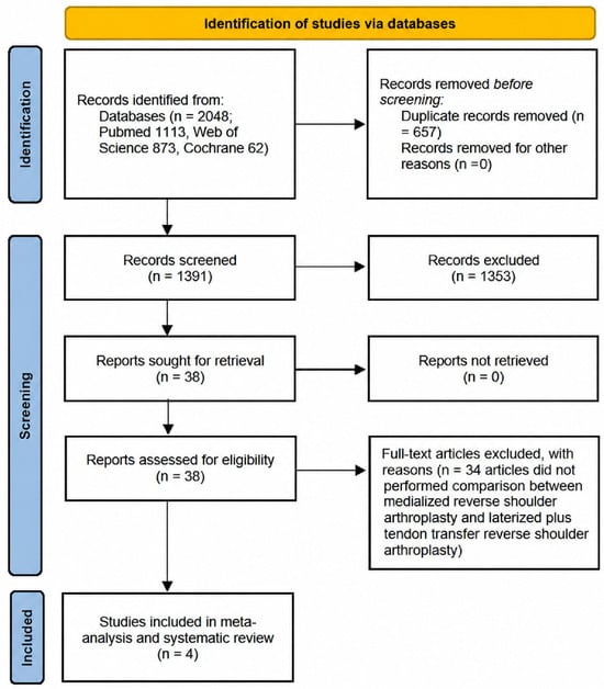
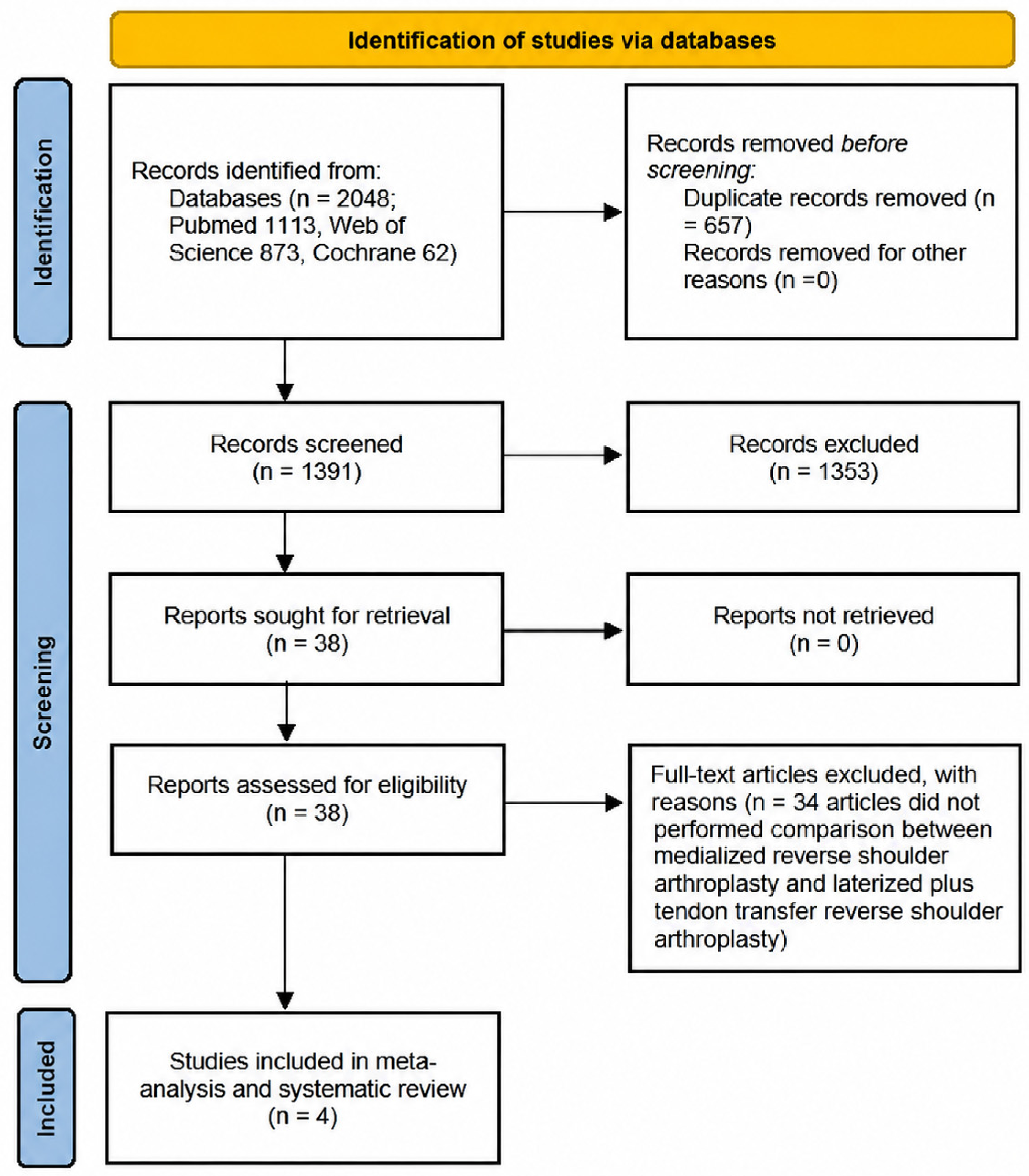
The literature search retrieved 2048 articles. Title and abstract screening led to the exclusion of 1353 articles. Following the screening process, four comparative studies met the inclusion criteria for the meta-analysis: one Level I randomized controlled trial [16] and three Level III retrospective comparative studies [17,18,19]. The selection process is summarized in the PRISMA flowchart in Figure 1. The final pooled cohort consisted of 151 shoulders evaluated across four comparative studies. Patients were stratified into two cohorts: 78 shoulders in the RSA group and 73 shoulders in the LDT group. Baseline demographic characteristics were consistently homogeneous across all included studies. No statistically significant differences were observed between the RSA and LDT groups at baseline in any of the individual trials. Detailed demographic data for each included study are provided in Table 1.
Figure 1.
PRISMA flowchart of the study selection process.
Table 1.
Details of the included studies.
3.2. Implant Design and Characteristics
To confirm the effective use of lateralized RSA implants, a rigorous assessment of prosthetic features was conducted, confirming that modern implants characterized by specific lateralization strategies designed to optimize posterior deltoid tension without soft-tissue augmentation were used in the RSA group. Specifically, Young et al. and Warnhoff et al. [16,18] employed humeral stems with a reduced neck-shaft angle (135° and 145°, respectively), ensuring significant humeral lateralization. Merolla et al. [17] utilized a curved onlay stem (145° inclination), achieving lateralization through the geometry of the epiphysis. Wiater et al. [19] obtained implant lateralization on the glenoid side using lateralized baseplates and glenospheres.
In the LDT group, classic Grammont-style non-lateralizing implants were used. In particular, Merolla et al., Wiater et al., and Warnhoff et al. [17,18,19] utilized classic medialized 155° inlay stems, adding LDT to compensate for the lack of prosthetic lateralization. Conversely, Young et al. [16] used the same lateralized implant designs in the control group and in the intervention group. Because the same lateralized implant design was used in both groups, Young et al. provided the closest available comparison of the isolated additive effect of tendon transfer (Table 2).
Table 2.
Detailed summary of the reverse shoulder arthroplasty systems utilized.
3.3. Range of Motion
The analysis of postoperative active ROM evaluated forward elevation (FE), abduction (ABD), external rotation (ER), and internal rotation (IR).
- •
- Data regarding active FE were reported in all four included studies. Postoperative FE did not differ significantly between groups. The RSA group achieved a weighted mean FE of 129.5° ± 21.0°, while the LDT group reached 132.8° ± 23.5° (std. MD: 0.12; 95% CI: −10.43 to 3.83; p > 0.05) (Table 3) (Figure 2) [16,18,19].Figure 2. Forest plot of the postoperative elevation. SD (standard deviation), CI (confidence interval), IV (interval value), Std (standard).
- •
- ABD was quantitatively synthesized from two studies. Postoperative analysis revealed no significant difference between the techniques: the RSA group achieved a weighted mean of 121.5° ± 21.5° versus 130.5° ± 25.5° in the LDT group (std. MD: −0.37; 95% CI: −21.23 to 3.23; p >0.05) (Table 3) (Figure 3) [17,18].Figure 3. Forest plot of the postoperative abduction. SD (standard deviation), CI (confidence interval), IV (interval value), Std (standard).
- •
- The analysis of active ER was stratified based on arm position. Comparison of ER at 0° of abduction (adduction) was reported in three studies. The pooled analysis showed no statistically significant difference, with the RSA group achieving a weighted mean of 16.3° ± 15.1° compared to 12.1° ± 14.8° in the LDT group (std. MD: 0.2; 95% CI: −1.45 to 9.85; p > 0.05). ER at 90° of abduction was reported in one study [16]. No statistically significant difference was reported between the RSA group (80° ± 33°) and the LDT group (70° ± 22°) (p > 0.05) (Table 3) (Figure 4) [17,18,19].Figure 4. Forest plot of the postoperative external rotation. SD (standard deviation), CI (confidence interval), IV (interval value), Std (standard).
- •
- Internal Rotation (IR) outcomes were assessed across all studies using various metrics, including vertebral levels and functional scales. Differences in postoperative IR outcomes generally favored the RSA group, although the methods of assessment varied and the results were not pooled. Specifically, Wiater et al. [19] reported a statistically significant difference between the RSA and LDT groups regarding the pre-to-postoperative change (p = 0.01): while the RSA group showed a preservation or slight improvement in IR (+ 0.77 vertebral levels), the LDT group experienced a functional decline (−0.29 vertebral levels). Regarding functional assessment, two studies [17,18] analyzed IR using specific scores. Warnhoff et al. [18] reported a significantly higher rate of negative Apley scratch tests in the RSA group (67%) compared to the LDT group (23%) (p < 0.05). Similarly, Merolla et al. [17] found significantly higher Constant sub-scores for internal rotation in the RSA group (p = 0.004). Young et al. found no significant difference using vertebral levels for evaluation (Table 3) [16].
Table 3.
Comparison of postoperative range of motion and functional outcomes.
3.4. Resolution of Clinical Signs
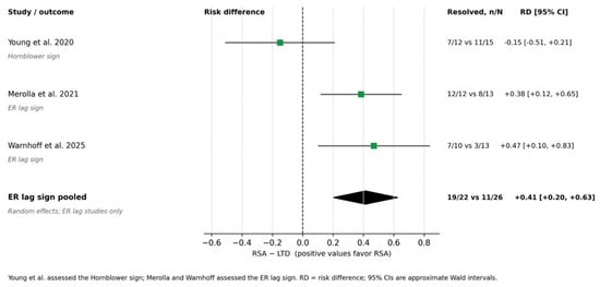
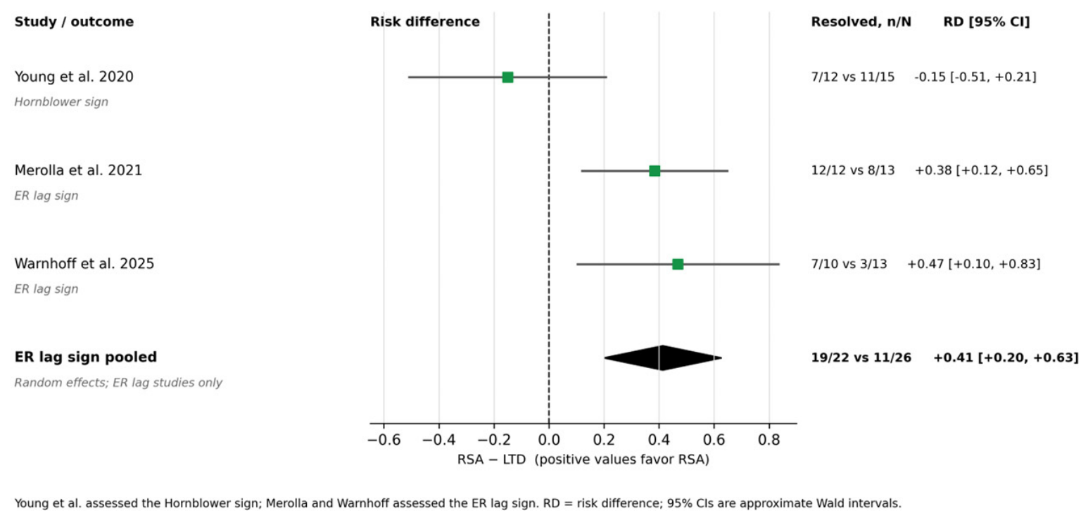
The resolution of ER deficit clinical signs (hornblower or ERLS) was reported in three studies [16,17,18]. While 100% of patients presented with positive clinical signs preoperatively, the pooled analysis showed a higher resolution rate in the RSA group of 76.5% (26/34 patients) compared to 53.7% (22/41 patients) in the LDT group. The pooled analysis revealed a statistically significant benefit for the RSA group (95% CI: 0.20 to 0.63; p = 0.04) (Table 4 and Figure 5).
Table 4.
Postoperative resolution rates of preoperative clinical signs of horizontal instability.
Figure 5.
Forest plot of the postoperative ER lag sign. RSA (reverse shoulder arthroplasty); LDT (latissimus dorsi transfer); ER (external rotation); RD (risk difference); CI (confidence interval); n (number of patients with resolution); N (total number of patients with a preoperative positive sign).
3.5. Functional Scores
The American Shoulder and Elbow Surgeons (ASES) score was reported in two studies [16,19]. The pooled analysis revealed no statistically significant difference between the two techniques (76.3 ± 23.5 in the RSA group vs. 77.1 ± 20.3 in the LDT group; p = 0.86). The Constant score was reported in two studies [17,18]. Similarly, no statistically significant difference was observed at final follow-up (62.7 ± 7.7 in the RSA group vs. 58.1 ± 15.1 in the LDT group; p = 0.12). Crucially, both groups demonstrated substantial improvements from preoperative baseline values, exceeding the minimal clinically important difference (MCID) (Table 3).
3.6. Safety and Operative Efficiency
Safety profiles were broadly comparable across the included studies, with no statistically significant differences detected (p > 0.05). In the RSA cohort, a single traumatic clavicle fracture was reported [18]. In contrast, the LDT cohort showed one dislocation [16] and one case requiring arthroscopic debridement [18], while Merolla et al. [17] and Wiater et al. [19] reported no complications in either group.
With respect to operative efficiency, the addition of a tendon transfer was consistently associated with increased surgical time. Warnhoff et al. [18] quantified this difference, demonstrating a mean increase of 55 min for the combined procedure (135 ± 26 vs. 80 ± 15 min; p < 0.001). This finding is in line with observations from Wiater et al. [19] and Merolla et al. [17], who also noted prolonged operative times attributable to the additional technical steps required for tendon harvesting and fixation.
3.7. Risk of Bias Assessment
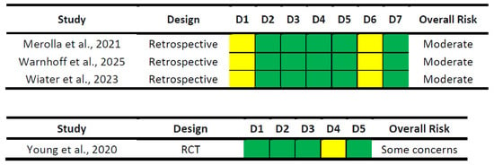
The RCT by Young et al. [16] was evaluated using the Cochrane RoB 2 tool and was classified as raising “some concerns.” While the randomization process and allocation concealment were robust (low risk), the inherent inability to blind patients and personnel to the tendon transfer procedure—which requires distinct incisions and rehabilitation protocols—introduced a potential detection bias in the measurement of subjective outcomes (Domain 4) (Figure 6).
Figure 6.
Risk of bias of the included studies according to the Risk of Bias in Non-randomized Studies of Interventions (ROBINS-I) tool for non-randomized comparative trials and the Cochrane Collaboration Risk of Bias tool Version 2 (ROB) for the only randomized study.
The three non-randomized comparative studies [17,18,19] were assessed using the ROBINS-I tool across seven domains. These studies were consistently classified as having a “moderate” overall risk of bias. Analysis of the specific domains indicated that the primary sources of bias were confounding (Domain 1), as the allocation to the transfer group was often influenced by surgeon preference or the severity of preoperative deformity, and measurement of outcomes (Domain 6), due to the lack of blinded assessment. However, the risk was judged to be low in the remaining domains: selection of participants (Domain 2), classification of interventions (Domain 3), deviations from intended interventions (Domain 4), missing data (Domain 5), and selection of the reported result (Domain 7); see Figure 5. Finally, according to the GRADE (Grading of Recommendations Assessment, Development and Evaluation) approach, the overall certainty of evidence for the primary outcomes was rated as “low” to “moderate”. This rating reflects the presence of non-randomized evidence and the limited cumulative sample size (imprecision), despite the high consistency of clinical findings across the included studies.
4. Discussion
The most significant finding of this systematic review and meta-analysis is that no statistically significant difference in active ER was detected between lateralized RSA alone and RSA combined with LDT for patients with CTA and ER deficit. The pooled data from Level I and III studies demonstrate that implant lateralization effectively resolves the ER lag sign while obtaining functional scores comparable to the LDT procedure [16,17,18,19]. Moreover, the “lateralization-only” strategy avoids the morbidity associated with tendon transfer, offering superior preservation of internal rotation and a more streamlined surgical workflow. These results potentially support the shift from a “soft-tissue substitution” philosophy with LDT to a “biomechanical compensation” model with RSA lateralization.
Historically, the Grammont design—characterized by a medialized center of rotation and a 155° neck-shaft angle—reliably increased the deltoid lever arm, restoring abduction and elevation, but failed to address the posterior force couple, resulting in a failure to restore external rotation [20]. Several cohorts of patients treated with a medialized implant (155° NSA) without tendon transfer exhibited a persistent failure in restoring ER [3,21,22]. In fact, theoretically, this mechanical deficiency necessitated an LDT to act as an external rotator substitute [23]. Nonetheless, the existing literature and the results of this meta-analysis suggest that a critical role in the restoration of ER may be played by implant geometry. In fact, the RSA group of this meta-analysis—utilizing lateralized stems or glenospheres as per Warnhoff et al., Merolla et al., and Wiater et al. [17,18,19]—achieved consistent ER restoration (mean +12° to +19°) with a higher recovery rate of the ER lag sign [18]. This comparison provides clinical proof that the posterior deltoid has the intrinsic capacity to act as an external rotator but only if mechanically potentiated by implant lateralization (humeral or glenoidal). This aspect has been further demonstrated by Merolla’s electromyographic analysis, showing that lateralization restores the resting length and increases the posterior moment arm of the posterior deltoid [17]. However, the independent effect of each prosthetic design feature remains difficult to establish, given the wide variety of implant configurations and designs currently available on the market, which complicates the assessment of their individual contributions to clinical outcomes.
In the literature, the retrospective study by Berglund et al. reported that lateralized RSA without tendon transfer (135° neck-shaft angle, glenosphere lateralization) restored ER from −21° preoperatively to 28° postoperatively (p < 0.001) at a mean follow-up of 43 months (range 24–77 months), with no correlation between teres minor fatty infiltration and ER improvement. A recent meta-analysis by Hones et al., which included both medialized and lateralized implants, reported outcomes generally consistent with the findings of the current meta-analysis, including superior postoperative ER and Constant scores for lateralized RSA alone, as well as lower rates of nerve injury and dislocation. However, direct comparison with the results of this meta-analysis is limited by differences in inclusion criteria and implant stratification. The present work therefore complements existing evidence by providing a more clinically homogeneous and implant-specific analysis [21].
Of particular importance is the Level I evidence from Young et al. [16], which demonstrated that adding a tendon transfer to a lateralized prosthesis does not yield clinical improvement. The fact that Young et al. [16] found no significant difference in ASES scores or active ER between the two groups suggests a functional “ceiling effect”: once the posterior deltoid has been mechanically optimized through lateralization, the addition of an external force vector via LDT appears not only ineffective but also subject to diminishing returns [24]. Clinically, these findings do not support the routine addition of LDT to every lateralized RSA, as the decision should remain individualized. If an ER deficit is effectively corrected by the immediate static tensioning of the posterior deltoid provided by lateralization, the tendon transfer procedure may introduce biological variables—such as tendon healing, tensioning errors, and the necessity for neuromuscular re-education—which may result in a higher rate of persistent ER lag signs in the LDT groups [18,24]. However, of note, the interpretation of the results presented by Young et al. has been questioned by Boileau et al., who pointed out that a reduced NSA might not be enough to consider the implants lateralized, potentially confounding the isolated effect of the tendon transfer.
Another critical aspect is that the tendon transfer is not merely “neutral” regarding internal rotation (IR) but actively detrimental. Harvesting the latissimus dorsi—one of the shoulder’s primary internal rotators—may alter the rotational balance. This was reported by Wiater et al. [19], who documented a divergent trajectory: patients treated with lateralized RSA alone gained function (+ 0.77 vertebral levels), while those undergoing transfer lost function (−0.29 levels, p = 0.01). This difference may have functional relevance, as also highlighted by Flury et al.: in their transfer group, the ability to reach the lumbosacral junction collapsed from 92% preoperatively to 38% postoperatively [3]. Conversely, lateralized-only implants were associated with more favorable IR outcomes in some studies, although assessment methods differed [18,19,24]. Biomechanically, this can be explained by the fact that implant lateralization maintains the native insertion of the internal rotators and tension on the anterior deltoid and subscapularis, facilitating the mechanics essential for activities of daily living such as perineal hygiene, tucking in a shirt, and independent toileting. Sacrificing these daily necessities to chase external rotation (which the prosthesis alone can already provide) could represent a questionable clinical trade-off [18,24]. In this light, the literature highlights the tangible biological and logistical cost of tendon transfer. In particular, the data from Flury et al. offer detailed insight into its morbidity. They documented that the addition of LDT significantly extended operative time by a mean of 26 min (p = 0.003) and was associated with a 23% complication rate (including radial nerve palsy and tendon re-tear), compared to only 8% in the RSA-alone group [3]. Similarly, Warnhoff et al., Wiater et al., and Merolla et al. [17,18,19] noted increased surgical duration and complexity. These data suggest that, to reduce complications given the similar clinical results, the streamlined, implant-based solution could be favored over the more complex soft-tissue reconstruction.
An additional safety concern specific to LDT is the potential risk of anterior instability. Harvesting the latissimus dorsi and teres major requires an incision through the pectoralis major tendon. When combined with an irreparable subscapularis—a frequent finding in CTA with an ER deficit—the resulting loss of anterior soft-tissue restraint may predispose patients to anterior dislocation. This concern is supported by the meta-analysis by Hones et al., which reported a dislocation rate of 2.8% with LDT versus 0.8% with lateralized RSA alone. Therefore, LDT should be used with particular caution in patients with an irreparable subscapularis or compromised anterior soft-tissue restraint [24].
Furthermore, a crucial consideration concerns the durability of mechanically restored ER. The mean follow-up of the 151 shoulders included in this meta-analysis ranged from 24 to 77 months across the primary studies [21]. Berglund et al. found no decline in ER over a mean of 43 months, and the studies in this meta-analysis showed stable results within this time frame [21]. However, the theoretical concern remains that over a longer period (beyond 10 years), age-related muscle atrophy and fatty infiltration could progressively reduce the mechanical advantage provided by lateralization. In other words, what is the fate of this “mechanical” external rotation, knowing that it may wear out over time? Conversely, Alonso-Rodriguez Piedra et al. demonstrated, using fine-needle EMG at a mean follow-up of 80 months, that transferred latissimus dorsi and teres major muscles remain functionally active as external rotators over time, achieving at least 50% of the strength of the contralateral shoulder [23]. Thus, while lateralized RSA alone provides excellent mid-term results (>10 years), the actual durability of mechanical ER remains unknown, and the included comparative studies do not establish the comparative long-term durability of lateralized RSA alone versus RSA combined with LDT.
Despite its important findings, this meta-analysis has some limitations: the first limitation is the reliance on predominantly retrospective data (Level III), with only one RCT available. This introduces an inherent selection bias (confounding by indication), as the decision to perform a tendon transfer in the historical cohorts was often driven by surgeon preference or the perceived severity of the preoperative deformity, rather than randomization. In addition, in three studies the comparison involved lateralized RSA without LDT versus predominantly medialized RSA with LDT; because implant design and tendon transfer status changed simultaneously, their independent effects cannot be disentangled. Second, heterogeneity in prosthetic design—encompassing both humeral (135°–145° NSA) and glenoidal lateralization—precluded the analysis of specific implant features, although all intervention devices shared the biomechanical goal of increasing the posterior deltoid moment arm. Furthermore, variability in outcome measurement methods necessitated stratified analyses rather than a single pooled effect size. Another specific limitation concerns the influence of the quality of the teres minor on the success of lateralization. Although we aimed to investigate the impact of muscle atrophy, the lack of standardized reporting of Goutallier grades across most included studies—with the exception of Warnhoff et al. [18]—precluded a pooled stratified analysis. Consequently, it remains unclear whether severe atrophy (Goutallier 3–4) represents a biological boundary where prosthetic compensation alone becomes insufficient, warranting further investigation in focused prospective cohorts.
5. Conclusions
Lateralized RSA was not associated with statistically significant differences in clinical scores or active external rotation compared with RSA combined with latissimus dorsi transfer. However, RSA without LDT was associated with higher rates of ER lag sign resolution and better internal rotation. In light of these findings, and considering the greater surgical invasiveness associated with LDT, lateralized RSA without LDT may be considered in patients with ER deficits.
Supplementary Materials
The following supporting information can be downloaded at: https://www.mdpi.com/article/10.3390/jcm15229117/s1, Table S1: PRISMA checklist.
Author Contributions
Conceptualization, L.M.O., P.F. and D.P.; methodology, L.M.O.; formal analysis, L.M.O.; investigation, L.M.O., P.F. and D.P.; resources, L.M.O.; data curation, L.M.O., F.S. and D.P.; writing—original draft preparation, L.M.O.; writing—review and editing, P.F, D.P., F.S. and J.K.; visualization, F.S. and C.C.; supervision, G.F. and C.C.; project administration, C.C.; funding acquisition, C.C. All authors have read and agreed to the published version of the manuscript.
Funding
This research received no external funding.
Institutional Review Board Statement
Not applicable.
Informed Consent Statement
Not applicable.
Data Availability Statement
No new data were created or analyzed in this study.
Acknowledgments
During the preparation of this work, the authors used Grammarly for the purpose of checking grammar, spelling, and basic English language corrections. After using this tool, the authors reviewed and edited the content as needed and take full responsibility for the content of the publication.
Conflicts of Interest
The authors declare no conflict of interest.
References
- Franceschi, F.; Giovannetti de Sanctis, E.; Gupta, A.; Athwal, G.S.; Di Giacomo, G. Reverse shoulder arthroplasty: State-of-the-art. J. Isakos 2023, 8, 306–317. [Google Scholar] [CrossRef] [Scilit] [PubMed]
- Sellers, T.R.; Abdelfattah, A.; Frankle, M.A. Massive Rotator Cuff Tear: When to Consider Reverse Shoulder Arthroplasty. Curr. Rev. Musculoskelet. Med. 2018, 11, 131–140. [Google Scholar] [CrossRef] [Scilit] [PubMed]
- Flury, M.; Kwisda, S.; Kolling, C.; Audigé, L. Latissimus dorsi muscle transfer reduces external rotation deficit at the cost of internal rotation in reverse shoulder arthroplasty patients: A cohort study. J. Shoulder Elb. Surg. 2019, 28, 56–64. [Google Scholar] [CrossRef] [Scilit] [PubMed]
- Hones, K.M.; Gutowski, C.T.; Rakauskas, T.R.; Bindi, V.E.; Simcox, T.; Wright, J.O.; Schoch, B.S.; Wright, T.W.; Werthel, J.-D.; King, J.J.; et al. Outcomes of lateralized reverse total shoulder arthroplasty versus latissimus dorsi transfer for external rotation deficit: A systematic review and meta-analysis. Clin. Shoulder Elb. 2024, 27, 464–478. [Google Scholar] [CrossRef] [Scilit] [PubMed]
- Boileau, P.; Chuinard, C.; Roussanne, Y.; Neyton, L.; Trojani, C. Modified latissimus dorsi and teres major transfer through a single delto-pectoral approach for external rotation deficit of the shoulder: As an isolated procedure or with a reverse arthroplasty. J. Shoulder Elb. Surg. 2007, 16, 671–682. [Google Scholar] [CrossRef] [Scilit] [PubMed]
- Neyton, L.; Gossing, L.; Gasbarro, G.; Kirsch, J.M. Modified L’Episcopo tendon transfer for isolated loss of active external rotation. J. Shoulder Elb. Surg. 2020, 29, 2587–2594. [Google Scholar] [CrossRef] [Scilit] [PubMed]
- Schönweger, F.; Oldrini, L.M.; Feltri, P.; Filardo, G.; Candrian, C. Stemmed VS stemless total shoulder arthroplasty: A systematic review and meta-analysis. Arch. Orthop. Trauma Surg. 2024, 145, 3. [Google Scholar] [CrossRef] [Scilit] [PubMed]
- Boileau, P.; Rumian, A.P.; Zumstein, M.A. Reversed shoulder arthroplasty with modified L’Episcopo for combined loss of active elevation and external rotation. J. Shoulder Elb. Surg. 2010, 19, 20–30. [Google Scholar] [CrossRef] [Scilit] [PubMed]
- Marigi, E.M.; Harstad, C.; Elhassan, B.; Sanchez-Sotelo, J.; Wieser, K.; Kriechling, P. Reverse shoulder arthroplasty after failed tendon transfer for irreparable posterosuperior rotator cuff tears. J. Shoulder Elb. Surg. 2022, 31, 763–771. [Google Scholar] [CrossRef] [Scilit] [PubMed]
- Oldrini, L.M.; Sangiorgio, A.; Feltri, P.; Marbach, F.; Filardo, G.; Candrian, C. Proximal humerus fractures: Deltopectoral open reduction and internal fixation vs deltosplit minimally invasive plate osteosynthesis: Which surgical approach provides superior results? EFORT Open Rev. 2023, 8, 662–671. [Google Scholar] [CrossRef] [Scilit] [PubMed]
- Valenti, P.; Zampeli, F.; Caruso, G.; Nidtahar, I.; Martinez-Catalan, N.; Kazum, E. Proximal humeral bone defect in reverse shoulder arthroplasty combined with latissimus-dorsi transfer is not related with a poor outcome. Orthop. Traumatol. Surg. Res. 2022, 108, 103263. [Google Scholar] [CrossRef] [Scilit] [PubMed]
- Oldrini, L.M.; Feltri, P.; Albanese, J.; Marbach, F.; Filardo, G.; Candrian, C. PHILOS Synthesis for Proximal Humerus Fractures Has High Complications and Reintervention Rates: A Systematic Review and Meta-Analysis. Life 2022, 12, 311. [Google Scholar] [CrossRef] [Scilit] [PubMed]
- Tan, A.K.S.; Chung, I.W.; Lee, W.Q.; Lie, D.T.T. Latissimus dorsi and teres major transfer in reverse shoulder arthroplasty: A systematic review. JSES Rev. Rep. Tech. 2024, 4, 379–384. [Google Scholar] [CrossRef] [Scilit] [PubMed]
- Wey, A.; Dunn, J.C.; Kusnezov, N.; Waterman, B.R.; Kilcoyne, K.G. Improved external rotation with concomitant reverse total shoulder arthroplasty and latissimus dorsi tendon transfer: A systematic review. J. Orthop. Surg. 2017, 25, 2309499017718398. [Google Scholar] [CrossRef] [Scilit] [PubMed]
- Wan, X.; Wang, W.; Liu, J.; Tong, T. Estimating the sample mean and standard deviation from the sample size, median, range and/or interquartile range. BMC Med. Res. Methodol. 2014, 14, 135. [Google Scholar] [CrossRef] [Scilit] [PubMed]
- Young, B.L.; Connor, P.M.; Schiffern, S.C.; Roberts, K.M.; Hamid, N. Reverse shoulder arthroplasty with and without latissimus and teres major transfer for patients with combined loss of elevation and external rotation: A prospective, randomized investigation. J. Shoulder Elb. Surg. 2020, 29, 874–881. [Google Scholar] [CrossRef] [Scilit] [PubMed]
- Merolla, G.; Cuoghi, F.; Athwal, G.S.; Parel, I.; Filippi, M.V.; Cutti, A.G.; Fabbri, E.; Padolino, A.; Paladini, P.; Catani, F.; et al. A kinematic and electromyographic comparison of a Grammont-style reverse arthroplasty combined with a l’Episcopo transfer compared to a lateralized humeral component reverse for restoration of active external rotation. Int. Orthop. 2021, 45, 2061–2069. [Google Scholar] [CrossRef] [Scilit] [PubMed]
- Warnhoff, M.; Moroder, P.; Audigé, L.; Spagna, G.; Ameziane, Y.; Schneller, T.; Scheibel, M.; Freislederer, F. Lateralized Reverse Shoulder Arthroplasty vs. Medialized Design with Latissimus Dorsi Transfer for Cuff Tear Arthropathy with Loss of External Rotation and ER Lag Sign. J. Clin. Med. 2025, 14, 5979. [Google Scholar] [CrossRef] [Scilit] [PubMed]
- Wiater, J.M.; Oshikoya, O.; Shields, E.; Vara, A.D.; Cavinatto, L.; Koueiter, D.M. Concomitant latissimus dorsi tendon transfer during reverse total shoulder arthroplasty does not improve active external rotation or clinical outcomes in patients with external rotation deficit. J. Shoulder Elb. Surg. 2023, 32, 1016–1021. [Google Scholar] [CrossRef] [Scilit] [PubMed]
- Boileau, P.; Watkinson, D.J.; Hatzidakis, A.M.; Balg, F. Grammont reverse prosthesis: Design, rationale, and biomechanics. J. Shoulder Elb. Surg. 2005, 14, 147s–161s. [Google Scholar] [CrossRef] [Scilit] [PubMed]
- Berglund, D.D.; Rosas, S.; Triplet, J.J.; Kurowicki, J.; Horn, B.; Levy, J.C. Restoration of External Rotation Following Reverse Shoulder Arthroplasty without Latissimus Dorsi Transfer. JBJS Open Access 2018, 3, e0054. [Google Scholar] [CrossRef] [Scilit] [PubMed]
- Passaretti, D.; Ascani, C.; Ponzo, A.; Marchioni, B.; De Cupis, M.; Scacchi, M.; Pasqualetto, M.; Ascani, J.; Candela, V.; Gumina, S. Lateralized versus medialized glenoid implants in reverse total shoulder arthroplasty for proximal humerus fractures. Comparison between trabecular metal and comprehensive Zimmer Biomet glenoid implants. J. Shoulder Elb. Surg. 2026, 35, 1201–1208. [Google Scholar] [CrossRef] [Scilit] [PubMed]
- Alonso-Rodriguez Piedra, J.; Souza Virgolino, B.; Gamez Baños, F.; Miranda Elstein, Q.; Ventura Parellada, C.; Mora Guix, J.M. Reverse total shoulder arthroplasty with latissimus dorsi and teres major transfer: Biomechanical and electromyographical outcomes. Eur. J. Orthop. Surg. Traumatol. 2023, 33, 1003–1012. [Google Scholar] [CrossRef] [Scilit] [PubMed]
- Hones, K.M.; Rakauskas, T.R.; Wright, J.O.; King, J.J.; Wright, T.W.; Werthel, J.D.; Schoch, B.S.; Hao, K.A. Outcomes of Reverse Total Shoulder Arthroplasty with Latissimus Dorsi Tendon Transfer for External Rotation Deficit: A Systematic Review and Meta-Analysis. JBJS Rev. 2023, 11. [Google Scholar] [CrossRef] [Scilit] [PubMed]







Disclaimer/Publisher’s Note: The statements, opinions and data contained in all publications are solely those of the individual author(s) and contributor(s) and not of MDPI and/or the editor(s). MDPI and/or the editor(s) disclaim responsibility for any injury to people or property resulting from any ideas, methods, instructions or products referred to in the content. |
© 2026 by the authors. Licensee MDPI, Basel, Switzerland. This article is an open access article distributed under the terms and conditions of the Creative Commons Attribution (CC BY) license.