


Abstract
Background/Objectives: Multivessel coronary artery disease (MVCD) determines prognosis and revascularization strategy. The CRP × 100/HDL-C atherogenic index (AtI) integrates systemic inflammation and anti-atherogenic lipoprotein parameters. We evaluated its capacity to predict MVCD in patients undergoing invasive coronary angiography (CA). Materials and Methods: Retrospective cross-sectional study of a cohort of 204 patients with suspected ischemic heart disease. The 173 patients who underwent invasive CA as a diagnostic test were classified as having no significant lesions (n = 55), single-vessel disease (n = 69), or MVCD (n = 49). The new AtI was measured as CRP (mg/dL) × 100/HDL-C (mg/dL). Kruskal-Wallis with Bonferroni correction, ROC analysis with DeLong confidence intervals, adjusted logistic regression (log2-transformed), and Harrell bootstrap optimism correction (1000 iterations) were applied. Results: The AtI showed a progressive gradient: median 0.73 (IQR 0.57–1) in the no-lesions group, 1.71 (IQR 0.95–2.44) in the single-vessel group, and 2.86 (IQR 1.79–6.67) in the MVCD group (Kruskal-Wallis p < 0.001), Spearman ρ = 0.596 (p < 0.001), and all Bonferroni-corrected pairwise comparisons significant. AUC for MVCD prediction: 0.818 (95% CI 0.753–0.883), superior to Castelli I (0.685; DeLong p = 0.003), Castelli II (0.626; p < 0.001), and the plasma atherogenic index (0.708; p = 0.013). At cut-off ≥2.00 (Youden criterion): sensitivity 73.5%, specificity 78.2%, NPV 88.2%. The AtI was independently associated with MVCD in adjusted logistic regression (OR 2.84 per doubling; 95% CI 1.95–4.13; p < 0.001). Adding the AtI to a classical risk factor model increased AUC from 0.652 to 0.836 (ΔAUC +0.184; 95% CI 0.093–0.275; p < 0.001; optimism-corrected AUC 0.812). Conclusions: The CRP × 100/HDL-C AtI is independently associated with MVCD and adds discrimination to a clinical model. It is a simple and inexpensive marker of angiographic extent; a cut-off of ≥2 had an NPV of 88.2% (86.5% after optimism correction), and its clinical usefulness requires external and prospective validation.
1. Introduction
Ischemic heart disease (IHD) has been the leading cause of morbidity and mortality worldwide for the last 20 years [1], despite numerous advances in both pharmacologic treatment and percutaneous coronary interventions. Dyslipidemia is one of the main cardiovascular risk factors (CVRF) that lead to IHD, as the causal role of cholesterol bound to low-density lipoproteins (LDL-C) and the formation of an atherosclerotic plaque has been established. Current guidelines emphasize LDL reduction, but they also acknowledge that IHD is still present in a large subgroup of patients that have normal LDL but have elevated residual risk [2]. These patients normally have elevated triglycerides, low values of cholesterol bound to high-density lipoproteins (HDL-C), and elevated C-reactive protein (CRP) as a marker for systemic inflammation. This residual risk has been associated with IHD and with multivessel coronary disease (MVCD). MVCD, defined as a significant stenosis (>70% in epicardial coronary arteries or more than 50% in the left main trunk) in 2 or more coronary territories, is present in many patients undergoing coronary angiography for an acute coronary syndrome, determining both the revascularization strategy and the prognosis of the patient [3,4].
Atherogenic indexes (AtI), ratios between different cholesterol fractions, are accessible tools used for risk stratification, but they offer an incomplete view on atherosclerosis pathogenesis, as they do not include parameters related to inflammation. The classic AtI only includes parameters related to circulating lipids, as they are the result of a ratio between atherogenic particles and anti-atherogenic particles. They include Castelli I (total cholesterol/HDL-C), Castelli II (LDL-C/HDL-C), and the atherogenic index of plasma (AIP): log 10 (triglycerides/HDL-C). Their association with MVCD has been reported previously [5,6,7].
Our group has developed a new AtI integrates both the lipid residual risk and the inflammatory residual risk, defined as CRP (mg/dL) × 100/HDL-C (mg/dL). This index has been associated both with carotid atherosclerosis [8] and coronary heart disease [9] in previous studies, but its relationship specifically with MVCD has not been adequately evaluated before. An exploratory subgroup analysis [9] in the same cohort detected an association between the new AtI and MVCD, but it was methodologically limited as the index was used as a dichotomous variable, no adjusted multivariable model was fitted, and analyses were not confined to coronary angiography (CA), which is the reference standard for defining MVCD.
The present study aims to address these limitations and to evaluate the association of the AtI with MVCD and its incremental value over traditional CVRF and to compare its discrimination with classical AtI.
2. Materials and Methods
2.1. Study Population
We conducted a retrospective cross-sectional study of a single center cohort of 204 patients younger than 65 years, with no previous cardiac history, admitted to cardiology for suspicion of IHD. The patients were identified retrospectively from hospital records, and the cohort was described in our previous study [9]. The present study included a subset of 173 patients from that original cohort, with a different outcome (association with MVCD), a different statistical treatment (AtI as a continuous variable with an adjusted multivariable model), and a different subpopulation (only patients with invasive CA).
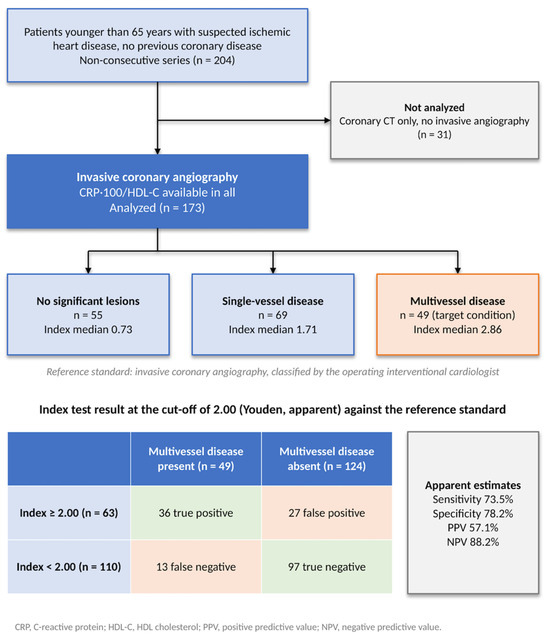
Of the original 204 patients, 31 had their coronary anatomy assessed by computerized tomography (CT) and were excluded; the present study was done on 173 patients that underwent CA (Figure 1).
Figure 1.
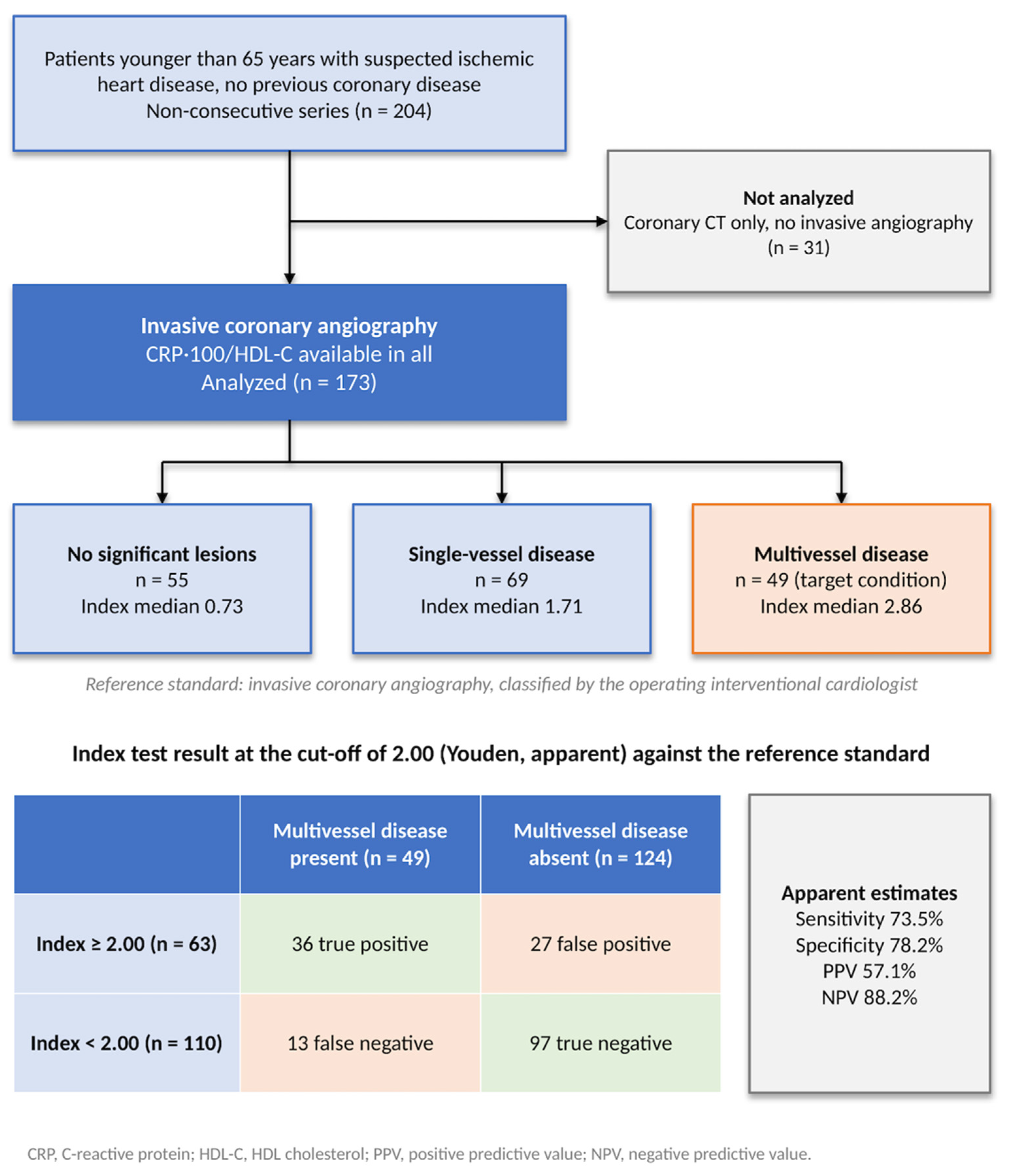
Flow diagram. Patients with suspected coronary artery disease (n = 204): 31 with coronary CT only were excluded; 173 underwent CA and were analyzed (no significant lesions 55, single-vessel disease 69, MVCD 49). The 2 × 2 table shows the AtI at the apparent cut-off of 2.00 against the reference standard.
2.2. Coronary Angiography
Coronary angiography was performed on a Philips Allura Xper FD20 system (Philips Healthcare, Best, The Netherlands), and angiograms were classified by the interventional cardiologist who performed each procedure. The choice of diagnostic modality between coronary CT and invasive CA was done by the attending cardiologist at that moment on clinical grounds. A significant stenosis was defined as a >70% diameter reduction in a major epicardial coronary artery or >50% in the left main coronary artery. Groups were classified according to the number of affected territories: (1) no significant lesions, (2) single vessel disease, or (3) MVCD (two or more territories). Functional assessment of intermediate lesions (50–70%) was not performed systematically.
2.3. Clinical Variables and Laboratory Analysis
Demographic data, cardiovascular risk factors, NYHA functional class, medication use, clinical presentation (unstable angina, non-ST elevation myocardial infarction (NSTEMI), ST elevation myocardial infarction (STEMI), chronic coronary syndrome), and ECG findings were recorded. Laboratory analysis, including fasting glucose, HbA1c, lipid profile (total cholesterol, LDL-C, HDL-C, and triglycerides), and C-reactive protein, was obtained from blood samples drawn on admission in patients admitted with acute coronary syndrome and up to 3 days before the procedure in patients studied electively; therefore, all samples preceded angiography and percutaneous coronary intervention. Total cholesterol, HDL-C, and triglycerides were measured directly with an enzymatic assay with specific detergents, and LDL-C was calculated with the Friedewald formula. CRP was measured by immunoturbidimetry on an Abbott platform (not high-sensitivity; reference interval 0.00–0.50 mg/dL; lower limit of quantification 0.02 mg/dL). The AtI was calculated as the ratio between CRP × 100 (mg/dL)/HDL-C and was compared with the traditional AtI Castelli I (total cholesterol/HDL-C), Castelli II (LDL-C/HDL-C) and AIP (log10 triglycerides/HDL-C).
2.4. Statistical Analysis
Categorical variables are reported as n (%) and compared using the Pearson chi-squared test. Continuous variables were tested for normality using the Shapiro–Wilk test and are reported as median (interquartile range, IQR). They are compared using the Kruskal-Wallis test, with Bonferroni-corrected pairwise Mann-Whitney comparisons for the main AtI. Spearman’s ρ was used to assess the correlation of the AtI with angiographic extent (0, 1, or ≥2 territories).
Discrimination of the AtI for MVCD was assessed by Receiver Operating Characteristic (ROC) analysis, with 95% CI by DeLong’s method. Area under the curve (AUC) were compared with the paired DeLong test (curves from the same patients) against Castelli I, Castelli II, and the AIP (chosen because they are the best-known atherogenic indices in routine clinical practice); these three p-values are reported unadjusted, and after Holm adjustment all remained significant with a p < 0.05 (largest adjusted p = 0.013). The cut-off point was 2.00 and was chosen with the Youden index in our same sample, so sensitivity, specificity, positive predictive value (PPV), and negative predictive value (NPV) are apparent estimates. To quantify the optimism, cut-off selection and evaluation were evaluated with 1000 bootstrap resamples, and the performance of each resample cut-off point in the original data was compared with its apparent performance, and the mean difference was subtracted (Harrell’s method for bootstrap optimism correction). The cut-off point differs from the value ≥ 1 reported in the original cohort [9] because the outcome and subpopulation were different. Because the AtI is right-skewed, we assessed the linearity of its association with the log odds of MVCD using cubic splines and likelihood-ratio tests. As the association was not linear (see Section 3 ), the AtI was log2-transformed, and odds ratios are expressed per doubling of the index. Logistic regression modelled MVCD with the log2-transformed AtI and four pre-specified clinical covariates: age, sex, diabetes mellitus, and hypertension (model A). Incremental value was determined as the change in AUC (paired DeLong test) when the index was added to the same model compared to the model without it. Internal validation was obtained with Harrell’s method for bootstrap optimism correction of the C statistic of model A, refitting the model in each resample; the AUC of the AtI alone requires no refitting. Calibration used the Hosmer-Lemeshow test (10 groups) and assessed for collinearity using variance inflation methods. With 49 events and 5 predictors, the events-per-variable (EPV) was 9.8, below the threshold of 10, so results should be interpreted cautiously. Sensitivity analysis comprised a model with seven classical CVRF (age, sex, diabetes, hypertension, smoking, dyslipidemia + overweight/obesity), a model adjusted for NSTEMI/STEMI, and a model excluding NSTEMI/STEMI as CRP rises after myocardial infarction. Additionally, post hoc analyses were added: stratification by NSTEMI/STEMI with an interaction test, exclusion of patients with CRP > 5 mg/dL, chronic obstructive pulmonary disease or prior cancer (to exclude other causes for CRP elevation), additional adjustment (smoking and LDL-C; clinical presentation, smoking, statin therapy, triglycerides and creatinine), and inclusion of the patients assessed only by CT as patients without MVCD. Penalized (Firth) logistic regression was used as a check in models with few events per predictor. The statistical analysis was performed on Stata v16 (StataCorp, College Station, TX, USA), and p < 0.05 was considered significant. Claude (Anthropic, model Sonnet 5.5) was used in October 2026 as a support tool for English grammar and translation, cross-referencing bibliographic references, and providing pre-submission review. All text was written by authors, and they take responsibility for the content.
3. Results
3.1. Baseline Characteristics
The study population included 173 patients (median age 55, IQR 49–60; 78.6% male). Coronary anatomy showed no significant lesions in 55 patients (31.8%), single-vessel disease in 69 (39.9%), and MVCD in 49 (28.3%). Groups were comparable in age (p = 0.057) and statin use (p = 0.530) (Table 1). Male sex (58%, 88%, 88%; p < 0.001) and triglycerides increased with the angiographic extent of the coronary lesions, as did NSTEMI/STEMI at admission (16%, 58%, 61%; p < 0.001). Active smoking (20%, 41%, 39%; p = 0.036) was also more frequent in patients with lesions, and diabetes showed a similar trend, although it was not significant (7%, 16%, 24%; p = 0.054). CRP increased and HDL-C decreased across groups, both with p < 0.001. LDL-C was similar among all groups (p = 0.777). The clinical presentation was unstable angina in 42 patients (24.3%), NSTEMI in 56 (32.4%), STEMI in 23 (13.3%), and another presentation (chronic coronary syndrome or chest pain) in 52 (30.1%).
Table 1.
Baseline characteristics by angiographic result.
3.2. Atherogenic Index Gradient Across Angiographic Groups
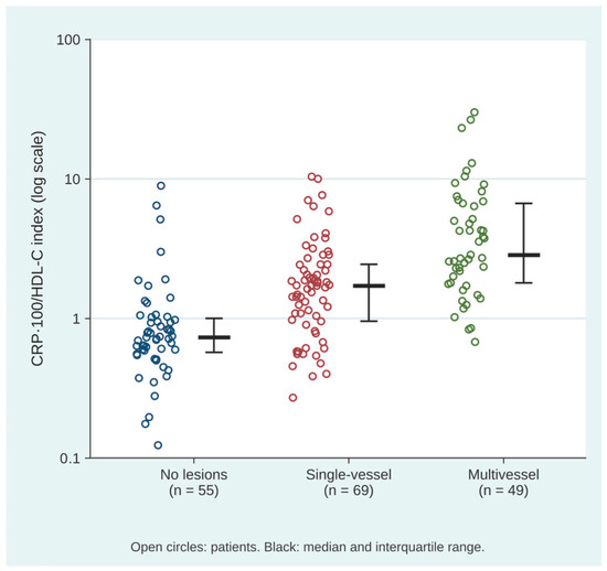
The AtI increased progressively with angiographic extent of coronary artery disease (Kruskal-Wallis H = 61.1, df = 2, p < 0.001): 0.73 (0.57–1.00), 1.71 (0.95–2.44), and 2.86 (1.79–6.67), showing a 3.9-fold difference between the no-lesions groups and the multivessel group (Figure 2). Bonferroni-corrected pairwise comparisons were significant: no lesions vs. single vessel disease (U = 952; p < 0.001), no lesions vs. MVCD (p < 0.001), and single vessel vs. MVCD (U = 869; p < 0.001). Spearman’s ρ was 0.596 (p < 0.001).
Figure 2.
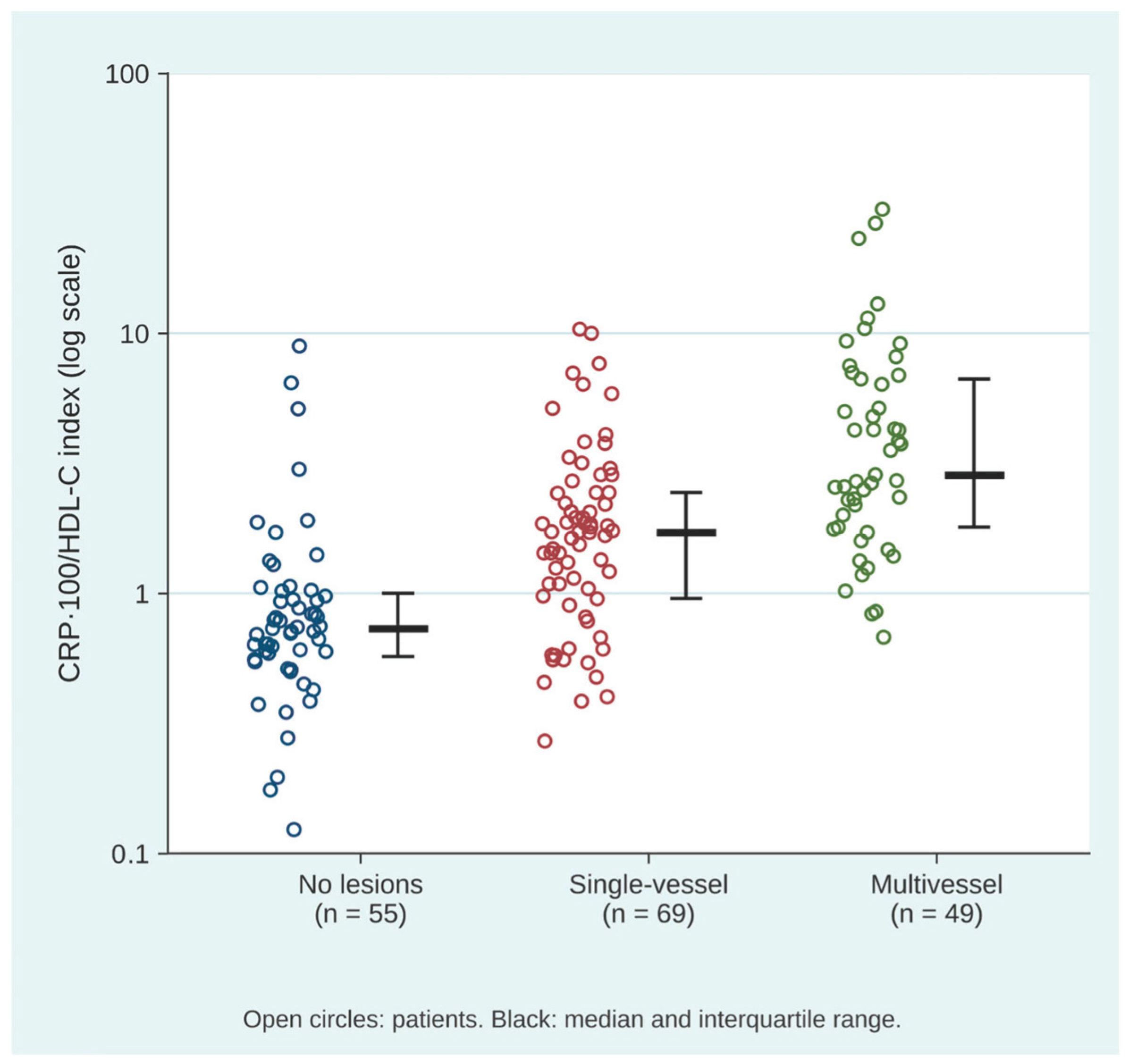
Scatter plot. The CRP × 100/HDL-C index by angiographic group (logarithmic scale). Each dot represents one patient. Black bars show the median and interquartile range (0.73 [0.57–1.00], 1.71 [0.95–2.44], and 2.86 [1.79–6.67]; Kruskal-Wallis p < 0.001).
3.3. Discrimination for MVCD
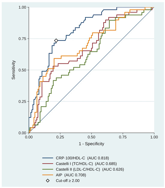
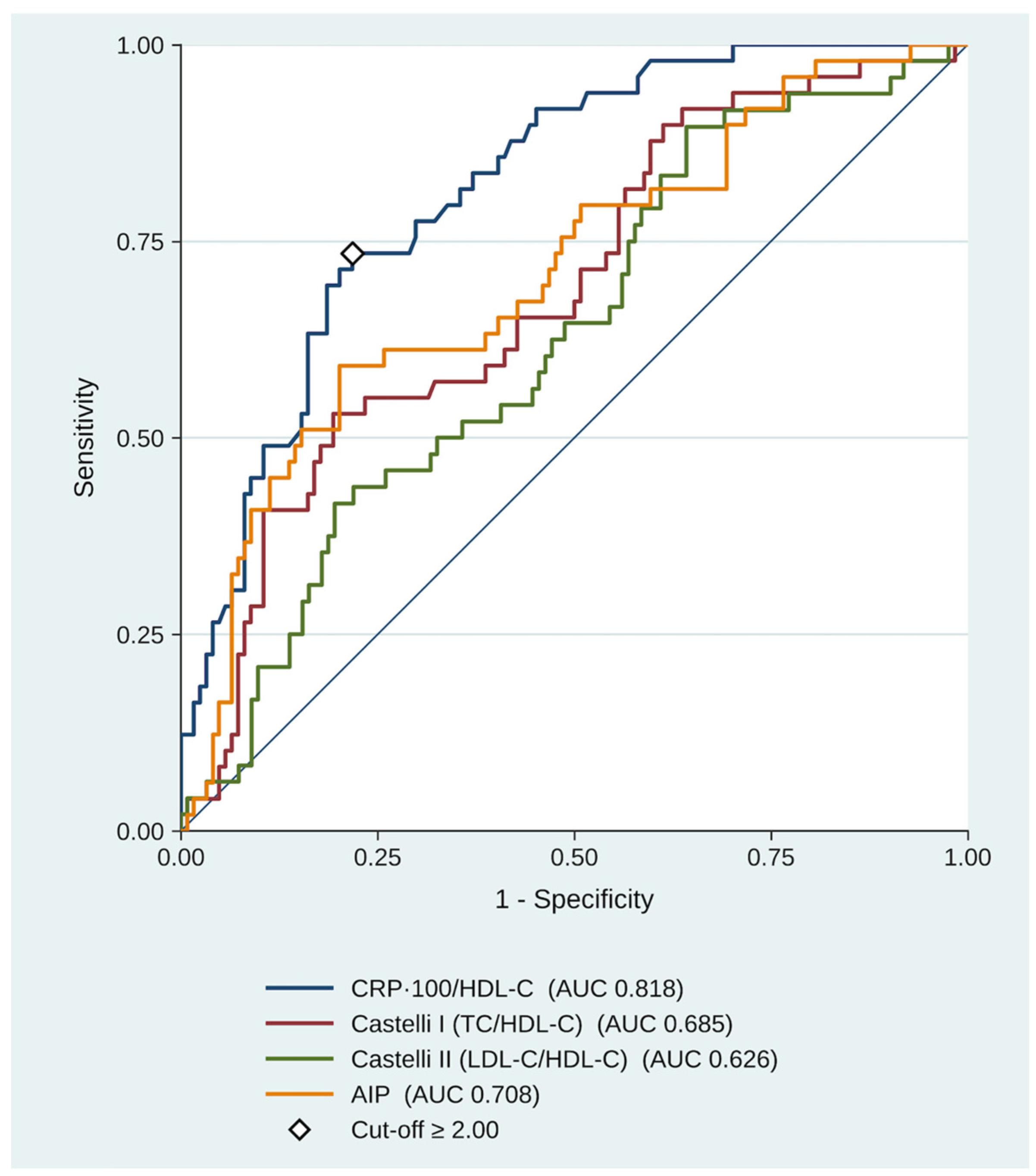
The AUC for MVCD was 0.818 (95% CI 0.753–0.883), as shown in Figure 3.
Figure 3.
ROC curves for MVCD (n = 173; Castelli II, n = 171). The AUC of the AtI was 0.818 (95% CI 0.753–0.883); paired DeLong p = 0.003, p < 0.001, and p = 0.013 against Castelli I, Castelli II, and AIP, respectively. The diamond marks the Youden cut-off of 2.00.
By paired DeLong tests, the AtI had a higher AUC than Castelli I (ΔAUC +0.132; 95% CI 0.046–0.219; p = 0.003), Castelli II (ΔAUC +0.190; 95% CI 0.100–0.280; p < 0.001), and the AIP (ΔAUC +0.110; 95% CI 0.024–0.196; p = 0.013), as shown in Table 2.
Table 2.
AUC for MVCD (n = 173).
At the Youden cut-off ≥2.00 (apparent estimates), sensitivity was 73.5% (95% CI 59.7–83.8), specificity was 78.2% (95% CI 70.2–84.6), PPV was 57.1% (95% CI 44.9–68.6), and NPV 88.2% (95% CI 80.8–93.0); positive and negative likelihood ratios (LR) were 3.37 (95% CI 2.32–4.90) and 0.34 (95% CI 0.21–0.55). After optimism correction, sensitivity was 69.8% and specificity was 77.1% (Youden index 0.469 versus 0.517 apparent), with PPV 55.1% and NPV 86.5%.
3.4. Independent and Incremental Value
As shown in Table 3, the AtI was independently associated with MVCD (OR 2.84 per doubling; 95% CI 1.95–4.13; p < 0.001). The association was not linear on the original scale (likelihood-ratio test of a spline term against a linear term, p = 0.002), and the log2-transformed index fitted the data better and did not differ from the spline (p = 0.63). With the index per unit, the OR was 1.49 (95% CI 1.25–1.78). Age was also associated with MVCD (OR 1.08; 95% CI 1.013–1.146; p = 0.017); sex, diabetes, and hypertension were not independently associated. Adding the AtI to a model with the four clinical covariates (model A) raised the AUC from 0.652 (95% CI 0.564–0.740) to 0.836 (95% CI 0.776–0.896) with a ΔAUC +0.184; 95% CI 0.093–0.275; p < 0.001. In another model with seven classical risk factors (model B), the AUC went from 0.671 (95% CI 0.586–0.757) to 0.840 (95% CI 0.780–0.901) with a ΔAUC +0.169; 95% CI 0.080–0.258; p < 0.001. The index alone had an AUC of 0.818, lower than the composite of model A with the index (0.836), so most of the discrimination came from the index. The optimism-corrected C statistic of model A was 0.812 (apparent 0.836), indicating modest overfitting. Hosmer-Lemeshow χ2 was 11.5 (p = 0.176), and variance inflation factors were ≤1.13.
Table 3.
Multivariable logistic regression for MVCD (n = 173; 49 events).
3.5. Sensitivity Analysis
NSTEMI/STEMI was present in 45.7% of patients (16.4%, 58.0%, and 61.2% across groups; p < 0.001). The association persisted after adjusting for NSTEMI/STEMI in the whole cohort (OR 2.80 per doubling; 95% CI 1.88–4.16). It was also present in the 94 patients without NSTEMI/STEMI (AUC 0.843, 95% CI 0.749–0.937; OR 4.71 per doubling, 95% CI 2.11–10.50; Firth penalized logistic regression OR 3.78, 95% CI 1.86–7.68) and in the 79 patients with NSTEMI/STEMI (AUC 0.745, 95% CI 0.636–0.853; OR 2.18 per doubling, 95% CI 1.38–3.43), as shown in Table 4. The median CRP in patients with MVCD was 0.70 mg/dL without NSTEMI/STEMI and 1.40 mg/dL with it. In the 52 patients that did not present with unstable angina (neither NSTEMI/STEMI nor unstable angina), the AUC was 0.908 (95% CI 0.823–0.993; unadjusted Firth OR 5.49, 95% CI 1.85–16.32), although this estimate is very imprecise as reflected in the wide CI. The atherogenic index by NSTEMI/STEMI interaction was not statistically significant (p = 0.12), and the AUCs of the two groups did not have statistically significant differences (p = 0.18). Therefore, the association was present in both groups, and we cannot demonstrate that it differs between them. After excluding patients with CRP > 5 mg/dL, chronic obstructive pulmonary disease, or cancer (a total of 9 patients), the results were similar (AUC 0.809, 95% CI 0.741–0.877; OR per doubling 2.77, 95% CI 1.89–4.06). After adding active smoking and LDL-C, we obtained similar results (OR per doubling 2.94, 95% CI 1.98–4.37), and after a more extensive adjustment including clinical presentation, triglycerides, creatinine, and statin therapy (OR 2.93, 95% CI 1.90–4.53; Firth penalized logistic regression OR 2.60, 95% CI 1.74–3.88; 4 events per predictor), the results were also similar. In another sensitivity analysis including the 31 patients that were assessed only by CT (none of whom had MVCD), as patients without MVCD, the results did not change (n = 204; AUC 0.827, 95% CI 0.766–0.887; OR 2.81, 95% CI 1.97–4.00). These additional analyses were post hoc and are exploratory.
Table 4.
Sensitivity analysis for the AtI and MVCD.
4. Discussion
In this study of patients younger than 65 years studied for suspicion of IHD with invasive CA, the AtI was associated with angiographic extent (medians 0.73, 1.71, and 2.86) and was independently associated with MVCD (OR 2.84 per doubling, 95% CI 1.95–4.13) with an AUC of 0.818. It had better discrimination than the traditional indexes Castelli I, Castelli II, and the AIP, and adding the AtI to a clinical model comprising age, sex, diabetes, and hypertension showed an increase in AUC from 0.652 to 0.836. These preliminary findings suggest an association between AtI and MVCD and serve to generate hypotheses to corroborate and validate in other studies.
Atherogenic indexes and their association with coronary artery disease have been studied before [5], but the classical AtI are built only with lipid fractions and do not include inflammation, which is a plausible explanation as to why the new AtI seems to have a better performance than Castelli I (AUC 0.685), Castelli II (AUC 0.626), and the AIP (AUC 0.708). For the traditional AtI, a meta-analysis of 51 studies found that AIP is associated with coronary artery disease [6] (OR 2.79) and multivessel disease (OR 2.04, 95% CI 1.50–2.77). Another study [10] of 450 patients undergoing coronary computerized tomography found that the AIP independently predicted MVCD (OR 2.35), and pericoronary adipose tissue may be an underlying mechanism, as this type of visceral fat is proinflammatory, adding an inflammatory signal that augments atherosclerosis and leads to coronary artery disease.
The new AtI is associated with MVCD, either by a low HDL-C in the denominator (which reflects anti-atherogenic lipoproteins) or by a high CRP in the numerator as a marker of systemic inflammation. The AtI should be interpreted as a risk marker that integrates both lipid residual risk and inflammatory residual risk, which could explain why its performance is greater than each component separately. Results with CRP are inconclusive, as previous Mendelian randomization analysis based on individual patient data shows that genetically raised CRP was not associated with coronary risk (RR 1.00, 95% CI 0.90–1.13) [11], but in other studies hs-CRP was found to be higher in patients with acute coronary syndrome and was found to be an independent predictor of a high SYNTAX score [12]. In regard to HDL-C, genetically raised HDL-C was also not associated with myocardial infarction [13] (OR 0.93, 95% CI 0.68–1.26). Combining the two parameters has biologic plausibility to have a better performance, as both lipids and inflammation are involved in the pathogenesis of coronary artery disease.
Indeed, inflammation is necessary for the formation of the atherosclerotic plaque, and it is intimately related to the lipid hypothesis of coronary artery disease, as cholesterol accumulation inside macrophages promotes Toll-like receptor signaling and the inflammasome activation [14], which perpetuates the cycle. Previous studies targeting the inflammatory pathway, like the CANTOS trial [15], showed that inhibiting interleukin-1β mediated inflammation reduced cardiovascular events. Currently, there have been many advances in pharmacology resulting in new lipid-lowering agents, but cardiovascular disease remains the leading cause of mortality, and events persist even in patients who have guideline-controlled LDL with statins and other drugs [16]. Other studies, also done in patients with controlled baseline LDL-C, show that elevated CRP is associated with major adverse cardiovascular events and that the incidence rises from persistently low to persistently high inflammation [17,18].
The role of inflammation in atherosclerosis is no longer a fringe theory or just a causal finding in a minor study, like it was considered a few years ago, as in 2025 the American College of Cardiology presented an official scientific statement acknowledging the causal role of inflammation and concluding that measuring residual inflammation with hs-CRP was strongly predictive of recurrent cardiovascular events in patients treated with statins [19], and even though hs-CRP is already used to stratify risk in primary prevention guidelines, there were no recommendations to measure it in secondary prevention [20]. The concept of residual inflammatory risk, therefore, has a solid basis in the available literature and is compatible with our finding that LDL-C did not differ across angiographic groups (p = 0.777) but that the AtI had a stepwise increase in relation to affected vessel territories.
Our results are consistent with earlier reports of the index by our group, although the current study has a different outcome and subpopulation. Earlier reports showed an association with carotid atherosclerosis in 282 patients [8] and an AUC of 0.795 for coronary artery disease and OR 1.59 [9]. In this study, with the AtI as a continuous variable and invasive CA as the only diagnostic modality, the AtI has an OR of 2.84 per doubling in relation with MVCD.
The CRP/HDL ratio has been explored before in other populations. Luo et al. [21] showed that in patients undergoing CA the hs-CRP/HDL-C ratio was correlated with the Gensini score and was an independent predictor of coronary artery disease, with an AUC of 0.662. In another study, the CRP/HDL-C ratio was higher in patients with metabolic syndrome but was inferior to the AIP [22]. Han et al. [23] used the AtI in patients with cardiovascular-kidney metabolic syndrome and showed the CRP/HDL-C ratio was associated with all-cause mortality in these patients. These studies differ from the one done by our group in outcome, population, and CRP assay, so their effect is not directly comparable, but the findings among the different studies do suggest that there is an association between the AtI and coronary artery disease. Other composite indices that integrate inflammation have been proposed. The advanced lung cancer inflammation index, which combines serum albumin, body mass index, and the neutrophil-to-lymphocyte ratio, predicted all-cause mortality in 1171 patients with STEMI undergoing primary percutaneous coronary intervention [24]. In our study, we could not calculate it because serum albumin was not available. A direct comparison of the CRPx100/HDL-C index with such inflammatory indices, using clinical outcomes, should be addressed in further studies.
The study limitations include its reduced sample size, the single-center and cross-sectional design, and the lack of hs-CRP. In our findings, the index alone had an AUC of 0.818, and adding it to a clinical model raised the AUC from 0.652 to 0.836, but these findings are a cross-sectional determination of the coronary anatomy in patients suspected of coronary artery disease, so the results should be considered as hypothesis-generating and not conclusive evidence. The rationale for the inclusion of patients younger than 65 years was that inflammation is a key driver of acute coronary syndromes in these patients, but this age limit and the exclusion of patients with prior cardiac disease could lead to a selection bias that may limit the external validity of the results. Also, men were overrepresented in the MVCD, but the index association was adjusted for sex, and sex itself was not independently associated with MVCD (OR 1.19, 95% CI 0.39–3.67). The Youden cut-off values were generated from the same cohort, which also limits the generalization of the results. To address this limitation, we did Bootstrap optimism correction, which showed a very modest reduction in the NPV from 88.2 to 86.5%, the sensitivity from 73.5% to 69.8%, and the specificity from 78.2 to 77.1%, suggesting that the results are consistent and are not overly affected by optimism. Several of the statistical techniques were chosen to limit this over-optimism, such as the Bootstrap, paired DeLong comparisons with Holm adjustments, and the reporting of both apparent and corrected estimates. Bootstrap correction is an internal validation and does not replace validation in an independent cohort, which constitutes the next step in future studies.
Another limitation is the lack of hs-CRP. This should be addressed in confirmatory studies, preferentially multicentric studies that incorporate serial CRP measurements to control for the acute rise in CRP after myocardial infarction. Median CRP values were 0.4 mg/dL in patients without lesions, 0.6 mg/dL in patients with single-vessel disease, and 1.1 mg/dL in patients with MVCD. These values are above the range generally used to define low-grade inflammation (hs-CRP 1–3 mg/L), so the index should be interpreted as reflecting systemic inflammation, which includes the response to acute myocardial injury, and not specifically chronic low-grade inflammation. In our study, NSTEMI or STEMI was present in 45.7% of patients and was more frequent with greater angiographic extent (16%, 58%, and 61%), so part of the association could reflect the inflammatory response to myocardial injury rather than the systemic inflammation associated with atherosclerosis. The association persisted after adjustment for NSTEMI/STEMI in a pre-specified sensitivity analysis (OR 2.80 per doubling; 95% CI 1.88–4.16). In post-hoc stratified analyses it was present both without NSTEMI/STEMI (n = 94; AUC 0.843; OR 4.71 per doubling; 95% CI 2.11–10.50) and with NSTEMI/STEMI (n = 79; AUC 0.745; OR 2.18 per doubling; 95% CI 1.38–3.43); the difference between the two groups was not statistically significant (interaction p = 0.12). The association was also observed in the 52 patients without any acute coronary syndrome (9 events; AUC 0.908), with a very imprecise estimate and wide CI. Because the subgroups were small (19 and 30 events), these analyses are only exploratory and cannot exclude a lower performance of the index in the acute setting. This has also been reported in earlier studies of the AtI in relation with carotid and coronary atherosclerosis, although this finding should be corroborated and validated in other independent, multicentric, and prospective studies incorporating hs-CRP. Angiography was performed at the attending cardiologist’s decision and, in about 70% of patients, in the setting of an acute coronary syndrome, so selection bias cannot be completely excluded. Patients assessed only by CT were excluded, although none of them had MVCD, and including them did not change the results. Another limitation is that stenoses were graded visually by the performing operator, who was not blinded to clinical data, without quantitative analysis or core laboratory review. The CRPx100/HDL-C index was calculated retrospectively for this study, so the operators could not have known its value when grading the angiograms, which compensates a possible selection bias by the operator. In our routine clinical practice, a continuous anatomical score such as the SYNTAX or Gensini score is not reported systematically, and it is only calculated in a few cases in which the Heart Team needs it to choose between coronary artery bypass grafting and percutaneous coronary intervention. None of the patients included in this study had a continuous anatomical score, as none were evaluated by the Heart Team. Also, only significant lesions were included in this analysis, and functional assessment (FFR/iFR) was not performed systematically.
CRP was measured before angiography, but the time from symptom onset was not recorded, so in acute myocardial infarction the sample may have been drawn before CRP reached its peak. Patients with acute infection were not excluded and leukocyte counts were not available, so we cannot rule out that at least part of the elevation of CRP is due to other causes, as CRP (whether high-sensitivity or not) is not specific to atherosclerotic inflammation. The assay used in this study is not high-sensitivity and has limited resolution at low values, which may be frequent in patients without lesions. Despite additional adjustment, residual confounding cannot be excluded, and the extended models had fewer than 10 events per predictor, so results should be interpreted cautiously. Several analyses were added post hoc following peer review.
5. Conclusions
In patients undergoing invasive CA, the CRPx100/HDL-C ratio was independently associated with MVCD and discriminated it with an AUC of 0.818, higher than traditional lipid indices like Castelli I, Castelli II, and the AIP. It is a simple and inexpensive marker of angiographic extent that adds discrimination to clinical factors. Whether it can impact clinical decisions, for example, in prioritizing invasive coronary angiography in stable patients, requires prospective validation. The findings in this study are currently exploratory, and the hypothesis should be tested in future studies.
Author Contributions
Conceptualization, V.V.-G. and Ó.F.-A.; Methodology, V.V.-G.; Validation, V.V.-G., Ó.F.-A., F.J.F.S. and N.V.-G.; Formal Analysis, V.V.-G. and Ó.F.-A.; Investigation, all authors; Resources, Ó.F.-A.; Data Curation, V.V.-G. and Ó.F.-A.; Writing, all authors; Original Draft Preparation, V.V.-G., Ó.F.-A., F.J.F.S., N.V.-G., M.d.C.L.d.P. and V.J.-O.; Writing, Review and Editing, all authors; Visualization, V.V.-G.; Supervision Ó.F.-A.; Project Administration, V.V.-G. All authors have read and agreed to the published version of the manuscript.
Funding
The article processing charges for this paper were funded by the Universidad Católica de Valencia.
Institutional Review Board Statement
Ethical review and approval were waived for this study, as it involved the fully anonymized retrospective analysis of routinely collected clinical data from a single center registry, with no access to or storage of patient identifiers. Formal ethics committee review was not required under Spanish legislation (Ley 14/2007 de Investigación Biomédica, Art. 2.d) for this category of retrospective anonymized secondary data analysis. All procedures were conducted in accordance with the Declaration of Helsinki.
Informed Consent Statement
Patient consent was waived due to the retrospective and fully anonymized nature of the data analysis.
Data Availability Statement
The raw data that support the findings of this study are available from the corresponding author upon reasonable request, subject to applicable data protection regulations (Ley Orgánica 3/2018, de 5 de diciembre, de Protección de Datos Personales).
Conflicts of Interest
The authors declare no conflicts of interest.
References
- Roth, G.A.; Mensah, G.A.; Johnson, C.O.; Addolorato, G.; Ammirati, E.; Baddour, L.M.; Barengo, N.C.; Beaton, A.Z.; Benjamin, E.J.; Benziger, C.P.; et al. Global Burden of Cardiovascular Diseases and Risk Factors, 1990–2019: Update from the GBD 2019 Study. J. Am. Coll. Cardiol. 2020, 76, 2982–3021, Correction in J. Am. Coll. Cardiol. 2021, 77, 1958–1959. [Google Scholar] [CrossRef] [Scilit] [PubMed]
- Wadström, B.N.; Pedersen, K.M.; Wulff, A.B.; Nordestgaard, B.G. Elevated remnant cholesterol, plasma triglycerides, and cardiovascular and non-cardiovascular mortality. Eur. Heart J. 2023, 44, 1432–1445. [Google Scholar] [CrossRef] [Scilit] [PubMed]
- Neumann, F.-J.; Sousa-Uva, M.; Ahlsson, A.; Alfonso, F.; Banning, A.P.; Benedetto, U.; Byrne, R.A.; Collet, J.-P.; Falk, V.; Head, S.J.; et al. 2018 ESC/EACTS guidelines on myocardial revascularization. Eur. Heart J. 2019, 40, 87–165. [Google Scholar] [CrossRef] [Scilit] [PubMed]
- Mehta, S.R.; Wood, D.A.; Storey, R.F.; Mehran, R.; Bainey, K.R.; Nguyen, H.; Meeks, B.; Di Pasquale, G.; López-Sendón, J.; Faxon, D.P.; et al. Complete revascularization with multivessel PCI for myocardial infarction. N. Engl. J. Med. 2019, 381, 1411–1421. [Google Scholar] [CrossRef] [Scilit] [PubMed]
- González, Á.A.L.; Ledo, Y.I.R.; Herrero, M.T.V.; Llinás, M.G.; Salvá, M.T.; Fe, B.R. Atherogenic indices in workers from different occupational sectors of the Spanish Mediterranean area. Clín. Investig. Arterioscler. 2015, 27, 118–128. [Google Scholar]
- Assempoor, R.; Daneshvar, M.S.; Taghvaei, A.; Abroy, A.S.; Azimi, A.; Nelson, J.R.; Hosseini, K. Atherogenic index of plasma and coronary artery disease: A systematic review and meta-analysis of observational studies. Cardiovasc. Diabetol. 2025, 24, 35. [Google Scholar] [CrossRef] [Scilit] [PubMed]
- Ridker, P.M. Residual inflammatory risk: Addressing the obverse side of the atherosclerosis prevention coin. Eur. Heart J. 2016, 37, 1720–1722. [Google Scholar] [CrossRef] [Scilit] [PubMed]
- Fabregat-Andrés, Ó.; Pérez-De-Lucía, P.; Vallejo-García, V.E.; Vera-Ivars, P.; Valverde-Navarro, A.A.; Tormos, J.M. New atherogenic index for the prediction of carotid atherosclerosis based on the non-ultrasensitive c-reactive protein/HDL ratio. Clin. Investig. Arterioscler. 2024, 36, 12–21. [Google Scholar] [CrossRef] [Scilit]
- Vallejo-Garcia, V.; Fabregat-Ándres, Ó.; Sargues, F.J.F.; Valtueña-Gimeno, N.; Cejalvo-Lapeña, D.; Gumbau, G.V.; Tormos, J.M. Diagnostic value of the novel non-high-sensitivity CRP/HDL atherogenic index for detection of significant coronary artery disease. Rev. Clín. Esp. 2026; in press.
- Du, H.; Zheng, J.; Yao, Y.; Zhou, Q.; Li, L. Pericoronary adipose tissue inflammation mediates the atherogenic effects of lipids on multivessel coronary artery disease: A CCTA-based radiomics analysis. Front. Cardiovasc. Med. 2025, 12, 1629984. [Google Scholar] [CrossRef] [Scilit] [PubMed]
- C Reactive Protein Coronary Heart Disease Genetics Collaboration (CCGC). Association between C reactive protein and coronary heart disease: Mendelian randomisation analysis based on individual participant data. BMJ 2011, 342, d548. [Google Scholar] [CrossRef] [Scilit] [PubMed]
- Karadeniz, M.; Duran, M.; Akyel, A.; Yarlıoğlueş, M.; Öcek, A.H.; Çelik, I.E.; Kılıç, A.; Yalcin, A.A.; Ergün, G.; Murat, S.N. High Sensitive CRP Level Is Associated with Intermediate and High Syntax Score in Patients with Acute Coronary Syndrome. Int. Heart J. 2015, 56, 377–380. [Google Scholar] [CrossRef] [Scilit] [PubMed]
- Voight, B.F.; Peloso, G.M.; Orho-Melander, M.; Frikke-Schmidt, R.; Barbalic, M.; Jensen, M.K.; Hindy, G.; Hólm, H.; Ding, E.L.; Johnson, T.; et al. Plasma HDL cholesterol and risk of myocardial infarction: A mendelian randomisation study. Lancet 2012, 380, 572–580, Erratum in Lancet 2012, 367, 189. [Google Scholar] [CrossRef] [Scilit] [PubMed]
- Tall, A.R.; Yvan-Charvet, L. Cholesterol, inflammation and innate immunity. Nat. Rev. Immunol. 2015, 15, 104–116. [Google Scholar] [CrossRef] [Scilit] [PubMed]
- Ridker, P.M.; Everett, B.M.; Thuren, T.; MacFadyen, J.G.; Chang, W.H.; Ballantyne, C.; Fonseca, F.; Nicolau, J.; Koenig, W.; Anker, S.D.; et al. Antiinflammatory therapy with canakinumab for atherosclerotic disease. N. Engl. J. Med. 2017, 377, 1119–1131. [Google Scholar] [CrossRef] [Scilit] [PubMed]
- Cannon, C.P.; Blazing, M.A.; Giugliano, R.P.; McCagg, A.; White, J.A.; Théroux, P.; Darius, H.; Lewis, B.S.; Ophuis, T.O.; Jukema, J.W.; et al. Ezetimibe added to statin therapy after acute coronary syndromes (IMPROVE-IT). N. Engl. J. Med. 2015, 372, 2387–2397. [Google Scholar] [CrossRef] [Scilit] [PubMed]
- Guedeney, P.; Claessen, B.E.; Kalkman, D.N.; Aquino, M.; Sorrentino, S.; Giustino, G.; Farhan, S.; Vogel, B.; Sartori, S.; Montalescot, G.; et al. Residual inflammatory risk in patients with low LDL cholesterol levels undergoing PCI. J. Am. Coll. Cardiol. 2019, 73, 2401–2409. [Google Scholar] [CrossRef] [Scilit] [PubMed]
- Ridker, P.M.; Danielson, E.; Fonseca, F.A.H.; Genest, J.; Gotto, A.M.; Kastelein, J.J.P.; Koenig, W.; Libby, P.; Lorenzatti, A.J.; MacFadyen, J.G.; et al. Rosuvastatin to prevent vascular events in men and women with elevated CRP (JUPITER). N. Engl. J. Med. 2008, 359, 2195–2207. [Google Scholar] [CrossRef] [Scilit] [PubMed]
- Mensah, G.A.; Arnold, N.; Prabhu, S.D.; Ridker, P.M.; Welty, F.K. Inflammation and cardiovascular disease: 2025 ACC Scientific Statement. J. Am. Coll. Cardiol. 2025, 87, 1381–1404. [Google Scholar] [CrossRef] [Scilit] [PubMed]
- Mehta, A.; Blumenthal, R.S.; Gluckman, T.J.; I Feldman, D.; Kohli, P. High-sensitivity CRP in atherosclerotic cardiovascular disease: To measure or not to measure? US Cardiol. Rev. 2025, 19, e06. [Google Scholar] [CrossRef] [Scilit] [PubMed]
- Luo, H.; Kou, T.; Yin, L. High-sensitivity C-reactive protein to HDL-C ratio. Int. Heart J. 2021, 62, 1221–1229. [Google Scholar] [CrossRef] [Scilit] [PubMed]
- Jialal, I.; Adams-Huet, B.; Remaley, A.T. A comparison of the ratios of CRP and TG to HDL-C as biomarkers of metabolic syndrome in African Americans and non-Hispanic Whites. J. Diabetes Complicat. 2022, 36, 108231. [Google Scholar]
- Han, F.; Guo, H.; Zhang, H.; Zheng, Y. Hs-CRP/HDL-C can predict the risk of all cause mortality in cardiovascular-kidney-metabolic syndrome stage 1-4 patients. Front. Endocrinol. 2025, 16, 1552219. [Google Scholar] [CrossRef] [Scilit] [PubMed]
- Trimarchi, G.; Pizzino, F.; Lilli, A.; De Caterina, A.R.; Esposito, A.; Dalmiani, S.; Mazzone, A.; Di Bella, G.; Berti, S.; Paradossi, U. Advanced Lung Cancer Inflammation Index as Predictor of All-Cause Mortality in ST-Elevation Myocardial Infarction Patients Undergoing Primary Percutaneous Coronary Intervention. J. Clin. Med. 2024, 13, 6059. [Google Scholar] [CrossRef] [Scilit] [PubMed]

Disclaimer/Publisher’s Note: The statements, opinions and data contained in all publications are solely those of the individual author(s) and contributor(s) and not of MDPI and/or the editor(s). MDPI and/or the editor(s) disclaim responsibility for any injury to people or property resulting from any ideas, methods, instructions or products referred to in the content. |
© 2026 by the authors. Licensee MDPI, Basel, Switzerland. This article is an open access article distributed under the terms and conditions of the Creative Commons Attribution (CC BY) license.