


Abstract
Background: Although diabetes mellitus is traditionally viewed as a systemic metabolic disorder, growing evidence indicates that it also affects the upper airways through vascular, inflammatory, and neuro-sensory mechanisms. The sinonasal mucosa, highly vascularized and immunologically active, may represent an early target of diabetic microangiopathy and immune–metabolic imbalance. Objectives: Our objectives are to synthesize current evidence on the rhinologic manifestations of DM, with a focus on chronic rhinosinusitis, olfactory dysfunction, and other nasal disorders, and to identify the main pathophysiologic and clinical patterns linking diabetes to sinonasal disease. Results: Evidence suggests that DM, particularly type 2 DM, increases susceptibility to CRSwNP and modulates the sinonasal microbiome toward Gram-negative predominance. Surgical outcomes after endoscopic sinus surgery are generally comparable between diabetics and non-diabetics when perioperative care is optimized. Olfactory dysfunction occurs more frequently and severely in diabetic patients, likely reflecting the combined effects of chronic inflammation, vascular compromise, and insulin resistance. Additional manifestations include recurrent epistaxis, delayed mucociliary clearance, and chronic cough. Allergic rhinitis appears to not be increased, and maybe even inversely related, especially among users of DPP-4 inhibitors. Conclusions: Diabetes intersects with rhinologic health through immune–metabolic, vascular, and epithelial pathways that may shape susceptibility, disease phenotype, and neurosensory decline. Future research should focus on disentangling type-specific mechanisms, metabolic biomarkers, and longitudinal outcomes, with the aim of developing precision-based approaches to rhinologic assessment and management in diabetic patients.
Keywords:
diabetes mellitus; chronic rhinosinusitis; olfactory dysfunction; sinusitis; rhinitis; rhinology; DM 1. Introduction
1.1. Diabetes Mellitus Type 1
Type 1 diabetes mellitus (T1D or T1DM) is an autoimmune disease characterized by the progressive destruction of pancreatic β-cells by autoreactive CD4+ and CD8+ T lymphocytes and macrophages, leading to absolute insulin deficiency [1,2,3,4,5]. It accounts for approximately 5–10% of all diabetes cases worldwide [2] and remains the predominant form in childhood and adolescence, although a considerable proportion of diagnoses occur in adulthood. The 2024 global prevalence was estimated at around 9 million individuals, including 1.8 million under the age of 20 [1].
The incidence of T1DM shows remarkable geographic variability, with extremely high rates in Northern European countries and very low rates in Asia and South America (up to 350–400-fold differences between regions) [4,5]. According to the WHO DIAMOND Project and the EURODIAB study, the incidence among children under 15 years ranges from 0.1 per 100,000 in China and Venezuela to over 36 per 100,000 in Finland.
Over recent decades, the incidence of T1DM has been rising globally by approximately 2–5% per year, with the steepest increases observed among young children and in regions previously classified as low-incidence. This rapid rise cannot be explained by genetic changes alone and likely reflects environmental influences acting upon susceptible genotypes.
From a pathogenetic standpoint, T1DM progresses through distinct stages: two asymptomatic preclinical phases (stages 1 and 2) characterized by the appearance of multiple islet autoantibodies against β-cell antigens such as GAD65, IA-2, and insulin, followed by stage 3, marked by hyperglycaemia and insulin dependence [1].
Overall, T1DM is a chronic, immune-mediated condition with a steadily increasing incidence worldwide and substantial implications for public health due to its acute and chronic complications [6,7,8,9]. Its multifactorial aetiology, at the intersection of genetics, immune dysregulation, and environmental exposure, continues to be a major focus of translational and epidemiological research.
1.2. Diabetes Mellitus Type 2
Type 2 diabetes mellitus (T2D or T2DM) is a complex, multifactorial metabolic disorder characterized by chronic hyperglycaemia resulting from a combination of insulin resistance and a progressive decline in pancreatic β-cell function [10,11,12,13]. It represents approximately 90% of all diabetes cases globally and is increasingly diagnosed across all age groups, including adolescents [13]. According to the International Diabetes Federation, in 2021 there were an estimated 537 million adults living with diabetes worldwide, with projections suggesting a rise to 783 million by 2045 [14]. T2DM is therefore considered one of the “big four” non-communicable diseases alongside cardiovascular disease, cancer, and chronic respiratory disease.
The aetiology of T2DM involves a complex interplay between genetic, environmental, and lifestyle factors. Obesity remains the most significant modifiable risk factor, contributing to more than 50% of global disability-adjusted life years (DALYs) related to T2DM. Excess caloric intake, high-fat and high-sugar diets, and sedentary behaviour promote adipose tissue dysfunction, lipotoxicity, and low-grade inflammation, which collectively drive insulin resistance and β-cell failure [15,16]. At the cellular level, mitochondrial dysfunction, oxidative stress, and endoplasmic reticulum stress further exacerbate insulin resistance and impair glucose metabolism.
Genetic predisposition contributes substantially to T2DM risk, with heritability estimates ranging from 30 to 70% [17,18].
Epidemiological studies show that T2DM disproportionately affects low- and middle-income countries, where more than 80% of cases occur [19]. The prevalence rises markedly with age and is slightly higher in men, though women face a greater lifetime risk. Sex hormones and body-fat distribution contribute to these differences: men typically develop T2DM at a lower BMI but with greater visceral fat, whereas women show stronger associations between obesity and diabetes risk. In addition to obesity and genetics, psychosocial and behavioural factors such as socioeconomic disadvantage, chronic stress, poor diet, and sleep deprivation play important roles in disease onset and progression.
From a mechanistic perspective, T2DM is not a uniform disease entity but a heterogeneous spectrum encompassing variable degrees of β-cell dysfunction and insulin resistance [20,21].
1.3. An Overview of Nasal Disorders
1.3.1. Inflammatory and Infectious Disorders
Allergic rhinitis (AR) and chronic rhinosinusitis (CRS) represent the most common chronic inflammatory diseases of the upper airways, while acute and fungal rhinosinusitis illustrate their infectious spectrum.
AR is an IgE-mediated Th2-driven disorder characterized by eosinophilic inflammation, epithelial barrier dysfunction, and cytokine release (IL-4, IL-5, IL-13). Beyond Th2, lymphocytes, ILC2, Tfh, and regulatory T/B cells contribute to chronicity through impaired IL-10–mediated tolerance. Allergen-specific immunotherapy remains the only disease-modifying treatment, and biologics targeting IgE or IL-4Rα benefit severe cases [22,23,24,25,26].
CRS, defined by sinonasal inflammation persisting > 12 weeks, is divided into CRS with nasal polyps (CRSwNP) (type 2/eosinophilic) and CRS without nasal polyps (CRSsNP) (type 1/3/neutrophilic) forms. Barrier dysfunction, reduced mucociliary clearance, and microbial dysbiosis sustain chronicity, while Staphylococcus aureus colonization and persistent tissue-resident Th2/Th17 cells drive recurrence despite corticosteroid therapy or surgery [27,28,29,30].
Acute bacterial sinusitis (ABS) is a secondary bacterial complication of viral infections, affecting ~2% of adults and up to 13% of children. It is linked to mucosal edema and impaired mucociliary clearance that facilitate growth of Streptococcus pneumoniae and Haemophilus influenzae. Diagnosis is mainly clinical, and antibiotic therapy is reserved for confirmed bacterial cases [31,32,33,34,35,36].
Fungal rhinosinusitis (FRS) includes non-invasive forms—saprophytic infestation, fungal ball, and allergic fungal rhinosinusitis (AFRS)—and invasive forms (acute, chronic, granulomatous). AFRS occurs in atopic, immunocompetent hosts as a Th2-mediated allergic response to dematiaceous fungi, while invasive forms affect immunocompromised or diabetic patients, causing angioinvasion, tissue necrosis, and high mortality, predominantly due to Mucorales or Aspergillus species [37,38,39].
Overall, these conditions exemplify how immune dysregulation, epithelial dysfunction, and microbial imbalance interact to sustain sinonasal inflammation, pathways that may be further amplified in diabetes through vascular injury, oxidative stress, and impaired host defence.
1.3.2. Olfactory Dysfunction
Olfactory dysfunction (OD) affects up to 20% of the population and arises from diverse causes including inflammatory sinonasal disease, viral infections, head trauma, neurodegenerative disorders, and metabolic conditions such as diabetes. The olfactory mucosa functions as both a sensory and immune barrier, containing specialized epithelial, neuronal, and immune cells. The not very frequently mentioned blood–olfactory barrier (BOB) restricts systemic antibody access, rendering the olfactory mucosa particularly vulnerable to pathogens and inflammation [40,41,42,43,44].
Local immune responses involving macrophages, T cells, and plasma cells protect against infection but may also contribute to chronic smell loss. OD thus reflects a complex interaction between immune regulation, epithelial integrity, and neural function, mechanisms that may be further impaired in metabolic diseases such as diabetes.
Despite the high global burden of DM, knowledge of its rhinologic manifestations remains limited and methodologically inconsistent. Existing studies differ substantially in diabetes classification, rarely distinguish between type 1 and type 2 disease, and apply non-uniform criteria for sinonasal outcomes, particularly for OD, which is often assessed subjectively rather than through validated testing.
Consequently, the overall clinical significance of diabetes-related rhinologic involvement remains incompletely defined.
Against this background, the present narrative review aims to critically synthesize the available human adult literature published between 1973 and 2025 and indexed in PubMed on the association between diabetes mellitus and rhinologic disorders.
2. Materials and Methods
2.1. Research and Screening of the Literature
A comprehensive literature search was conducted to explore the association between diabetes mellitus (type 1 and type 2) and rhinologic diseases with a predominant vascular or immunological component. The search was performed in PubMed, covering the period from 1973 to 2025. The following Boolean string was used:
(“diabetes mellitus” OR “diabetes” OR “type 1 diabetes” OR “type 2 diabetes” OR “DM1” OR “DM2” OR “insulin-dependent diabetes” OR “non-insulin-dependent diabetes”)
AND
(“rhinitis” OR “allergic rhinitis” OR “non-allergic rhinitis” OR “vasomotor rhinitis” OR “atrophic rhinitis” OR “chronic rhinitis” OR “acute rhinitis” OR “infectious rhinitis” OR “sinusitis” OR “chronic sinusitis” OR “rhinorrhea” OR “nasal obstruction” OR “nasal congestion” OR “nasal discharge” OR “nasal dryness” OR “nasal crusting” OR “nasal polyps” OR “nasal inflammation” OR “nasal infection” OR “nasal mucosa” OR “nasal complications” OR “nose disease” OR “nose diseases” OR “sinonasal disease” OR “sinonasal inflammation” OR “olfactory dysfunction” OR “olfactory disorder” OR “olfactory impairment” OR “olfactory loss” OR “smell disorder” OR “smell loss” OR “anosmia” OR “hyposmia” OR “dysosmia” OR “parosmia” OR “phantosmia”)
PubMed was chosen as the primary database because it represents the most authoritative and widely used source for biomedical and clinical research, offering extensive coverage of the peer-reviewed literature in endocrinology and otorhinolaryngology. The use of MeSH (Medical Subject Headings) terms allows precise identification of clinically relevant studies, which was aligned with the narrative- and pathophysiology-oriented scope of this review.
Without applying any filters, the Boolean search string retrieved 961 records from PubMed, including preprints, non-English publications, animal studies, and studies involving pediatric populations.
Database filters were then applied to exclude duplicate records and review articles and to restrict the search to human studies. Although filters for adult populations were applied, animal and pediatric studies were not always reliably excluded due to database indexing limitations.
These records were therefore excluded during a first manual screening based on titles and abstracts, together with studies unrelated to rhinologic or diabetic outcomes.
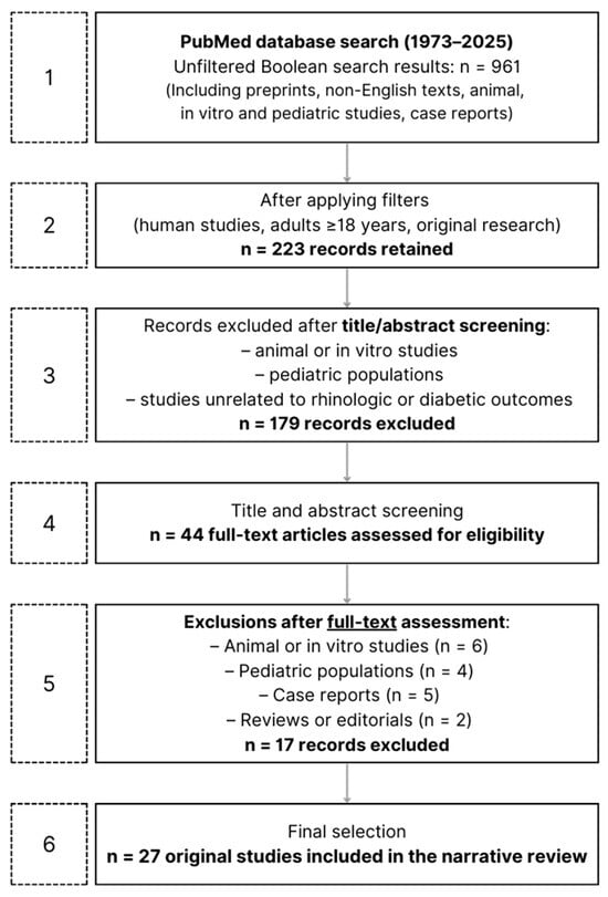
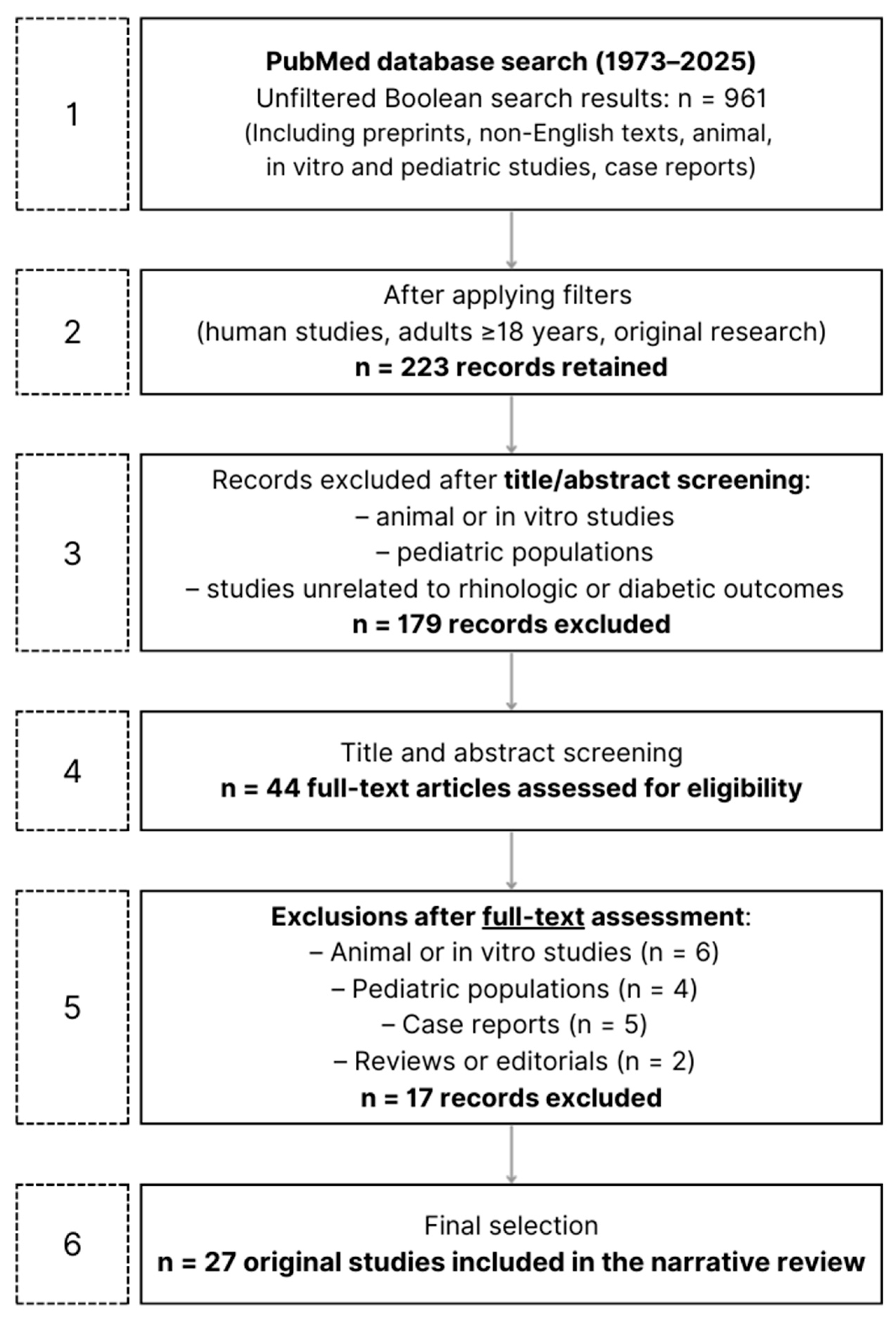
This screening yielded 44 articles eligible for full-text assessment. Among these 44 articles, 17 studies were excluded only after full-text assessment because the study design or population (animal, in vitro, or pediatric) was not clearly identifiable from the title or abstract, which primarily reported outcomes in adult human populations (Figure 1).
Figure 1.
Flow diagram summarizing the literature search strategy and study selection process. The initial unfiltered Boolean search on PubMed (1973–2025) yielded 961 records, including preprints, non-English texts, animal, in vitro, and pediatric studies, and case reports (Step 1). After application of database filters to restrict the search to human studies, adults ≥ 18 years, and original research, 223 records were retained (Step 2). Records were then screened by title and abstract, and studies clearly unrelated to rhinologic or diabetic outcomes, as well as animal/in vitro and pediatric studies identifiable at this stage, were excluded (Step 3), resulting in 44 full-text articles assessed for eligibility (Step 4). A subsequent manual full-text assessment led to the exclusion of additional studies, including animal or in vitro studies, pediatric populations, case reports, and reviews or editorials, when the study design or population was not clearly identifiable from the title or abstract alone (Step 5). Following this process, 27 original studies were included in the final narrative review (Step 6).
Since the literature exploring the vascular and immunological interplay between diabetes and rhinologic disorders remains limited and heterogeneous, an extended time frame (1973–2025) was selected to encompass both early pathophysiological reports and contemporary studies, thereby providing a comprehensive overview of the topic’s evolution over time. Given the heterogeneity of study designs and outcome measures, a narrative synthesis approach was adopted, focusing on mechanistic and clinical evidence linking diabetes-related metabolic, vascular, and immune alterations to rhinologic manifestations. Selected studies were qualitatively analyzed and grouped into predefined thematic categories according to the main rhinologic domain investigated. Within each category, findings were descriptively compared with attention to study design, population characteristics, diabetes phenotype (when specified), and proposed immunological, vascular, or metabolic mechanisms. No quantitative synthesis or formal assessment of risk of bias was performed.
2.2. Inclusion and Exclusion Criteria
Inclusion criteria comprised original research (clinical, observational, or experimental) investigating any form of DM (type 1 or type 2) in association with rhinologic or sinonasal diseases in adult human populations (≥18 years). Studies were required to provide vascular, immunological, or inflammatory data relevant to nasal or sinonasal pathology. Exclusion criteria included animal or in vitro studies, paediatric cohorts, review articles, editorials, case reports, and studies unrelated to the investigated association.
3. Review of the Literature
To facilitate comprehension, the literature review was organized into three main sections: inflammatory and infectious rhinologic disorders, olfactory dysfunction, and other miscellaneous manifestations involving the nasal and sinonasal tract.
3.1. Inflammatory and Infectious Rhinologic Disorders
Across inflammatory conditions, DM shows a selective and often nuanced relationship with sinonasal disease. In a large, population-based analysis published in 2022, the authors of [45] identified a robust association between DM and CRSwNP—but not with CRSsNP—and noted more prolonged smell dysfunction among diabetic patients with CRS, aligning with the broader concept that immune–metabolic dysregulation in diabetes can amplify chronic inflammation and susceptibility to infection. Extending this inflammatory–infectious axis into the operative setting, a study from 2014 [46] reported that CRS patients with diabetes harbour a distinct sinus microbiologic signature, enriched for Pseudomonas aeruginosa and other Gram-negatives, and experience less quality-of-life improvement six months after FESS, despite similar preoperative severity. Together, these findings suggest that diabetic terrain may shape both the inflammatory phenotype (polyposis) and the infectious ecology (Gram-negative predominance) of CRS.
Surgical outcomes themselves, however, are not uniformly worse in diabetes. In a matched nested case–control study of medically recalcitrant CRS from 2015 [47], comparable postoperative gains in symptoms and quality of life between diabetic and non-diabetic patients were found, with neither insulin use nor glycaemic indices predicting outcome. These data imply that when perioperative care is optimized, diabetes per se does not preclude meaningful benefit from ESS, even if microbiological patterns differ.
A prospective multicentre cohort study [48] of 241 CRS patients undergoing ES examined the impact of obesity and diabetes on surgical outcomes. Diabetes was more frequent in overweight and obese individuals but did not preclude significant postoperative quality-of-life improvement. However, obese patients showed a smaller relative gain. The authors suggested that the shared pro-inflammatory milieu of obesity and diabetes may influence CRS severity and recovery, even though a direct causal role of diabetes was not demonstrated. More recent evidence indicates that T2DM and higher fasting blood glucose levels are not only more prevalent among patients with recurrent CRS but also serve as independent predictors of postoperative recurrence, supporting the notion that metabolic status may shape postoperative disease trajectories [49].
Beyond chronic disease, historical clinical observations highlight a propensity toward more severe acute infections in diabetic hosts. Already in 1987, acute bacterial sinusitis in adults with diabetes was described as clinically harder to control, frequently requiring IV antibiotics and, not infrequently, surgical drainage—an experience attributed to impaired neutrophil function and microangiopathy [50].
At the colonization–infection interface, a study published in 2015 [51] showed that while nasal Staphylococcus aureus carriage rates in diabetes approximate the general population, carriage in diabetic patients strongly predicts subsequent S. aureus hospitalization, underscoring heightened vulnerability once colonized.
Fungal disease offers an illustration of this vulnerability. In a contemporary cohort spanning the COVID-19 era, a very high DM prevalence was found among invasive fungal rhinosinusitis (especially acute invasive forms), reinforcing diabetes as a dominant risk factor for angioinvasive disease by Mucorales and related species [52]. These observations argue for aggressive, integrated diagnostic pathways in diabetic patients presenting with rapidly progressive sinonasal disease.
Allergic pathways present a more complex, sometimes counterintuitive picture. In a study based on nationally representative data [53], a bidirectional inverse association between AR and DM was observed, while another study published in 2011 [54] showed no link between T1DM and rhinologic allergic disease in twins. Pharmacologic data hint that immune modulation may be operative: in a large propensity-matched cohort of T2DM [55] a lower incidence of AR among users of DPP-4 inhibitors was reported, with a dose–response effect, consistent with CD26-mediated dampening of Th2 activity.
At the genetic level, a study published in 2023 [56] showed no causal signal from T1DM to nasal polyps in bidirectional Mendelian randomization, suggesting that the DM–CRSwNP link seen epidemiologically may reflect type 2-predominant metabolic–inflammatory mechanisms rather than autoimmunity.
Collectively, epidemiology and clinical series converge on a model in which type 2–skewed metabolic inflammation and host–defence impairment in diabetes are associated with
- CRSwNP susceptibility;
- Altered sinonasal microbiology with Gram-negative enrichment;
- Greater severity of bacterial or fungal infections.
while surgical benefit remains achievable with optimized care.
Allergic phenotypes appear to not be increased (and may even be inversely associated) with antidiabetic immunomodulators potentially protective against AR.
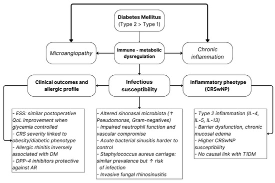
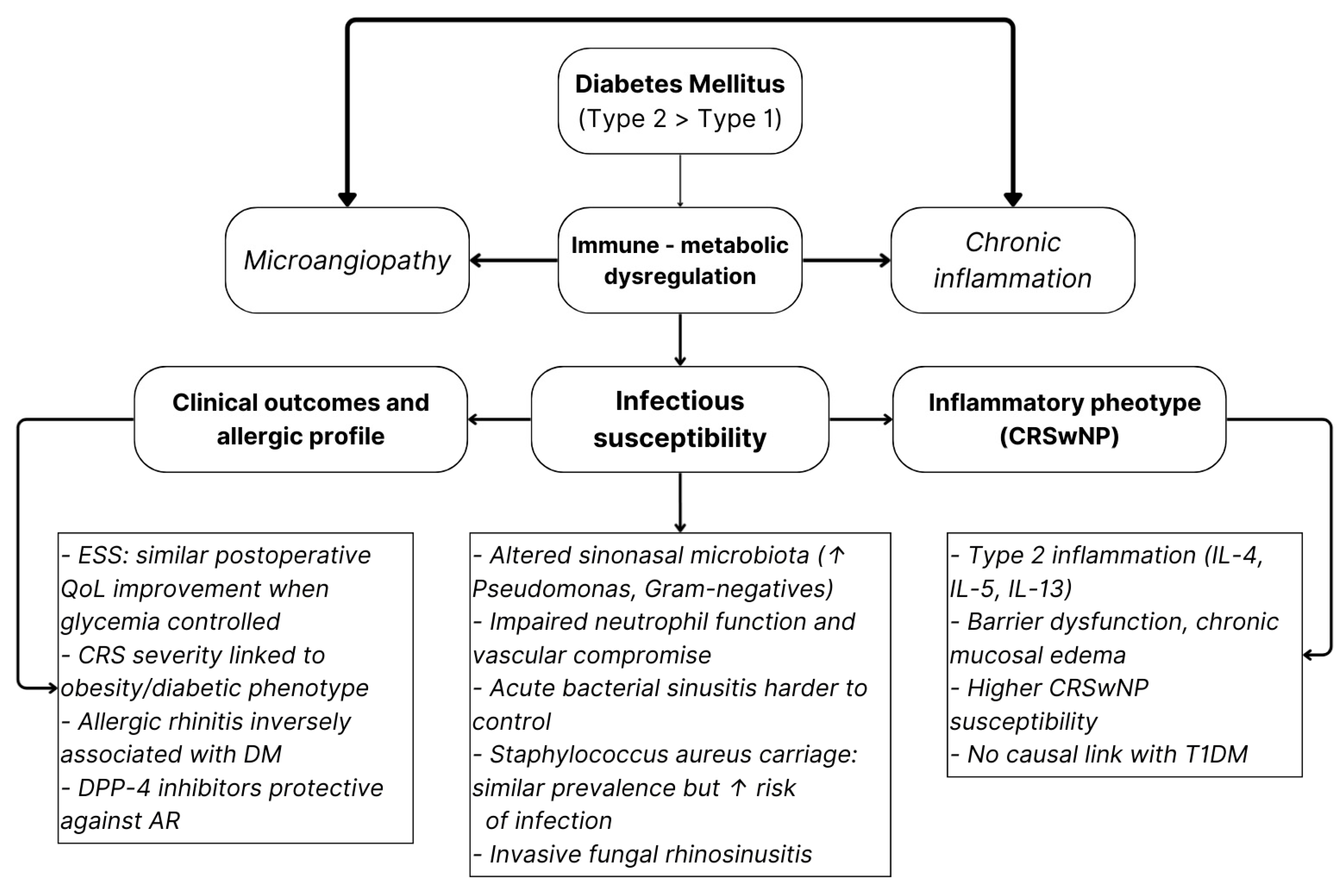
A schematic overview of the proposed pathophysiologic links between diabetes and rhinologic disease is shown in Figure 2. The model highlights how metabolic inflammation and vascular compromise may promote CRSwNP susceptibility, microbiota alterations, and infectious vulnerability.
Figure 2.
Conceptual model illustrating how DM—particularly type 2—impacts sinonasal health through immune–metabolic dysregulation. Arrows depict the proposed multidirectional interactions and downstream effects linking metabolic imbalance to inflammation, infectious susceptibility, and CRS phenotype [45,46,47,48,49,50,51,52,53,54,55].
Studies focusing on the association between DM and inflammatory or infectious sinonasal diseases are summarized in Table 1.
Table 1.
Association between diabetes mellitus and inflammatory/infectious sinonasal diseases.
A total of 12 studies were identified.
The column “Association direction” indicates whether the study found a significant or suggestive relationship between diabetes and the rhinologic condition (Positive) or no/inverse association (Negative). Specifically, “Positive” denotes a higher prevalence, severity, or worse outcomes of sinonasal disease among diabetic patients, while “Negative” refers to the absence of an association or an inverse relationship (i.e., lower prevalence or milder disease in diabetic patients).
3.2. Olfactory Disorders
Olfaction sits at the crossroads of local sinonasal inflammation and systemic metabolic–vascular health. In CRS cohorts, diabetes mellitus independently predicted poorer olfactory performance, reflected by lower TDI and odor identification scores, even after accounting for the dominant effect of nasal polyps [57]. This finding is consistent with evidence of more prolonged olfactory dysfunction in diabetic patients with CRS [45].
Population-based studies underscore the importance of distinguishing crude from adjusted associations. Analyses of KNHANES data showed that the apparent excess of self-reported olfactory dysfunction (OD) in individuals with diabetes mellitus disappears after multivariable adjustment, suggesting confounding by concomitant rhinologic and vascular factors [58]. In a large Swedish population-based cohort of 1387 adults, diabetes was not associated with overall olfactory impairment but was significantly linked to complete anosmia (OR = 2.6), indicating a selective predisposition to severe olfactory loss alongside established factors such as nasal polyps, older age, and male sex [59]. Longitudinal data with follow-up of up to 12 years further demonstrated that DM is associated with a faster decline in odour identification, independent of age and vascular comorbidity, supporting the concept of an accelerated neurosensory trajectory in diabetes [60].
Earlier population studies suggested that OD correlates more strongly with macrovascular disease than with glycaemic control or microvascular complications, raising the possibility that ischemic involvement of olfactory pathways plays a substantive role [61]. Consistent with this interpretation, metabolic analyses from NHANES identified insulin resistance, as measured by HOMA-IR, rather than hyperglycaemia per se, as the factor most closely associated with smell loss in older adults [62].
Clinical observations further indicate that chemosensory dysfunction may reflect disease burden: higher rates of phantosmia and more severe olfactory impairment have been reported among insulin-treated individuals, with greater dysfunction observed in those receiving more intensive therapy [63]. In older adults, T2DM has been identified as an independent risk factor for OD and incorporated as a key predictor in the simplified four-item Concise Aging adults Smell Test (4-CAST) screening tool [64]. Moreover, among elderly outpatients with T2DM, poorer odour identification has been associated with lower Mini-Mental State Examination scores, highlighting the neurocognitive relevance of olfactory testing in this population [65].
In line with the broader impact of diabetes on chemosensory pathways, adults with T1DM have been shown to exhibit markedly reduced olfactory performance, with hyposmia affecting up to 70% of patients [66]. The observed associations with peripheral neuropathy and retinopathy further support the interpretation of OD as part of the wider neurovascular spectrum of diabetic complications.
Taken together, the available evidence supports diabetes mellitus—particularly insulin-resistant, vascularly burdened phenotypes—as a permissive risk environment for olfactory deterioration, shaped by the combined influence of local CRS-related inflammation, systemic macrovascular compromise, and altered metabolic signalling. From a clinical perspective, olfactory assessment may represent a low-cost sentinel marker of broader micro- and macro-organ involvement in diabetic patients (Table 2).
Table 2.
Association between diabetes mellitus and olfactory disorders.
3.3. Other Rhinologic Manifestations
Beyond CRS and OD, diabetes interfaces with several additional rhinologic endpoints. Evidence from a large retrospective cohort identified diabetes as a modest but statistically significant risk factor for recurrent spontaneous epistaxis, plausibly mediated by vascular fragility and atherosclerotic changes within the nasal mucosa, in keeping with the broader vascular paradigm discussed above [67]. Mucosal clearance may also be impaired in diabetic patients: assessment of nasal mucociliary clearance using the saccharin transit test demonstrated that diabetes mellitus, particularly when coexisting with hypertension, is independently associated with delayed nasal mucociliary transport, a dysfunction that may enhance susceptibility to infection and promote persistent inflammation [68].
Cough phenotypes in older adults appear sensitive to glycaemic control. In community-dwelling seniors, uncontrolled DM (HbA1c ≥ 8%) was independently associated with frequent, chronic-persistent, and nocturnal cough, even after adjusting for AR, asthma, and smoking, supporting a link between hyperglycaemia, airway inflammation/hyperresponsiveness, and symptom burden [69]. In the COVID-19 setting, anosmia was reported as a relatively frequent symptom overall but without specific associations between DM and rhinologic symptoms [70]; thus, existing data do not establish a DM-specific predisposition to COVID-related smell loss.
Large-scale population-based survey data further showed that DM, without specification of type, was reported as a comorbidity in 10.9% of individuals who contracted COVID-19, within a cohort of 15,166 adults assessed across multiple regions. Among infected participants, anosmia and ageusia were among the most frequently reported symptoms, affecting 68.4% of cases; however, no analyses specifically addressed the relationship between diabetes and olfactory or gustatory dysfunction [71].
Vascular integrity, epithelial transport, and systemic metabolic control likely converge to influence bleeding risk, mucosal clearance, and cough in DM. Evidence remains heterogeneous, but signals are consistent with micro-/macrovascular and metabolic contributions across these domains.
3.4. Overall Conclusions and Implications
Taken together, the available literature supports a convergent interpretative framework in which DM, particularly T2DM, affects rhinologic health through three interrelated mechanisms: (1) immune–metabolic dysregulation promoting chronic mucosal inflammation and altered sinonasal microbiology; (2) vascular and neurosensory impairment contributing to progressive olfactory dysfunction and a shift toward more severe phenotypes; and (3) epithelial and vascular mucosal alterations that impair clearance and increase tissue fragility. Within this context, surgical outcomes appear generally comparable to those of non-diabetic patients, provided that metabolic status and systemic risk factors are adequately considered, underscoring the potential relevance of integrated metabolic and rhinologic management.
A global summary of the main findings of all the studies included in this narrative review is provided in Table 3.
Table 3.
Global findings among the studies included.
4. Discussion
A specific limitation of this narrative review is the restriction of the literature search to PubMed. We acknowledge that relevant studies indexed exclusively in other databases may have been missed; however, PubMed was deliberately selected as the primary source for this narrative review because it provides comprehensive coverage of peer-reviewed biomedical and clinical research and ensures standardized indexing through MeSH terms.
4.1. Methodological Limitations of the Current Evidence
Despite the growing number of studies exploring the relationship between DM and rhinologic conditions, several methodological limitations substantially weaken the strength and generalizability of the available evidence.
A major limitation is the marked heterogeneity of study designs and populations. Most available evidence derives from cross-sectional or retrospective cohort studies, which inherently preclude causal inference. Only a limited number of investigations adopted prospective or longitudinal designs capable of assessing temporal relationships. Furthermore, study populations range from small tertiary-centre cohorts to large national health surveys, resulting in variable diagnostic accuracy and potential sampling bias.
This lack of robust evidence is particularly evident in the field of OD. A recent scoping review [72] identified only three eligible studies out of more than 3600 screened records, highlighting the extreme paucity of data despite the high prevalence of OD among patients with diabetes. A broader narrative review [73], although summarizing decades of research on DM and OD, similarly reinforces how limited and fragmented the evidence base remains.
4.2. Variability in Diabetes Definition and Metabolic Characterization
Another critical limitation concerns the definition of diabetes itself. In several studies, diabetes was self-reported [45,57,58,59,62], relying on participant recall or previous physician diagnosis without biochemical confirmation. Even when laboratory parameters were included, diagnostic thresholds and classification between T1DM and T2DM were often unclear (19 of the 27 studies included in this review), limiting phenotype-specific interpretation. Measures of glycemic control (HbA1c) and disease duration were incorporated in only a minority of studies and were inconsistently associated with outcomes.
4.3. Inconsistency in Rhinologic Outcome Assessment
Similarly, the definition and assessment of rhinologic outcomes varied widely across studies. Many population-based investigations [53,58] relied on self-reported symptoms or prior diagnoses, whereas only a limited number employed objective diagnostic criteria, imaging, or validated questionnaires. This issue is particularly relevant for OD, which was frequently assessed subjectively rather than through psychophysical testing such as Sniffin’ Sticks or the Smell Identification Test. As a result, the true prevalence and severity of olfactory impairment in diabetic populations may be either over- or underestimated.
4.4. Confounding Factors and Population Heterogeneity
Inadequate control of confounding factors represents an additional source of variability. While some studies adjusted for key metabolic and vascular covariates, others failed to account for important mediators such as obesity, hypertension, smoking, or chronic inflammation, all of which independently influence sinonasal health. The extent of adjustment often determined whether associations between diabetes and rhinologic outcomes remained statistically significant [58].
Geographic and ethnic variability further complicates interpretation. The majority of epidemiological evidence in this narrative review originates from Asian populations, particularly from East Asia, with relatively few studies conducted in Western countries.
Genetic susceptibility, environmental exposure, dietary habits, prevalence of metabolic comorbidities, and healthcare access differ substantially across regions and ethnic groups and may modulate both diabetes phenotype and rhinologic disease expression. Consequently, the magnitude and clinical relevance of observed associations may not be directly generalizable to other populations.
Finally, inter-study inconsistency in diagnostic tools, outcome reporting, and follow-up duration limits meta-analytic synthesis. Standardized instruments such as SNOT-22 or validated olfactory tests were infrequently used, and postoperative or longitudinal outcomes were rarely correlated with metabolic control.
4.5. Pathophysiological Framework and Interpretative Considerations
Beyond individual rhinologic phenotypes, the available evidence supports a convergent pathophysiological framework linking diabetes to sinonasal disease through immune–metabolic dysregulation, vascular impairment, and epithelial barrier dysfunction.
Chronic low-grade inflammation and insulin resistance alter innate and adaptive immune responses, promoting persistent mucosal inflammation, impaired host defense, and increased susceptibility to infection.
In parallel, diabetes-related micro- and macrovascular damage may compromise mucosal perfusion and neural integrity, contributing to delayed tissue repair, impaired mucociliary clearance, and accelerated olfactory decline. Epithelial barrier dysfunction further facilitates microbial dysbiosis and perpetuates local inflammation. This triad mirrors the systemic impact of diabetes on organs such as the retina, kidney, and peripheral nervous system, supporting the concept of rhinologic involvement as part of a multisystem disease continuum rather than an isolated manifestation.
4.6. Clinical Implications and Future Directions
Although multiple associations between DM and rhinologic manifestations have been reported, causal relationships remain partially unestablished. In this context, emerging human evidence suggests that the nasal microbiota may represent an active biological interface between systemic metabolic alterations and local sinonasal inflammation, with disease-specific and metabolically sensitive microbial signatures being associated with inflammatory severity and mucosal dysfunction [74,75].
In addition, a systematic and longitudinal assessment of the nasal microbiological profile may represent a promising research direction, allowing evaluation of how sinonasal microbial communities evolve over time in relation to glycaemic control, metabolic status, and disease progression.
The integration of metabolic and inflammatory biomarkers, including HbA1c, markers of insulin resistance, systemic inflammatory mediators, and indicators of endothelial dysfunction, may help clarify dose–response relationships and temporal associations.
Advanced imaging techniques, such as high-resolution MRI or functional imaging of olfactory pathways, may further elucidate microvascular and neurosensory involvement.
The emerging evidence also supports the potential role of multidisciplinary management strategies integrating endocrinology and otorhinolaryngology. Olfactory testing represents a low-cost, non-invasive tool that could be incorporated into routine assessment of diabetic patients, potentially serving as an early marker of neurovascular or systemic involvement, analogous to screening approaches used for diabetic retinopathy or neuropathy. Optimized metabolic control should be considered an integral component of rhinologic management, particularly in patients with chronic rhinosinusitis or olfactory dysfunction, alongside standard medical or surgical treatments.
Future research should move beyond cross-sectional and retrospective designs toward well-powered prospective and longitudinal studies capable of capturing temporal relationships between metabolic control and rhinologic outcomes.
Large cohort studies with adequate follow-up would allow evaluation of disease progression, treatment response, and causal pathways.
The systematic adoption of standardized and validated rhinologic assessment tools, such as SNOT-22 and psychophysical olfactory tests, is essential to improve comparability across studies.
Clear differentiation between T1DM and T2DM is crucial, as these conditions differ substantially in pathophysiology, inflammatory burden, and vascular involvement, and may require phenotype-specific preventive and therapeutic strategies.
5. Conclusions
While several experimental and mechanistic studies have investigated diabetes-related biological pathways, the body of clinically oriented and methodologically comparable human evidence specifically focused on rhinologic manifestations remains limited. Over nearly five decades of available research, only a limited number of original studies have explored this intersection, underscoring a substantial gap in research relative to the global burden of diabetes.
The few available investigations, although heterogeneous in design and population, converge on the notion that diabetes—particularly T2DM—affects sinonasal health through intertwined immune–metabolic, vascular, and epithelial mechanisms.
These mechanisms appear to
- Promote CRSwNP susceptibility and alter sinus microbiota toward Gram-negative enrichment;
- Accelerate olfactory decline via vascular and neuro-sensory compromise. Mirroring the established multidisciplinary pathways used to detect retinopathy in diabetes, olfactory testing might emerge as a similarly valuable tool, one that could be implemented early, perhaps even before symptom onset, to identify individuals at risk of neuro-sensory decline;
- Contribute to secondary rhinologic issues such as recurrent epistaxis, impaired mucociliary clearance, and increased infection severity.
Future research should address current methodological limitations, particularly the lack of standardized diagnostic tools, small sample sizes, and insufficient differentiation between T1DM and T2DM. Well-powered prospective studies integrating metabolic parameters with objective rhinologic assessments are needed to clarify causal mechanisms. Recognizing rhinologic manifestations as an underexplored component of diabetes may also support earlier detection of systemic vascular and neural involvement and foster multidisciplinary management strategies.
Author Contributions
All authors have equally contributed to the writing, conceptualization and screening process for this review. All authors have read and agreed to the published version of the manuscript.
Funding
This research received no external funding.
Institutional Review Board Statement
Not applicable.
Informed Consent Statement
Not applicable.
Data Availability Statement
No new data were created or analyzed in this study.
Conflicts of Interest
The authors declare no conflicts of interest.
Abbreviations
The following abbreviations are used in this manuscript:
| AIFRS | Acute Invasive Fungal Rhinosinusitis |
| AFRS | Allergic Fungal Rhinosinusitis |
| AR | Allergic Rhinitis |
| ABS | Acute Bacterial Sinusitis |
| BOB | Blood–Olfactory Barrier |
| BSIT | Brief Smell Identification Test |
| CD26 | Cluster of Differentiation 26 |
| CGFRS | Chronic Granulomatous Fungal Rhinosinusitis |
| CIFRS | Chronic Invasive Fungal Rhinosinusitis |
| CRS | Chronic Rhinosinusitis |
| CRSsNP | Chronic Rhinosinusitis without Nasal Polyps |
| CRSwNP | Chronic Rhinosinusitis with Nasal Polyps |
| DALYs | Disability-Adjusted Life Years |
| DM | Diabetes Mellitus |
| DPP-4 | Dipeptidyl Peptidase-4 |
| ESS | Endoscopic Sinus Surgery |
| FESS | Functional Endoscopic Sinus Surgery |
| GWAS | Genome-Wide Association Study |
| HbA1c | Glycated Hemoglobin |
| HOMA-IR | Homeostatic Model Assessment for Insulin Resistance |
| IDDM | Insulin-Dependent Diabetes Mellitus |
| IL | Interleukin |
| ILC2 | Type 2 Innate Lymphoid Cells |
| KNHANES | Korea National Health and Nutrition Examination Survey |
| MRSA | Methicillin-Resistant Staphylococcus aureus |
| NHANES | National Health and Nutrition Examination Survey |
| NIDDM | Non-Insulin-Dependent Diabetes Mellitus |
| OD | Olfactory Dysfunction |
| QOL/QoL | Quality of Life |
| S. aureus | Staphylococcus aureus |
| SIT-40 | Smell Identification Test–40 |
| SNOT-22 | Sino-Nasal Outcome Test–22 |
| T1D/T1DM | Type 1 Diabetes Mellitus |
| T2D/T2DM | Type 2 Diabetes Mellitus |
| Tfh | T Follicular Helper Cells |
| Th1/Th2/Th17 | T Helper Cells |
| Tregs | Regulatory T Cells |
References
- Bell, K.J.; Lain, S.J. The Changing Epidemiology of Type 1 Diabetes: A Global Perspective. Diabetes Obes. Metab. 2025, 27, 3–14. [Google Scholar] [CrossRef] [Scilit] [PubMed]
- Maahs, D.M.; West, N.A.; Lawrence, J.M.; Mayer-Davis, E.J. Epidemiology of type 1 diabetes. Endocrinol. Metab. Clin. N. Am. 2010, 39, 481–497. [Google Scholar] [CrossRef] [Scilit] [PubMed]
- Forlenza, G.P.; Rewers, M. The epidemic of type 1 diabetes: What is it telling us? Curr. Opin. Endocrinol. Diabetes Obes. 2011, 18, 248–251. [Google Scholar] [CrossRef] [Scilit]
- Frese, T.; Sandholzer, H. The Epidemiology of Type 1 Diabetes Mellitus; InTech: London, UK, 2013. [Google Scholar] [CrossRef] [Scilit]
- Gillespie, K.M. Type 1 diabetes: Pathogenesis and prevention. CMAJ Can. Med. Assoc. J. J. L’association Medicale Can. 2006, 175, 165–170. [Google Scholar] [CrossRef] [Scilit] [PubMed]
- Desai, S.; Deshmukh, A. Mapping of Type 1 Diabetes Mellitus. Curr. Diabetes Rev. 2020, 16, 438–441. [Google Scholar] [CrossRef] [Scilit]
- Lernmark, A. Type 1 diabetes. Clin. Chem. 1999, 45, 1331–1338. [Google Scholar] [CrossRef] [Scilit]
- Vallianou, N.G.; Stratigou, T.; Geladari, E.; Tessier, C.M.; Mantzoros, C.S.; Dalamaga, M. Diabetes type 1: Can it be treated as an autoimmune disorder? Rev. Endocr. Metab. Disord. 2021, 22, 859–876. [Google Scholar] [CrossRef] [Scilit]
- Bispham, J.A.; Hughes, A.S.; Driscoll, K.A.; McAuliffe-Fogarty, A.H. Novel Challenges in Aging with Type 1 Diabetes. Curr. Diabetes Rep. 2020, 20, 15. [Google Scholar] [CrossRef] [Scilit]
- Javeed, N.; Matveyenko, A.V. Circadian Etiology of Type 2 Diabetes Mellitus. Physiology 2018, 33, 138–150. [Google Scholar] [CrossRef] [Scilit]
- Fletcher, B.; Gulanick, M.; Lamendola, C. Risk factors for type 2 diabetes mellitus. J. Cardiovasc. Nurs. 2002, 16, 17–23. [Google Scholar] [CrossRef] [Scilit]
- Singh, A.; Shadangi, S.; Gupta, P.K.; Rana, S. Type 2 Diabetes Mellitus: A Comprehensive Review of Pathophysiology, Comorbidities, and Emerging Therapies. Compr. Physiol. 2025, 15, e70003. [Google Scholar] [CrossRef] [Scilit]
- Magliano, D.J.; Boyko, E.J. IDF Diabetes Atlas 10th edition scientific committee. In IDF Diabetes Atlas, 10th ed.; International Diabetes Federation: Brussels, Belgium, 2021. [Google Scholar]
- Kautzky-Willer, A.; Harreiter, J.; Pacini, G. Sex and Gender Differences in Risk, Pathophysiology and Complications of Type 2 Diabetes Mellitus. Endocr. Rev. 2016, 37, 278–316. [Google Scholar] [CrossRef] [Scilit]
- Gloyn, A.L.; Drucker, D.J. Precision medicine in the management of type 2 diabetes. Lancet Diabetes Endocrinol. 2018, 6, 891–900. [Google Scholar] [CrossRef] [Scilit]
- Loos, R.J.F.; Yeo, G.S.H. The genetics of obesity: From discovery to biology. Nat. Rev. Genet. 2022, 23, 120–133. [Google Scholar] [CrossRef] [Scilit] [PubMed]
- Trang, K.; Grant, S.F.A. Genetics and epigenetics in the obesity phenotyping scenario. Rev. Endocr. Metab. Disord. 2023, 24, 775–793. [Google Scholar] [CrossRef] [Scilit]
- Dunachie, S.; Chamnan, P. The double burden of diabetes and global infection in low and middle-income countries. Trans. R. Soc. Trop. Med. Hyg. 2019, 113, 56–64. [Google Scholar] [CrossRef] [Scilit] [PubMed]
- Guber, C. Type 2 diabetes. Lancet 2005, 365, 1347. [Google Scholar] [CrossRef] [Scilit] [PubMed]
- Hillson, R. Treating people with type 2 diabetes. Br. J. Hosp. Med. 2013, 74, 186. [Google Scholar] [CrossRef] [Scilit]
- Taylor, R. Type 2 diabetes: Etiology and reversibility. Diabetes Care 2013, 36, 1047–1055. [Google Scholar] [CrossRef] [Scilit]
- Siddiqui, Z.A.; Walker, A.; Pirwani, M.M.; Tahiri, M.; Syed, I. Allergic rhinitis: Diagnosis and management. Br. J. Hosp. Med. 2022, 83, 1–9. [Google Scholar] [CrossRef] [Scilit]
- Czech, E.J.; Overholser, A.; Schultz, P. Allergic Rhinitis. Prim. Care 2023, 50, 159–178. [Google Scholar] [CrossRef] [Scilit]
- Ponda, P.; Carr, T.; Rank, M.A.; Bousquet, J. Nonallergic Rhinitis, Allergic Rhinitis, and Immunotherapy: Advances in the Last Decade. J. Allergy Clin. Immunol. Pract. 2023, 11, 35–42. [Google Scholar] [CrossRef] [Scilit]
- Bernstein, D.I.; Schwartz, G.; Bernstein, J.A. Allergic Rhinitis: Mechanisms and Treatment. Immunol. Allergy Clin. N. Am. 2016, 36, 261–278. [Google Scholar] [CrossRef] [Scilit] [PubMed]
- Zhang, Y.; Lan, F.; Zhang, L. Update on pathomechanisms and treatments in allergic rhinitis. Allergy 2022, 77, 3309–3319. [Google Scholar] [CrossRef] [Scilit] [PubMed]
- Volpe, S.; Irish, J.; Palumbo, S.; Lee, E.; Herbert, J.; Ramadan, I.; Chang, E.H. Viral infections and chronic rhinosinusitis. J. Allergy Clin. Immunol. 2023, 152, 819–826. [Google Scholar] [CrossRef] [Scilit]
- Min, H.K.; Lee, S.; Kim, S.; Son, Y.; Park, J.; Kim, H.J.; Lee, J.; Lee, H.; Smith, L.; Rahmati, M.; et al. Global Incidence and Prevalence of Chronic Rhinosinusitis: A Systematic Review. Clin. Exp. Allergy J. Br. Soc. Allergy Clin. Immunol. 2025, 55, 52–66. [Google Scholar] [CrossRef] [Scilit] [PubMed]
- Koo, M.S.; Moon, S.; Rha, M.S. Mucosal Inflammatory Memory in Chronic Rhinosinusitis. Cells 2024, 13, 1947. [Google Scholar] [CrossRef] [Scilit]
- Michalik, M.; Krawczyk, B. Chronic Rhinosinusitis-Microbiological Etiology, Potential Genetic Markers, and Diagnosis. Int. J. Mol. Sci. 2024, 25, 3201. [Google Scholar] [CrossRef] [Scilit]
- Caballero, T.M.; Altillo, B.S.A.; Milstone, A.M. Acute Bacterial Sinusitis: Limitations of Test-Based Treatment. JAMA 2023, 330, 326–327. [Google Scholar] [CrossRef] [Scilit]
- Wilcox, J.; Lobo, D.; Anderson, S. Sinusitis. Prim. Care 2025, 52, 87–97. [Google Scholar] [CrossRef] [Scilit]
- Gleckman, R.A. Acute bacterial sinusitis. Hosp. Pract. 1986, 21, 92–102. [Google Scholar]
- Rosenfeld, R.M.; Piccirillo, J.F.; Chandrasekhar, S.S.; Brook, I.; Ashok Kumar, K.; Kramper, M.; Orlandi, R.R.; Palmer, J.N.; Patel, Z.M.; Peters, A.; et al. Clinical practice guideline (update): Adult sinusitis. Otolaryngol. Head Neck Surg. Off. J. Am. Acad. Otolaryngol. Head Neck Surg. 2015, 152, S1–S39. [Google Scholar] [CrossRef] [Scilit] [PubMed]
- Sande, M.A.; Gwaltney, J.M. Acute community-acquired bacterial sinusitis: Continuing challenges and current management. Clin. Infect. Dis. Off. Publ. Infect. Dis. Soc. Am. 2004, 39, S151–S158. [Google Scholar] [CrossRef] [Scilit]
- Feldt, B.; Dion, G.R.; Weitzel, E.K.; McMains, K.C. Acute sinusitis. South. Med. J. 2013, 106, 577–581. [Google Scholar] [CrossRef] [Scilit] [PubMed]
- Montone, K.T. Pathology of Fungal Rhinosinusitis: A Review. Head Neck Pathol. 2016, 10, 40–46. [Google Scholar] [CrossRef] [Scilit]
- Luong, A.; Marple, B.F. Allergic fungal rhinosinusitis. Curr. Allergy Asthma Rep. 2004, 4, 465–470. [Google Scholar] [CrossRef] [Scilit]
- Epstein, V.A.; Kern, R.C. Invasive fungal sinusitis and complications of rhinosinusitis. Otolaryngol. Clin. N. Am. 2008, 41, 497–524. [Google Scholar] [CrossRef] [Scilit]
- Hummel, T.; Whitcroft, K.L.; Andrews, P.; Altundag, A.; Cinghi, C.; Costanzo, R.M.; Damm, M.; Frasnelli, J.; Gudziol, H.; Gupta, N.; et al. Position paper on olfactory dysfunction. Rhinology. Suppl. 2017, 54, 1–30. [Google Scholar] [CrossRef] [Scilit]
- Hendriks, A.P. Olfactory dysfunction. Rhinology 1988, 26, 229–251. [Google Scholar]
- Wellford, S.A.; Moseman, E.A. Olfactory immunology: The missing piece in airway and CNS defence. Nat. Rev. Immunol. 2024, 24, 381–398. [Google Scholar] [CrossRef] [Scilit]
- Song, J.; Wang, M.; Wang, C.; Zhang, L. Olfactory dysfunction in chronic rhinosinusitis: Insights into the underlying mechanisms and treatments. Expert Rev. Clin. Immunol. 2023, 19, 993–1004. [Google Scholar] [CrossRef] [Scilit] [PubMed]
- Davidson, T.M.; Murphy, C. Olfactory impairment. West. J. Med. 1993, 159, 71–72. [Google Scholar]
- Nam, J.S.; Roh, Y.H.; Kim, J.; Chang, S.W.; Ha, J.G.; Park, J.J.; Fahad, W.A.; Yoon, J.H.; Kim, C.H.; Cho, H.J. Association between diabetes mellitus and chronic rhinosinusitis with nasal polyps: A population-based cross-sectional study. Clin. Otolaryngol. Off. J. ENT-UK Off. J. Neth. Soc. Oto-Rhino Laryngol. Cervico Facial Surg. 2022, 47, 167–173. [Google Scholar] [CrossRef] [Scilit] [PubMed]
- Zhang, Z.; Adappa, N.D.; Lautenbach, E.; Chiu, A.G.; Doghramji, L.; Howland, T.J.; Cohen, N.A.; Palmer, J.N. The effect of diabetes mellitus on chronic rhinosinusitis and sinus surgery outcome. Int. Forum Allergy Rhinol. 2014, 4, 315–320. [Google Scholar] [CrossRef] [Scilit]
- Hajjij, A.; Mace, J.C.; Soler, Z.M.; Smith, T.L.; Hwang, P.H. The impact of diabetes mellitus on outcomes of endoscopic sinus surgery: A nested case-control study. Int. Forum Allergy Rhinol. 2015, 5, 533–540. [Google Scholar] [CrossRef] [Scilit]
- Steele, T.O.; Mace, J.C.; DeConde, A.S.; Xiao, C.C.; Storck, K.A.; Gudis, D.A.; Schlosser, R.J.; Soler, Z.M.; Smith, T.L. Does comorbid obesity impact quality of life outcomes in patients undergoing endoscopic sinus surgery? Int. Forum Allergy Rhinol. 2015, 5, 1085–1094. [Google Scholar] [CrossRef] [Scilit] [PubMed]
- Yuan, X.; Meng, L.; Liu, L.; Zhang, B.; Xie, S.; Zhong, W.; Jia, J.; Zhang, H.; Jiang, W.; Xie, Z. Hyperglycemia and type 2 diabetes mellitus associate with postoperative recurrence in chronic rhinosinusitis patients. Eur. Arch. Oto Rhino Laryngol. Off. J. Eur. Fed. Oto-Rhino-Laryngol. Soc. (EUFOS) Affil. Ger. Soc. Oto-Rhino-Laryngol. Head Neck Surg. 2025, 282, 1289–1299. [Google Scholar] [CrossRef] [Scilit]
- Jackson, R.M.; Rice, D.H. Acute bacterial sinusitis and diabetes mellitus. Otolaryngol. Head Neck Surg. Off. J. Am. Acad. Otolaryngol. Head Neck Surg. 1987, 97, 469–473. [Google Scholar] [CrossRef] [Scilit]
- Hart, J.; Hamilton, E.J.; Makepeace, A.; Davis, W.A.; Latkovic, E.; Lim, E.M.; Dyer, J.R.; Davis, T.M. Prevalence, risk factors and sequelae of Staphylococcus aureus carriage in diabetes: The Fremantle Diabetes Study Phase II. J. Diabetes Its Complicat. 2015, 29, 1092–1097. [Google Scholar] [CrossRef] [Scilit]
- Soundarya, R.; Deepa, H.C.; Prakash, P.Y.; Geetha, V. Fungal Rhinosinusitis: An integrated diagnostic approach. Ann. Diagn. Pathol. 2025, 75, 152415. [Google Scholar] [CrossRef] [Scilit]
- Lee, T.K.; Jeon, Y.J.; Jung, S.J. Bi-directional association between allergic rhinitis and diabetes mellitus from the national representative data of South Korea. Sci. Rep. 2021, 11, 4344. [Google Scholar] [CrossRef] [Scilit]
- Thomsen, S.F.; Duffy, D.L.; Kyvik, K.O.; Skytthe, A.; Backer, V. Relationship between type 1 diabetes and atopic diseases in a twin population. Allergy 2011, 66, 645–647. [Google Scholar] [CrossRef] [Scilit]
- Chen, H.H.; Li, S.Y.; Chen, W.; Kao, C.H. Association between Dipeptidyl Peptidase-4 Inhibitors and Allergic Rhinitis in Asian Patients with Diabetes. Int. J. Environ. Res. Public Health 2019, 16, 1323. [Google Scholar] [CrossRef] [Scilit]
- Chen, S.; Tan, L.; Qin, D.; Lv, H.; Liu, K.; Xu, Y.; Wu, X.; Huang, J.; Xu, Y. The causal relationship between multiple autoimmune diseases and nasal polyps. Front. Immunol. 2023, 14, 1228226. [Google Scholar] [CrossRef] [Scilit]
- Schlosser, R.J.; Smith, T.L.; Mace, J.C.; Alt, J.; Beswick, D.M.; Mattos, J.L.; Payne, S.; Ramakrishnan, V.R.; Soler, Z.M. Factors driving olfactory loss in patients with chronic rhinosinusitis: A case control study. Int. Forum Allergy Rhinol. 2020, 10, 7–14. [Google Scholar] [CrossRef] [Scilit]
- Roh, D.; Lee, D.H.; Kim, S.W.; Kim, S.W.; Kim, B.G.; Kim, D.H.; Shin, J.H. The association between olfactory dysfunction and cardiovascular disease and its risk factors in middle-aged and older adults. Sci. Rep. 2021, 11, 1248. [Google Scholar] [CrossRef] [Scilit] [PubMed]
- Brämerson, A.; Johansson, L.; Ek, L.; Nordin, S.; Bende, M. Prevalence of olfactory dysfunction: The skövde population-based study. Laryngoscope 2004, 114, 733–737. [Google Scholar] [CrossRef] [Scilit] [PubMed]
- Ekström, I.; Larsson, M.; Rizzuto, D.; Fastbom, J.; Bäckman, L.; Laukka, E.J. Predictors of Olfactory Decline in Aging: A Longitudinal Population-Based Study. J. Gerontol. Ser. A Biol. Sci. Med. Sci. 2020, 75, 2441–2449. [Google Scholar] [CrossRef] [Scilit] [PubMed]
- Weinstock, R.S.; Wright, H.N.; Smith, D.U. Olfactory dysfunction in diabetes mellitus. Physiol. Behav. 1993, 53, 17–21. [Google Scholar] [CrossRef] [Scilit]
- Min, J.Y.; Min, K.B. Insulin resistance and the increased risk for smell dysfunction in US adults. Laryngoscope 2018, 128, 1992–1996. [Google Scholar] [CrossRef] [Scilit]
- Chan, J.Y.K.; García-Esquinas, E.; Ko, O.H.; Tong, M.C.F.; Lin, S.Y. The Association Between Diabetes and Olfactory Function in Adults. Chem. Senses 2017, 43, 59–64. [Google Scholar] [CrossRef] [Scilit]
- Soler, Z.M.; Gregoski, M.J.; Kohli, P.; LaPointe, K.A.; Schlosser, R.J. Development and validation of the four-item Concise Aging adults Smell Test to screen for olfactory dysfunction in older adults. Int. Forum Allergy Rhinol. 2025, 15, 250–257. [Google Scholar] [CrossRef] [Scilit]
- Sanke, H.; Mita, T.; Yoshii, H.; Yokota, A.; Yamashiro, K.; Ingaki, N.; Onuma, T.; Someya, Y.; Komiya, K.; Tamura, Y.; et al. Relationship between olfactory dysfunction and cognitive impairment in elderly patients with type 2 diabetes mellitus. Diabetes Res. Clin. Pract. 2014, 106, 465–473. [Google Scholar] [CrossRef] [Scilit]
- Falkowski, B.; Chudziński, M.; Jakubowska, E.; Duda-Sobczak, A. Association of olfactory function with the intensity of self-reported physical activity in adults with type 1 diabetes. Pol. Arch. Intern. Med. 2017, 127, 476–480. [Google Scholar] [CrossRef] [Scilit]
- Abrich, V.; Brozek, A.; Boyle, T.R.; Chyou, P.H.; Yale, S.H. Risk factors for recurrent spontaneous epistaxis. Mayo Clin. Proc. 2014, 89, 1636–1643. [Google Scholar] [CrossRef] [Scilit] [PubMed]
- Proença de Oliveira-Maul, J.; Barbosa de Carvalho, H.; Goto, D.M.; Maia, R.M.; Fló, C.; Barnabé, V.; Franco, D.R.; Benabou, S.; Perracini, M.R.; Jacob-Filho, W.; et al. Aging, diabetes, and hypertension are associated with decreased nasal mucociliary clearance. Chest 2013, 143, 1091–1097. [Google Scholar] [CrossRef] [Scilit]
- Song, W.J.; Morice, A.H.; Kim, M.H.; Lee, S.E.; Jo, E.J.; Lee, S.M.; Han, J.W.; Kim, T.H.; Kim, S.H.; Jang, H.C.; et al. Cough in the elderly population: Relationships with multiple comorbidity. PLoS ONE 2013, 8, e78081. [Google Scholar] [CrossRef] [Scilit]
- Alasia, D.; Owhonda, G.; Maduka, O.; Nwadiuto, I.; Arugu, G.; Tobin-West, C.; Azi, E.; Oris-Onyiri, V.; Urang, I.J.; Abikor, V.; et al. Clinical and epidemiological characteristics of 646 hospitalised SARS-Cov-2 positive patients in Rivers State Nigeria: A prospective observational study. Pan Afr. Med. J. 2021, 38, 25. [Google Scholar] [CrossRef] [Scilit]
- Schaalan, M.; Abou Warda, A.E.; Osman, S.M.; Fathy, S.; Sarhan, R.M.; Boshra, M.S.; Sarhan, N.; Gaber, S.; Ali, A.M.A. The Impact of Sociodemographic, Nutritional, and Health Factors on the Incidence and Complications of COVID-19 in Egypt: A Cross-Sectional Study. Viruses 2022, 14, 448. [Google Scholar] [CrossRef] [Scilit] [PubMed]
- Sivakumar, R.; White, J.; Villwock, J. Olfactory dysfunction in type II diabetes: Therapeutic options and lessons learned from other etiologies A scoping review. Prim. Care Diabetes 2022, 16, 543–548. [Google Scholar] [CrossRef] [Scilit] [PubMed]
- Zaghloul, H.; Pallayova, M.; Al-Nuaimi, O.; Hovis, K.R.; Taheri, S. Association between diabetes mellitus and olfactory dysfunction: Current perspectives and future directions. Diabet. Med. A J. Br. Diabet. Assoc. 2018, 35, 41–52. [Google Scholar] [CrossRef] [Scilit] [PubMed]
- Cho, S.W.; Kim, D.Y.; Choi, S.; Won, S.; Kang, H.R.; Yi, H. Microbiome profiling of uncinate tissue and nasal polyps in patients with chronic rhinosinusitis using swab and tissue biopsy. PLoS ONE 2021, 16, e0249688. [Google Scholar] [CrossRef] [Scilit] [PubMed]
- Toro-Ascuy, D.; Cárdenas, J.P.; Zorondo-Rodríguez, F.; González, D.; Silva-Moreno, E.; Puebla, C.; Nunez-Parra, A.; Reyes-Cerpa, S.; Fuenzalida, L.F. Microbiota Profile of the Nasal Cavity According to Lifestyles in Healthy Adults in Santiago, Chile. Microorganisms 2023, 11, 1635. [Google Scholar] [CrossRef] [Scilit] [PubMed]

Disclaimer/Publisher’s Note: The statements, opinions and data contained in all publications are solely those of the individual author(s) and contributor(s) and not of MDPI and/or the editor(s). MDPI and/or the editor(s) disclaim responsibility for any injury to people or property resulting from any ideas, methods, instructions or products referred to in the content. |
© 2026 by the authors. Licensee MDPI, Basel, Switzerland. This article is an open access article distributed under the terms and conditions of the Creative Commons Attribution (CC BY) license.