


Abstract
Male infertility represents a major clinical challenge. Despite standard genetic testing (karyotyping, Y-chromosome azoospermia factor (AZF) microdeletion testing, and CFTR variant analysis), the molecular cause remains unidentified in a substantial proportion of patients. These patients are consequently diagnosed with idiopathic male infertility. This review provides a comprehensive overview of the current evidence regarding the application of whole-exome sequencing (WES) in the molecular diagnosis and clinical management of idiopathic male infertility. WES can identify pathogenic variants associated with quantitative spermatogenic defects, qualitative abnormalities of sperm motility and morphology, and pre-testicular causes related to hypogonadotropic hypogonadism. When defining the cohort strictly as patients with true idiopathic non-obstructive azoospermia (NOA) who remain undiagnosed after standard testing, the pooled diagnostic yield of WES is approximately 10–15%. Estimates vary due to differences in cohort selection, variant interpretation, and the range of genes analysed. Establishing a precise molecular diagnosis improves genetic counselling, helps predict the likelihood of successful sperm retrieval via testicular sperm extraction (TESE) or microdissection TESE (micro-TESE), and informs treatment planning for assisted reproductive technologies. However, routine clinical implementation remains limited by the high frequency of variants of uncertain significance, the absence of standardised diagnostic pipelines, unequal access to testing, and ethical concerns. Emerging “all-in-one” diagnostic strategies, multi-omics integration, and whole-genome or long-read sequencing hold promise for improving genomic diagnostics. However, broader adoption will ultimately require ongoing standardisation and functional validation of identified variants.
1. Introduction
Infertility is a major global health problem estimated to affect around 17.5% of people of reproductive age [1]. Male factors are involved in approximately 50% of cases, either alone or in combination with female factors [2,3,4]. Male infertility is phenotypically heterogeneous and may present as a quantitative abnormality, such as oligozoospermia or azoospermia, or as a qualitative defect affecting sperm motility or morphology, termed asthenozoospermia and teratozoospermia, respectively [3].
However, it is not just a reproductive issue; it carries severe psychological and social burdens. Patients often experience profound psychological distress and social stigma, leading to a diminished quality of life. In men, a diagnosis of infertility may contribute to social isolation and low self-esteem and is associated with an increased risk of anxiety, depression, and sexual dysfunction [5]. Infertile men may also have an increased risk of certain malignancies [2,3,6]. Current evidence supports a shared genetic aetiology, suggesting that variants responsible for impaired spermatogenesis may also increase susceptibility to tumourigenesis, although a direct causal chain linking spermatogenic variants to cancer development has not yet been firmly established [3,6].
Standard genetic testing, comprising karyotype analysis, Y-chromosome azoospermia factor (AZF) microdeletion testing, and CFTR variants analysis, identifies the cause in only a minority of patients [6,7]. Consequently, approximately 40% of cases remain without a precise aetiological diagnosis and are classified as idiopathic [6]. The authoritative systematic review by the International Male Infertility Genomics Consortium (IMIGC) explicitly classified 120 genes with moderate, strong, or definitive evidence of association with 104 male infertility phenotypes, a catalogue that continues to expand with ongoing curation [8]. Addressing this diagnostic gap by identifying the monogenic causes using whole-exome sequencing (WES) has therefore become a major research priority in reproductive medicine [3,4,6,7].
2. Literature Search Strategy
To ensure methodological transparency while maintaining the format of a narrative review, we conducted a targeted literature search. We queried the PubMed database and Google Scholar, focusing primarily on articles published between 2020 and 2026, with the search started in May 2026 and finalised in July 2026. While the review of clinical WES diagnostic yields and guidelines focused on this recent timeframe, foundational primary literature was included regardless of publication date. The search strategy utilised combinations of terms including “male infertility”, “whole-exome sequencing”, “WES”, “non-obstructive azoospermia”, “teratozoospermia”, and “monogenic causes”. Articles were included if they provided original genomic data, allowed for the synthesis of overall and phenotype-specific WES diagnostic yields (e.g., for NOA, OA, and teratozoospermia), investigated relevant functional validations, or offered systematic evidence synthesis (e.g., gene–disease validity curation or clinical practice guidelines) informing the review’s conclusions. Studies focusing exclusively on female infertility or purely environmental factors were excluded. In addition, the quality of this narrative review was self-assessed using the Scale for the Assessment of Narrative Review Articles (SANRA) checklist (Document S1).
3. Aetiology of Male Infertility: Classification
Clinically, male infertility is viewed through two lenses: anatomical-functional and genetic-environmental. The anatomical–functional classification distinguishes obstructive azoospermia (OA), caused by obstruction of the seminal outflow tract despite preserved spermatogenesis, from non-obstructive azoospermia (NOA), in which the primary defect affects spermatogenesis within the seminiferous tubules. NOA may be further divided into pre-testicular causes associated with dysfunction of the hypothalamic–pituitary–gonadal axis and testicular causes arising from primary damage to gonadal tissue [9].
The genetic–environmental classification describes the nature of the causative factor. Genetic causes include chromosomal abnormalities, Y-chromosome AZF microdeletions, single-gene variants, and epigenetic alterations affecting the expression of genes involved in spermatogenesis [10,11]. Environmental contributors include exposure to toxins, increased scrotal temperature, radiation, endocrine-disrupting chemicals, smoking, and obesity. Genetic and environmental factors rarely act independently; the infertility phenotype commonly reflects their interaction [11].
Current guidelines from the American Urological Association and the American Society for Reproductive Medicine (AUA/ASRM) recommend targeted genetic testing, including karyotype and AZF microdeletion analysis, primarily for patients with azoospermia or severe oligozoospermia. Routine detection of genetic causes is therefore restricted to a narrowly defined group of patients [12]. Men in whom imaging, hormonal assessment, and initial genetic testing do not identify a cause are diagnosed with idiopathic infertility [10]. Idiopathic infertility is, however, not a distinct disease; it simply highlights the limits of current diagnostic testing. A proportion of these patients are likely to have an undetected monogenic cause that is not captured by standard tests [7,10]. They therefore represent the principal population in which the diagnostic use of WES may be considered.
4. Limitations of Standard Genetic Diagnostics
The standard genetic testing pathway for men with severe fertility disorders comprises karyotype analysis, Y-chromosome AZF microdeletion testing, and CFTR variants analysis (Table 1). According to the current AUA/ASRM guidelines, karyotyping is indicated in men with azoospermia or a sperm concentration below 5 million/mL when accompanied by elevated concentration of follicle-stimulating hormone (FSH), testicular atrophy, or evidence of impaired spermatogenesis. AZF microdeletion analysis is indicated in men with azoospermia or a sperm concentration of ≤1 million/mL under similar clinical circumstances [12]. CFTR testing is recommended for men with congenital absence of the vas deferens or idiopathic obstructive azoospermia [3,12]. These eligibility criteria restrict standard genetic testing to a relatively small group of severely affected patients.
Table 1.
Indications, utility and limitations of standard first-line genetic tests vs. WES in male infertility.
Even among eligible patients, the combined diagnostic yield of these tests remains limited. Routinely investigated genetic abnormalities are estimated to explain only 4–9.2% of all cases of male infertility. In NOA, where the genetic contribution is particularly high, standard testing identifies the cause in no more than approximately 25% of cases [7]. Each test also has a restricted scope. Karyotyping detects numerical and structural chromosomal abnormalities, AZF testing examines three defined regions of the long arm of the Y chromosome, and CFTR analysis evaluates a single gene associated primarily with obstructive infertility caused by congenital absence of the vas deferens [3,12]. These tests do not detect variants in the many other genes with confirmed or probable associations with abnormalities of spermatogenesis, sperm motility, or morphology. The number of such genes continues to increase with advances in next-generation sequencing (NGS) [3,4].
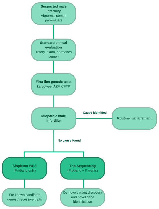
In many men with abnormal semen parameters and normal standard genetic findings, the molecular cause remains unidentified because the available tests were not designed to detect the full range of monogenic disorders [4,6]. The difference between the probable prevalence of monogenic disease and the detection rate of routine testing supports the use of more comprehensive approaches such as WES. Proposed genetic tests in male infertility are summarised in Figure 1.
Figure 1.
Proposed diagnostic algorithm based on genetic tests in infertile males. Figure created using Canva Pro (Canva Pty Ltd., Sydney, Australia; https://www.canva.com).
5. Methodological Principles of WES
WES involves the selective enrichment and sequencing of exons, which comprise the protein-coding regions of the genome. It is one of several genomic diagnostic approaches, alongside whole-genome sequencing (WGS) and targeted gene panels, each of which differs in scope, cost, and clinical application [13]. Although the exome accounts for only 1–2% of the genome, it contains most variants currently known to cause Mendelian disease. WES achieves a broad diagnostic yield of approximately 25–35% across diverse populations with suspected rare diseases [14], but the diagnostic yield specific to idiopathic non-obstructive azoospermia (NOA) after negative standard first-line testing (karyotype, AZF, and CFTR variants) typically rests between 10% and 15% [6,7].
WES serves as a middle ground between WGS and targeted gene panels (Table 2). It is less expensive and generally easier to analyse than WGS but does not comprehensively cover non-coding or regulatory regions and is less reliable for detecting large structural rearrangements. WGS therefore usually has a somewhat higher diagnostic yield, although the difference is modest in many clinical indications [14]. Targeted panels provide greater sequencing depth at selected loci, providing high coverage at a low cost, but they cannot identify candidate genes outside the predefined list. Thus, while suitable for well-defined phenotypes, their diagnostic value is restricted in highly heterogeneous conditions [13]. The clinical selection strategy increasingly favours WES for conditions such as male infertility; a genome-wide approach not only offers broad coverage but importantly allows for periodic data re-analysis as new gene-disease associations are rapidly established in the literature, thereby preventing diagnostic dead-ends [4,14,15].
Table 2.
Comparison of targeted genetic panels testing vs. WES and WGS.
In male infertility, WES combined with the analysis of single-nucleotide variants (SNVs) and copy-number variants (CNVs) has been proposed as a first-line strategy that could consolidate several separate genetic investigations. Conventional karyotyping would nevertheless remain necessary for the detection of balanced translocations and low-level mosaicism, which are not reliably identified by WES [7].
Once the physical sequencing is complete, the resulting data must be processed to identify causative mutations. The bioinformatic workflow converts the raw sequencing reads into a clinically relevant set of candidate variants. It includes alignment to the reference genome, calling of SNVs and small insertions or deletions, and sequential filtering based on population frequency, predicted functional effects, compatibility with the expected inheritance pattern, and the strength of the association between the affected gene and the patient’s phenotype.
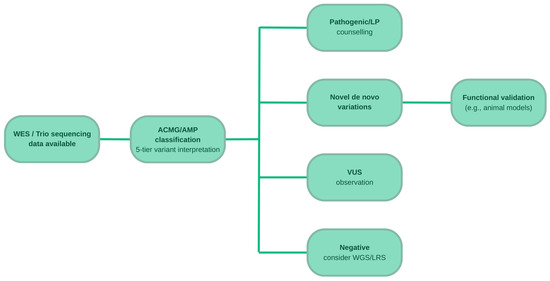
Variants are subsequently classified according to the American College of Medical Genetics and Genomics/Association for Molecular Pathology (ACMG/AMP) framework as pathogenic, likely pathogenic, of uncertain significance, likely benign, or benign (Figure 2).
Figure 2.
Proposed algorithm of male infertility diagnosis according to the type of variant. Variant classification based on American College of Medical Genetics and Genomics/Association for Molecular Pathology recommendations. Figure created using Canva Pro (Canva Pty Ltd., Sydney, Australia; https://www.canva.com).
This classification system is routinely used in WES studies of male infertility. In an Italian cohort, pathogenic or likely pathogenic variants identified using this approach provided a molecular diagnosis in 12% of patients with previously idiopathic infertility [6]. In clinical practice, reports generally focus on pathogenic and likely pathogenic variants. Variants of uncertain significance (VUS), which account for a substantial proportion of WES findings, require additional evidence and should not independently guide clinical decisions. Consistent application of classification criteria is particularly important when evaluating variants in newly proposed candidate genes.
Study design also affects the types of variants that can be detected. Most WES studies of male infertility have used case–control designs in which variants identified in patients are compared with population databases or cohorts of fertile men. This approach is primarily suited to identifying recessive and X-linked variants. Trio sequencing, which analyses the patient and both parents, permits direct identification of de novo variants that are absent from parental genomes. A study of 185 trios comprising infertile men and their unaffected parents found significant enrichment of deleterious de novo variants in genes intolerant to loss of function. These findings strongly imply that de novo variants may account for a previously underestimated proportion of severe male infertility [15].
6. Genetic Basis of Infertility Revealed by WES
WES studies have contributed to the systematic identification and classification of genes associated with specific male-infertility phenotypes. A systematic review by the International Male Infertility Genomics Consortium (IMIGC) used an adapted semi-quantitative framework analogous to that applied by ClinGen to assess the strength of gene–disease associations. The review identified 120 genes with moderate, strong, or definitive evidence of association with 104 infertility phenotypes [8]. This framework helps distinguish genes with established clinical relevance from candidate genes supported only by isolated case reports, which is essential when designing diagnostic panels and interpreting WES findings [3].
The genes identified through WES can be considered in three principal phenotypic groups (Table 3). Clinical validity of these genes is further supported by corresponding functional validation in animal models (Table 4). The first includes quantitative disorders of spermatogenesis, particularly NOA and severe oligozoospermia. Genes associated with meiotic arrest, including TEX11, STAG3, MEIOB, and M1AP, are prominent in this group. Pathogenic variants in these genes may prevent the production of mature gametes despite preservation of the spermatogonial pool [3,16].
The second group comprises disorders of sperm motility. Multiple morphological abnormalities of the sperm flagella (MMAF) are among the best-characterised phenotypes in this category. Since DNAH1 gene was first associated with MMAF, more than 40 genes encoding proteins involved in the axoneme, centrosome, and intraflagellar transport have been described. These include members of the CFAP family such as CFAP43, CFAP44, CFAP69 and the genes DNAH2, DNAH6, and DNAH17 [17].
The third group includes abnormalities of sperm-head morphology. These encompass well-characterised monogenic phenotypes, including globozoospermia, most associated with deletion of DPY19L2 gene, and macrozoospermia, caused by recurrent variants in AURKC gene. WES has also identified candidate genes associated with the broader and genetically heterogeneous phenotype of teratozoospermia [18].
WES can additionally identify pre-testicular causes of infertility associated with dysfunction of the hypothalamic–pituitary–gonadal axis. Congenital hypogonadotropic hypogonadism (CHH), including normosmic CHH and Kallmann syndrome, results from GnRH deficiency and is one of the few genetically determined causes of male infertility for which causal hormonal treatment is available. More than 40 genes have been associated with CHH, including ANOS1, GNRHR, FGFR1, KISS1R, and TAC3. Oligogenic inheritance is relatively common: in some patients, the phenotype results from variants in two or more genes. WES is well suited to detecting this type of genetic architecture because its analysis is not restricted to a small predefined panel [19].
However, these clinical phenotypes frequently overlap in practice. Some genes are associated with combined abnormalities of sperm number, motility, and morphology, reflecting molecular pathways involved in multiple stages of spermatogenesis and spermiogenesis [4].
Table 3.
Clinical validity of candidate gene–disease relationships (GDRs) for male-infertility phenotypes, comparing the systematic reviews of Houston et al. [8] and Zhao et al. [20]. Score/classification pairs follow the five-tier system of Smith et al. [21]: no evidence (<3), limited (3–8), moderate (9–12), strong (13–15), definitive (>15).
Table 4.
Animal-model support for each gene–disease relationship, independently verified against primary literature.
7. Diagnostic Yield of WES Across Different Phenotypes
The diagnostic yield of WES in male infertility depends on the phenotype, cohort size and selection criteria, and the bioinformatic and interpretative methods used. When defining the cohort strictly as patients with true idiopathic NOA who remain undiagnosed after standard testing, the pooled diagnostic yield of WES is approximately 15% [50]. However, substantially higher detection rates (ranging from 27% to over 50%) have been reported for rare, highly selected qualitative phenotypes, particularly in cohorts enriched for consanguinity [4,51].
NOA is the most extensively studied phenotype. A meta-analysis of nine studies including 1728 men with idiopathic NOA estimated a pooled diagnostic yield of 15% (95% CI: 10–20%). Because of considerable methodological heterogeneity, the authors cautioned that this estimate represents an average across different clinical settings rather than a precise value applicable to every population [50]. The largest individual WES cohort in NOA was reported by the international GEMINI consortium and included more than 1000 men with clinically confirmed disease. A probable recessive cause was identified in approximately 20% of participants. Twenty-one genes received additional support through a two-stage variant-burden analysis involving an independent cohort of more than 2000 cases and nearly 11,600 fertile controls [51].
Similar yields have been reported for other quantitative spermatogenic disorders. In a cohort of 521 men with idiopathic spermatogenic failure analysed using a panel of 638 candidate genes, a molecular diagnosis was established in 12% of participants. The yield did not differ significantly between men with azoospermia and those with oligozoospermia [52]. A diagnostic yield of 12% was also reported in the Italian cohort described above [6]. Together, these studies suggest that the yield in severe quantitative disorders of spermatogenesis is commonly approximately 10–15%.
Estimates are more variable for qualitative sperm abnormalities. WES identified a molecular diagnosis in 14.6% of patients with unexplained heterogeneous abnormalities of the sperm head. In small, highly selected cohorts with acephalic spermatozoa syndrome, reported yields ranged from 27.3% to 58.3%. Studies of asthenozoospermia and MMAF have likewise produced variable results; however, higher diagnostic yields in MMAF are often driven by cohorts enriched for consanguinity and the presence of highly recurrent variants within specific gene families, such as DNAH and CFAP [4].
Diagnostic yield varies considerably between centres primarily due to differing eligibility criteria, with some cohorts restricted to strictly defined, true idiopathic infertility, while others include broader, more heterogeneous populations (Table 5). Studies also differ in the number and selection of analysed genes, from limited sets of well-established genes to candidate panels containing several hundred genes. In rare qualitative phenotypes, small cohorts make diagnostic-yield estimates particularly sensitive to individual cases and limit their generalisability [50].
Table 5.
Diagnostic yield of WES across infertility phenotypes.
8. Clinical Significance of an Identified Molecular Diagnosis
Identifying the molecular cause of infertility directly alters clinical management by providing clear prognostic insights and guiding therapeutic interventions [53]. Establishing a precise diagnosis enables informative genetic counselling, as patients carrying pathogenic variants face a risk of transmitting male infertility to their potential progeny if assisted reproduction is successful [54].
A molecular diagnosis also serves as a powerful prognostic tool for predicting the likelihood of successful sperm retrieval by testicular sperm extraction (TESE) [50,53]. Pathogenic variants in meiotic genes are associated with extremely low retrieval rates. For instance, reported TESE success rates are as low as 8.3% in cases of Sertoli cell-only syndrome and 8.7% in meiotic arrest [50]. Incorporating well-validated genes into pre-TESE prognostic assessments adds substantial clinical utility, as men with specific genetic defects in meiotic pathways are theoretically unlikely to yield viable sperm [50]. Notable genes with a defined biological basis for poor TESE outcomes include:
TEX11: Mutations in this gene impair the assembly and function of the synaptonemal complex. This causes severe defects in chromosome synapsis during the pachytene stage, resulting in meiotic arrest and predominantly negative TESE outcomes [50].
STAG3: This gene encodes a component of the meiosis-specific cohesin complex that is essential for maintaining chromatid cohesion, DNA repair, and synapsis between homologous chromosomes [54].
MSH4: This gene plays a critical biological role in the reciprocal recombination and segregation of homologous chromosomes during meiosis [54].
TERB1, MEIOB, and SPO11: These are critical meiotic genes; loss-of-function mutations disrupting these genes have been strongly linked to meiotic arrest and uniformly negative TESE outcomes in studied cohorts [53].
Histopathological evidence of meiotic arrest strongly correlates with variants in these meiosis-involved genes, reinforcing the diagnostic and prognostic relevance of these findings [53]. Identifying a relevant variant before surgery can therefore help patients avoid an invasive procedure when the probability of success is minimal [50,54]. This approach not only spares patients from futile surgeries but also reduces their exposure to potential long-term adverse effects of TESE, such as androgen deficiency. Ultimately, a known genetic prognosis allows for more targeted clinical decision-making, including the potential for earlier inclusion in donor programs [53,54].
9. Challenges and Limitations
While WES offers undeniable diagnostic value, its routine use in male infertility remains limited by methodological, organisational, and ethical challenges.
The sheer volume of VUS remains the primary interpretative bottleneck. In the Italian cohort described above, only 12 of 189 classified variants were considered pathogenic or likely pathogenic, whereas most of the remaining variants were classified as VUS [6]. This imbalance reflects the relatively recent development of research into monogenic male infertility and the limited functional validation of newly identified variants. To cross this functional validation gap, the gold standard remains a mouse knockout model that faithfully reproduces the predicted infertility or spermatogenic phenotype; genes with strong human statistical evidence but lacking animal model support frequently remain classified as candidates [8,16]. The field also increasingly employs a reverse logic: when a mouse knockout causes infertility but lacks a human report, it becomes a strong candidate for a novel human male infertility gene [16]. Additionally, variant interpretation in male infertility requires adaptation of standard ACMG/AMP rules. Computational evidence leans heavily on conservation and in silico predictors, while loss-of-function (LoF) intolerance metrics, such as the pLI score, significantly strengthen a LoF claim [15]. A variant’s reporting is ultimately dictated by the gene’s disease-validity tier, underscoring the importance of IMIGC classifications [3,8]. VUS should therefore not be used independently to guide decisions such as abandoning TESE or changing the infertility-treatment strategy. Their uncertain interpretation limits the clinical utility of some WES findings and may require re-evaluation as new evidence becomes available.
Population ancestry represents an additional challenge in genomic variant interpretation. Reference population databases, including gnomAD, provide essential allele-frequency information for variant classification; however, the representation of global populations remains uneven. Consequently, the absence or low frequency of a variant in a reference database may be difficult to interpret when the relevant population is underrepresented, potentially contributing to uncertainty in variant classification. Ancestry-aware interpretation and consideration of population-specific allele frequencies are therefore important when applying population-based ACMG/AMP criteria. Expanding the representation of diverse populations in genomic reference databases and male infertility cohorts will be essential for improving the accuracy and equity of genetic diagnosis [55].
Beyond these interpretative challenges, there are technological limitations to standard short-read WES when applied to NOA. Because WES relies almost exclusively on blood-derived DNA, it is fundamentally blind to gonadal or testicular mosaicism, which may drive spermatogenic failure in some men [13]. Short-read WES also struggles significantly with the detection of complex structural variants and copy-number changes, and it cannot evaluate deep-intronic regulatory or non-coding variants that might impact gene expression [4,13]. While the promise of an “all-in-one” genomic test is appealing, standard short-read WES cannot reliably call common structural rearrangements such as partial AZFc microdeletions (e.g., gr/gr or b2/b3 deletions); thus, dedicated calling algorithms, sequence-tagged site PCR (STS-PCR), or digital droplet PCR remain essential alongside WES [7,56].
The absence of a standardised set of genes for WES analysis presents a further challenge. Despite the work of initiatives such as the IMIGC and the adaptation of ClinGen-based methods [8], no uniform and widely accepted clinical gene set for male infertility has been established [54]. Centres differ in the number and choice of genes analysed, contributing to variation in reported diagnostic yields.
Cost and access also limit implementation. Although sequencing costs continue to decrease, and cost-effectiveness studies in other rare diseases suggest that WES may reduce healthcare expenditure per additional diagnosis [57], access remains geographically uneven and dependent on national reimbursement policies [58]. Some centres also lack the bioinformatic infrastructure and clinical genetics expertise needed to interpret results in a condition with substantial phenotypic heterogeneity.
Ethical concerns arise from the potential identification of secondary findings unrelated to infertility but relevant to the patient’s health, including variants associated with cancer predisposition [59]. This issue may be particularly relevant given reports of increased cancer risk among infertile men [2,3,6]. Pre-test genetic counselling should address the possibility of secondary findings and establish the patient’s preferences regarding disclosure. Consequently, pre-test counselling must be exhaustive, adding complexity to the informed consent process.
10. Future Perspectives
The next era of andrological genetics will inevitably rely on converging multi-omics data and falling sequencing costs.
One area of development is the use of “all-in-one” strategies that combine SNV and CNV analysis within a single WES assay. Such strategies may consolidate much of the current first-line genetic pathway, including testing for AZF microdeletions and CFTR variants, while reducing the time and cost associated with separate investigations. Conventional karyotyping would still be required to detect balanced translocations and low-level mosaicism [7].
WES may also be integrated with transcriptomic, epigenomic, and proteomic data. Systematic reviews indicate that different omics approaches identify largely distinct and only partially overlapping molecular factors associated with infertility. Analysis of coding variants alone is therefore unlikely to capture the full spectrum of causes of spermatogenic failure. Combining WES with transcriptomic data from testicular biopsies or epigenetic profiles of sperm may help resolve cases that remain unexplained after exome sequencing [60,61].
WGS and long-read sequencing (LRS) may further expand diagnostic capability. By covering non-coding and regulatory regions, WGS could eventually serve as a comprehensive first-line test encompassing abnormalities currently assessed by karyotyping, AZF microdeletion analysis, and WES [62]. LRS enables the analysis of long, continuous DNA fragments and may improve the detection of complex structural rearrangements, discrimination between highly homologous genes and pseudogenes, and determination of the cis or trans configuration of variants without parental samples. In an early application relevant to infertile men, LRS increased molecular detection by more than 30% relative to a standard NGS panel within the group of variants analysed [63]. Although cost and bioinformatic complexity currently limit routine use, LRS may complement WES when a structural cause is suspected or when short-read sequencing produces an ambiguous result.
11. Conclusions
Whole-exome sequencing is an effective tool for investigating the molecular basis of idiopathic male infertility and addresses part of the diagnostic gap left by standard genetic testing. Although its diagnostic yield varies according to phenotype and study methodology, a confirmed molecular diagnosis can improve genetic counselling, inform assessment of risk to offspring, help predict the outcome of TESE or micro-TESE, and support treatment planning using assisted reproductive technologies.
Important limitations remain, including the high frequency of VUS, the lack of standardised gene sets and analytical pipelines, unequal access to testing, and ethical concerns related to secondary findings. Future diagnostic strategies are likely to combine WES-based SNV and CNV analysis with other omics approaches, WGS, and LRS. With further validation and standardisation, comprehensive genomic profiling may become a part of routine diagnostic pathways for male infertility.
Supplementary Materials
The following supporting information can be downloaded at: https://www.mdpi.com/article/10.3390/jcm15187281/s1, Document S1: SANRA (Scale for the Assessment of Narrative Review Articles) checklist. Reference [64] is cited in the Supplementary Materials.
Author Contributions
Conceptualization, F.K., I.Z., I.K., P.M. and W.Z.; methodology, F.K., A.G., I.Z., I.K. and P.M.; literature search and data curation, F.K., A.G., O.B., M.Ś. and J.G.; writing—original draft preparation, F.K.; writing—review and editing, F.K., A.G., O.B., M.Ś., J.G., I.Z., I.K. and P.M.; supervision, I.Z., I.K., P.M. and W.Z. All authors have read and agreed to the published version of the manuscript.
Funding
This research received no external funding.
Institutional Review Board Statement
Not applicable.
Informed Consent Statement
Not applicable.
Data Availability Statement
No new data were created or analyzed in this study. Data sharing is not applicable to this article.
Acknowledgments
The authors would like to acknowledge the Medical University of Lublin for providing access to the necessary research databases and literature.
Conflicts of Interest
The authors declare no conflicts of interest.
Abbreviations
The following abbreviations are used in this manuscript:
| ACMG/AMP | American College of Medical Genetics and Genomics/Association for Molecular Pathology |
| AD | Autosomal dominant |
| AR | Autosomal recessive |
| ART | Assisted reproductive technologies |
| AUA/ASRM | American Urological Association/American Society for Reproductive Medicine |
| AZF | Azoospermia factor |
| CBAVD | Congenital bilateral absence of the vas deferens |
| CHH | Congenital hypogonadotropic hypogonadism |
| CI | Confidence interval |
| ClinGen | Clinical Genome Resource |
| CNV | Copy-number variant |
| CRISPR/Cas9 | Clustered regularly interspaced short palindromic repeats/CRISPR-associated protein 9 |
| DNA | Deoxyribonucleic acid |
| DSB | DNA double-strand break |
| FSH | Follicle-stimulating hormone |
| GDR | Gene-disease relationship |
| gnomAD | Genome Aggregation Database |
| GnRH | Gonadotropin-releasing hormone |
| ICSI | Intracytoplasmic sperm injection |
| IMIGC | International Male Infertility Genomics Consortium |
| LOEUF | Loss-of-function observed/expected upper bound fraction |
| LRS | Long-read sequencing |
| Micro-TESE | Microdissection testicular sperm extraction |
| MMAF | Multiple morphological abnormalities of the sperm flagella |
| NGS | Next-generation sequencing |
| NOA | Non-obstructive azoospermia |
| OA | Obstructive azoospermia |
| OMIM | Online Mendelian Inheritance in Man |
| PCR | Polymerase chain reaction |
| pLI | Probability of loss-of-function intolerance |
| SNV | Single-nucleotide variant |
| STS-PCR | Sequence-tagged site polymerase chain reaction |
| TESE | Testicular sperm extraction |
| VUS | Variant of uncertain significance |
| WES | Whole-exome sequencing |
| WGS | Whole-genome sequencing |
| XL | X-linked |
References
- Infertility Prevalence Estimates, 1990–2021, 1st ed.; World Health Organization: Geneva, Switzerland, 2023.
- Fallara, G.; Pozzi, E.; Belladelli, F.; Boeri, L.; Capogrosso, P.; Corona, G.; D’Arma, A.; Alfano, M.; Montorsi, F.; Salonia, A. A Systematic Review and Meta-Analysis on the Impact of Infertility on Men’s General Health. Eur. Urol. Focus 2024, 10, 98–106. [Google Scholar] [CrossRef] [Scilit] [PubMed]
- Stallmeyer, B.; Dicke, A.-K.; Tüttelmann, F. How Exome Sequencing Improves the Diagnostics and Management of Men with Non-Syndromic Infertility. Andrology 2025, 13, 1011–1024. [Google Scholar] [CrossRef] [Scilit] [PubMed]
- Riera-Escamilla, A.; Nagirnaja, L. Utility of Exome Sequencing in Primary Spermatogenic Disorders: From Research to Diagnostics. Andrology 2025, 13, 999–1010. [Google Scholar] [CrossRef] [Scilit] [PubMed]
- Braverman, A.M.; Davoudian, T.; Levin, I.K.; Bocage, A.; Wodoslawsky, S. Depression, Anxiety, Quality of Life, and Infertility: A Global Lens on the Last Decade of Research. Fertil. Steril. 2024, 121, 379–383. [Google Scholar] [CrossRef] [Scilit] [PubMed]
- Quarantani, G.; Sorgente, A.; Alfano, M.; Pipitone, G.B.; Boeri, L.; Pozzi, E.; Belladelli, F.; Pederzoli, F.; Ferrara, A.M.; Montorsi, F.; et al. Whole Exome Data Prioritization Unveils the Hidden Weight of Mendelian Causes of Male Infertility. A Report from the First Italian Cohort. PLoS ONE 2023, 18, e0288336. [Google Scholar] [CrossRef] [Scilit] [PubMed]
- Oud, M.S.; de Leeuw, N.; Smeets, D.F.C.M.; Ramos, L.; van der Heijden, G.W.; Timmermans, R.G.J.; van de Vorst, M.; Hofste, T.; Kempers, M.J.E.; Stokman, M.F.; et al. Innovative All-in-One Exome Sequencing Strategy for Diagnostic Genetic Testing in Male Infertility: Validation and 10-Month Experience. Andrology 2025, 13, 1078–1092. [Google Scholar] [CrossRef] [Scilit] [PubMed]
- Houston, B.J.; Riera-Escamilla, A.; Wyrwoll, M.J.; Salas-Huetos, A.; Xavier, M.J.; Nagirnaja, L.; Friedrich, C.; Conrad, D.F.; Aston, K.I.; Krausz, C.; et al. A Systematic Review of the Validated Monogenic Causes of Human Male Infertility: 2020 Update and a Discussion of Emerging Gene–Disease Relationships. Hum. Reprod. Update 2021, 28, 15–29. [Google Scholar] [CrossRef] [Scilit] [PubMed]
- Tharakan, T.; Luo, R.; Jayasena, C.N.; Minhas, S. Non-Obstructive Azoospermia: Current and Future Perspectives. Fac. Rev. 2021, 10, 7. [Google Scholar] [CrossRef] [Scilit] [PubMed]
- Bhattacharya, I.; Sharma, S.S.; Majumdar, S.S. Etiology of Male Infertility: An Update. Reprod. Sci. 2024, 31, 942–965. [Google Scholar] [CrossRef] [Scilit] [PubMed]
- Imran, M.; Zia, R.; Arshad, M.; Fayyaz, F.; Haider, T.; Tabraiz, A.; Arshad, I.; Sharif, M.A.; Javed, B. Exploration of the Genetic and Environmental Determinants of Male Infertility: A Comprehensive Review. Egypt. J. Med. Hum. Genet. 2025, 26, 68. [Google Scholar] [CrossRef] [Scilit]
- Brannigan, R.E.; Hermanson, L.; Kaczmarek, J.; Kim, S.K.; Kirkby, E.; Tanrikut, C. Updates to Male Infertility: AUA/ASRM Guideline (2024). J. Urol. 2024, 212, 789–799. [Google Scholar] [CrossRef] [Scilit] [PubMed]
- Kernohan, K.D.; Boycott, K.M. The Expanding Diagnostic Toolbox for Rare Genetic Diseases. Nat. Rev. Genet. 2024, 25, 401–415. [Google Scholar] [CrossRef] [Scilit] [PubMed]
- Albuquerque, A.L.B.; dos Santos, G.G.; Sadok, S.H.; Antonello, B.B.; de Jesus, L.M.; de Carvalho, M.E.A.; Mutarelli, A.; Ribeiro, P.V.Z. Diagnostic Yield of Genome Sequencing Versus Exome Sequencing in Pediatric Patients with Rare Phenotypes: A Systematic Review and Meta-Analysis. Am. J. Med. Genet. Part A 2025, 197, e64146. [Google Scholar] [CrossRef] [Scilit] [PubMed]
- Oud, M.S.; Smits, R.M.; Smith, H.E.; Mastrorosa, F.K.; Holt, G.S.; Houston, B.J.; de Vries, P.F.; Alobaidi, B.K.S.; Batty, L.E.; Ismail, H.; et al. A de Novo Paradigm for Male Infertility. Nat. Commun. 2022, 13, 154. [Google Scholar] [CrossRef] [Scilit] [PubMed]
- Kasak, L.; Laan, M. Monogenic Causes of Non-Obstructive Azoospermia: Challenges, Established Knowledge, Limitations and Perspectives. Hum. Genet. 2021, 140, 135–154. [Google Scholar] [CrossRef] [Scilit] [PubMed]
- Zhou, Y.; Yu, S.; Zhang, W. The Molecular Basis of Multiple Morphological Abnormalities of Sperm Flagella and Its Impact on Clinical Practice. Genes 2024, 15, 1315. [Google Scholar] [CrossRef] [Scilit] [PubMed]
- Li, Y.; Wang, Y.; Wen, Y.; Zhang, T.; Wang, X.; Jiang, C.; Zheng, R.; Zhou, F.; Chen, D.; Yang, Y.; et al. Whole-Exome Sequencing of a Cohort of Infertile Men Reveals Novel Causative Genes in Teratozoospermia That Are Chiefly Related to Sperm Head Defects. Hum. Reprod. 2022, 37, 152–177. [Google Scholar] [CrossRef] [Scilit] [PubMed]
- Louden, E.D.; Poch, A.; Kim, H.-G.; Ben-Mahmoud, A.; Kim, S.-H.; Layman, L.C. Genetics of Hypogonadotropic Hypogonadism—Human and Mouse Genes, Inheritance, Oligogenicity, and Genetic Counseling. Mol. Cell. Endocrinol. 2021, 534, 111334. [Google Scholar] [CrossRef] [Scilit] [PubMed]
- Zhao, Q.; Peng, H.; Chen, J.; Zhang, H.; Ma, Y.; Jiang, H. A Systematic Review and Evidence Assessment of Monogenic Gene-Disease Relationships in Human Male Infertility. Front. Endocrinol. 2025, 16, 1643543. [Google Scholar] [CrossRef] [Scilit] [PubMed]
- Smith, E.D.; Radtke, K.; Rossi, M.; Shinde, D.N.; Darabi, S.; El-Khechen, D.; Powis, Z.; Helbig, K.; Waller, K.; Grange, D.K.; et al. Classification of Genes: Standardized Clinical Validity Assessment of Gene–Disease Associations Aids Diagnostic Exome Analysis and Reclassifications. Hum. Mutat. 2017, 38, 600–608. [Google Scholar] [CrossRef] [Scilit] [PubMed]
- Yang, F.; Gell, K.; van der Heijden, G.W.; Eckardt, S.; Leu, N.A.; Page, D.C.; Benavente, R.; Her, C.; Höög, C.; McLaughlin, K.J.; et al. Meiotic Failure in Male Mice Lacking an X-Linked Factor. Genes Dev. 2008, 22, 682–691. [Google Scholar] [CrossRef] [Scilit] [PubMed]
- Winters, T.; McNicoll, F.; Jessberger, R. Meiotic Cohesin STAG3 Is Required for Chromosome Axis Formation and Sister Chromatid Cohesion. EMBO J. 2014, 33, 1256–1270. [Google Scholar] [CrossRef] [Scilit] [PubMed]
- Hopkins, J.; Hwang, G.; Jacob, J.; Sapp, N.; Bedigian, R.; Oka, K.; Overbeek, P.; Murray, S.; Jordan, P.W. Meiosis-Specific Cohesin Component, Stag3 Is Essential for Maintaining Centromere Chromatid Cohesion, and Required for DNA Repair and Synapsis between Homologous Chromosomes. PLoS Genet. 2014, 10, e1004413. [Google Scholar] [CrossRef] [Scilit] [PubMed]
- Bolcun-Filas, E.; Speed, R.; Taggart, M.; Grey, C.; de Massy, B.; Benavente, R.; Cooke, H.J. Mutation of the Mouse Syce1 Gene Disrupts Synapsis and Suggests a Link between Synaptonemal Complex Structural Components and DNA Repair. PLoS Genet. 2009, 5, e1000393. [Google Scholar] [CrossRef] [Scilit] [PubMed]
- Yuan, L.; Liu, J.-G.; Zhao, J.; Brundell, E.; Daneholt, B.; Höög, C. The Murine SCP3 Gene Is Required for Synaptonemal Complex Assembly, Chromosome Synapsis, and Male Fertility. Mol. Cell 2000, 5, 73–83. [Google Scholar] [CrossRef] [Scilit] [PubMed]
- Xu, H.; Beasley, M.D.; Warren, W.D.; van der Horst, G.T.J.; McKay, M.J. Absence of Mouse REC8 Cohesin Promotes Synapsis of Sister Chromatids in Meiosis. Dev. Cell 2005, 8, 949–961. [Google Scholar] [CrossRef] [Scilit] [PubMed]
- Bannister, L.A.; Reinholdt, L.G.; Munroe, R.J.; Schimenti, J.C. Positional Cloning and Characterization of Mouse Mei8, a Disrupted Allele of the Meiotic Cohesin Rec8. Genesis 2004, 40, 184–194. [Google Scholar] [CrossRef] [Scilit] [PubMed]
- Souquet, B.; Abby, E.; Hervé, R.; Finsterbusch, F.; Tourpin, S.; Le Bouffant, R.; Duquenne, C.; Messiaen, S.; Martini, E.; Bernardino-Sgherri, J.; et al. MEIOB Targets Single-Strand DNA and Is Necessary for Meiotic Recombination. PLoS Genet. 2013, 9, e1003784. [Google Scholar] [CrossRef] [Scilit] [PubMed]
- Luo, M.; Yang, F.; Leu, N.A.; Landaiche, J.; Handel, M.A.; Benavente, R.; La Salle, S.; Wang, P.J. MEIOB Exhibits Single-Stranded DNA-Binding and Exonuclease Activities and Is Essential for Meiotic Recombination. Nat. Commun. 2013, 4, 2788. [Google Scholar] [CrossRef] [Scilit] [PubMed]
- Libby, B.J.; De La Fuente, R.; O’Brien, M.J.; Wigglesworth, K.; Cobb, J.; Inselman, A.; Eaker, S.; Handel, M.A.; Eppig, J.J.; Schimenti, J.C. The Mouse Meiotic Mutation Mei1 Disrupts Chromosome Synapsis with Sexually Dimorphic Consequences for Meiotic Progression. Dev. Biol. 2002, 242, 174–187. [Google Scholar] [CrossRef] [Scilit] [PubMed]
- Libby, B.J.; Reinholdt, L.G.; Schimenti, J.C. Positional Cloning and Characterization of Mei1, a Vertebrate-Specific Gene Required for Normal Meiotic Chromosome Synapsis in Mice. Proc. Natl. Acad. Sci. USA 2003, 100, 15706–15711. [Google Scholar] [CrossRef] [Scilit] [PubMed]
- Arango, N.A.; Li, L.; Dabir, D.; Nicolau, F.; Pieretti-Vanmarcke, R.; Koehler, C.; McCarrey, J.R.; Lu, N.; Donahoe, P.K. Meiosis I Arrest Abnormalities Lead to Severe Oligozoospermia in Meiosis 1 Arresting Protein (M1ap)-Deficient Mice. Biol. Reprod. 2013, 88, 76. [Google Scholar] [CrossRef] [Scilit] [PubMed]
- Tang, S.; Wang, X.; Li, W.; Yang, X.; Li, Z.; Liu, W.; Li, C.; Zhu, Z.; Wang, L.; Wang, J.; et al. Biallelic Mutations in CFAP43 and CFAP44 Cause Male Infertility with Multiple Morphological Abnormalities of the Sperm Flagella. Am. J. Hum. Genet. 2017, 100, 854–864. [Google Scholar] [CrossRef] [Scilit] [PubMed]
- Coutton, C.; Vargas, A.S.; Amiri-Yekta, A.; Kherraf, Z.-E.; Ben Mustapha, S.F.; Le Tanno, P.; Wambergue-Legrand, C.; Karaouzène, T.; Martinez, G.; Crouzy, S.; et al. Mutations in CFAP43 and CFAP44 Cause Male Infertility and Flagellum Defects in Trypanosoma and Human. Nat. Commun. 2018, 9, 686. [Google Scholar] [CrossRef] [Scilit] [PubMed]
- He, X.; Li, W.; Wu, H.; Lv, M.; Liu, W.; Liu, C.; Zhu, F.; Li, C.; Fang, Y.; Yang, C.; et al. Novel Homozygous CFAP69 Mutations in Humans and Mice Cause Severe Asthenoteratospermia with Multiple Morphological Abnormalities of the Sperm Flagella. J. Med. Genet. 2019, 56, 96–103. [Google Scholar] [CrossRef] [Scilit] [PubMed]
- Neesen, J. Disruption of an Inner Arm Dynein Heavy Chain Gene Results in Asthenozoospermia and Reduced Ciliary Beat Frequency. Hum. Mol. Genet. 2001, 10, 1117–1128. [Google Scholar] [CrossRef] [Scilit] [PubMed]
- Ben Khelifa, M.; Coutton, C.; Zouari, R.; Karaouzène, T.; Rendu, J.; Bidart, M.; Yassine, S.; Pierre, V.; Delaroche, J.; Hennebicq, S.; et al. Mutations in DNAH1, Which Encodes an Inner Arm Heavy Chain Dynein, Lead to Male Infertility from Multiple Morphological Abnormalities of the Sperm Flagella. Am. J. Hum. Genet. 2014, 94, 95–104. [Google Scholar] [CrossRef] [Scilit] [PubMed]
- Liu, C.; Miyata, H.; Gao, Y.; Sha, Y.; Tang, S.; Xu, Z.; Whitfield, M.; Patrat, C.; Wu, H.; Dulioust, E.; et al. Bi-Allelic DNAH8 Variants Lead to Multiple Morphological Abnormalities of the Sperm Flagella and Primary Male Infertility. Am. J. Hum. Genet. 2020, 107, 330–341. [Google Scholar] [CrossRef] [Scilit] [PubMed]
- Zhang, B.; Khan, I.; Liu, C.; Ma, A.; Khan, A.; Zhang, Y.; Zhang, H.; Kakakhel, M.B.S.; Zhou, J.; Zhang, W.; et al. Novel Loss-of-function Variants in DNAH17 Cause Multiple Morphological Abnormalities of the Sperm Flagella in Humans and Mice. Clin. Genet. 2021, 99, 176–186. [Google Scholar] [CrossRef] [Scilit] [PubMed]
- Pierre, V.; Martinez, G.; Coutton, C.; Delaroche, J.; Yassine, S.; Novella, C.; Pernet-Gallay, K.; Hennebicq, S.; Ray, P.F.; Arnoult, C. Absence of Dpy19l2, a New Inner Nuclear Membrane Protein, Causes Globozoospermia in Mice by Preventing the Anchoring of the Acrosome to the Nucleus. Development 2012, 139, 2955–2965. [Google Scholar] [CrossRef] [Scilit] [PubMed]
- Fujihara, Y.; Oji, A.; Larasati, T.; Kojima-Kita, K.; Ikawa, M. Human Globozoospermia-Related Gene Spata16 Is Required for Sperm Formation Revealed by CRISPR/Cas9-Mediated Mouse Models. Int. J. Mol. Sci. 2017, 18, 2208. [Google Scholar] [CrossRef] [Scilit] [PubMed]
- Lin, Y.-N.; Roy, A.; Yan, W.; Burns, K.H.; Matzuk, M.M. Loss of Zona Pellucida Binding Proteins in the Acrosomal Matrix Disrupts Acrosome Biogenesis and Sperm Morphogenesis. Mol. Cell. Biol. 2007, 27, 6794–6805. [Google Scholar] [CrossRef] [Scilit] [PubMed]
- Kimmins, S.; Crosio, C.; Kotaja, N.; Hirayama, J.; Monaco, L.; Höög, C.; van Duin, M.; Gossen, J.A.; Sassone-Corsi, P. Differential Functions of the Aurora-B and Aurora-C Kinases in Mammalian Spermatogenesis. Mol. Endocrinol. 2007, 21, 726–739. [Google Scholar] [CrossRef] [Scilit] [PubMed]
- Shang, Y.; Zhu, F.; Wang, L.; Ouyang, Y.-C.; Dong, M.-Z.; Liu, C.; Zhao, H.; Cui, X.; Ma, D.; Zhang, Z.; et al. Essential Role for SUN5 in Anchoring Sperm Head to the Tail. eLife 2017, 6, e28199. [Google Scholar] [CrossRef] [Scilit] [PubMed]
- Zhang, Y.; Yang, L.; Huang, L.; Liu, G.; Nie, X.; Zhang, X.; Xing, X. SUN5 Interacting With Nesprin3 Plays an Essential Role in Sperm Head-to-Tail Linkage: Research on Sun5 Gene Knockout Mice. Front. Cell Dev. Biol. 2021, 9, 684826. [Google Scholar] [CrossRef] [Scilit] [PubMed]
- Zhu, F.; Liu, C.; Wang, F.; Yang, X.; Zhang, J.; Wu, H.; Zhang, Z.; He, X.; Zhang, Z.; Zhou, P.; et al. Mutations in PMFBP1 Cause Acephalic Spermatozoa Syndrome. Am. J. Hum. Genet. 2018, 103, 188–199. [Google Scholar] [CrossRef] [Scilit] [PubMed]
- Nozawa, K.; Satouh, Y.; Fujimoto, T.; Oji, A.; Ikawa, M. Sperm-Borne Phospholipase C Zeta-1 Ensures Monospermic Fertilization in Mice. Sci. Rep. 2018, 8, 1315. [Google Scholar] [CrossRef] [Scilit] [PubMed]
- Hachem, A.; Godwin, J.; Ruas, M.; Lee, H.C.; Ferrer Buitrago, M.; Ardestani, G.; Bassett, A.; Fox, S.; Navarrete, F.; de Sutter, P.; et al. PLCζ Is the Physiological Trigger of the Ca2+ Oscillations That Induce Embryogenesis in Mammals but Conception Can Occur in Its Absence. Development 2017, 144, 2914–2924. [Google Scholar] [CrossRef] [Scilit] [PubMed]
- Zhou, F.; Li, Y.; Zhang, J.; Wang, X. Diagnostic Yield of Exome Sequencing in Nonobstructive Azoospermia (NOA): A Systematic Review and Meta-Analysis. PLoS ONE 2025, 20, e0338892. [Google Scholar] [CrossRef] [Scilit] [PubMed]
- Nagirnaja, L.; Lopes, A.M.; Charng, W.-L.; Miller, B.; Stakaitis, R.; Golubickaite, I.; Stendahl, A.; Luan, T.; Friedrich, C.; Mahyari, E.; et al. Diverse Monogenic Subforms of Human Spermatogenic Failure. Nat. Commun. 2022, 13, 7953. [Google Scholar] [CrossRef] [Scilit] [PubMed]
- Lillepea, K.; Juchnewitsch, A.-G.; Kasak, L.; Valkna, A.; Dutta, A.; Pomm, K.; Poolamets, O.; Nagirnaja, L.; Tamp, E.; Mahyari, E.; et al. Toward Clinical Exomes in Diagnostics and Management of Male Infertility. Am. J. Hum. Genet. 2024, 111, 877–895. [Google Scholar] [CrossRef] [Scilit] [PubMed]
- Sharifi, S.; Dursun, M.; Şahin, A.; Turan, S.; Altun, A.; Özcan, Ö.; Kalkanlı, A.; Çefle, K.; Öztürk, Ş.; Palanduz, Ş.; et al. Genetic Insights into Non-Obstructive Azoospermia: Implications for Diagnosis and TESE Outcomes. J. Assist. Reprod. Genet. 2025, 42, 1223–1237. [Google Scholar] [CrossRef] [Scilit] [PubMed]
- Podgrajsek, R.; Hodzic, A.; Maver, A.; Stimpfel, M.; Andjelic, A.; Miljanovic, O.; Ristanovic, M.; Novakovic, I.; Plaseska-Karanfilska, D.; Noveski, P.; et al. Genetic Testing for Monogenic Forms of Male Infertility Contributes to the Clinical Diagnosis of Men with Severe Idiopathic Male Infertility. World J. Mens Health 2025, 43, 908–917. [Google Scholar] [CrossRef] [Scilit] [PubMed]
- Gudmundsson, S.; Singer-Berk, M.; Watts, N.A.; Phu, W.; Goodrich, J.K.; Solomonson, M.; Rehm, H.L.; MacArthur, D.G.; O’Donnell-Luria, A. Variant Interpretation Using Population Databases: Lessons from gnomAD. Hum. Mutat. 2022, 43, 1012–1030. [Google Scholar] [CrossRef] [Scilit] [PubMed]
- Zikopoulos, A.; Katopodis, P.; Filiponi, M.; Zachariou, A.; Sesse, A.; Bouba, I.; Kostoulas, C.; Markoula, S.; Georgiou, I. Genetic and Epigenetic Risks of Male Infertility in ART. Int. J. Mol. Sci. 2025, 26, 11812. [Google Scholar] [CrossRef] [Scilit] [PubMed]
- Lavelle, T.A.; Feng, X.; Keisler, M.; Cohen, J.T.; Neumann, P.J.; Prichard, D.; Schroeder, B.E.; Salyakina, D.; Espinal, P.S.; Weidner, S.B.; et al. Cost-Effectiveness of Exome and Genome Sequencing for Children with Rare and Undiagnosed Conditions. Genet. Med. 2022, 24, 1349–1361. [Google Scholar] [CrossRef] [Scilit] [PubMed]
- Jobanputra, V.; Schroeder, B.; Rehm, H.L.; Shen, W.; Spiteri, E.; Nakouzi, G.; Taylor, S.; Marshall, C.R.; Meng, L.; Kingsmore, S.F.; et al. Advancing Access to Genome Sequencing for Rare Genetic Disorders: Recent Progress and Call to Action. npj Genom. Med. 2024, 9, 23. [Google Scholar] [CrossRef] [Scilit] [PubMed]
- Miller, D.T.; Lee, K.; Abul-Husn, N.S.; Amendola, L.M.; Brothers, K.; Chung, W.K.; Gollob, M.H.; Gordon, A.S.; Harrison, S.M.; Hershberger, R.E.; et al. ACMG SF v3.2 List for Reporting of Secondary Findings in Clinical Exome and Genome Sequencing: A Policy Statement of the American College of Medical Genetics and Genomics (ACMG). Genet. Med. 2023, 25, 100866. [Google Scholar] [CrossRef] [Scilit] [PubMed]
- Podgrajsek, R.; Hodzic, A.; Stimpfel, M.; Kunej, T.; Peterlin, B. Insight into the Complexity of Male Infertility: A Multi-Omics Review. Syst. Biol. Reprod. Med. 2024, 70, 73–90. [Google Scholar] [CrossRef] [Scilit] [PubMed]
- Wagner, A.O.; Turk, A.; Kunej, T. Towards a Multi-Omics of Male Infertility. World J. Mens Health 2023, 41, 272–288. [Google Scholar] [CrossRef] [Scilit] [PubMed]
- Ghieh, F.; Barbotin, A.-L.; Leroy, C.; Marcelli, F.; Swierkowsky-Blanchard, N.; Serazin, V.; Mandon-Pepin, B.; Vialard, F. Will Whole-Genome Sequencing Become the First-Line Genetic Analysis for Male Infertility in the near Future? Basic Clin. Androl. 2021, 31, 21. [Google Scholar] [CrossRef] [Scilit] [PubMed]
- Li, X.; Liu, C.; Liu, W.; Wang, X.; Liu, J.; Cai, F.; Lu, S. Long-Read Sequencing of CAH and ADPKD Provides Novel Insights Into the Genetic Diagnosis of Male Infertility. Reprod. Med. Biol. 2026, 25, e70038. [Google Scholar] [CrossRef] [Scilit] [PubMed]
- Baethge, C.; Goldbeck-Wood, S.; Mertens, S. SANRA—A scale for the quality assessment of narrative review articles. Res. Integr. Peer Rev. 2019, 4, 5. [Google Scholar] [CrossRef] [Scilit] [PubMed]

Disclaimer/Publisher’s Note: The statements, opinions and data contained in all publications are solely those of the individual author(s) and contributor(s) and not of MDPI and/or the editor(s). MDPI and/or the editor(s) disclaim responsibility for any injury to people or property resulting from any ideas, methods, instructions or products referred to in the content. |
© 2026 by the authors. Licensee MDPI, Basel, Switzerland. This article is an open access article distributed under the terms and conditions of the Creative Commons Attribution (CC BY) license.