


Abstract
Background: Tumor-induced osteomalacia (TIO) is a rare paraneoplastic syndrome caused by fibroblast growth factor 23 (FGF-23)-secreting tumors, typically of mesenchymal origin, leading to renal phosphate wasting and severe bone demineralization and fragility fractures. Diagnosing TIO remains a significant clinical challenge, particularly when coexisting mineral metabolism disorders, such as hypercalcemic hyperparathyroidism, are masking its clinical presentation. Case Presentation: A 74-year-old woman with fragility fractures, generalized bone pain, and nephrolithiasis was initially diagnosed with primary hyperparathyroidism due to concomitant hypercalcemia, hypophosphatemia, and elevated parathyroid hormone (PTH). Despite a successful parathyroidectomy, which normalized calcium levels, severe hypophosphatemia persisted due to renal phosphate wasting. High FGF-23 levels and subsequent functional imaging indicating a somatostatin receptor-positive lesion in the left popliteal fossa led to the diagnosis of TIO. Surgical resection immediately normalized FGF-23 levels, leading to a slower rise in phosphorus during follow-up. Histopathology revealed a tophaceous-like giant cell granulomatous reaction, recalling the earlier report by Prader. Conclusions: This case highlights that parathyroid disorders can coexist with TIO, and they may delay its diagnosis. In this circumstance, a high index of clinical suspicion is represented by the persistence of hypophosphatemia post-parathyroidectomy.
1. Introduction
Tumor-induced osteomalacia (TIO) is a rare paraneoplastic syndrome caused, in most cases, by benign and small phosphaturic mesenchymal tumors (PMTs) secreting fibroblast growth factor 23 (FGF-23), which are preferentially located in the soft tissues of the extremities, long bones and the nasopharynx [1,2]. In addition, FGF-23 excess has been documented also in association with various malignancies [3]. It is current opinion that other phosphaturic hormones such as matrix extracellular phosphoglycoproteins, fibroblast growth factor 7, and secreted Frizzled-related Protein 4 may also be secreted in association with certain tumors, although their role in disease pathogenesis remains less clearly defined [1,4].
Although there is a lack of data on the real prevalence and incidence of the disease, it is estimated that TIO affects 0.40–0.10 per 100,000 individuals in the general population [5,6]. It seems to have a median age of 40–45 years at onset, and to affect both genders equally [7].
Pathophysiologically, FGF-23 excess disrupts mineral homeostasis by downregulating sodium–phosphate cotransporters (NaPi-2a/2c) in the proximal tubule and suppressing renal 1-α-hydroxylase activity. This dual mechanism leads to severe renal phosphate wasting and impaired vitamin D metabolism [8]. The resulting chronic hypophosphatemia severely compromises bone mineralization, leading to osteomalacia, which presents with a nonspecific constellation of symptoms—including diffuse pain, muscle weakness, and recurrent fractures. These symptoms frequently lead to misdiagnoses, as they are often attributed to rheumatological or musculoskeletal disorders [9], resulting in a significant diagnostic delay [4,10]. Consistent wth this concept, misdiagnosis at presentation is reported in 87.5% to 95% of cases [1].
In this setting, laboratory findings are crucial for a TIO diagnosis. They include: hypophosphatemia due to renal phosphate wasting, hyperphosphaturia and a low rate of tubular maximum phosphate reabsorption to glomerular filtration rate (TmP/GFR), as well as low or inappropriately normal 1,25(OH)2vitaminD3, while parathyroid hormone (PTH) and calcium concentrations are usually normal at least in the early stages of the disease [1,10]. However, the laboratory workup can be further complicated by “masking” factors. The profound 1,25(OH)2vitaminD3 deficiency induced by FGF-23 often triggers secondary hyperparathyroidism, which can progress to autonomous tertiary hyperparathyroidism over time [10]. When hypercalcemia and elevated PTH coexist, clinicians may prematurely conclude a diagnosis of primary hyperparathyroidism, leading to surgical interventions that normalize calcium but leave the underlying TIO untreated.
Here, we report a challenging case of TIO where a coexisting parathyroid disease initially masked the underlying phosphaturic disorder. This report highlights the importance of a systematic diagnostic workup in unresolved hypophosphatemia, particularly after parathyroidectomy. To contextualize this case, we performed a review of the literature and a search in PubMed of other patients with TIO and tertiary hyperparathyroidism. For this purpose, we used the combined terms “TIO”, hypercalcemic hyperparathyroidism”, “primary hyperparathyroidism”, and “tertiary hyperparathyroidism”, and we selected only English-written articles, while we excluded a few reports in other languages and/or abstracts.
2. Case Report
A 74-year-old patient was referred to our Endocrinology Unit with a long-standing history (~30 years) of bone pain, recurrent fragility fractures, nephrolithiasis and progressive muscle weakness. Biochemical evaluation revealed hypercalcemia (11.1 mg/dL; r.r. 8.5–10.5), severe hypophosphatemia (1.8 mg/dL; r.r 2.5–4.5), and elevated PTH (298 pg/mL; r.r. < 73), with sufficient 25(OH)vitamin D levels (36 ng/mL), as shown in Table 1. A diagnosis of primary hyperparathyroidism was made. The neck ultrasound and 99mTc-MIBI scintigraphy identified a hyperfunctioning lesion at the lower pole of the right thyroid lobe.
Table 1.
Laboratory tests.
The patient underwent parathyroid surgery, starting with the minimally invasive excision of the enlarged parathyroid gland that was localized preoperatively in the right inferior parathyroid gland. However, intraoperative PTH (ioPTH) monitoring showed an insufficient decline, with levels dropping from 536.7 pg/mL to only 413 pg/mL after 10 min from the excision of the right lesion. This necessitated a conversion to a bilateral neck exploration, which revealed a second pathological gland on the left side. Following its resection, ioPTH levels successfully dropped to 104.9 pg/mL after 10 min (>50% decrease from the highest pre-excision value [11]), indicating the complete removal of all hyperfunctioning parathyroid tissue, based on the Miami criteria [11]. Histopathology revealed an unusual combination: a parathyroid carcinoma on the right side and a parathyroid adenoma on the left. The lesion that was diagnosed as a carcinoma exhibited a solid trabecular pattern composed of round cells with finely granular cytoplasm (TTF1−, PTH+). The neoplasm was surrounded by a thick fibrous capsule with multiple foci of capsular infiltration and extracapsular extension into surrounding tissues. Notably, vascular invasion was identified, as evidenced by intravascular neoplastic emboli staining positive for PTH and CD31. The Ki67 proliferative index was 5%, while mitoses and necrosis were absent. To ensure oncologic radicality, after discussion in our multidisciplinary tumor board, a total thyroidectomy was subsequently performed. Total thyroidectomy rather than right lobectomy was chosen due to the presence of chronic lymphocytic thyroiditis and a left thyroid nodule. Genetic screening for multiple endocrine neoplasia (MEN)1 and MEN2A was negative.
During post-surgical follow-up, calcitriol (0.5 μg twice daily) and calcium carbonate (500 mg twice daily) were prescribed to support calcium homeostasis and bone remineralization in the early postoperative phase. While calcium and PTH levels normalized, as shown in Table 1, severe hypophosphatemia persisted (1.8 mg/dL) together with asthenia and bone pain. The persistence of hypophosphatemia prompted further metabolic evaluation, showing inappropriate phosphaturia (638 mg/24 h) and a significantly reduced TmP/GFR (1.15 mg/dL; r.r. 2.6–3.8). The presence of renal phosphate wasting with normal PTH levels effectively excluded recurrent hyperparathyroidism, pointing towards an FGF-23-mediated process. At this stage, oral potassium/sodium phosphate salts at the final dosage of five tablets daily were added to the therapy. Intact FGF-23 was found to be elevated (214 pg/mL; r.r. 23.5–95.4), supporting a diagnosis of TIO, as shown in Table 1.
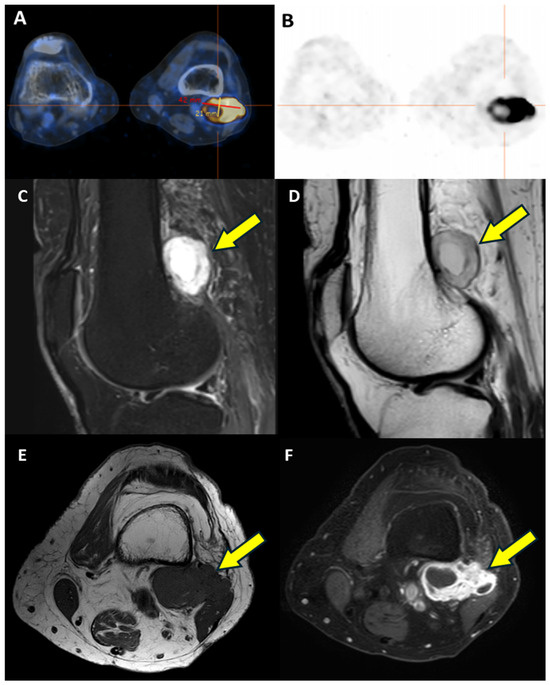
A subsequent 68Ga-DOTATOC PET/CT localized a hypermetabolic lesion (42 mm × 21 mm × 41 mm; SUV max 7.2) in the left popliteal fossa (Figure 1). The magnetic resonance imaging (MRI) confirmed a multiloculated lesion, measuring 22 mm × 43 mm × 30 mm, consisting of a solid peripheral rim and a multichambered gelatinous core in contact with the lateral femoral cortex (Figure 1).
Figure 1.
Imaging localization of the phosphaturic lesion. (A,B) 68Ga-DOTATOC PET/CT imaging showing intense focal radiopharmaceutical uptake in the left popliteal fossa. (C–F) MRI showed a well-defined multilobulated lesion (yellow arrow) with sharp margins, measuring 22 mm × 43 mm on the axial plane and 30 mm in longitudinal extension, within the soft tissues of the left popliteal fossa, slightly above the left lateral femoral condyle. The lesion appeared heterogeneously hyperintense on sagittal STIR (C) and T2-weighted TSE sequences (D) and hypointense on axial T1-weighted TSE images (E). After gadolinium-based contrast administration, T1-weighted TSE fat-sat images (F) showed intense peripheral and septal enhancement, with a multiloculated appearance and a non-enhancing core. The lesion was adjacent to the femoral cortex, popliteal neurovascular structures, and biceps femoris, without evidence of infiltration or regional lymphadenopathy.
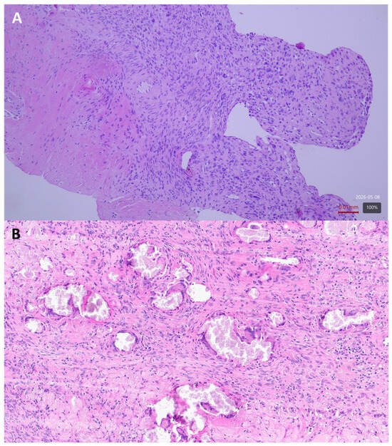
To complete the preoperative workup, the patient underwent a core needle biopsy of the mass. Histopathological analysis showed a proliferation of round cells with slightly polymorphic nuclei and multinucleated giant cells, embedded in a sclerotic and grungy calcified matrix. No mitotic figures were observed. Immunohistochemistry was performed to assess the nature of the lesion, demonstrating negativity for broad-spectrum cytokeratins (AE1/AE3 and CAM5.2), PTH, GATA3, TTF1, CD163, chromogranin, synaptophysin, SOX10, CD34, HMB-45, and ERG (the latter being expressed only in vascular endothelia). A low proliferation index was confirmed by Ki67 staining, while CD56 showed granular cytoplasmic positivity with variable intensity (Figure 2).
Figure 2.
Histopathology of the popliteal fossa mass. (A) Microphotograph of the needle biopsy performed on the popliteal mass showing the proliferation of mononuclear histiocytoid elements with intervening multinucleated giant cells, reactive-appearing vascular structures, and a dense sclerotic matrix in a picture initially interpreted as a tenosynovial giant cell tumor. (B) Microphotograph of the surgical specimen demonstrating the presence of calcified needle-like material associated with a giant cell granulomatous reaction. Hematoxylin and eosin, original magnification ×10 (A) and ×20 (B).
Overall, the combination of clinical, biochemical, imaging, and histological data suggested the presence of a phosphaturic mesenchymal tumor with giant cell tumor-like features causing TIO. After discussion in a multidisciplinary tumor board, the patient underwent a complete surgical resection of the lesion through a posterior approach to the knee. Intraoperatively, a well-circumscribed multiloculated soft-tissue mass with a gelatinous component was identified in the popliteal fossa, in close relationship with the distal posterior femoral cortex, without macroscopic invasion of the surrounding neurovascular structures. The mass was removed by marginal en bloc excision, achieving a complete macroscopic resection, with microscopically negative margins confirmed on histopathological examination. Histopathological evaluation revealed a complex lesion characterized by fibrosis and giant-cell granulomatous inflammation associated with multifocal deposits of amorphous, needle-like calcified material. Overall, the morphological features were consistent with a tophaceous-like giant-cell granulomatous reaction. Of note, FGF-23 immunohistochemistry was not performed on either the core needle biopsy or the final surgical specimen, as the anti-FGF-23 antibody is not routinely available in our pathology department. Nevertheless, postoperatively, intact FGF-23 levels normalized immediately, leading to a progressive rise in serum phosphate (2.6 mg/dL); consequently, oral potassium/sodium phosphate supplementation was withdrawn. The patient reported the resolution of bone pain and a rapid recovery of motor function. Having said that, the final diagnosis of TIO was established through a multidisciplinary correlation of clinical, biochemical, imaging, and pathological findings.
3. Discussion
This case highlights how challenging the management of TIO can be, as in our case it was masked by hypercalcemic hyperparathyroidism due to a parathyroid carcinoma that delayed its diagnosis.
TIO is notoriously difficult to diagnose; the literature reports a median diagnostic delay ranging from 2.9 years in specialized centers [10] up to 28 years [12]. This has been put down to several factors: (1) clinicians might not be familiar with this rare paraneoplastic syndrome; (2) clinical presentation is nonspecific and often misinterpreted; (3) tumors are frequently very small and difficult to locate; and (4) biochemical features such as the elevation of calcium and PTH are misinterpreted as primary hyperparathyroidism [13]. Our case highlights that parathyroid disorders (hypercalcemic hyperparathyroidism) can coexist with TIO, leading to a misdiagnosis of primary hyperparathyroidism. Clinicians must maintain a high index of suspicion for TIO if hypophosphatemia persists after parathyroidectomy.
The underlying mechanism of hypercalcemic hyperparathyroidism in patients with TIO involves FGF-23-mediated inhibition of 1,25(OH)2vitaminD3 synthesis, which reduces intestinal calcium absorption and triggers compensatory PTH secretion. Over time, this chronic stimulation may lead to parathyroid autonomy. In addition, exogenous phosphate supplementation also stimulates parathyroid activity through sequestration of calcium [13].
The association between TIO and hypercalcemic hyperparathyroidism is gaining increasing attention. Recently, Ni et al. evaluated the prevalence, clinical characteristics and risk factors for hyperparathyroidism in a cohort of 91 patients with TIO [14]. They found that hyperparathyroidism was present in 45% of the patients with TIO, and that 41.6% had secondary hyperparathyroidism while 3.5% had tertiary hyperparathyroidism. Of note, patients with tertiary hyperparathyroidism had the longest disease duration and a higher rate of phosphate and calcitriol supplementation.
Table 2 summarizes the existing case reports of patients presenting with hypercalcemic hyperparathyroidism associated with TIO. Our systematic review of the literature identified 27 cases. The median age at diagnosis was 46 years (range 13–68). The median disease duration before recognizing the metabolic complication was 8 years (range: 3–38). In the majority of cases (76.9%, 20/26), hyperparathyroidism was identified after the onset of TIO symptoms. Regarding histopathology, benign lesions predominated, with adenoma (40.7%) and hyperplasia (33.3%) being the most frequent findings. Remarkably, no cases of parathyroid carcinoma were previously documented in this specific clinical setting, distinguishing our report as the first to describe such a rare malignancy in a patient with TIO-induced tertiary hyperparathyroidism. Consistent with this, our case is a reminder that parathyroid surgery serves not only as a therapy but also as a fundamental diagnostic tool.
Table 2.
Association between TIO and hypercalcemic (tertiary) hyperparathyroidism.
Furthermore, in our case, histopathological analysis of the popliteal mass revealed a complex lesion characterized by a prominent giant-cell granulomatous reaction. This morphologic appearance validates Prader’s landmark early observation that such giant-cell-rich lesions can indeed be the source of FGF-23-mediated phosphaturia [34]. Historically, TIO was described in 1947 by Robert McCance [35], who reported the case of a patient with pain and weakness associated with low phosphate levels that improved only after a tumor found in the femur was resected [35]. However, the link between tumor and osteomalacia was established in 1959 by Prader, who postulated the production of a rachitogenic substance by a giant cell reparative granuloma of bone, given that the resection of the tumor led to the cure of the rickets [34]. The discovery of FGF-23 later provided the molecular basis for TIO, which is now recognized as a paraneoplastic syndrome caused, in most cases, by small phosphaturic mesenchymal tumors (PMTs).
PMTs most commonly arise in the soft tissues of the extremities and in the appendicular skeleton, although craniofacial bones and paranasal sinuses may also be involved. Histologically, they are typically composed of bland round-to-spindle cells, which are responsible for FGF-23 production, embedded in a variably vascular and extracellular matrix-rich stroma, often with characteristic “grungy” calcifications. Additional features may include multinucleated giant cells, fibrohistiocytic areas, myxoid matrix, woven bone formation, and, less frequently, adipocytic components. However, the relative proportions of neoplastic cells, vessels, matrix, calcifications, and giant cells vary considerably from case to case, making the diagnosis challenging, particularly in soft-tissue lesions. Indeed, a prominent giant-cell-rich or granulomatous reaction can heavily obscure the underlying neoplasm, leading to frequent diagnostic pitfalls. For instance, the most common misdiagnoses for PMTs include hemangiopericytoma, hemangioma, osteosarcoma, ossifying fibroma, giant cell tumor of bone, osteoblastoma, or purely reactive granulomas [2,4].
To overcome the limited diagnostic specificity of immunohistochemistry, combined immunoreactivity for FGF-23 and somatostatin receptors (SSTR2A), the latter being highly expressed in most PMTs [36], may increase diagnostic confidence [37]. Notably, SSTR2 expression might also be detected with somatostatin receptor-based imaging [38]. Somatostatin receptor-based imaging remains the gold standard for TIO localization, providing also crucial diagnostic support especially when the prominent giant-cell component or granulomatous-like features of the lesion might otherwise obscure its underlying neoplastic, phosphaturic nature [37,38]. In addition, it has been recently demonstrated that 68Ga-DOTATATE PET/CT has superior diagnostic accuracy compared to 18F-FDG PET/CT for the localization of TIO [38,39]. Consistent with this, in our patient, 68Ga-DOTATOC PET/CT was instrumental in accurately localizing the potential phosphaturic tumor.
Regarding their clinical behavior, PMTs exhibit a broad and often unpredictable biological spectrum [40] and complete excision remains the treatment of choice [41,42]. While the majority are benign and slow-growing, they can range from indolent lesions to locally aggressive tumors and, rarely (<10%), malignant forms capable of recurrence or distant metastasis [43]. According to Folpe et al. [40], histologically malignant PMTs are characterized by focal areas of high cellularity, high nuclear grade, and mitotic activity of >5/10 HPF, a high Ki-67 proliferation index and p53 expression, resembling an undifferentiated spindle cell sarcoma. In our case, the lesion displayed a favorable risk profile, lacking any of these features. This benign nature was further supported by the intraoperative findings of a well-circumscribed mass and the subsequent histological confirmation of microscopically negative resection margins.
Long-term surveillance is recommended [42,44], although no standard protocols have been universally established. In this clinical context, we implemented a follow-up strategy combining regular biochemical monitoring with local radiological reassessment via MRI starting 6 months after surgery. Clinically, fasting serum phosphate levels should be measured initially every 3–6 months and annually thereafter [10]. From an imaging perspective, an MRI of the surgical site is indicated 6 months postoperatively; subsequent MRI scans or 68Ga-DOTATOC PET/CT may be considered in the event of recurrent hypophosphatemia, rising FGF-23 levels, or in the presence of equivocal radiological findings. Nevertheless, given the broad biological spectrum of the disease, follow-up strategies must always be tailored to the individual patient’s presentation.
4. Conclusions
TIO is an extremely challenging condition. This case sheds light on the fact that hypercalcemic parathyroid disorders can coexist with TIO, and they may delay its diagnosis. Early integration of a meticulous biochemical follow-up and functional imaging is essential to prevent diagnostic delays and ensure the timely surgical resolution of this debilitating condition.
Author Contributions
Conceptualization, G.M., A.T. and S.B.; methodology, S.B.; investigation, G.M. and A.T.; resources and patient management, C.D., C.R., L.M., V.C. and S.B.; data curation, A.P.; writing—original draft preparation, G.M., A.T. and S.B.; writing—review and editing, A.P., C.D., L.M., B.F. and V.C.; visualization, A.P. and M.M.; supervision, S.B. All authors have read and agreed to the published version of the manuscript.
Funding
This research received no external funding.
Institutional Review Board Statement
Ethical review and approval were waived for this study due to the retrospective nature of this single-case report, which involved standard diagnostic and therapeutic procedures and did not introduce any deviations from established clinical protocols. In accordance with local institutional guidelines, formal IRB approval is not strictly required for single, non-interventional case reports, provided that patient anonymity is maintained and explicit written informed consent is obtained.
Informed Consent Statement
Written informed consent has been obtained from the patient to publish this paper.
Data Availability Statement
Data are contained within the article.
Conflicts of Interest
The authors declare no conflicts of interest.
Abbreviations
The following abbreviations are used in this manuscript:
| 18F-FDG | Fluorine-18 fluorodeoxyglucose |
| 68Ga | Gallium-68 |
| 99mTc-MIBI | Technetium-99m sestamibi |
| AE1/AE3 | Multi-cytokeratin cocktail AE1/AE3 |
| CAM5.2 | Low molecular weight cytokeratin CAM5.2 |
| CD31 | Cluster of differentiation 31 (endothelial cell adhesion molecule) |
| CD34 | Cluster of differentiation 34 |
| CD56 | Cluster of differentiation 56 (neural cell adhesion molecule) |
| CD163 | Cluster of differentiation 163 |
| ERG | ETS-related gene |
| FGF-23 | Fibroblast growth factor 23 |
| GATA3 | GATA binding protein 3 |
| HMB-45 | Human melanoma black 45 |
| HPF | High-power field |
| ioPTH | Intraoperative parathyroid hormone |
| Ki67 | Ki-67 proliferation index |
| MEN 1 | Multiple endocrine neoplasia type 1 |
| MEN2A | Multiple endocrine neoplasia type 2A |
| MRI | Magnetic resonance imaging |
| NaPi | Sodium–phosphate cotransporter |
| PET/CT | Positron emission tomography/Computed tomography |
| PMT | Phosphaturic mesenchymal tumor |
| PTH | Parathyroid hormone |
| PTX | Parathyroidectomy |
| r.r. | Reference range |
| SOX10 | SRY-box transcription factor 10 |
| TIO | Tumor-induced osteomalacia |
| TmP/GFR | Tubular maximum reabsorption rate of phosphate to glomerular filtration rate |
| TTF1 | Thyroid transcription factor 1 |
References
- Minisola, S.; Fukumoto, S.; Xia, W.; Corsi, A.; Colangelo, L.; Scillitani, A.; Pepe, J.; Cipriani, C.; Thakker, R.V. Tumor-induced Osteomalacia: A Comprehensive Review. Endocr. Rev. 2023, 44, 323–353. [Google Scholar] [CrossRef] [Scilit]
- Chong, W.H.; Molinolo, A.A.; Chen, C.C.; Collins, M.T. Tumor-induced osteomalacia. Endocr. Relat. Cancer 2011, 18, R53–R77. [Google Scholar] [CrossRef] [Scilit] [PubMed]
- Bouraima, F.; Sapin, V.; Kahouadji, S.; Pickering, M.E.; Pereira, B.; Bouvier, D.; Oris, C. Tumor-Induced Osteomalacia in Patients With Malignancy: A Meta-analysis and Systematic Review of Case Reports. J. Clin. Endocrinol. Metab. 2023, 108, 3031–3040. [Google Scholar] [CrossRef] [Scilit] [PubMed]
- Boland, J.M.; Tebben, P.J.; Folpe, A.L. Phosphaturic mesenchymal tumors: What an endocrinologist should know. J. Endocrinol. Investig. 2018, 41, 1173–1184. [Google Scholar] [CrossRef] [Scilit]
- Abrahamsen, B.; Smith, C.D.; Minisola, S. Epidemiology of Tumor-Induced Osteomalacia in Denmark. Calcif. Tissue Int. 2021, 109, 147–156. [Google Scholar] [CrossRef] [Scilit]
- Endo, I.; Fukumoto, S.; Ozono, K.; Namba, N.; Inoue, D.; Okazaki, R.; Yamauchi, M.; Sugimoto, T.; Minagawa, M.; Michigami, T.; et al. Nationwide survey of fibroblast growth factor 23 (FGF23)-related hypophosphatemic diseases in Japan: Prevalence, biochemical data and treatment. Endocr. J. 2015, 62, 811–816. [Google Scholar] [CrossRef] [Scilit]
- Rendina, D.; Abate, V.; Cacace, G.; D’Elia, L.; De Filippo, G.; Del Vecchio, S.; Galletti, F.; Cuocolo, A.; Strazzullo, P. Tumor-induced Osteomalacia: A Systematic Review and Individual Patient’s Data Analysis. J. Clin. Endocrinol. Metab. 2022, 107, e3428–e3436. [Google Scholar] [CrossRef] [Scilit]
- Juppner, H. Phosphate and FGF-23. Kidney Int. Suppl. 2011, 79, S24–S27. [Google Scholar] [CrossRef] [Scilit]
- Alvarez-Rivas, N.; Lugo-Rodriguez, G.; Maneiro, J.R.; Iniguez-Ubiaga, C.; Melero-Gonzalez, R.B.; Iglesias-Cabo, T.; Carmona, L.; Garcia-Porrua, C.; de Toro-Santos, F.J. Tumor-induced osteomalacia: A systematic literature review. Bone Rep. 2024, 21, 101772. [Google Scholar] [CrossRef] [Scilit] [PubMed]
- Brandi, M.L.; Clunie, G.P.R.; Houillier, P.; Jan de Beur, S.M.; Minisola, S.; Oheim, R.; Seefried, L. Challenges in the management of tumor-induced osteomalacia (TIO). Bone 2021, 152, 116064. [Google Scholar] [CrossRef] [Scilit]
- Stenner, E.; Sandic, S.; Dobrinja, C.; Ruscio, M.; Bernardi, S. The Total Testing Process of Intra-Operative Parathyroid Hormone. A Narrative Review. Clin. Lab. 2020, 66, 229–237. [Google Scholar] [CrossRef] [Scilit]
- Bosman, A.; Palermo, A.; Vanderhulst, J.; De Beur, S.M.J.; Fukumoto, S.; Minisola, S.; Xia, W.; Body, J.J.; Zillikens, M.C. Tumor-Induced Osteomalacia: A Systematic Clinical Review of 895 Cases. Calcif. Tissue Int. 2022, 111, 367–379. [Google Scholar] [CrossRef] [Scilit] [PubMed]
- Huang, Q.L.; Feig, D.S.; Blackstein, M.E. Development of tertiary hyperparathyroidism after phosphate supplementation in oncogenic osteomalacia. J. Endocrinol. Investig. 2000, 23, 263–267. [Google Scholar] [CrossRef] [Scilit]
- Ni, X.; Liu, W.; Zhang, D.; Li, X.; Chi, Y.; Feng, J.; Jin, C.; Pang, Q.; Gong, Y.; Cui, L.; et al. Hyperparathyroidism in a Large Cohort of Chinese Patients With Tumor-induced Osteomalacia. J. Clin. Endocrinol. Metab. 2023, 108, 1224–1235. [Google Scholar] [CrossRef] [Scilit]
- Olefsky, J.; Kempson, R.; Jones, H.; Reaven, G. “Tertiary” hyperparathyroidism and apparent “cure” of vitamin-D-resistant rickets after removal of an ossifying mesenchymal tumor of the pharynx. N. Engl. J. Med. 1972, 286, 740–745. [Google Scholar] [CrossRef] [Scilit]
- Wyman, A.L.; Paradinas, F.J.; Daly, J.R. Hypophosphataemic osteomalacia associated with a malignant tumour of the tibia: Report of a case. J. Clin. Pathol. 1977, 30, 328–335. [Google Scholar] [CrossRef] [Scilit]
- Pollack, J.A.; Schiller, A.L.; Crawford, J.D. Rickets and myopathy cured by removal of a nonossifying fibroma of bone. Pediatrics 1973, 52, 364–371. [Google Scholar] [CrossRef] [Scilit] [PubMed]
- Firth, R.G.; Grant, C.S.; Riggs, B.L. Development of hypercalcemic hyperparathyroidism after long-term phosphate supplementation in hypophosphatemic osteomalacia. Report of two cases. Am. J. Med. 1985, 78, 669–673. [Google Scholar] [CrossRef] [Scilit] [PubMed]
- Reid, I.R.; Teitelbaum, S.L.; Dusso, A.; Whyte, M.P. Hypercalcemic hyperparathyroidism complicating oncogenic osteomalacia: Effect of successful tumor resection on mineral homeostasis. Am. J. Med. 1987, 83, 350–354. [Google Scholar] [CrossRef] [Scilit]
- Heylen, A.; Dasnoy, D.; Hustin, J.; Pochet, J.M. Tumor-related osteomalacia followed after treatment by hyperparathyroidism. Rev. Rhum. Engl. Ed. 1999, 66, 53–57. [Google Scholar]
- Sato, K.; Obara, T.; Yamazaki, K.; Kanbe, M.; Nakajima, K.; Yamada, A.; Yanagisawa, T.; Kato, Y.; Nishikawa, T.; Takano, K. Somatic mutations of the MEN1 gene and microsatellite instability in a case of tertiary hyperparathyroidism occurring during high phosphate therapy for acquired, hypophosphatemic osteomalacia. J. Clin. Endocrinol. Metab. 2001, 86, 5564–5571. [Google Scholar] [CrossRef]
- Tartaglia, F.; Minisola, S.; Sgueglia, M.; Blasi, S.; Brunelli, D.; Degli Effetti, E.; Maturo, A.; Cola, A.; Custureri, F.; Campana, F.P. Tumor-induced hypophosphatemic osteomalacia associated with tertiary hyperparathyroidism: A case report. G. Chir. 2006, 27, 9–13. [Google Scholar]
- Lyritis, G.; Giannikou, P.; Economopoulos, D.; Samdanis, V.; Tournis, S. Autonomous Hyperparathyroidism Following Long-term Phosphate treatment for Tumor-Inducd Osteomalacia. Endocrinol. 2007, 17, 263–266. [Google Scholar]
- Nawrot-Wawrzyniak, K.; Varga, F.; Nader, A.; Roschger, P.; Sieghart, S.; Zwettler, E.; Roetzer, K.M.; Lang, S.; Weinkamer, R.; Klaushofer, K.; et al. Effects of tumor-induced osteomalacia on the bone mineralization process. Calcif. Tissue Int. 2009, 84, 313–323. [Google Scholar] [CrossRef] [Scilit]
- Markou, A.; Tsiama, V.; Tournis, S.; Papanastasiou, L.; Tsiavos, V.; Dassou, A.; Vlachou, V.; Papaliodi, E.; Asimaki, N.; Zografos, G.; et al. Coexistence of tumor-induced osteomalacia and primary hyperparathyroidism. Endocr. Pract. 2011, 17, 144–148. [Google Scholar] [CrossRef] [Scilit] [PubMed]
- Elfenbein, D.M.; Weber, T.J.; Scheri, R.P. Tumor-induced osteomalacia masking primary hyperparathyroidism. Surgery 2012, 152, 1256–1258. [Google Scholar] [CrossRef] [Scilit]
- Bhadada, S.K.; Palnitkar, S.; Qiu, S.; Parikh, N.; Talpos, G.B.; Rao, S.D. Deliberate total parathyroidectomy: A potentially novel therapy for tumor-induced hypophosphatemic osteomalacia. J. Clin. Endocrinol. Metab. 2013, 98, 4273–4278. [Google Scholar] [CrossRef] [Scilit] [PubMed]
- Hu, S. Cinacalcet for management of tertiary hyperparathyroidism associated with chronic treatment of hypophosphatemia in an adult with tumor-induced osteomalacia. AACE Clin. Case Rep. 2015, 1, e225–e229. [Google Scholar] [CrossRef] [Scilit]
- Koikawa, K.; Okada, Y.; Torimoto, K.; Tanaka, Y. Hyperparathyroidism Which Developed after Resection of a Fibroblast Growth Factor 23-producing Tumor. Intern. Med. 2020, 59, 2523–2527. [Google Scholar] [CrossRef] [Scilit]
- Kumar, S. Tumor-induced rickets-osteomalacia: An enigma. J. Pediatr. Endocrinol. Metab. 2020, 33, 1097–1103. [Google Scholar] [CrossRef] [Scilit]
- Kilbane, M.T.; Crowley, R.; Heffernan, E.; D’Arcy, C.; O’Toole, G.; Twomey, P.J.; McKenna, M.J. High bone turnover and hyperparathyroidism after surgery for tumor-induced osteomalacia: A case series. Bone Rep. 2021, 15, 101142. [Google Scholar] [CrossRef] [Scilit]
- Salim, M.; Behairy, M.S.; Barengolts, E. TIO Associated with Hyperparathyroidism: A Rarity, a Rule, or a Novel HPT-PMT Syndrome-A Case Study with Literature Review. Case Rep. Endocrinol. 2021, 2021, 5172131. [Google Scholar] [CrossRef] [Scilit]
- Brociek-Pilczynska, A.; Brodowska-Kania, D.; Szczygielski, K.; Lorent, M.; Zielinski, G.; Kowalewski, P.; Jurkiewicz, D. A rare combination of tumor-induced osteomalacia caused by sinonasal glomangiopericytoma and coexisting parathyroid adenoma: Case report and literature review. BMC Endocr. Disord. 2022, 22, 31. [Google Scholar] [CrossRef] [Scilit] [PubMed]
- Prader, A.; Illig, R.; Uehlinger, E.; Stalder, G. Rickets following bone tumor. Helv. Paediatr. Acta 1959, 14, 554–565. [Google Scholar]
- McCance, R.A. Osteomalacia with Looser’s nodes (Milkman’s syndrome) due to a raised resistance to vitamin D acquired about the age of 15 years. Q. J. Med. 1947, 16, 33–46. [Google Scholar]
- Houang, M.; Clarkson, A.; Sioson, L.; Elston, M.S.; Clifton-Bligh, R.J.; Dray, M.; Ranchere-Vince, D.; Decouvelaere, A.V.; de la Fouchardiere, A.; Gill, A.J. Phosphaturic mesenchymal tumors show positive staining for somatostatin receptor 2A (SSTR2A). Hum. Pathol. 2013, 44, 2711–2718. [Google Scholar] [CrossRef] [Scilit]
- Liu, Y.; He, H.; Zhang, C.; Zeng, H.; Tong, X.; Liu, Q. Phosphaturic Mesenchymal Tumors: Rethinking the Clinical Diagnosis and Surgical Treatment. J. Clin. Med. 2022, 12, 252. [Google Scholar] [CrossRef] [Scilit] [PubMed]
- Hoong, C.W.S.; Sfeir, J.G.; Drake, M.T.; Broski, S.M. Gallium-68-DOTATATE PET/CT for phosphaturic mesenchymal tumor localization in suspected tumor-induced osteomalacia. JBMR Plus 2025, 9, ziaf040. [Google Scholar] [CrossRef] [Scilit]
- El-Maouche, D.; Sadowski, S.M.; Papadakis, G.Z.; Guthrie, L.; Cottle-Delisle, C.; Merkel, R.; Millo, C.; Chen, C.C.; Kebebew, E.; Collins, M.T. (68)Ga-DOTATATE for Tumor Localization in Tumor-Induced Osteomalacia. J. Clin. Endocrinol. Metab. 2016, 101, 3575–3581. [Google Scholar] [CrossRef] [Scilit] [PubMed]
- Folpe, A.L.; Fanburg-Smith, J.C.; Billings, S.D.; Bisceglia, M.; Bertoni, F.; Cho, J.Y.; Econs, M.J.; Inwards, C.Y.; Jan de Beur, S.M.; Mentzel, T.; et al. Most osteomalacia-associated mesenchymal tumors are a single histopathologic entity: An analysis of 32 cases and a comprehensive review of the literature. Am. J. Surg. Pathol. 2004, 28, 1–30. [Google Scholar] [CrossRef] [Scilit]
- Maier, J.P.; Krapohl, M.A.; Herget, G.W.; Reising, K.; Fullgraf, H.; Bronsert, P.; Braig, D.; Schmal, H. Surgical treatment as a key determinant of outcome in phosphaturic mesenchymal tumors of the bone and soft tissue: A systematic review and case series. EFORT Open Rev. 2025, 10, 829–841. [Google Scholar] [CrossRef] [Scilit] [PubMed]
- Jan de Beur, S.M.; Minisola, S.; Xia, W.B.; Abrahamsen, B.; Body, J.J.; Brandi, M.L.; Clifton-Bligh, R.; Collins, M.; Florenzano, P.; Houillier, P.; et al. Global guidance for the recognition, diagnosis, and management of tumor-induced osteomalacia. J. Intern. Med. 2023, 293, 309–328. [Google Scholar] [CrossRef] [Scilit] [PubMed]
- Li, X.; Jiang, Y.; Huo, L.; Wu, H.; Liu, Y.; Jin, J.; Yu, W.; Lv, W.; Zhou, L.; Xia, Y.; et al. Nonremission and Recurrent Tumor-Induced Osteomalacia: A Retrospective Study. J. Bone Miner. Res. 2020, 35, 469–477. [Google Scholar] [CrossRef] [Scilit] [PubMed]
- Pannu, C.D.; Baxter, D.; Anwar, H. Spinal phosphaturic mesenchymal tumors: A rare etiology causing tumor-induced osteomalacia-a review of experience at a UK tertiary referral center and literature review. Eur. Spine J. 2024, 33, 1911–1920. [Google Scholar] [CrossRef] [Scilit]

Disclaimer/Publisher’s Note: The statements, opinions and data contained in all publications are solely those of the individual author(s) and contributor(s) and not of MDPI and/or the editor(s). MDPI and/or the editor(s) disclaim responsibility for any injury to people or property resulting from any ideas, methods, instructions or products referred to in the content. |
© 2026 by the authors. Licensee MDPI, Basel, Switzerland. This article is an open access article distributed under the terms and conditions of the Creative Commons Attribution (CC BY) license.