


Abstract
Background/Objectives: Mandibular reconstruction following trauma or oncologic resection is crucial for restoring function and aesthetics. While autologous bone grafting remains the gold standard, it presents challenges such as donor site morbidity and graft availability. Bone tissue engineering (BTE) offers an innovative alternative, integrating scaffolds, osteogenic cells, and bioactive factors to regenerate functional bone. This systematic review evaluates BTE strategies for mandibular reconstruction, focusing on critical-sized defects in large animal models and their translational potential for clinical applications. Methods: A systematic review was performed following PRISMA guidelines. Eligible studies involved large animal models and critical-sized mandibular defects treated with at least two BTE components (scaffold, osteogenic cells, or growth factors). Quality and bias assessments were conducted using ARRIVE guidelines and SYRCLE tools. Results: Of the 6088 studies screened, 27 met the inclusion criteria, focusing on critical-sized mandibular defects in large animal models such as pigs, sheep, and dogs. Common scaffolds included β-tricalcium phosphate (β-TCP), poly-lactic-co-glycolic acid (PLGA), and polycaprolactone (PCL), frequently combined with bone marrow-derived mesenchymal stem cells (BMSCs) and growth factors like recombinant human bone morphogenetic protein-2 (rhBMP-2). Preclinical outcomes demonstrated effective bone regeneration, vascularization, and biomechanical restoration. Advanced strategies, including in vivo bioreactors and 3D-printed scaffolds, further enhanced regeneration. However, challenges such as incomplete scaffold degradation, hypoxic conditions within constructs, and variability in growth factor efficacy and dose optimization were observed, emphasizing the need for further refinement to ensure consistent outcomes. Conclusions: BTE shows promise in mandibular reconstruction, achieving bone regeneration and functional restoration in preclinical models of critical-sized defects. However, challenges such as scaffold optimization, vascularization enhancement, and protocol standardization require further investigation to facilitate clinical translation. These findings emphasize the need for refinement to achieve consistent, scalable outcomes for clinical use.
1. Introduction
Mandibular resection, often necessitated by oncologic disease or trauma, leads to substantial functional and aesthetic impairments, severely affecting mastication, deglutition, speech, and in severe cases, even airway patency. These deficits impact essential functions such as chewing, swallowing, and speaking, and in severe cases, can lead to critical complications like airway collapse, significantly diminishing social interactions and consequently the patient’s quality of life [1,2]. Reconstructing the mandible is essential for patient rehabilitation and restoring both form and function [2].
Despite bone tissue’s intrinsic ability to regenerate, the challenge of repairing large mandibular defects remains challenging [3]. Autologous bone grafting or microvascular bone transplantation, using donor sites such as the iliac crest, fibula, scapula, or even calvaria, is still the gold standard for reconstruction [2,4]. However, this approach is associated with several drawbacks, including donor site morbidity, extended hospitalization, embolism risks, limited graft availability, intraoperative complications and graft loss [3,4,5]. These limitations have spurred the development of alternative strategies, particularly within the field of bone tissue engineering (BTE) [4,6].
BTE combines biodegradable scaffolds, osteogenic stem cells, and bioactive factors to create three-dimensional constructs that mimic autologous bone [3,4,7,8,9]. These constructs promote bone regeneration through the patient’s self-derived cells, ultimately integrating into the skeletal system [3]. The translation of these promising technologies from the laboratory bench to clinical practice is highly challenging, requiring robust preclinical evidence, especially in animal models, before advancing to clinical application [3].
While immunocompromised rodent models are valuable for studying ectopic bone formation due to their ability to support human tissue engraftment, their small size limits their utility for investigating the orthotopic integration of BTE constructs in load-bearing regions such as the mandible [3]. Large animal models, such as swine, sheep, and dogs, provide a more appropriate platform for evaluating BTE strategies in the context of critical-sized bone defects (CSBDs) [3,10]. These models enable the study of essential factors, such as vascularization within the core of implanted constructs and the biomechanical properties required for functional bone regeneration in load-bearing environments [3,11,12,13].
CSBDs are defined as injuries that surpass the bone’s intrinsic capacity for spontaneous regeneration, requiring surgical intervention for repair [14,15,16]. In mandibular studies, CSBDs are highly variable across species and are influenced by defect size, geometry, and continuity factors. For instance, canine mandibular CSBDs often range from 5 to 25 mm in diameter, with defect volumes between 810 and 2000 mm3 [15,17], whereas in pigs, defects exceeding 12–20 mm in diameter and 2800 mm3 in volume are considered critical [18]. In primate models, mandibular CSBDs are typically defined as exceeding 20 mm in diameter and 3000 mm3 in volume [18]. The periosteum, a critical reservoir of osteoprogenitor cells and growth factors, plays a central role in bone healing. Its removal in segmental defects significantly compromises regenerative outcomes, underscoring the importance of periosteal preservation [19]. Large animal models such as pigs, sheep, and dogs offer anatomical and physiological similarities to humans, providing an essential bridge between preclinical research and clinical applications.
This systematic review aims to assess the current strategies and outcomes of mandibular critical-sized defect reconstruction using TE approaches in large animal models, providing insights into their potential for future clinical applications. Analyzing unique methodologies, including scaffold designs, biomaterials, large animal models, and cell types, provides insights for future clinical applications.
2. Materials and Methods
2.1. Protocol and Registration
This systematic review was prospectively registered on 27 September 2023 with the International Prospective Register of Systematic Reviews in Health and Social Care (PROSPERO, National Institute for Health Research, York, UK; CRD42023456866). The study adheres to the Preferred Reporting Items for Systematic Reviews and Meta-Analyses (PRISMA) guidelines [20].
2.2. Eligibility Criteria
The PICO framework informed the selection criteria for articles [20]. The study population (P) encompassed large animal models (e.g., swine, sheep, dogs), with the intervention (I) focusing on the reconstruction of critical-sized mandibular bone defects through a tissue engineering approach. The comparison (C) involved reconstructions utilizing bone grafts, implantable materials, or untreated groups. The primary outcome (O) was the histological evaluation of newly formed bone tissue, while secondary outcomes included radiographic imaging, biomechanical testing, and qualitative clinical assessments. The inclusion criteria further specified original studies on large animals, mandibular reconstruction using tissue engineering as the primary treatment, critical-sized bone defects exceeding ≥ 20 mm in diameter (no spontaneous healing despite surgical stabilization and requires further surgical intervention [14]), and articles published in English within the last 20 years. For an approach to be classified as tissue-engineered, studies were required to meet at least two of the following criteria: incorporation of a scaffold material, application of osteogenic cells, or use of osteoinductive growth factors. Exclusions comprised in vitro studies, clinical studies, conference abstracts, review papers, letters to the editor, absence of histological analysis, repair through vascularized free flap surgery, studies on periodontal or alveolar regeneration, dental implant studies, and those implementing distraction osteogenesis.
2.3. Databases and Search Criteria
Systematic searches were conducted in the MEDLINE/PubMed, Embase, Web of Science, and Cochrane Library databases until 27 September 2024. Search terms were constructed to identify articles related to mandibular reconstruction in large animal models using tissue engineering approaches, while a filter was applied to exclude human studies. The following terms were used to conduct the search: (mandi* OR jaw OR stomatognathic) AND (tissue engineering OR bioengineering OR bioimplant OR biomaterials OR cells OR MSC OR BMSC OR scaffold OR grafts OR growth factor OR bone morphogenic protein OR BMP OR prefabricated OR β-tricalcium phosphate OR β-TCP OR intercellular signaling peptides OR intracellular signaling proteins) AND (large animal model OR sheep OR goat OR porcine OR pig OR swine OR minipig OR dog OR canine OR primates OR chimpanzee OR macaques OR baboon OR cow OR bovine OR horse OR equine) AND (reconstruct* OR segment* OR hemimandibulectomy OR defect OR critical OR defect OR damage).
2.4. Data Collection Process
The identified articles were screened independently by two reviewers (E.O. and Y.S.) using the Rayyan.ai research collaboration platform. Conflicting decisions were resolved through a third independent reviewer (F.G.). Preselected articles were downloaded, read in full, and assessed against the inclusion/exclusion criteria by the same reviewers. Disagreements were resolved through discussion with a third reviewer (F.G.).
2.5. Data Extraction
Different authors employed a data extraction sheet to systemically extract information from included studies (E.O., A.G., and Y.S.). The data extracted from the articles included the animal model (breed/strain, age, gender, and weight), the characteristics of the study (groups, interventions, tissue engineering approach, defect size, fixation of reconstruction, time points of the studies, complications, and postoperative monitoring), outcome measurements (imaging techniques, histological analysis, histomorphometry, immunohistochemistry, and molecular biology), and the conclusions drawn by the respective authors.
2.6. Quality and Risk of Bias Assessment
Compliance with the Animal Research Reporting In Vivo Experiments (ARRIVE) guidelines was assessed to assess the quality of the included studies. The ARRIVE guidelines were developed to guide the reporting of animal studies and thereby improve their reproducibility, inform future researchers, and enable peer review [21]. The ARRIVE guidelines provide a checklist containing 20 items. A grading system adopted by Schwarz et al. [22] was implemented to assess the included studies’ conformity with the ARRIVE guidelines.
Additionally, the risk of bias in the included studies was analyzed according to the Systematic Review Center for Laboratory Animal Experimentation (SYRCLE) tool, which was developed to provide a standardized risk of bias assessment for animal studies [23].
3. Results
3.1. Search Results
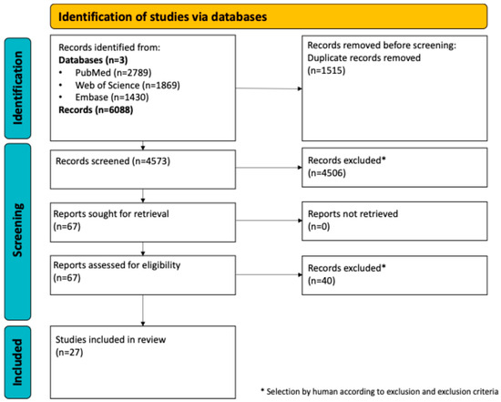
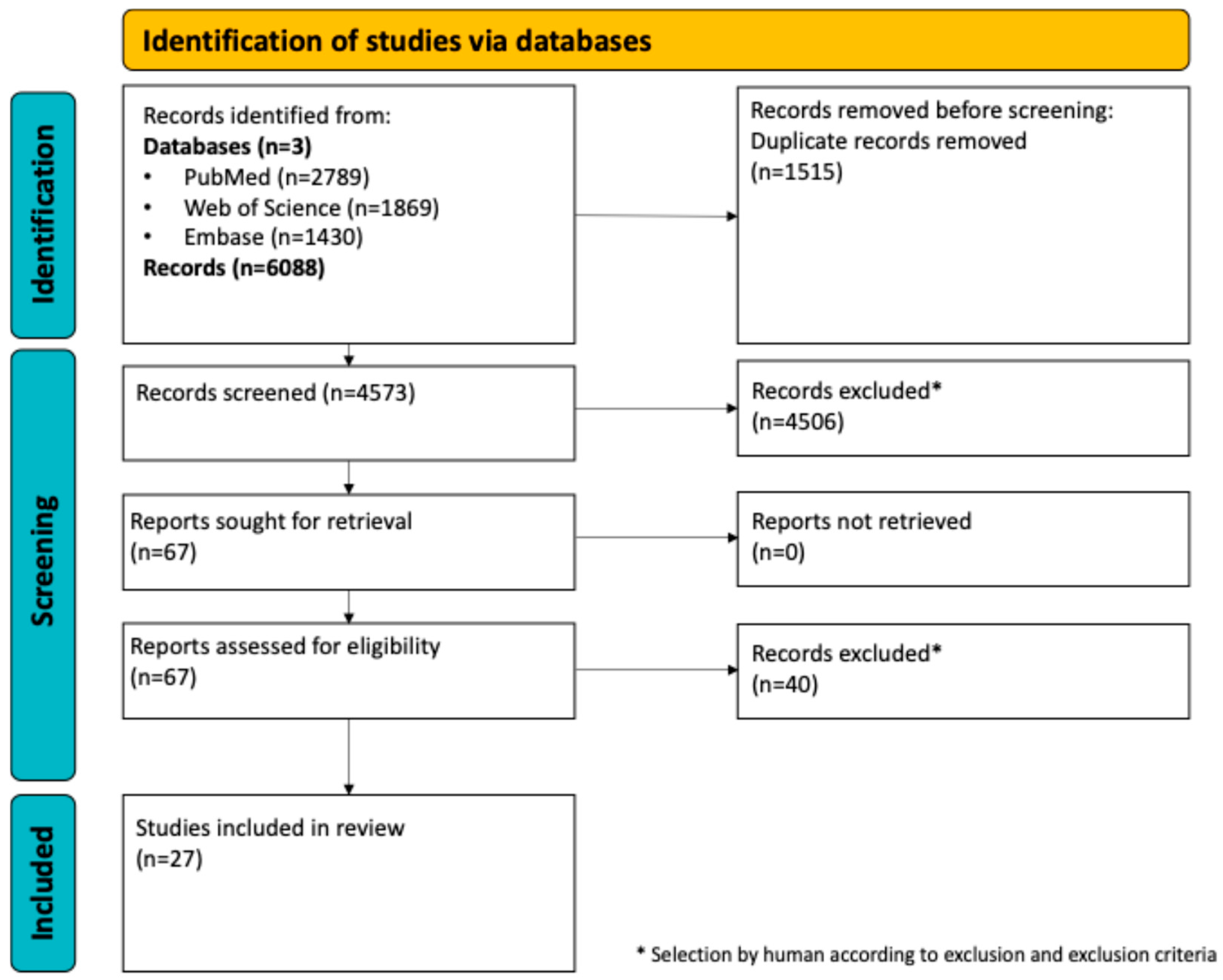
The electronic search yielded 6088 articles. This included 2789 through PubMed, 1869 through Web of Science, and 1430 from Embase. In total, 1515 articles were excluded as duplicates and another 4506 were removed after screening the titles and abstracts. The remaining 67 articles underwent full-text review, resulting in 40 exclusions due to inconsistencies with the inclusion/exclusion criteria. Twenty-seven articles were finally included in this systematic review. The PRISMA flow chart demonstrating the screening process can be seen in Figure 1.
Figure 1.
PRISMA flowchart.
3.2. Study Design, Animal Model, and Defect Characteristics
The included studies employed a variety of study designs and large animal models to evaluate tissue engineering strategies for mandibular reconstruction, focusing on critical-sized defects.
Study Design: Most studies implemented comparative experimental designs involving treatment and control groups. Treatment groups received scaffold-based constructs, often combined with osteogenic cells and/or bioactive molecules, while controls included untreated defects or conventional grafts [24,25]. Follow-up intervals ranged from 4 weeks to 12 months, allowing longitudinal evaluation of bone regeneration [26,27].
Animal Models: Porcine models, particularly Göttingen and Yucatan minipigs, were employed in 9 studies [26,28,29,30,31,32,33,34,35] due to their craniofacial similarity to humans [30,32]. Canine models (Beagle, Mongrel) were used in 8 studies [25,36,37,38,39,40,41,42], ovine models in 6, [24,27,43,44,45,46], and non-human primate models (Rhesus Macaque monkeys) in 3 studies [47,48,49]. A single study utilized a caprine model (n = 1) [50].
Defect Characteristics: Critical-sized defects were primarily located in the body of the mandible, with defect dimensions tailored to species-specific thresholds for spontaneous healing. Defects ranged in size from 20 mm in canine models [25] to 50 mm in porcine models [46]. Segmental defects were the most commonly employed, often created surgically and stabilized using titanium plates or fixation devices to mimic clinical scenarios [28,34]. The absence of the periosteum was a defining feature in many studies, particularly for evaluating the regenerative potential of scaffolds under challenging conditions [24,45].
This section highlights the diversity in animal models and defect designs across the included studies, emphasizing their importance in evaluating the translational potential of tissue-engineered strategies for mandibular reconstruction. Table 1 summarizes detailed information on the animal models, defect characteristics, and study designs.
Table 1.
Animal models and defect characteristics [9,24,25,26,27,28,29,30,31,32,33,34,35,36,37,39,40,41,42,43,44,45,46,47,48,49,50].
3.3. Bone Tissue Engineering Strategies
The BTE approaches in the included studies typically utilized combinations of biomaterials, cellular components, and bioactive factors to address critical-sized mandibular defects in large animal models.
Biomaterials: A diverse range of biomaterials was employed, with β-tricalcium phosphate (β-TCP) being the most commonly used. Pure β-TCP scaffolds were utilized in numerous studies [25,37,42], while others combined β-TCP with additional materials, such as hydroxyapatite or polymers, to enhance their properties [31,32,34]. Poly(lactic-co-glycolic) acid (PLGA) scaffolds, such as those used by Abukawa et al., were also frequently employed [26]. To create porous, biocompatible constructs, PCL scaffolds, including laser-sintered and 3D-printed PCL/β-TCP composites, were explored for their structural and osteoconductive advantages [33,37]. Additionally, naturally occurring materials like coral scaffolds demonstrated promise as osteoconductive and biodegradable matrices [50]. These varied materials underscore the breadth of scaffolding strategies employed in BTE.
Cellular Components: Osteogenic cells were integral to many BTE approaches, with BMSCs being the most widely used cell type across studies [24,25,27]. BMSCs were typically harvested and seeded onto scaffolds, enhancing osteogenesis and bone regeneration. In some cases, autologous osteoblasts were employed, such as in studies by Henkel et al. [31], highlighting their potential for direct bone-forming applications. Adipose tissue-derived mesenchymal stem cells (AMSCs) were used in some studies, demonstrating their versatility as an alternative cell source [33,34]. The integration of cellular components across these studies highlights their crucial role in enhancing the osteoinductive potential of biomaterials.
Growth Factors: Various studies employed growth factors to enhance osteogenesis and bone healing within tissue-engineered constructs. Recombinant human bone morphogenetic protein-2 (rhBMP-2) was the most commonly used growth factor and was incorporated into scaffolds in several studies [28,29,37,48,49]. These studies demonstrated enhanced bone regeneration and osteoinductive properties, with dose-dependent effects observed in some cases [28]. Another growth factor, recombinant human osteogenic protein-1 (RhOP-1), was used by Wang et al. [40], resulting in improved bone formation and mechanical properties. In vivo bioreactor techniques utilizing local muscle flaps to deliver growth factors were applied in some studies, enhancing vascularization and bone remodeling [43,46,49]. For example, Zhou et al. [49] reported better bone regeneration using the rhBMP-2-incorporated scaffold prefabricated within a latissimus dorsi muscle bioreactor compared to the rhBMP-2-incorporated scaffold alone. Similarly, Tatara et al. [46] demonstrated the efficacy of periosteum-based bioreactors in generating vascularized bone tissue. Platelet-rich plasma (PRP) combined with a polycaprolactone scaffold and porcine adipose-derived stem cells was another effective growth factor strategy, augmenting osteoconductive and osteoinductive properties [33]. Additionally, factor XIII, tested in a single study, did not significantly promote osteogenesis [45]. Overall, growth factors played a pivotal role in enhancing bone regeneration across various tissue-engineered approaches, particularly when combined with advanced scaffolds and cellular components.
The BTE strategies of the included studies can be seen in detail in Table 2.
Table 2.
Bone tissue engineering strategies [9,24,25,26,27,28,29,30,31,32,33,34,35,36,37,39,40,41,42,43,44,45,46,47,48,49,50].
3.4. Outcome Measures
Histological Analysis: Histological analysis was the primary outcome parameter to evaluate bone formation, scaffold degradation, and tissue integration across the included studies. Most studies employed hematoxylin and eosin (H&E) staining as a standard method for assessing new bone formation and scaffold biocompatibility [25,26,34]. Additionally, advanced staining techniques, such as Masson’s trichrome [37,41] and Alizarin Red complex staining [40,49], provided detailed insights into mineralization patterns and osteoid deposition. Histomorphometric analyses were conducted in several studies to quantify bone volume and integration rates [28,46]. These assessments consistently highlighted improved bone formation in groups with cellular components or growth factor treatments compared to scaffolds alone.
Biomechanical Testing: Biomechanical testing was performed in a subset of studies to evaluate the regenerated bone’s mechanical properties and functional integrity. Three-point bending tests were frequently used to assess the stiffness and load-bearing capacity of tissue-engineered constructs [40,42]. Other methods, such as compressive strength and Young’s modulus testing, provided additional insights into the functional recovery of bone [33,36]. The results from these tests often correlated with histological findings, demonstrating that constructs with enhanced bone regeneration also exhibited superior mechanical properties [29,33].
Imaging Techniques, Immunohistochemistry, and Molecular Biology: Imaging modalities, including micro-computed tomography (CT), X-ray radiography, and CT scans, were widely used to evaluate the morphology and volume of regenerated bone [27,34,48]. Micro-CT provided high-resolution assessments of bone structure and scaffold integration, while dual-energy X-ray absorptiometry (DXA) scans were employed in some studies for bone density analysis [25]. Immunohistochemical techniques, such as staining for collagen type I and von Willebrand factor (vWF), were used to assess osteogenesis and vascularization [28,43]. Molecular analyses, including qRT-PCR for osteogenic markers and cytokine profiling, further elucidated the underlying biological processes driving bone regeneration [29,33]. These complementary techniques provided a comprehensive understanding of both the structural and molecular outcomes of mandibular reconstruction.
The methods of outcome assessment are detailed in Table 3.
Table 3.
Outcome assessment [9,24,25,26,27,28,29,30,31,32,33,34,35,36,37,39,40,41,42,43,44,45,46,47,48,49,50].
3.5. Assessment of Adherence to the ARRIVE Guidelines
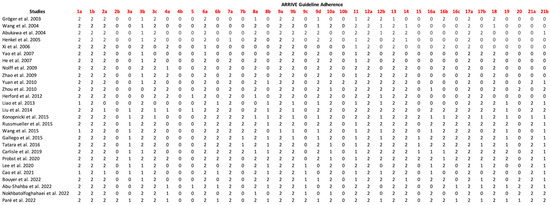
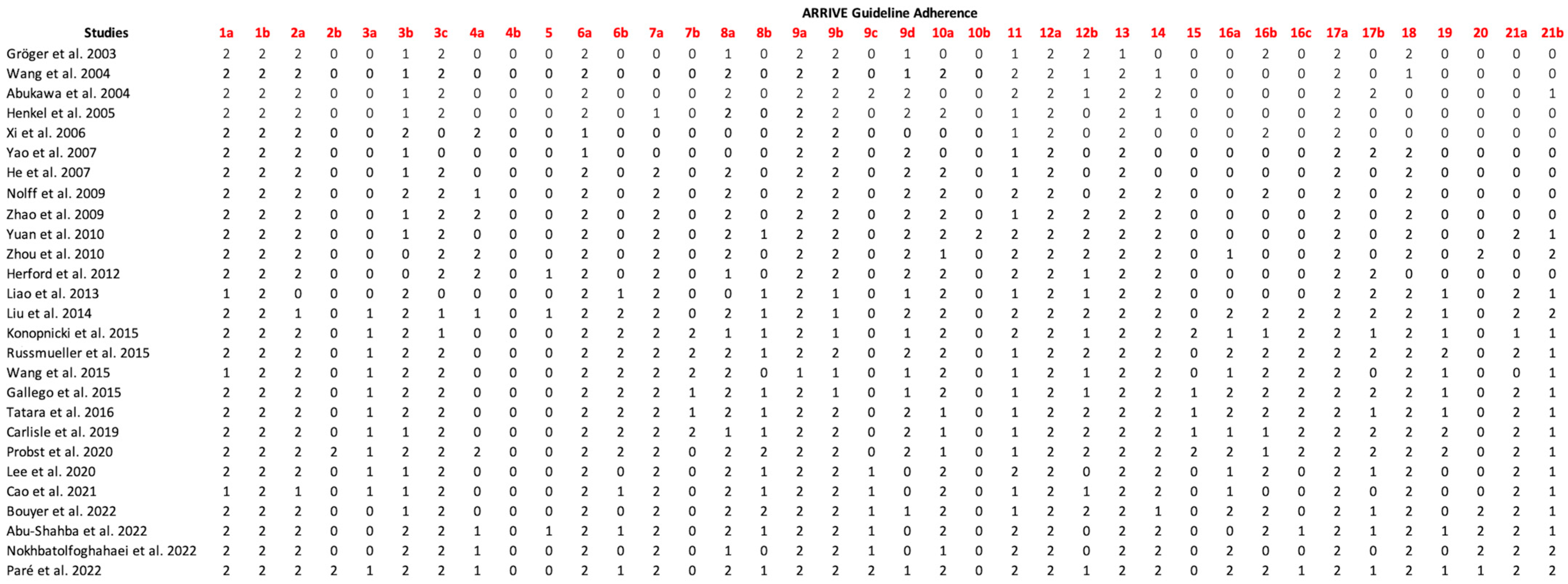
There has been a marked improvement in the reporting of animal experiments, with a growing emphasis on reproducibility and transparency, particularly since the development of the ARRIVE guidelines in 2010. Most papers in the dataset provided detailed and transparent insights into their methodologies, with seven studies offering comprehensive reporting across nearly all guideline categories [27,29,34,38,44,45,46]. These studies demonstrated profound insights into their approach. By contrast, four studies, primarily published before 2010, lacked essential details in their reporting [30,36,41,50].
Several topics outlined in the ARRIVE 2.0 guidelines were consistently addressed across all studies, reflecting an established baseline for good reporting practices. Study design details, including the groups compared (1a), the experimental unit (1b), the sample size (2a), the inclusion and exclusion criteria (3b), the outcome measures (6a), and the experimental procedures (9a, 9b), were thoroughly documented and clearly defined, along with discussions on research objectives (13) and scientific implications (17a).
However, some critical aspects were frequently omitted, particularly those related to the rationale behind methodological choices, such as the calculation of the sample size (2b) or the effect size (10b). Techniques to avoid confounding effects (4b) and details about housing and husbandry conditions (15) were also often missing, raising concerns about potential bias and variability in animal welfare. The results of the ARRIVE guidelines evaluation are shown in Figure 2.
Figure 2.
ARRIVE guidelines adherence [9,24,25,26,27,28,29,30,31,32,33,34,35,36,37,39,40,41,42,43,44,45,46,47,48,49,50].
3.6. Risk of Bias Assessment
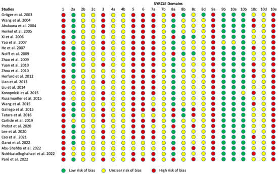
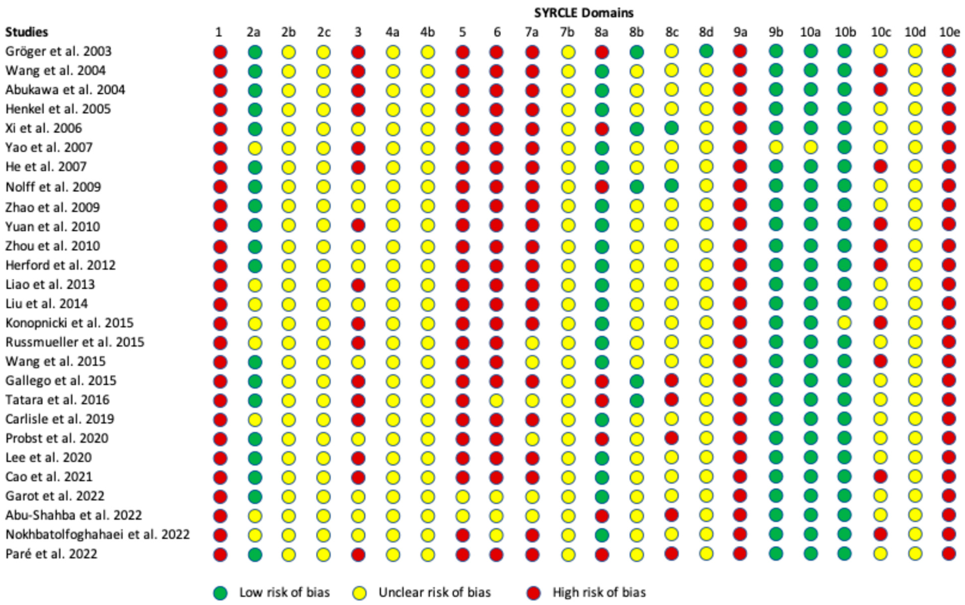
The evaluation of the risk of bias, based on the SYRCLE assessment tool, is shown in Figure 3. Most papers demonstrated a moderate risk level, with four studies providing detailed information to minimize biases in selection, performance, detection, attrition, and reporting [24,25,28,50]. All published reports included the expected outcomes, and there were no indications of “study contamination” or “inappropriate influence of funders.” Additionally, most studies balanced the distribution of relevant baseline characteristics between the intervention and control groups.
Figure 3.
Risk of bias assessment using the SYRCLE tool [9,24,25,26,27,28,29,30,31,32,33,34,35,36,37,39,40,41,42,43,44,45,46,47,48,49,50].
However, none of the papers provided detailed study protocols, including information about the allocation/randomization processes or steps for managing dropouts and replacements in the study and control groups. Furthermore, most studies lacked detailed information regarding the blinding of caregivers, investigators, and outcome assessors, raising concerns about potential biases in these areas.
3.7. Conclusions Drawn by the Included Studies
As shown in Table 4, tissue engineering approaches have made significant progress in developing constructs for bone regeneration, with a particular focus on mandibular reconstruction. Among the materials studied, β-tricalcium phosphate (β-TCP) emerges as a leading scaffold material due to its ability to support osteogenesis and promote the formation of mature bone structures [24,29,31,32,34,36,37,39,40,45,47,48]. Modifying the scaffold composition further enhances its regenerative capabilities, as demonstrated by HA/β-TCP matrices composed of 60% hydroxyapatite and 40% β-TCP [31,41], which exhibited superior bioactivity and osteoconductive properties compared to conventional hydroxyapatite ceramics. The integration of BMSCs with scaffolds shows remarkable outcomes in bone regeneration. When paired with β-TCP or other advanced scaffolds, BMSCs have promoted uniform radiodensity, active osteoblast and osteocyte activity, and seamless integration with the host bone [24,25,26,27,36,38,42,45,50]. Constructs using GFP-labeled BMSCs have effectively bridged critical-sized defects, achieving biomechanical properties comparable to native bone [42]. Similarly, cryopreserved bone-derived osteoblasts (CBOs) have demonstrated comparable efficacy to fresh osteoblasts, offering a practical solution for bone regeneration, particularly in scenarios involving reduced regenerative capacity due to aging or limited tissue availability [40].
Table 4.
Conclusions Drawn by the Included Studies [9,24,25,26,27,28,29,30,31,32,33,34,35,36,37,39,40,41,42,43,44,45,46,47,48,49,50].
Incorporating rhBMP-2 into scaffolds further enhances osteoinduction, vascularization, and overall bone formation [28,29,37,39,47,48,49]. Additional supplements, such as β-glycerophosphate sodium, dexamethasone, and PRP, have also been shown to enhance osteogenic differentiation and scaffold remodeling, further optimizing regenerative outcomes [33,50]. In vivo muscle-based bioreactors, particularly those incorporating vascularized periosteal flaps, have proven to be an effective strategy for scaffold prefabrication. These bioreactors facilitate vascularization and remodeling while minimizing surgical complexity [39,41,43,46,47,49]. Innovative imaging modalities have been pivotal in evaluating these advancements. Techniques such as 18F-FDG PET/CT [47] and recently developed deep learning-enhanced micro-CT [44] provide reliable and precise methods for tracking bone regeneration, vascularization, and scaffold performance.
4. Discussion
This systematic review critically evaluated current BTE strategies for mandibular reconstruction, with an emphasis on approaches targeting CSBDs in large animal models. The review identifies key translational milestones, summarizes major preclinical advancements, and outlines remaining barriers to clinical application. While autologous bone grafting remains the clinical gold standard, it is fraught with challenges, including donor site morbidity, limited graft availability, and long-term complications such as graft resorption [4,51,52,53]. These issues underscore the urgent need for innovative strategies to improve patient outcomes and reduce the associated morbidity. BTE offers a promising alternative, leveraging biodegradable scaffolds, osteogenic cells, and bioactive factors to regenerate functional bone. This systematic review highlights significant advancements in BTE strategies for mandibular reconstruction. These include the development of 3D-printed scaffolds, the incorporation of MSCs, and the use of growth factors. These approaches have significantly improved bone formation, vascularization, and scaffold integration in preclinical models of critical-sized mandibular defects [51,53,54]. However, challenges such as inconsistent vascularization, variability in scaffold degradation, and translational limitations remain hurdles that must be addressed.
4.1. Historical Perspective: The Evolution of Mandibular Bone Tissue Engineering
4.1.1. Early Efforts and Initial Challenges
The early phase of BTE focused on using simple biomaterials like β-TCP and HA for mandibular reconstruction. These scaffolds provided basic structural support but suffered from significant limitations, including poor vascularization, incomplete scaffold degradation, and inconsistent bone regeneration [52,53]. The mismatch between scaffold resorption and new bone formation often accumulates residual material, impairing the healing process. Additionally, the lack of biological activity of these materials limited their ability to stimulate robust osteogenesis and integration [52,53,54].
4.1.2. Integration of Cellular Components
Multiple studies have consistently demonstrated that autologous cell seeding—particularly with BMSCs—enhances osteogenic potential, resulting in increased bone volume, improved vascularization, and superior mechanical strength compared to acellular scaffolds.
The inclusion of BMSCs and AMSCs marked a pivotal advancement in BTE. BMSCs enhance osteogenesis by differentiating into osteoblasts and secreting osteogenic factors, while AMSCs provide a versatile and readily available cell source. Multiple studies have consistently demonstrated that autologous cell seeding—particularly with BMSCs—enhances osteogenic potential, resulting in increased bone volume, improved vascularization, and superior mechanical strength compared to acellular scaffolds [38,39,45]. For instance, constructs combining β-TCP scaffolds with BMSCs achieved seamless integration with native bone and superior mechanical stability, paving the way for more effective tissue-engineered constructs [24,34,51,54].
4.1.3. Emergence of Growth Factor Strategies
The addition of bioactive molecules, particularly rhBMP-2, revolutionized the field by enhancing the osteoinductive potential of scaffolds. rhBMP-2 promotes robust and consistent bone regeneration, addressing key limitations of earlier scaffold designs [28,29,37,48]. However, studies have shown that growth factor dose optimization is crucial to minimize complications such as ectopic bone formation and variable efficacy [28]. PRP was another possibility, providing localized delivery of cytokines and growth factors that augmented osteogenic differentiation and scaffold integration; however, its mechanisms remain poorly understood, its composition is highly heterogeneous, and it is more commonly applied in other regenerative contexts, such as the treatment of osteoarthritis [33,52,53,55].
4.1.4. Technological Advancements
Technological innovations such as 3D printing and in vivo bioreactors have transformed scaffold design and prefabrication. Notably, 3D-printed scaffolds allow for precise customization to defect geometry, ensuring an anatomical fit and enhanced functional outcomes [56,57,58,59,60]. Bioreactors that leverage vascularized tissues for scaffold maturation before implantation have significantly improved vascularization and bone regeneration [39,46]. Furthermore, advanced imaging techniques like micro-CT and PET scans provide detailed assessments of scaffold integration, bone volume, and vascularization, refining BTE strategies and improving their reliability [28,34,37,39,44,47,51,53,54].
Histological analysis is vital in validating imaging results and assessing the maturity and quality of newly formed bone in mandibular tissue engineering. Notably, all of the included studies evaluated histological findings, as this was a requirement of the inclusion criteria. By providing detailed insights into bone microstructure and cellular composition, histology complements imaging techniques such as micro-CT, ensuring a more comprehensive evaluation of bone regeneration. This dual approach enhances the reliability of outcome characterization, allowing researchers to better understand the biological processes underlying successful reconstruction. Advanced techniques such as immunohistochemistry and fluoroscopic microscopy provide valuable insights into the rates and maturity of bone formation in mandibular tissue engineering. These methods allow for the detailed analysis of specific markers, such as type I collagen, and serial in vivo radiolabeling to track bone development over time. By offering a deeper understanding of the biological processes involved in regeneration, these techniques complement traditional imaging and histological assessments, providing a more nuanced evaluation of bone quality and the effectiveness of tissue-engineered constructs. Materials used in mandibular reconstruction must withstand the complex biomechanical forces of mastication, including tension, compression, and torsion. Despite this critical requirement, only a limited number of studies have conducted biomechanical testing on newly formed bone [28,34,43,46,47]. The variability in testing approaches and outcome measures across studies has hindered direct comparisons and the development of standardized evaluation methods. A promising approach to assess implant success involves comparing the biomechanical properties of the reconstructed region with those of the contralateral mandible, offering a more reliable benchmark for functional restoration.
There is a critical need for standardized measurements, such as micro-CT and histological analysis, to improve the comparability of outcomes across studies in mandibular tissue engineering [51]. Consistent use of objective metrics, including trabecular thickness, separation, bone mineral density, and percentage of newly formed bone, would enable more reliable comparisons between treatment approaches. Standardizing these methods would enhance the reproducibility of findings and facilitate the development of evidence-based protocols for effective bone regeneration.
4.2. Current State of Mandibular Bone Tissue Engineering
4.2.1. Key Achievements
Significant advancements in BTE include integrating cellular and molecular strategies, such as BMSCs and rhBMP-2, with innovative scaffolds. Preclinical studies have demonstrated that these approaches significantly enhance bone formation, vascularization, and biomechanical strength. For example, β-TCP and PCL scaffolds seeded with BMSCs resulted in superior bone integration and mechanical properties compared to controls [28,34,43,47]. Studies employing advanced scaffold designs, such as composite and 3D-printed constructs, have also shown promising outcomes in restoring both structure and function in critical-sized mandibular defects [28,32,37,46,53,54].
4.2.2. Persistent Challenges
Despite these advancements, challenges persist. Scaffold degradation rates often fail to match the pace of new bone formation, leading to residual material accumulation or insufficient structural support during healing. Additionally, variability in the efficacy of growth factors, such as rhBMP-2, has been reported, with dose-dependent inconsistencies and complications such as ectopic bone formation [28]. Hypoxic conditions within larger constructs limit nutrient diffusion and cell viability, posing a critical barrier to effective bone regeneration [51,52,54].
4.2.3. Animal Models
Large animal models play an essential role in mandibular tissue engineering due to their close anatomical, physiological, and biomechanical resemblances to humans. Despite their importance, significant variability in study designs and a lack of standardized approaches often hinder direct comparisons between studies, limiting the ability to draw conclusive insights regarding efficacy [32,61,62]. Regulatory agencies mandate large animal models for preclinical efficacy testing of medical and dental implants in their final human form [32]. Large animal models, including pigs, dogs, sheep, and goats, are preferred for mandibular tissue engineering due to their craniofacial anatomical and biomechanical similarities to humans. These models allow for testing critical-sized defects in a context mimicking human conditions, enabling more clinically relevant outcomes [63,64]. Studies have highlighted that critical-sized mandibular defects pose a significant challenge due to their poor intrinsic healing capacity and the frequent complications associated with internal fixation in large animals, such as plate failures [15,16]. Despite these challenges, large animal models provide a platform to evaluate the regenerative potential of tissue-engineered constructs and their mechanical stability under physiologically relevant forces. Moreover, their size allows for testing human-scale implants and surgical techniques, which is not feasible in small animal models [65,66].
One of the key issues with large animal models is the considerable heterogeneity in study designs, including differences in species/strain selection, defect location, defect size, surgical procedures, and experimental outcome measures. For instance, what constitutes a critical-sized defect varies among species is not consistently defined, which complicates the standardization of preclinical models [15]. Furthermore, while pigs, dogs, and sheep exhibit moderate similarities to human bone properties, differences in bone remodeling, composition, and healing responses remain. For example, Aerssens et al. demonstrated that dog bone is most comparable to human bone in terms of mechanical competence and composition [67]. Small animal models, such as rats and mice, are often used due to their lower costs and widespread availability; however, they fail to replicate the human mandible’s anatomical, biomechanical, and physiological complexities. While useful for initial exploratory studies, these models do not provide the scale or functional relevance required for testing human-sized implants or complex regenerative strategies. By contrast, large animal models allow for testing materials and techniques under conditions that better mimic human clinical scenarios, making them the only way to advance mandibular tissue engineering.
Another limitation is the predominant use of young, healthy, large animals, which does not accurately reflect the demographics of human patients who often present with comorbidities such as advanced age, impaired vascular function, diabetes or radiotherapy in cancer cases. These factors significantly influence bone healing and regenerative outcomes, emphasizing the need for more representative models [68,69,70]. Additionally, immunological differences within and between species, such as varying disease susceptibilities among sheep breeds, further complicate the extrapolation of preclinical findings to humans [71,72]. Selecting an appropriate large animal model is critical for obtaining clinically relevant preclinical data. An ideal model should minimize morbidity, ensure reproducibility, and closely replicate the targeted clinical condition. For mandibular tissue engineering, this includes anatomical and biomechanical similarities and consideration of the biological mechanisms underlying bone regeneration [73]. For example, the reduced efficacy of rhBMP-2 in human orofacial bone regeneration compared to some animal models highlights the importance of selecting models that accurately represent human physiology [74]. Additional considerations include the potential influence of systemic factors, such as age and comorbidities, and the inclusion of strain information for large animals, which is often underreported but has been shown to impact experimental results [72,75]. To improve the utility of large animal models in mandibular tissue engineering, future studies should prioritize standardizing protocols and addressing existing gaps in knowledge. This includes developing age-appropriate and disease-representative models, defining critical-sized defects for specific species, and ensuring consistent strain and breed information reporting. Furthermore, integrating advanced imaging and molecular analysis techniques could provide deeper insights into bone regeneration’s cellular and molecular mechanisms, enhancing the predictive nature of preclinical studies. By addressing these challenges, large animal models can better fulfill their role as a bridge between small animal studies and human clinical trials, ultimately advancing the field of mandibular bone tissue engineering [66,75,76]. The heterogeneity among the included studies—particularly in terms of defect size, animal model, and fixation strategy—presents a notable challenge when attempting to synthesize consistent conclusions. Variability in defect dimensions reflects species-specific anatomical considerations and directly influences the regenerative demands placed on tissue-engineered constructs. For instance, a 20 mm defect in a canine mandible may differ substantially in biomechanical loading and biological response compared to a similarly sized defect in a porcine or ovine model [14,17,18,19]. Likewise, differences in fixation methods, ranging from rigid titanium plates to resorbable systems, alter the mechanical environment of the defect site and can significantly impact outcomes in bone regeneration. The choice of animal model itself introduces further variability, as differences in bone metabolism, immune response, and oral biomechanics can influence both the speed and quality of healing. Despite this heterogeneity, the systematic comparison of methodologies across studies—as provided in this review—offers a valuable framework for identifying the most commonly used and effective parameters. By highlighting trends in defect size, anatomical location, fixation techniques, and outcome measures, this review contributes to the development of more consistent experimental designs. In doing so, it supports the establishment of standardized protocols for large animal models in mandibular tissue engineering, which are essential for improving comparability, reproducibility, and translational potential in future preclinical research.
4.3. Future Outlook and Directions
4.3.1. Technological Innovations
Emerging technologies, including biofabrication, smart biomaterials, and personalized scaffolds, hold immense potential to address these challenges. Innovations such as 3D bioprinting enable the creation of complex, patient-specific constructs, while smart biomaterials can dynamically respond to the local microenvironment to promote regeneration [56,57,58,59,60]. Artificial intelligence (AI) offers additional potential for optimizing study design and predicting outcomes [51,54,77]. AI is poised to revolutionize the field of mandibular bone tissue engineering by enhancing every stage of the regenerative workflow—from scaffold design and biomaterial optimization to fabrication, personalized therapy, and clinical translation [51,54,77,78]. In scaffold design, AI-driven algorithms can simulate the interplay between material properties, mechanical loading, and cellular responses, enabling the development of patient-specific constructs with optimized porosity, degradation rates, and mechanical stability. These models can predict scaffold performance under physiological conditions, ensuring a better match with native bone biomechanics and enhancing long-term integration with host tissues [79,80,81,82]. Moreover, AI facilitates the selection and development of next-generation biomaterials by analyzing complex biological datasets to identify favorable chemical interactions and predict biocompatibility outcomes. This capability has accelerated the emergence of smart materials—such as shape-memory polymers and stimuli-responsive composites—that can dynamically adapt to the microenvironment or enable controlled therapeutic release [79,80,82,83]. AI also supports the integration of drug delivery systems into scaffolds, optimizing dosage, timing, and release profiles for enhanced osteoinductive efficacy while minimizing complications [79,80,82]. In fabrication, AI significantly enhances the precision and reproducibility of 3D and 4D printing. By optimizing parameters like printing speed, temperature, and material deposition pattern, AI reduces variability and improves the scalability of scaffold production [79,80,81]. These advancements are especially valuable for manufacturing complex, load-bearing structures like the mandible, where dimensional accuracy and mechanical reliability are critical. The emerging field of 4D printing, enhanced by AI, allows for the fabrication of stimuli-responsive scaffolds that undergo functional transformations post-implantation, offering new opportunities for minimally invasive procedures and patient-specific adaptability [79]. AI’s potential also extends to bridging the translational gap between preclinical research and clinical application. Predictive modeling and advanced simulations can reduce reliance on animal studies by identifying failure points early in the design phase and optimizing scaffold prototypes before clinical testing [81,83]. Furthermore, AI enhances the personalization of regenerative therapies by integrating imaging data, genetic profiles, and clinical history to create individualized treatment strategies. These personalized approaches are particularly relevant for mandibular reconstruction, where anatomical variation and defect complexity often require custom solutions [80,82,83]. Ultimately, AI introduces a paradigm shift toward iterative, data-driven development in bone tissue engineering. Through continuous learning from clinical outcomes and real-world applications, AI can refine scaffold designs and regenerative strategies over time, driving the field toward more effective, reproducible, and patient-centered solutions [81,82]. As computational tools mature and become more integrated into clinical practice, AI will become a cornerstone in the development of next-generation maxillofacial regenerative therapies [78,81,82].
4.3.2. Translational and Clinical Applications
Bridging the gap between preclinical research and clinical application requires large-scale, standardized animal studies and well-designed clinical trials. These efforts should focus on validating scaffold designs, optimizing growth factor delivery, and ensuring consistent outcomes.
Mandibular tissue engineering faces critical limitations that hinder progress and widespread clinical application. One major challenge is the significant heterogeneity in study designs and evaluation methods. Differences in animal models, surgical techniques, materials used, and outcome measurement approaches make it difficult to compare results across studies and draw meaningful conclusions. This lack of standardization limits the reproducibility of findings and delays the development of universally accepted protocols for mandibular reconstruction. Moreover, there is an insufficient understanding of the cellular and molecular mechanisms that underpin successful bone regeneration. The complex interplay between scaffolds, stem cells, and growth factors—known as the regenerative triad—remains poorly characterized. For example, while scaffolds such as calcium phosphates show excellent osteoconductivity, the mechanisms by which they interact with mesenchymal stem cells (MSCs) and bioactive molecules like BMPs to promote bone formation are not fully understood. This knowledge gap limits the ability to optimize scaffold composition, cellular delivery, and growth factor administration to achieve predictable outcomes. Overcoming these limitations requires concerted efforts to standardize experimental protocols and invest in research on the biological processes driving regeneration. A deeper understanding of these mechanisms will enable the identification of the most effective combinations of materials, cells, and signaling molecules, paving the way for reliable and clinically translatable tissue engineering solutions for mandibular reconstruction. From a translational perspective, the evolution of patient-centered innovations across OMS highlights the need to integrate technical advances with patient comfort and outcomes. For example, recent research into conscious sedation protocols for routine procedures such as third molar extractions demonstrates the field’s growing emphasis on individualized care and perioperative experience [84]. Similarly, future clinical applications of BTE must also consider not only regenerative efficacy but also patient safety, operative time, and recovery profiles.
Regulatory and ethical considerations must also be addressed to facilitate the clinical translation of these strategies [51,52]. The lack of standardized protocols for scaffold composition, cell seeding, and growth factor delivery contributes to outcome variability across studies, further hindering clinical translation.
4.3.3. Interdisciplinary Approaches
Advancing BTE will require collaborative efforts across biomaterials science, molecular biology, and surgical research. Interdisciplinary approaches can help to develop comprehensive solutions that address the multifaceted challenges of mandibular reconstruction. Engaging regulatory bodies early in the development process is critical for ensuring the clinical viability of new BTE technologies [51,52].
5. Conclusions
Mandibular TE has made significant advancements, with innovations in scaffold design, cellular therapy, and growth factor delivery offering promising solutions to address the limitations of autologous bone grafting. However, persistent challenges, including inconsistent scaffold degradation, vascularization, variability in growth factor efficacy, and a lack of standardized evaluation methods, continue to hinder clinical translation. Due to their anatomical and physiological similarities to humans, large animal models play an essential role in bridging preclinical findings to clinical applications. Yet, variability in study designs and the absence of standardized protocols limit their utility. Future efforts must focus on developing standardized criteria for critical-sized defects, incorporating age-appropriate and disease-representative animal models, and leveraging emerging technologies such as 3D printing and artificial intelligence. By addressing these challenges through interdisciplinary collaboration and regulatory alignment, mandibular TE holds the potential to significantly enhance patient outcomes and transform the field of maxillofacial reconstruction.
Author Contributions
Conceptualization, F.P.S.G. and Y.M.S.; methodology, F.P.S.G. and Y.M.S.; formal analysis, Y.M.S., P.E., E.O., A.J.G., F.W. and F.P.S.G.; data extraction, Y.M.S., P.E., E.O. and A.J.G.; writing—original draft preparation, Y.M.S.; writing—review and editing, Y.M.S., P.E., F.W. and F.P.S.G.; visualization, Y.M.S., P.E., E.O. and A.J.G.; supervision, F.P.S.G.; funding acquisition, F.P.S.G. All authors have read and agreed to the published version of the manuscript.
Funding
This research was funded by in part by the Massachusetts General Hospital, Department of Oral and Maxillofacial Surgery Education Research Fund (Boston, MA, USA).
Institutional Review Board Statement
Not applicable.
Informed Consent Statement
Not applicable.
Data Availability Statement
All data generated or analyzed during this study are included in the published article. Additional information can be provided by the corresponding author upon reasonable request.
Conflicts of Interest
The authors declare no conflicts of interest.
References
- Chandu, A.; Sun, K.C.V.; DeSilva, R.N.; Smith, A.C.H. The Assessment of Quality of Life in Patients Who Have Undergone Surgery for Oral Cancer: A Preliminary Report. J. Oral Maxillofac. Surg. 2005, 63, 1606–1612. [Google Scholar] [CrossRef] [Scilit] [PubMed]
- Molteni, G.; Gazzini, L.; Sacchetto, A.; Nocini, R.; Comini, L.V.; Arietti, V.; Locatello, L.G.; Mannelli, G. Mandibular Reconstruction in Head and Neck Cancer: Which Is the Gold Standard? Eur. Arch. Otorhinolaryngol. 2023, 280, 3953–3965. [Google Scholar] [CrossRef] [Scilit]
- McGovern, J.A.; Griffin, M.; Hutmacher, D.W. Animal Models for Bone Tissue Engineering and Modelling Disease. Dis. Models Mech. 2018, 11, dmm033084. [Google Scholar] [CrossRef] [Scilit] [PubMed]
- Mueller, M.-L.; Ottensmeyer, M.P.; Thamm, J.R.; Schmelzeisen, R.; Troulis, M.J.; Guastaldi, F.P.S. Increased Osteogenic Activity of Dynamic Cultured Composite Bone Scaffolds: Characterization and In Vitro Study. J. Oral Maxillofac. Surg. 2022, 80, 303–312. [Google Scholar] [CrossRef] [Scilit]
- Sakkas, A.; Schramm, A.; Winter, K.; Wilde, F. Risk Factors for Post-Operative Complications after Procedures for Autologous Bone Augmentation from Different Donor Sites. J. Cranio-Maxillofac. Surg. 2018, 46, 312–322. [Google Scholar] [CrossRef] [Scilit] [PubMed]
- Emara, A.; Shah, R. Recent Update on Craniofacial Tissue Engineering. J. Tissue Eng. 2021, 12, 204173142110037. [Google Scholar] [CrossRef] [Scilit]
- Deng, Y.; Jiang, C.; Li, C.; Li, T.; Peng, M.; Wang, J.; Dai, K. 3D Printed Scaffolds of Calcium Silicate-Doped β-TCP Synergize with Co-Cultured Endothelial and Stromal Cells to Promote Vascularization and Bone Formation. Sci. Rep. 2017, 7, 5588. [Google Scholar] [CrossRef] [Scilit]
- Kang, Y.; Kim, S.; Fahrenholtz, M.; Khademhosseini, A.; Yang, Y. Osteogenic and Angiogenic Potentials of Monocultured and Co-Cultured Human-Bone-Marrow-Derived Mesenchymal Stem Cells and Human-Umbilical-Vein Endothelial Cells on Three-Dimensional Porous Beta-Tricalcium Phosphate Scaffold. Acta Biomater. 2013, 9, 4906–4915. [Google Scholar] [CrossRef] [Scilit]
- Liu, X.; Zhao, N.; Liang, H.; Tan, B.; Huang, F.; Hu, H.; Chen, Y.; Wang, G.; Ling, Z.; Liu, C.; et al. Bone Tissue Engineering Scaffolds with HUVECs/hBMSCs Cocultured on 3D-Printed Composite Bioactive Ceramic Scaffolds Promoted Osteogenesis/Angiogenesis. J. Orthop. Transl. 2022, 37, 152–162. [Google Scholar] [CrossRef] [Scilit]
- Rossi, N.; Hadad, H.; Bejar-Chapa, M.; Peretti, G.M.; Randolph, M.A.; Redmond, R.W.; Guastaldi, F.P.S. Bone Marrow Stem Cells with Tissue-Engineered Scaffolds for Large Bone Segmental Defects: A Systematic Review. Tissue Eng. Part B Rev. 2023, 29, 457–472. [Google Scholar] [CrossRef] [Scilit]
- Bi, M.; Yang, K.; Yu, T.; Wu, G.; Li, Q. Cell-Based Mechanisms and Strategies of Co-Culture System Both in Vivo and Vitro for Bone Tissue Engineering. Biomed. Pharmacother. 2023, 169, 115907. [Google Scholar] [CrossRef] [Scilit] [PubMed]
- Simunovic, F.; Finkenzeller, G. Vascularization Strategies in Bone Tissue Engineering. Cells 2021, 10, 1749. [Google Scholar] [CrossRef] [Scilit]
- Unger, R.E.; Ghanaati, S.; Orth, C.; Sartoris, A.; Barbeck, M.; Halstenberg, S.; Motta, A.; Migliaresi, C.; Kirkpatrick, C.J. The Rapid Anastomosis between Prevascularized Networks on Silk Fibroin Scaffolds Generated in Vitro with Cocultures of Human Microvascular Endothelial and Osteoblast Cells and the Host Vasculature. Biomaterials 2010, 31, 6959–6967. [Google Scholar] [CrossRef] [Scilit]
- Nauth, A.; Schemitsch, E.; Norris, B.; Nollin, Z.; Watson, J.T. Critical-Size Bone Defects: Is There a Consensus for Diagnosis and Treatment? J. Orthop. Trauma 2018, 32, S7–S11. [Google Scholar] [CrossRef] [Scilit]
- Schemitsch, E.H. Size Matters: Defining Critical in Bone Defect Size! J. Orthop. Trauma 2017, 31, S20–S22. [Google Scholar] [CrossRef] [Scilit]
- Schmitz, J.P.; Hollinger, J.O. The Critical Size Defect as an Experimental Model for Craniomandibulofacial Nonunions. Clin. Orthop. Relat. Res. 1986, 205, 299–308. [Google Scholar] [CrossRef] [Scilit]
- Marei, H.F.; Mahmood, K.; Almas, K. Critical Size Defects for Bone Regeneration Experiments in the Dog Mandible: A Systematic Review. Implant. Dent. 2018, 27, 135–141. [Google Scholar] [CrossRef] [Scilit] [PubMed]
- Dalfino, S.; Savadori, P.; Piazzoni, M.; Connelly, S.T.; Giannì, A.B.; Del Fabbro, M.; Tartaglia, G.M.; Moroni, L. Regeneration of Critical-Sized Mandibular Defects Using 3D-Printed Composite Scaffolds: A Quantitative Evaluation of New Bone Formation in In Vivo Studies. Adv. Healthc. Mater. 2023, 12, 2300128. [Google Scholar] [CrossRef] [Scilit] [PubMed]
- Wei, J.; Chen, X.; Xu, Y.; Shi, L.; Zhang, M.; Nie, M.; Liu, X. Significance and Considerations of Establishing Standardized Critical Values for Critical Size Defects in Animal Models of Bone Tissue Regeneration. Heliyon 2024, 10, e33768. [Google Scholar] [CrossRef] [Scilit]
- Moher, D. Preferred Reporting Items for Systematic Reviews and Meta-Analyses: The PRISMA Statement. Ann. Intern. Med. 2009, 151, 264. [Google Scholar] [CrossRef]
- Kilkenny, C.; Browne, W.J.; Cuthill, I.C.; Emerson, M.; Altman, D.G. Improving Bioscience Research Reporting: The ARRIVE Guidelines for Reporting Animal Research. PLoS Biol. 2010, 8, e1000412. [Google Scholar] [CrossRef] [Scilit]
- Schwarz, F.; Iglhaut, G.; Becker, J. Quality Assessment of Reporting of Animal Studies on Pathogenesis and Treatment of Peri-implant Mucositis and Peri-implantitis. A Systematic Review Using the ARRIVE Guidelines. J. Clin. Periodontol. 2012, 39, 63–72. [Google Scholar] [CrossRef] [Scilit] [PubMed]
- Hooijmans, C.R.; Rovers, M.M.; De Vries, R.B.; Leenaars, M.; Ritskes-Hoitinga, M.; Langendam, M.W. SYRCLE’s Risk of Bias Tool for Animal Studies. BMC Med. Res. Methodol. 2014, 14, 43. [Google Scholar] [CrossRef] [Scilit] [PubMed]
- Nolff, M.C.; Gellrich, N.-C.; Hauschild, G.; Fehr, M.; Bormann, K.-H.; Rohn, K.; Spalthoff, S.; Rücker, M.; Kokemüller, H. Comparison of Two Beta-Tricalcium Phosphate Composite Grafts Used for Reconstruction of Mandibular Critical Size Bone Defects. Vet. Comp. Orthop. Traumatol. 2009, 22, 96–102. [Google Scholar] [PubMed]
- Zhao, J.; Zhang, Z.; Wang, S.; Sun, X.; Zhang, X.; Chen, J.; Kaplan, D.L.; Jiang, X. Apatite-Coated Silk Fibroin Scaffolds to Healing Mandibular Border Defects in Canines. Bone 2009, 45, 517–527. [Google Scholar] [CrossRef] [Scilit]
- Abukawa, H.; Shin, M.; Williams, W.B.; Vacanti, J.P.; Kaban, L.B.; Troulis, M.J. Reconstruction of Mandibular Defects with Autologous Tissue-Engineered Bone. J. Oral Maxillofac. Surg. 2004, 62, 601–606. [Google Scholar] [CrossRef] [Scilit]
- Gallego, L.; Pérez-Basterrechea, M.; García-Consuegra, L.; Álvarez-Viejo, M.; Megías, J.; Novoa, A.; Costilla, S.; Meana, Á.; Junquera, L. Repair of Segmental Mandibular Bone Defects in Sheep Using Bone Marrow Stromal Cells and Autologous Serum Scaffold: A Pilot Study. J. Clin. Periodontol. 2015, 42, 1143–1151. [Google Scholar] [CrossRef] [Scilit]
- Bouyer, M.; Garot, C.; Machillot, P.; Vollaire, J.; Fitzpatrick, V.; Morand, S.; Boutonnat, J.; Josserand, V.; Bettega, G.; Picart, C. 3D-Printed Scaffold Combined to 2D Osteoinductive Coatings to Repair a Critical-Size Mandibular Bone Defect. Mater. Today Bio 2021, 11, 100113. [Google Scholar] [CrossRef] [Scilit]
- Carlisle, P.; Guda, T.; Silliman, D.T.; Burdette, A.J.; Talley, A.D.; Alvarez, R.; Tucker, D.; Hale, R.G.; Guelcher, S.A.; BrownBaer, P.R. Localized Low-dose rhBMP-2 Is Effective at Promoting Bone Regeneration in Mandibular Segmental Defects. J. Biomed. Mater. Res. 2019, 107, 1491–1503. [Google Scholar] [CrossRef] [Scilit]
- Gröger, A.; Kläring, S.; Merten, H.-A.; Holste, J.; Kaps, C.; Sittinger, M. Tissue Engineering of Bone for Mandibular Augmentation in Immunocompetent Minipigs: Preliminary Study. Scand. J. Plast. Reconstr. Surg. Hand Surg. 2003, 37, 129–133. [Google Scholar] [CrossRef] [Scilit]
- Henkel, K.-O.; Gerber, T.; Dörfling, P.; Gundlach, K.K.H.; Bienengräber, V. Repair of Bone Defects by Applying Biomatrices with and without Autologous Osteoblasts. J. Cranio-Maxillofac. Surg. 2005, 33, 45–49. [Google Scholar] [CrossRef] [Scilit] [PubMed]
- Konopnicki, S.; Sharaf, B.; Resnick, C.; Patenaude, A.; Pogal-Sussman, T.; Hwang, K.-G.; Abukawa, H.; Troulis, M.J. Tissue-Engineered Bone With 3-Dimensionally Printed β-Tricalcium Phosphate and Polycaprolactone Scaffolds and Early Implantation: An In Vivo Pilot Study in a Porcine Mandible Model. J. Oral Maxillofac. Surg. 2015, 73, 1016.e1–1016.e11. [Google Scholar] [CrossRef] [Scilit]
- Liao, H.-T.; Chen, J.-P.; Lee, M.-Y. Bone Tissue Engineering with Adipose-Derived Stem Cells in Bioactive Composites of Laser-Sintered Porous Polycaprolactone Scaffolds and Platelet-Rich Plasma. Materials 2013, 6, 4911–4929. [Google Scholar] [CrossRef] [Scilit]
- Probst, F.A.; Fliefel, R.; Burian, E.; Probst, M.; Eddicks, M.; Cornelsen, M.; Riedl, C.; Seitz, H.; Aszódi, A.; Schieker, M.; et al. Bone Regeneration of Minipig Mandibular Defect by Adipose Derived Mesenchymal Stem Cells Seeded Tri-Calcium Phosphate-Poly(D,L-Lactide-Co-Glycolide) Scaffolds. Sci. Rep. 2020, 10, 2062. [Google Scholar] [CrossRef] [Scilit]
- Wang, H.; Springer, I.N.G.; Schildberg, H.; Acil, Y.; Ludwig, K.; Rueger, D.R.; Terheyden, H. Carboxymethylcellulose-stabilized Collagenous rhOP-1 Device—A Novel Carrier Biomaterial for the Repair of Mandibular Continuity Defects. J. Biomed. Mater. Res. 2004, 68A, 219–226. [Google Scholar] [CrossRef] [Scilit] [PubMed]
- He, Y.; Zhang, Z.; Zhu, H.; Qiu, W.; Jiang, X.; Guo, W. Experimental Study on Reconstruction of Segmental Mandible Defects Using Tissue Engineered Bone Combined Bone Marrow Stromal Cells With Three-Dimensional Tricalcium Phosphate. J. Craniofac. Surg. 2007, 18, 800–805. [Google Scholar] [CrossRef] [Scilit] [PubMed]
- Lee, S.; Choi, D.; Shim, J.-H.; Nam, W. Efficacy of Three-Dimensionally Printed Polycaprolactone/Beta Tricalcium Phosphate Scaffold on Mandibular Reconstruction. Sci. Rep. 2020, 10, 4979. [Google Scholar] [CrossRef]
- Liu, C.; Tan, X.; Luo, J.; Liu, H.; Hu, M.; Yue, W. Reconstruction of Beagle Hemi-Mandibular Defects with Allogenic Mandibular Scaffolds and Autologous Mesenchymal Stem Cells. PLoS ONE 2014, 9, e105733. [Google Scholar] [CrossRef] [Scilit]
- Nokhbatolfoghahaei, H.; Bastami, F.; Farzad-Mohajeri, S.; Rezai Rad, M.; Dehghan, M.M.; Bohlouli, M.; Farajpour, H.; Nadjmi, N.; Khojasteh, A. Prefabrication Technique by Preserving a Muscular Pedicle from Masseter Muscle as an in Vivo Bioreactor for Reconstruction of Mandibular Critical-sized Bone Defects in Canine Models. J. Biomed. Mater. Res. 2022, 110, 1675–1686. [Google Scholar] [CrossRef] [Scilit]
- Wang, S.; Zhao, J.; Zhang, W.; Ye, D.; Zhang, X.; Zou, D.; Zhang, X.; Sun, X.; Sun, S.; Zhang, W.; et al. Comprehensive Evaluation of Cryopreserved Bone-Derived Osteoblasts for the Repair of Segmental Mandibular Defects in Canines. Clin. Implant. Dent. Relat. Res. 2015, 17, 798–810. [Google Scholar] [CrossRef] [Scilit]
- Yao, J.F.; Zhang, Y.Z.; Bao, C.Y.; Sun, L.Y.; Hao, X.M.; Fan, H.S.; Zhang, X.D. Reconstructing Box-Like Bone Defect of Mandible with In Vivo Tissue Engineering Bone. Key Eng. Mater. 2007, 330–332, 1165–1168. [Google Scholar] [CrossRef] [Scilit]
- Yuan, J.; Zhang, W.J.; Liu, G.; Wei, M.; Qi, Z.L.; Liu, W.; Cui, L.; Cao, Y.L. Repair of Canine Mandibular Bone Defects with Bone Marrow Stromal Cells and Coral. Tissue Eng. Part A 2010, 16, 1385–1394. [Google Scholar] [CrossRef] [Scilit] [PubMed]
- Abu-Shahba, A.G.; Wilkman, T.; Kornilov, R.; Adam, M.; Salla, K.M.; Lindén, J.; Lappalainen, A.K.; Björkstrand, R.; Seppänen-Kaijansinkko, R.; Mannerström, B. Periosteal Flaps Enhance Prefabricated Engineered Bone Reparative Potential. J. Dent. Res. 2022, 101, 166–176. [Google Scholar] [CrossRef] [Scilit] [PubMed]
- Paré, A.; Charbonnier, B.; Veziers, J.; Vignes, C.; Dutilleul, M.; De Pinieux, G.; Laure, B.; Bossard, A.; Saucet-Zerbib, A.; Touzot-Jourde, G.; et al. Standardized and Axially Vascularized Calcium Phosphate-Based Implants for Segmental Mandibular Defects: A Promising Proof of Concept. Acta Biomater. 2022, 154, 626–640. [Google Scholar] [CrossRef] [Scilit]
- Russmueller, G.; Moser, D.; Spassova, E.; Plasenzotti, R.; Poeschl, P.W.; Seemann, R.; Becker, S.; Pirklbauer, K.; Eder-Czembirek, C.; Czembirek, C.; et al. Tricalcium Phosphate-Based Biocomposites for Mandibular Bone Regeneration—A Histological Study in Sheep. J. Cranio-Maxillofac. Surg. 2015, 43, 696–704. [Google Scholar] [CrossRef] [Scilit]
- Tatara, A.M.; Koons, G.L.; Watson, E.; Piepergerdes, T.C.; Shah, S.R.; Smith, B.T.; Shum, J.; Melville, J.C.; Hanna, I.A.; Demian, N.; et al. Biomaterials-Aided Mandibular Reconstruction Using in Vivo Bioreactors. Proc. Natl. Acad. Sci. USA 2019, 116, 6954–6963. [Google Scholar] [CrossRef] [Scilit]
- Cao, S.; Li, S.; Geng, Y.; Kapat, K.; Liu, S.; Perera, F.H.; Li, Q.; Terheyden, H.; Wu, G.; Che, Y.; et al. Prefabricated 3D-Printed Tissue-Engineered Bone for Mandibular Reconstruction: A Preclinical Translational Study in Primate. ACS Biomater. Sci. Eng. 2021, 7, 5727–5738. [Google Scholar] [CrossRef] [Scilit]
- Herford, A.S.; Lu, M.; Buxton, A.N.; Kim, J.; Henkin, J.; Boyne, P.J.; Caruso, J.M.; Rungcharassaeng, K.; Hong, J. Recombinant Human Bone Morphogenetic Protein 2 Combined With an Osteoconductive Bulking Agent for Mandibular Continuity Defects in Nonhuman Primates. J. Oral Maxillofac. Surg. 2012, 70, 703–716. [Google Scholar] [CrossRef] [Scilit]
- Zhou, M.; Peng, X.; Mao, C.; Xu, F.; Hu, M.; Yu, G. Primate Mandibular Reconstruction with Prefabricated, Vascularized Tissue-Engineered Bone Flaps and Recombinant Human Bone Morphogenetic Protein-2 Implanted in Situ. Biomaterials 2010, 31, 4935–4943. [Google Scholar] [CrossRef] [Scilit]
- Xi, Q.; Bu, R.-F.; Liu, H.-C.; Mao, T.-Q. Reconstruction of Caprine Mandibular Segmental Defect by Tissue Engineered Bone Reinforced by Titanium Reticulum. Chin. J. Traumatol. 2006, 9, 67–71. [Google Scholar]
- Basyuni, S.; Ferro, A.; Santhanam, V.; Birch, M.; McCaskie, A. Systematic Scoping Review of Mandibular Bone Tissue Engineering. Br. J. Oral Maxillofac. Surg. 2020, 58, 632–642. [Google Scholar] [CrossRef] [Scilit] [PubMed]
- Fishero, B.; Kohli, N.; Das, A.; Christophel, J.; Cui, Q. Current Concepts of Bone Tissue Engineering for Craniofacial Bone Defect Repair. Craniomaxillofac. Trauma Reconstr. 2015, 8, 23–30. [Google Scholar] [CrossRef] [Scilit]
- Hatt, L.P.; Thompson, K.; Helms, J.A.; Stoddart, M.J.; Armiento, A.R. Clinically Relevant Preclinical Animal Models for Testing Novel Cranio-maxillofacial Bone 3D-printed Biomaterials. Clin. Transl. Med. 2022, 12, e690. [Google Scholar] [CrossRef] [Scilit] [PubMed]
- Hurley, C.M.; McConn Walsh, R.; Shine, N.P.; O’Neill, J.P.; Martin, F.; O’Sullivan, J.B. Current Trends in Craniofacial Reconstruction. Surgeon 2023, 21, e118–e125. [Google Scholar] [CrossRef] [Scilit]
- Sillmann, Y.M.; Monteiro, J.L.G.C.; Haugstad, M.; Burris, B.; Keith, D.A.; Handa, S.; Guastaldi, F.P.S. Intra-Articular Injection of Orthobiologics for Temporomandibular Joint Osteoarthritis: A Systematic Review of Randomized Controlled Trials. Int. J. Oral Maxillofac. Surg. 2025; Epub ahead of print. [CrossRef] [Scilit]
- Guastaldi, F.P.S. Bone Tissue Engineering: Bench to Bedside Using 3D Printing; Springer International Publishing AG: Cham, Switzerland, 2022; ISBN 978-3-030-92014-2. [Google Scholar]
- Guastaldi, F.P.S.; Matheus, H.R.; Faloni, A.P.D.S.; De Almeida-Filho, E.; Cominotte, M.A.; Moretti, L.A.C.; Verzola, M.H.A.; Marcantonio, E.; De Almeida, J.M.; Guastaldi, A.C.; et al. A New Multiphase Calcium Phosphate Graft Material Improves Bone Healing—An in Vitro and in Vivo Analysis. J. Biomed. Mater. Res. 2022, 110, 2686–2704. [Google Scholar] [CrossRef] [Scilit] [PubMed]
- Guastaldi, F.P.S.; Takusagawa, T.; Monteiro, J.L.G.C.; He, Y.; Ye, Q.; Troulis, M.J. 3D Printing for Oral and Maxillofacial Regeneration. In Bone Tissue Engineering; Guastaldi, F.P.S., Mahadik, B., Eds.; Springer International Publishing: Cham, Switzerland, 2022; pp. 93–119. ISBN 978-3-030-92013-5. [Google Scholar]
- Hadad, H.; Boos Lima, F.B.; Shirinbak, I.; Porto, T.S.; Chen, J.E.; Guastaldi, F.P. The Impact of 3D Printing on Oral and Maxillofacial Surgery. J. 3D Print. Med. 2023, 7, 3DP007. [Google Scholar] [CrossRef] [Scilit]
- Matheus, H.R.; Hadad, H.; Guastaldi, F.P.S. The State of 3D Printing of Bioengineered Customized Maxillofacial Bone Scaffolds: A Narrative Review. Front. Oral Maxillofac. Med. 2024, 6, 5. [Google Scholar] [CrossRef] [Scilit]
- Bolander, M.E. Regulation of Fracture Repair by Growth Factors. Exp. Biol. Med. 1992, 200, 165–170. [Google Scholar] [CrossRef] [Scilit]
- Guillaume, O.; Geven, M.A.; Varjas, V.; Varga, P.; Gehweiler, D.; Stadelmann, V.A.; Smidt, T.; Zeiter, S.; Sprecher, C.; Bos, R.R.M.; et al. Orbital Floor Repair Using Patient Specific Osteoinductive Implant Made by Stereolithography. Biomaterials 2020, 233, 119721. [Google Scholar] [CrossRef] [Scilit]
- Lemperle, S.M.; Calhoun, C.J.; Curran, R.W.; Holmes, R.E. Bony Healing of Large Cranial and Mandibular Defects Protected from Soft-Tissue Interposition: A Comparative Study of Spontaneous Bone Regeneration, Osteoconduction, and Cancellous Autografting in Dogs. Plast. Reconstr. Surg. 1998, 101, 660–672. [Google Scholar] [CrossRef] [Scilit]
- Li, Y.; Chen, S.-K.; Li, L.; Qin, L.; Wang, X.-L.; Lai, Y.-X. Bone Defect Animal Models for Testing Efficacy of Bone Substitute Biomaterials. J. Orthop. Transl. 2015, 3, 95–104. [Google Scholar] [CrossRef] [Scilit] [PubMed]
- Pilawski, I.; Tulu, U.S.; Ticha, P.; Schüpbach, P.; Traxler, H.; Xu, Q.; Pan, J.; Coyac, B.R.; Yuan, X.; Tian, Y.; et al. Interspecies Comparison of Alveolar Bone Biology, Part I: Morphology and Physiology of Pristine Bone. JDR Clin. Transl. Res. 2021, 6, 352–360. [Google Scholar] [CrossRef] [Scilit] [PubMed]
- Spicer, P.P.; Kretlow, J.D.; Young, S.; Jansen, J.A.; Kasper, F.K.; Mikos, A.G. Evaluation of Bone Regeneration Using the Rat Critical Size Calvarial Defect. Nat. Protoc. 2012, 7, 1918–1929. [Google Scholar] [CrossRef] [Scilit]
- Aerssens, J.; Boonen, S.; Lowet, G.; Dequeker, J. Interspecies Differences in Bone Composition, Density, and Quality: Potential Implications for in Vivo Bone Research*. Endocrinology 1998, 139, 663–670. [Google Scholar] [CrossRef] [PubMed]
- Clark, D.; Nakamura, M.; Miclau, T.; Marcucio, R. Effects of Aging on Fracture Healing. Curr. Osteoporos. Rep. 2017, 15, 601–608. [Google Scholar] [CrossRef] [Scilit]
- Stegen, S.; Van Gastel, N.; Carmeliet, G. Bringing New Life to Damaged Bone: The Importance of Angiogenesis in Bone Repair and Regeneration. Bone 2015, 70, 19–27. [Google Scholar] [CrossRef] [Scilit]
- Wancket, L.M. Animal Models for Evaluation of Bone Implants and Devices: Comparative Bone Structure and Common Model Uses. Vet. Pathol. 2015, 52, 842–850. [Google Scholar] [CrossRef] [Scilit]
- Akhter, M.P.; Fan, Z.; Rho, J.Y. Bone Intrinsic Material Properties in Three Inbred Mouse Strains. Calcif. Tissue Int. 2004, 75, 416–420. [Google Scholar] [CrossRef] [Scilit]
- Begg, D.J.; Purdie, A.C.; De Silva, K.; Dhand, N.K.; Plain, K.M.; Whittington, R.J. Variation in Susceptibility of Different Breeds of Sheep to Mycobacterium Avium Subspecies Paratuberculosis Following Experimental Inoculation. Vet. Res. 2017, 48, 36. [Google Scholar] [CrossRef] [Scilit]
- Barnes, G.L.; Kostenuik, P.J.; Gerstenfeld, L.C.; Einhorn, T.A. Growth Factor Regulation of Fracture Repair. J. Bone Miner. Res. 1999, 14, 1805–1815. [Google Scholar] [CrossRef] [Scilit]
- Li, F.; Yu, F.; Liao, X.; Wu, C.; Wang, Y.; Li, C.; Lou, F.; Li, B.; Yin, B.; Wang, C.; et al. Efficacy of Recombinant Human BMP2 and PDGF-BB in Orofacial Bone Regeneration: A Systematic Review and Meta-Analysis. Sci. Rep. 2019, 9, 8073. [Google Scholar] [CrossRef] [Scilit] [PubMed]
- Haffner-Luntzer, M.; Kovtun, A.; Rapp, A.E.; Ignatius, A. Mouse Models in Bone Fracture Healing Research. Curr. Mol. Biol. Rep. 2016, 2, 101–111. [Google Scholar] [CrossRef] [Scilit]
- Monteiro, J.L.G.C.; Sillmann, Y.M.; Kambakhsh, T.M.; Bei, M.; Guastaldi, F.P.S. Molecular Mechanisms of Temporomandibular Joint Degeneration in Large Animal Models. Int. J. Oral Maxillofac. Surg. 2025; Epub ahead of print. [CrossRef] [Scilit]
- Sillmann, Y.M.; Monteiro, J.L.G.C.; Eber, P.; Baggio, A.M.P.; Peacock, Z.S.; Guastaldi, F.P.S. Empowering Surgeons: Will Artificial Intelligence Change Oral and Maxillofacial Surgery? Int. J. Oral Maxillofac. Surg. 2025, 54, 179–190. [Google Scholar] [CrossRef] [Scilit] [PubMed]
- Eber, P.; Sillmann, Y.M.; Guastaldi, F.P.S. The Transformative Potential of Artificial Intelligence in Oral and Maxillofacial Surgery. J. Oral Maxillofac. Surg. 2025, 83, 402–405. [Google Scholar] [CrossRef] [Scilit]
- Gharehdaghi, N.; Nokhbatolfoghahaei, H.; Khojasteh, A. 4D Printing of Smart Scaffolds for Bone Regeneration: A Systematic Review. Biomed. Mater. 2025, 20, 012003. [Google Scholar] [CrossRef] [Scilit]
- Kolomenskaya, E.; Butova, V.; Poltavskiy, A.; Soldatov, A.; Butakova, M. Application of Artificial Intelligence at All Stages of Bone Tissue Engineering. Biomedicines 2023, 12, 76. [Google Scholar] [CrossRef] [Scilit]
- Mackay, B.S.; Marshall, K.; Grant-Jacob, J.A.; Kanczler, J.; Eason, R.W.; Oreffo, R.O.C.; Mills, B. The Future of Bone Regeneration: Integrating AI into Tissue Engineering. Biomed. Phys. Eng. Express 2021, 7, 052002. [Google Scholar] [CrossRef] [Scilit]
- Wu, Y.; Ding, X.; Wang, Y.; Ouyang, D. Harnessing the Power of Machine Learning into Tissue Engineering: Current Progress and Future Prospects. Burn. Trauma 2024, 12, tkae053. [Google Scholar] [CrossRef] [Scilit]
- De Carvalho, A.B.G.; Rahimnejad, M.; Oliveira, R.L.M.S.; Sikder, P.; Saavedra, G.S.F.A.; Bhaduri, S.B.; Gawlitta, D.; Malda, J.; Kaigler, D.; Trichês, E.S.; et al. Personalized Bioceramic Grafts for Craniomaxillofacial Bone Regeneration. Int. J. Oral Sci. 2024, 16, 62. [Google Scholar] [CrossRef] [Scilit]
- Rosa, A.; Ronsivalle, V.; Fiorillo, L.; Arcuri, C. Different Uses of Conscious Sedation for Managing Dental Anxiety During Third-Molar Extraction: Clinical Evidence and State of the Art. J. Craniofac. Surg. 2024, 35, 2524–2530. [Google Scholar] [CrossRef] [Scilit]

Disclaimer/Publisher’s Note: The statements, opinions and data contained in all publications are solely those of the individual author(s) and contributor(s) and not of MDPI and/or the editor(s). MDPI and/or the editor(s) disclaim responsibility for any injury to people or property resulting from any ideas, methods, instructions or products referred to in the content. |
© 2025 by the authors. Licensee MDPI, Basel, Switzerland. This article is an open access article distributed under the terms and conditions of the Creative Commons Attribution (CC BY) license (https://creativecommons.org/licenses/by/4.0/).