


Abstract
Objectives: to investigate the association of IL-18 and CXCL10 levels with disease severity in Vietnamese patients infected with Dengue virus. Methods: A total of 295 serum samples were collected from patients with clinical presentation of Dengue virus infection during the 2022–2023 outbreaks in Hanoi, Vietnam. Clinical and laboratory parameters were recorded at the time of admission. IL-18 and CXCL-10 were measured by standard ELISA assays. The clinical outcome of Dengue infection was classified into three groups according to the WHO 2009 criteria. Results: Among the 295 patients, 140 were diagnosed with Dengue without warning signs, 134 with Dengue with warning signs, and 21 with severe Dengue according to the WHO 2009 criteria. Patients with SD had a greater proportion of warning signs than those with DwoWS and DwWS (bleeding: 57.1% versus 47.8% and 1.4%, p < 0.001) and (hepatomegaly: 23.8% versus 9.7% and 1.4%, p < 0.001). Higher levels of liver enzymes were observed in the SD group than in the DwoWS and DwWS groups (p < 0.001). Of note, elevated levels of IL-18 and CXCL-10 levels were found in the SD group compared to the DwoWS and DwWS groups (p < 0.01 and p = 0.037). Further ROC analysis revealed that the cut-off values for distinguishing severe Dengue from non-severe Dengue for IL-18 and CXCL-10 were both 669.65 pg/mL, whereas that of CXCL-10 was 3739.5 pg/mL. Conclusions: The present study showed that IL-18 and CXCL-10 are associated with Dengue severity. This finding suggests that IL-18 and CXCL-10 may be biomarkers for disease progression and disease severity in Dengue infection.
1. Introduction
Infection by Dengue virus remains a significant challenge, and unsolved problems in many tropical and subtropical countries influence significant morbidity and mortality [1]. DENV virus belongs to the Flaviviridae family and is transmitted by the Aedes mosquito species. There are four distinct serotypes (DENV-1, DENV-2, DENV-3, and DENV-4), and different serotypes circulate in different geographical regions. The clinical spectrum of Dengue infection varies from asymptomatic infection and mild febrile illness to severe Dengue (SD) and death [2]. The pathogenesis of Dengue is a complex interplay between host and viral factors. It has been shown that clinical outcome and disease severity depend on immune responses to each DENV serotype [3]. Individuals infected with one serotype do not have protection against other serotypes, and secondary infection with Dengue may result in more severe illness. DENV infects a variety of cell types, including epithelial cells, endothelial cells, immune cells, and hepatocytes. The activation of immune system contributes to chemokine and cytokine production. However, the excessive release of inflammatory mediators is associated with organ damage and disease progression [3]. There has been substantial evidence suggesting that an increase in cytokine and chemokine levels, such as IFNγ, TNFα, IL-8, IL-18, CXCL-10, in Dengue infection is associated with disease severity [4,5]. The 2009 WHO Dengue guideline recommended the use of warning signs such as early indicators of plasma leakage and organ failure in SD [6]. An important aspect of the disease’s progression to SD commonly occurs after the febrile phase, i.e., between days 4 and 6 of illness. It is essential to identify patients at risk of progressing to SD [7,8]. The potential biomarkers for predicting early Dengue disease severity during the febrile phase therefore play a major role in improving case management and reducing the health-care burden of Dengue in endemic regions. However, it still poses significant challenges for the early diagnosis of severe Dengue in resource-limited settings. Dengue is endemic in Vietnam, with thousands of cases reported per year. Recently, a large outbreak of Dengue fever occurred in Hanoi, with nearly 37.651 cases of Dengue infection and 7 deaths reported during the 2017 outbreak [9]. In this study, we aim to investigate the IL-18 and CXCL-10 levels among adult patients in a tertiary care hospital during the 2022–2023 outbreaks in Hanoi, Vietnam, and to analyze their associations with disease severity.
2. Materials and Methods
2.1. Patients
We studied 295 patients with suspected Dengue infection who were admitted into the Center of Tropical diseases, Bach Mai Hospital, during the 2022–2023 outbreak in Hanoi, Vietnam. A diagnosis of Dengue infection was defined as patients having positive NS1 Ag alone or IgM antibodies or IgG antibodies using SD BIOLINE Dengue Duo NS1 Ag and the IgG/IgM test (Standard Diagnostic Inc., Yongin-si, Republic of Korea) according to the manufacturer’s instructions. Dengue serotypes were detected by previously published protocols [10]. Clinical data and laboratory parameters were recorded, and clinical outcome were classified according to the WHO’s guidelines as Dengue without warning signs (DwoWS), Dengue with warning signs (DwWS), or severe Dengue (SD) [6]. Plasma samples were collected during the acute phase of Dengue infection (0–6 days) and then stored at −70 °C until use. Patients were given informed consent, and the study protocol was approved by the local ethics committee (7210/QĐ-BM. Date of approval was 31 December 2024).
2.2. Quantification of IL-18 and CXCL-10
Quantification of IL-18 and CXCL-10 in the plasma samples was performed using the commercially available Human CXCL10/IP-10 DuoSet ELISA (Catalog #: DY266), and Total IL-18 Duoset ELISA (Catalog #: DY318-05), R&D, Minneapolis, MN, USA. The tests were performed in accordance with the manufacturer’s instructions. All these samples were tested in duplicate. Internal quality control standards were included in each assay, and the standard curve was used for estimation of cytokine concentration (pg/mL).
2.3. Statistical Analysis
Statistical analyses were performed with SPSS 20.0 (IBM, Armonk, NY, USA). For categorical variables, a Chi-square test or Fisher’s exact tests were used when appropriate, and Student’s T tests and the Mann–Whitney U test was used for quantitative variables. Correlation of key laboratory parameters with IL-18 and CXCL-10 levels was estimated by Spearman’s correlation. Receiver operating characteristic curve analysis was used to determine the cut-off values of IL-18 and CXCL-10 for predicting severe Dengue. p values < 0.05 were considered statistically significant.
3. Results
3.1. Baseline Characteristics and Laboratory Parameters of Patients with Dengue
In the present study, 295 patients suspected to have Dengue were recruited from the Center of tropical diseases, Bach Mai hospital in Hanoi, Vietnam. Patients were divided into three groups according to the WHO’s guidelines [6]. There were 140 patients with Dengue fever, 134 patients with Dengue with warning signs, and 21 cases of severe Dengue. Clinical and laboratory parameters of the three patient groups are presented in Table 1. There was no significant difference in relation to gender in the three patient groups. The median ages in patients with severe Dengue were higher than the median ages of those in the DwoWS and DwWS groups (58 versus 41.5 and 38 years, p < 0.011). Among the clinical manifestations noted, fever manifestations were more common in DwoWS than in the DwWS and SD groups (59.3% versus 29.1% and 38.1%, p < 0.001). In contrast, days of fever and days of hospital stay in the DwWS and SD groups were longer than in the DwoWS group. Compared with the groups of DwoWS and DwWS, patients with SD had a greater proportion of warning signs (bleeding 57.1% versus 47.8% and 1.4%, p < 0.001, and hepatomegaly 23.8% versus 9.7% and 1.4%, p < 0.001). Abdominal pain was a more common symptom in patients with SD as compared with those of the DwoWS and DwWS groups (28.6% versus 6.0% and 3.6%, p < 0.001). A comparison of laboratory characteristics showed that patients with SD had higher mean levels of liver enzymes than patients with DwWS and DwoWS did (ALT 366.5 IU/l versus 54.5 IU/l and 35 IU/l, p < 0.001, and AST 1040 IU/l versus 104 IU/l and 55 IU/l, p < 0.001). There was no significant difference in renal function between the Dengue groups. Regarding hematological parameters, there was a significant decrease in platelet counts among the three Dengue groups. The lowest mean platelet counts were reported in patients with SD as compared with those in the DwoWS and DwWS groups (34 G/l versus 37.5 G/l and 100.5 G/l, p < 0.001). Conversely, patients with DwoWS and DwWS had lower WB counts than patients with SD (3.87 G/l and 3.73 G/l versus 7.21 G/l, p < 0.001). Among the 295 blood samples of patients suspected to have Dengue, Dengue serotypes were successfully detected in 223 samples. The distribution of Dengue serotypes according to Dengue severity is presented in Table 2. DENV-2 was predominant either as single or mixed infections. We also observed a higher proportion of DENV-2 in the SD group as compared to the DwoWS and DwWS groups, whereas DENV-1 had higher proportions in the DwoWS and DwWS groups (p = 0.032). Of note, DENV-4 and co-infection of DENV1-4 were only detected in the DwoWS and DwWS groups.
Table 1.
Characteristics of patients with Dengue infection.
Table 2.
Distribution of Dengue serotypes by Dengue severity.
3.2. Levels of IL-18 and CXCL-10 Are Associated with Dengue Severity
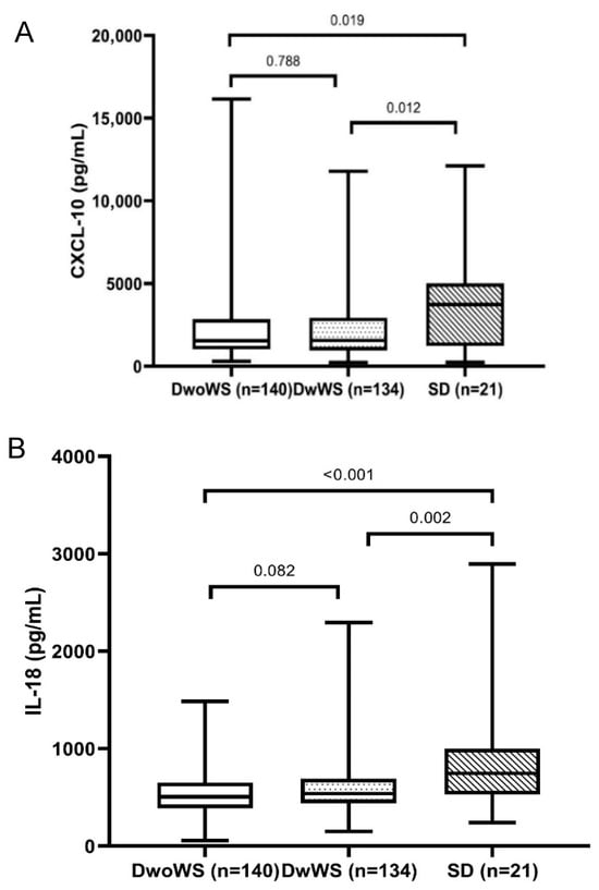
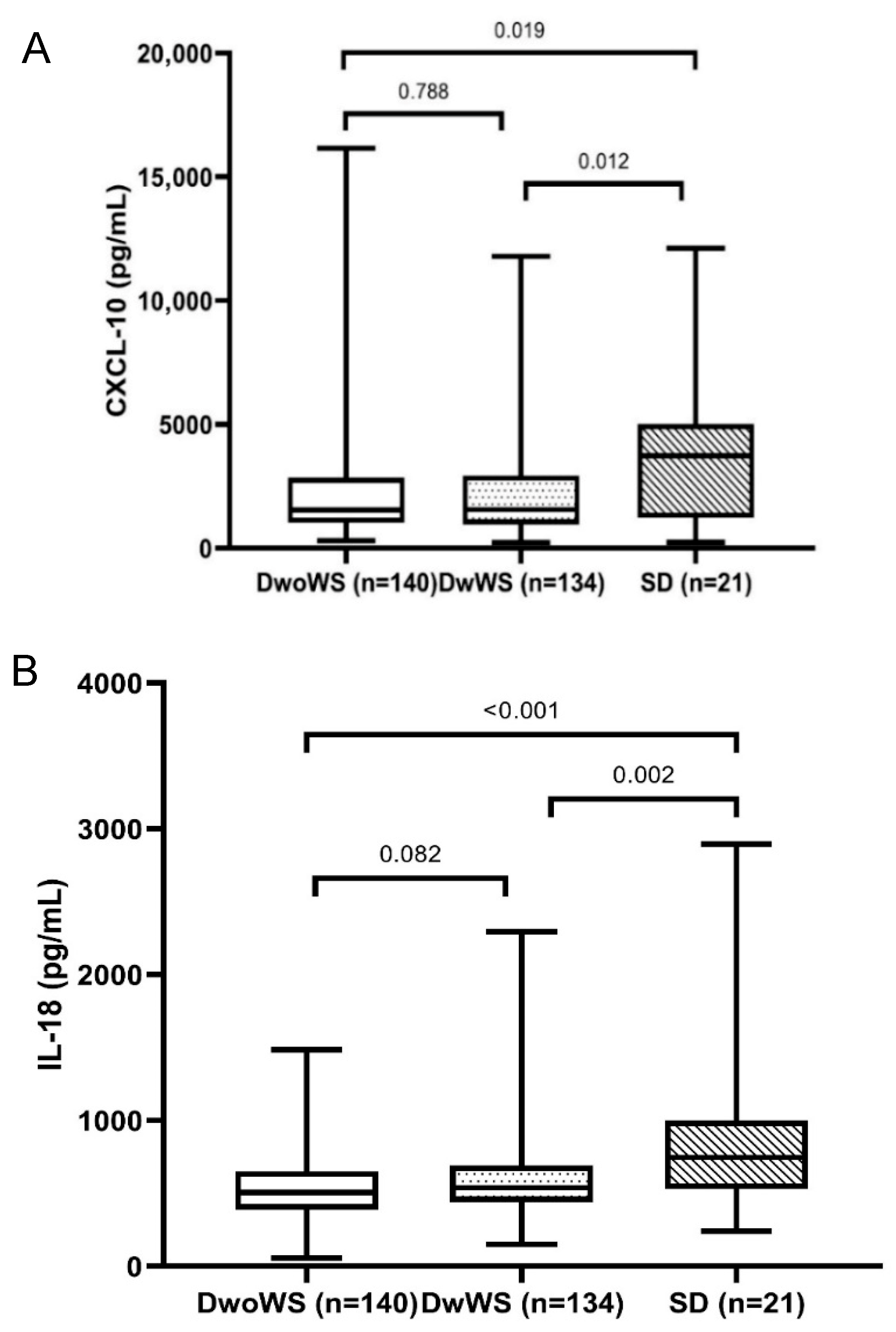
We measured and compared the levels of IL-18 and CXCL-10 with the different severity levels of Dengue. The results are presented in Table 2 and Figure 1. The mean levels of IL-18 pro-inflammatory cytokine in patients with SD were significantly higher as compared with those of the DwoWS (870.64 ± 550.93 pg/mL versus 546.26 ± 227.20 pg/mL, p < 0.001) and DwWS (870.64 ± 550.93 pg/mL versus 588.63 ± 247.78 pg/mL, p = 0.002) groups. However, there was no significant difference in IL-18 levels between the DwoWS and DwWS groups (588.63 ± 247.78 pg/mL versus 546.26 ± 227.20 pg/mL, p = 0.082). Similarly, significantly higher levels of CXCL-10 were detected in the plasma of patients with SD than in the DwoWS and DwWS groups (4013.48 ± 3554.50 pg/mL versus 2282.05 ± 2189.76 pg/mL, p = 0.019 and 2279.58 ± 2073 pg/mL, p = 0.012) (Table 3).
Figure 1.
Comparison of CXCL-10 (A) and IL-18 (B) levels according to Dengue severity. Dengue without warning signs (DwoWS), Dengue with warning signs (DwWS), or severe Dengue (SD).
Table 3.
Association between plasma IL-18 and CXCL-10 concentrations and Dengue severity.
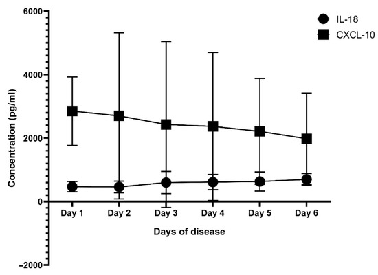
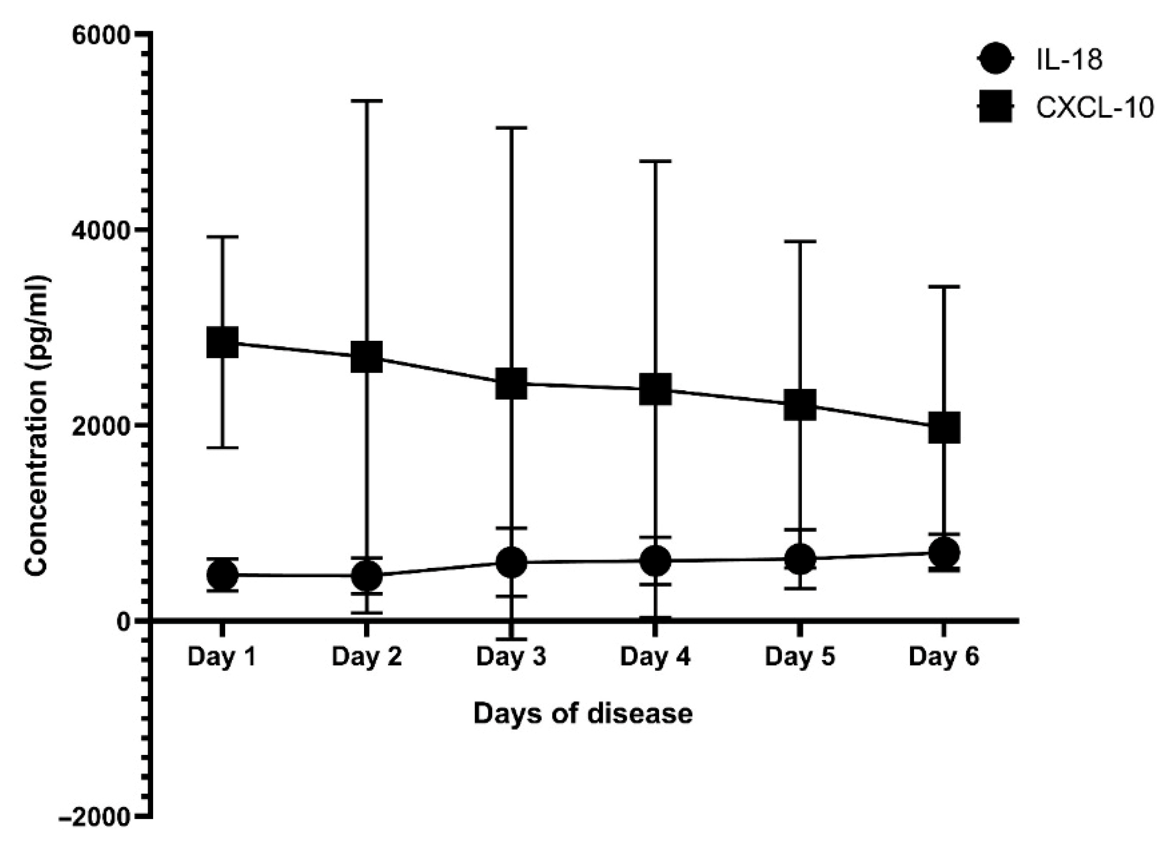
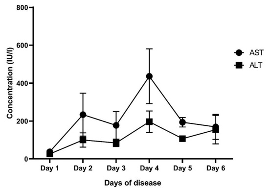
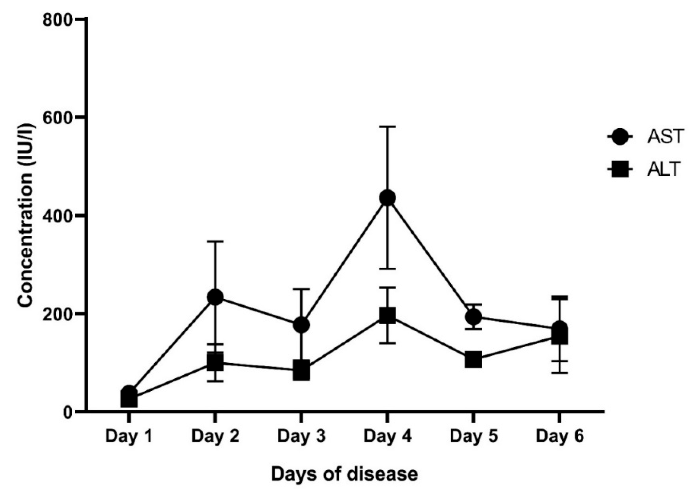
Next, we analyzed the correlation of IL-18 and CXCL-10 with key laboratory parameters. The results are presented in Table 4. There was a positive correlation of IL-18 and CXCL-10 levels with liver enzymes in all patients, but when stratified in each group, we only observed the correlation of liver enzymes with IL-18, whereas CXCL-10 only had a positive correlation with AST levels in the DwoWS (r = 0.219; p = 0.016) and DwWS (r = 0.196; p = 0.03) groups, but there was no correlation in the SD group (p > 0.05). In contrast, platelet counts were negatively correlated with IL-18 levels in all patients (r = −0.361; p < 0.001), DwoWS (r = −0.448; p < 0.001), and DwWS (r = −0.216; p = 0.012). Of note, no correlation was found between CXCL-10 and key laboratory parameters in the SD group. Interestingly, when we analyzed the change in mean IL-18, CXCL-10, and liver enzyme levels according to the days of disease, we observed a consistent elevated IL-18 level during the course of disease, whereas CXCL-10 increased early in the febrile phase of the disease. In contrast, the elevation of liver enzymes tended to peak late in the course of disease (Figure 2 and Figure 3).
Table 4.
The correlation of IL-18 and CXCL-10 with laboratory parameters according to Dengue severity.
Figure 2.
The mean levels of IL-18 and CXCL-10 at different days of disease. AST: aspartate aminotransferase; ALT: alanine aminotransferase.
Figure 3.
The mean levels of AST and ALT at different days of the disease. AST: Aspartate aminotransferase; ALT: Alanine aminotransferase.
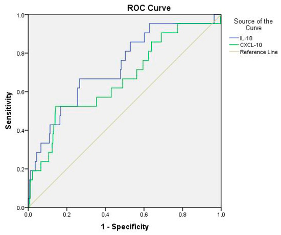
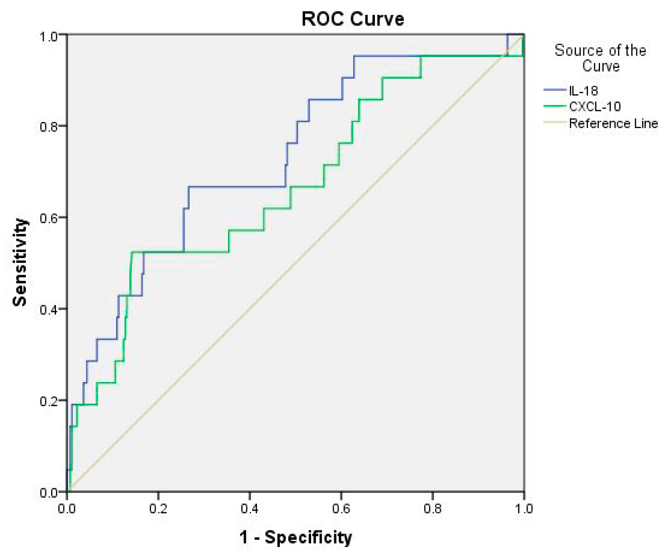
Furthermore, we analyzed the ROC curve to calculate cut-off values of IL-18 and CXCL-10 levels for predicting the severity of Dengue infection. The analysis revealed that the cut-off values of IL-18 were 669.65 (sensitivity: 66.7%; specificity: 73.4%; AUC: 0.729), and CXCL-10 levels were 3739.5 pg/mL (sensitivity: 52.4%; specificity: 85.8%; AUC: 0.665), which provided acceptable discrimination between DwoWS/DwWS and SD. Results are presented in Table 5 and Figure 4.
Table 5.
AUC values obtained from ROC analysis for IL-18 and CXCL-10 associated with Dengue severity.
Figure 4.
ROC curve analysis of IL-18 and CXCL-10 concentrations in predicting severe Dengue fever.
4. Discussions
In this study, we included 295 patients with clinical manifestations of Dengue infection in a tertiary hospital in Hanoi, Vietnam, and analyzed clinical and laboratory findings as related to disease severity. Vietnam is an endemic for Dengue, with an increasing incidence of Dengue reported in recent years, and Dengue infection occurs year-round, with the time of peak outbreak associated with the rainy season, from June to December, every year [11,12]. There have been endemic outbreaks of Dengue in Hanoi with the circulation of all four Dengue serotypes, but Dengue 1 and Dengue 2 are the most predominant serotypes [13,14,15]. In this study, based on the WHO’s 2009 classification [6], we showed that the proportion of severe Dengue was 7.11% (21/295). This is slightly higher than that observed in a previous study of patients admitted to a Referral Hospital in Hanoi [16] but lower than that reported by Huy et al., which showed a rate of 11.4% [12]. Of note, we found significant differences in clinical presentations and laboratory parameters between groups of patients—DwoWS, DwWS, and SD. Patients with SD exhibited a higher frequency of warning signs, including bleeding, hepatomegaly, and abdominal pain, as compared to those with DwoWS and DwWS. Similarly, levels of liver enzymes were elevated in patients with SD compared to those with DwoWS and DwWS (ALT 366.5 IU/l versus 54.5 IU/l and 35 IU/l, p < 0.001, and AST 1040 IU/l versus 104 IU/l and 55 IU/l, p < 0.001). These findings are consistent with previous studies [17], which explained that organ dysfunction and liver damage is common in Dengue infection and is recognized as a main indicator of SD [18]. In addition, abnormalities in hematological parameters showed a significant decline of platelet counts in the SD group, which resulted in an increased risk of bleeding and Dengue shock syndrome. Conversely, white blood cell (WB) counts were notably higher in patients with SD (7.21 G/l versus 3.87 G/l and 3.73 G/l in DwoWS and DwWS, respectively). Therefore, in clinical practice, marked indicators of routine blood tests during the course of Dengue infection could help to distinguish SD from non-SD. Of note, we showed a significant association of Dengue serotypes with disease severity, with a higher proportion of DENV-2 in SD (Table 2). Although all serotypes are the cause of Dengue disease with different manifestations, it has been found that the Asian strain of DENV-2 has a higher likelihood of causing severe disease than the American strain. This could be explained by the fact that the Asian strain of DENV-2 replicates more efficiently in human cells and can infect new mosquitos more effectively, resulting in increased transmission and a larger Dengue endemic outbreak [1,19].
Interestingly, in this study, we measured the levels of IL-18 and CXCL-10 and analyzed the potential association of these biomarkers with liver enzymes and disease severity. In fact, the role of IL-18, a pro-inflammatory cytokine associated with the Th1 immune response, has been implicated in various viral infections [20]. It has been shown that the interaction of virus and host factors results in the production and release of cytokine pro-inflammatory, anti-viral, and immunoregulatory cytokines that contribute to hepatic inflammation [3]. Importantly, the liver is an immune organ, and it plays a central role in the immune response to infections [21,22]. However, a hyperinflammatory response and overactive immune response trigger an excessive release of pro-inflammatory cytokines, which may contribute to vascular permeability, thereby contributing to the severity of the disease. In agreement with other studies [5,23], our study indicated that the mean level of IL-18 was significantly higher in patients with SD (870.64 ± 550.93 pg/mL) compared to the DwoWS (546.26 ± 227.20 pg/mL) and DwWS (588.63 ± 247.78 pg/mL) groups (p < 0.001). Furthermore, we also showed a positive correlation of IL-18 with liver enzymes in all patients, irrespective of severity (Table 4). It has been reported that high elevated liver enzymes are predictors of severe Dengue; however, these indicators appear later in the course of disease. In fact, progression to severe disease commonly occurs after the febrile phase (day 1 to 3), between days 4 and 6 of illness (critical phase). Hence, it was difficult to distinguish between SD and non-severe Dengue in the early stage of infection. In this study, we observed that the elevation of IL-18 levels seems to be early, while liver enzyme levels tend to peak late in the course of disease (Figure 2 and Figure 3). Therefore, the use of IL-18 could predict early disease progression to more severe Dengue. Although there have been several reports that used ROC analysis of AST and ALT to distinguish between severe Dengue and non-severe Dengue, providing the values of AUC in so doing, sensitivity and specificity were different [24,25,26]. This finding suggests that IL-18 may be a potential biomarker for disease progression and liver involvement in Dengue infection. Similarly, CXCL-10, a chemokine, plays an important role in attracting immune cells to sites of infection and immune activation and may thereby contribute to a cascade of cytokine production in Dengue infection [27]. It has been reported in various studies that elevation of IP-10 levels is clearly associated with plasma leakage and liver injury, especially in secondary Dengue infection [5,28,29]. However, CXCL-10 inhibits DENV replication by binding to heparan sulfate on the host membrane, promoting viral clearance [30,31]. In this study, we observed that patients with SD had significantly higher levels of CXCL-10 than those with DwoWS and DwWS (Table 2 and Figure 1). In addition, we revealed a positive correlation of CXCL-10 with liver enzymes in all patients with Dengue. This finding indicates the influence of CXCL-10 on liver enzyme increase and is linked to an indicator of disease severity.
Furthermore, based on the analysis of ROC curves, we determined the cut-off values of IL-18 and CXCL-10 for predicting Dengue severity. The cut-off value of IL-18 was 669.65 pg/mL (sensitivity: 66.7%; specificity: 73.4%; AUC: 0.729), whereas CXCL-10 was 3739.5 pg/mL (sensitivity: 52.4%; specificity: 85.8%; AUC: 0.665) (Table 5 and Figure 4). To the best of our knowledge, there is little information about the role of IL-18 and CXCL-10 in relation to disease severity among Vietnamese patients with Dengue infection. We suggest that these biomarkers are able to support clinicians in early diagnoses of severe Dengue, potentially aiding them in making proper clinical decisions in the monitoring and management of Dengue infection. Further studies should explore the mechanistic role of IL-18 and CXCL10 in Dengue pathogenesis and their potential as therapeutic targets for managing severe cases of the disease.
The present study had several limitations. This was a single-center and cross-sectional design study. Blood samples of patients with Dengue were only collected at a time point of admission for measuring of IL-18 and CXCL-10; therefore, we did not monitor dynamic changes in IL-18 and CXCL-10 levels during the course of the disease. In addition, we did not determine the history status of Dengue infection, which could have led to a difference in results. Finally, the number of patients with SD was small. Therefore, prospective studies with a larger sample size and molecular characterization of the Dengue virus will be necessary to validate our results.
5. Conclusions
This study indicates that IL-18 and CXCL-10 levels are significantly higher in SD compared to DwoWS and DwWS and were associated with disease severity among Vietnamese patients with Dengue infection. These findings suggest that IL-18 and CXCL-10 may be biomarkers for disease progression and disease severity in Dengue infection.
Author Contributions
Conceptualization: Conceptualization, L.H.T., C.D.D. and S.X.H.; methodology, S.X.H. and C.D.D.; software, B.H.N.; validation, P.M.N.; formal analysis, L.H.T. and B.H.N.; investigation, L.H.T., C.D.D. and S.X.H.; resources, L.H.T. and S.X.H.; data curation, M.D.C.; writing—original draft preparation, L.H.T.; writing—review and editing, C.D.D. and S.X.H.; visualization, L.T.N. and C.X.H.; supervision, C.D.D. and S.X.H.; project administration, C.D.D. All authors have read and agreed to the published version of the manuscript.
Funding
This research received no external funding.
Institutional Review Board Statement
The local ethics committee of Bach Mai hospital, Hanoi, Vietnam, approved this study with code 7210/QĐ-BM. The date of approval was 31 December 2024.
Informed Consent Statement
Informed consent was obtained from all subjects involved in the study.
Data Availability Statement
Data are available upon reasonable request.
Acknowledgments
The authors thank all the patients who participated in this study. We thank Nguyen Thuy Linh for their excellent laboratory assistance in this study.
Conflicts of Interest
The authors declare no conflicts of interest.
Abbreviations
The following abbreviations are used in this manuscript:
| IL-18 | Interleukin 18 |
| CXCL-10 | chemokines C-X-C motif ligand-10 |
| WHO | World Health Organization |
References
- Guzman, M.G.; Gubler, D.J.; Izquierdo, A.; Martinez, E.; Halstead, S.B. Dengue Infection. Nat. Rev. Dis. Primers 2016, 2, 16055. [Google Scholar] [CrossRef] [Scilit]
- Paz-Bailey, G.; Adams, L.E.; Deen, J.; Anderson, K.B.; Katzelnick, L.C. Dengue. Lancet 2024, 403, 667–682. [Google Scholar] [CrossRef] [Scilit] [PubMed]
- Screaton, G.; Mongkolsapaya, J.; Yacoub, S.; Roberts, C. New Insights into the Immunopathology and Control of Dengue Virus Infection. Nat. Rev. Immunol. 2015, 15, 745–759. [Google Scholar] [CrossRef] [Scilit] [PubMed]
- Patro, A.R.K.; Mohanty, S.; Prusty, B.K.; Singh, D.K.; Gaikwad, S.; Saswat, T.; Chattopadhyay, S.; Das, B.K.; Tripathy, R.; Ravindran, B. Cytokine Signature Associated with Disease Severity in Dengue. Viruses 2019, 11, 34. [Google Scholar] [CrossRef] [Scilit] [PubMed]
- Fiestas Solórzano, V.E.; da Costa Faria, N.R.; Dos Santos, C.F.; Corrêa, G.; Cipitelli, M.d.C.; Dornelas Ribeiro, M.; de Souza, L.J.; Damasco, P.V.; da Cunha, R.V.; Dos Santos, F.B.; et al. Different Profiles of Cytokines, Chemokines and Coagulation Mediators Associated with Severity in Brazilian Patients Infected with Dengue Virus. Viruses 2021, 13, 1789. [Google Scholar] [CrossRef] [Scilit]
- World Health Organization. Dengue: Guidelines for Diagnosis, Treatment, Prevention and Control: New Edition; WHO Guidelines Approved by the Guidelines Review Committee; World Health Organization: Geneva, Switzerland, 2009; ISBN 978-92-4-154787-1. [Google Scholar]
- Robinson, M.; Einav, S. Towards Predicting Progression to Severe Dengue. Trends Microbiol. 2020, 28, 478–486. [Google Scholar] [CrossRef] [Scilit]
- Tejo, A.M.; Hamasaki, D.T.; Menezes, L.M.; Ho, Y.-L. Severe Dengue in the Intensive Care Unit. J. Intensive Med. 2024, 4, 16–33. [Google Scholar] [CrossRef] [Scilit]
- Dang, T.T.; Pham, M.H.; Bui, H.V.; Van Le, D. Whole Genome Sequencing and Genetic Variations in Several Dengue Virus Type 1 Strains from Unusual Dengue Epidemic of 2017 in Vietnam. Virol. J. 2020, 17, 7. [Google Scholar] [CrossRef] [Scilit]
- Lanciotti, R.S.; Calisher, C.H.; Gubler, D.J.; Chang, G.J.; Vorndam, A.V. Rapid Detection and Typing of Dengue Viruses from Clinical Samples by Using Reverse Transcriptase-Polymerase Chain Reaction. J. Clin. Microbiol. 1992, 30, 545–551. [Google Scholar] [CrossRef] [Scilit]
- Lee, H.S.; Nguyen-Viet, H.; Nam, V.S.; Lee, M.; Won, S.; Duc, P.P.; Grace, D. Seasonal Patterns of Dengue Fever and Associated Climate Factors in 4 Provinces in Vietnam from 1994 to 2013. BMC Infect. Dis. 2017, 17, 218. [Google Scholar] [CrossRef] [Scilit]
- Huy, B.V.; Hoa, L.N.M.; Thuy, D.T.; Van Kinh, N.; Ngan, T.T.D.; Duyet, L.V.; Hung, N.T.; Minh, N.N.Q.; Truong, N.T.; Chau, N.V.V. Epidemiological and Clinical Features of Dengue Infection in Adults in the 2017 Outbreak in Vietnam. Biomed Res. Int. 2019, 2019, 3085827. [Google Scholar] [CrossRef] [Scilit]
- Lytton, S.D.; Nematollahi, G.; van Tong, H.; Xuan Anh, C.; Hung, H.V.; Hoan, N.X.; Diez, G.; Schumacher, T.; Landt, O.; Melchior, W.; et al. Predominant Secondary Dengue Infection among Vietnamese Adults Mostly without Warning Signs and Severe Disease. Int. J. Infect. Dis. 2020, 100, 316–323. [Google Scholar] [CrossRef] [Scilit]
- Phadungsombat, J.; Vu, H.T.T.; Nguyen, Q.T.; Nguyen, H.T.V.; Nguyen, H.T.N.; Dang, B.T.; Nakayama, E.E.; Ishizaki, A.; Ichimura, H.; Shioda, T.; et al. Molecular Characterization of Dengue Virus Strains from the 2019–2020 Epidemic in Hanoi, Vietnam. Microorganisms 2023, 11, 1267. [Google Scholar] [CrossRef] [Scilit]
- Anh, D.D.; The, N.T.; My, T.N.; Linh, L.T.K.; Hoan, N.X.; Kremsner, P.G.; Toan, N.L.; Song, L.H.; Velavan, T.P. Epidemiology and Genotype Dynamics of Dengue in Hospitalized Patients in Northern Vietnam between 2020–2022. Open Forum Infect. Dis. 2024, 12, ofae753. [Google Scholar] [CrossRef] [Scilit]
- Taylor, W.R.; Fox, A.; Pham, K.T.; Le, H.N.M.; Tran, N.T.H.; Tran, G.V.; Nguyen, B.T.; Nguyen, M.V.; Nguyen, L.T.; Yacoub, S.; et al. Dengue in Adults Admitted to a Referral Hospital in Hanoi, Vietnam. Am. J. Trop. Med. Hyg. 2015, 92, 1141–1149. [Google Scholar] [CrossRef] [Scilit] [PubMed]
- Trung, D.T.; Thao, L.T.T.; Hien, T.T.; Hung, N.T.; Vinh, N.N.; Hien, P.T.D.; Chinh, N.T.; Simmons, C.; Wills, B. Liver Involvement Associated with Dengue Infection in Adults in Vietnam. Am. J. Trop. Med. Hyg. 2010, 83, 774–780. [Google Scholar] [CrossRef] [Scilit] [PubMed]
- Imad, H.A.; Phumratanaprapin, W.; Phonrat, B.; Chotivanich, K.; Charunwatthana, P.; Muangnoicharoen, S.; Khusmith, S.; Tantawichien, T.; Phadungsombat, J.; Nakayama, E.; et al. Cytokine Expression in Dengue Fever and Dengue Hemorrhagic Fever Patients with Bleeding and Severe Hepatitis. Am. J. Trop. Med. Hyg. 2020, 102, 943–950. [Google Scholar] [CrossRef] [Scilit] [PubMed]
- Bhatt, P.; Sabeena, S.P.; Varma, M.; Arunkumar, G. Current Understanding of the Pathogenesis of Dengue Virus Infection. Curr. Microbiol. 2021, 78, 17–32. [Google Scholar] [CrossRef] [Scilit]
- Pirhonen, J.; Sareneva, T.; Kurimoto, M.; Julkunen, I.; Matikainen, S. Virus Infection Activates IL-1β and IL-18 Production in Human Macrophages by a Caspase-1-Dependent Pathway. J. Immunol. 1999, 162, 7322–7329. [Google Scholar] [CrossRef] [Scilit]
- Racanelli, V.; Rehermann, B. The Liver as an Immunological Organ. Hepatology 2006, 43, S54–S62. [Google Scholar] [CrossRef] [Scilit]
- Protzer, U.; Maini, M.K.; Knolle, P.A. Living in the Liver: Hepatic Infections. Nat. Rev. Immunol. 2012, 12, 201–213. [Google Scholar] [CrossRef] [Scilit]
- Patra, G.; Saha, B.; Mukhopadhyay, S. The Relationship between Changes in IL-2/IL-18 and Liver Enzyme with Dengue Severity. Cytokine 2021, 148, 155713. [Google Scholar] [CrossRef] [Scilit]
- Lee, L.K.; Gan, V.C.; Lee, V.J.; Tan, A.S.; Leo, Y.S.; Lye, D.C. Clinical Relevance and Discriminatory Value of Elevated Liver Aminotransferase Levels for Dengue Severity. PLoS Negl. Trop. Dis. 2012, 6, e1676. [Google Scholar] [CrossRef] [Scilit] [PubMed]
- Zhang, H.; Xie, Z.; Xie, X.; Ou, Y.; Zeng, W.; Zhou, Y. A Novel Predictor of Severe Dengue: The Aspartate Aminotransferase/Platelet Count Ratio Index (APRI). J. Med. Virol. 2018, 90, 803–809. [Google Scholar] [CrossRef] [Scilit]
- Alam, R.; Fathema, K.; Yasmin, A.; Roy, U.; Hossen, K.; Rukunuzzaman, M. Prediction of Severity of Dengue Infection in Children Based on Hepatic Involvement. JGH Open 2024, 8, e13049. [Google Scholar] [CrossRef] [Scilit]
- Rathakrishnan, A.; Wang, S.M.; Hu, Y.; Khan, A.M.; Ponnampalavanar, S.; Lum, L.C.S.; Manikam, R.; Sekaran, S.D. Cytokine Expression Profile of Dengue Patients at Different Phases of Illness. PLoS ONE 2012, 7, e52215. [Google Scholar] [CrossRef] [Scilit]
- Patra, G.; Mallik, S.; Saha, B.; Mukhopadhyay, S. Assessment of Chemokine and Cytokine Signatures in Patients with Dengue Infection: A Hospital-Based Study in Kolkata, India. Acta Trop. 2019, 190, 73–79. [Google Scholar] [CrossRef] [Scilit]
- Gowri Sankar, S.; Alwin Prem Anand, A. Cytokine IP-10 and GM-CSF Are Prognostic Biomarkers for Severity in Secondary Dengue Infection. Hum. Immunol. 2021, 82, 438–445. [Google Scholar] [CrossRef] [Scilit] [PubMed]
- Chen, J.-P.; Lu, H.-L.; Lai, S.-L.; Campanella, G.S.; Sung, J.-M.; Lu, M.-Y.; Wu-Hsieh, B.A.; Lin, Y.-L.; Lane, T.E.; Luster, A.D.; et al. Dengue Virus Induces Expression of CXC Chemokine Ligand 10/IFN-Gamma-Inducible Protein 10, Which Competitively Inhibits Viral Binding to Cell Surface Heparan Sulfate. J. Immunol. 2006, 177, 3185–3192. [Google Scholar] [CrossRef] [Scilit] [PubMed]
- Ip, P.-P.; Liao, F. Resistance to Dengue Virus Infection in Mice Is Potentiated by CXCL10 and Is Independent of CXCL10-Mediated Leukocyte Recruitment. J. Immunol. 2010, 184, 5705–5714. [Google Scholar] [CrossRef] [Scilit]

Disclaimer/Publisher’s Note: The statements, opinions and data contained in all publications are solely those of the individual author(s) and contributor(s) and not of MDPI and/or the editor(s). MDPI and/or the editor(s) disclaim responsibility for any injury to people or property resulting from any ideas, methods, instructions or products referred to in the content. |
© 2025 by the authors. Licensee MDPI, Basel, Switzerland. This article is an open access article distributed under the terms and conditions of the Creative Commons Attribution (CC BY) license (https://creativecommons.org/licenses/by/4.0/).