


Abstract
Background/Objectives: The role of artificial intelligence (AI) in dentistry is becoming increasingly significant, particularly in diagnosis and treatment planning. This study aimed to assess the sensitivity, specificity, accuracy, and precision of AI-driven software in analyzing dental panoramic radiographs (DPRs) in patients with permanent dentition. Methods: Out of 638 DPRs, 600 fulfilled the inclusion criteria. The radiographs were analyzed by AI software and two researchers. The following variables were assessed: (1) missing tooth, (2) root canal filling, (3) endodontic lesion, (4) implant, (5) abutment, (6) pontic, (7) crown, (8) and sound tooth. Results: The study revealed very high performance metrics for the AI algorithm in detecting missing teeth, root canal fillings, and implant abutment crowns, all greater than 90%. However, it demonstrated moderate sensitivity and precision in identifying endodontic lesions and the lowest precision (65.30%) in detecting crowns. Conclusions: AI software can be a valuable tool in clinical practice for diagnosis and treatment planning but may require additional verification by clinicians, especially for identifying endodontic lesions and crowns. Due to some limitations of the study, further research is recommended.
1. Introduction
Radiography plays an important role in diagnostics and treatment planning in modern dentistry. It provides information on anatomy, pathologies, and treatment outcomes. One of the most widely used is a dental panoramic radiograph (DPR). This extraoral technique produces a comprehensive view of dental arches, maxilla, mandible, temporomandibular joints, and partially maxillary sinuses [1]. It is used in many fields of dentistry. In orthodontics, it allows the detection of dental anomalies, evaluation of general dental health, and observation of treatment. DPR also supports maxillofacial surgery in diagnosing dental impactions, dental and mandibular fractures, cysts, and tumors [2]. It involves a small dose of ionizing radiation compared to a dose of cone beam computed tomography (CBCT), which, depending on the scan mode, is about three to six times higher than for DPRs [3,4]. Panoramic radiographs are widely used in epidemiological and screening studies, providing information on the general oral health of large groups of patients [5]. However, they have limitations such as superimposition, distortions, and ghost images [6,7]. Superimposition is the overlapping of structures in the X-ray path and can be caused by tongue rings or developing permanent teeth in the primary dentition [2]. The level of distortion depends on the machine type and the distance between the film and the patient [8]. Ghost images may result from anatomical structures like the cervical spine reflecting over the lower incisor. These artifacts can also be caused by jewelry, including earrings [2]. DPRs lack fine details compared to intraoral radiographs [9]. Therefore, it is not sufficient for examining, for example, proximal dental caries [10]. Similar to other radiographic examinations, DPRs have limited inter- and intra-examiner reliability [11]. Inter-examiner reliability describes the level of agreement among independent examiners when applying a test to the same patient [12]. Intra-examiner reliability is the consistency of an examiner in documenting the same conditions over time [13].
In addition to radiographic imaging, visual-tactile examination, caries detection dye, transillumination, pulp vitality testing, and probing pocket depths are also valuable diagnostic tests in dentistry, crucial for obtaining an accurate diagnosis [14,15]. Early detection of pathological conditions allows for timely interventions and effective dental prevention. Diagnostic tests are also important for monitoring oral health, evaluating treatment outcomes, and assessing risks for dental diseases. They can also be used to screen populations, providing data that inform about the need to implement certain actions and strategies [16].
Artificial intelligence (AI) is the ability of machines to perform tasks traditionally associated with human intelligence, such as learning and problem-solving [17]. It is increasingly applied in dentistry and, according to Thurzo et al., is implemented mainly in radiology and orthodontics [18]. This technology can also be used in detecting root fractures, analyzing the anatomy of the root canal system, and aiding clinicians with working length determination [19]. Machine learning in CAD/CAM software can help manufacture well-made fixed and removable dental restorations [18]. Dental radiography such as CBCT, intraoral, panoramic, and cephalometric radiographs provides large data sets for developing AI-based software [20]. When evaluating DPRs, these algorithms achieve an accuracy of about 90% in detecting caries, osteoporosis, maxillary sinusitis, periodontal bone loss, and teeth identification and numbering on DPRs. Detection of periapical lesions is also characterized by high specificity and sensitivity above 90% [21].
According to the study by Gunec et al., AI can generate faster, more accurate diagnoses than junior dentists with one or two years of experience in detecting periapical lesions [22]. Early detection of pathologies allows rapid implementation of appropriate treatment. AI is also a promising tool in medical screening, as it provides accurate and cost-effective results in less time than traditional methods [23]. Its ability to analyze large data sets facilitates population oral health surveillance [24]. This technology also reduces the risk of human errors resulting from, for example, examiner fatigue [25]. By obtaining a second opinion from the program, dentists can validate their radiograph evaluation and better explain the rationale of the planned treatment.
In the overview of reviews from 2023, twelve systematic reviews regarding the application of AI in the automatic evaluation of DPRs were analyzed [21]. Comparing different algorithms was challenging due to the heterogeneity of the studies, with differences in performance metrics reaching 55%. Possible causes include applying different AI on sample sizes ranging from fifty-five to over a thousand DPRs. The algorithms were used to identify periapical lesions; root canal fillings; and metal- and resin-based restorations, crowns, or implants [26,27,28,29]. Currently, there are very few studies with large sample sizes that assess multiple parameters. This deficiency became the basis for designing and implementing the study reported in this paper.
This study aims to validate AI software as a diagnostic tool and evaluate its sensitivity, specificity, precision, and accuracy in assessing permanent teeth on DPRs. The null hypothesis is stated as follows: “The accuracy of AI software in detecting dental conditions on DPRs is equal to that of human analysis”.
2. Materials and Methods
2.1. Study Design
This prospective, double-gate study on diagnostic accuracy followed the Standards for Reporting of Diagnostic Accuracy Studies (STARD) and the Checklist for Artificial Intelligence in Medical Imaging (CLAIM) checklists. The research was approved by the Bioethics Committee in Kielce at the Świętokrzyska Chamber of Physicians (approval number: 2.3/2023) and was conducted according to the principles of the Declaration of Helsinki. The National Clinical Trial number assigned to this study is NCT06258798. Patients fulfilling specific eligibility criteria were included. Data was collected in real-time, after the study had begun. The research established two gates with distinct eligibility criteria for AI and human, essential for accurately comparing their diagnostic performances.
2.2. Study Population
Patients included in the study were admitted to the radiology department in Kielce, a central European city with about 200,000 inhabitants, and the capital of Świętokrzyskie province in Poland. The eligibility criteria are presented in Table 1. The participant sampling was consecutive. A single reference standard was used for all patients.
Table 1.
Inclusion and exclusion criteria.
2.3. Setting
DPRs were taken with the device Carestream CS 9600 (Carestream Health, Rochester, New York, NY, USA) with adjustable exposure conditions set to 60–90 kV and 2–15 mA. Only appropriate quality X-rays, which were performed according to the criteria of the Polish Ministry of Health, were analyzed. The AI algorithm used in the study was integrated with CS Imaging software (version 8, Carestream Dental LLC, Atlanta, GA, USA). After DPRs had been anonymized, the software analyzed the uploaded X-rays (index test). The report included the location of missing teeth, root canal fillings, endodontic lesions, dental crowns, pontics, implants, and implant abutment crowns. Then, radiographs were analyzed by two independent clinical evaluators (N.T. and M.C.) with 4 and 12 years of experience, respectively (reference test). Human analysis was considered the gold reference standard.
2.4. Study Size
The required sample size was calculated using the Sample Size Estimation for Diagnostic Accuracy Studies tool [30]. The highest sensitivity or specificity values from the latest publications were considered [21,26,27,29]. The sensitivity value was preferred, and if not available, specificity was chosen. In the recent overview of reviews, the highest sensitivity of detecting missing teeth was 98.1%, while the specificity of identifying endodontic lesions reached 90.24% [21]. According to Kazimierczak et al., the sensitivity of automatic detection of root canal fillings on DPRs achieved 90.7% [26]. Identifying crowns presented a specificity of 95.73% [27]. In the study of Başaran et al., the sensitivity of detecting implants, implant abutment crowns, and pontics was 96.15%, 89.47%, and 77.38%, respectively [29]. We assumed a 5% type I error and a marginal error of 5%.
The actual sample size was 600 DPRs due to the capabilities of researchers—19,200 tests were performed by AI and investigators for each variable. This size was significantly larger than the estimated size for variables: missing (186), root canal filling (2048), endodontic treatment (139), and crown (3470). However, it might be insufficient for the following variables: implant (29,937), implant abutment crown (103,403), and pontic (34,045).
2.5. Variables
Table 2 presents the analyzed variables and abbreviations used. The values of variables (1) missing teeth, (2) root canal filling, (3) endodontic lesion, (4) implant, (5) implant abutment crown, (6) pontic crown, (7) dental abutment crown, and (8) sound teeth for analysis performed by AI and investigators were grouped depending on the location of the tooth according to World Dental Federation (FDI) notation, ISO 3950. In this system, each tooth is given a unique two-digit number. The first digit indicates the quadrant of the mouth (the number 1 represents the upper right quadrant, 2—the upper left quadrant, 3—the lower left quadrant, and 4—the lower right quadrant). The second digit indicates the position of the tooth within that quadrant (the number 1,2—incisors, 3—canines, 4,5—premolars, 6,7,8—molars) [31].
Table 2.
Variables.
2.6. Analysis
The acquired data were analyzed in the Microsoft Office program (Microsoft Corporation, Redmond, WA, USA), Google Workspace (version 2024.05.31, Google LLC, Mountain View, CA, USA), and MedCalc software (version 23.0.1; MedCalc Software Ltd., Ostend, Belgium). Based on the analyses performed by AI and investigators, the number of true positive, true negative, false positive, and false negative values were calculated and then used to assess the sensitivity =, specificity =, precision =, and accuracy = of the AI program. TP, TN, FP, and FN denote true positives, true negatives, false positives, and false negatives, respectively. To evaluate the results, guideline definitions of “very high”, “high”, “moderate”, and “low” were used (Table A1) [32].
3. Results
3.1. Participants
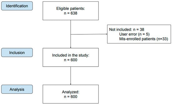
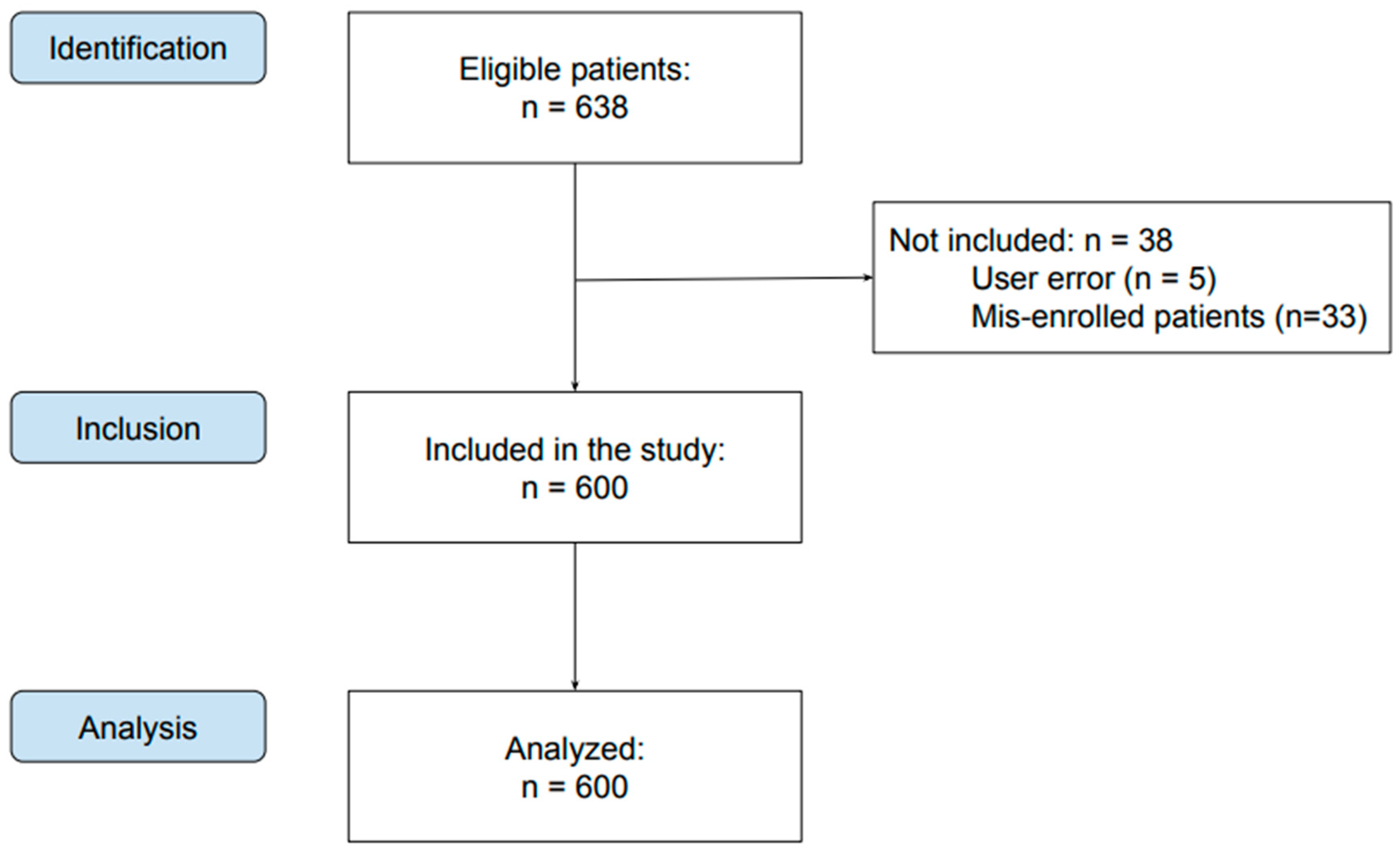
The study involved 638 patients. Data on 38 participants were not obtained due to user error (n = 5) or not meeting the eligibility criteria (n = 33). Finally, 600 patients were included in the study—337 females and 263 males (Figure 1). The youngest patient was 11 years old, and the oldest was 81. The average age was 34.78 (SD = 14.48) years.
Figure 1.
Flow diagram.
3.2. Test Results
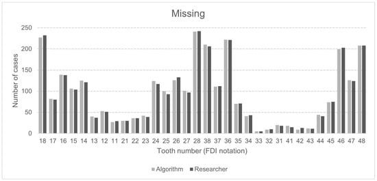
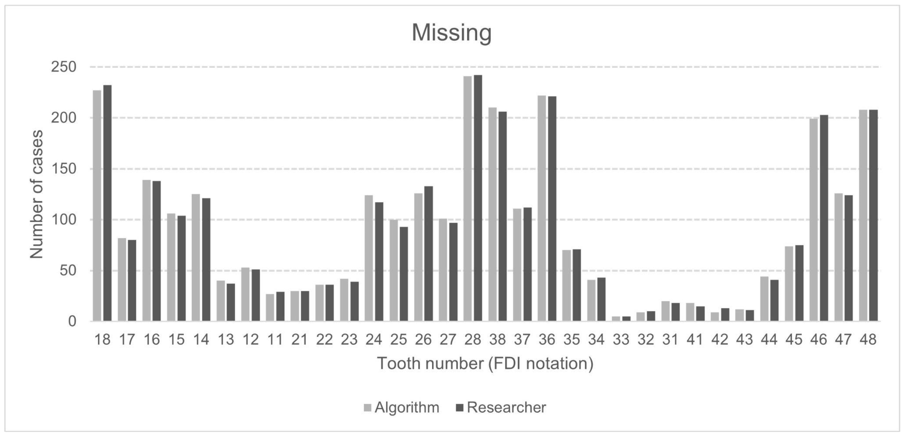
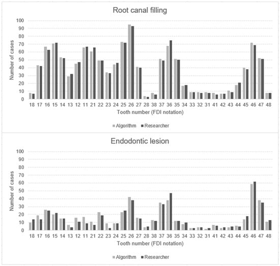
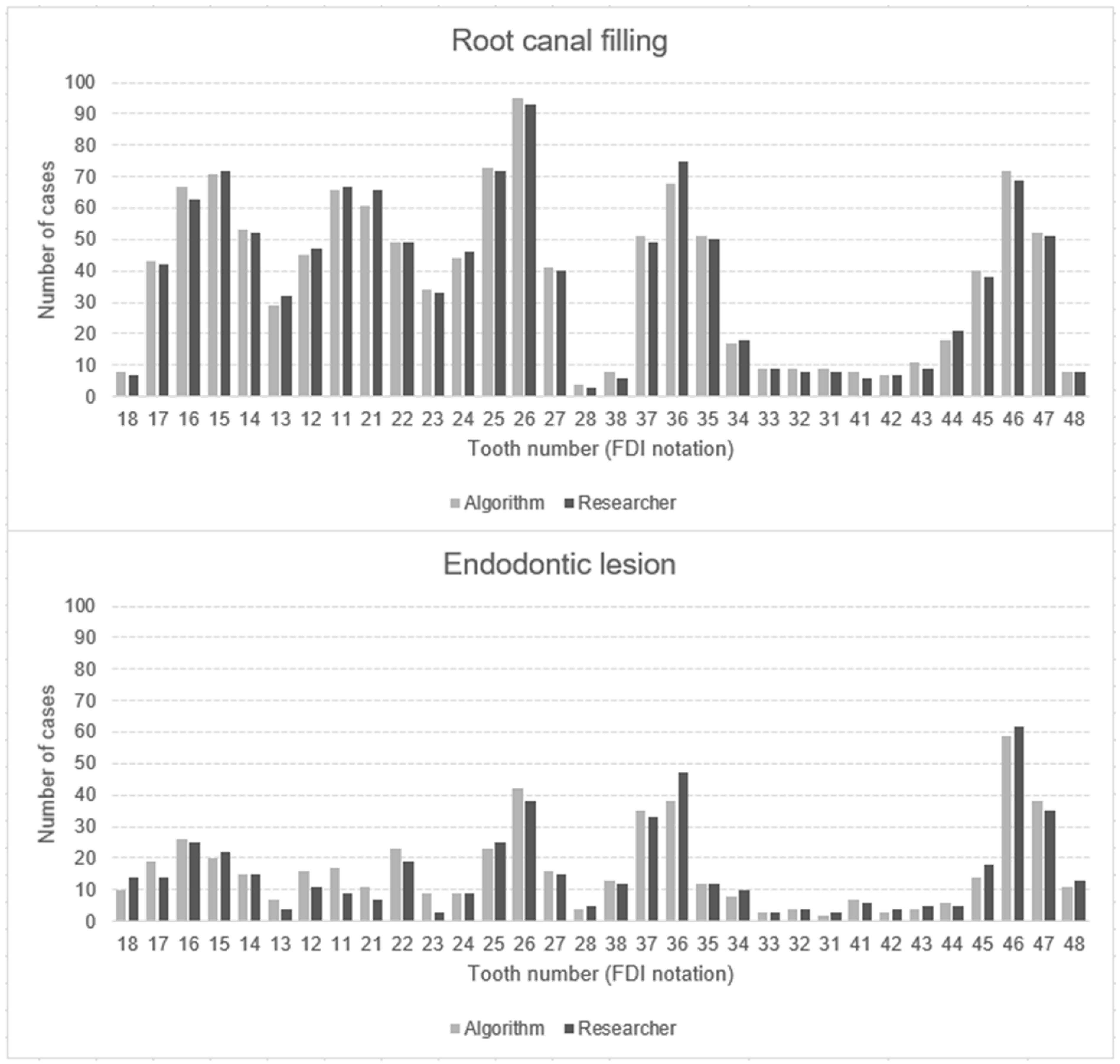
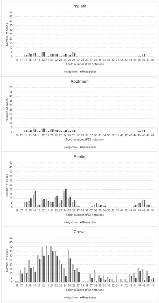
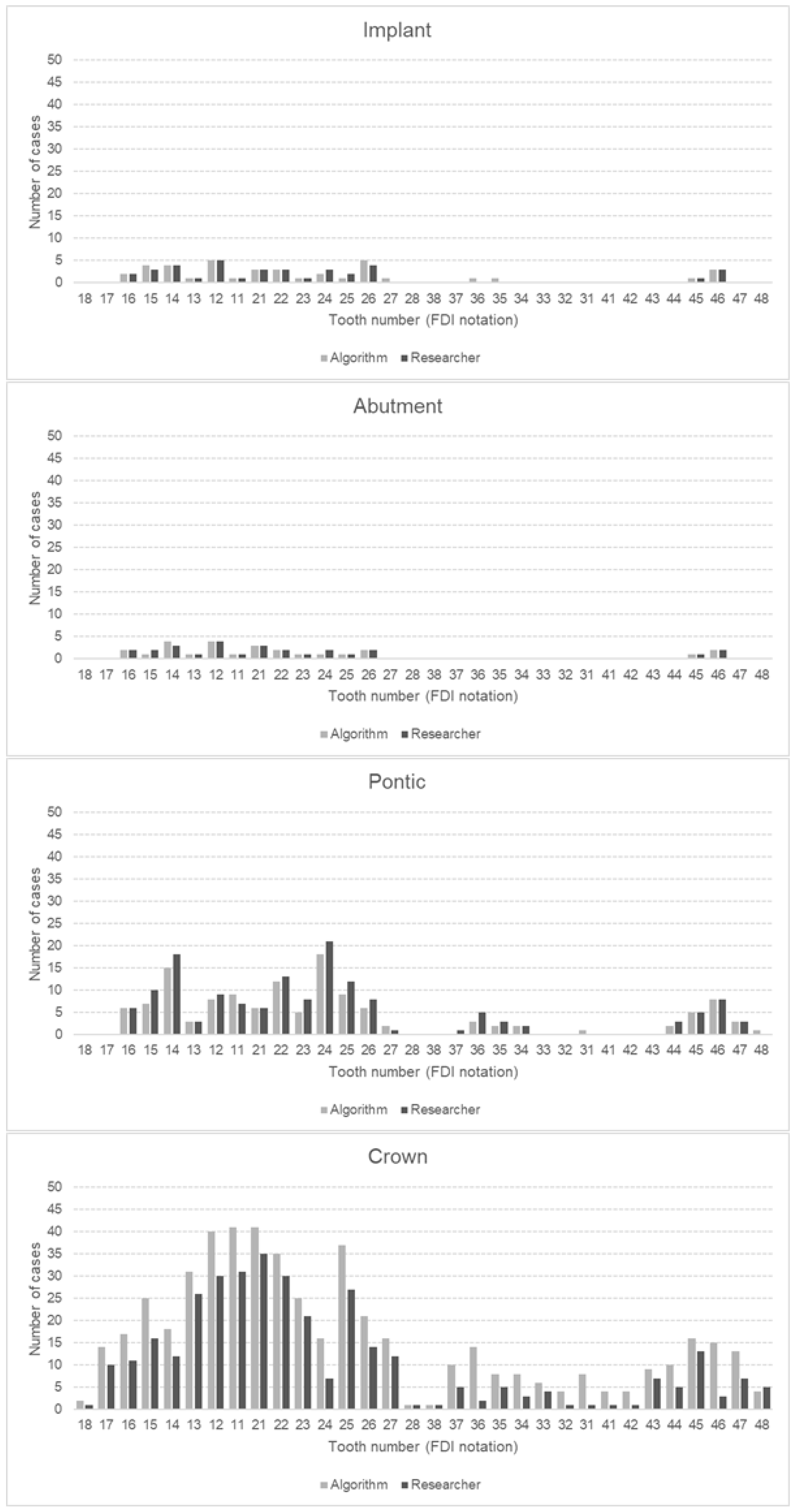
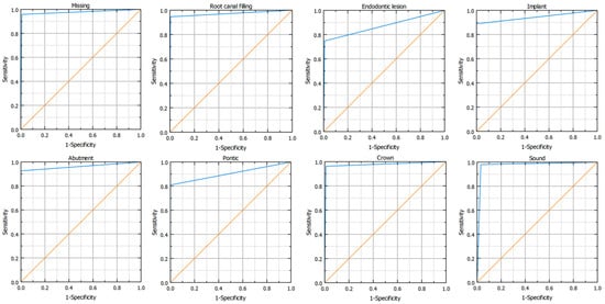
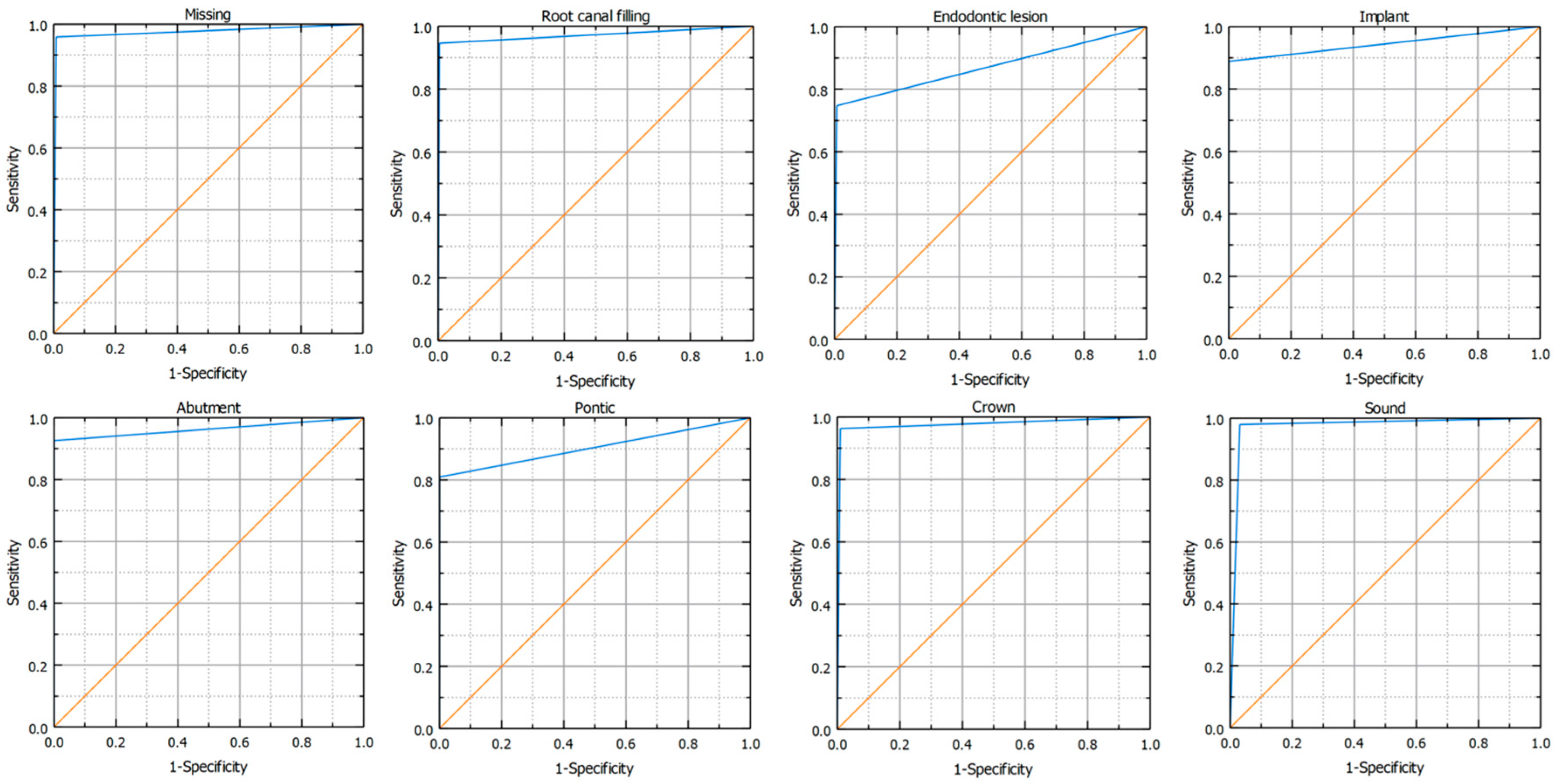
In total, both the principal investigator and the AI made 153,600 binary decisions for eight variables and 32 oral positions in 600 DPRs. The researcher had concerns assessing 281 (1.46%) of 19,200 oral locations. The second investigator verified these cases. Table 3 presents the raw results for individual variables and values of sensitivity, specificity, precision, and accuracy. The numbers of teeth with each diagnosis identified by AI and investigators are presented in Figure 2, Figure 3 and Figure 4. Figure 2 illustrates frequency of missing teeth according to their position in both dental arches. The distributions of teeth with endodontic lesions or root canal fillings are presented in Figure 3. Figure 4 provides a comprehensive overview of prosthetic restorations detected on DPRs, showing the comparison in the number of given diagnoses between humans and AI. The receiver operating characteristic curve (ROC) for each variable is presented in Figure 5.
Table 3.
Outcomes.
Figure 2.
Number of missing teeth detected by AI algorithm and human examiner, with positional detail in upper (18–28) and lower (38–48) dental arch. Position numbering according to ISO 3950:2016 FDI notation (version 2022, FDI World Dental Federation, Geneva, Switzerland) [31].
Figure 3.
Number of root canal fillings and endodontic lesions detected by AI algorithm and human examiner, with positional detail in upper (18–28) and lower (38–48) dental arch. Position numbering according to ISO 3950:2016 FDI notation (version 2022, FDI World Dental Federation, Geneva, Switzerland) [31].
Figure 4.
Number of implants, implant abutment crowns, pontic crowns, and dental abutment crowns detected by AI algorithm and human examiner, with positional detail in upper (18–28) and lower (38–48) dental arch. Position numbering according to ISO 3950:2016 FDI notation (version 2022, FDI World Dental Federation, Geneva, Switzerland) [31].
Figure 5.
ROC curve representations for the studied variables. The random classifier (diagonal line) is orange, and the ROC curve for the classifier is blue.
4. Discussion
4.1. Missing Teeth
Research in recent years has shown a very high effectiveness in identifying missing teeth using AI on panoramic X-rays [33,34,35]. According to Tuzoff et al., the performance of tested CNN-based architecture was comparable to that of experts, which can simplify the filling in of digital dental charts [36]. Vinayahalingam et al., after testing 200 DPRs by the Mask R-CNN architecture, achieved a precision of 99.7% [37]. The results of the present study are also promising. The algorithm detected missing teeth, achieving all analyzed performance metrics above 95%. The sensitivity and specificity were similar to the study from 2024, where CNNs were used for automatic evaluation [38]. The algorithm in the present study marked 22 more teeth as missing than the researchers did. The most common diagnosis differences between AI and clinicians occurred in teeth 24, 25, and 26, while no differences were observed in teeth 21, 22, 33, and 48. Problems with the correct identification of missing teeth for both AI and dentists may occur in the residual dentition when the remaining teeth move toward the edentulous space, leading to their displacement from the proper position (Popov–Godon phenomenon). The algorithm also did not correctly identify teeth when one tooth covered the other, which occurred in the case of crowding or the presence of an impacted tooth. Sometimes, the developing wisdom tooth buds were not detected by the software, either.
4.2. Endodontic Lesions and Root Canal Fillings
All analyzed performance metrics for detecting root canal fillings (RCFs) were very high, with values above 94%. These metrics indicate significantly good reliability in making accurate predictions. Other studies also present good results in identifying endodontically treated teeth [26,27]. In a study from 2024, another popular cloud-based AI software, Diagnocat, achieved high accuracy (90.72%) in detecting the probability of fillings [26]. Bonfanti Gris et al., using web-based software Denti.AI, achieved accuracy, specificity, and sensitivity above 90% as well [26,27]. The most common tooth with a RCF was 26. The first molars are the most susceptible to caries because of their early eruption and morphology [39]. Endodontic treatment was least frequently performed in the lower incisors. They are least likely to experience caries because of the large amount of saliva produced by the submandibular salivary glands [40].
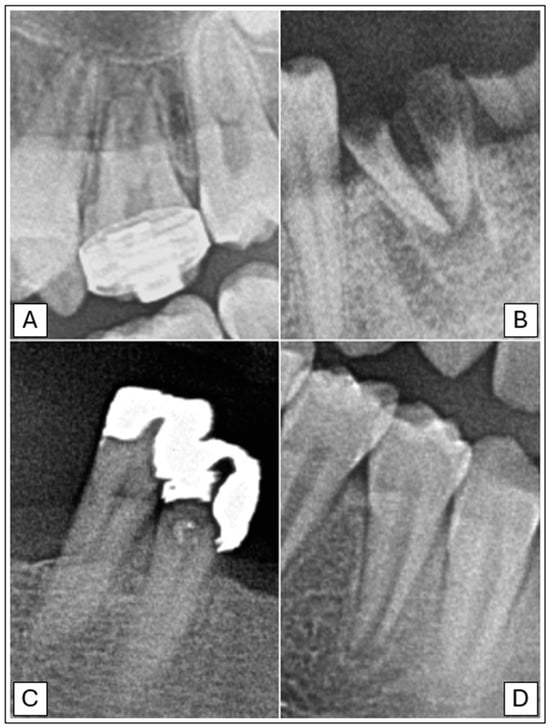
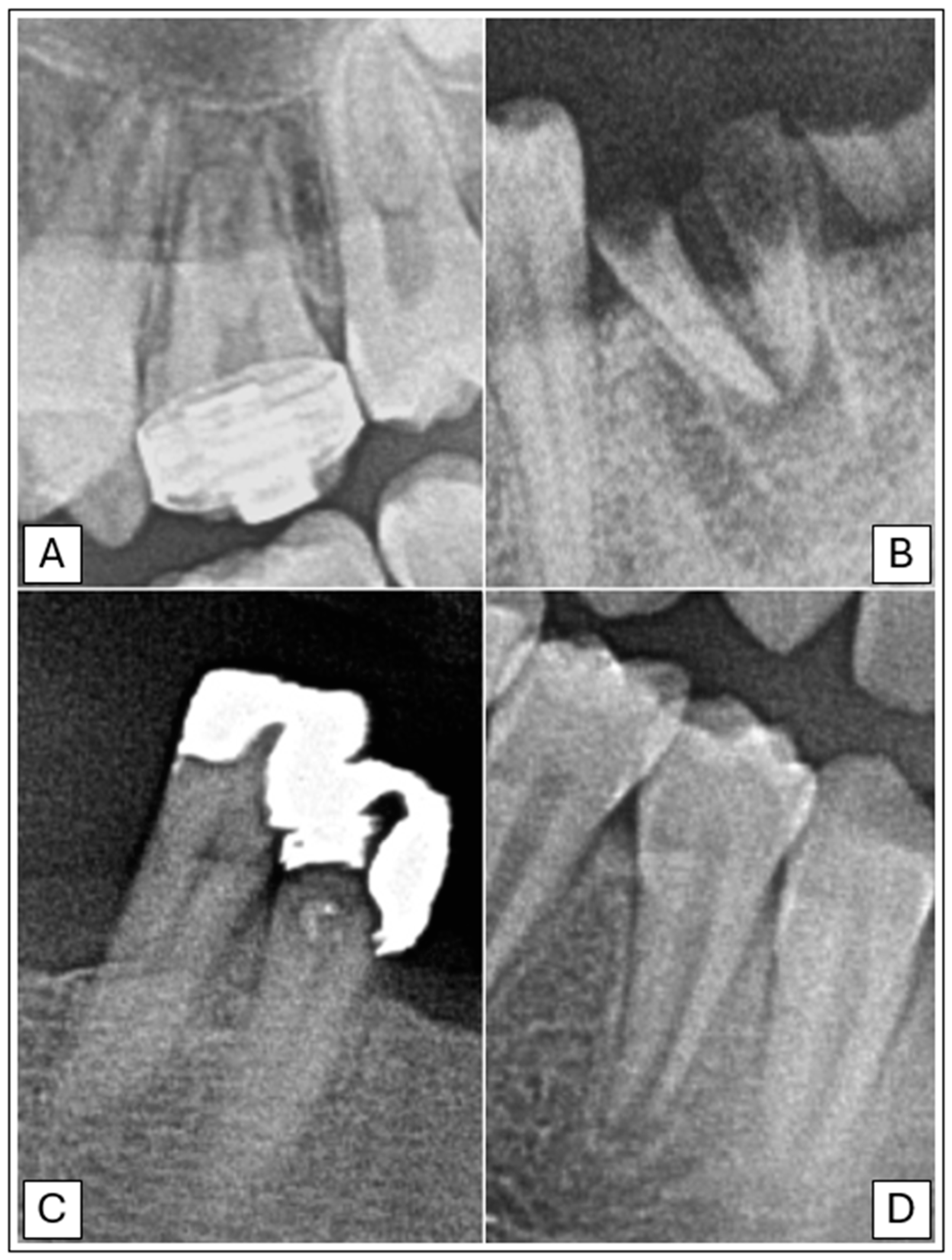
Lower values of sensitivity (74.75%) and precision (72.47%) were observed in detecting periapical lesions. However, these values are higher than those obtained in other studies, where they achieved 39% and 56%, respectively [11,38]. The reason for the lower value of the indicators may be due to the use of DPR instead of CBCT. Many studies confirmed the higher accuracy of CBCT in detecting periapical lesions, identifying about one-third more periapical lesions [41,42]. A recent study by Kazimierczak et al. has shown that the sensitivity and precision of identifying this pathology were 44.45% and 59.19% higher in the case of AI software analyzing CBCT rather than in DPRs. The specificity and accuracy were high in the case of both these images (above 97%) [43]. There was also a study from 2023 where a model consisting of two convolutional neural networks (CNNs) performed well and detected periapical lesions with accuracy, sensitivity, and specificity above 81% [28]. In the present study, the algorithm sometimes incorrectly identified artifacts, teeth with an undeveloped root apex, and anatomical structures such as the mental foramen or the mandibular canal as periapical lesions (Figure 6). A few times, the software indicated the presence of a lesion in a tooth adjacent to the actual one.
Figure 6.
Examples of teeth misdiagnosed by AI software. (A) Tooth 26 with an orthodontic band, mistakenly identified as a tooth with a crown. (B) Tooth 36, not treated endodontically, incorrectly diagnosed as a tooth with root canal filling. (C) Teeth 44 and 45 with extensive, highly radiopaque dental fillings, wrongly identified as teeth with crowns. (D) Tooth 44 with incomplete root development, misdiagnosed as a tooth with a periapical lesion.
4.3. Prosthetics (Crowns, Pontics, Implants, and Implant Abutment Crowns)
The tested algorithm generally performed well in detecting prosthetic elements. All of them achieved very high specificity, above 99%. Moderate precision was observed in identifying crowns. The common problem for the AI was identifying extensive dental fillings, direct veneers, and molar bands as crowns (Figure 6). Nevertheless, the sensitivity, specificity, and accuracy were very high (above 99%). Similar good results in detecting crowns were also obtained in another study where a pre-trained CNN, available online from 2017, was used to analyze 300 DPRs. The sensitivity and precision were 89.53%, while specificity achieved almost 96% [27]. The precision in the study by Altan, where the YOLOv4 model was used, was moderate and reached 74%. However, detecting bridges became more successful, resulting in a precision of 84% [44]. In the present study, all performance metrics for detecting pontics were high, reaching up to 99.95%. A few times the algorithm wrongly indicated teeth as pontics. Another study from 2022 used the AI Model CranioCatch based on the deep CNN method to analyze 1084 DPRs. The detection of pontics was characterized by moderate sensitivity at 77.38% but high precision at 87.83% [29].
The prevalence of implants and implant abutment crowns in the study was low, 0.19% and 0.14%, respectively. Analyzed performance metrics achieved high and very high values. Various studies have also obtained good results for detecting implants. Vinayahalingam et al. achieved a precision of 97.9% [37]. In the study by Başaran et al., the sensitivity and precision were even higher, reaching 96.15% and 92.59%, respectively. However, the algorithm showed slightly lower metrics in detecting implant abutment crowns, with about 3% and 10% decreases [29]. In the present study, all parameters regarding the identification of implant-supported crowns were very high, above 92%. Occasionally, in the present study the algorithm mistakenly identified post and core restorations as implants.
4.4. Limitations
The study only included correctly performed DPRs. Radiographs followed the criteria of the Polish Ministry of Health [45]. However, any qualitative deviation increases the risk of incorrect AI evaluation.
Radiographs of only Caucasian patients were acquired. Additionally, they were performed in one imaging diagnostics center. The lack of diversity due to analyzing a narrow population may limit the generalizability of results. Nevertheless, a typical radiology department was chosen in a medium-sized central European city.
DPRs provide valuable information about teeth and the maxillofacial skeleton. However, they lack details that can be obtained from CBCT or intraoral radiographs. For example, periapical lesions can only be detected on DPRs when the mineral loss of bone reaches 30–50% [46]. Therefore, initial lesions may have been omitted. In the periapical area, condensing osteitis can also be observed, which radiologically appears as a concentric radio-opaque area [47]. Although treatment is only advised if symptoms indicate the need, the algorithm did not include these conditions as periapical lesions. Moreover, superimposition and distortion, which may occur on DPRs, can lead to errors in analysis [2].
In the case of the variables implant, implant abutment crown, and pontic, there is a risk of obtaining an inaccurate result due to a sample size smaller than calculated with the Sample Size Estimation for Diagnostic Accuracy Studies tool [30].
The validity of a study depends on the reliability of the investigator [13]. Human evaluation may be fallible because of examiners’ different experiences or individual perceptions. Fatigue, anxiety, or stress can also affect diagnostic accuracy. More examiners undergoing the same training procedure, called standardization, would objectify the results.
4.5. Strengths and Future Perspectives
The strength of this study lies in its large sample size. While most researchers evaluate the reliability of AI algorithms using usually up to 300 radiographs, this study includes more than 600 DPRs, which enhances the validity and generalizability of the findings [25,27,38]. The study delivers a comprehensive comparison of diagnostic accuracy between AI and humans. The results suggest that this technology may serve as a second opinion tool for dental professionals, improving the accuracy of diagnoses and treatments. However, it is necessary to continuously develop the AI algorithms by training them on big data sets and improving the quality of data.
5. Conclusions
In summary, the AI algorithm performed well in automatically evaluating DPRs. It achieved very high performance metrics in detecting missing teeth, root canal fillings, and implant abutment crowns, indicating that AI-based software can be a reliable tool for analyzing panoramic radiographs. However, the moderate precision of identifying endodontic lesions and crowns suggest that clinicians should further verify these findings. Due to the limitations of the study, further research is recommended to develop AI algorithms.
Author Contributions
Conceptualization, N.T. and M.S. (Maciej Sikora); methodology, N.T. and K.C.; software, N.T. and K.C.; validation, M.C.; formal analysis, N.T. and K.C.; investigation, N.T. and M.C.; resources, M.S. (Marcin Sielski); data curation, N.T.; writing—original draft preparation, N.T.; writing—review and editing, N.T., K.C., M.C., M.S. (Marcin Sielski) and M.S. (Maciej Sikora); visualization, N.T.; supervision, M.S. (Maciej Sikora); project administration, M.S. (Maciej Sikora). All authors have read and agreed to the published version of the manuscript.
Funding
This research received no external funding.
Institutional Review Board Statement
This study was conducted in accordance with the Declaration of Helsinki and approved by the Bioethics Committee in Kielce at the Świętokrzyska Chamber of Physicians (approval number: 2.3/2023; 31 August 2023). The study protocol was registered in the ClinicalTrials.gov database: NCT06258798.
Informed Consent Statement
Patient consent was waived due to the inclusion in the research material of only dental panoramic radiographs previously performed following therapeutic needs and then anonymized. No further radiographs or other interventions were performed for this study.
Data Availability Statement
All synthesized data are available in the article. The raw data supporting the conclusions of this article will be made available by the authors on request.
Conflicts of Interest
The authors declare no conflicts of interest.
Appendix A
Table A1.
Guideline definitions of the levels of performance metrics.
References
- Różyło-Kalinowska, I. Panoramic radiography in dentistry. Clin. Dent. Rev. 2021, 5, 26. [Google Scholar] [CrossRef] [Scilit]
- Farman, A.G. Panoramic Radiology. Seminars on Maxillofacial Imaging and Interpretation; Springer: Berlin/Heidelberg, Germany; New York, NY, USA, 2007; ISBN 978-3-540-46229-3. [Google Scholar]
- Deman, P.; Atwal, P.; Duzenli, C.; Thakur, Y.; Ford, N.L. Dose measurements for dental cone-beam CT: A comparison with MSCT and panoramic imaging. Phys. Med. Biol. 2014, 59, 3201. [Google Scholar] [CrossRef] [Scilit]
- Signorelli, L.; Patcas, R.; Peltomäki, T.; Schätzle, M. Radiation dose of cone-beam computed tomography compared to conventional radiographs in orthodontics. J. Orofac. Orthop. 2016, 77, 9–15. [Google Scholar] [CrossRef] [Scilit]
- Choi, J.-W. Assessment of panoramic radiography as a national oral examination tool: Review of the literature. Imaging Sci. Dent. 2011, 41, 1–6. [Google Scholar] [CrossRef] [Scilit]
- Kweon, H.H.-I.; Lee, J.-H.; Youk, T.; Lee, B.-A.; Kim, Y.-T. Panoramic radiography can be an effective diagnostic tool adjunctive to oral examinations in the national health checkup program. J. Periodontal Implant. Sci. 2018, 48, 317. [Google Scholar] [CrossRef] [Scilit]
- Perschbacher, S. Interpretation of panoramic radiographs. Aust. Dent. J. 2012, 57, 40–45. [Google Scholar] [CrossRef] [Scilit] [PubMed]
- Kayal, R.A. Distortion of digital panoramic radiographs used for implant site assessment. J. Orthod. Sci. 2016, 5, 117–120. [Google Scholar] [CrossRef] [Scilit] [PubMed]
- Kiser, J.R. Reduce Your Panoramic Imaging Errors. Dimens. Dent. Hyg. 2020, 18, 36–39. [Google Scholar]
- Akkaya, N.; Kansu, Ö.; Kansu, H.; Çağirankaya, L.; Arslan, U. Comparing the accuracy of panoramic and intraoral radiography in the diagnosis of proximal caries. Dentomaxillofac. Radiol. 2006, 35, 170–174. [Google Scholar] [CrossRef] [Scilit]
- İçöz, D.; Terzioğlu, H.; Özel, M.A.; Karakurt, R. Evaluation of an Artificial Intelligence System for the Diagnosis of Apical Periodontitis on Digital Panoramic Images. Niger. J. Clin. Pract. 2023, 26, 1085. [Google Scholar] [CrossRef] [Scilit]
- Laslett, M. Chapter 28—Evidence-based clinical testing of the lumbar spine and pelvis. In Movement, Stability & Lumbopelvic Pain, 2nd ed.; Vleeming, A., Mooney, V., Stoeckart, R., Wilson, P., Eds.; Churchill Livingstone: Edinburgh, UK, 2007; pp. 405–425. ISBN 978-0-443-10178-6. [Google Scholar]
- Dye, B.A. 12—Epidemiology and Research Design in Dental Public Health. In Burt and Eklund’s Dentistry, Dental Practice, and the Community, 7th ed.; Mascarenhas, A.K., Okunseri, C., Dye, B.A., Eds.; W.B. Saunders: St. Louis, MO, USA, 2021; pp. 118–130. ISBN 978-0-323-55484-8. [Google Scholar]
- Macey, R.; Walsh, T.; Riley, P.; Glenny, A.-M.; Worthington, H.V.; O’Malley, L.; Clarkson, J.E.; Ricketts, D. Visual or visual-tactile examination to detect and inform the diagnosis of enamel caries. Cochrane Database Syst. Rev. 2021, 2021, CD014546. [Google Scholar] [CrossRef] [Scilit]
- Heitz-Mayfield, L.J.A. Conventional diagnostic criteria for periodontal diseases (plaque-induced gingivitis and periodontitis). Periodontol. 2000 2024, 95, 10–19. [Google Scholar] [CrossRef] [Scilit] [PubMed]
- Ribeiro, M.F.A.; Oliveira, M.C.M.; Leite, A.C.; Bruzinga, F.F.B.; Mendes, P.A.; Grossmann, S.d.M.C.; de Araújo, V.E.; Souto, G.R. Assessment of screening programs as a strategy for early detection of oral cancer: A systematic review. Oral. Oncol. 2022, 130, 105936. [Google Scholar] [CrossRef] [Scilit]
- Mohd Ali, J.; Hussain, M.A.; Tade, M.O.; Zhang, J. Artificial Intelligence techniques applied as estimator in chemical process systems—A literature survey. Expert. Syst. Appl. 2015, 42, 5915–5931. [Google Scholar] [CrossRef] [Scilit]
- Thurzo, A.; Urbanová, W.; Novák, B.; Czako, L.; Siebert, T.; Stano, P.; Mareková, S.; Fountoulaki, G.; Kosnáčová, H.; Varga, I. Where Is the Artificial Intelligence Applied in Dentistry? Systematic Review and Literature Analysis. Healthcare 2022, 10, 1269. [Google Scholar] [CrossRef] [Scilit]
- Aminoshariae, A.; Kulild, J.; Nagendrababu, V. Artificial Intelligence in Endodontics: Current Applications and Future Directions. J. Endod. 2021, 47, 1352–1357. [Google Scholar] [CrossRef] [Scilit]
- Putra, R.H.; Doi, C.; Yoda, N.; Astuti, E.R.; Sasaki, K. Current applications and development of artificial intelligence for digital dental radiography. Dentomaxillofac. Radiol. 2022, 51, 20210197. [Google Scholar] [CrossRef] [Scilit]
- Turosz, N.; Chęcińska, K.; Chęciński, M.; Brzozowska, A.; Nowak, Z.; Sikora, M. Applications of artificial intelligence in the analysis of dental panoramic radiographs: An overview of systematic reviews. Dentomaxillofac. Radiol. 2023, 52, 20230284. [Google Scholar] [CrossRef] [Scilit] [PubMed]
- Güneç, H.G.; Ürkmez, E.Ş.; Danaci, A.; Dilmaç, E.; Onay, H.H.; Aydin, K.C. Comparison of artificial intelligence vs. junior dentists’ diagnostic performance based on caries and periapical infection detection on panoramic images. Quant. Imaging Med. Surg. 2023, 13, 7494503–7497503. [Google Scholar] [CrossRef] [Scilit]
- Alowais, S.A.; Alghamdi, S.S.; Alsuhebany, N.; Alqahtani, T.; Alshaya, A.I.; Almohareb, S.N.; Aldairem, A.; Alrashed, M.; Bin Saleh, K.; Badreldin, H.A.; et al. Revolutionizing healthcare: The role of artificial intelligence in clinical practice. BMC Med. Educ. 2023, 23, 689. [Google Scholar] [CrossRef] [Scilit]
- Turosz, N.; Chęcińska, K.; Chęciński, M.; Rutański, I.; Sielski, M.; Sikora, M. Oral Health Status and Treatment Needs Based on Artificial Intelligence (AI) Dental Panoramic Radiograph (DPR) Analysis: A Cross-Sectional Study. J. Clin. Med. 2024, 13, 3686. [Google Scholar] [CrossRef] [Scilit] [PubMed]
- Leite, A.F.; Gerven, A.V.; Willems, H.; Beznik, T.; Lahoud, P.; Gaêta-Araujo, H.; Vranckx, M.; Jacobs, R. Artificial intelligence-driven novel tool for tooth detection and segmentation on panoramic radiographs. Clin. Oral. Investig. 2021, 25, 2257–2267. [Google Scholar] [CrossRef] [Scilit] [PubMed]
- Kazimierczak, W.; Wajer, R.; Wajer, A.; Kalka, K.; Kazimierczak, N.; Serafin, Z. Evaluating the Diagnostic Accuracy of an AI-Driven Platform for Assessing Endodontic Treatment Outcomes Using Panoramic Radiographs: A Preliminary Study. J. Clin. Med. 2024, 13, 3401. [Google Scholar] [CrossRef] [Scilit] [PubMed]
- Bonfanti-Gris, M.; Garcia-Cañas, A.; Alonso-Calvo, R.; Salido Rodriguez-Manzaneque, M.P.; Pradies Ramiro, G. Evaluation of an Artificial Intelligence web-based software to detect and classify dental structures and treatments in panoramic radiographs. J. Dent. 2022, 126, 104301. [Google Scholar] [CrossRef] [Scilit] [PubMed]
- Ba-Hattab, R.; Barhom, N.; Osman, S.A.A.; Naceur, I.; Odeh, A.; Asad, A.; Al-Najdi, S.A.R.N.; Ameri, E.; Daer, A.; Silva, R.L.B.D.; et al. Detection of Periapical Lesions on Panoramic Radiographs Using Deep Learning. Appl. Sci. 2023, 13, 1516. [Google Scholar] [CrossRef] [Scilit]
- Başaran, M.; Çelik, Ö.; Bayrakdar, I.S.; Bilgir, E.; Orhan, K.; Odabaş, A.; Aslan, A.F.; Jagtap, R. Diagnostic charting of panoramic radiography using deep-learning artificial intelligence system. Oral. Radiol. 2022, 38, 363–369. [Google Scholar] [CrossRef] [Scilit]
- Akoglu, H. User’s guide to sample size estimation in diagnostic accuracy studies. Turk. J. Emerg. Med. 2022, 22, 177–185. [Google Scholar] [CrossRef] [Scilit]
- ISO Standard No. 3950:2016; Dentistry—Designation System for Teeth and Areas of the Oral Cavity. International Organization for Standardization: Geneva, Switzerland, 2016. Available online: https://www.iso.org/standard/68292.html (accessed on 15 September 2024).
- Evans, H.J.; Gibson, N.A.; Bennett, J.; Chan, S.Y.; Gavlak, J.; Harman, K.; Ismail-Koch, H.; Kingshott, R.N.; Langley, R.; Morley, A.; et al. British Thoracic Society guideline for diagnosing and monitoring paediatric sleep-disordered breathing. Thorax 2023, 78, s1–s27. [Google Scholar] [CrossRef] [Scilit]
- Khanagar, S.B.; Al-ehaideb, A.; Maganur, P.C.; Vishwanathaiah, S.; Patil, S.; Baeshen, H.A.; Sarode, S.C.; Bhandi, S. Developments, application, and performance of artificial intelligence in dentistry—A systematic review. J. Dent. Sci. 2021, 16, 508–522. [Google Scholar] [CrossRef] [Scilit]
- Umer, F.; Habib, S.; Adnan, N. Application of deep learning in teeth identification tasks on panoramic radiographs. Dentomaxillofac. Radiol. 2022, 51, 20210504. [Google Scholar] [CrossRef] [Scilit]
- Hung, K.; Montalvao, C.; Tanaka, R.; Kawai, T.; Bornstein, M.M. The use and performance of artificial intelligence applications in dental and maxillofacial radiology: A systematic review. Dentomaxillofac. Radiol. 2020, 49, 20190107. [Google Scholar] [CrossRef] [Scilit] [PubMed]
- Tuzoff, D.V.; Tuzova, L.N.; Bornstein, M.M.; Krasnov, A.S.; Kharchenko, M.A.; Nikolenko, S.I.; Sveshnikov, M.M.; Bednenko, G.B. Tooth detection and numbering in panoramic radiographs using convolutional neural networks. Dentomaxillofac. Radiol. 2019, 48, 20180051. [Google Scholar] [CrossRef] [Scilit]
- Vinayahalingam, S.; Goey, R.; Kempers, S.; Schoep, J.; Cherici, T.; Moin, D.A.; Hanisch, M. Automated chart filing on panoramic radiographs using deep learning. J. Dent. 2021, 115, 103864. [Google Scholar] [CrossRef] [Scilit] [PubMed]
- Zadrożny, Ł.; Regulski, P.; Brus-Sawczuk, K.; Czajkowska, M.; Parkanyi, L.; Ganz, S.; Mijiritsky, E. Artificial Intelligence Application in Assessment of Panoramic Radiographs. Diagnostics 2022, 12, 224. [Google Scholar] [CrossRef] [Scilit]
- Agrawal, S.K.; Bhagat, T.; Shrestha, A. Dental Caries in Permanent First Molar and Its Association with Carious Primary Second Molar among 6–11-Year-Old School Children in Sunsari, Nepal. Int. J. Dent. 2023, 2023, 9192167. [Google Scholar] [CrossRef] [Scilit]
- Khattak, O.; Iqbal, A.; Nadeem Baig, M.; Magar, S.P.; Magar, S.S.; Mehmood Khan, A.; Osamah Molla, F.; Alhabib, S.; Alruwaili, S. Prevalence of Caries in Anterior Teeth in Adults; An Epidemiology Study. PJMHS 2021, 15, 3421–3423. [Google Scholar] [CrossRef] [Scilit]
- Lofthag-Hansen, S.; Huumonen, S.; Gröndahl, K.; Gröndahl, H.-G. Limited cone-beam CT and intraoral radiography for the diagnosis of periapical pathology. Oral Surg. Oral Med. Oral Pathol. Oral Radiol. Endod. 2007, 103, 114–119. [Google Scholar] [CrossRef] [Scilit]
- Ramis-Alario, A.; Tarazona-Alvarez, B.; Cervera-Ballester, J.; Soto-Peñaloza, D.; Peñarrocha-Diago, M.; Peñarrocha-Oltra, D.; Peñarrocha-Diago, M. Comparison of diagnostic accuracy between periapical and panoramic radiographs and cone beam computed tomography in measuring the periapical area of teeth scheduled for periapical surgery. A cross-sectional study. J. Clin. Exp. Dent. 2019, 11, e732–e738. [Google Scholar] [CrossRef] [Scilit]
- Kazimierczak, W.; Wajer, R.; Wajer, A.; Kiian, V.; Kloska, A.; Kazimierczak, N.; Janiszewska-Olszowska, J.; Serafin, Z. Periapical Lesions in Panoramic Radiography and CBCT Imaging—Assessment of AI’s Diagnostic Accuracy. J. Clin. Med. 2024, 13, 2709. [Google Scholar] [CrossRef] [Scilit]
- Altan, B.; Gunec, H.G.; Cinar, S.; Kutal, S.; Gulum, S.; Aydin, K.C. Detecting Prosthetic Restorations Using Artificial Intelligence on Panoramic Radiographs. Sci. Program. 2022, 2022, 6384905. [Google Scholar] [CrossRef] [Scilit]
- Obwieszczenie Ministra Zdrowia z dnia 10 listopada 2015 r. w Sprawie Ogłoszenia Wykazu Wzorcowych Procedur Radiologicznych z Zakresu Radiologii—Diagnostyki Obrazowej i Radiologii Zabiegowej; Dziennik Urzędowy Ministra Zdrowia: Warsaw, Poland, 2015; p. 43. Available online: https://dziennikmz.mz.gov.pl/legalact/2015/78/ (accessed on 15 September 2024).
- Johnson, M. DDS Saving Teeth with Endodontic Treatment: Lesion Size and Predictability of Healing. Available online: https://www.perioimplantadvisory.com/periodontics/article/14178968/saving-teeth-with-endodontic-treatment-lesion-size-and-predictability-of-healing (accessed on 13 September 2024).
- Chapter 9—Management of pulpal and periradicular disease. In Restorative Dentistry, 2nd ed.; Walmsley, A.D., Walsh, T.F., Lumley, P.J., Burke, F.J.T., Shortall, A.C.C., Hayes-Hall, R., Pretty, I.A., Eds.; Churchill Livingstone: Edinburgh, UK, 2007; pp. 89–113. ISBN 978-0-443-10246-2. [Google Scholar]

Disclaimer/Publisher’s Note: The statements, opinions and data contained in all publications are solely those of the individual author(s) and contributor(s) and not of MDPI and/or the editor(s). MDPI and/or the editor(s) disclaim responsibility for any injury to people or property resulting from any ideas, methods, instructions or products referred to in the content. |
© 2024 by the authors. Licensee MDPI, Basel, Switzerland. This article is an open access article distributed under the terms and conditions of the Creative Commons Attribution (CC BY) license (https://creativecommons.org/licenses/by/4.0/).