


Abstract
Background/Objectives: Retinal metastasis (RM) is an exceptionally rare clinical condition, comprising less than 1% of ocular metastasis cases. This study aims to investigate the clinical features of RM originating from systemic carcinoma. Methods: A systematic review (PROSPERO ID: CRD42022344724). Results: The systematic review revealed 58 cases involving 61 eyes. Most of the cases (82.8%) had a known primary malignancy before RM was diagnosed. The main primary sites were the lung (45.8%), gastrointestinal tract (23.7%), and breast (16.9%). The lung was the most common (80.0%) carcinoma in cases with unknown primary sites. The median survival after RM diagnosis was 11 months. The main clinical patterns were patchy retinal infiltrates (35.7%), an elevated retinal mass (63.8%), and punctate retinal infiltrates (7.1%). Subretinal fluid, vitreous seeding, and choroidal invasion were noted in 57.4%, 41.0%, and 6.6% of eyes, respectively. Key multimodal imaging features were early hypofluorescence with mid-to-late hyperfluorescence on fluorescein angiography (84.6%) and hyper-reflectiveness on optical coherence tomography (70.8%). A final best-corrected visual acuity of ≤20/200 was noted in 66.7% of the eyes. Conclusions: Patchy retinal infiltrates, an elevated retinal mass, and punctate retinal infiltrates are the major clinical patterns of RM from systemic carcinoma. The primary carcinoma sites are the lung, gastrointestinal tract, and breast. Systemic and visual prognoses were unfavorable.
1. Introduction
Most cases of ocular metastasis from systemic cancer appear in the uveal tracts (>98%), especially the choroid. On the contrary, retinal metastasis (RM) is extremely rare, accounting for <1% of cases of ocular metastasis [1]. This rarity may be attributed to the difference in vascularization between the choroid and retina. The retina is supplied by a single central retinal artery (CRA) that provides blood to the inner layers and is distributed throughout the retina, whereas the highly vascularized choroid is supplied by multiple choroidal arteries.
RM is an umbrella term referring to the invasion of the retina by remote systemic malignant cells. Among various types of cancer, carcinoma is the most typical entity of solid organ tumors. Meanwhile, carcinomatous RM is also more commonly seen than RM from melanoma, lymphoma, or leukemia cells [2,3,4,5,6,7]. Given the rarity of RM, the literature on RM includes isolated case reports and a few case series. Those studies have enrolled patients with heterogeneous clinical entities and have applied inconsistent inclusion and exclusion criteria [2,3,8]. Since the clinical outcomes and biological behaviors associated with systemic malignancies of different cell types vary widely, analyzing a single type of malignancy is crucial to obtaining a precise and in-depth understanding of RM.
This systematic review investigates RM from systemic carcinoma and describes this disease entity’s clinical features and outcomes.
2. Materials and Methods
In accordance with preferred reporting items for systematic reviews and meta-analyses (PRISMA) recommendations [9], we selected articles from PubMed, CINAHL, and Google Scholar that were published on or before May 31, 2024. This systematic review has been prospectively registered in PROSPERO (registration ID: CRD42022344724), entitled “Systemic and Ocular Features of Retinal Metastatic Carcinoma”. We used 4 search terms to identify eligible articles: “retinal metastasis”, “retina metastasis”, “metastasis to the retina”, and “metastatic retinal mass”. Three reviewers (LUW, THH, and YRH) reviewed the titles and abstracts independently. After removing duplicate articles, the full texts of the remaining articles were obtained and thoroughly reviewed. Disagreement between the reviewers was resolved through the re-evaluation of the manuscript and further discussion. Prespecified inclusion and exclusion criteria are shown in Table 1 and applied to ensure study quality. Only studies that provided sufficient relevant information on patients’ demographic profiles, main ocular features, adjunctive ocular features, and systemic features of RM were enrolled. Only peer-reviewed English language publications were reviewed. All the collected data were compiled into an Excel spreadsheet (Excel for Mac version 16.48, Microsoft, Washington, DC, USA), and descriptive statistics were conducted on the spreadsheet. A Kaplan–Meier survival analysis was performed using jamovi (the jamovi Project. n.d. jamovi (Version 2.3.26) [Computer Software]. https://www.jamovi.org (accessed on 30 June 2024)).
Table 1.
Inclusion and exclusion criteria.
3. Results
3.1. Systemic Features
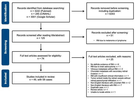
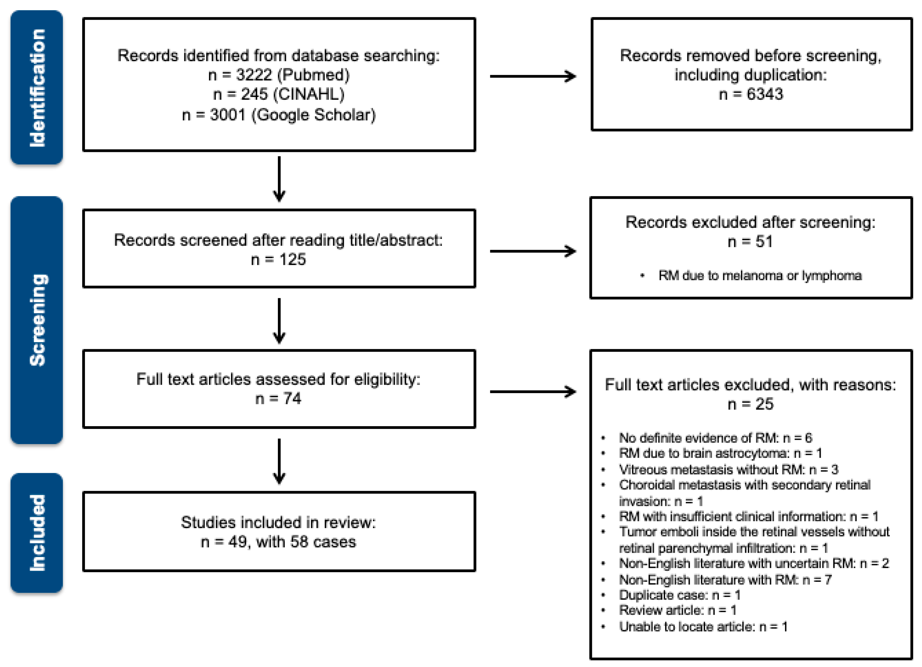
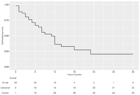
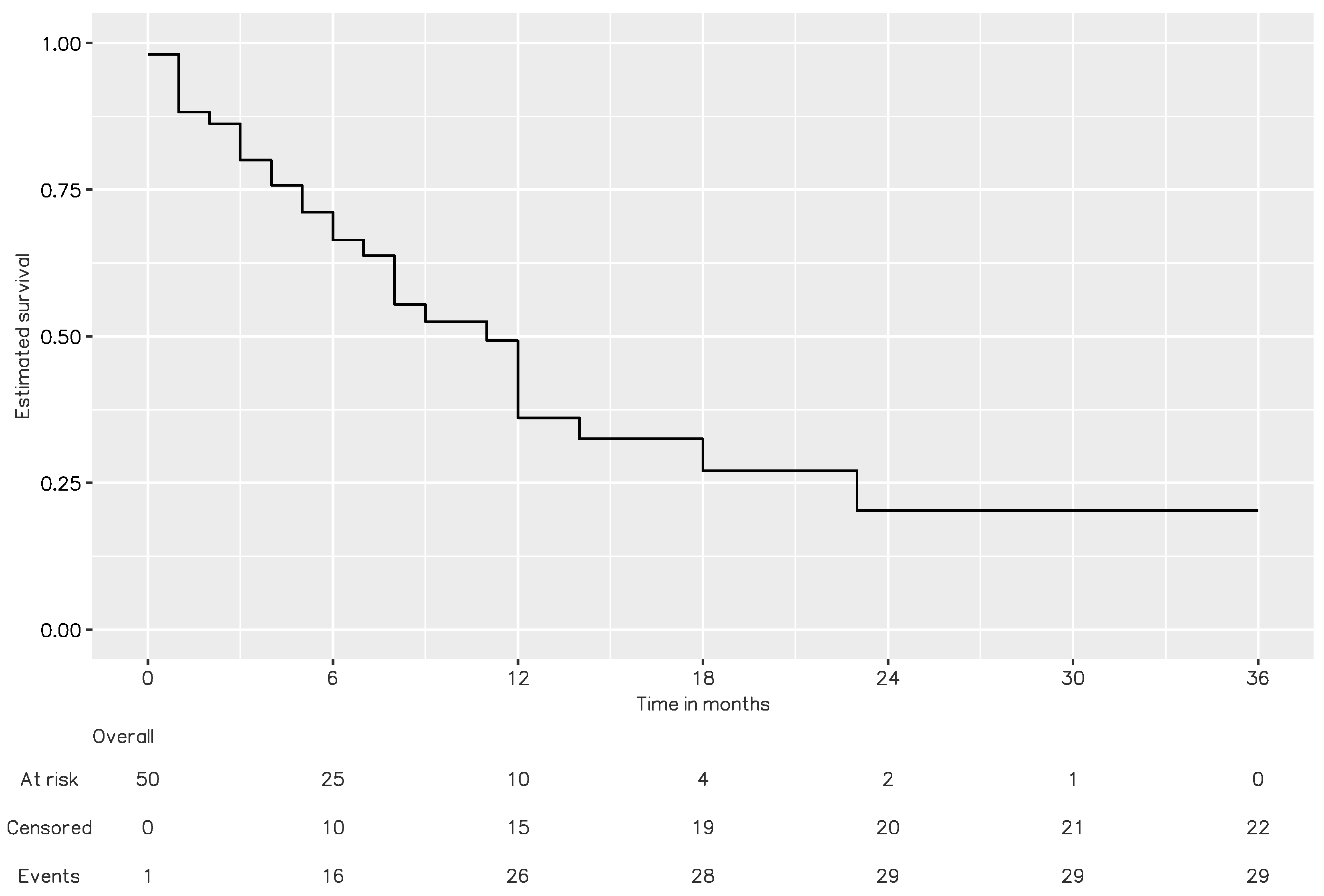
Overall, 49 studies involving 58 cases were eligible for review and the PRISMA flow diagram is shown as Figure 1 (the complete literature list is provided in the Supplementary Table S1). The patients’ systemic profiles are described in Table 2. The studies included 36 men (62.1%) and 22 women (37.9%) with a median age of 59 years (IQR = 13.5; range = 15–80). Most (n = 55, 94.8%) presented with unilateral RM. The three most common primary carcinoma sites were the lung (n = 27, 45.8%), GI tract (n = 14, 23.7%), and breast (n = 10, 16.9%). The patient’s primary diagnosis, metastasis, and mortality profiles are summarized in Table 3. Primary carcinoma diagnosis preceded ocular manifestations in 82.8% of the cases (n = 48), with varied intervals (median = 18.0 months; IQR = 27.0; range = 1–214). Among the cases in which ocular manifestations preceded the primary diagnosis (n = 10, 17.2%), lung cancer (n = 8, 80.0%) was the most common primary diagnosis. Systemic metastasis was noted in 45 (86.3%) patients. Twenty-two patients (59.4%) had an involvement of more than two solid organs, excluding lymph node metastasis. The lymph nodes, lungs, and brain were the three main sites involved (51.2%, 46.3%, and 46.3%, respectively). Considering the treatment after RM diagnosed, 82.4% received chemotherapy among 17 patients with systemic treatment according to the primary carcinoma and 71.4% received radiotherapy for 28 patients receiving local treatment. Overall, 56.8% of the patients died during the follow-up. A Kaplan–Meier survival analysis (Figure 2) revealed that the median interval from RM diagnosis to death was 11 months.
Figure 1.
PRISMA flow diagram.
Table 2.
Basic demographic data and primary carcinoma sites of patients with retinal metastatic carcinoma (RM).
Table 3.
Primary diagnosis, metastasis, and mortality profiles of patients with RM from systemic carcinoma.
Figure 2.
Kaplan–Meier survival analysis of RM from systematic review. The median interval from RM diagnosis to death was 11 months.
3.2. Ocular Features
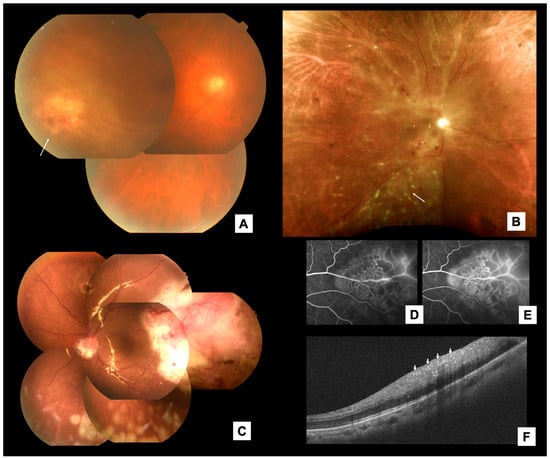
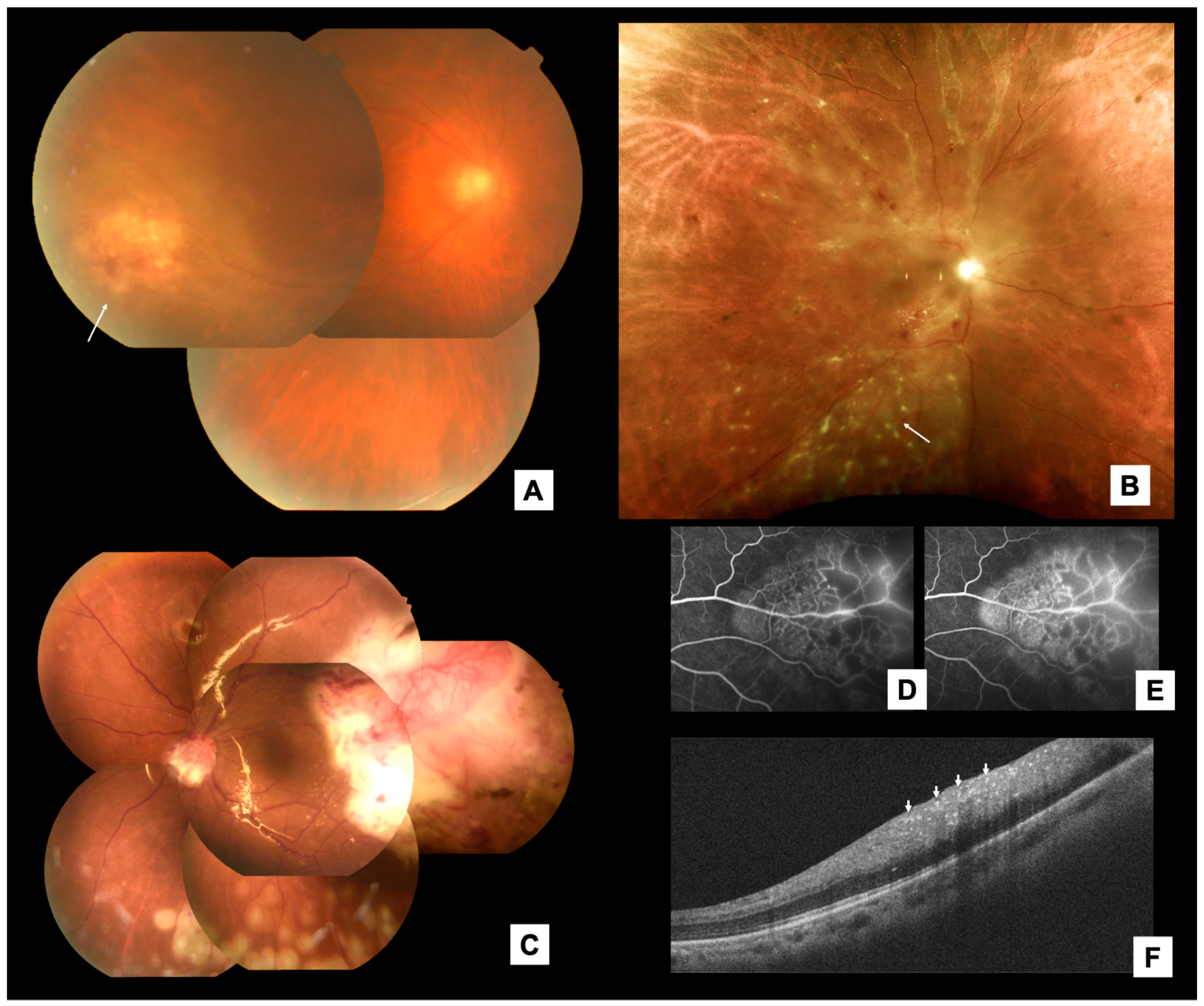
The patients’ ocular features are summarized in Table 4. We divided the clinical appearance of RM into three main clinical patterns, as in Figure 3: [12] patchy retinal infiltrates (n = 20, 35.7%); an elevated retinal mass (n = 20, 63.8%); and punctate retinal infiltrates (n = 4, 7.1%). Subretinal fluid (SRF) was noted in 35 (57.4%) eyes. Serous RD (SRD), rhegmatogenous retinal detachment (RRD), and tractional RD (TRD) were noted in 47.5%, 8.2%, and 1.6% of eyes, respectively. Vitreous seeds were noted in 25 (41.0%) eyes. Choroidal invasion was noted in four (6.6%) eyes. A pathological diagnosis was obtained in 43 (70.5%) eyes. Pars plana vitrectomy (PPV) with a vitreous/retinal/SRF biopsy was performed on 23 (53.5%) eyes. A fine-needle aspiration biopsy was performed on seven (16.3%) eyes. An initial misdiagnosis was noted in 21 cases (37.5%), of which 13 cases (61.9%) were recorded as infectious retinitis. The mean best corrected visual acuity (BCVA) logarithm of the minimum angle of resolution (logMAR) at presentation and the end of follow-up was 1.06 (Snellen equivalent: 20/230) and 1.73 (Snellen equivalent: 20/1074), respectively. A Snellen BCVA of ≤20/200 was noted in 18 (37.5%) and 22 (66.7%) eyes at the initial and final visits, respectively.
Table 4.
Ocular features of RM from systemic carcinoma (61 eyes).
Figure 3.
Representative of 3 retinal patterns and multimodal image findings of RM. Adapted from Chen [12]. [Figure 1]. (A) Patchy retinal infiltrate. (B) Punctate retinal infiltrate. (C) An elevated retinal mass with tortuous retinal neovascularization. (D,E) Intrinsic vascularity on fluorescein angiography at mid-phase (2:49) and late phase (9:18). (F) Inner retina hyper-reflective changes (arrows) on OCT.
The multimodal imaging findings were investigated. The most common FA pattern was early hypofluorescence with mid/late-phase hyperfluorescence (lesion staining/leakage) (n = 22, 84.6%). Intrinsic intralesional vascularity was noted in 10 (38.5%) eyes. The lesions were predominantly hyper-reflective (n = 17, 70.8%) on OCT, hyperechoic (n = 9, 100%) on a B-scan ultrasonography, and hypo-autofluorescent (n = 5, 100%).
4. Discussion
Cases of RM were first reported before 1960 [13,14,15]; since then, oncologic therapies, multimodal ophthalmic imaging, and surgical procedures have advanced considerably. However, the management of RM still needs to be improved. As RM is rare, relevant information on ocular presentations, protocols of vitreoretinal biopsies, primary tumor and systemic metastasis profiles, and prognosis are scarce. Although most of the publications included in our review were case reports, Shields et al. presented four cases of histologically diagnosed RM from systemic carcinoma among a series of eight cases of RM [2]. More recently, Gascon et al. presented a series of one biopsy-confirmed and two clinically presumed cases of carcinomatous RM [3]. The current study presents a rigorous systematic review with predefined criteria.
This systematic review revealed three key systemic features of RM. First, similar to uveal metastasis, lung and breast carcinoma are the most common types of RM among male and female patients, respectively. Meanwhile, cases of RM originating in the GI tract are among the three leading diagnoses in both male and female patients. Therefore, we suggest that the examinations of chest/abdominal CT, mammography/breast echography, and panendoscopy should be prioritized in cases without cancer history. Second, the intervals from primary cancer diagnosis to RM varied widely: 38.4% of the patients presented with RM within 1 year of their primary diagnosis, and 35.9% presented with RM more than 2 years after their primary diagnosis. Therefore, a long or relatively stable preceding malignancy does not preclude the possibility of RM. In nearly 20% of the cases, ocular manifestations preceded the primary diagnosis; among them, lung cancer was the most common primary diagnosis (80%), reflecting its aggressive nature. Third, the systemic prognoses of patients with RM are often poor. Systemic metastasis was common (86.3%) and was often multiple (59.4%). The Kaplan–Meier analysis revealed that the median interval from RM diagnosis to death was 11 months. This information is crucial to decisions related to holistic care systemic management and communication among physicians, patients, and family members.
Our systematic review also revealed several key ocular characteristics of RM. Three distinct clinical patterns were identified. Although the diagnosis of RM is relatively straightforward when RM manifests as an elevated retinal mass, the other two patterns can masquerade as uveitis entities [16]. Patchy retinal infiltrates may be accompanied by a retinal hemorrhage and exudates, often mistaken for infectious retinitis [11,17,18,19]. When such lesions appear in patients with cancer who are immunocompromised, cytomegaloviral or toxoplasmic retinitis is often suspected. Punctate retinal infiltrates, the least common of the three patterns, can be mistaken as noninfectious uveitis, such as those that appear in sarcoidosis or Behcet disease. Physicians should be aware of the possibility of RM when initial investigations or treatment responses are suboptimal.
The biological nature of RM can be assessed using the clinical information and multimodal imaging findings collected in this study. Most retinal lesions occurred in the posterior pole or midperiphery, with predominant inner-retinal hyper-reflective infiltration shown on OCT, reflecting tumor invasion through the CRA. Since the CRA is a terminal branch of the systemic circulation, RM presumably occurs in the case of a large metastatic tumor burden, the entry of malignant cells into the CRA, and the partial disruption of the inner blood–retinal barrier (BRB), which explains the rarity, multiple metastases, and poor systemic prognosis of RM. Interestingly, choroidal involvement was uncommon (6.6%). As the hypoautofluorescence profile indicates, outer retinopathy or retinal pigment epithelium (RPE) disturbance is uncommon. The intact tight junction of the RPE may protect against cancer cell breakthrough. Meanwhile, tumors with selected adhesion molecules may be more adapted to cross the BRB and invade the retinal tissues. Finally, some cases with concurrent chorioretinal metastasis were possibly ignored when the choroidal component was more impressive and easily diagnosed.
The hypermetabolic state of these carcinoma cells can be demonstrated by the late fluorescent staining/leakage (84.6%) with intrinsic vascularity (38.5%). This may explain the aggressive intraocular behaviors of RM. Although SRD is more common, RRD [3,20] and TRD [17] are also possible. First, as reported by Taubenslag et al. [11], retinal breaks can form due to the contraction of the retinal tumor. Second, densely infiltrated vitreous skirt contraction from tumor infiltration may lead to retinal breaks. Third, prior tumor tumefaction may cause breaks at the tumor’s border. These three mechanisms may contribute to the relatively large number (8.2%) of RRD in RM compared to < 1% RRD in choroidal metastases [21]. Following RM, tumor invasion to the iris with neovascular glaucoma is not uncommon. As reported by Saornil et al. [22], Spraul et al. [10], and Koenig et al. [23], the diagnosis was established by examining the enucleated painful blind eye. This reflects the poor visual prognoses of eyes with RM.
Whether pathological evidence is required for RM diagnosis remains unclear. Physicians should evaluate the risks and benefits of surgical procedures on a case-by-case basis. Systemic workup and collaboration with the oncologist are also crucial. In most cases, however, histological evidence from ocular sampling provides important information to guide further management. Indirect ophthalmoscopy and operating microscope-guided fine-needle aspiration biopsies with 25-gauge needles, as described by Shields et al., are well-established procedures with low complication rates [24]. However, because of advances in the vitrectomy systems, PPV remains the most common procedure for obtaining the specimen. It is beneficial for cases with dense vitreous infiltration with a poor fundal view and with vitreoretinal complications such as RRD or TRD and for small retinal lesion biopsies, which require precise intraoperative localization. In cases of significant vitreous infiltration, a vitreous biopsy suffices. Sometimes, a retinal biopsy with the following surgical techniques may be necessary. A limited retinectomy with a tumor-free margin and en bloc retinal tumor excision can be performed in far-peripheral giant retinal tear RRD due to cancer cell infiltration or the highly elevated retinal mass with bullous SRD [25]. In most posterior lesions, however, tumor excision may result in permanent structural complications [26]. Therefore, a conservative intralesional incisional biopsy is often more appropriate [18,27]. In some cases, by involving vitreoretinal surface membranes or cell aggregates, membranes/aggregates can be lifted using forceps and gently aspirated using a backflush needle. Finally, a chandelier endoillumination-assisted vitrectomy with the creation of focal RD with a 41-gauge cannula followed by local retinectomy [28] is another possible option.
The management of RM remains challenging. Although systemic chemotherapy is usually used to treat multiple organ metastases, it may be ineffective for intraocular lesions. For patients with advanced cancer, maintaining spiritual well-being and quality of life is crucial. Moreover, multidisciplinary management is necessary to reach a proper treatment [29]. If a patient’s remaining visual acuity can be salvaged and a painful blind eye can be prevented, the patient’s fear and anxiety can be minimized. Therefore, ocular therapy should be administered using simple, noninvasive, and short procedures. Although an intravitreal injection (IVI) of anti-vascular endothelial growth factor (VEGF) is a short and simple procedure [3], it is invasive, and its efficacy depends on the primary tumor type. If IVI of anti-VEGF fails, injecting other chemotherapy agents into the eye is often not feasible because of insufficient information regarding the dosage and retinal toxicity of these medications. Photodynamic therapy is another feasible option [3,30]. As most RM lesions are vascularized according to FA profiles, the effect of the tumor size control and reduction of exudation can be anticipated. Since an ophthalmologist administers photodynamic therapy, a detailed explanation and follow-up are preferable. Finally, the external beam radiation therapy is widely used to control intraocular tumors [28,31,32,33,34]. It is an advantageous, non-invasive treatment option for concurrent brain metastasis. However, patients who undergo such therapy should be monitored for secondary radiation retinopathy. Overall, individually tailored treatment plans are crucial.
This study has some limitations. First, with the prespecified criteria for systematic reviews, some publications were inevitably excluded because of insufficient clinical data. Due to the generally poor systemic condition of patients with RM, missing data for various clinical parameters and multimodal imaging were common. Meanwhile, misdiagnosis was possible in some reports without pathological evidence. Second, because this study focused only on RM from systemic carcinoma, other major sources of RM, such as melanoma or lymphoma, were excluded. Therefore, comparing carcinomatous RM with other different types of RM was not possible. Nevertheless, this study has several strengths of note. The information presented in this study provides clear insights into the various clinicopathological findings of RM. In addition, by focusing only on metastatic carcinoma, we avoided the confusion surrounding the inconsistent definitions of RM employed in previous studies and limited our analysis to a uniform disease entity. The data were extracted from three major databases using stringent prespecified criteria. A complete presentation of clinical features, prognosis, biopsy methods, and treatment options were summarized herein.
5. Conclusions
RM is a rare but severe disease. The main primary carcinoma sites were the lung, GI tract, and breast, and the three main ocular manifestations were patchy retinal infiltrates, an elevated retinal mass, and punctate retinal infiltrates. These manifestations may present as uveitis masquerade syndromes. Although pathological diagnosis is preferable, combining systemic information and multimodal imaging may support the clinical diagnosis. The treatment of RM should be individually tailored and focus on holistic care and quality of life.
Supplementary Materials
The following supporting information can be downloaded at: https://www.mdpi.com/article/10.3390/jcm13206037/s1, Table S1: Complete List of the Literature in Systematic Review.
Author Contributions
Conceptualization, Y.-R.H.; methodology, Y.-R.H.; software, L.-U.W. and T.-H.H.; validation, F.-T.C., Y.-J.C., J.-K.W. and Y.-R.H.; formal analysis, L.-U.W. and T.-H.H.; investigation, L.-U.W., T.-H.H. and Y.-R.H.; resources, F.-T.C., Y.-J.C., J.-K.W. and Y.-R.H.; data curation, L.-U.W., T.-H.H. and Y.-R.H.; writing—original draft preparation, L.-U.W. and T.-H.H.; writing—review and editing, Y.-R.H.; visualization, L.-U.W. and T.-H.H. All authors have read and agreed to the published version of the manuscript.
Funding
This research received no external funding.
Data Availability Statement
Data are unavailable due to privacy or ethical restrictions.
Conflicts of Interest
The authors declare no conflicts of interest. The authors have no commercial or proprietary interests in any product or instrument mentioned in the manuscript.
References
- Shields, C.L.; Kalafatis, N.E.; Gad, M.; Sen, M.; Laiton, A.; Silva, A.M.V.; Agrawal, K.; Lally, S.E.; Shields, J.A. Metastatic tumours to the eye. Review of metastasis to the iris, ciliary body, choroid, retina, optic disc, vitreous, and/or lens capsule. Eye 2023, 37, 809–814. [Google Scholar] [CrossRef] [Scilit]
- Shields, C.L.; McMahon, J.F.; Atalay, H.T.; Hasanreisoglu, M.; Shields, J.A. Retinal metastasis from systemic cancer in 8 cases. JAMA Ophthalmol. 2014, 132, 1303–1308. [Google Scholar] [CrossRef] [Scilit]
- Gascon, P.; Matet, A.; Gualino, V.; Denis, D.; Nguyen, A.M.; Papegaey, M.; Levy, N.; Arnould, T.; Kodjikian, L.; Mathis, T. CLINICAL FEATURES OF RETINAL METASTASES: New Cases Integrated in a Systematic Review of the Literature. Retina 2022, 42, 1370–1383. [Google Scholar] [CrossRef] [Scilit]
- Rali, A.; Xu, L.T.; Craven, C.; Cohen, J.B.; Yeh, S.; Grossniklaus, H.E.; O’Keefe, G.D. Diagnostic retinal biopsy in the management of secondary non-CNS vitreoretinal lymphoma masquerading as viral retinitis: A case report. Int. J. Retin. Vitr. 2021, 7, 58. [Google Scholar] [CrossRef] [Scilit]
- Ryan, M.E.; Shantha, J.G.; Grossniklaus, H.E.; Yeh, S. Secondary Vitreoretinal Lymphoma Masquerading as Acute Retinal Necrosis. Ophthalmic Surg. Lasers Imaging Retin. 2015, 46, 1048–1050. [Google Scholar] [CrossRef] [Scilit]
- Szeto, S.K.; Mohamed, S.; Tsang, C.W.; Chan, C.K. Massive retinal infiltrates as the presenting sign of chronic myeloid leukemia: Clinical and imaging features of leukemic retinopathy. Eur. J. Ophthalmol. 2021, 33, 11206721211048792. [Google Scholar] [CrossRef] [Scilit]
- Yee, C.; Scruggs, B.A.; Flaxel, C.J. Retinal infiltration in a case of chronic myeloid leukemia. Retin. Cases Brief. Rep. 2022, 17, 690–693. [Google Scholar] [CrossRef] [Scilit]
- Ozcan, G.; Gunduz, A.K.; Mirzayev, I.; Saglik, A. Presumed Retinal Metastasis from Lung Adenocarcinoma: A Case Report and Literature Review. Case Rep. Ophthalmol. Med. 2021, 2021, 6615284. [Google Scholar] [CrossRef] [Scilit]
- Moher, D.; Shamseer, L.; Clarke, M.; Ghersi, D.; Liberati, A.; Petticrew, M.; Shekelle, P.; Stewart, L.A.; Group, P.-P. Preferred reporting items for systematic review and meta-analysis protocols (PRISMA-P) 2015 statement. Syst. Rev. 2015, 4, 1. [Google Scholar] [CrossRef]
- Spraul, C.W.; Lang, G.E.; Grossniklaus, H.E.; Lang, G.K. Metastatic adenocarcinoma to the retina in a patient with Muir-Torre syndrome. Am. J. Ophthalmol. 1995, 120, 248–250. [Google Scholar] [CrossRef] [Scilit]
- Taubenslag, K.J.; Kim, S.J.; Attia, A.; Abel, T.W.; Nickols, H.H.; Ancell, K.K.; Daniels, A.B. Retinal metastasis from unknown primary: Diagnosis, management, and clinicopathologic correlation. Digit. J. Ophthalmol. 2015, 21, 71–75. [Google Scholar] [CrossRef] [Scilit]
- Chen, C.L.; Chan, W.C.; Chen, Y.C.; Hsu, Y.R. Retinal metastasis from systemic carcinoma: Clinical, multimodal imaging, and pathological characteristics from a multicenter case series. Graefes Arch. Clin. Exp. Ophthalmol. 2024, 262, 3403–3407. [Google Scholar] [CrossRef] [Scilit]
- Kennedy, R.J.; Rummel, W.D.; Mc, C.J.; Hazard, J.B. Metastatic carcinoma of the retina; report of a case and the pathologic findings. AMA Arch. Ophthalmol. 1958, 60, 12–18. [Google Scholar] [CrossRef] [Scilit]
- Duke, J.R.; Walsh, F.B. Metastatic carcinoma to the retina. Am. J. Ophthalmol. 1959, 47, 44–48. [Google Scholar] [CrossRef] [Scilit]
- Smoleroff JW, A.S. Metastatic carcinoma of the retina: Report of a case, with pathologic observation. Arch. Ophthalmol. 1934, 12, 359–365. [Google Scholar] [CrossRef] [Scilit]
- Hsu, Y.R.; Wang, L.U.; Chen, F.T.; Wang, J.K.; Huang, T.L.; Chang, P.Y.; Chen, Y.J. Clinical Manifestations and Implications of Nonneoplastic Uveitis Masquerade Syndrome. Am. J. Ophthalmol. 2022, 238, 75–85. [Google Scholar] [CrossRef] [Scilit]
- Mano, F.; LoBue, S.A.; Chang, K.C.; Mano, T. Multimodal imaging of retinal metastasis masquerading as an acute retinal necrosis. Int. J. Retin. Vitr. 2018, 4, 43. [Google Scholar] [CrossRef] [Scilit]
- Singh, R.P.; Steinle, N.C.; Bedi, R.; Kaiser, P.; Lowder, C.Y. Retinal infiltrates secondary to metastatic squamous cell carcinoma masquerading as infectious retinitis. Retin. Cases Brief. Rep. 2014, 8, 333–335. [Google Scholar] [CrossRef] [Scilit]
- Shehada, R.; Turfa, R.; Ibrahimi, A.K.H.; Khzouz, J.; Gharaibeh, A.; Yousef, Y.A.; Mohammad, M. Metastatic esophageal carcinoma to the eye masquerading as inflammatory vitreo-retinitis: A case report and literature review. Taiwan. J. Ophthalmol. 2023, 13, 398–401. [Google Scholar] [CrossRef] [Scilit]
- Ucgul, A.Y.; Ozdek, S.; Hasanreisoglu, M.; Aydin, K.; Atalay, H.T. Rhegmatogenous retinal detachment secondary to isolated retinal metastasis from small cell lung carcinoma: An uncommon complication. Eur. J. Ophthalmol. 2021, 31, NP11–NP14. [Google Scholar] [CrossRef] [Scilit]
- Pokawattana, N.; Prasert, W.; Silpa-Archa, S. Coexistence of choroidal melanoma and rhegmatogenous retinal detachment: A case report and literature review. Ann. Med. Surg. 2023, 85, 5709–5715. [Google Scholar] [CrossRef] [Scilit]
- Saornil, M.A.; Blanco, G.; Sarasa, J.L.; Gonzalez-Sansegundo, C.; Rabano, G. Isolated metastasis of gastric adenocarcinoma to the retina: First presentation of systemic disease. Acta Ophthalmol. Scand. 2004, 82, 86–88. [Google Scholar] [CrossRef] [Scilit]
- Koenig, R.P.; Johnson, D.L.; Monahan, R.H. Bronchogenic Carcinoma with Metastases to the Retina. Am. J. Ophthalmol. 1963, 56, 827–829. [Google Scholar] [CrossRef] [Scilit]
- Shields, J.A.; Shields, C.L.; Ehya, H.; Eagle, R.C., Jr.; De Potter, P. Fine-needle aspiration biopsy of suspected intraocular tumors. The 1992 Urwick Lecture. Ophthalmology 1993, 100, 1677–1684. [Google Scholar] [CrossRef] [Scilit]
- Apte, R.S.; Dibernardo, C.; Pearlman, J.R.; Patel, S.; Schachat, A.P.; Green, W.R.; Gehlbach, P. Retinal metastasis presenting as a retinal hemorrhage in a patient with adenocarcinoma of the cecum. Arch. Ophthalmol. 2005, 123, 850–853. [Google Scholar] [CrossRef] [Scilit]
- Tetsumoto, A.; Imai, H.; Miki, A.; Nakamura, M. A Case of Retinal Metastasis of Laryngeal Squamous Cell Carcinoma with Histopathologic Confirmation by Vitrectomy-Assisted En Block Extraction. Retin. Cases Brief. Rep. 2020, 14, 127–130. [Google Scholar] [CrossRef] [Scilit]
- Praidou, A.; Jacob, S.; Irion, L.; Sivaraj, R.; Groenewald, C.; Coupland, S.E.; Heimann, H. Retinal and vitreous metastases from hepatocholangiocarcinoma. BMC Cancer 2017, 17, 430. [Google Scholar] [CrossRef] [Scilit]
- Payne, J.F.; Rahman, H.T.; Grossniklaus, H.E.; Bergstrom, C.S. Retinal metastasis simulating cytomegalovirus retinitis. Ophthalmic Surg. Lasers Imaging 2012, 43, e90–e93. [Google Scholar] [CrossRef] [Scilit]
- Savino, G.; Piccinni, F.; Pagliara, M.M.; Sammarco, M.G.; Caputo, C.G.; Moro, A.; Barbera, G.; Tagliaferri, L.; Fionda, B.; Schinzari, G.; et al. Multidisciplinary ocular and periocular cancers meetings: Implementation in a tertiary referral center and analysis over a 12-months period. BMC Ophthalmol. 2022, 22, 497. [Google Scholar] [CrossRef] [Scilit]
- Rundle, P.; Rennie, I. Photodynamic therapy for solitary retinal metastasis from breast carcinoma. Eye 2006, 20, 1410–1412. [Google Scholar] [CrossRef] [Scilit]
- Hutchison, B.M.; McAllister, I.L.; Barry, C.J. Bowel carcinoma metastatic to the retina. Clin. Exp. Ophthalmol. 2001, 29, 438–439. [Google Scholar] [CrossRef]
- Whalen, K.E.; Eagle, R.C., Jr.; Vrabec, T.R. Case of Metastatic Urothelial Carcinoma of the Retina and Vitreous. Retin. Cases Brief. Rep. 2018, 12, 177–180. [Google Scholar] [CrossRef] [Scilit]
- Vakharia, P.; Yonekawa, Y.; Randhawa, S. Management of Retinal Whitening With Vitritis. JAMA Ophthalmol. 2016, 134, 835–836. [Google Scholar] [CrossRef] [Scilit]
- Nookala, R.; Batchu, V.V.; Lee, H.M.; Loghmani, A.; Chhabra, G.S. Difficult Diagnosis of Colon Adenocarcinoma Metastasis to Retina: A Case Report and Literature Review. Int. J. Hematol. Oncol. Stem Cell Res. 2016, 10, 186–190. [Google Scholar]

Disclaimer/Publisher’s Note: The statements, opinions and data contained in all publications are solely those of the individual author(s) and contributor(s) and not of MDPI and/or the editor(s). MDPI and/or the editor(s) disclaim responsibility for any injury to people or property resulting from any ideas, methods, instructions or products referred to in the content. |
© 2024 by the authors. Licensee MDPI, Basel, Switzerland. This article is an open access article distributed under the terms and conditions of the Creative Commons Attribution (CC BY) license (https://creativecommons.org/licenses/by/4.0/).