


Abstract
The aim of this meta-meta-analysis was to assess the current evidence regarding the effect of physical therapy (PT) interventions on pain and functional variables in temporomandibular disorders (TMD). We conducted an umbrella systematic review (SR) and four meta-meta-analyses (MMA) and created an evidence map to determine the effectiveness of PT on pain intensity and maximum mouth opening in patients with TMD. The quality of the included SR was assessed with the AMSTAR 2, and the risk of bias with ROBIS. Of the 31 SR included in the umbrella SR, only 10 were included in the MMA. The MMA showed moderate effects for manual therapy and therapeutic exercise, and large effects for low-level laser therapy on improving pain intensity and maximum mouth opening in patients with TMD, with a limited to moderate quality of evidence. The overlapping analyses showed only a slight overlap for all the MMA according to the corrected covered area (range from 0.07 to 0.2), 23.1% to 41.6%. This umbrella SR showed that manual therapy and exercise interventions, as well as low-level laser therapy interventions, are effective in the reduction in pain intensity and improvement of maximum mouth opening in TMD. This article presents a synthesis of the available evidence related to the various physical therapy interventions used in patients presenting with temporomandibular disorders. These results could help clinicians to select the optimal intervention for their patients and to reject those that are less useful.
1. Introduction
Among the pathological conditions of the orofacial region, temporomandibular disorders (TMD) are among the most frequent once dental caries and periodontal diseases have been ruled out [1]. These represent a group of musculoskeletal disorders affecting the masticatory muscles, temporomandibular joints (TMJ), and related structures [2,3]; thus, they are classified as follows according to the Diagnostic Criteria for Temporomandibular Disorders (DC/TMD) Axis I, (1) pain-related disorders (including myofascial pain, myofascial pain with referral, local myalgia, arthralgia and headache attributed to TMD), and (2) TMJ intra-articular disorders (including disc displacement with or without reduction, degenerative joint disease and subluxation) [4]. The prevalence of TMD within the general population ranges from 10.8% to 55.9%, for which women (65.8%) between the age of 25 and 45 comprise the highest population [5,6,7]. TMD are characterized by orofacial pain as the main symptom, as well as limitation of mandibular movements [2,3]. Other clinical manifestations are muscle or joint tenderness on palpation, joint sounds and otologic complaints such as ear fullness, tinnitus and dizziness [2,3,8]. It has been described as an association between the presence of headache and TMD, the presence of headache among patients with TMD being more frequent [6,9]. Some authors also refer to the possible association with the cervical region, since it has been demonstrated that the treatment of this region could have a positive influence in patients with pain-related TMD [9]. However, the possible association between the TMD and the spinal posture is still a controversial topic [10].
Due to TMD complexity and multifactorial behavior, there is no single approach to treat these patients; thus, the literature suggests applying multimodal treatment, combining conservative therapies such as therapeutic exercise (TE), therapeutic education (TEd), cognitive-behavioral therapies, manual therapy (MT), and laser therapy, with invasive therapies such as dry needling (DN) [1]. Currently, conservative treatment has been recommended as the first choice for TMD [11,12,13]. A recent systematic review (SR) and meta-analysis suggests that among the conservative interventions, those that showed the most benefits for pain relief in patients with TMD are laser therapy and splint therapy [13]. However, a multimodal biopsychosocial behavior-based and multidisciplinary approach could be optimal [11,12,14]. To date, there is a significant body of evidence on the effects of various physical therapy (PT) interventions on the treatment of TMD, focused mainly on the effects of MT, TE, TEd, and electrotherapy on pain and functional variables; however, no compilation of all SR on the treatment of these patients has yet been performed.
Although it has been shown in various SR that a number of PT approaches are safe and simple interventions that could benefit patients with TMD, there remain controversies around the quality of evidence from these studies [15,16,17,18,19,20,21,22,23]. On the other hand, SR are usually published, and there is a lack of a compilation of all that information, which is why umbrella reviews are being carried out currently [24]. With umbrella reviews and meta-meta-analysis, we can perform a compilation of all the information of the different SR in order to achieve the most reliable evidence possible.
Due to the controversies around the effectiveness of PT treatment for patients with TMD, a joint assessment of the effects of various PT interventions through an umbrella SR and mapping the results to create a rapid and didactic view of the effects and target populations of these effects appears necessary to answer our research question: “What is the effect of PT interventions on the outcomes of patients with TMD?” The aim of this study was therefore to develop a mapping and umbrella review with a meta-meta-analysis (MMA) to synthesize and critically evaluate the current evidence for the effect of PT on TMD, specifically on pain and functional variables such as pain intensity (PI) and maximum mouth opening (MMO).
2. Materials and Methods
This study was conducted in accordance with the Preferred Reporting Items for Overviews of Systematic Reviews including the harm checklist, which consists of 27 items (56 sub-items), followed by a 5-stage process flow diagram (identification, screening, eligibility, inclusion, and separation of relevant studies) [25]. The protocol of this systematic review and meta-analysis was registered in PROSPERO (CRD42021234520).
2.1. Review Inclusion Criteria
The inclusion criteria we employed were based on the PICOS method, which regards the 5 essential elements of the research question in evidence-based practice: population, intervention, control, outcomes, and study design [25,26,27].
2.1.1. Population
The participants selected for the articles were adults (18 years and older older) diagnosed by any type of health professional (doctor, dentist, and physical therapist) with any type of TMD including myogenous and arthrogenous TMD according to DC/TMD or RDC/TMD [4,28].
2.1.2. Intervention and Control
Any type of physiotherapeutic intervention performed in isolation or combined with other treatment techniques was accepted. Control group interventions could be placebo, standard care treatment, or any other type of physiotherapeutic intervention.
2.1.3. Outcomes
The outcomes employed to assess the results and effects of the various physiotherapeutic techniques in TMD were PI and MMO.
2.1.4. Study Design
We selected systematic reviews with or without a meta-analysis of randomized controlled trials (RCTs), controlled clinical trials, or a combination of both, given that they are primary studies from which useful information can be extracted. There were no restrictions for any specific language as recommended by the international criteria [29].
2.2. Search Strategy
The SR search was conducted on 5 databases—PubMed, PEDro, Scielo, and LILACS—from inception to 26 April 2021; and on EBSCOhost from inception to January 2021. The last search was conducted on 26 April 2021. The search strategy combined medical subject headings (MeSH) and non-MeSH terms, adding Boolean operators (AND and OR) to combine them, and “systematic review” and “meta-analysis” were the filters added in the various databases when possible.
Keywords were as follows: “physical therapy”, “manual therapy”, “exercise”, “dry needling”, “soft tissue therapy”, “soft tissue mobilization”, “massage”, “massage therapy”, “therapeutic exercise”, “education”, “laser therapy”, “oral myofunctional therapy”, “relaxation”, “relaxation techniques”, “biofeedback”, “cold”, “thermotherapy”, “cryotherapy”, “heat”, “TENS”, “cold therapy”, “heat therapy”, “laser”, “electrotherapy”, “ultrasound therapy”, “microwave”, “temporomandibular joint”, “temporomandibular disorders”, “chronic temporomandibular disorders”, “temporomandibular disorder”, “myofascial masticatory pain”, “masticatory muscle pain”, “masticatory pain”, “headache attributed to TMD”, “headache attributed to temporomandibular disorder”, “disc displacement with reduction”, “temporomandibular degenerative joint disease”, and “temporomandibular subluxation”.
Terms and Boolean operators were adapted to the requirements of each database when necessary. We searched on a source of grey literature, such as Google Scholar, by hand-searching from inception to January 2021. We searched by reference checks of the SR included, we contacted an expert on the topic, and we manually searched in PubMed for SR according to various specific types of physiotherapy interventions. The search process was conducted by 3 independent reviewers (M.A., S.H. and C.J.) using the same methodology. To develop the search strategy, they were helped by an experienced researcher and expert on the topic (A.P.). Differences that emerged during this phase were resolved by consensus. The screening of the articles was performed by hand-searching, and we contacted authors by email if the full text was not available.
Appendix, Table A1 shows the search strategies for each database.
2.3. Selection Criteria and Data Extraction
In the first phase, 3 independent reviewers (M.A., S.H. and C.J.) performed a data analysis, assessing the relevance of the reviews regarding the study question, the objectives, and the inclusion and exclusion criteria. The analysis was performed based on information from each study’s title, abstract, and keywords. If there was no consensus or if the abstract did not contain enough information, the full text was reviewed.
In the second phase, the full text was reviewed to assess whether the studies met all the inclusion criteria. Differences between reviewers were resolved by discussion, and consensus was moderated by a fourth reviewer (A.P.). Data described in Section 3 were extracted by means of a structured protocol that ensured that the most relevant information was obtained from each study [30].
To structure the information extracted from each SR, 2 different data extraction tables were planned, the first dedicated to general SR information (number of studies included, study population, intervention performed, meta-analysis variables, and study results), and the second dedicated to the interventions studied (type of intervention, description, dosage, and follow-up time points). The specific inclusion criteria followed at each of the phases of the umbrella SR are those described in Section 2.1.
2.4. Methodological Quality Assessment
Two independent reviewers (M.A. and A.P.) assessed the methodological quality of the SR (with or without meta-analysis), assessing each of the studies based on the Modified Quality Assessment Scale for Systematic Reviews (AMSTAR 2) [31]. This approach has proven to be a valid and reliable tool for assessing the methodological quality of SR, comprising 16 domains with the simple answer options “yes” or “no”, and “partial yes” when there was partial adherence to the standard. Each answer adds 0 (no), 1 (partial yes), or 2 (yes) points to the total score, which can range from 0 to 32 points. Seven domains are considered critical, and 4 confidence levels emerge from their assessment: “high”, “moderate”, “low”, and “critically low” [31].
We calculated the kappa coefficient (k) and percentage agreement scores to assess reliability prior to any consensus and estimated the inter-rater reliability using k: (1) k > 0.7 indicates a high level of agreement between reviewers; (2) k 0.5–0.7 indicates a moderate level of agreement; and (3) k < 0.5 indicates a low level of agreement [32]. Disagreements on the final quality assessment score were resolved by consensus and, if necessary, by a third reviewer.
2.5. Risk of Bias Assessment
We assessed the risk of bias with the Risk of Bias in Systematic Reviews (ROBIS) tool [33], which consists of 3 phases: (1) relevance assessment (optional); (2) identification of concerns with the review process through 4 domains related to the study eligibility criteria: identification and selection of studies, data collection, study appraisal, and synthesis and findings; and (3) judgement on the risk of bias. The ROBIS tool includes signaling questions to evaluate specific domains to help judge the systematic review’s risk of bias, which could be answered as “yes”, “probably yes”, “probably no”, “no”, or “no information”. The risk of bias is therefore judged as “low”, “high”, or “unclear” [33].
Two independent reviewers (M.A. and A.P.) evaluated the risk of bias in the selected studies. Disagreements were resolved through consensus and, if necessary, by a third reviewer. We estimated the inter-rater reliability using the same kappa coefficient cutoffs described in the previous section.
2.6. Evidence Map
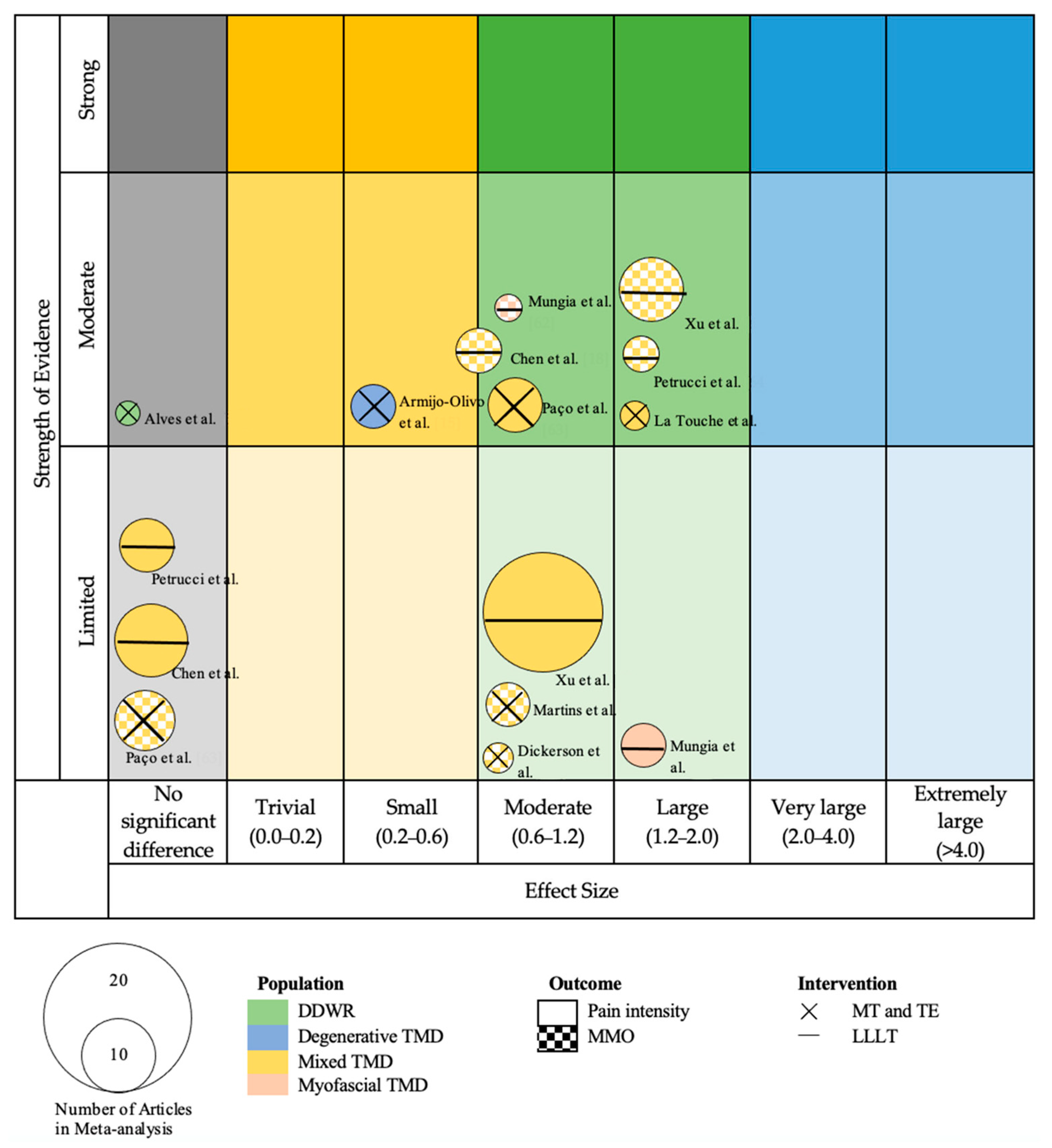
We created a visual map of the scientific evidence for each SR with meta-analysis to visually display the information as a bubble plot. The review information is based on 4 dimensions [34]:
- Number of studies (bubble size): The size of each bubble is directly proportional to the number of original studies included in each of the SR.
- Study population, intervention, and outcome variable (bubble aspect): The study population determined each bubble’s color. The outcome variable was represented by a different background within each bubble of each study (smooth background for PI and chequered background for MMO). The study intervention is represented by the symbol inside each bubble: “X” for combined MT and TE, and “-” for low-level laser therapy (LLLT).
- Effect size (x-axis): Each of the reviews was classified according to the effect size. When the effect size showed benefits for the intervention group, the intervention was classified as “trivial” (standard mean difference [SMD] between 0.0 and 0.2); “small” (SMD between 0.2 and 0.6); “moderate” (SMD between 0.2 and 1.2), “large” (SMD between 0.0 and 0.2); “very large” (SMD between 2.0 and 4.0); and “extremely large” (SMD more than 4.0). When the effect of the intervention was not superior to the control, it was classified as “no significant difference”.
- Strength of evidence (y-axis): The reviews were sorted into the following 4 categories according to the 2018 Physical Activity Guidelines Advisory Committee Grading Criteria (PAGAC) approach [35]: strong strength of evidence, moderate strength of evidence, limited strength of evidence, or strength of evidence unable to be assigned.
2.7. Qualitative Analysis
As described above, we relied on the assessments of each SR (with or without MA) for the methodological quality of the primary studies, using the AMSTAR for the included reviews and the PAGAC for assessing the evidence across SR. For the PAGAC analysis, the team assessed the strength of evidence based on the description of the findings provided in the studies and discussed as a group how to reconcile it according to Miake-Lye et al. [34].
2.8. Data Synthesis and Analysis
We performed the statistical analysis using meta-analyses with interactive explanations (MIX, version 1.7) [36]. We employed the same inclusion criteria for the review, adding 2 criteria: (1) results contained detailed information on the comparative statistical data (mean, standard deviation, and/or 95% confidence interval [CI]) of the main variables; and (2) data for the analyzed variables were represented in at least 2 studies.
We present the summary statistics in the form of forest plots [37], which consist of a weighted compilation of all SMDs and corresponding 95% CIs reported by each study, and provide an indication of heterogeneity among the studies. We examined the statistical significance of the pooled SMDs using Hedges’ g to account for a possible overestimation of the true population effect size in small studies [38]. The magnitude of g was interpreted according to a 4-point scale: (1) <0.20, negligible effect; (2) 0.20 to 0.49, small effect; (3) 0.50 to 0.79, moderate effect; and (4) ≥0.80, large effect [39]. We estimated the degree of heterogeneity among the studies by employing Cochran’s Q statistic test (p < 0.1 was considered significant) and the inconsistency index (I2) [40]. An I2 > 25% is considered to represent low heterogeneity, while an I2 > 50% is considered medium, and an I2 > 75% is considered to represent large heterogeneity [41]. The I2 index is complementary to the Q test, although it has a similar problem with power as the Q test with a small number of studies [41].
A study was therefore considered heterogeneous when it fulfilled one or both of the following conditions: (1) the Q-test was significant (p < 0.1); and (2) the result of I2 was >75%. To obtain a pooled estimate of the effect in the meta-analysis of the heterogeneous studies, we employed a random-effects model [42]. In addition, we decided to perform an analysis of the overlaps in the primary studies included in the final MMA, to assess the possibility that the data obtained for the effects of the interventions were over- or underestimated.
We analyzed the overlap of primary trials included at each of the meta-meta-analysis. Two methods were used, on one hand the percentage of overlapping (% overlap) was calculated as number of duplicated trials divided by the number of include trials. On the other hand, as recommended by Pieper et al., we calculated the “corrected covered area” (CCA) as a measure of overlapping: CAA = (N − r)/(r*c − r), where “N” is the number of included publications including double counting of overlapped trials, “r” is the number of included trials, “c” is the number of meta-analyses. The interpretation of CCA used to interpret the degree of overlap was 0–5 slight, 6–10 moderate, 11–15 high, >15 very high. We included a citation matrix to graphically represent the overlap [43]. We have also added a sensitivity analysis to include one more overlapping contrast as recommended by some authors [44].
3. Results
The research screening process is shown in the form of a flow chart (Figure 1). We initially found 324 articles, with 255 remaining after removing duplicates. Following title and abstract screening, we selected 90 full-text review articles for further evaluation. Ultimately, 31 SR met the inclusion criteria. The Appendix, Table A2 shows the reasons for exclusion of each article in the full-text screening process. The publication dates of the included SR ranged from 2006 to 2021.
Fifteen of the included studies were SR [16,17,20,21,22,45,46,47,48,49,50,51,52,53,54], whereas the other sixteen were SR and meta-analyses [15,18,19,55,56,57,58,59,60,61,62,63,64,65,66,67]. Table 1 list the characteristics of the included articles, and Table 2 list the characteristics of the interventions analyzed in each SR.
After the data extraction, we could only conduct 4 independent MMA, which included 10 of the 31 SR: (1) a meta-analysis including 4 studies regarding MT and TE on PI [15,57,60,63]; (2) a meta-analysis including 3 studies regarding MT and TE on MMO [19,61,63]; (3) a meta-analysis including 4 studies regarding LLLT on PI [18,62,64,67]; and (4) a meta-analysis including 4 studies regarding LLLT on MMO [18,62,64,67].
3.1. Characteristics of the Included Systematic Reviews
Our umbrella SR included 31 SR (with or without MA), amounting to 463 original studies, 446 RCTs, and 17 non-RCTs included in 3 SR [49,51,54], with a total of 17,611 participants.
In terms of the population in the SR, all of them included patients with various diagnoses of TMD: 8 SR (n = 4286) included myalgia TMD [16,21,46,55,58,60,62,66]; 3 SR (n = 684) included intra-articular TMD [47,56,57]; 17 SR (n = 11,571) included any type of TMD [15,17,18,19,22,45,46,48,49,50,52,54,59,61,63,64,67]; and 2 SR (n = 850); 2 SR (n = 643) included headache attributed to TMD [58,65] and 2 SR (n = 850) included mixed TMD [20,53].
Referring to the diagnostic criteria used in the primary studies of the included SR, 18 (57%) SR used the research DC/TMD or the DC/TMD criteria [15,16,21,45,46,47,48,49,50,51,55,56,59,60,62,63,65,66]. In the remaining 13 SR, the diagnosis was made with non-standardized criteria based on signs and symptoms, such as the Helkimo index or Fonseca’s questionnaire, or it was not described [15,17,18,19,22,52,53,54,57,58,61,64,67].
In all the SR, there was at least one PT intervention group in the analysis of the primary studies. In nine SR (n = 5304) the intervention group used MT, either passive or active techniques focused on the TMJ or masticatory muscles, associated or not with other MT techniques such as cervical MT, or associated or not with other interventions such as TE [15,17,19,20,46,47,57,60,61]. In nine SR (n = 5195), the intervention group used TE, focused mainly on the TMJ, masticatory muscles, and postural correction, and associated or not with MT [15,19,20,22,46,47,51,58,60]. In three SR (n = 1570), the intervention group used DN, mainly for myofascial TMD [21,55,66]. In eight SR (n = 5510), the intervention group used LLLT, mainly comparing it with control groups and between various laser applications [18,52,53,54,59,62,64,67]. Only in one SR (n = 153) did the intervention group use biofeedback training [45]. In seven SR (n = 4302), the intervention group included various PT interventions, and the analysis was performed focused on each intervention and the controls or comparisons between interventions [16,48,49,50,56,63,65].
Figure 1.
Flow chart of the research screening process.
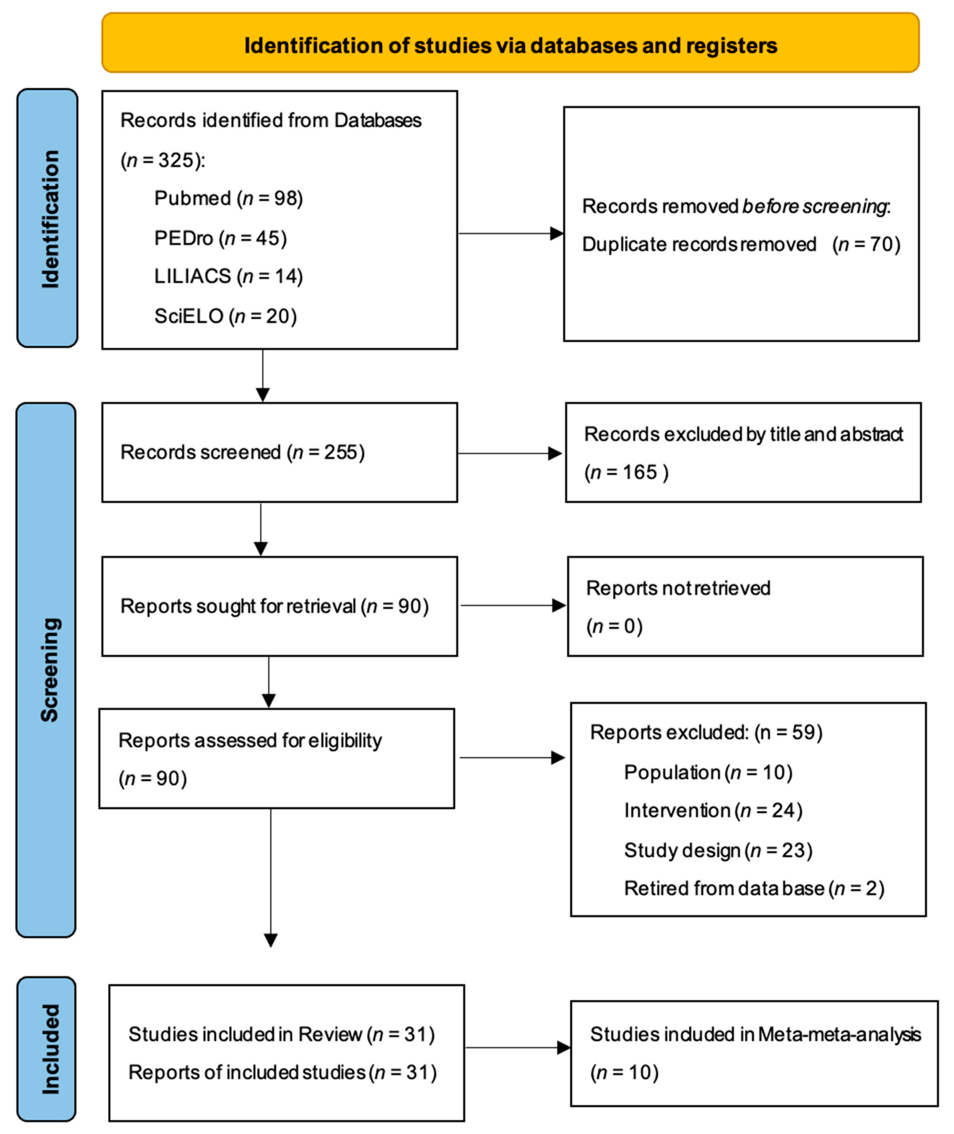
Table 1.
Characteristics of the included systematic reviews.
Table 2.
Intervention’s characteristics of the included systematic reviews.
3.2. Methodological Quality Results
Following the recommendations of Shea et al. [31] for the use of the AMSTAR 2 instrument, individual item ratings were not combined to create an overall score; however, we analyzed the methodological quality based on critical and non-critical items.
The AMSTAR 2 scores of overall confidence in the results of the 31 SR ranged from “critically low” to “low”. Only five (16.1%) SR scored as “low” and were considered the highest-quality studies in this umbrella SR [15,55,60,64,65]. The critical items with the lowest scores were those related to the methods established prior to the performance of the review, the use of a comprehensive literature search strategy, providing a list of excluded primary studies, and justifying the exclusions. The critical items that were completed and marked as “yes” in more than 50% were items 9 (80.64%) and 13 (61.29%), in which the presence of a satisfactory technique for assessing the risk of bias in the primary studies is assessed, as well as the risk of bias itself when discussing the results of the review. The remaining critical items were not completed in more than half of the included studies, and none of the SR included reported the sources of funding for the primary studies included in each review.
Table 3 shows the results of the methodological quality assessment using the AMSTAR 2. The inter-rater reliability of the methodological quality assessment was high (k = 0.9).
Table 3.
Quality assessment scores and resultant reliability statistics (AMSTAR 2).
3.3. Risk of Bias Results
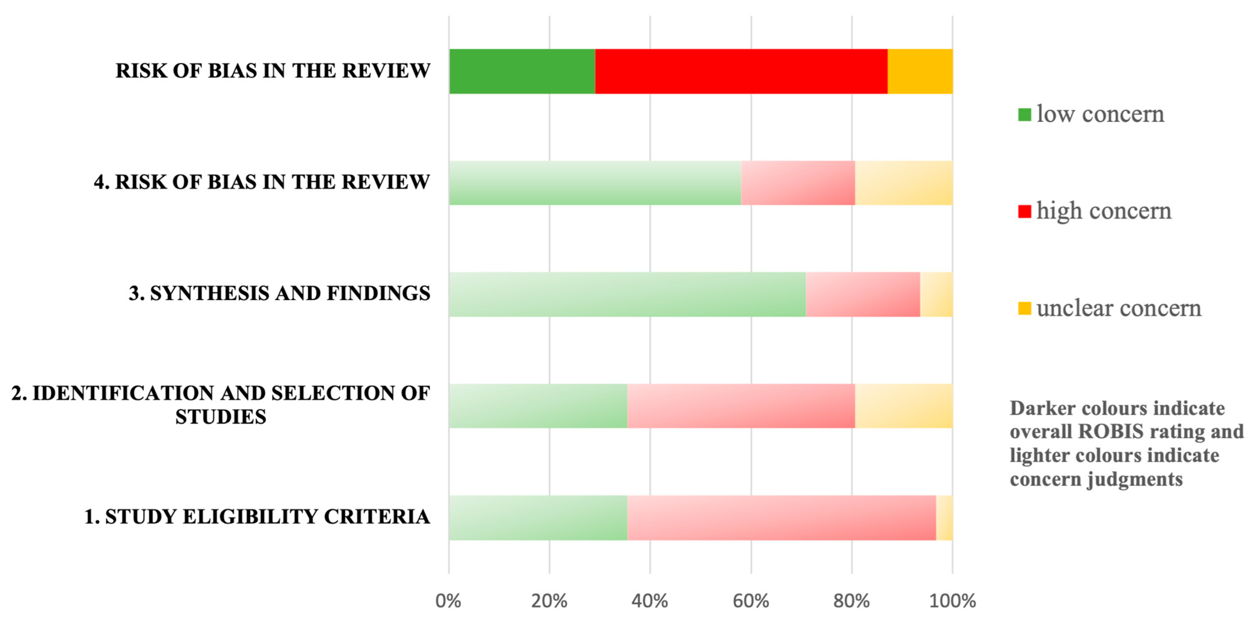
Table 4 and Figure 2 show the results of the risk of bias assessment using ROBIS. Of the 31 SR, 19 (61.3%) had a high risk of bias [16,18,19,20,45,46,50,51,52,53,54,58,59,61,62,63,64,65,66]; 10 (32.3%) had a low risk of bias [15,17,21,47,48,49,55,57,60,67]; and the remaining 2 had an unclear risk of bias [22,56].
Table 4.
Risk of bias assessment in systematic reviews through the ROBIS scale.
Figure 2.
Risk of bias assessment with the ROBIS tool.
The domains “Synthesis of Findings” and “Data Collection and Study Appraisal” had the lowest risk of bias, given that 24 (77.4%) SR scored with a “low concern” for both domains. In contrast, the domains “Study Eligibility Criteria” and “Identification and Selection of Studies” had the highest risk of bias, given that 21 (67.74%) and 19 (61.29%) of the SR, respectively, scored a “high concern” for both domains.
The inter-rater reliability for the risk of bias assessment was moderate (k = 0.6). We specifically analyzed the sources of funding for each SR, and only 9 of the 31 SR described that information [15,17,20,50,54,58,59,63,67]. Detailed information about the funding is shown in the Appendix, Table A3.
3.4. Evidence Map
Figure 3 presents the results of the evidence map for the 10 SR included in the MMA.
Figure 3.
Mapping based on effect size of MMA and strength of the evidence. On the x-axis, the studies are classified according to the effect size of the meta-analysis with standard mean difference (SMD). On the y-axis, they are categorized according to the strength of evidence based on the Physical Activity Guidelines Advisory Committee (PAGAC) [15,18,19,57,60,61,62,63,64,67]. Green bubble: Disc displacement with reduction; blue bubble: degenerative temporomandibular disorder; yellow bubble: mixed temporomandibular disorder; orange bubble: myofascial temporomandibular disorder; X: manual therapy and therapeutic exercise; −: low-level laser therapy.
3.5. Effects of Manual Therapy and Therapeutic Exercise Interventions
3.5.1. Isolated Manual Therapy Interventions
Three SR examined isolated MT interventions and analyzed their effects on various types of TMD [17,57,61]. All of them found that MT alone, or in association with other conservative interventions, is an effective approach for reducing PI and increasing the MMO and pressure pain threshold in patients with TMD.
The types of MT studied were massage therapy, mobilization/manipulation of the TMJ, and mobilization/manipulation of the upper cervical spine, all of which showed positive effects on patients with TMD, except for the C7-T1 thrust manipulation intervention, which showed low evidence of non-significant differences when comparing the effects of placebo on the pressure pain threshold in 1 SR [17]. The MT intervention also showed the same beneficial effects as botulinum toxin (BTX) injection on MMO and PI in patients with TMD [17]. One SR studied MT interventions specifically focused on myofascial TMD and found that MT was superior to no treatment and confers greater benefits when added to home therapy than home therapy alone; however, it was not more efficient than BTX treatment [46]. When MT was combined with counselling techniques, its effectiveness was noticeably higher. However, MT alone was not superior to counselling techniques, and it did not confer greater benefits to patients with myofascial TMD when added to these techniques. Finally, only 1 SR analyzed the effects of MT on the treatment of patients with disc displacement without a reduction in the TMJ and found that, based on the analysis of 2 studies, at 8 weeks follow-up, the treatment of patients with manual TMJ therapy, nonsteroidal anti-inflammatory drugs (NSAIDs), and occlusal techniques was not superior to controls for PI and MMO. However, at 60 months follow-up, the conservative intervention had the same effects as the treatment with arthroscopic surgery and arthroplasty [57].
Isolated MT interventions consisted mainly of the application of intra- and extra-oral soft tissue techniques of the masticatory musculature and joint tissue techniques addressed to the TMJ and upper cervical spine, comparing their effect with standard medical treatment, including medication (NSAIDs) and/or oral splint or TMJ surgery, and sham MT or controls. Two SR analyzed the number of sessions applied in the treatment, which varied from 1 to 24 [17,61].
Finally, only 1 SR specifically analyzed the frequency of MT interventions and the number of MT techniques used, revealing that 1 to 5 MT techniques were used, and that the frequency of the sessions ranged from 1 to 3 sessions per week [61].
3.5.2. Isolated Therapeutic Exercise Interventions
Three SR examined isolated TE interventions [22,51,58]. Two of the SR analyzed their effects on different combined types of TMD. One found that oral myofunctional therapy was not superior to controls or stabilization appliance (occlusal splint), it does not confer more benefits to self-care intervention, and it brings enhanced benefits to LLLT treatment [22]. In the other SR, positive effects were found for exercise therapy based on the emphasis on global exercises when added to body awareness and breathing techniques on reducing PI in the short term, with no superiority found between global postural re-education, postural exercises, and static segmental stretching [51]. Finally, only 1 SR analyzed the effects of TE interventions specifically on myofascial TMD and headache attributed to patients with TMD and found that TE based on stretching and postural relaxation was superior to controls and placebo on decreasing PI and headache frequency; however, the addition of a TE program to other conservative modalities did not show greater benefits in these outcome variables [58].
In terms of the interventions analyzed in the SR, isolated TE interventions consisted mainly of applying TMJ repetitive mobility exercises, cervical and TMJ motor control exercises, stretching exercises, and postural control exercise, comparing them with no treatment, self-management, and placebo exercises. Only 1 SR [22] included an analysis of TE interventions based on stomatognathic system functions and lingual exercises.
Two SR [22,51] analyzed the number of sessions applied in the treatment, which varied from 8 to 16. One SR [51] specifically analyzed the frequency of TE sessions and the duration of each session, finding that the frequency of the sessions ranged from 1 to 2 sessions per week, and the duration of each session varied from 15 to 45 min.
3.5.3. Combined Manual Therapy and Therapeutic Exercise Interventions
Six SR examined MT and TE interventions in isolation or in combination [15,19,20,46,47,60]. All of them found positive results for MT and TE interventions on MMO and PI, and that the combined interventions could be beneficial and play a role in the TMD approach. Two SR found positive effects from the inclusion of MT and TE in the cervical region on PI and MMO. The first [15] found that MT and TE in the cervical spine had positive effects on PI and MMO in myofascial TMD; the second [60] found the same in myofascial TMD and mixed TMD; and also that the combination of a cervical intervention with a TMJ intervention had better results. Only 1 SR [47] analyzed the effect of MT and TE on disc displacement without reduction and found that the greatest benefits were found by combining both interventions.
In terms of the interventions analyzed in the SR, combined MT and TE interventions consisted mainly of applying soft tissue techniques on the masticatory muscles; articular mobilization, traction, and manipulation techniques on the TMJ and the cervical spine; TE based on motor control of the mandibular and cervical movements; repetitive opening exercises of the TMJ; isometric exercises of the masticatory and cervical muscles; and postural control exercises. Two SR [20,60] included interventions based on neuromuscular and nerve tissue techniques or TMJ neurodynamics, and only 1 SR [15] analyzed interventions including manipulation of the thoracic spine. Interventions were compared normally with splint therapy, medication, BTX, surgery, placebo or sham, and no treatment.
In the various SR, the total time of the sessions ranged from 10 to 60 min, the frequency of the intervention varied from 2 to 6 times per day and from 0.5 to 5 sessions per week, and the total intervention time was between 1 week and 12 months.
3.5.4. Results of Quantitative Meta-Meta-Analysis of Combined Manual Therapy and Therapeutic Exercise Interventions
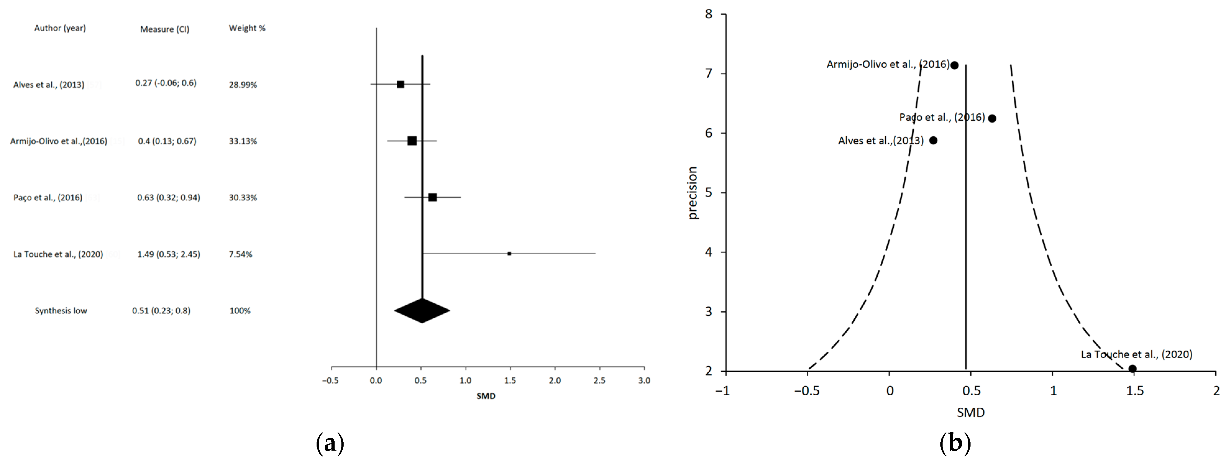
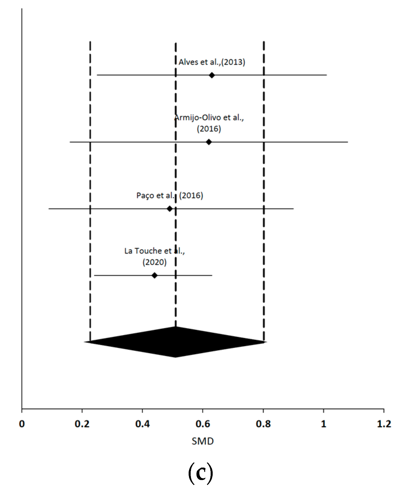
Regarding the quantitative analysis, the MMA of MT and TE on PI in patients with TMD revealed a statistically significant moderate effect size [15,57,60,63] (SMD = 0.51; 95% CI 0.8 to 0.23; p < 0.001). Given that the I2 was <75% (I2 = 57%), we considered that there was no evidence of heterogeneity. However, based on Cochran’s Q statistical test (Q = 6.97, p = 0.07), the heterogeneity among the reviews was considered significant. Although the shape of the funnel plot appeared to be asymmetrical in the dominant model, the sensitivity exclusion analysis suggested that no reviews significantly affected the pooled SMD. Forest, funnel and sensitivity plots are shown in Figure 4. The % of overlap in this MMA was 23.1% and the CAA = 0.07 (slight overlap). The overlapping of RCT is shown in Appendix, Table A4.
Figure 4.
(a) The forest plot summarizes the results of included studies (standardized mean differences (SMD) and weight). The small squares represent the point estimate of the effect size and sample size. The lines on either side of the box represent a 95% confidence interval [15,57,60,63]. (b) Funnel plot aims to assess the existence of publication bias. (c) Sensitivity plot.
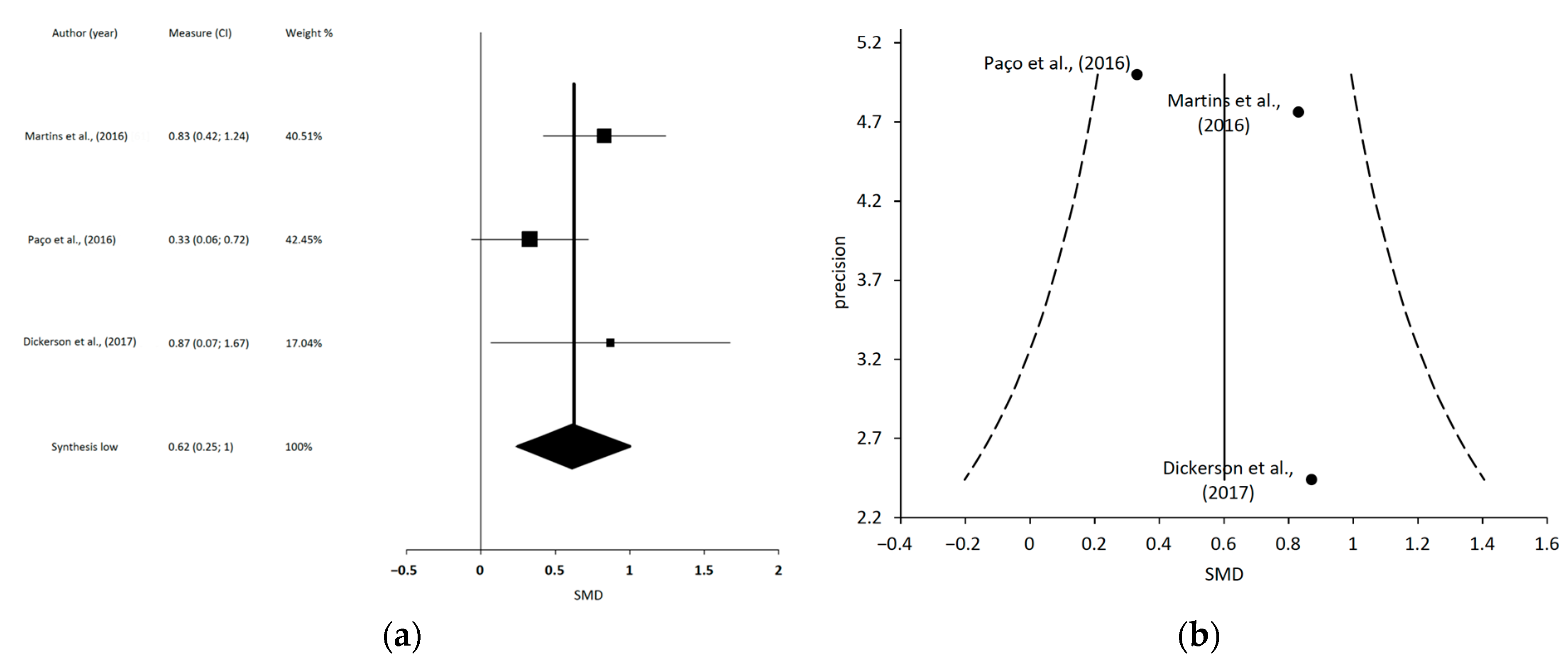
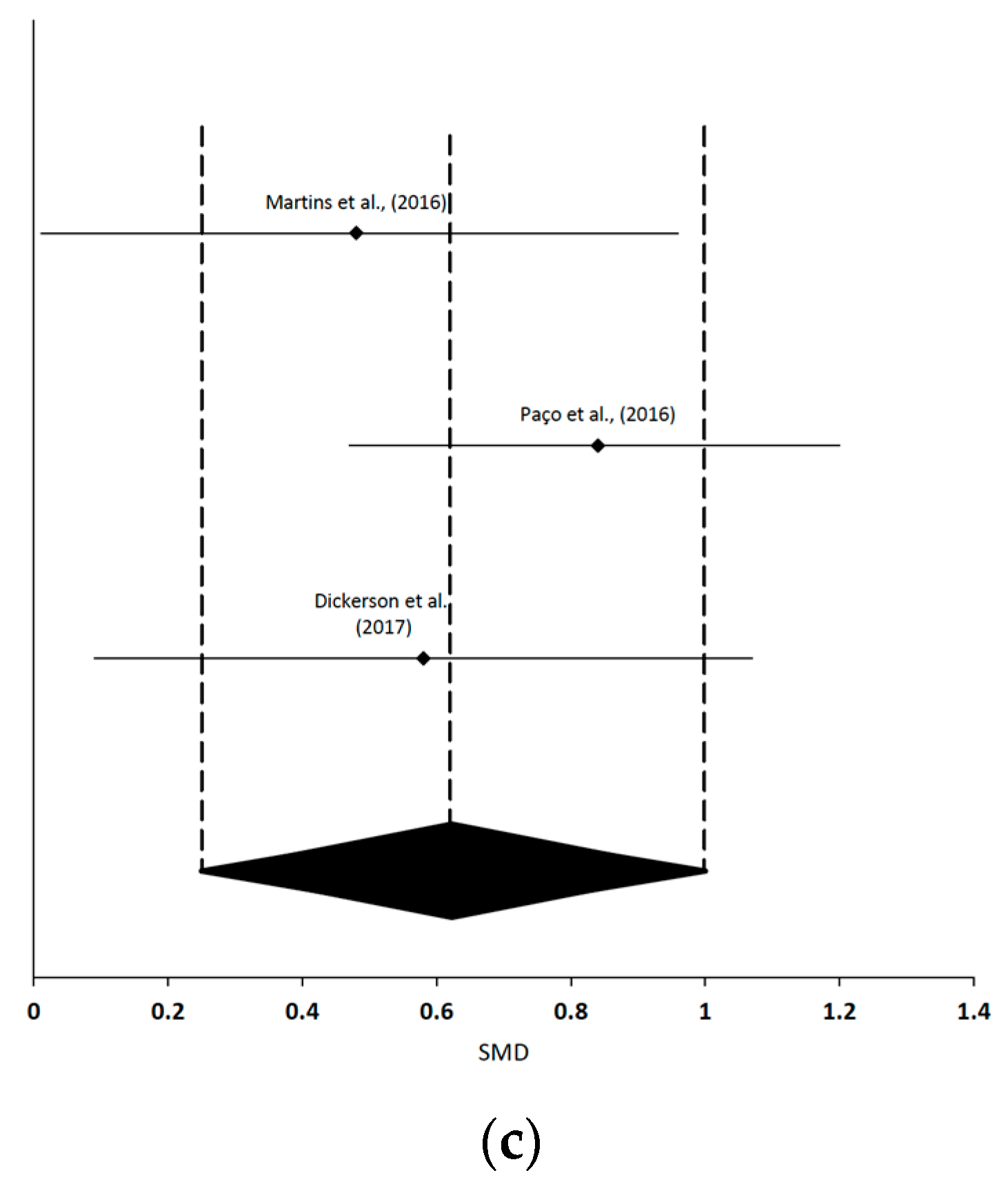
The MMA of MT and TE on MMO in patients with TMD revealed a statistically significant moderate effect size [19,61,63] (SMD = 0.62; 95% CI 0.25 to 1.00; p < 0.001). Given that the I2 was <75% (I2 = 42%), and considering the results of Cochran’s Q statistical test (Q = 3.46 p = 0.18), the heterogeneity among the reviews was not considered significant. The shape of the funnel plot appeared to be symmetrical in the dominant model. The sensitivity exclusion analysis suggested that no review significantly affected the pooled SMD. Forest, funnel and sensitivity plots are shown in Figure 5. The % of overlap was 33.3% and the CCA = 0.08 (slight overlap). The overlapping of RCT is shown in Appendix, Table A5.
Figure 5.
Synthesis forest, funnel and sensitivity plots of combined manual therapy and therapeutic exercise intervention on maximum mouth opening variable in patients with TMD [19,61,63]. (a) The forest plot summarizes the results of included studies (standardized mean differences (SMD) and weight). The small squares represent the point estimate of the effect size and sample size. The lines on either side of the box represent a 95% confidence interval. (b) Funnel plot aims to assess the existence of publication bias. (c) Sensitivity plot.
3.6. Effect of LLLT Interventions
Eight SR analyzed the effect of LLLT interventions on the management of TMD [18,52,53,54,59,62,64,67]. Seven of the SR evaluated the effects of LLLT on PI compared with placebo, and four SR found significant differences favoring LLLT for reducing PI at immediate post-intervention term (0 to 4 weeks) [52,53,62,67]. Five SR found no significant differences for reducing PI in the short term (2 to 4 weeks) [18,53,59,64,67]. Six studies evaluated the effect of LLLT on MMO compared with placebo, finding significant differences favoring LLLT in increasing MMO in the short term (0 to 4 weeks) [18,52,53,62,64,67] and 1 SR found no significant differences in MMO at immediate post-intervention [62].
In terms of the interventions analyzed in the SR, the main types of lasers used were GaAIAs, HeNe, GaAs, Nd: YAG, diode laser InGaAlP, infrared laser, AlGasAs, and gallium aluminum semiconductor LLL, with the control interventions being placebo, sham laser, or NSAIDs (piroxicam). Wavelengths ranged from 632 to 1064 nm, and dosage ranged from 0.3 to 144 J/cm2. Total intervention time varied from 10 s to 45 min, applied from 1 to 12 times per week, with a total number of sessions ranging from 1 to 24.
Quantitative Meta-Meta-Analysis of LLLT Intervention Results
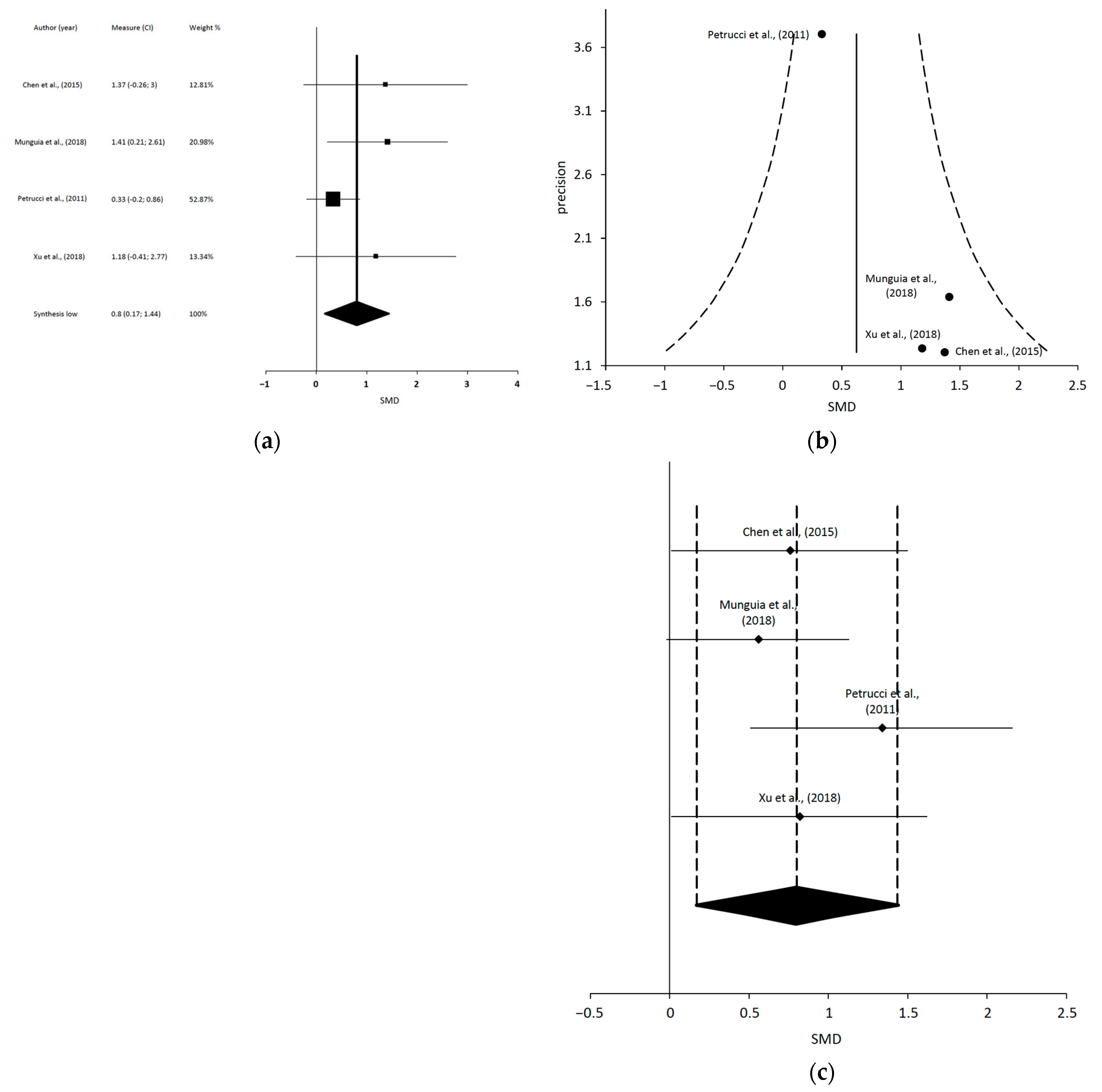
Regarding the quantitative analysis, the MMA of LLLT on PI in patients with TMD revealed a statistically significant large effect size [18,62,64,67] (SMD = 0.8; 95% CI 1.44 to 0.17; p < 0.001). Given that the I2 was <75% (I2 = 27%), and considering the results of Cochran’s Q statistical test (Q = 4.12, p = 0.25), the heterogeneity among the reviews was not considered significant. The shape of the funnel plot appeared to be symmetrical in the dominant model. The sensitivity exclusion analysis suggested that the review of Muguia et al. influences the pooled SMD [62]. Forest, funnel and sensitivity plots are shown in Figure 6. The % of overlap was 41.6% and the CCA = 0.13 (slight overlap). The overlapping of RCT is shown in Appendix, Table A6.
Figure 6.
Synthesis forest and funnel plot of low-level laser therapy on pain intensity variable [18,62,64,67]. (a) The forest plot summarizes the results of included studies (standardized mean differences (SMD) and weight). The small squares represent the point estimate of the effect size and sample size. The lines on either side of the box represent a 95% confidence interval. (b) Funnel plot aims to assess the existence of publication bias. (c) Sensitivity plot.
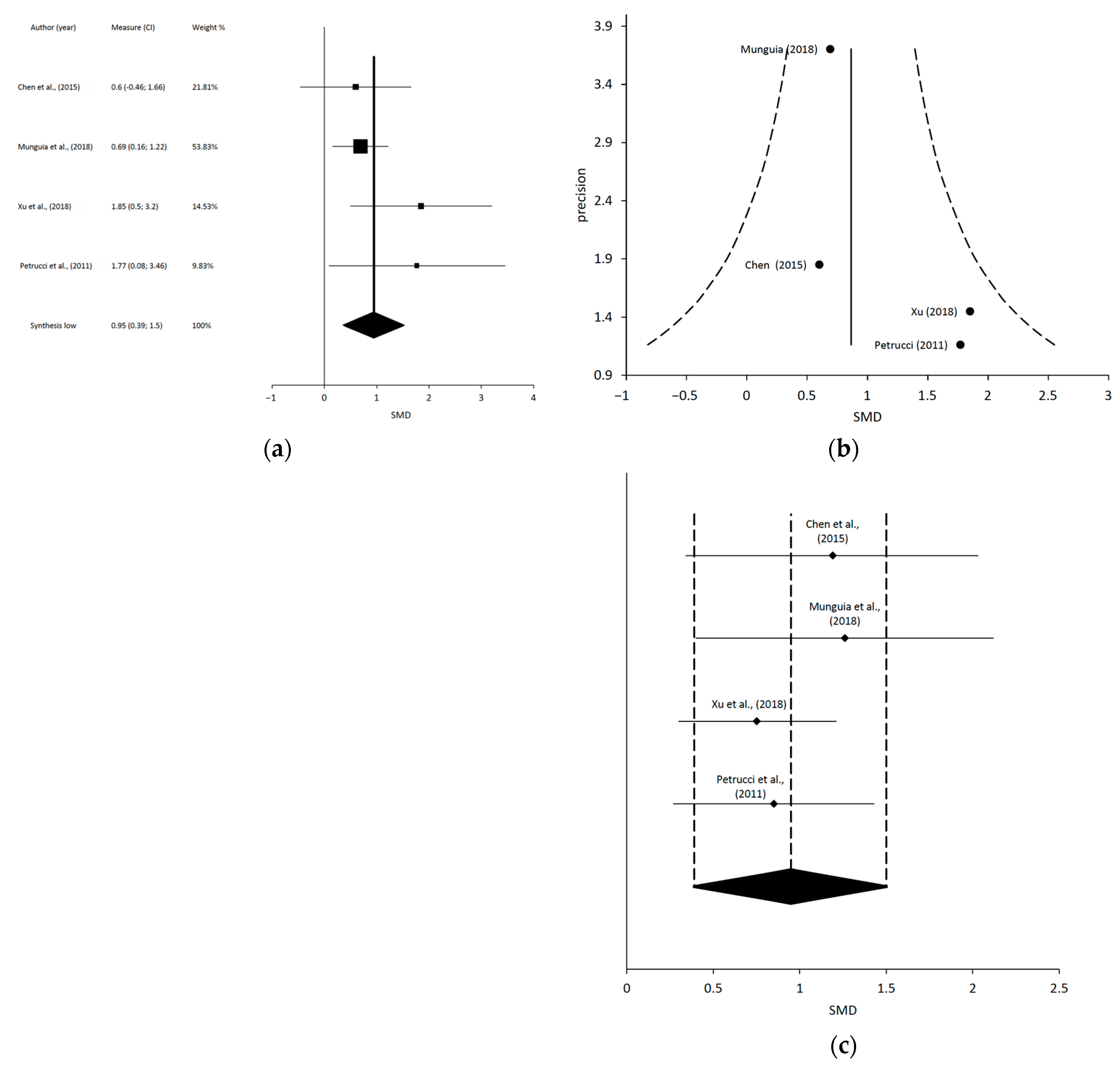
The MMA of LLLT on MMO in patients with TMD revealed a statistically significant large effect size (SMD = 0.95; 95% CI 1.5 to 0.39; p < 0.001). Given that the I2 was <75% (I2 = 21%) and considering the results of Cochran’s Q statistical test (Q = 3.81 p = 0.28), the heterogeneity among the reviews was not considered significant. The shape of the funnel plot appeared to be symmetrical in the dominant model. The sensitivity exclusion analysis suggested that no review significantly affected the pooled SMD. Forest and funnel plots are shown in Figure 7. The % of overlap was 35.7% and the CCA = 0.2 (slight overlap). The overlapping of RCT is shown in Appendix, Table A7.
Figure 7.
Synthesis forest and funnel plot of low-level laser therapy on maximum mouth opening variable [18,62,64,67]. (a) The forest plot summarizes the results of included studies (standardized mean differences (SMD) and weight). The small squares represent the point estimate of the effect size and sample size. The lines on either side of the box represent a 95% confidence interval. (b) Funnel plot aims to assess the existence of publication bias. (c) Sensitivity plot.
3.7. Effect of Dry Needling Interventions
Three studies evaluated the effect of DN on the management of TMD [21,55,66]. Two SR found no significant differences between DN and sham DN on reducing PI in the short and intermediate term (2 weeks to 6 months), and 1 SR found better results for DN [21]. One SR found better results for DN than other wet needling interventions on reducing PI [66], and 1 SR found better results for DN than active placebo in increasing MMO in the intermediate term (2 weeks to 6 months) [55].
In terms of the interventions analyzed in the SR, DN consisted of the direct needling by a monofilament needle without any chemical agent injected directly into the masticatory or cervical muscles, and was compared with sham DN, wet needling, LLLT, and acupuncture. DN was compared with wet needling in 2 RSs [21,55], which were intramuscular injections with substances such as BTX, lidocaine, or granisetron, which were used in the masticatory muscles, and found the same positive effects for both interventions. Every SR studied the comparison of DN and sham DN, and contradictory results were found. Two SR found that DN was superior to sham DN [21,55]; however, 1 SR found no statistically significant difference in PI and MMO between both interventions [66]. In terms of the characteristics of the interventions applied, only 1 SR [66] described the total treatment time of the intervention, which was from 20 to 30 min, once a week for 1 to 5 weeks; and only 1 SR [55] described the total treatment sessions, which ranged from 1 to 4.
Despite 3 SR analyzed DN interventions an MMA could not be performed due to the inconsistencies in the results, the variability of the variables, and the interventions included.
3.8. Effect of Combined Physical Therapy Interventions
Seven SR analyzed the effect of combined PT interventions in patients with TMD [16,48,49,50,56,63,65]. When comparing combined PT interventions with placebo, no intervention, or usual care, four SR found significant differences in PI and MMO favoring PT interventions, mainly based on MT, TE, DN, LLLT, muscular awareness relaxation therapy, and self-management [48,49,50,63]. However, three SR found no significant differences [16,56,65]. Both the intervention and comparison groups analyzed in the SR were heterogeneous.
Interventions consisted of MT; TE (oral exercises, posture correction); occlusal splint therapy; electrotherapy modalities such as LLLT, transcutaneous electrical nerve stimulation (TENS), biofeedback, and photobiomodulation therapy; TEd (behavioral education, counselling); invasive interventions such as DN, acupuncture, oral injections, and surgeries; relaxation training; and counselling, all of which were applied in isolation or in combination.
Despite the number of SR that analyzed combined physical therapy interventions (7), an MMA could not be performed due to the heterogeneity of the interventions and variables and inconsistencies in the results.
3.9. Effect of Biofeedback Interventions
One SR of 10 RCTs [45] analyzed the effect of biofeedback training on the treatment of TMD, showing positive effects in reducing PI, reducing masticatory muscle activity, and decreasing frequency of sleep bruxism events, with a very low to moderate quality of evidence. Biofeedback training can be applied as visual biofeedback, vibratory biofeedback, audio biofeedback, or contingent electrical stimulation, and was compared with TENS treatment, occlusal splint therapy, and a control group. Total session time varied from 20 min to 8 h. The intervention lasted between 2 nights and 12 weeks, with a total number of sessions ranging from 2 to 42 sessions.
MMA could not be performed due to number of SR.
3.10. Grades of Evidence
The results of the grades of evidence of the MMA variables are shown in Table 5.
Table 5.
Grades of evidence of meta-meta-analyzed variables (PAGAC).
4. Discussion
The aim of this umbrella SR was to study, synthesize, and critically evaluate the current evidence on the effects of PT interventions in patients with TMD, specifically on pain and functional variables such as PI and MMO. We collected and synthesized evidence to assist with the decision-making process in the physical therapy and medical clinic setting. We specifically analyzed the presence of overlaps in the total included primary studies of the MMA, and we found a percentage of duplicities between 23.1% and 41.6%; moreover, the analysis of the CCA showed a slight overlap for all four MMA performed.
Various physical therapy interventions have been shown to be effective in improving pain and function in patients with TMD. Our MMA showed that combined interventions including MT and TE had statistically significant moderate effects on the improvement of pain, with a moderate grade of evidence; and on MMO, with a limited grade of evidence. Moreover, LLLT interventions had a statistically significant large effect on pain improvement, with a limited grade of evidence; and on MMO, with a moderate grade of evidence.
Two important symptoms of patients with TMD are pain and a reduction in MMO; thus, the use of therapeutic interventions that improve them appears critical. Interventions involving manual therapy and therapeutic exercise have been shown to be effective on improving these variables in patients with TMD; however, there is great diversity as to what the interventions consist of. From among the SR that analyzed these combined interventions and could be meta-meta-analyzed [15,19,57,60,61,63], the prescription of therapeutic exercise varied considerably, ranging from general exercises involving the whole body based on postural correction, to specific exercises of the TMJ and masticatory muscles, or even for the cervical spine. On the other hand, the use of manual therapy techniques was also diverse, including joint techniques such as mobilizations or manipulations of the cervical spine and/or TMJ, neural tissue mobilization techniques of the craniomandibular region, and soft tissue techniques such as compression and massage of painful muscles. In this sense, it is possible that the diversity of techniques found in the various SR is due to the diversity of the type of TMD. Different types of TMD are analyzed in the same SR, and therefore it could be that the use of certain techniques directed towards a specific tissue was not specifically analyzed, which would perhaps improve the effect of that specific intervention. This result highlights the importance of subclassifying patients with TMD so that we can make the decision of the choice of manual therapy techniques and exercise prescription towards the maximum effect for improving patients’ health. We should not forget that clinical reasoning based on the analysis of the symptoms’ source could be relevant for the choice of manual therapy technique, which is essentially how the DC/TMD subclassifies patients. There are two main general etiopathogenesis for the TMD which are myogenous and arthrogenous, they show with different clinical signs and symptoms, and treatment approach may be also different. However, many studies include different types of TMD or even mixed TMD in their samples. Another important topic to point out is the possible process of central sensitization that some patients could undergo when suffering chronic pain or in this case a chronic TMD, which could lead to a great pain expansion and overlapping of symptoms such as headache and fibromyalgia [68,69,70]. Additionally, psychological distress can contribute to the chronicity of the TMD, thus multidisciplinary management is necessary in these patients [68].
In future research, it will be essential to always classify and subclassify patients with TMD using clear criteria, such as according to the DC/TMD, as well as clearly define the chronicity of the TMD, so that the decision-making process and the impact of interventions could be analyzed, and their effectiveness in each subgroup can be compared.
In the studies included in the MMA, there was a lack of data regarding the intensity, dosage, and application frequency of manual therapy and exercise. A proper subclassification of TMD would also help in terms of finding the most appropriate therapy for each patient and the minimum effective dose. Another important aspect to note is the high frequency with which manual therapy is combined with therapeutic exercise in the scientific literature in the rehabilitation of patients with TMD. In this aspect, it is relevant to note that most research groups recommend the combination of both therapeutic tools for the management of these patients; their use in isolation does not appear to be the best choice.
Regarding usefulness, the combination of manual therapy and therapeutic exercise appears to be a useful intervention for the management of these patients regardless of the type of TMD; it has thus been proposed as a first-line treatment. Additionally, LLLT interventions have been shown to be effective for improving PI and MMO in patients with TMD, and the application appears to be more homogeneous. Among the SR that analyzed LLLT and could be meta-meta-analyzed [18,62,64,67], several types of LLLT were used. Although there was variability in the intensity and dose of application, the number of sessions was not greater than 20, with a weekly frequency of 1 to 12 sessions, applying the laser to the masticatory musculature and/or TMJ, with satisfactory results in terms of PI reduction and improvement in MMO in most of the SR. Unfortunately, due to the variability in the application of LLLT, MT, and TE, we cannot make a specific proposal of parameters for its application.
Given that the LLLT interventions’ follow-up time points were mostly in the short term, with a maximum follow-up of 3 months, we could not analyze the effects of these interventions on TMD. However, given that the follow-ups of the MT and TE interventions were conducted in the short, medium, and long terms, it could be interesting to investigate whether both interventions could be used in combination, using LLLT as a facilitating tool for the first phase of the whole rehabilitation process, with MT and ET as the main intervention. However, given that the combination of these interventions has not been analyzed in this umbrella SR, it is not possible to make a statement on this point.
4.1. Clinical Implications
As for the clinical application of this type of intervention, it was difficult to obtain sufficient information to generate concrete indications based on the data of this review; however, there are some indications that can be extracted to facilitate clinicians’ decision making.
First, this review suggests that conservative physiotherapy interventions using combined MT and TE or LLLT are the most studied interventions and are effective in the management of patients with TMD, and, given that these are conservative and low-cost interventions, they could be the best choice for the first line of treatment for these patients.
Second, subclassification of patients with TMD appears fundamental to improving decision making in terms of the choice of techniques and parameters in prescribing treatment, mainly considering the most likely source of symptoms.
Third, given that this review suggests that LLLT interventions achieve greater effects in the short term, but combined MT and TE achieve greater effects in the medium and long term on improving pain intensity and MMO, it is possible that the best intervention for these patients is a combination of both techniques, using LLLT in the initial phases (2–4 weeks) in combination with MT + TE, and maintaining the latter two for the medium to long term.
For the treatment planning and dosage, considering the data obtained from both interventions in this umbrella SR, we should consider sessions of between 30 and 60 min, with a frequency of 1 to 3 sessions per week, to be able to combine the interventions, dedicating less than 10 min to the LLLT application and 10 to 45 min to MT and TE, as well as being combined with home exercise.
Although the prescription of MT and TE appears simple to apply in the clinical field of physiotherapy, their effectiveness depends on the context in terms of the clinician and the patient, and always within a biobehavioral framework in which a multitude of social and psychological factors are involved and can enhance or worsen the results of the intervention.
4.2. Limitations
This umbrella SR has several limitations. The first is the lack of information on the interventions carried out in some RCTs in SR. Second, the review did not allow us to analyze the impact of educational and behavioral interventions, even though these interventions are used repeatedly among the reviews as part of the treatment, such as LLLT, MT, or TE, a fact that could influence the effect found in the application of the techniques. As a final limitation, given that we could not independently analyze acute and chronic TMD in the included SR, it was not possible to perform an analysis of the interventions’ effects in terms of chronicity in these different populations.
5. Conclusions
The present umbrella and mapping SR with MMA provides an overview of the effect of physical therapy interventions on PI and MMO variables in TMD. The results showed that the application of LLLT and combined MT and TE interventions have a positive effect on improving pain and functional variables in patients with TMD. The MMA showed the positive results of LLLT on PI and MMO, with a large effect size, and for combined MT and TE on PI and MMO, with a moderate effect size. The quality of evidence was limited for LLLT on PI and MT and TE on MMO, and moderate for LLLT on MMO and MT and TE on PI. The application of these interventions appears to be the first line in the treatment of TMD, and the combination of interventions should be considered in the clinical setting and in future studies.
Author Contributions
Conceptualization, R.L.T. and A.P.-A.; methodology, R.L.T., A.P.-A. and M.G.-A.; software, S.A.-D.-P.; validation, M.A.-P., S.H.-H., C.J.-A., M.G.-A., S.A.-D.-P., R.L.T. and A.P.-A.; formal analysis, R.L.T. and S.A.-D.-P.; investigation, M.A.-P., S.H.-H., C.J.-A., M.G.-A., S.A.-D.-P., R.L.T. and A.P.-A.; resources, R.L.T., A.P.-A. and M.G.-A.; data curation, M.A.-P., S.H.-H. and C.J.-A.; writing—original draft preparation, M.A.-P., S.H.-H., C.J.-A., M.G.-A., S.A.-D.-P., R.L.T. and A.P.-A.; writing—review and editing, M.A.-P., S.H.-H., C.J.-A., M.G.-A., S.A.-D.-P., R.L.T. and A.P.-A.; visualization, M.A.-P., S.H.-H., C.J.-A., M.G.-A., S.A.-D.-P., R.L.T. and A.P.-A.; supervision, R.L.T. and A.P.-A.; project administration, R.L.T. and A.P.-A. All authors have read and agreed to the published version of the manuscript.
Funding
This research received no external funding.
Institutional Review Board Statement
Not applicable.
Informed Consent Statement
Not applicable.
Data Availability Statement
All our data are published in the review, and can be found in the other paper we have reviewed.
Conflicts of Interest
The authors declare no conflict of interest.
Appendix A
Table A1.
Search strategies in each database.
Table A2.
Reasons of exclusion of full-text screening process.
Table A3.
Funding information of included systematic reviews.
Table A4.
Overlap of clinical trials of the MMA of combined manual therapy and therapeutic exercise interventions on pain intensity.
Table A5.
Overlap of clinical trials of the MMA of combined manual therapy and therapeutic exercise interventions on mouth opening.
Table A6.
Overlap of clinical trials of the MMA of LLLT interventions on pain intensity.
Table A7.
Overlap of clinical trials of the MMA of LLLT interventions on mouth opening.
References
- List, T.; Jensen, R.H. Temporomandibular Disorders: Old Ideas and New Concepts. Cephalalgia 2017, 37, 692–704. [Google Scholar] [CrossRef] [Scilit] [PubMed]
- Dubner, R.; Ohrbach, R.; Dworkin, S.F. The Evolution of TMD Diagnosis: Past, Present, Future. J. Dent. Res. 2016, 95, 1093–1101. [Google Scholar] [CrossRef] [Scilit]
- Marim, G.C.; Machado, B.C.Z.; Trawitzki, L.V.V.; de Felício, C.M. Tongue Strength, Masticatory and Swallowing Dysfunction in Patients with Chronic Temporomandibular Disorder. Physiol. Behav. 2019, 210, 112616. [Google Scholar] [CrossRef] [Scilit]
- Schiffman, E.; Ohrbach, R.; Truelove, E.; Look, J.; Anderson, G.; Goulet, J.-P.; List, T.; Svensson, P.; Gonzalez, Y.; Lobbezoo, F.; et al. Diagnostic Criteria for Temporomandibular Disorders (DC/TMD) for Clinical and Research Applications: Recommendations of the International RDC/TMD Consortium Network* and Orofacial Pain Special Interest Group. J. Oral Facial Pain Headache 2014, 28, 6–27. [Google Scholar] [CrossRef] [Scilit]
- Talaat, W.M.; Adel, O.I.; al Bayatti, S. Prevalence of Temporomandibular Disorders Discovered Incidentally during Routine Dental Examination Using the Research Diagnostic Criteria for Temporomandibular Disorders. Oral Surgery Oral Med. Oral Pathol. Oral Radiol. 2018, 125, 250–259. [Google Scholar] [CrossRef] [Scilit]
- Wieckiewicz, M.; Grychowska, N.; Nahajowski, M.; Hnitecka, S.; Kempiak, K.; Charemska, K.; Balicz, A.; Chirkowska, A.; Zietek, M.; Winocur, E. Prevalence and Overlaps of Headaches and Pain-Related Temporomandibular Disorders Among the Polish Urban Population. J. Oral Facial Pain Headache 2020, 34, 31–39. [Google Scholar] [CrossRef] [Scilit] [PubMed]
- Valesan, L.F.; Da-Cas, C.D.; Réus, J.C.; Denardin, A.C.S.; Garanhani, R.R.; Bonotto, D.; Januzzi, E.; de Souza, B.D.M. Prevalence of Temporomandibular Joint Disorders: A Systematic Review and Meta-Analysis. Clin. Oral Investig. 2021, 25, 441–453. [Google Scholar] [CrossRef] [Scilit] [PubMed]
- Kusdra, P.M.; Stechman-Neto, J.; De Leão, B.L.C.; Martins, P.F.A.; Lacerda, A.B.M.d.; Zeigelboim, B.S. Relationship between Otological Symptoms and TMD. Int. Tinnitus J. 2018, 22, 30–34. [Google Scholar] [CrossRef] [Scilit]
- Calixtre, L.B.; Oliveira, A.B.; de Sena Rosa, L.R.; Armijo-Olivo, S.; Visscher, C.M.; Alburquerque-Sendín, F. Effectiveness of Mobilisation of the Upper Cervical Region and Craniocervical Flexor Training on Orofacial Pain, Mandibular Function and Headache in Women with TMD. A Randomised, Controlled Trial. J. Oral Rehabil. 2019, 46, 109–119. [Google Scholar] [CrossRef] [Scilit]
- Ferrillo, M.; Marotta, N.; Giudice, A.; Calafiore, D.; Curci, C.; Fortunato, L.; Ammendolia, A.; de Sire, A. Effects of Occlusal Splints on Spinal Posture in Patients with Temporomandibular Disorders: A Systematic Review. Healthcare 2022, 10, 739. [Google Scholar] [CrossRef] [Scilit]
- Gil-Martínez, A.; Paris-Alemany, A.; López-de-Uralde-Villanueva, I.; la Touche, R. Management of Pain in Patients with Temporomandibular Disorder (TMD): Challenges and Solutions. J. Pain Res. 2018, 11, 571–587. [Google Scholar] [CrossRef] [Scilit] [PubMed]
- Furquim, B.D.; Flamengui, L.M.S.P.; Conti, P.C.R. TMD and Chronic Pain: A Current View. Dent. Press J. Orthod. 2015, 20, 127–133. [Google Scholar] [CrossRef] [Scilit] [PubMed]
- Ferrillo, M.; Nucci, L.; Giudice, A.; Calafiore, D.; Marotta, N.; Minervini, G.; d’Apuzzo, F.; Ammendolia, A.; Perillo, L.; de Sire, A. Efficacy of Conservative Approaches on Pain Relief in Patients with Temporomandibular Joint Disorders: A Systematic Review with Network Meta-Analysis. Cranio 2022. [Google Scholar] [CrossRef] [Scilit] [PubMed]
- Florjański, W.; Orzeszek, S. Role of Mental State in Temporomandibular Disorders: A Review of the Literature. Dent. Med. Probl. 2021, 58, 127–133. [Google Scholar] [CrossRef] [Scilit] [PubMed]
- Armijo-Olivo, S.; Pitance, L.; Singh, V.; Neto, F.; Thie, N.; Michelotti, A. Effectiveness of Manual Therapy and Therapeutic Exercise for Temporomandibular Disorders: Systematic Review and Meta-Analysis. Phys. Ther. 2016, 96, 9–25. [Google Scholar] [CrossRef] [Scilit] [PubMed]
- Brochado, F.T.; De Jesus, L.H.; Martins, M.D.; Chaves, K.D. Non-Invasive Therapies for Management of Temporomandibular Disorders: A Systematic Review. Clin. Biomed. Res. 2019, 39, 230–243. [Google Scholar] [CrossRef] [Scilit]
- Calixtre, L.B.; Moreira, R.F.C.; Franchini, G.H.; Alburquerque-Sendín, F.; Oliveira, A.B. Manual Therapy for the Management of Pain and Limited Range of Motion in Subjects with Signs and Symptoms of Temporomandibular Disorder: A Systematic Review of Randomised Controlled Trials. J. Oral Rehabil. 2015, 42, 847–861. [Google Scholar] [CrossRef] [Scilit]
- Chen, J.; Huang, Z.; Ge, M.; Gao, M. Efficacy of Low-Level Laser Therapy in the Treatment of TMDs: A Meta-Analysis of 14 Randomised Controlled Trials. J. Oral Rehabil. 2015, 42, 291–299. [Google Scholar] [CrossRef] [Scilit]
- Dickerson, S.M.; Weaver, J.M.; Boyson, A.N.; Thacker, J.A.; Junak, A.A.; Ritzline, P.D.; Donaldson, M.B. The Effectiveness of Exercise Therapy for Temporomandibular Dysfunction: A Systematic Review and Meta-Analysis. Clin. Rehabil. 2017, 31, 1039–1043. [Google Scholar] [CrossRef] [Scilit]
- Herrera-Valencia, A.; Ruiz-Muñoz, M.; Martin-Martin, J.; Cuesta-Vargas, A.; González-Sánchez, M. Effcacy of Manual Therapy in TemporomandibularJoint Disorders and Its Medium-and Long-TermEffects on Pain and Maximum Mouth Opening: A Systematic Review and Meta-Analysis. J. Clin. Med. 2020, 9, 3404. [Google Scholar] [CrossRef] [Scilit]
- Machado, E.; Machado, P.; Wandscher, V.F.; Marchionatti, A.M.E.; Zanatta, F.B.; Kaizer, O.B. A Systematic Review of Different Substance Injection and Dry Needling for Treatment of Temporomandibular Myofascial Pain. Int. J. Oral Maxillofac. Surg. 2018, 47, 1420–1432. [Google Scholar] [CrossRef] [Scilit]
- Melis, M.; di Giosia, M.; Zawawi, K.H. Oral Myofunctional Therapy for the Treatment of Temporomandibular Disorders: A Systematic Review. Cranio® 2019, 40, 41–47. [Google Scholar] [CrossRef] [Scilit] [PubMed]
- Shukla, D.; Muthusekhar, M. Efficacy of Low-Level Laser Therapy in Temporomandibular Disorders: A Systematic Review. Natl. J. Maxillofac. Surg. 2016, 7, 62. [Google Scholar] [CrossRef] [Scilit] [PubMed]
- Cerón, L.; Pacheco, M.; Delgado Gaete, A.; Bravo Torres, W.; Astudillo Rubio, D. Therapies for Sleep Bruxism in Dentistry: A Critical Evaluation of Systematic Reviews. Dent. Med. Probl. 2022. [Google Scholar] [CrossRef] [Scilit] [PubMed]
- Bougioukas, K.I.; Liakos, A.; Tsapas, A.; Ntzani, E.; Haidich, A.B. Preferred Reporting Items for Overviews of Systematic Reviews Including Harms Checklist: A Pilot Tool to Be Used for Balanced Reporting of Benefits and Harms. J. Clin. Epidemiol. 2018, 93, 9–24. [Google Scholar] [CrossRef] [Scilit] [PubMed]
- Methley, A.M.; Campbell, S.; Chew-Graham, C.; McNally, R.; Cheraghi-Sohi, S. PICO, PICOS and SPIDER: A Comparison Study of Specificity and Sensitivity in Three Search Tools for Qualitative Systematic Reviews. BMC Health Serv. Res. 2014, 14, 579. [Google Scholar] [CrossRef] [Scilit]
- Amir-Behghadami, M.; Janati, A. Population, Intervention, Comparison, Outcomes and Study (PICOS) Design as a Framework to Formulate Eligibility Criteria in Systematic Reviews. Emerg. Med. J. 2020, 37, 387. [Google Scholar] [CrossRef] [Scilit]
- Schiffman, E.; Ohrbach, R. Executive Summary of the Diagnostic Criteria for Temporomandibular Disorders for Clinical and Research Applications. J. Am. Dent. Assoc. 2016, 147, 438–445. [Google Scholar] [CrossRef] [Scilit]
- Moher, D. CONSORT: An Evolving Tool to Help Improve the Quality of Reports of Randomized Controlled Trials. Consolidated Standards of Reporting Trials. JAMA 1998, 279, 1489–1491. [Google Scholar] [CrossRef] [Scilit]
- Higgins, J.P.; Thomas, J.; Chandler, J.; Cumpston, M.; Li, T.; Page, M.J.; Welch, V.A. (Eds.) Cochrane Handbook for Systematic Reviews of Interventions; Version 5; The Cochrane Collaboration: London, UK, 2011. [Google Scholar]
- Shea, B.J.; Reeves, B.C.; Wells, G.; Thuku, M.; Hamel, C.; Moran, J.; Moher, D.; Tugwell, P.; Welch, V.; Kristjansson, E.; et al. AMSTAR 2: A Critical Appraisal Tool for Systematic Reviews That Include Randomised or Non-Randomised Studies of Healthcare Interventions, or Both. BMJ 2017, 358, j4008. [Google Scholar] [CrossRef] [Scilit]
- McHugh, M.L. Interrater Reliability: The Kappa Statistic. Biochem. Med. 2012, 22, 276–282. [Google Scholar] [CrossRef] [Scilit]
- Whiting, P.; Savović, J.; Higgins, J.P.T.; Caldwell, D.M.; Reeves, B.C.; Shea, B.; Davies, P.; Kleijnen, J.; Churchill, R. ROBIS: A New Tool to Assess Risk of Bias in Systematic Reviews Was Developed. J. Clin. Epidemiol. 2016, 69, 225–234. [Google Scholar] [CrossRef] [Scilit] [PubMed]
- Miake-Lye, I.M.; Hempel, S.; Shanman, R.; Shekelle, P.G. What Is an Evidence Map? A Systematic Review of Published Evidence Maps and Their Definitions, Methods, and Products. Syst. Rev. 2016, 5, 1–21. [Google Scholar] [CrossRef] [Scilit] [PubMed]
- Dipietro, L.; Campbell, W.W.; Buchner, D.M.; Erickson, K.I.; Powell, K.E.; Bloodgood, B.; Hughes, T.; Day, K.R.; Piercy, K.L.; Vaux-Bjerke, A.; et al. Physical Activity, Injurious Falls, and Physical Function in Aging: An Umbrella Review. Med. Sci. Sports Exerc. 2019, 51, 1303–1313. [Google Scholar] [CrossRef] [Scilit] [PubMed]
- Bax, L.; Yu, L.-M.M.; Ikeda, N.; Tsuruta, H.; Moons, K.G.M.M. Development and Validation of MIX: Comprehensive Free Software for Meta-Analysis of Causal Research Data. BMC Med. Res. Methodol. 2006, 6, 50. [Google Scholar] [CrossRef] [Scilit] [PubMed]
- Lewis, S.; Clarke, M. Forest Plots: Trying to See the Wood and the Trees. BMJ 2001, 322, 1479–1480. [Google Scholar] [CrossRef] [Scilit]
- Hedges, L.V. Estimation of Effect Size from a Series of Independent Experiments. Psychol. Bull. 1982, 92, 490–499. [Google Scholar] [CrossRef]
- Cohen, J. A Power Primer. Psychol. Bull. 1992, 112, 155–159. [Google Scholar] [CrossRef]
- Higgins, J.P.T.T.; Thompson, S.G.; Deeks, J.J.; Altman, D.G. Measuring Inconsistency in Meta-Analyses. BJM 2003, 327, 557–560. [Google Scholar] [CrossRef] [Scilit]
- Huedo-Medina, T.B.; Sánchez-Meca, J.; Marín-Martínez, F.; Botella, J. Assessing Heterogeneity in Meta-Analysis: Q Statistic or I2 Index? Psychol. Methods 2006, 11, 193–206. [Google Scholar] [CrossRef] [Scilit]
- DerSimonian, R.; Laird, N. Meta-Analysis in Clinical Trials. Control Clin. Trials 1986, 7, 177–188. [Google Scholar] [CrossRef] [Scilit] [PubMed]
- Pieper, D.; Antoine, S.L.; Mathes, T.; Neugebauer, E.A.M.; Eikermann, M. Systematic Review Finds Overlapping Reviews Were Not Mentioned in Every Other Overview. J. Clin. Epidemiol. 2014, 67, 368–375. [Google Scholar] [CrossRef] [Scilit] [PubMed]
- Lunny, C.; Pieper, D.; Thabet, P.; Kanji, S. Managing Overlap of Primary Study Results across Systematic Reviews: Practical Considerations for Authors of Overviews of Reviews. BMC Med. Res. Methodol. 2021, 21, 1–14. [Google Scholar] [CrossRef] [Scilit]
- Florjanski, W.; Malysa, A.; Orzeszek, S.; Smardz, J.; Olchowy, A.; Paradowska-Stolarz, A.; Wieckiewicz, M. Evaluation of Biofeedback Usefulness in Masticatory Muscle Activity Management-A Systematic Review. J. Clin. Med. 2019, 8, 766. [Google Scholar] [CrossRef] [Scilit]
- de Melo, L.; Bezerra de Medeiros, A.; Campos, M.; Bastos Machado de Resende, C.; Barbosa, G.; de Almeida, E. Manual Therapy in the Treatment of Myofascial Pain Related to Temporomandibular Disorders: A Systematic Review. J. Oral Facial Pain Headache 2020, 34, 141–148. [Google Scholar] [CrossRef] [Scilit]
- La Touche, R.; Boo-Mallo, T.; Zarzosa-Rodríguez, J.; Paris-Alemany, A.; Cuenca-Martínez, F.; Suso-Martí, L. Manual Therapy and Exercise in Temporomandibular Joint Disc Displacement without Reduction. A Systematic Review. Cranio® 2022, 40, 440–450. [Google Scholar] [CrossRef] [Scilit] [PubMed]
- McNeely, M.L.; Armijo Olivo, S.; Magee, D.J. A Systematic Review of the Effectiveness of Physical Therapy Interventions for Temporomandibular Disorders. Phys. Ther. 2006, 86, 710–725. [Google Scholar] [CrossRef] [Scilit] [PubMed]
- Medlicott, M.S.; Harris, S.R. A Systematic Review of the Effectiveness of Exercise, Manual Therapy, Electrotherapy, Relaxation Training, and Biofeedback in the Management of Temporomandibular Disorder. Phys. Ther. 2006, 86, 955–973. [Google Scholar] [CrossRef] [Scilit]
- Randhawa, K.; Bohay, R.; Côté, P.; van der Velde, G.; Sutton, D.; Wong, J.J.; Yu, H.; Southerst, D.; Varatharajan, S.; Mior, S.; et al. The Effectiveness of Noninvasive Interventions for Temporomandibular Disorders: A Systematic Review by the Ontario Protocol for Traffic Injury Management (OPTIMa) Collaboration. Clin. J. Pain 2016, 32, 260–278. [Google Scholar] [CrossRef] [Scilit]
- Melo, M.M.S.; Pataro, S.M.S. Eficácia Da Reeducação Postural Global Na Dor de Indivíduos Com Disfunção Temporomandibular: Uma Revisão Sistemática. Rev. Pesqui. Fisioter 2018, 8, 437–447. [Google Scholar] [CrossRef] [Scilit]
- Tunér, J.; Hosseinpour, S.; Fekrazad, R. Photobiomodulation in Temporomandibular Disorders. Photobiomodul. Photomed. Laser Surg. 2019, 37, 826–836. [Google Scholar] [CrossRef] [Scilit]
- Lanas-Teran, G.; Vallejo-Rosero, K.; Zindel-Deboni, M.C.; Naclério-Homem, M.G. Is Low-Level Laser Therapy (Lllt) Effective in Relieving the Symptoms of Temporomandibular Disorders (TMDS)? A Systematic Review. Rev. Fac. Odontol. Univ. Antioq. 2019, 31, 136–146. [Google Scholar] [CrossRef] [Scilit]
- Zwiri, A.; Alrawashdeh, M.A.; Khan, M.; Ahmad, W.M.A.W.; Kassim, N.K.; Ahmed Asif, J.; Suan Phaik, K.; Husein, A.; Ab-Ghani, Z. Effectiveness of the Laser Application in Temporomandibular Joint Disorder: A Systematic Review of 1172 Patients. Pain Res. Manag. 2020, 2020, 1–11. [Google Scholar] [CrossRef] [Scilit]
- Al-Moraissi, E.A.; Alradom, J.; Aladashi, O.; Goddard, G.; Christidis, N. Needling Therapies in the Management of Myofascial Pain of the Masticatory Muscles: A Network Meta-Analysis of Randomised Clinical Trials. J. Oral Rehabil. 2020, 47, 910–922. [Google Scholar] [CrossRef] [Scilit] [PubMed]
- Al-Moraissi, E.A.; Wolford, L.M.; Ellis, E.; Neff, A. The Hierarchy of Different Treatments for Arthrogenous Temporomandibular Disorders: A Network Meta-Analysis of Randomized Clinical Trials. J. Cranio-Maxillofac. Surg. 2020, 48, 9–23. [Google Scholar] [CrossRef] [Scilit] [PubMed]
- Alves, B.M.F.; Macedo, C.R.; Januzzi, E.; Grossmann, E.; Atallah, Á.N.; Peccin, S. Mandibular Manipulation for the Treatment of Temporomandibular Disorder. J. Craniofac. Surg. 2013, 24, 488–493. [Google Scholar] [CrossRef] [Scilit] [PubMed]
- Fricton, J.; Velly, A.; Ouyang, W.; Look, J.O. Does Exercise Therapy Improve Headache? A Systematic Review with Meta-Analysis. Curr. Pain Headache Rep. 2009, 13, 413–419. [Google Scholar] [CrossRef] [Scilit]
- Jing, G.; Zhao, Y.; Dong, F.; Zhang, P.; Ren, H.; Liu, J.; Liu, Y.; Yu, C.; Hu, J.; Bao, G.; et al. Effects of Different Energy Density Low-Level Laser Therapies for Temporomandibular Joint Disorders Patients: A Systematic Review and Network Meta-Analysis of Parallel Randomized Controlled Trials. Lasers Med. Sci. 2021, 36, 1101–1108. [Google Scholar] [CrossRef] [Scilit]
- La Touche, R.; Martínez García, S.; Serrano García, B.; Proy Acosta, A.; Adraos Juárez, D.; Fernández Pérez, J.J.; Angulo-Díaz-Parreño, S.; Cuenca-Martínez, F.; Paris-Alemany, A.; Suso-Martí, L. Effect of Manual Therapy and Therapeutic Exercise Applied to the Cervical Region on Pain and Pressure Pain Sensitivity in Patients with Temporomandibular Disorders: A Systematic Review and Meta-Analysis. Pain Med. 2020, 21, 2373–2384. [Google Scholar] [CrossRef] [Scilit]
- Martins, W.R.; Blasczyk, J.C.; Aparecida Furlan de Oliveira, M.; Lagôa Gonçalves, K.F.; Bonini-Rocha, A.C.; Dugailly, P.-M.; de Oliveira, R.J. Efficacy of Musculoskeletal Manual Approach in the Treatment of Temporomandibular Joint Disorder: A Systematic Review with Meta-Analysis. Man. Ther. 2016, 21, 10–17. [Google Scholar] [CrossRef] [Scilit]
- Munguia, F.; Jang, J.; Salem, M.; Clark, G.; Enciso, R. Efficacy of Low-Level Laser Therapy in the Treatment of Temporomandibular Myofascial Pain: A Systematic Review and Meta-Analysis. J. Oral Facial Pain Headache 2018, 32, 287–297. [Google Scholar] [CrossRef] [Scilit] [PubMed]
- Paço, M.; Peleteiro, B.; Duarte, J.; Pinho, T. The Effectiveness of Physiotherapy in the Management of Temporomandibular Disorders: A Systematic Review and Meta-Analysis. J. Oral Facial Pain Headache 2016, 30, 210–220. [Google Scholar] [CrossRef] [Scilit] [PubMed]
- Petrucci, A.; Sgolastra, F.; Gatto, R.; Mattei, A.; Monaco, A. Effectiveness of Low-Level Laser Therapy in Temporomandibular Disorders: A Systematic Review and Meta-Analysis. J. Oral Amp; Facial Pain Headache 2011, 25, 298–307. [Google Scholar]
- van der Meer, H.A.; Calixtre, L.B.; Engelbert, R.H.H.; Visscher, C.M.; van der Sanden, M.W.N.; Speksnijder, C.M. Effects of Physical Therapy for Temporomandibular Disorders on Headache Pain Intensity: A Systematic Review. Musculoskelet. Sci. Pr. 2020, 50, 102277. [Google Scholar] [CrossRef] [Scilit] [PubMed]
- Vier, C.; deAlmeida, M.B.; Neves, M.L.; Santos, A.R.S.d.; Bracht, M.A. The Effectiveness of Dry Needling for Patients with Orofacial Pain Associated with Temporomandibular Dysfunction: A Systematic Review and Meta-Analysis. Braz. J. Phys. Ther. 2019, 23, 3–11. [Google Scholar] [CrossRef] [Scilit]
- Xu, G.Z.; Jia, J.; Jin, L.; Li, J.H.; Wang, Z.Y.; Cao, D.Y. Low-Level Laser Therapy for Temporomandibular Disorders: A Systematic Review with Meta-Analysis. Pain Res. Manag. 2018, 2018, 1–13. [Google Scholar] [CrossRef] [Scilit]
- Maísa Soares, G.; Rizzatti-Barbosa, C.M. Chronicity Factors of Temporomandibular Disorders: A Critical Review of the Literature. Braz. Oral Res. 2015, 29, 1–6. [Google Scholar] [CrossRef] [Scilit]
- Zakrzewska, J.M. Temporomandibular Disorders, Headaches and Chronic Pain. J. Pain Palliat Care Pharmacother 2015, 29, 61–63, discussion 63. [Google Scholar] [CrossRef] [Scilit]
- Vierck, C.J. Mechanisms Underlying Development of Spatially Distributed Chronic Pain (Fibromyalgia). Pain 2006, 124, 242–263. [Google Scholar] [CrossRef] [Scilit]











Disclaimer/Publisher’s Note: The statements, opinions and data contained in all publications are solely those of the individual author(s) and contributor(s) and not of MDPI and/or the editor(s). MDPI and/or the editor(s) disclaim responsibility for any injury to people or property resulting from any ideas, methods, instructions or products referred to in the content. |
© 2023 by the authors. Licensee MDPI, Basel, Switzerland. This article is an open access article distributed under the terms and conditions of the Creative Commons Attribution (CC BY) license (https://creativecommons.org/licenses/by/4.0/).