
The Role of Hi-Tech Devices in Assessment of Corneal Healing in Patients with Neurotrophic Keratopathy

 , and
, and 
Abstract
:1. Introduction
2. Materials and Methods
3. Results
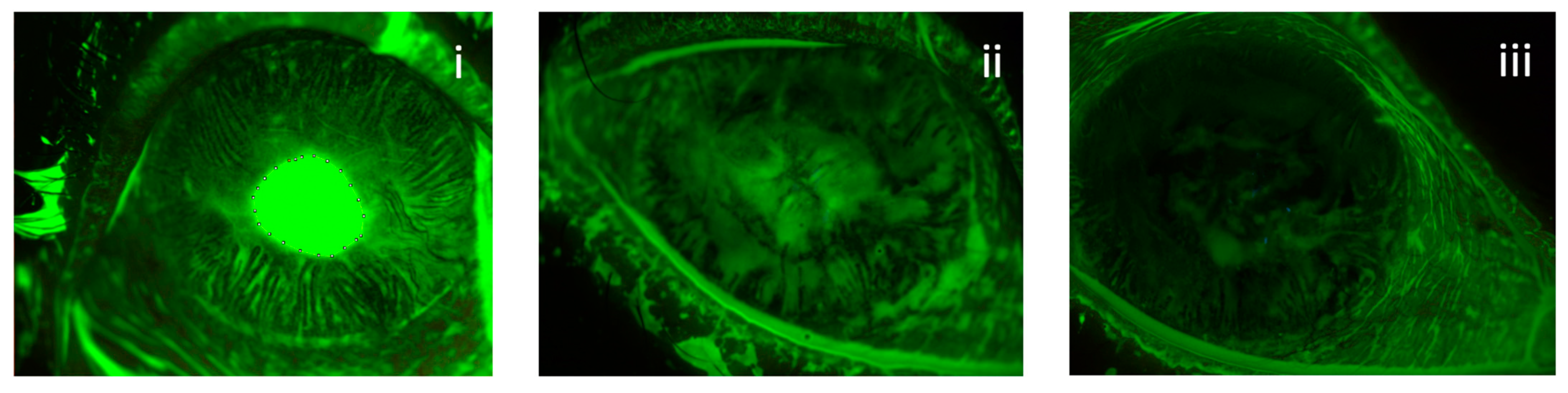
3.1. Area of Ulcers
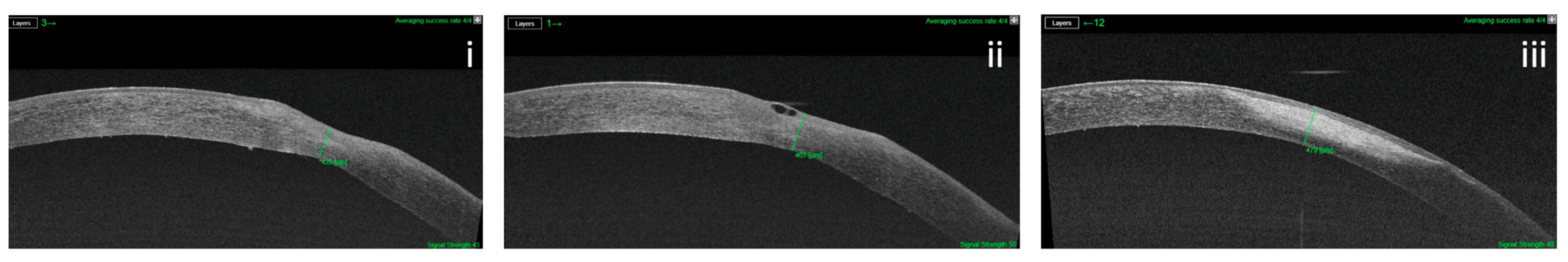
3.2. Corneal Thickness
3.3. Corneal Nerves
4. Discussion
Author Contributions
Funding
Institutional Review Board Statement
Informed Consent Statement
Data Availability Statement
Conflicts of Interest
References
- Ang, M.; Baskaran, M.; Werkmeister, R.M.; Chua, J.; Schmidl, D.; Aranha Dos Santos, V.; Garhöfer, G.; Mehta, J.S.; Schmetterer, L. Anterior segment optical coherence tomography. Prog. Retin. Eye Res. 2018, 66, 132–156. [Google Scholar] [CrossRef]
- Lim, S.-H. Clinical Applications of Anterior Segment Optical Coherence Tomography. J. Ophthalmol. 2015, 2015, 605729. [Google Scholar] [CrossRef]
- Venkateswaran, N.; Galor, A.; Wang, J.; Karp, C.L. Optical coherence tomography for ocular surface and corneal diseases: A review. Eye Vis. 2018, 5, 13. [Google Scholar] [CrossRef] [PubMed] [Green Version]
- Martin, R.; Sridhar, M. Anterior segment optical coherence tomography for evaluation of cornea and ocular surface. Indian J. Ophthalmol. 2018, 66, 367–372. [Google Scholar] [CrossRef] [PubMed]
- Czajkowski, G.; Kaluzny, B.; Laudencka, A.; Malukiewicz, G.; Kałużny, J.J. Tear Meniscus Measurement by Spectral Optical Coherence Tomography. Optom. Vis. Sci. 2012, 89, 336–342. [Google Scholar] [CrossRef] [PubMed]
- Soliman, W.; Mohamed, T.A. Spectral domain anterior segment optical coherence tomography assessment of pterygium and pinguecula. Acta Ophthalmol. 2012, 90, 461–465. [Google Scholar] [CrossRef] [PubMed]
- Randleman, J.B.; Woodward, M.; Lynn, M.J.; Stulting, R.D. Risk Assessment for Ectasia after Corneal Refractive Surgery. Ophthalmology 2008, 115, 37–50.e4. [Google Scholar] [CrossRef]
- Chow, V.W.; Agarwal, T.; Vajpayee, R.B.; Jhanji, V. Update on diagnosis and management of Descemet’s membrane detachment. Curr. Opin. Ophthalmol. 2013, 24, 356–361. [Google Scholar] [CrossRef]
- Konstantopoulos, A.; Kuo, J.; Anderson, D.; Hossain, P. Assessment of the Use of Anterior Segment Optical Coherence Tomography in Microbial Keratitis. Am. J. Ophthalmol. 2008, 146, 534–542.e2. [Google Scholar] [CrossRef]
- Bonnet, C.; Debillon, L.; Al-Hashimi, S.; Hoogewoud, F.; Monnet, D.; Bourges, J.-L.; Brézin, A. Anterior segment optical coherence tomography imaging in peripheral ulcerative keratitis, a corneal structural description. BMC Ophthalmol. 2020, 20, 205. [Google Scholar] [CrossRef]
- Schwarz, C.; Dang Burgener, N.P.; Dosso, A.A. OCT Visante observation of the progression of a perforated neurotrophic cornea ulcer treated with amniotic membrane grafts. J. Fr. Ophtalmol. 2008, 31, 419–421. [Google Scholar] [CrossRef]
- Sheha, H.; Tighe, S.; Hashem, O.; Hayashida, Y. Update on Cenegermin Eye Drops in the Treatment of Neurotrophic Keratitis. Clin. Ophthalmol. 2019, 13, 1973–1980. [Google Scholar] [CrossRef] [PubMed] [Green Version]
- García-Marqués, J.V.; Martínez-Albert, N.; Talens-Estarelles, C.; García-Lázaro, S.; Cerviño, A. Repeatability of Non-invasive Keratograph Break-Up Time measurements obtained using Oculus Keratograph 5M. Int. Ophthalmol. 2021, 41, 2473–2483. [Google Scholar] [CrossRef] [PubMed]
- Best, N.; Drury, L.; Wolffsohn, J.S. Clinical evaluation of the Oculus Keratograph. Contact Lens Anterior Eye 2012, 35, 171. [Google Scholar] [CrossRef] [PubMed]
- Wang, M.T.M.; Craig, J.P. Comparative Evaluation of Clinical Methods of Tear Film Stability Assessment: A Randomized Crossover Trial. JAMA Ophthalmol. 2018, 136, 291–294. [Google Scholar] [CrossRef] [PubMed]
- Tian, L.; Qu, J.-H.; Zhang, X.-Y.; Sun, X.-G. Repeatability and Reproducibility of Noninvasive Keratograph 5M Measurements in Patients with Dry Eye Disease. J. Ophthalmol. 2016, 2016, 8013621. [Google Scholar] [CrossRef] [PubMed] [Green Version]
- García-Montero, M.; del Viejo, L.R.; Lorente-Velázquez, A.; Martínez-Alberquilla, I.; Hernández-Verdejo, J.L.; Madrid-Costa, D. Repeatability of Noninvasive Keratograph 5M Measurements Associated With Contact Lens Wear. Eye Contact Lens 2019, 45, 377–381. [Google Scholar] [CrossRef]
- Alfaro-Juárez, A.; Caro-Magdaleno, M.; Montero-Iruzubieta, J.; Fernández-Palacín, A.; Muñoz-Morales, A.; Castilla-Martino, M.A.; Spínola-Muñoz, C.; de la Rua, E.R. Keratograph 5M As a Useful and Objective Tool for Evaluating the Ocular Surface in Limbal Stem Cell Deficiency. Clin. Ophthalmol. 2019, 13, 2025–2033. [Google Scholar] [CrossRef] [Green Version]
- Cruzat, A.; Qazi, Y.; Hamrah, P. In Vivo Confocal Microscopy of Corneal Nerves in Health and Disease. Ocul. Surf. 2017, 15, 15–47. [Google Scholar] [CrossRef] [PubMed] [Green Version]
- Roszkowska, A.M.; Wylęgała, A.; Gargano, R.; Spinella, R.; Inferrera, L.; Orzechowska-Wylęgała, B.; Aragona, P. Impact of corneal parameters, refractive error and age on density and morphology of the subbasal nerve plexus fibers in healthy adults. Sci. Rep. 2021, 11, 6076. [Google Scholar] [CrossRef]
- Al-Aqaba, M.A.; Dhillon, V.K.; Mohammed, I.; Said, D.G.; Dua, H.S. Corneal nerves in health and disease. Prog. Retin. Eye Res. 2019, 73, 100762. [Google Scholar] [CrossRef] [PubMed]
- Ruiz-Lozano, R.E.; Hernandez-Camarena, J.C.; Loya-Garcia, D.; Merayo-Lloves, J.; Rodriguez-Garcia, A. The molecular basis of neurotrophic keratopathy: Diagnostic and therapeutic implications. A review. Ocul. Surf. 2021, 19, 224–240. [Google Scholar] [CrossRef] [PubMed]
- Dua, H.S.; Said, D.G.; Messmer, E.M.; Rolando, M.; Benitez-Del-Castillo, J.M.; Hossain, P.N.; Shortt, A.J.; Geerling, G.; Nubile, M.; Figueiredo, F.C.; et al. Neurotrophic keratopathy. Prog. Retin. Eye Res. 2018, 66, 107–131. [Google Scholar] [CrossRef] [PubMed] [Green Version]
- Araki, K.; Ohashi, Y.; Kinoshita, S.; Hayashi, K.; Kuwayama, Y.; Tano, Y. Epithelial wound healing in the denervated cornea. Curr. Eye Res. 1994, 13, 203–211. [Google Scholar] [CrossRef] [PubMed]
- Bonini, S.; Rama, P.; Olzi, D.; Lambiase, A. Neurotrophic keratitis. Eye 2003, 17, 989–995. [Google Scholar] [CrossRef] [PubMed] [Green Version]
- Mackie, I. Neuroparalytic keratitis. In 1246 Current Ocular Therapy; Fraunfelder, F., Roy, F.H., Meyer, S.M., Eds.; Saunders: Philadephia, PA, USA, 1995; pp. 452–454. [Google Scholar]
- Sacchetti, M.; Lambiase, A. Diagnosis and management of neurotrophic keratitis. Clin. Ophthalmol. 2014, 8, 571–579. [Google Scholar] [CrossRef] [PubMed] [Green Version]
- Huang, D.; Swanson, E.A.; Lin, C.P.; Schuman, J.S.; Stinson, W.G.; Chang, W.; Hee, M.R.; Flotte, T.; Gregory, K.; Puliafito, C.A.; et al. Optical coherence tomography. Science 1991, 254, 1178–1181. [Google Scholar] [CrossRef] [Green Version]
- Nanji, A.A.; Sayyad, F.E.; Galor, A.; Dubovy, S.; Karp, C.L. High-Resolution Optical Coherence Tomography as an Adjunctive Tool in the Diagnosis of Corneal and Conjunctival Pathology. Ocul. Surf. 2015, 13, 226–235. [Google Scholar] [CrossRef] [Green Version]
- Gasser, T.; Romano, V.; Seifarth, C.; Bechrakis, N.E.; Kaye, S.B.; Steger, B. Morphometric characterisation of pterygium associated with corneal stromal scarring using high-resolution anterior segment optical coherence tomography. Br. J. Ophthalmol. 2017, 101, 660–664. [Google Scholar] [CrossRef]
- Lluch, S.; Julio, G.; Pujol, P.; Merindano, D. What biomarkers explain about pterygium OCT pattern. Graefes Arch. Clin. Exp. Ophthalmol. 2016, 254, 143–148. [Google Scholar] [CrossRef]
- Maeda, N. Optical Coherence Tomography for Corneal Diseases. Eye Contact Lens 2010, 36, 254–259. [Google Scholar] [CrossRef] [PubMed]
- Matalia, H.; Francis, M.; Gangil, T.; Chandapura, R.S.; Kurian, M.; Shetty, R.; Nelson, E.J.R.; Sinha Roy, A. Noncontact Quantification of Topography of Anterior Corneal Surface and Bowman’s Layer With High-Speed OCT. J. Refract. Surg. 2017, 33, 330–336. [Google Scholar] [CrossRef] [PubMed]
- Fuentes, E.; Sandali, O.; El Sanharawi, M.; Basli, E.; Hamiche, T.; Goemaere, I.; Borderie, V.; Bouheraoua, N.; Laroche, L. Anatomic Predictive Factors of Acute Corneal Hydrops in Keratoconus: An Optical Coherence Tomography Study. Ophthalmology 2015, 122, 1653–1659. [Google Scholar] [CrossRef] [PubMed]
- Li, Y.; Chamberlain, W.; Tan, O.; Brass, R.; Weiss, J.L.; Huang, D. Subclinical keratoconus detection by pattern analysis of corneal and epithelial thickness maps with optical coherence tomography. J. Cataract. Refract. Surg. 2016, 42, 284–295. [Google Scholar] [CrossRef] [PubMed] [Green Version]
- Ortiz, S.; Pérez-Merino, P.; Alejandre, N.; Gambra, E.; Jimenez-Alfaro, I.; Marcos, S. Quantitative OCT-based corneal topography in keratoconus with intracorneal ring segments. Biomed. Opt. Express 2012, 3, 814–824. [Google Scholar] [CrossRef] [PubMed] [Green Version]
- Pahuja, N.; Shroff, R.; Pahanpate, P.; Francis, M.; Veeboy, L.; Shetty, R.; Nuijts, R.M.M.A.; Sinha Roy, A. Application of high resolution OCT to evaluate irregularity of Bowman’s layer in asymmetric keratoconus. J. Biophotonics 2017, 10, 701–707. [Google Scholar] [CrossRef] [PubMed]
- Sandali, O.; El Sanharawi, M.; Temstet, C.; Hamiche, T.; Galan, A.; Ghouali, W.; Goemaere, I.; Basli, E.; Borderie, V.; Laroche, L. Fourier-domain optical coherence tomography imaging in keratoconus: A corneal structural classification. Ophthalmology 2013, 120, 2403–2412. [Google Scholar] [CrossRef] [PubMed]
- Su, J.P.; Li, Y.; Tang, M.; Liu, L.; Pechauer, A.D.; Huang, D.; Liu, G. Imaging the anterior eye with dynamic-focus swept-source optical coherence tomography. J. Biomed. Opt. 2015, 20, 126002. [Google Scholar] [CrossRef] [PubMed] [Green Version]
- Siebelmann, S.; Scholz, P.; Sonnenschein, S.; Bachmann, B.; Matthaei, M.; Cursiefen, C.; Heindl, L.M. Anterior segment optical coherence tomography for the diagnosis of corneal dystrophies according to the IC3D classification. Surv. Ophthalmol. 2018, 63, 365–380. [Google Scholar] [CrossRef]
- Sacchetti, M.; Lambiase, A. Neurotrophic factors and corneal nerve regeneration. Neural Regen. Res. 2017, 12, 1220–1224. [Google Scholar] [CrossRef]
- Versura, P.; Giannaccare, G.; Pellegrini, M.; Sebastiani, S.; Campos, E.C. Neurotrophic keratitis: Current challenges and future prospects. Eye Brain 2018, 10, 37–45. [Google Scholar] [CrossRef] [PubMed] [Green Version]
- Hamrah, P.; Cruzat, A.; Dastjerdi, M.H.; Zheng, L.; Shahatit, B.M.; Bayhan, H.A.; Dana, R.; Pavan-Langston, D. Corneal Sensation and Subbasal Nerve Alterations in Patients with Herpes Simplex Keratitis: An In Vivo Confocal Microscopy Study. Ophthalmology 2010, 117, 1930–1936. [Google Scholar] [CrossRef] [PubMed] [Green Version]
- Mastropasqua, L.; Nubile, M.; Lanzini, M.; Calienno, R.; Dua, H.S. In vivo microscopic and optical coherence tomography classification of neurotrophic keratopathy. J. Cell. Physiol. 2019, 234, 6108–6115. [Google Scholar] [CrossRef] [PubMed]
- Turkoglu, E.; Celik, E.; Alagoz, G. A Comparison of the Efficacy of Autologous Serum Eye Drops with Amniotic Membrane Transplantation in Neurotrophic Keratitis. Semin. Ophthalmol. 2014, 29, 119–126. [Google Scholar] [CrossRef] [PubMed]
- Matsumoto, Y.; Dogru, M.; Goto, E.; Ohashi, Y.; Kojima, T.; Ishida, R.; Tsubota, K. Autologous serum application in the treatment of neurotrophic keratopathy. Ophthalmology 2004, 111, 1115–1120. [Google Scholar] [CrossRef] [PubMed]
- Quinto, G.G.; Campos, M.S.D.Q.; Behrens, A. Autologous serum for ocular surface diseases. Arq. Bras. Oftalmol. 2008, 71 (Suppl. 6), 47–54. [Google Scholar] [CrossRef] [PubMed] [Green Version]
- López-Plandolit, S.; Morales, M.-C.; Freire, V.; Etxebarría, J.; Durán, J.A. Plasma Rich in Growth Factors as a Therapeutic Agent for Persistent Corneal Epithelial Defects. Cornea 2010, 29, 843–848. [Google Scholar] [CrossRef] [PubMed]
- Jeng, B.H.; Dupps, W.J., Jr. Autologous serum 50% eyedrops in the treatment of persistent corneal epithelial defects. Cornea 2009, 28, 1104–1108. [Google Scholar] [CrossRef]
- Rao, K.; Leveque, C.; Pflugfelder, S.C. Corneal nerve regeneration in neurotrophic keratopathy following autologous plasma therapy. Br. J. Ophthalmol. 2010, 94, 584–591. [Google Scholar] [CrossRef] [Green Version]
- Aifa, A.; Gueudry, J.; Portmann, A.; Delcampe, A.; Muraine, M. Topical Treatment with a New Matrix Therapy Agent (RGTA) for the Treatment of Corneal Neurotrophic Ulcers. Investig. Opthalmol. Vis. Sci. 2012, 53, 8181–8185. [Google Scholar] [CrossRef] [Green Version]
- Guerra, M.; Marques, S.; Gil, J.Q.; Campos, J.; Ramos, P.; Rosa, A.M.; Quadrado, M.J.; Murta, J. Neurotrophic Keratopathy: Therapeutic Approach Using a Novel Matrix Regenerating Agent. J. Ocul. Pharmacol. Ther. 2017, 33, 662–669. [Google Scholar] [CrossRef] [PubMed]
- Dunn, S.P.; Heidemann, D.G.; Chow, C.Y.C.; Crockford, D.; Turjman, N.; Angel, J.; Allan, C.B.; Sosne, G. Treatment of Chronic Nonhealing Neurotrophic Corneal Epithelial Defects with Thymosin Beta 4. Ann. N. Y. Acad. Sci. 2010, 1194, 199–206. [Google Scholar] [CrossRef] [PubMed]
- Lambiase, A.; Rama, P.; Bonini, S.; Caprioglio, G.; Aloe, L. Topical Treatment with Nerve Growth Factor for Corneal Neurotrophic Ulcers. N. Engl. J. Med. 1998, 338, 1174–1180. [Google Scholar] [CrossRef] [PubMed]
- Bonini, S.; Lambiase, A.; Rama, P.; Filatori, I.; Allegretti, M.; Chao, W.; Mantelli, F.; Bonini, S.; Lambiase, A.; Rama, P.; et al. Phase I Trial of Recombinant Human Nerve Growth Factor for Neurotrophic Keratitis. Ophthalmology 2018, 125, 1468–1471. [Google Scholar] [CrossRef] [PubMed] [Green Version]
- Bonini, S.; Lambiase, A.; Rama, P.; Sinigaglia, F.; Allegretti, M.; Chao, W.; Mantelli, F.; REPARO Study Group. Phase II Randomized, Double-Masked, Vehicle-Controlled Trial of Recombinant Human Nerve Growth Factor for Neurotrophic Keratitis. Ophthalmology 2018, 125, 1332–1343. [Google Scholar] [CrossRef] [Green Version]
- Di Zazzo, A.; Varacalli, G.; Mori, T.; Coassin, M. Long-term restoration of corneal sensitivity in neurotrophic keratopathy after rhNGF treatment. Eur. J. Ophthalmol. 2022, 32, NP15–NP18. [Google Scholar] [CrossRef] [PubMed]
- Pflugfelder, S.C.; Massaro-Giordano, M.; Perez, V.L.; Hamrah, P.; Deng, S.X.; Espandar, L.; Foster, C.S.; Affeldt, J.; Seedor, J.A.; Afshari, N.A.; et al. Topical Recombinant Human Nerve Growth Factor (Cenegermin) for Neurotrophic Keratopathy: A Multicenter Randomized Vehicle-Controlled Pivotal Trial. Ophthalmology 2020, 127, 14–26. [Google Scholar] [CrossRef] [PubMed] [Green Version]
- Mastropasqua, L.; Lanzini, M.; Dua, H.S.; D’Uffizi, A.; Di Nicola, M.; Calienno, R.; Bondì, J.; Said, D.G.; Nubile, M. In Vivo Evaluation of Corneal Nerves and Epithelial Healing after Treatment with Recombinant Nerve Growth Factor for Neurotrophic Keratopathy. Am. J. Ophthalmol. 2020, 217, 278–286. [Google Scholar] [CrossRef] [PubMed]
- Fung, S.S.M.; Catapano, J.; Elbaz, U.; Zuker, R.M.; Borschel, G.H.; Ali, A. In Vivo Confocal Microscopy Reveals Corneal Reinnervation after Treatment of Neurotrophic Keratopathy with Corneal Neurotization. Cornea 2018, 37, 109–112. [Google Scholar] [CrossRef] [PubMed]

Publisher’s Note: MDPI stays neutral with regard to jurisdictional claims in published maps and institutional affiliations. |
© 2022 by the authors. Licensee MDPI, Basel, Switzerland. This article is an open access article distributed under the terms and conditions of the Creative Commons Attribution (CC BY) license (https://creativecommons.org/licenses/by/4.0/).
Share and Cite
Inferrera, L.; Aragona, E.; Wylęgała, A.; Valastro, A.; Latino, G.; Postorino, E.I.; Gargano, R.; Orzechowska-Wylęgała, B.; Wylęgała, E.; Roszkowska, A.M. The Role of Hi-Tech Devices in Assessment of Corneal Healing in Patients with Neurotrophic Keratopathy. J. Clin. Med. 2022, 11, 1602. https://doi.org/10.3390/jcm11061602
Inferrera L, Aragona E, Wylęgała A, Valastro A, Latino G, Postorino EI, Gargano R, Orzechowska-Wylęgała B, Wylęgała E, Roszkowska AM. The Role of Hi-Tech Devices in Assessment of Corneal Healing in Patients with Neurotrophic Keratopathy. Journal of Clinical Medicine. 2022; 11(6):1602. https://doi.org/10.3390/jcm11061602
Chicago/Turabian StyleInferrera, Leandro, Emanuela Aragona, Adam Wylęgała, Antonio Valastro, Gianluigi Latino, Elisa I. Postorino, Romana Gargano, Bogusława Orzechowska-Wylęgała, Edward Wylęgała, and Anna M. Roszkowska. 2022. "The Role of Hi-Tech Devices in Assessment of Corneal Healing in Patients with Neurotrophic Keratopathy" Journal of Clinical Medicine 11, no. 6: 1602. https://doi.org/10.3390/jcm11061602
APA StyleInferrera, L., Aragona, E., Wylęgała, A., Valastro, A., Latino, G., Postorino, E. I., Gargano, R., Orzechowska-Wylęgała, B., Wylęgała, E., & Roszkowska, A. M. (2022). The Role of Hi-Tech Devices in Assessment of Corneal Healing in Patients with Neurotrophic Keratopathy. Journal of Clinical Medicine, 11(6), 1602. https://doi.org/10.3390/jcm11061602