Article

Increased Circulating Malondialdehyde-Modified Low-Density Lipoprotein Level Is Associated with High-Risk Plaque in Coronary Computed Tomography Angiography in Patients Receiving Statin Therapy

1
Department of Cardiovascular Medicine, Okayama University Graduate School of Medicine, Dentistry and Pharmaceutical Sciences, Okayama 700-8558, Japan
2
Department of Cardiovascular Medicine, Japanese Red Cross Okayama Hospital, Okayama 700-8607, Japan
*
Author to whom correspondence should be addressed.
J. Clin. Med. 2021, 10(7), 1480; https://doi.org/10.3390/jcm10071480
Submission received: 17 March 2021 / Revised: 30 March 2021 / Accepted: 30 March 2021 / Published: 2 April 2021
(This article belongs to the Special Issue The Prevention and Treatment of Atherosclerosis)

Abstract

:
Objective: To evaluate the association of serum malondialdehyde low-density lipoprotein (MDA-LDL), an oxidatively modified LDL, with the prevalence of high-risk plaques (HRP) determined with coronary computed tomography angiography (CTA) in statin-treated patients. Methods: This study was a single-center retrospective cohort comprising 268 patients (mean age 67 years, 58% men) with statin therapy and who underwent coronary CTA for suspected stable coronary artery disease. Patients were classified into two groups according to median MDA-LDL level or median LDL-C level. Coronary CTA-verified HRP was defined when two or more characteristics, including positive remodeling, low-density plaques, and spotty calcification, were present. Results: Patients with HRP had higher MDA-LDL (p = 0.011), but not LDL-C (p = 0.867) than those without HRP. High MDA-LDL was independently associated with HRP (odds ratio 1.883, 95% confidential interval 1.082–3.279) after adjustment for traditional risk factors. Regarding incremental value of MDA-LDL for predicting CTA-verified HRP, addition of serum MDA-LDL levels to the baseline model significantly increased global chi-square score from 26.1 to 32.8 (p = 0.010). Conclusions: A high serum MDA-LDL level is an independent predictor of CTA-verified HRP, which can lead to cardiovascular events in statin-treated patients.

1. Introduction

Cardiovascular disease is the leading global cause of adult mortality and morbidity [1]. Many studies have demonstrated a beneficial effect of low-density lipoprotein (LDL)-lowering therapies by statins on cardiovascular outcomes [2,3]. Lowering low-density lipoprotein cholesterol (LDL-C) is the primary target in cardiovascular disease prevention; however, despite effective LDL-lowering treatment, substantial patients remain at cardiovascular risk [4]. Among several lipid markers, serum malondialdehyde low-density lipoprotein (MDA-LDL), which is an oxidatively modified LDL, has been reported to be associated with coronary plaque vulnerability [5], adverse clinical outcomes after percutaneous coronary intervention [6], and the incidence of acute coronary syndrome [7]. However, the clinical relevance of serum MDA-LDL levels in patients receiving statin therapy for cardiovascular events has not been fully elucidated.
Coronary computed tomography angiography (CTA) is used to noninvasively evaluate coronary artery disease [8]. In addition to the evaluation of stenosis, coronary CTA identifies characteristics of plaque composition. Many observational follow-up studies have demonstrated the association between high-risk plaques (HRP) by coronary CTA and cardiovascular events [9,10]. Although statins contribute to the stabilization of plaques [11], HRP is often detected by coronary CTA even in patients receiving statin therapy.
Therefore, we hypothesized that serum MDA-LDL levels in patients with statin therapy is involved in the prevalence of HRP by coronary CTA. The aim of this study was to clarify the association between serum MDA-LDL levels and the prevalence of HRP, which increases the likelihood of acute coronary events in patients with suspected stable coronary artery disease receiving statin therapy.

2. Materials and Methods

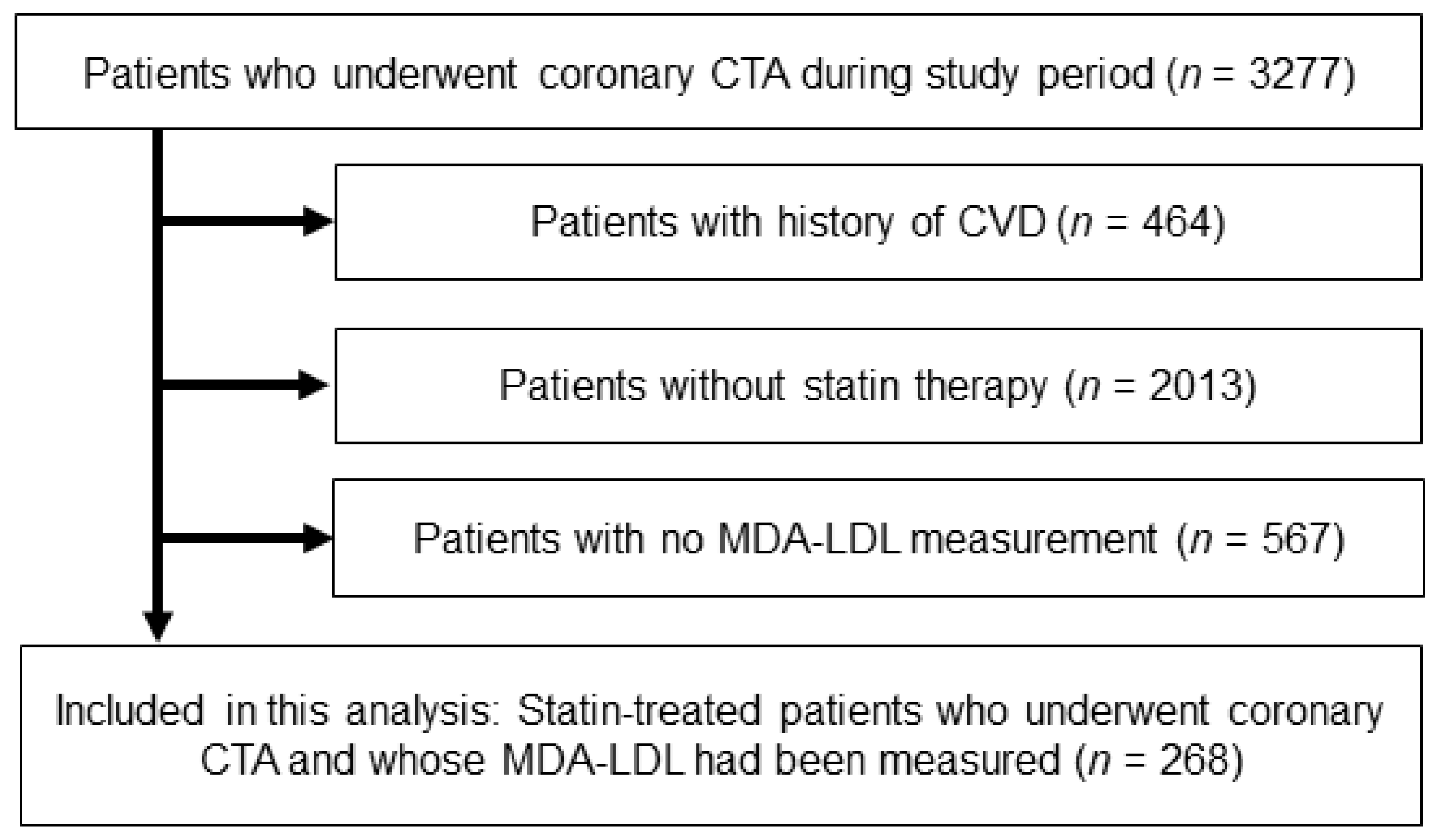
2.1. Study Population and Risk Assessment

This single-center retrospective study included 268 outpatients who underwent coronary CTA from August 2011 to December 2018 at Okayama University Hospital. A flow diagram of this study is shown in Figure 1. Participants had no history of coronary artery disease but had been taking statins.
Hypertension was defined as systolic blood pressure (BP) ≥140 mmHg or diastolic BP ≥90 mmHg, and/or the use of antihypertensive medication. Diabetes mellitus was defined as a fasting blood glucose concentration of ≥126 mg/dL or postprandial blood glucose concentration of ≥200 mg/dL, and/or the use of insulin or oral hypoglycemic medication. medication. Smoking was defined as a self-reported history of current smoking. This study was conducted according to the principles of the Declaration of Helsinki and approved by the ethics committees of Okayama University Graduate School of Medicine, Dentistry and Pharmaceutical Sciences. All patients enrolled in the study provided written informed consent.

2.2. Blood Sampling and the Measurement of MDA-LDL

Blood samples were collected from the antecubital vein after fasting overnight on the day of coronary CTA. MDA-LDL levels were measured using an enzyme-linked immunosorbent assay kit (Sekisui Medical Co., Tokyo, Japan), as described previously [12].

2.3. Acquisition and Analyses of Coronary CTA Image

CT images were acquired using a Somatom Definition Flash scanner (Siemens Medical Solutions, Munich, Germany) as described previously [13]. Coronary artery plaques were evaluated on axial and curved multiplanar reformatted images using commercially available cardiac reconstruction software (Virtual Place, Raijin; AZE Inc., Tokyo, Japan). Interpretation of coronary CTA was evaluated by two experienced cardiologists. Significant coronary artery stenosis was defined as a luminal narrowing of >50%. Coronary plaque was defined as a structure >1 mm2 located within the vessel wall, and plaque density was calculated for all lesions [14]. Plaques with a CT attenuation number <50 Hounsfield units were defined as low-density plaques. Positive remodeling, which indicates an enlarged vessel to compensate for atherosclerotic change, was assessed visually on multiplanar reformatted images that were reconstructed in long-axis and short-axis views of the vessel. Positive remodeling was defined as a threshold of 1.1 for the maximal diameter of the vessel. Spotty calcification was defined as a calcium burden length <1.5 times the vessel diameter and a width of less than two-thirds of the vessel diameter. CT-verified high-risk plaques were defined when two or more plaque characteristics, including positive remodeling, low-density plaques, and spotty calcification, were present [14].

2.4. Outcome Data

Follow-up clinical information was obtained from a review of the medical records or telephone interviews by attending physicians. Cardiovascular events were defined as a composite of cardiac death and acute coronary syndrome. Cardiac death was defined as death from myocardial infarction, cardiogenic shock, cardiac failure, or ventricular arrhythmias. Acute coronary syndrome included myocardial infarction and unstable angina. Non-fatal myocardial infarction was defined using the criteria of typical acute chest pain and persistent ST-segment elevation or positive cardiac enzymes. Unstable angina pectoris was defined as typical acute chest pain with negative cardiac enzymes if coronary artery disease could not be excluded as the cause of symptoms in accordance with current guidelines [15].

2.5. Statistical Analysis

Continuous variables were expressed as mean ± standard deviation or median with interquartile range. Dichotomous variables were expressed as numbers and percentages. Differences in continuous variables between the two groups were analyzed using Student’s t-test and the Mann–Whitney U-test, as appropriate. Categorical data were compared by χ2 analysis and Fisher’s test, as appropriate. Patients were classified into two groups based on the median serum MDA-LDL level (93 U/L): high MDA-LDL group (≥93 U/L, n = 139) and low MDA-LDL group (<93 U/L, n = 129), or LDL-C level (104 mg/dL): high LDL-C group (≥104 mg/dL, n = 136) and low MDA-LDL group (<104 mg/dL, n = 132). Associations between serum MDA-LDL and each variable were assessed using Pearson’s correlation coefficient. Univariate and multivariate logistic regression analyses were performed to evaluate the association between serum MDA-LDL levels and CTA-verified HRP. A receiver operating characteristic (ROC) curve was generated to evaluated diagnostic value to predict the presence of HRP. The increased discriminative value after the addition of serum MDA-LDL levels to the baseline model in predicting the presence of HRP was assessed by the global chi-square test and ROC curve analysis. ROC curves were built based on a logistic regression model, and the Delong test was used to compare the C-statistics. The net reclassification improvement and integrated discrimination improvement were also calculated. The baseline model consisted of established clinical risk factors with p < 0.05 by univariate logistic regression analysis. Cumulative survival estimates were calculated using the Kaplan–Meier method and compared with the log-rank test. A Cox proportional hazard model was used to identify whether serum MDA-LDL was associated with cardiovascular events. All reported p-values were two-sided, and p < 0.05 was considered statistically significant. Statistical analyses were performed using SPSS statistical software (Version 24; IBM Corp., Armonk, NY, USA).

3. Results

3.1. Patient Characteristics

The baseline characteristics of the patients are summarized in Table 1. The mean age was 67 years, and 58% of the patients were men. A share of 76% of patients had hypertension and 46% had diabetes mellitus. Age, sex, prevalence of hypertension, diabetes mellitus, current smoking status, medications, renal function, hemoglobin A1c, and high-sensitivity CRP did not differ between the high and low MDA-LDL groups. Patients with high MDA-LDL had greater body mass index (p = 0.016), and higher levels of LDL-C (p < 0.001) and triglyceride (p < 0.001), than those with low MDA-LDL. Patients with high MDA-LDL had greater body mass index (p = 0.016), and higher levels of LDL-C (p < 0.001) and triglyceride (p < 0.001), and a lower proportion of patients achieved LDL-C <70 mg/dL, than those with low MDA-LDL.
In addition, simple correlation coefficients for the association between serum MDA-LDL and other lipid variables were analyzed. Serum MDA-LDL levels were significantly positively associated with total cholesterol (r = 0.52, p < 0.001), LDL-C (r = 0.60, p < 0.001), and log-transformed triglyceride (r = 0.34, p < 0.001), and were significantly inversely associated with high-density lipoprotein cholesterol (HDL) (r = −0.14, p = 0.025).

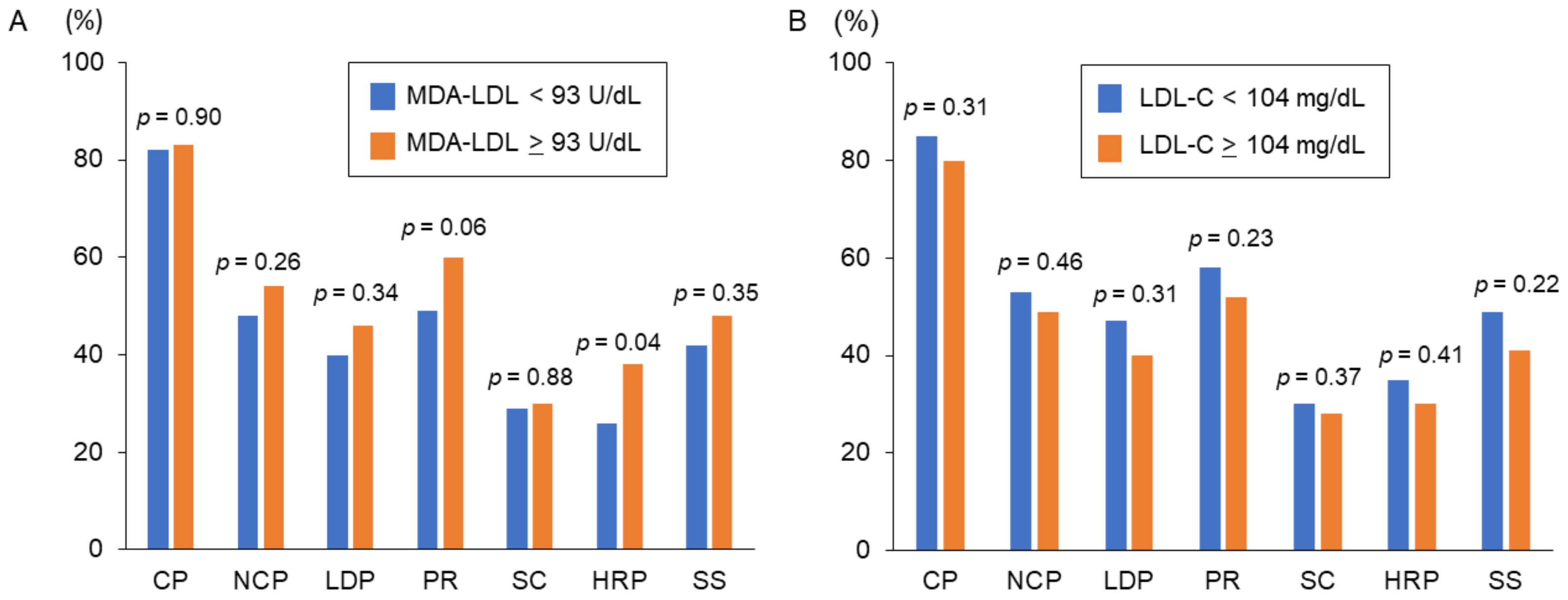
3.2. MDA-LDL and Coronary CTA Findings

Among all patients, the prevalence of HRP and significant stenosis presented in 87 patients (32%) and 120 patients (45%), respectively. As shown in Figure 2A, the prevalence of HRP in the high MDA-LDL group was significantly greater than that in the low MDA-LDL group (p = 0.040). However, no differences in the prevalence of calcified plaques (p = 0.904), non-calcified plaques (p = 0.267), low-density plaque (p = 0.344), positive remodeling (p = 0.057), spotty calcification (p = 0.883), and significant stenosis (p = 0.355) were found between the high and low MDA-LDL groups. Figure 2B shows no differences in the prevalence of HRP (p = 0.4111) and significant stenosis (p = 0.229), or other plaque features, between the high and low LDL-C groups.
As shown in Table 2, patients with HRP had a higher prevalence of male gender (p < 0.001), hypertension (p = 0.031), and diabetes mellitus (p = 0.028) compared with patients without HRP. Patients with HRP had lower HDL-C levels (p = 0.014) and serum MDA-LDL levels (p = 0.011), whereas LDL-C levels did not differ between patients with and without HRP. Although patients with significant stenosis had higher prevalence of male gender (p < 0.001) and diabetes mellitus (p = 0.039) compared with patients without significant stenosis, serum levels of LDL-C and MDA-LDL did not differ between patients with and without significant stenosis.
As shown in Table 3, in univariate logistic regression analysis, male sex, hypertension, diabetes mellitus, HDL, and MDA-LDL, but not LDL-C, were significantly associated with the presence of HRP. In multivariate logistic regression analysis, MDA-LDL was independently associated with the presence of HRP with an odds ratio of 1.883 (95% confidence interval 1.082–3.279, p = 0.025). When serum MDA-LDL levels were added to the baseline model, global chi-square scores significantly increased from 24.0 to 31.6 (p = 0.006). The net reclassification improvement and integrated discrimination improvement were significantly improved by 0.282 (p = 0.029) and 0.030 (p = 0.008), respectively. However, the increase in the C-statistic was not significant (0.68 to 0.70, p = 0.352).

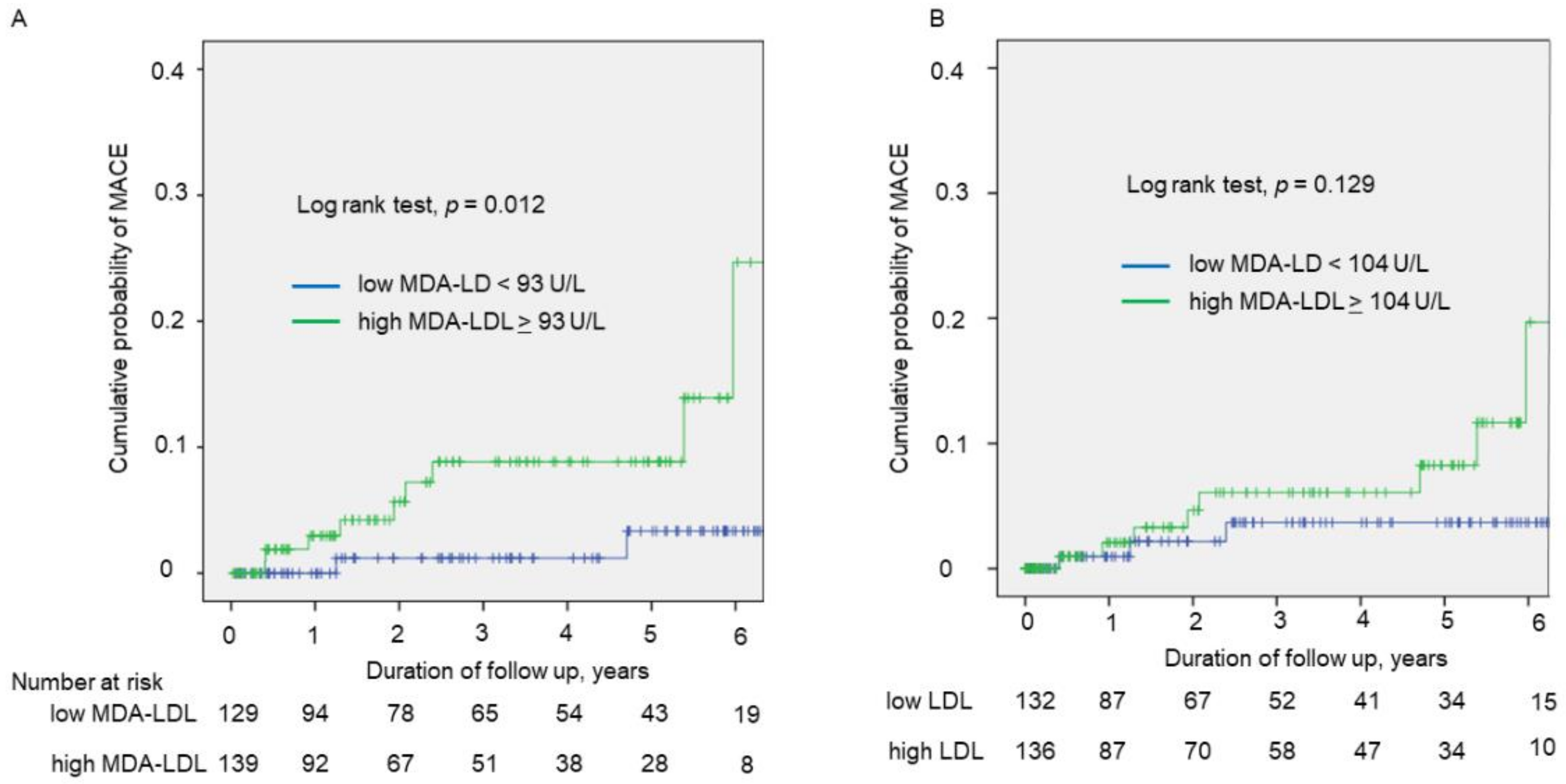
3.3. Prognostic Impact of HRP and MDA-LDL for Cardiovascular Events

During a median follow-up period of 2.5 years, 11 cardiovascular events (two cardiac deaths, nine acute coronary syndrome) occurred. Kaplan–Meier curves showed that the high MDA-LDL group had more cardiovascular events than the low MDA-LDL group (p = 0.012, log-rank test) (Figure 3A), whereas no significant difference in the incidence of cardiovascular events was observed between patients with the high and low LDL-C groups (Figure 3B). Cox univariate regression analysis showed that high MDA-LDL (HR 5.717, 95%CI 1.225–26.670, p = 0.027), but not high LDL-C (hazard ratio 2.697, 95% confidence interval 0.715–10.183, p = 0.143), was significantly associated with cardiovascular events. In age and sex adjusted Cox multivariate regression analysis, high MDA-LDL was independently associated with cardiovascular events (hazard ratio 5.865, 95% confidence interval 1.250–27.512, p = 0.025).

4. Discussion

This study demonstrated that serum MDA-LDL, but not LDL-C, was significantly associated with the presence of HRP and had a moderate incremental value to predict HRP over traditional risk factors in statin-treated patients with suspected coronary artery disease. In addition, high MDA-LDL was shown to be a possible predictor for cardiovascular events defined as a composite of cardiac death and ACS in statin-treated patients.
Although increased oxidized LDL, including serum MDA-LDL, is known to be a predictor for atherosclerotic cardiovascular diseases [16], the clinical impact of serum MDA-LDL in patients receiving statin therapy has not been clarified. High triglyceride levels and low HDL levels have been known as risk markers in patients treated with lipid-lowering therapy [17]. These lipid abnormalities are associated with an increase in small dense LDL [18], which are more susceptible to oxidative modification compared with LDL. Even after considering triglyceride and HDL-C, this study clearly demonstrated that serum MDA-LDL is a relevant biomarker for the presence of HRP in statin-treated patients.
Oxidized LDL plays a critical role in plaque vulnerability. Oxidized LDL through lectin-like oxidized LDL receptor-1, which is the major receptor for oxidized LDL in endothelial cells, contributes to increased matrix metalloproteinase activity [19]. In addition, elevated concentrations of oxidized LDL induce apoptosis in vascular smooth muscle cells [20]. Enhanced matrix metalloproteinase production and apoptosis of vascular smooth muscle cells contribute to plaque instability [21]. Several previous clinical studies reported that serum MDA-LDL levels have been associated with thin-cap fibroatheroma identified by frequency-domain optical coherence tomography or with tissue characteristics evaluated by integrated backscatter intravascular ultrasound, both of which are considered to be indicators of plaque vulnerability [5,22]. However, these studies only included patients with obstructive coronary artery disease. In addition, previous studies have reported an association between serum MDA-LDL and plaque vulnerability in lesions targeted for revascularization.
Our findings suggest that serum MDA-LDL is an independent factor for cardiovascular events such as cardiac death and acute coronary syndrome. Similar to our results, a recent clinical study demonstrated that small dense LDL, which is easily oxidized to MDA-LDL, was strongly associated with myocardial infarction [23]. In fact, oxidized LDL is also reported to be involved in platelet activation [24]. The significant association between MDA-LDL and HRP shown in this study may partly explain the role of MDA-LDL as a predictor of cardiovascular events; nevertheless, further studies are required to clarify whether there is a causal relationship.
Statin therapies have been shown to decrease not only LDL but also serum MDA-LDL [25]. In fact, the current study demonstrated that LDL-C had a significant correlation with serum MDA-LDL, and the proportion of patients achieving LDL-C < 70 mg/dL in the low MDA-LDL group was significantly greater than that in the high MDA-LDL group. In addition, inflammation has been considered as a residual risk in statin-treated patients, and a previous study reported that oxidized LDL-induced interleukins (IL)-1β secretion promotes foam cell formation [26]. Recently, a clinical trial showed that antibody therapy against IL-1β significantly reduced cardiovascular events without lowering lipid or blood pressure [27]. Further studies are needed to investigate whether additional anti-inflammatory treatment in patients with high MDA-LDL will reduce cardiovascular events.
Our study has some limitations that need to be addressed. First, this was a single-center study and the number of patients was relatively small. Patient selection may have been biased and a prospective study would be preferable. Second, we included only Japanese patients with suspected stable coronary artery diseasse; the results cannot be applied to other ethnic groups and the general population. Third, the follow-up study was relatively short and a small number of cardiovascular events were documented. To determine the impact of MDA-LDL on cardiovascular death and acute coronary syndrome in statin-treated patients, a large long-term study is warranted.

5. Conclusions

Our study demonstrated that serum MDA-LDL was significantly associated with HRP and had an incremental value to predict HRP over traditional risk factors in statin-treated patients with suspected stable coronary artery disease. Our results suggest the measurement of serum MDA-LDL is useful in identifying patients likely to have HRP who are at high risk. Further studies with larger sample size are needed to prove the association between serum MDA-LDL and cardiovascular events in statin-treated patients.

Author Contributions

Conceptualization, K.I., T.M. (Toru Miyoshi) and H.I.; methodology, K.I. and T.M. (Takashi Miki); formal analysis, K.I., T.M. (Toru Miyoshi) and K.O.; investigation, K.I., K.O. and T.M. (Takashi Miki); writing—original draft preparation, K.I., T.M. (Toru Miyoshi) and T.M. (Takashi Miki).; writing—review and editing, H.I. All authors have read and agreed to the published version of the manuscript.

Funding

This research received no external funding.

Institutional Review Board Statement

This study was conducted according to the principles of the Declaration of Helsinki and approved by the ethics committees of Okayama University Graduate School of Medicine, Dentistry and Pharmaceutical Sciences.

Informed Consent Statement

Patient consent was waived due to the low-risk nature of the study and the inability to obtain consent directly from all the study subjects.

Data Availability Statement

The data presented in this study are available on request from the corresponding author. The data are not publicly available due to privacy.

Conflicts of Interest

The authors declare no conflict of interest.

References

  1. Timmis, A.; Townsend, N.; Gale, C.; Grobbee, R.; Maniadakis, N.; Flather, M.; Wilkins, E.; Wright, L.; Vos, R.; Bax, J.; et al. European Society of Cardiology: Cardiovascular Disease Statistics 2017. Eur. Heart J. 2018, 39, 508–579. [Google Scholar] [CrossRef] [Green Version]
  2. Ridker, P.M.; Pradhan, A.; MacFadyen, J.G.; Libby, P.; Glynn, R.J. Cardiovascular benefits and diabetes risks of statin therapy in primary prevention: An analysis from the JUPITER trial. Lancet 2012, 380, 565–571. [Google Scholar] [CrossRef] [Green Version]
  3. Mills, E.J.; Rachlis, B.; Wu, P.; Devereaux, P.J.; Arora, P.; Perri, D. Primary prevention of cardiovascular mortality and events with statin treatments: A network meta-analysis involving more than 65,000 patients. J. Am. Coll. Cardiol. 2008, 52, 1769–1781. [Google Scholar] [CrossRef] [Green Version]
  4. Reith, C.; Armitage, J. Management of residual risk after statin therapy. Atherosclerosis 2016, 245, 161–170. [Google Scholar] [CrossRef] [PubMed] [Green Version]
  5. Matsuo, Y.; Kubo, T.; Okumoto, Y.; Ishibashi, K.; Komukai, K.; Tanimoto, T.; Ino, Y.; Kitabata, H.; Hirata, K.; Imanishi, T.; et al. Circulating malondialdehyde-modified low-density lipoprotein levels are associated with the presence of thin-cap fibroatheromas determined by optical coherence tomography in coronary artery disease. Eur. Heart J. Cardiovasc. Imaging 2013, 14, 43–50. [Google Scholar] [CrossRef] [PubMed] [Green Version]
  6. Ito, T.; Fujita, H.; Tani, T.; Ohte, N. Malondialdehyde-modified low-density lipoprotein is a predictor of cardiac events in patients with stable angina on lipid-lowering therapy after percutaneous coronary intervention using drug-eluting stent. Atherosclerosis 2015, 239, 311–317. [Google Scholar] [CrossRef] [PubMed]
  7. Holvoet, P.; Collen, D.; Van de Werf, F. Malondialdehyde-modified LDL as a marker of acute coronary syndromes. JAMA 1999, 281, 1718–1721. [Google Scholar] [CrossRef] [PubMed] [Green Version]
  8. Xie, J.X.; Cury, R.C.; Leipsic, J.; Crim, M.T.; Berman, D.S.; Gransar, H.; Budoff, M.J.; Achenbach, S.; Hartaigh, B.O.; Callister, T.Q.; et al. The Coronary Artery Disease-Reporting and Data System (CAD-RADS): Prognostic and Clinical Implications Associated With Standardized Coronary Computed Tomography Angiography Reporting. JACC Cardiovasc. Imaging 2018, 11, 78–89. [Google Scholar] [CrossRef] [PubMed]
  9. Bamberg, F.; Sommer, W.H.; Hoffmann, V.; Achenbach, S.; Nikolaou, K.; Conen, D.; Reiser, M.F.; Hoffmann, U.; Becker, C.R. Meta-analysis and systematic review of the long-term predictive value of assessment of coronary atherosclerosis by contrast-enhanced coronary computed tomography angiography. J. Am. Coll. Cardiol. 2011, 57, 2426–2436. [Google Scholar] [CrossRef] [Green Version]
  10. Versteylen, M.O.; Kietselaer, B.L.; Dagnelie, P.C.; Joosen, I.A.; Dedic, A.; Raaijmakers, R.H.; Wildberger, J.E.; Nieman, K.; Crijns, H.J.; Niessen, W.J.; et al. Additive value of semiautomated quantification of coronary artery disease using cardiac computed tomographic angiography to predict future acute coronary syndrome. J. Am. Coll. Cardiol. 2013, 61, 2296–2305. [Google Scholar] [CrossRef]
  11. Puri, R.; Nicholls, S.J.; Shao, M.; Kataoka, Y.; Uno, K.; Kapadia, S.R.; Tuzcu, E.M.; Nissen, S.E. Impact of statins on serial coronary calcification during atheroma progression and regression. J. Am. Coll. Cardiol. 2015, 65, 1273–1282. [Google Scholar] [CrossRef] [PubMed]
  12. Amioka, N.; Miyoshi, T.; Otsuka, H.; Yamada, D.; Takaishi, A.; Ueeda, M.; Hirohata, S.; Ito, H. Serum malondialdehyde-modified low-density lipoprotein levels on admission predict prognosis in patients with acute coronary syndrome undergoing percutaneous coronary intervention. J. Cardiol. 2019, 74, 258–266. [Google Scholar] [CrossRef] [PubMed]
  13. Osawa, K.; Miyoshi, T.; Yamauchi, K.; Koyama, Y.; Nakamura, K.; Sato, S.; Kanazawa, S.; Ito, H. Nonalcoholic Hepatic Steatosis Is a Strong Predictor of High-Risk Coronary-Artery Plaques as Determined by Multidetector CT. PLoS ONE 2015, 10, e0131138. [Google Scholar] [CrossRef] [PubMed]
  14. Ichikawa, K.; Miyoshi, T.; Osawa, K.; Miki, T.; Nakamura, K.; Ito, H. Prognostic Value of Coronary Computed Tomographic Angiography in Patients With Nonalcoholic Fatty Liver Disease. JACC Cardiovasc. Imaging 2020, 13, 1628–1630. [Google Scholar] [CrossRef]
  15. Anderson, J.L.; Adams, C.D.; Antman, E.M.; Bridges, C.R.; Califf, R.M.; Casey, D.E., Jr.; Chavey, W.E., 2nd; Fesmire, F.M.; Hochman, J.S.; Levin, T.N.; et al. 2011 ACCF/AHA Focused Update Incorporated Into the ACC/AHA 2007 Guidelines for the Management of Patients With Unstable Angina/Non-ST-Elevation Myocardial Infarction: A report of the American College of Cardiology Foundation/American Heart Association Task Force on Practice Guidelines. Circulation 2011, 123, e426–e579. [Google Scholar] [CrossRef]
  16. Gao, S.; Zhao, D.; Wang, M.; Zhao, F.; Han, X.; Qi, Y.; Liu, J. Association Between Circulating Oxidized LDL and Atherosclerotic Cardiovascular Disease: A Meta-analysis of Observational Studies. Can. J. Cardiol. 2017, 33, 1624–1632. [Google Scholar] [CrossRef] [PubMed]
  17. Matsuura, Y.; Kanter, J.E.; Bornfeldt, K.E. Highlighting Residual Atherosclerotic Cardiovascular Disease Risk. Arterioscler. Thromb. Vasc. Biol. 2019, 39, e1–e9. [Google Scholar] [CrossRef]
  18. Fan, J.; Liu, Y.; Yin, S.; Chen, N.; Bai, X.; Ke, Q.; Shen, J.; Xia, M. Small dense LDL cholesterol is associated with metabolic syndrome traits independently of obesity and inflammation. Nutr. Metab. 2019, 16, 7. [Google Scholar] [CrossRef]
  19. Hu, C.; Dandapat, A.; Sun, L.; Chen, J.; Marwali, M.R.; Romeo, F.; Sawamura, T.; Mehta, J.L. LOX-1 deletion decreases collagen accumulation in atherosclerotic plaque in low-density lipoprotein receptor knockout mice fed a high-cholesterol diet. Cardiovasc. Res. 2008, 79, 287–293. [Google Scholar] [CrossRef]
  20. Kume, N.; Kita, T. Apoptosis of vascular cells by oxidized LDL: Involvement of caspases and LOX-1 and its implication in atherosclerotic plaque rupture. Circ. Res. 2004, 94, 269–270. [Google Scholar] [CrossRef] [Green Version]
  21. Kattoor, A.J.; Pothineni, N.V.K.; Palagiri, D.; Mehta, J.L. Oxidative Stress in Atherosclerosis. Curr. Atheroscler. Rep. 2017, 19, 42. [Google Scholar] [CrossRef]
  22. Ikenaga, H.; Kurisu, S.; Kono, S.; Sumimoto, Y.; Watanabe, N.; Shimonaga, T.; Higaki, T.; Iwasaki, T.; Mitsuba, N.; Ishibashi, K.; et al. Impact of Malondialdehyde-Modified Low-Density Lipoprotein on Tissue Characteristics in Patients With Stable Coronary Artery Disease- Integrated Backscatter-Intravascular Ultrasound Study. Circ. J. 2016, 80, 2173–2182. [Google Scholar] [CrossRef] [PubMed] [Green Version]
  23. Duran, E.K.; Aday, A.W.; Cook, N.R.; Buring, J.E.; Ridker, P.M.; Pradhan, A.D. Triglyceride-Rich Lipoprotein Cholesterol, Small Dense LDL Cholesterol, and Incident Cardiovascular Disease. J. Am. Coll. Cardiol. 2020, 75, 2122–2135. [Google Scholar] [CrossRef] [PubMed]
  24. Marwali, M.R.; Hu, C.P.; Mohandas, B.; Dandapat, A.; Deonikar, P.; Chen, J.; Cawich, I.; Sawamura, T.; Kavdia, M.; Mehta, J.L. Modulation of ADP-induced platelet activation by aspirin and pravastatin: Role of lectin-like oxidized low-density lipoprotein receptor-1, nitric oxide, oxidative stress, and inside-out integrin signaling. J. Pharmacol. Exp. Ther. 2007, 322, 1324–1332. [Google Scholar] [CrossRef] [PubMed] [Green Version]
  25. Nishikido, T.; Oyama, J.; Keida, T.; Ohira, H.; Node, K. High-dose statin therapy with rosuvastatin reduces small dense LDL and MDA-LDL: The Standard versus high-dose therApy with Rosuvastatin for lipiD lowering (SARD) trial. J. Cardiol. 2016, 67, 340–346. [Google Scholar] [CrossRef] [PubMed] [Green Version]
  26. Liu, W.; Yin, Y.; Zhou, Z.; He, M.; Dai, Y. OxLDL-induced IL-1 beta secretion promoting foam cells formation was mainly via CD36 mediated ROS production leading to NLRP3 inflammasome activation. Inflamm. Res. 2014, 63, 33–43. [Google Scholar] [CrossRef]
  27. Ridker, P.M.; Everett, B.M.; Thuren, T.; MacFadyen, J.G.; Chang, W.H.; Ballantyne, C.; Fonseca, F.; Nicolau, J.; Koenig, W.; Anker, S.D.; et al. Antiinflammatory Therapy with Canakinumab for Atherosclerotic Disease. N. Engl. J. Med. 2017, 377, 1119–1131. [Google Scholar] [CrossRef]
Figure 1. Study flowchart. CTA, computed tomography angiography; CVD, cardiovascular disease; MDA-LDL, malondialdehyde-modified low-density lipoprotein.
Jcm 10 01480 g001
Figure 2. The prevalence of coronary plaque characteristics according to MDA-LDL and LDL-C. (A) Patients were divided into two groups based on the median value of MDA-LDL. (B) Patients were divided into two groups based on the median value of LDL-C. MDA-LDL, malondialdehyde-modified low-density lipoprotein; LDL-C, low-density lipoprotein cholesterol; CP, calcified plaque; NCP, non-calcified plaque; LDP, low density plaque; PR, positive remodeling; SC, spotty calcification; HRP, high-risk plaque: SS, significant stenosis.
Jcm 10 01480 g002
Figure 3. Kaplan–Meier curves of cumulative incidence of cardiovascular events. Kaplan–Meier curves (A) according to low or high MDA-LDL, and (B) according to low or high LDL-C. MDA-LDL, malondialdehyde-modified low-density lipoprotein; LDL-C, low-density lipoprotein cholesterol.
Jcm 10 01480 g003
Table 1. Baseline patient demographic and clinical characteristics according to high or low levels of MDA-LDL.
All PatientsHigh MDA-LDL
(≥93 U/L)
Low MDA-LDL
(<93 U/L)
p-Value
n268139129
Age, year67 ± 1167 ± 1168 ± 110.521
Male sex154 (58)76 (55)78 (61)0.338
Body mass index, kg/m224.6 ± 4.125.2 ± 3.924.0 ± 4.20.016
Hypertension203 (76)109 (78)94 (73)0.290
Diabetes mellitus122 (46)59 (42)63 (49)0.294
Current Smoker70 (26)36 (26)34 (26)0.932
Medications
Beta blockers71 (27)37 (27)34 (26)0.933
CCBs122 (46)70 (50)52 (40)0.099
ACE-Is or ARBs135 (50)73 (53)62 (48)0.466
Oral antihyperglycemic drugs86 (32)45 (32)41 (32)0.918
Ezetimibe15 (6)10 (7)5 (4)0.293
Statin intensity, low/moderate *160 (60)/108 (40)76 (55)/63 (45)84 (65)/45 (35)0.082
Statin type, dosage
Atorvastatin, 5 mg/10 mg18/495/2613/23
Fluvastatin, 20 mg/40 mg4/13/11/0
Pitavastatin, 1 mg/2 mg/4 mg14/28/46/13/38/15/1
Pravastatin, 5 mg/10 mg9/365/214/15
Rosuvastatin, 2.5 mg/5 mg/10 mg68/24/230/19/238/5/0
Simvastatin, 5 mg/10 mg8/34/14/2
Laboratory findings
Creatinine, mg/dL0.89 ± 0.780.85 ± 0.640.93 ± 0.920.397
eGFR, mL/min/1.73 m268 ± 1868 ± 1769 ± 190.665
Total cholesterol, mg/dL186 ± 40203 ± 40168 ± 31<0.001
LDL cholesterol, mg/dL107 ± 32122 ± 3390 ± 22<0.001
HDL cholesterol, mg/dL58 ± 1757 ± 1760 ± 170.080
Triglyceride, mg/dL121 (88, 171)150 (104, 198)101 (76, 130)<0.001
MDA-LDL, U/L96 ± 35122 ± 2869 ± 15<0.001
HbA1c, %6.6 ± 1.36.6 ± 1.46.6 ± 1.30.968
hsCRP, mg/dL0.08 (0.05, 0.18)0.08 (0.04, 0.16)0.08 (0.05, 0.20)0.608
Patients achieving LDLcholesterol <70 mg/dL24 (9)1 (1)23 (18)<0.001
Data are presented as mean ± standard deviation, number (%), or median (25th, 75th percentile). MDA-LDL, malondialdehyde low-density lipoprotein cholesterol; CCBs, calcium channel blockers; ACE-Is, angiotensin-converting enzyme inhibitors; ARBs, angiotensin receptor blockers; eGFR, estimated glomerular filtration rate; LDL, low-density lipoprotein; HDL, high-density lipoprotein; HbA1c, glycated hemoglobin A1c; hsCRP, high-sensitivity C-reactive protein. * No patients received high-intensity statins.
Table 2. Comparison of patient characteristics according to coronary CTA findings.
VariablesHigh-Risk PlaqueSignificant Stenosis
PresentAbsentp ValuePresentAbsentp Value
n87181 119148
Age, year68 ± 1067 ± 120.74768 ± 1167 ± 120.489
Male sex64 (74)90 (50)<0.00184 (70)70 (47)<0.001
Body mass index, kg/m224.7 ± 3.424.5 ± 4.40.71824.4 ± 4.124.7 ± 4.10.450
Hypertension73 (84)130 (76)0.03193 (78)110 (74)0.546
Diabetes mellitus48 (55)74 (41)0.02863 (53)59 (40)0.039
Current smoker29 (33)41 (23)0.06232 (27)38 (25)0.854
Oral antihyperglycemic drugs32 (37)54 (30)0.25442 (35)44 (30)0.333
Creatinine, mg/dL1.00 ± 0.990.83 ± 0.650.0870.91 ± 0.710.88 ± 0.830.066
Total cholesterol, mg/dL186 ± 45187 ± 380.834183 ± 41188 ± 380.300
LDL cholesterol, mg/dL107 ± 37107 ± 300.867106 ± 34107 ± 310.728
HDL cholesterol, mg/dL55 ± 1660 ± 170.01458 ± 1858 ± 160.905
Triglyceride, mg/dL130 (90, 182)117 (85, 170)0.206118 (86, 158)122 (89, 183)0.155
MDA-LDL, U/L105 ± 4092 ± 320.01196 ± 3396 ± 360.979
HbA1c, %6.7 ± 1.36.5 ± 1.40.3476.6 ± 1.36.5 ± 1.40.122
hsCRP, mg/dL0.08
(0.05, 0.16)
0.08
(0.04, 0.19)
0.9070.09
(0.05, 0.200)
0.07
(0.040, 0.160)
0.132
Table 3. Univariate and multivariate predictors of the presence of HRP.
UnivariateMultivariate
Odds Ratio (95%CI)p ValueOdds Ratio (95%CI)p Value
Age, per 1 year1.004 (0.981–1.028)0.746
Male2.814 (1.609–4.918)<0.0012.749 (1.502–1.502)0.001
Hypertension2.046 (1.060–3.947)0.0332.027 (1.022–4.019)0.049
Diabetes Mellitus1.780 (1.062–2.982)0.0291.630 (0.941–2.824)0.081
Current smoker1.707 (0.970–3.006)0.064
HDL cholesterol, per 1 mg/dL0.980 (0.964–0.996)0.0150.994 (0.977–1.012)0.531
LDL cholesterol, >104 mg/dL0.807 (0.483–1.347)0.412
Triglyceride *, per 1 index1.454 (0.854–2.475)0.168
MDA-LDL, >93 U/L1.722 (1.024–2.897)0.0411.883 (1.082–3.279)0.025
hsCRP *, per 1 index0.983 (0.793–1.219)0.878
HRP, high-risk plaques; LDL, low-density lipoprotein; HDL-C, high-density lipoprotein; MDA-LDL, malondialdehyde low-density lipoprotein cholesterol; hsCRP, high sensitivity C-reactive protein. * Triglyceride and hsCRP were logarithm-transformed.
Publisher’s Note: MDPI stays neutral with regard to jurisdictional claims in published maps and institutional affiliations.

