


Abstract
Severe Acute Respiratory Syndrome Coronavirus 2 (SARS-CoV-2) infection induces heterogeneous immune responses that influence both acute disease severity and long-term immune remodeling. A key question in the context of infection and vaccination is whether SARS-CoV-2 exerts direct oncogenic effects or instead acts as a transient immunological stressor capable of reinforcing tumor-permissive pathways. Current evidence does not support classical viral oncogenesis. Rather, severe infection is characterized by early interferon (IFN) imbalance followed by NF-κB-dominant inflammatory amplification, promoting sustained IL-6/JAK–STAT3 and MAPK signaling, chronic cytokine production, metabolic reprogramming, and impaired antitumor immune surveillance. At the molecular level, viral structural proteins modulate host signaling networks. The spike (S1) protein engages TLR2/TLR4–MyD88 pathways, activating NF-κB and MAPK cascades, while the membrane (M) protein reinforces NF-κB–STAT3 circuits linked to epithelial–mesenchymal transition and inflammatory gene expression. These mechanisms intensify pre-existing oncogenic signaling without initiating malignant transformation. Tissue-specific responses are further shaped by IFN competence, renin–angiotensin system balance, and metabolic context. In parallel, immune evasion programs shared by chronic viral infection and cancer, including checkpoint upregulation, impaired antigen presentation, and suppressive myeloid expansion, may be transiently reinforced following severe infection. In contrast, SARS-CoV-2 vaccination induces spatially restricted, self-limited innate activation without sustained inflammatory signaling or persistent antigen exposure. By preventing severe disease and chronic immune dysregulation, vaccination interrupts pathways hypothesized to intersect with cancer biology, with no evidence of increased cancer incidence. Ongoing longitudinal studies are required to clarify the long-term oncologic implications of post-infectious immune remodeling.
1. Introduction
The emergence of Coronavirus Disease 2019 (COVID-19), caused by the Severe Acute Respiratory Syndrome Coronavirus 2 (SARS-CoV-2), has presented a global health challenge with far-reaching consequences on public health. Beyond its immediate acute respiratory manifestations, a growing body of evidence indicates that SARS-CoV-2 infection can lead to long-term immunological sequelae, fundamentally altering host immune responses, exacerbating pre-existing conditions or contributing to novel pathologies [1]. The interplay between viral infection, immune evasion, and carcinogenesis is well established in oncology. Chronic viral infections have long been recognized as significant etiological drivers of multiple malignancies, establishing a precedent for evaluating SARS-CoV-2 within a cancer-relevant biological framework [2]. Mechanistically, persistent viral infections promote sustained inflammation, immune dysregulation, oxidative stress, and impaired immune surveillance, thereby creating a biological environment permissive to malignant transformation [3]. Similar processes have been documented in COVID-19, where prolonged cytokine production, oxidative stress, metabolic reprogramming, and T-cell exhaustion may collectively contribute to a pro-tumorigenic milieu [4]. Given these overlaps, an important question emerges as to whether SARS-CoV-2 functions as a direct oncogenic agent or instead represents a transient infection that induces cancer-relevant immune and inflammatory reprogramming in susceptible individuals. In this review, we trace the immunological trajectory of SARS-CoV-2 infection from early interferon imbalance to chronic inflammatory remodeling, examine its convergence with tumor-associated signaling networks, analyze tissue-specific tumor interactions, and contrast these effects with vaccination.
2. Acute and Chronic Immune Responses to SARS-CoV-2
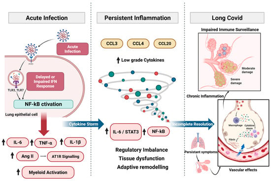
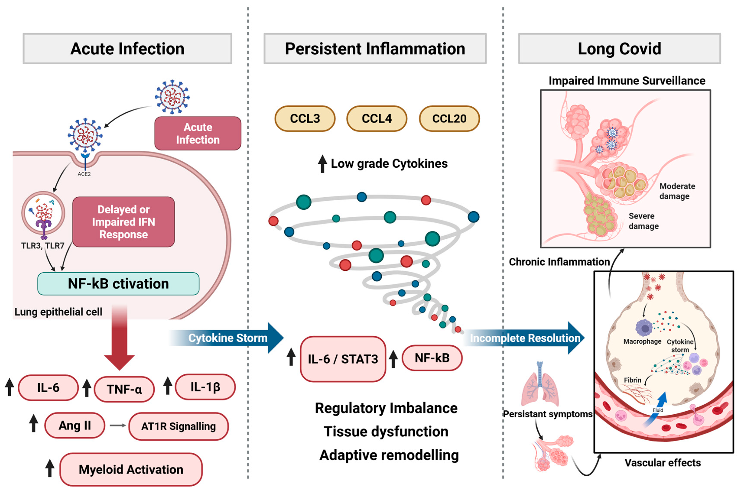
SARS-CoV-2 induces immune responses ranging from effective antiviral control to profound dysregulation [5]. The timing and magnitude of innate and adaptive activation determine whether immune homeostasis is restored or whether inflammatory, metabolic, and exhaustion programs persist. These divergent trajectories underline chronic pathology, impaired tissue repair, and altered immune surveillance with potential oncogenic relevance (Figure 1).
Figure 1.
Immune trajectory from acute SARS-CoV-2 infection to chronic immune remodeling. Acute SARS-CoV-2 infection triggers antiviral responses shaped by the balance between early type I/III IFN signaling and NF-κB-driven inflammation. In mild disease, timely IFN responses promote immune resolution. In severe infection, delayed or impaired IFN responses permit sustained NF-κB activation, amplification of IL-6, TNF-α, and IL-1β, Ang II–AT1R signaling, and myeloid activation, contributing to cytokine storm and JAK/STAT3 engagement. Incomplete resolution may lead to persistent low-grade cytokine and chemokine activity, regulatory imbalance, and tissue dysfunction. Long COVID reflects downstream chronic inflammation, impaired immune surveillance, vascular effects, and adaptive immune remodeling. Created with BioRender.com.
2.1. Acute Immune Activation and Cytokine Storm
SARS-CoV-2 is recognized by Pattern-Recognition Receptors (PRRs) in airway epithelial cells, macrophages, and dendritic cells, inducing type I and III interferons (IFNs) alongside early pro-inflammatory mediators [6]. In mild or asymptomatic infection, timely IFN signaling restricts viral replication and promotes coordinated adaptive immunity [5]. In severe COVID-19, however, type I/III IFN responses are often delayed or attenuated, impairing early viral control and prolonging innate immune activation. Sustained PRR engagement amplifies Nuclear Factor-kappa B (NF-κB)-dependent signaling, increasing production of interleukin (IL)-6, IL-1β, Tumor Necrosis Factor (TNF)-α, and related mediators [7]. Beyond antiviral effects, type I IFNs normally constrain myeloid activation and regulate cytokine balance. Their insufficiency shifts immunity toward NF-κB-dominant inflammatory amplification. Additional pathways reinforce this inflammatory state. SARS-CoV-2-mediated downregulation of Angiotensin-Converting Enzyme (ACE) 2 enhances Angiotensin (Ang) II–Angiotensin type 1 receptor (AT1R) signaling and Metalloprotease 17 (ADAM17) activation, promoting TNF-α release and generation of soluble IL-6 receptor α [8]. IL-6 trans-signaling via glycoprotein (gp)130–Janus Kinase (JAK)–Signal Transducers and Activators of Transcription (STAT) 3 establishes positive feedback with NF-κB, sustaining inflammation even as viral replication declines [9,10]. Viral RNA and spike protein may further activate Toll-like Receptor (TLR) 2/Myeloid differentiation primary response 88 (MyD88) pathways, augmenting NF-κB-driven cytokine production [11,12]. Clinically, convergence of impaired IFN responses and amplified inflammatory signaling culminates in cytokine release syndrome (CRS), characterized by elevated IL-6, TNF-α, IL-1β, Granulocyte Macrophage Colony-Stimulating Factor (GM-CSF), and chemokines [13]. The pathological significance depends less on peak cytokine levels than on persistence of self-reinforcing NF-κB/JAK–STAT signaling that promotes endothelial activation, vascular leak, and Acute Respiratory Distress Syndrome (ARDS) [8,14].
2.2. Transition from Acute to Chronic Immune Dysregulation
As outlined in Section 2.1, early IFN imbalance establishes an NF-κB-dominant inflammatory trajectory that can impair timely resolution, thereby predisposing a subset of individuals to persistent immune dysregulation and chronic inflammatory remodeling [1,14,15,16]. In most patients, contraction of effector responses restores immune equilibrium following viral clearance. However, when acute infection is marked by sustained NF-κB activation, IL-6–STAT3 engagement, and prolonged myeloid stimulation, inflammatory programs may fail to fully resolve. A post-acute immune state can emerge through three partially overlapping mechanisms: (i) residual inflammatory signaling, (ii) persistent perturbation of regulatory pathways, and (iii) adaptive immune remodeling.
Residual inflammation is reflected by continued expression of chemokines such as Macrophage Inflammatory Protein (MIP)-1α (CCL3), Macrophage Inflammatory Protein (MIP)-1β (CCL4), and Macrophage Inflammatory Protein (MIP)-3 (CCL20), indicating ongoing immune cell trafficking and tissue surveillance rather than autonomous inflammatory escalation [17,18]. More consequential is sustained low-grade activation of NF-κB- and STAT3-associated transcriptional programs. Even as circulating cytokines decline, persistent engagement of these pathways may maintain a pro-inflammatory milieu and influence stromal and endothelial function. Prolonged antigen exposure and inflammatory signaling can further drive T-cell exhaustion, altered memory formation, and progressive reduction in naïve lymphocyte pools [15,19]. Thus, the post-acute immune landscape reflects structural reorganization of regulatory circuits rather than simple persistence of acute-phase cytokines. Functionally, this transition manifests low-grade inflammation, altered lymphocyte composition, and impaired immune resolution capacity. The biological impact derives from the stability of dysregulated signaling networks that modulate tissue homeostasis and immune surveillance.
2.3. Persistent Inflammation and Immune Remodeling in Long COVID
Long COVID represents a remodeled immune state in which inflammatory resolution remains incomplete after acute infection. Multiple cohorts demonstrate persistent low-grade elevation of cytokines and chemokines, including IL-6, IL-1β, TNF-α, Monocyte Chemoattractant Protein (MCP)-1 (CCL2), Interferon Gamma-Induced Protein (IP)-10 (CXCL10), and CCL4, up to 6–12 months post-infection [20]. Many of these mediators likely reflect continued immune surveillance or delayed tissue repair rather than autonomous cytokine amplification. More biologically significant is sustained low-level NF-κB and JAK/STAT activity despite normalization of systemic cytokine levels. Persistent IL-6-associated signaling and checkpoint ligand expression in subsets of patients suggest incomplete restoration of immune equilibrium [17,21]. This regulatory imbalance may influence endothelial activation, stromal remodeling, and metabolic adaptation. Cellular analyses reveal reduced naïve T- and B-cell pools, expansion of activated monocytes, and enrichment of exhausted or terminally differentiated T-cell subsets months after infection [19,22]. In some phenotypes, including Myalgic Encephalomyelitis/Chronic Fatigue Syndrome (ME/CFS)-like presentations, elevated Galectin-9, Artemin, and autoantibodies indicate convergence of immune activation and exhaustion [23]. Clinically, persistent inflammation correlates with fatigue, cognitive dysfunction, dyspnea, dysautonomia, and chronic pain [24]. Proposed mechanisms include neuroinflammation, microvascular injury, and immunothrombosis [25]. Even when circulating cytokines normalize, extracellular vesicle-associated inflammatory mediators and neuronal injury markers such as Neurofilament Light chain (NfL) and Glial Fibrillary Acidic Protein (GFAP) may remain detectable [26,27], supporting compartmentalized immune activity. Taken together, long COVID reflects sustained regulatory perturbation and adaptive remodeling rather than continuation of acute cytokine storm.
2.4. T-Cell Dysfunction, Exhaustion and Impaired Immune Surveillance
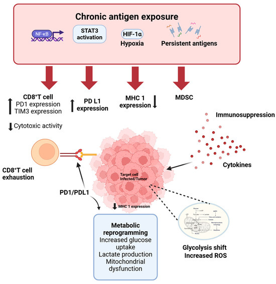
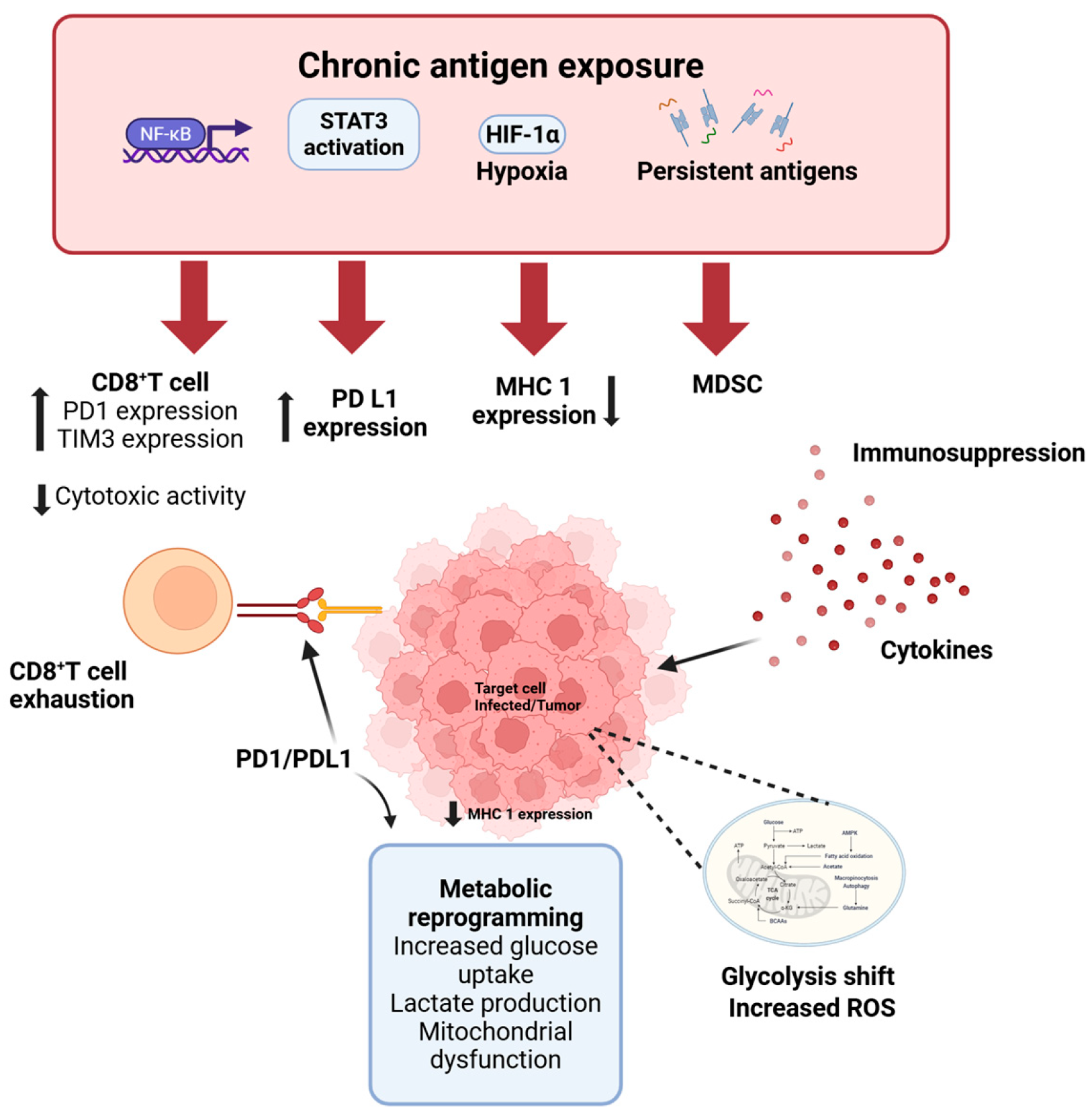
Among the most consequential features of post-acute immune remodeling is persistent T-cell dysfunction. During acute severe disease, lymphopenia affects both CD4+ and CD8+ subsets and correlates with mortality [26,28]. This depletion likely reflects inflammatory redistribution, apoptosis, metabolic stress, and sustained antigen exposure. Increased expression of inhibitory receptors, including Programmed Cell Death (PD)-1, T Cell Immunoglobulin Mucin (TIM-3), Lymphocyte Activation Gene (LAG)-3, and Cytotoxic T-Lymphocyte-Associated (CTLA) protein-4, is common [29,30]. However, elevated inhibitory receptor expression during acute infection may represent transient activation rather than bona fide exhaustion. Consistent with this interpretation, many T cells maintain proliferative potential and cytokine-producing capacity [29,31]. Prolonged inflammatory signaling and persistent antigen exposure may instead drive bona fide exhaustion in susceptible individuals. Exhaustion is characterized by impaired proliferation, reduced effector cytokine production, stable inhibitory receptor expression, transcriptional and epigenetic reprogramming, and metabolic dysfunction [29,32]. Severe and some long-COVID cohorts demonstrate expansion of CD28−CD57+ or Terminally Differentiated Effector Memory (TEMRA) CD8+ populations with reduced regenerative capacity [26,33]. Longitudinal studies indicate persistence of reduced naïve T-cell pools and exhausted or senescent CD8+ subsets beyond the acute phase [15,19,34,35,36,37]. Circulating markers of activation and exhaustion correlate with fatigue and cognitive dysfunction [38], suggesting functional relevance. These adaptive alterations may compromise immune surveillance. Persistent inhibitory receptor-high and poorly regenerative T-cell compartments could attenuate the capacity to detect and eliminate transformed cells, particularly in predisposed or immunologically vulnerable hosts. These immune alterations establish the systemic context in which tumor-associated effects must be interpreted.
3. Convergence on Tumor-Permissive Signaling Networks
Beyond systemic immune remodeling, SARS-CoV-2 infection engages intracellular signaling pathways that regulate inflammation, proliferation, survival, angiogenesis, and immune evasion, core processes in tumor biology. Rather than initiating de novo transformation, SARS-CoV-2 appears to modulate and intensify pre-existing oncogenic signaling circuits, particularly those involving NF-κB, Janus Kinase (JAK)/Signal Transducers and Activators of Transcription (STAT) 3, Mitogen-Activated Protein Kinase (MAPK)/Extracellular Signal-Regulated Protein Kinases (ERK), and TLR2–MyD88 pathways.
3.1. Viral Protein-Mediated Engagement of Host Signaling
SARS-CoV-2 structural proteins can activate inflammatory and stress-response pathways independent of productive replication. The spike (S1) subunit functions as a Pathogen-Associated Molecular Pattern (PAMP)-like ligand that engages NF-κB and MAPK/ERK signaling in epithelial, endothelial, and myeloid cells [11,39,40,41,42], promoting ERK1/2 and p38 phosphorylation, Activator Protein (AP-1)-dependent transcription, and production of IL-6, IL-8, and TNF-α. In macrophages and epithelial cells, spike additionally signals through TLR2/TLR4–MyD88, amplifying NF-κB activation and inflammasome responses [11,43]. The membrane (M) protein similarly modulates oncogenic signaling. In breast cancer models, M expression activates NF-κB and downstream STAT3 signaling, inducing Epithelial–Mesenchymal Transition (EMT) transcription factors and pro-migratory gene programs [44]. NF-κB inhibition attenuates these effects, positioning it upstream of JAK/STAT3 [45]. Although largely derived from in vitro systems, these data suggest that viral proteins potentiate inflammatory and EMT-associated pathways central to tumor progression.
3.2. NF-κB–STAT3 Feedback and Inflammatory Stabilization
NF-κB acts as a central hub linking viral sensing to tumor-associated inflammatory programs. Multiple SARS-CoV-2 proteins, including spike, Open Reading Frame (ORF) 3a, and Non-Structural Protein (NSP) 6, activate canonical NF-κB signaling via Transforming Growth Factor (TGF)-β-activated kinase (TAK) 1– Iκ B kinase (IKK) complexes [46,47,48], inducing IL-6 and other mediators involved in proliferation, angiogenesis, and EMT. IL-6-mediated JAK/STAT3 activation establishes a reinforcing feedback loop in which NF-κB-driven cytokine production sustains STAT3 signaling, and STAT3 stabilizes inflammatory and survival gene expression. This reciprocal NF-κB–STAT3 axis is characteristic of tumor-promoting microenvironments and may be engaged during severe or persistent infection. TLR2 serves as an upstream trigger. Upon engagement by spike and envelope proteins, it initiates MyD88-dependent NF-κB activation, thereby driving downstream IL-6/STAT3 signaling [12]. Thus, antiviral sensing pathways can intersect with and transiently reinforce tumor-associated inflammatory circuits.
3.3. MAPK/ERK as an Amplification Node
SARS-CoV-2 infection robustly engages the MAPK network, particularly ERK1/2 and p38 signaling pathways [45,49,50]. Early after infection, rapid activation of the Raf–MEK-ERK cascade occurs, and pharmacologic MEK inhibition attenuates both viral replication and cytokine production [51]. Spike-dependent Epidermal Growth Factor (EGFR)–C-RAF–MEK–ERK signaling further links viral sensing to proliferative and survival pathways [52]. In parallel, p38 activation amplifies inflammatory cytokine production. Pharmacologic inhibition attenuates IL-6, IL-8, and CXCL10 levels while preserving interferon responses [53]. Sustained MAPK activity is reinforced by suppression of regulatory phosphatases such as Dual-Specificity Protein Phosphatase (DUSP)1 and DUSP5, thereby prolonging ERK, p38, and NF-κB signaling [45,54]. Given that ERK regulates MYC expression, cell-cycle progression, and survival programs, and that p38 contributes to angiogenesis, fibrosis, and metastatic adaptation, infection-induced MAPK activation represents a mechanistic bridge between inflammatory stress and tumor-permissive microenvironments. These findings indicate pathway reinforcement within tumor-relevant signaling networks.
3.4. Integrated Network Perspective
The mechanisms outlined above reveal a tightly interconnected signaling architecture in which NF-κB, JAK/STAT3, and MAPK/ERK function as regulatory hubs integrating antiviral sensing with programs governing survival, angiogenesis, EMT, and immune checkpoint expression. When early IFN responses are blunted and NF-κB-dominant inflammation persists, this network may remain transiently reinforced, particularly in tissues already primed by oncogenic or inflammatory stress [46,49,50]. In this context, viral protein-mediated signaling can augment existing proliferative and survival programs without conferring autonomous transforming capacity.
The emergence of variants with altered transmissibility and immune evasion properties adds further complexity to the infection–cancer interface. Omicron sub-lineages, for example, exhibit differences in IFN antagonism, spike–receptor interactions, and inflammatory signatures that may influence the magnitude and duration of pathway engagement [55]. Whether such variant-specific differences translate into differential modulation of tumor-relevant signaling remains largely unknown. Comparative mechanistic analyses across variants therefore represent an important research direction.
4. Tumor Microenvironment and Tissue-Specific Cancer Responses
While SARS-CoV-2 is not a transforming virus, its driven immune remodeling and protein-mediated signaling can intersect with tumor microenvironments in tissue-specific ways. Tumors exhibit altered receptor expression, impaired antiviral signaling, metabolic stress, and pre-existing inflammatory niches, conditions that may modulate susceptibility to viral exposure and downstream signaling effects.
4.1. Direct or Abortive Infection of Cancer Cells
Experimental evidence indicates that selected cancer cell types can support SARS-CoV-2 entry and, in some contexts, productive or abortive infection. Hepatoma cell lines such as Huh7.5 and HepG2 permit viral replication with associated cytopathic effects, whereas non-transformed hepatic progenitors remain largely non-permissive, suggesting that malignant transformation and altered innate immune signaling influence cellular susceptibility [56]. Serial passages in hepatoma models promote selection of spike-adapted variants with enhanced replication efficiency and reduced reliance on canonical ACE2-mediated entry [38,56]. In glioblastoma models, permissiveness correlates with ACE2 expression and impaired IFN responses, and ACE2 overexpression further increases susceptibility [57]. In addition to ACE2, alternative receptors, including Asialoglycoprotein receptor (ASGR) 1, Dendritic Cell-Specific Intercellular adhesion molecule-3-Grabbing Non-integrin (DC-SIGN), Transmembrane protein (TMEM) 106B, and heparan sulfate-associated mechanisms, may facilitate viral entry within tumor-associated cellular contexts [55,58]. Importantly, viral exposure can induce phenotypic remodeling even in the absence of sustained replication. In hepatic progenitor models, spike exposure promotes partial dedifferentiation and syncytia formation despite limited viral propagation [56]. These findings suggest that tumor cells may undergo transient infection or signaling perturbation; however, there is no evidence for viral genome integration or direct oncogenic transformation.
4.2. Tumor Microenvironment Remodeling and Checkpoint Induction
In vivo studies and spatial transcriptomic analyses indicate that tumor tissues may harbor viral RNA or exhibit virus-associated response signatures following SARS-CoV-2 infection [59]. Within virus-enriched tumor regions, increased Programmed Cell Death Ligand (PD-L) 1 expression, features of T-cell dysfunction, and B-cell-rich immune niches have been reported, consistent with localized remodeling of immune architecture. In this setting, checkpoint upregulation most likely reflects cytokine-driven inflammatory signaling rather than viral transformation. Type I and II IFNs, NF-κB activation, and IL-6–STAT3 signaling are established inducers of PD-L1 transcription, and sustained cytokine exposure can reinforce adaptive immune resistance. Accordingly, tumor cell “remodeling” appears to represent inflammation-mediated phenotypic adaptation under persistent immune pressure. Prolonged exposure to IL-6, TNF-α, and related mediators may further promote EMT, angiogenesis, stromal activation, and metabolic reprogramming within the tumor microenvironment [56,59]. These processes overlap with the signaling networks described in Section 3 and could be amplified in tissues subjected to sustained inflammatory stress.
4.3. Tissue-Specific Divergence in Cancer Responses
SARS-CoV-2-associated effects vary substantially across tumor types. Divergent responses appear to be influenced by ACE2 and Transmembrane Protease, Serine (TMPRSS) 2 expression patterns, IFN competence, renin–angiotensin–aldosterone system (RAAS) signaling balance, and metabolic context [60]. In colorectal cancer models, where ACE2 and TMPRSS2 expressions are relatively high, viral exposure has been linked to enhanced inflammatory and pro-migratory signaling [61,62]. In contrast, prostate cancer models exhibit context-dependent susceptibility, potentially shaped by androgen-regulated TMPRSS2 expression and differences in interferon responsiveness [63,64]. Downregulation of ACE2 in ACE2-high gastrointestinal and lung tumors may amplify Ang II–AT1R signaling, thereby increasing Reactive Oxygen Species (ROS) production, hypoxia, angiogenesis, and inflammatory cytokine release. Conversely, baseline ACE2 expression in certain tumor contexts has been associated with reduced stemness and more favorable immune signatures, highlighting that ACE2 biology may exert tumor type-specific effects independent of viral infection [65,66]. IFN competence further modulates these outcomes. Tumors with impaired interferon signaling may permit sustained viral pathway engagement and inflammatory amplification, whereas interferon-high tumors preferentially induce truncated deltaACE2 isoforms that lack spike-binding capacity, thereby limiting additional viral entry [67,68].
4.4. Inflammation-Driven Reactivation of Dormant Metastases
Tissue-specific inflammatory remodeling may also influence metastatic dormancy. In murine breast cancer models, SARS-CoV-2-induced lung inflammation disrupted dormancy programs in disseminated tumor cells, triggering IL-6-dependent metastatic outgrowth without direct viral infection of tumor cells [69]. Infection-associated impairment of T-cell activation and altered CD4+/CD8+ dynamics contributed to sustained metastatic burden. Observational analyses in cancer survivors have suggested increased lung metastasis and cancer-related mortality following SARS-CoV-2 infection [69], although causality remains to be established. These findings emphasize that infection-induced tissue inflammation, rather than direct viral oncogenesis, may under specific conditions facilitate tumor progression. Across tumor contexts, SARS-CoV-2-associated effects appear to arise primarily from immune and inflammatory remodeling rather than direct genetic transformation. The magnitude and direction of these effects depend on tumor-intrinsic signaling programs, antiviral competence, RAAS balance, and the local immune milieu. Together, these observations support a model in which SARS-CoV-2 acts as a context-dependent modifier of tumor ecosystems, with consequences shaped by pre-existing vulnerabilities rather than uniform pro-oncogenic activity.
6. SARS-CoV-2 Vaccination as a Protective Modifier of Cancer-Relevant Immune Pathways
6.1. Mechanistic Contrast: Infection vs. Vaccination
SARS-CoV-2 vaccination provides a mechanistic contrast to natural infection with respect to antigen persistence, tissue dissemination, and engagement of inflammatory signaling networks. Natural infection involves replicating viruses and sustaining antigen production across multiple tissue compartments. In contrast, currently deployed mRNA and adenoviral vector vaccines encode spike alone and result in transient antigen expression primarily within professional antigen-presenting cells, without viral replication [69,78]. This distinction has important implications for immune remodeling. Whereas infection can sustain NF-κB, MAPK, and IL-6–STAT3 activation in epithelial, endothelial, and myeloid compartments, vaccine-induced innate activation is acute and self-limited, resolving as antigen is cleared. Clinical and immunogenicity studies in patients with solid and hematologic malignancies demonstrate effective spike-specific priming without persistent antigenemia or chronic inflammatory activation [69,78,79,80,81,82]. Adaptive responses also differ qualitatively. Prolonged antigen exposure during severe infection, particularly in immunocompromised hosts, is associated with lymphopenia and accumulation of inhibitory receptor-expressing T cells with exhaustion-like features [83,84,85]. Vaccination induces polyfunctional, Th1-skewed CD4+ and CD8+ T-cell responses targeting multiple spike epitopes [85,86,87], without establishing sustained exhaustion programs. Recent advances in RNA delivery platforms further refine this distinction. Extracellular vesicle (EV)-based mRNA therapeutics represent an emerging strategy aimed at enhancing targeted antigen delivery while minimizing systemic inflammatory exposure. EV-based systems may improve cellular specificity and reduce off-target distribution, thereby further restricting spike expression to professional APCs and limiting unintended tissue exposure [88]. Such approaches reinforce the principle that vaccine-induced antigen presentation is transient, spatially controlled, and mechanistically distinct from the widespread viral replication observed during infection.
In addition to vaccination, emerging antiviral strategies may further limit infection-driven immune remodeling. Small interfering RNA (siRNA)-based therapeutics targeting conserved regions of the SARS-CoV-2 genome represent a promising approach to suppress viral replication and accelerate viral clearance [89]. By directly reducing viral RNA levels, such strategies may prevent prolonged antigen exposure and downstream NF-κB/IL-6–STAT3 amplification, thereby mitigating chronic inflammatory circuits implicated in tumor-permissive immune states. Although still investigational, siRNA-based antivirals conceptually reinforce the importance of early viral control in preventing the transition from acute interferon imbalance to chronic NF-κB-dominant inflammation.
6.2. Impact on Cancer Patients
In patients with solid tumors and hematologic malignancies, vaccine-elicited cellular immune responses are often preserved even when humoral responses are attenuated by chemotherapy or B-cell-depleting therapies. Booster doses enhance both antibody and T-cell responses and are generally well tolerated in oncology populations [79,80,82,90]. Vaccine responsiveness varies across oncologic subgroups. Patients receiving B-cell-depleting agents, such as anti-CD20 monoclonal antibodies, frequently demonstrate impaired seroconversion, with protection relying predominantly on cellular immunity. The durability of T-cell-mediated protection in these settings remains incompletely defined, and optimal booster timing relative to immunosuppressive therapy continues to be actively investigated. These considerations highlight the need for individualized vaccination strategies and longitudinal immune monitoring in highly immunocompromised patients. Importantly, vaccination substantially reduces severe COVID-19, hospitalization, and mortality in cancer populations [78,81,82,91]. By preventing severe infection, vaccination mitigates the prolonged inflammatory signaling, immune dysregulation, and treatment disruption associated with SARS-CoV-2-driven immune remodeling. In this context, infection prevention itself represents a key strategy for limiting cancer-relevant immune perturbations.
6.3. Interaction with Immunotherapy
Mechanistic data suggest that transient vaccine-induced interferon signaling may, in certain contexts, intersect positively with antitumor immunity. Preclinical models demonstrate enhanced dendritic-cell priming and epitope spreading against tumor-associated antigens following mRNA vaccination [86]. Retrospective analyses have reported improved outcomes in subsets of patients with non-small-cell lung cancer and melanoma receiving immune checkpoint blockade around the time of vaccination, although these findings remain associative. Importantly, studies in patients receiving PD-1/PD-L1 inhibitors show preserved vaccine immunogenicity without increased immune-related adverse events [85,91,92]. These observations suggest that vaccine-induced immune activation is compatible with, and may occasionally synergize with, immune checkpoint therapy rather than exacerbating immune toxicity.
6.4. Epidemiologic Safety
Speculative concerns regarding long-term oncologic effects of repeated vaccination lack empirical support. Extensive pharmacovigilance and population-level analyses have not demonstrated increased cancer incidence following vaccination. Current evidence does not support vaccine-associated tumorigenesis, and such proposals remain hypothesis-generating rather than causal [93]. Natural infection and vaccination occupy opposite ends of the immune remodeling spectrum. Infection may impose prolonged antigen exposure, sustained inflammatory signaling, and adaptive exhaustion in susceptible individuals. Vaccination delivers a transient, spatially restricted antigen pulse that supports durable adaptive immunity without establishing persistent inflammatory niches. These mechanistic distinctions are essential when interpreting post-COVID immune alterations in cancer biology and reinforce the safety and clinical benefit of vaccination in oncology settings.
7. Clinical Implications
Cancer patients with SARS-CoV-2 infection experience disproportionately severe outcomes, reflecting baseline immune compromise, systemic inflammation, and vulnerability to treatment disruption. Across large registries, reported mortality rates have ranged from 17–33%, particularly among individuals with hematologic malignancies, advanced age, comorbidities, or recent cytotoxic or monoclonal antibody therapy [93,94]. Profound lymphopenia and elevated inflammatory markers—including IL-6, CRP, LDH, and neutrophil-to-lymphocyte ratio—predict both acute severity and prolonged recovery [95,96,97,98]. Beyond acute mortality, infection frequently disrupts oncologic care. Treatment delays of several weeks are common and may adversely affect tumor control in selected high-risk settings [99]. Persistent inflammatory signatures and post-acute sequelae raise concerns regarding sustained immune dysregulation in cancer survivors, although definitive evidence linking SARS-CoV-2 infection to accelerated tumor progression remains limited. Careful follow-up and risk stratification may therefore be warranted in vulnerable populations. Preventive strategies substantially mitigate these risks. In oncology populations, vaccination reduces severe disease, hospitalization, and mortality, thereby limiting infection-associated inflammatory remodeling and preserving treatment continuity [78,81,82,91]. Available data demonstrates preserved immunogenicity in many patients with solid tumors and acceptable safety profiles, including among those receiving immune checkpoint inhibitors. To date, no evidence supports increased cancer incidence following vaccination. The COVID–cancer interface thus underscores both vulnerability and opportunity: heightened susceptibility to infection necessitates proactive prevention, while mechanistic insights into inflammatory and immune regulatory networks inform risk stratification and therapeutic decision-making. Maintaining infection control and minimizing prolonged inflammatory stress are central to preserving oncologic outcomes.
8. Conclusions and Future Directions
SARS-CoV-2 does not fulfill the criteria of a classical oncogenic virus. It lacks dedicated transforming oncogenes, does not integrate into the host genome, and does not establish stable latency comparable to established oncoviruses such as HPV, HBV, or EBV [84,100]. Current evidence therefore does not support direct viral oncogenesis [66]. Nevertheless, SARS-CoV-2 infection perturbs immune and intracellular signaling networks that substantially overlap with tumor-promoting pathways [92]. Early interferon imbalance followed by sustained inflammatory amplification, centered on NF-κB, IL-6/JAK–STAT3, and MAPK signaling, may, under susceptible conditions, transiently reinforce tumor-supportive microenvironments [4,53]. Severe infections and prolonged post-acute syndromes are characterized by immune exhaustion, impaired cytotoxic surveillance, oxidative stress, fibrosis, and tissue remodeling, features that resemble microenvironmental hallmarks of cancer progression rather than viral transformation [101]. These observations support a model in which SARS-CoV-2 modulates pre-existing signaling circuits rather than initiating malignancy [66]. The biological impact likely depends on the magnitude and duration of inflammatory remodeling, host immune competence, tumor-intrinsic vulnerabilities, and tissue context. Vaccination provides a critical counterbalance. By delivering a transient, spatially restricted antigen exposure without sustained viral replication, vaccination prevents the prolonged inflammatory states implicated in tumor-relevant immune remodeling [53,92]. In doing so, it interrupts the cascade linking acute interferon imbalance to chronic NF-κB-dominant inflammation and immune dysfunction. Important research gaps remain. These include variant-specific differences in immune remodeling, the long-term oncologic consequences of post-acute inflammatory states, and the durability of protective immunity in profoundly immunosuppressed populations [102]. Addressing these questions will require longitudinal epidemiologic surveillance, mechanistic studies of persistent immune alterations, and biomarker-guided risk stratification in cancer survivors [102]. The appropriate stance is neither undue alarm nor premature reassurance, but rigorous, evidence-based vigilance [53,92]. Within this framework, SARS-CoV-2 is best understood not as an oncogenic virus, but as a transient immunological stressor whose influence on cancer biology is contingent on host vulnerability, inflammatory persistence, and effective preventive intervention.
Author Contributions
D.P.: conceptualization, writing—original draft preparation, writing—review and editing, visualization; A.R.: writing—original draft preparation, writing—review and editing, visualization. All authors have read and agreed to the published version of the manuscript.
Funding
This research received no external funding.
Institutional Review Board Statement
Not applicable.
Informed Consent Statement
Not applicable.
Data Availability Statement
No new data were created or analyzed in this study. Data sharing is not applicable to this article.
Acknowledgments
Illustrations are created using BioRender.com.
Conflicts of Interest
The authors declare no conflicts of interest.
References
- Mohandas, S.; Jagannathan, P.; Henrich, T.J.; Sherif, Z.A.; Bime, C.; Quinlan, E.; Portman, M.A.; Gennaro, M.; Rehman, J.; RECOVER Mechanistic Pathways Task Force. Immune mechanisms underlying COVID-19 pathology and post-acute sequelae of SARS-CoV-2 infection (PASC). Elife 2023, 12, e86014. [Google Scholar] [CrossRef] [Scilit] [PubMed]
- Echchannaoui, H.; Perreau, M.; Schild, H.; Theobald, M. Editorial: Understanding convergent evasion mechanisms in cancer and chronic infection: Implications for immunotherapy. Front. Immunol. 2024, 15, 1450555. [Google Scholar] [CrossRef] [Scilit] [PubMed]
- Dalton-Griffin, L.; Kellam, P. Infectious causes of cancer and their detection. J. Biol. 2009, 8, 67. [Google Scholar] [CrossRef] [Scilit] [PubMed]
- Ogarek, N.; Oboza, P.; Olszanecka-Glinianowicz, M.; Kocelak, P. SARS-CoV-2 infection as a potential risk factor for the development of cancer. Front. Mol. Biosci. 2023, 10, 1260776. [Google Scholar] [CrossRef] [Scilit]
- Tay, M.Z.; Poh, C.M.; Rénia, L.; MacAry, P.A.; Ng, L.F.P. The trinity of COVID-19: Immunity, inflammation and intervention. Nat. Rev. Immunol. 2020, 20, 363–374. [Google Scholar] [CrossRef] [Scilit]
- Lowery, S.A.; Sariol, A.; Perlman, S. Innate immune and inflammatory responses to SARS-CoV-2: Implications for COVID-19. Cell Host Microbe 2021, 29, 1052–1062. [Google Scholar] [CrossRef] [Scilit]
- Wang, J.; Jiang, M.; Chen, X.; Montaner, L.J. Cytokine storm and leukocyte changes in mild versus severe SARS-CoV-2 infection: Review of 3939 COVID-19 patients in China and emerging pathogenesis and therapy concepts. J. Leukoc. Biol. 2020, 108, 17–41. [Google Scholar] [CrossRef] [Scilit]
- Yang, L.; Xie, X.; Tu, Z.; Fu, J.; Xu, D.; Zhou, Y. The signal pathways and treatment of cytokine storm in COVID-19. Signal Transduct. Target. Ther. 2021, 6, 255. [Google Scholar] [CrossRef] [Scilit]
- Costela-Ruiz, V.J.; Illescas-Montes, R.; Puerta-Puerta, J.M.; Ruiz, C.; Melguizo-Rodríguez, L. SARS-CoV-2 infection: The role of cytokines in COVID-19 disease. Cytokine Growth Factor Rev. 2020, 54, 62–75. [Google Scholar] [CrossRef] [Scilit]
- Rowaiye, A.B.; Okpalefe, O.A.; Onuh, O.A.; Ogidigo, J.O.; Oladipo, O.H.; Ogu, A.C.; Oli, A.N.; Olofinase, S.; Onyekwere, O.; Abubakar, A.R.; et al. Attenuating the effects of novel COVID-19 (SARS-CoV-2) infection-induced cytokine storm and the implications. J. Inflamm. Res. 2021, 14, 1487–1510. [Google Scholar] [CrossRef] [Scilit]
- Jiang, Y.; Rubin, L.; Peng, T.; Liu, L.; Xing, X.; Lazarovici, P.; Zheng, W. Cytokine storm in COVID-19: From viral infection to immune responses, diagnosis and therapy. Int. J. Biol. Sci. 2022, 18, 459–472. [Google Scholar] [CrossRef] [Scilit] [PubMed]
- Khan, S.; Shafiei, M.S.; Longoria, C.; Schoggins, J.W.; Savani, R.C.; Zaki, H. SARS-CoV-2 spike protein induces inflammation via TLR2-dependent activation of the NF-κB pathway. eLife 2021, 10, e68563. [Google Scholar] [CrossRef] [Scilit] [PubMed]
- Rabaan, A.A.; Al-Ahmed, S.H.; Muhammad, J.; Khan, A.; Sule, A.A.; Tirupathi, R.; Al Mutair, A.; Alhumaid, S.; Dhawan, M.; Tiwari, R.; et al. Role of inflammatory cytokines in COVID-19 patients: A review on molecular mechanisms, immune functions, immunopathology and immunomodulatory drugs to counter cytokine storm. Vaccines 2021, 9, 436. [Google Scholar] [CrossRef] [Scilit] [PubMed]
- Davitt, E.; Davitt, C.; Mazer, M.B.; Areti, S.S.; Hotchkiss, R.S.; Remy, K.E. COVID-19 disease and immune dysregulation. Best Pract. Res. Clin. Haematol. 2022, 35, 101401. [Google Scholar] [CrossRef] [Scilit]
- Adhikari, A.; Maddumage, J.; Eriksson, E.M.; Annesley, S.J.; Lawson, V.A.; Bryant, V.L.; Gras, S. Beyond acute infection: Mechanisms underlying post-acute sequelae of COVID-19 (PASC). Med. J. Aust. 2024, 221, S40–S48. [Google Scholar] [CrossRef] [Scilit]
- Opsteen, S.; Files, J.K.; Fram, T.; Erdmann, N. The role of immune activation and antigen persistence in acute and long COVID. Clin. Transl. Immunol. 2023, 12, e1441. [Google Scholar] [CrossRef] [Scilit]
- Kallaste, A.; Kisand, K.; Aart, A.; Salumets, A.; Kisand, K.; Peterson, P.; Lember, M. Long COVID and Biomarker Dysregulation—A Shift Toward Immune Exhaustion? Medicina 2025, 61, 996. [Google Scholar] [CrossRef] [Scilit]
- Cruz, T.; Albacar, N.; Ruiz, E.; Lledo, G.M.; Perea, L.; Puebla, A.; Torvisco, A.; Mendoza, N.; Marrades, P.; Sellares, J.; et al. Persistence of dysfunctional immune response 12 months after SARS-CoV-2 infection and their relationship with pulmonary sequelae and long COVID. Respir. Res. 2025, 26, 200. [Google Scholar] [CrossRef] [Scilit]
- Kratzer, B.; Gattinger, P.; Trapin, D.; Ettel, P.; Körmöczi, U.; Rottal, A.; Stieger, R.B.; Sehgal, A.N.A.; Feichter, M.; Borochova, K.; et al. Differential decline of SARS-CoV-2-specific antibody levels, innate and adaptive immune cells, and shift of Th1/inflammatory to Th2 serum cytokine levels long after first COVID-19. Allergy 2024, 79, 2482–2501. [Google Scholar] [CrossRef] [Scilit]
- Phetsouphanh, C.; Darley, D.R.; Wilson, D.B.; Howe, A.; Munier, C.M.L.; Patel, S.K.; Juno, J.A.; Burrell, L.M.; Kent, S.J.; Dore, G.J.; et al. Immunological dysfunction persists for 8 months following initial mild-to-moderate SARS-CoV-2 infection. Nat. Immunol. 2022, 23, 210–216. [Google Scholar] [CrossRef] [Scilit] [PubMed]
- Peluso, M.J.; Deitchman, A.N.; Torres, L.; Iyer, N.S.; Munter, S.E.; Nixon, C.C.; Donatelli, J.; Thanh, C.; Takahashi, S.; Hakim, J.; et al. Long-term SARS-CoV-2-specific immune and inflammatory responses in individuals recovering from COVID-19 with and without post-acute symptoms. Cell Rep. 2021, 36, 109518. [Google Scholar] [CrossRef] [Scilit] [PubMed]
- Proal, A.D.; VanElzakker, M.B. Long COVID or post-acute sequelae of COVID-19 (PASC): An overview of biological factors that may contribute to persistent symptoms. Front. Microbiol. 2021, 12, 698169. [Google Scholar] [CrossRef] [Scilit] [PubMed]
- Saito, S.; Shahbaz, S.; Osman, M.; Redmond, D.; Bozorgmehr, N.; Rosychuk, R.J.; Lam, G.; Sligl, W.; Tervaert, J.W.C.; Elahi, S. Diverse immunological dysregulation, chronic inflammation, and impaired erythropoiesis in long COVID patients with chronic fatigue syndrome. J. Autoimmun. 2024, 147, 103267. [Google Scholar] [CrossRef] [Scilit]
- Low, R.N.; Low, R.J.; Akrami, A. A review of cytokine-based pathophysiology of long COVID symptoms. Front. Med. 2023, 10, 1011936. [Google Scholar] [CrossRef] [Scilit]
- Islam, M.S.; Wang, Z.; Abdel-Mohsen, M.; Chen, X.; Montaner, L.J. Tissue injury and leukocyte changes in post-acute sequelae of SARS-CoV-2: Review of 2833 post-acute patient outcomes per immune dysregulation and microbial translocation in long COVID. J. Leukoc. Biol. 2023, 113, 236–254. [Google Scholar] [CrossRef] [Scilit]
- Bachiller, S.; Vitallé, J.; Camprubí-Ferrer, L.; García, M.; Gallego, I.; López-García, M.; Galvá, M.I.; Cañizares, J.; Rivas-Jeremías, I.; Díaz-Mateos, M.; et al. SARS-CoV-2 post-acute sequelae linked to inflammation via extracellular vesicles. Front. Immunol. 2025, 16, 1501666. [Google Scholar] [CrossRef] [Scilit]
- Huang, Z.; Haile, K.; Gedefaw, L.; Lau, B.W.-M.; Jin, L.; Yip, S.P.; Huang, C.-L. Blood biomarkers as prognostic indicators for neurological injury in COVID-19 patients: A systematic review and meta-analysis. Int. J. Mol. Sci. 2023, 24, 15738. [Google Scholar] [CrossRef] [Scilit]
- Vazquez-Alejo, E.; Tarancon-Diez, L.; de la Sierra Espinar-Buitrago, M.; Genebat, M.; Calderón, A.; Pérez-Cabeza, G.; Magro-Lopez, E.; Leal, M.; Muñoz-Fernández, M.Á. Persistent exhausted T-cell immunity after severe COVID-19: 6-month evaluation in a prospective observational study. J. Clin. Med. 2023, 12, 3539. [Google Scholar] [CrossRef] [Scilit]
- Rha, M.S.; Shin, E.C. Activation or exhaustion of CD8+ T cells in patients with COVID-19. Cell. Mol. Immunol. 2021, 18, 2325–2333. [Google Scholar] [CrossRef] [Scilit]
- De Biasi, S.; Meschiari, M.; Gibellini, L.; Bellinazzi, C.; Borella, R.; Fidanza, L.; Gozzi, L.; Iannone, A.; Lo Tartaro, D.; Mattioli, M.; et al. Marked T cell activation, senescence, exhaustion and skewing towards TH17 in patients with COVID-19 pneumonia. Nat. Commun. 2020, 11, 3434. [Google Scholar] [CrossRef] [Scilit]
- Mishra, K.P.; Singh, M.; Saraswat, D.; Ganju, L.; Varshney, R. Dysfunctional state of T cells or exhaustion during chronic viral infections and COVID-19: A review. Viral Immunol. 2022, 35, 284–290. [Google Scholar] [CrossRef] [Scilit] [PubMed]
- Chen-Camaño, R.; DeAntonio, R.; López-Vergès, S. T-cell exhaustion in COVID-19: What do we know? Front. Immunol. 2025, 16, 1678149. [Google Scholar] [CrossRef] [Scilit] [PubMed]
- Ayoubkhani, D.; Bermingham, C.; Pouwels, K.B.; Glickman, M.; Nafilyan, V.; Zaccardi, F.; Khunti, K.; Alwan, N.A.; Walker, A.S. Trajectory of long COVID symptoms after COVID-19 vaccination: Community-based cohort study. BMJ 2022, 377, e069676. [Google Scholar] [CrossRef] [Scilit] [PubMed]
- Diao, B.; Wang, C.; Tan, Y.; Chen, X.; Liu, Y.; Ning, L.; Chen, L.; Li, M.; Liu, Y.; Wang, G.; et al. Reduction and functional exhaustion of T cells in patients with coronavirus disease 2019 (COVID-19). Front. Immunol. 2020, 11, 827. [Google Scholar] [CrossRef] [Scilit]
- Herrmann, M.; Schulze Zur Wiesch, J.; Pawelec, G.; Thimme, R.; Kochanek, M.; Thiel, A.; Lungem, S.; Schultze, J.L. Analysis of co-inhibitory receptor expression in COVID-19 infection compared to acute Plasmodium falciparum malaria: LAG-3 and TIM-3 correlate with T cell activation and course of disease. Front. Immunol. 2020, 11, 1870. [Google Scholar] [CrossRef] [Scilit]
- Kusnadi, A.; Ramírez-Suástegui, C.; Fajardo, V.; Chee, S.J.; Meckiff, B.J.; Simon, H.; Pelosi, E.; Seumois, G.; Ay, F.; Vijayanand, P.; et al. Severely ill COVID-19 patients display impaired exhaustion features in SARS-CoV-2-reactive CD8+ T cells. Sci. Immunol. 2021, 6, eabe4782. [Google Scholar] [CrossRef] [Scilit]
- Wiech, M.; Chrościcki, P.; Swatler, J.; Stepnik, D.; De Biasi, S.; Hampel, M.; Brewinska-Olchowik, M.; Maliszewska, A.; Sklinda, K.; Durlik, M.; et al. Remodeling of T cell dynamics during long COVID is dependent on severity of SARS-CoV-2 infection. Front. Immunol. 2022, 13, 886431. [Google Scholar] [CrossRef] [Scilit]
- Ueland, T.; Cox, R.J.; Michelsen, A.E.; Fjelltveit, E.B.; Otterdal, K.; Dahl, T.; Zhou, F.; Elyanow, R.; Aukrust, P.; Blomberg, B.; et al. Markers of T cell activation and exhaustion in plasma are associated with persistent symptoms up to 18 months following mild SARS-CoV-2 infection. Front. Immunol. 2025, 16, 1578208. [Google Scholar] [CrossRef] [Scilit]
- Alpalhão, M.; Ferreira, J.A.; Filipe, P. Persistent SARS-CoV-2 infection and the risk for cancer. Med. Hypotheses 2020, 143, 109882. [Google Scholar] [CrossRef] [Scilit]
- Forsyth, C.B.; Zhang, L.; Bhushan, A.; Swanson, B.; Zhang, L.; Mamede, J.I.; Voigt, R.M.; Shaikh, M.; Engen, P.A.; Keshavarzian, A. The SARS-CoV-2 S1 spike protein promotes MAPK and NF-κB activation in human lung cells and inflammatory cytokine production in human lung and intestinal epithelial cells. Microorganisms 2022, 10, 1996. [Google Scholar] [CrossRef] [Scilit] [PubMed]
- Olajide, O.A.; Iwuanyanwu, V.U.; Adegbola, O.D.; Al-Hindawi, A.A. SARS-CoV-2 spike glycoprotein S1 induces neuroinflammation in BV-2 microglia. Mol. Neurobiol. 2022, 59, 445–458. [Google Scholar] [CrossRef] [Scilit] [PubMed]
- Patra, T.; Meyer, K.; Geerling, L.; Isbell, T.S.; Hoft, D.F.; Brien, J.; Pinto, A.K.; Ray, R.B.; Ray, R. SARS-CoV-2 spike protein promotes IL-6 transsignaling by activation of angiotensin II receptor signaling in epithelial cells. PLoS Pathog. 2020, 16, e1009128. [Google Scholar] [CrossRef] [Scilit]
- Johnson, E.L.; Smith, J.P.; Nguyen, L.T.; Patel, R.R.; Lee, H.; Kim, D.; Romero, P.C.; Greenberg, M.L.; Nguyen, A.L.; Cruz, M.R.; et al. The S1 spike protein of SARS-CoV-2 upregulates the ERK/MAPK signaling pathway in DC-SIGN-expressing THP-1 cells. Cell Stress Chaperones 2024, 29, 227–234. [Google Scholar] [CrossRef] [Scilit] [PubMed]
- Nguyen, H.N.T.; Kawahara, M.; Vuong, C.K.; Fukushige, M.; Yamashita, T.; Ohneda, O. SARS-CoV-2 M protein facilitates malignant transformation of breast cancer cells. Front. Oncol. 2022, 12, 923467. [Google Scholar] [CrossRef] [Scilit]
- Nguyen, H.N.T.; Hansen, K.P.; Nguyen, M.A.; Martinez, C.R.; Hoang, P.T.; Carlson, A.L.; Pham, T.N.; Geng, X.; Nguyen, K.Q.; Palmer, A.J.; et al. Extracellular vesicles derived from SARS-CoV-2 M-protein-induced triple negative breast cancer cells promoted the ability of tissue stem cells supporting cancer progression. Front. Oncol. 2024, 14, 1346312. [Google Scholar] [CrossRef] [Scilit]
- Zhang, T.; Ma, C.; Zhang, Z.; Zhang, H.; Hu, H. NF-κB signaling in inflammation and cancer. Mol. Carcinog. 2021, 60, 497–512. [Google Scholar] [CrossRef] [Scilit]
- Mao, H.; Zhao, X.; Sun, S.C. NF-κB in inflammation and cancer. Cell. Mol. Immunol. 2025, 22, 811–839. [Google Scholar] [CrossRef] [Scilit]
- Nishitsuji, H.; Iwahori, S.; Ohmori, M.; Shimotohno, K.; Murata, T. Ubiquitination of SARS-CoV-2 NSP6 and ORF7a facilitates NF-κB activation. mBio 2022, 13, e00971-22. [Google Scholar] [CrossRef] [Scilit]
- Goel, S.; Ramaswamy, M.; Verma, P.; Kumar, D.; Singh, V.K.; Yadav, A.K.; Gupta, A.; Tiwari, R.; Sharma, P.; Agnihotri, P.; et al. SARS-CoV-2 switches “on” MAPK and NF-κB signaling via the reduction of nuclear DUSP1 and DUSP5 expression. Front. Pharmacol. 2021, 12, 631879. [Google Scholar] [CrossRef] [Scilit]
- Higgins, C.A.; Nilsson-Payant, B.E.; Bonaventure, B.; Kurland, A.P.; Ye, C.; Yaron, T.M.; Johnson, J.L.; Adhikary, P.; Golynker, I.; Panis, M.; et al. SARS-CoV-2 hijacks p38β/MAPK11 to promote virus replication. mBio 2023, 14, e0100723. [Google Scholar] [CrossRef] [Scilit]
- Schreiber, A.; Viemann, D.; Schöning, J.; Schloer, S.; Mecate Zambrano, A.; Brunotte, L.; Faist, A.; Schöfbänker, M.; Hrincius, E.; Hoffmann, H.; et al. The MEK1/2-inhibitor ATR-002 efficiently blocks SARS-CoV-2 propagation and alleviates pro-inflammatory cytokine/chemokine responses. Cell. Mol. Life Sci. 2022, 79, 65. [Google Scholar] [CrossRef] [Scilit] [PubMed]
- Engler, M.; Albers, D.; von Maltitz, P.; Groß, R.; Münch, J.; Cirstea, I.C. ACE2-EGFR-MAPK signaling contributes to SARS-CoV-2 infection. Life Sci. Alliance 2023, 6, e202201880. [Google Scholar] [CrossRef] [Scilit] [PubMed]
- Faist, A.; Schloer, S.; Mecate-Zambrano, A.; Janowski, J.; Schreiber, A.; Boergeling, Y.; Conrad, B.C.G.; Kumar, S.; Toebben, L.; Schughart, K.; et al. Inhibition of p38 signaling curtails the SARS-CoV-2 induced inflammatory response but retains the IFN-dependent antiviral defense of the lung epithelial barrier. Antiviral Res. 2023, 209, 105475. [Google Scholar] [CrossRef] [Scilit] [PubMed]
- Bouhaddou, M.; Memon, D.; Meyer, B.; White, K.M.; Rezelj, V.V.; Marrero, M.C.; Polacco, B.J.; Melnyk, J.E.; Ulferts, S.; Kaake, R.M.; et al. The global phosphorylation landscape of SARS-CoV-2 infection. Cell 2020, 182, 685–712. [Google Scholar] [CrossRef] [Scilit]
- Vizgirda, G.; Underwood, A.P.; Fahnøe, U.; Weis, N.; Ramirez, S.; Bukh, J. Spike substitutions E484D, P812R and Q954H mediate ACE2-independent entry of SARS-CoV-2 across different cell lines. PLoS ONE 2025, 20, e0326419. [Google Scholar] [CrossRef] [Scilit]
- Smirnova, O.A.; Antonova, O.M.; Stepanova, A.V.; Gushchin, V.A.; Kochetkov, S.N.; Logunov, D.Y.; Shmarov, M.M.; Tukhvatulin, A.I.; Naroditsky, B.S.; Gintsburg, A.L. SARS-CoV-2 establishes a productive infection in hepatoma and glioblastoma multiforme cell lines. Cancers 2023, 15, 632. [Google Scholar] [CrossRef] [Scilit]
- Vanhulle, E.; Broeckx, S.Y.; Michiels, J.; Vandeput, J.; De Maeyer, S.; Van Damme, E.; Naessens, E.; Van Gorp, H.; Delputte, P.L.; Nauwynck, H.J. SARS-CoV-2 permissive glioblastoma cell line for high throughput antiviral screening. Antiviral Res. 2022, 203, 105342. [Google Scholar] [CrossRef] [Scilit]
- Arora, P.; Kempf, A.; Nehlmeier, I.; Graichen, L.; Winkler, M.S.; Schulz, S.R.; Jäck, H.M.; Winkler, M.; Pöhlmann, S. Host cell lectins ASGR1 and DC-SIGN jointly with TMEM106B confer ACE2 independence and imdevimab resistance to SARS-CoV-2 pseudovirus with spike mutation E484D. J. Virol. 2025, 99, e01230-24. [Google Scholar] [CrossRef] [Scilit]
- Lau, M.C.; Shum, H.P.; Chan, J.W.M.; Li, T.; Leung, W.S.; Wu, A.K.L.; Tam, A.R.; Chan, W.M.; Ng, A.C.K.; Chan, P.K.S.; et al. Case report: Understanding the impact of persistent tissue-localization of SARS-CoV-2 on immune response activity via spatial transcriptomic analysis of two cancer patients with COVID-19 co-morbidity. Front. Immunol. 2022, 13, 978760. [Google Scholar] [CrossRef] [Scilit]
- Zhang, Z.; Li, L.; Li, M.; Wang, X. The SARS-CoV-2 host cell receptor ACE2 correlates positively with immunotherapy response and is a potential protective factor for cancer progression. Comput. Struct. Biotechnol. J. 2020, 18, 2438–2444. [Google Scholar] [CrossRef] [Scilit]
- Serwaa, A.; Adu-Gyamfi, E.A.; Agyei-Frimpong, J.; Obeng, C.; Asare, G.A.; Antwi-Boasiako, C.; Amponsah, J.A.; Danquah, K.O.; Agyeman, A.A.; Amankwah, E.A.; et al. In vitro analysis suggests that SARS-CoV-2 infection differentially modulates cancer-like phenotypes and cytokine expression in colorectal and prostate cancer cells. Sci. Rep. 2024, 14, 75718. [Google Scholar] [CrossRef] [Scilit] [PubMed]
- Li, K.; Shi, H.; Zhang, B.; Ou, X.; Ma, Q.; Chen, Y.; Shu, P.; Li, D.; Wang, Y. Myeloid-derived suppressor cells as immunosuppressive regulators and therapeutic targets in cancer. Signal Transduct. Target. Ther. 2021, 6, 362. [Google Scholar] [CrossRef] [Scilit] [PubMed]
- Cheng, M.T.K.; Patel, H.; White, M.; Harrison, S.; Li, X.; Nguyen, A.; Roberts, J.; Thompson, R.; Singh, N.; Zhao, Y.; et al. Determinants of SARS-CoV-2 outcomes in patients with cancer vs controls without cancer: A multivariable meta-analysis with genomic imputation. EClinicalMedicine 2025, 83, 103194. [Google Scholar] [CrossRef] [Scilit] [PubMed]
- Parise, R.; Franchi, M.; Gentile, S.; Trevisan, C.; Basso, D.; Plebani, M.; Rugge, M.; Zorzi, M. Health influence of SARS-CoV-2 (COVID-19) on cancer: A review. Acta Biochim. Biophys. Sin. 2022, 54, 1395–1405. [Google Scholar] [CrossRef] [Scilit]
- Montopoli, M.; Zumerle, S.; Vettor, R.; Rugge, M.; Zorzi, M.; Catapano, C.V.; Carbone, G.M.; Cavalli, A.; Pagano, F.; Ragazzi, E.; et al. Androgen-deprivation therapies for prostate cancer and risk of infection by SARS-CoV-2: A population-based study (N = 4532). Ann. Oncol. 2020, 31, 1040–1045. [Google Scholar] [CrossRef] [Scilit]
- Jahankhani, K.; Ahangari, F.; Adcock, I.M.; Mortaz, E. Possible cancer-causing capacity of COVID-19: Is SARS-CoV-2 an oncogenic agent? Biochimie 2023, 213, 130–138. [Google Scholar] [CrossRef] [Scilit]
- Chakravarty, D.; Chakravarty, D.; Nair, S.S.; Hammouda, N.; Ratnani, P.; Gharib, Y.; Wagaskar, V.; Mohamed, N.; Lundon, D.; Dovey, Z.; et al. Sex differences in SARS-CoV-2 infection rates and the potential link to prostate cancer. Commun. Biol. 2020, 3, 374. [Google Scholar] [CrossRef] [Scilit]
- Hirano, T.; Murakami, M. COVID-19: A new virus, but a familiar receptor and cytokine release syndrome. Immunity 2020, 52, 731–733. [Google Scholar] [CrossRef] [Scilit]
- Chia, S.B.; McDonnell, A.M.P.; Wang, X.; Lo, A.; Mak, S.Y.; Shyer, J.A.; Chen, L.; Xu, J.; Liu, M.; Janes, M.R.; et al. Respiratory viral infections awaken metastatic breast cancer cells in lungs. Nature 2025, 645, 496–506. [Google Scholar] [CrossRef] [Scilit]
- Mortezaee, K.; Majidpoor, J. CD8+ T cells in SARS-CoV-2 induced disease and cancer—Clinical perspectives. Front. Immunol. 2022, 13, 864298. [Google Scholar] [CrossRef] [Scilit] [PubMed]
- Lasser, S.A.; Ozbay Kurt, F.G.; Arkhypov, I.; Utikal, J.; Umansky, V. Myeloid-derived suppressor cells in cancer and cancer therapy. Nat. Rev. Clin. Oncol. 2024, 21, 147–164. [Google Scholar] [CrossRef] [Scilit] [PubMed]
- Veglia, F.; Sanseviero, E.; Gabrilovich, D.I. Myeloid-derived suppressor cells in the era of increasing myeloid cell diversity. Nat. Rev. Immunol. 2021, 21, 485–498. [Google Scholar] [CrossRef] [Scilit] [PubMed]
- Liu, J.; Wu, Y.; Gao, G.F. A structural voyage toward the landscape of humoral and cellular immune escapes of SARS-CoV-2. Immunol. Rev. 2025, 330, e70000. [Google Scholar] [CrossRef] [Scilit]
- Agerer, B.; Koblischke, M.; Gudipati, V.; Montaño-Gutierrez, L.F.; Smyth, M.; Popa, A.; Genger, J.-W.; Endler, L.; Florian, D.M.; Mühlgrabner, V.; et al. SARS-CoV-2 mutations in MHC-I-restricted epitopes evade CD8+ T cell responses. Sci. Immunol. 2021, 6, eabg6461. [Google Scholar] [CrossRef] [Scilit]
- Moriyama, M.; Lucas, C.; Monteiro, V.S.; Iwasaki, A. Enhanced inhibition of MHC-I expression by SARS-CoV-2 Omicron subvariants. Proc. Natl. Acad. Sci. USA 2023, 120, e2221652120. [Google Scholar] [CrossRef] [Scilit]
- Codo, A.C.; Davanzo, G.G.; Monteiro, L.B.; de Souza, G.F.; Muraro, S.P.; Virgilio-da-Silva, J.V.; Prodonoff, J.S.; Carregari, V.C.; de Biagi Junior, C.A.O.; Crunfli, F.; et al. Elevated glucose levels favor SARS-CoV-2 infection and monocyte response through a HIF-1α/glycolysis-dependent axis. Cell Metab. 2020, 32, 437–446.e5. [Google Scholar] [CrossRef] [Scilit]
- Guarnieri, J.W.; Angelin, A.; Ceglia, N.; Sinha, S.; Spangler, A.; Wnorowski, A.; Green, A.E.; Henao-Mejia, J.; Weirick, M.E.; Rathmell, J.C.; et al. SARS-CoV-2 mitochondrial metabolic and epigenomic reprogramming in COVID-19. Pharmacol. Res. 2024, 204, 107170. [Google Scholar] [CrossRef] [Scilit]
- Xiong, N.; Sun, Q. How does SARS-CoV-2 infection impact on immunity, progression and treatment of pan cancers. J. Med. Virol. 2023, 95, e28487. [Google Scholar] [CrossRef] [Scilit]
- Shen, W.; Guo, Y.; Ai, C.; Wang, X.; Li, G. The double-edged sword: How SARS-CoV-2 might fuel lung cancer: Investigating the potential oncogenic mechanisms of the novel coronavirus in lung carcinogenesis. Mol. Aspects Med. 2025, 106, 101413. [Google Scholar] [CrossRef] [Scilit]
- Stingi, A.; Cirillo, L. SARS-CoV-2 infection and cancer: Evidence for and against a role of SARS-CoV-2 in cancer onset. BioEssays 2021, 43, 2000289. [Google Scholar] [CrossRef] [Scilit] [PubMed]
- Wagner, A.; Weinberger, B.; Grubeck-Loebenstein, B.; Melchardt, T.; Neureiter, D.; Pleyer, L.; Hörl, C.; Neudorfer, C.; Schlick, K.; Rinnerthaler, G.; et al. SARS-CoV-2-mRNA booster vaccination reverses non-responsiveness and early antibody waning in immunocompromised patients–a phase four study comparing immune responses in patients with solid cancers, multiple myeloma and inflammatory bowel disease. Front. Immunol. 2022, 13, 889138. [Google Scholar] [CrossRef] [Scilit] [PubMed]
- Obermannova, R.L.; Havel, L.; Fiala, O.; Beran, O.; Šefr, R.; Svoboda, M.; Vokurka, S.; Hrabětová, M.; Petruželka, L.; Študentová, H.; et al. Patterns of SARS-CoV-2-specific humoral and cellular immune response in actively treated patients with solid cancer following prime BNT162b2 COVID-19 vaccination: Results from phase IV CoVigi trial. Ther. Adv. Med. Oncol. 2025, 17, 17588359251316224. [Google Scholar] [CrossRef] [Scilit] [PubMed]
- Addeo, A.; Shah, P.K.; Bordry, N.; Hudson, R.D.; Albracht, B.; Di Marco, M.; Kaklamani, V.; Dietrich, P.Y.; Taylor, B.S.; Simand, P.F.; et al. Immunogenicity of SARS-CoV-2 messenger RNA vaccines in patients with cancer. Cancer Cell 2021, 39, 1091–1098.e2. [Google Scholar] [CrossRef] [Scilit]
- Li, Y.; Zhang, H.; Zhao, Y.; Wang, L.; Liu, X.; Chen, J.; Sun, Q.; Zhou, M.; Huang, Y.; Wu, J.; et al. Impact of SARS-CoV-2 infection on clinical characteristics, antibody levels, and immune responses in patients with malignant hematological tumors. J. Chemother. 2026, 38, 54–65. [Google Scholar] [CrossRef] [Scilit]
- Mair, M.J.; Berger, J.M.; Mitterer, M.; Rinnerthaler, G.; Aigner, F.; Mader, R.M.; Greil, R.; Wolf, D.; Melchardt, T. Third dose of SARS-CoV-2 vaccination in hemato-oncological patients and health care workers: Immune responses and adverse events–a retrospective cohort study. Eur. J. Cancer 2022, 165, 184–194. [Google Scholar] [CrossRef] [Scilit]
- Grippin, A.J.; Wiesenfeld, J.; Suryawanshi, R.K.; Nair, M.; Hu, C.; Shyer, J.A.; Satpathy, A.T.; Jenkins, M.K.; Hegde, P.S.; Yoon, C.H.; et al. SARS-CoV-2 mRNA vaccines sensitize tumours to immune checkpoint blockade. Nature 2025, 647, 488–497. [Google Scholar] [CrossRef] [Scilit]
- Bertoletti, A.; Le Bert, N.; Qui, M.; Tan, A.T. SARS-CoV-2-specific T cells in infection and vaccination. Cell. Mol. Immunol. 2021, 18, 2307–2312. [Google Scholar] [CrossRef] [Scilit]
- Li, Q.; Wang, Y.; Zhang, Y.; Chen, X.; Zhao, J.; Liu, Z.; Sun, H.; Zhou, Y.; Liu, Y.; Wang, C. Extracellular vesicle-based mRNA therapeutics and vaccines. Exploration 2025, 5, 20240109. [Google Scholar] [CrossRef] [Scilit]
- Forgham, H.; Kakinen, A.; Qiao, R.; Davis, T.P. Keeping up with the COVID’s—Could siRNA-based antivirals be a part of the answer? Exploration 2022, 2, 20220012. [Google Scholar] [CrossRef] [Scilit]
- Tarke, A.; Sidney, J.; Methot, N.; Yu, E.D.; Zhang, Y.; Dan, J.M.; Goodwin, B.; Rubiro, P.; Sutherland, A.; Wang, E.; et al. Impact of SARS-CoV-2 variants on the total CD4+ and CD8+ T cell reactivity in infected or vaccinated individuals. Cell Rep. Med. 2021, 2, 100355. [Google Scholar] [CrossRef] [Scilit] [PubMed]
- Becerril-Gaitan, A.; Vaca-Cartagena, B.F.; Ferrigno, A.S.; Mesa-Chavez, F.; Barrientos-Gutiérrez, T.; Tagliamento, M.; Lambertini, M.; Cortes, J.; Curigliano, G.; Lanza, E.; et al. Immunogenicity and risk of SARS-CoV-2 infection after COVID-19 vaccination in patients with cancer: A systematic review and meta-analysis. Eur. J. Cancer 2022, 160, 243–260. [Google Scholar] [CrossRef] [Scilit] [PubMed]
- Jaiswal, A.; Shrivastav, S.; Kushwaha, H.R.; Chaturvedi, R.; Singh, R.P. Oncogenic potential of SARS-CoV-2—Targeting hallmarks of cancer pathways. Cell Commun. Signal. 2024, 22, 518. [Google Scholar] [CrossRef] [Scilit] [PubMed]
- Isidoro, C. SARS-CoV2 and anti-COVID-19 mRNA vaccines: Is there a plausible mechanistic link with cancer? Cancers 2025, 17, 3867. [Google Scholar] [CrossRef] [Scilit]
- Gosain, R.; Abdou, Y.; Singh, A.; Rana, N.; Puzanov, I.; Ernstoff, M.S. COVID-19 and cancer: A comprehensive review. Curr. Oncol. Rep. 2020, 22, 53. [Google Scholar] [CrossRef] [Scilit]
- Albiges, L.; Foulon, S.; Bayle, A.; Gachot, B.; Pommeret, F.; Willekens, C.; Stoclin, A.; Teghom, C.; Canouï, E.; Mariette, X.; et al. Determinants of the outcomes of patients with cancer infected with SARS-CoV-2: Results from the Gustave Roussy cohort. Nat. Cancer 2020, 1, 965–975. [Google Scholar] [CrossRef] [Scilit]
- García-Suárez, J.; de la Cruz, J.; Cedillo, Á.; Llamas, P.; Duarte, R.; Jiménez-Yuste, V.; Hernández-Rivas, J.Á.; Gil-Manso, R.; Kwon, M.; Sánchez-Godoy, P.; et al. Impact of hematologic malignancy and type of cancer therapy on COVID-19 severity and mortality: Lessons from a large population-based registry study. J. Hematol. Oncol. 2020, 13, 133. [Google Scholar] [CrossRef] [Scilit]
- Cai, G.; Gao, Y.; Zeng, S.; Yu, Y.; Liu, Y.; Wang, Y.; Wang, Q.; Xie, X.; Liu, D.; Zhang, Y.; et al. Immunological alteration in COVID-19 patients with cancer and its implications on mortality. Oncoimmunology 2021, 10, 1854424. [Google Scholar] [CrossRef] [Scilit]
- Dettorre, G.M.; Dolci, G.; Sita, R.; Roque, G.; Fiorentino, G.; Bonomi, L.; Gori, A.; De Toma, A.; Taramasso, L.; Costantini, M.; et al. Systemic pro-inflammatory response identifies patients with cancer with adverse outcomes from SARS-CoV-2 infection: The OnCovid Inflammatory Score. J. Immunother. Cancer 2021, 9, e002277. [Google Scholar] [CrossRef] [Scilit]
- Oppolzer, I.A.; Rausch, S.; Pichler, R.; Bektic, J.; Horninger, W.; Heidegger, I. Impact of SARS-CoV-2 pandemic on diagnosis of prostate cancer. Urol. Int. 2025, 109, 158–166. [Google Scholar] [CrossRef] [Scilit]
- Jafarzadeh, A.; Nemati, M.; Jafarzadeh, S.; Nozari, P.; Mortaz, E. SARS-CoV-2 infection: A possible risk factor for incidence and recurrence of cancers. Int. J. Hematol.-Oncol. Stem Cell Res. 2022, 16, 117. [Google Scholar] [CrossRef] [Scilit]
- Li, Y.S.; Ren, H.C.; Cao, J.H. Correlation of SARS-CoV-2 to cancer: Carcinogenic or anticancer? (Review). Int. J. Oncol. 2022, 60, 42. [Google Scholar] [CrossRef] [Scilit]
- Moujaess, E.; Kourie, H.R.; Ghosn, M. Cancer patients and research during COVID-19 pandemic: A systematic review of current evidence. Crit. Rev. Oncol. Hematol. 2020, 150, 102972. [Google Scholar] [CrossRef] [Scilit]
Disclaimer/Publisher’s Note: The statements, opinions and data contained in all publications are solely those of the individual author(s) and contributor(s) and not of MDPI and/or the editor(s). MDPI and/or the editor(s) disclaim responsibility for any injury to people or property resulting from any ideas, methods, instructions or products referred to in the content. |
© 2026 by the authors. Licensee MDPI, Basel, Switzerland. This article is an open access article distributed under the terms and conditions of the Creative Commons Attribution (CC BY) license.