


Abstract
Respiratory viral infections remain a leading cause of morbidity, hospitalization, and healthcare burden among children worldwide. While traditional injectable vaccines have been effective in preventing severe disease, they are often limited in their ability to induce strong mucosal immunity at the initial sites of viral entry. Mucosal vaccination has therefore been proposed as a promising strategy to improve local and systemic immune responses and to potentially decrease pathogen transmission. The aim of this study is to review recent advances in mucosal vaccine development for pediatric populations, with an emphasis on the immunological basis of mucosal protection, features related to the age of the developing immune system, and the most relevant vaccine platforms, including intranasal, oral, viral-vectored, and nanoparticle-based approaches. We discuss recent advances in mucosal vaccine development against the main respiratory viral pathogens affecting children, including respiratory syncytial virus (RSV), influenza viruses, and SARS-CoV-2, and review available preclinical and clinical evidence to support their use. Finally, we present major challenges limiting the widespread implementation of mucosal vaccines in pediatric populations, including safety, variability of immune responses, and regulatory considerations. We conclude by highlighting future directions and emerging technologies that may aid the development of safe, effective, and long-lasting mucosal vaccination strategies for the prevention of respiratory viral infections in children.
1. Introduction
1.1. Burden of Respiratory Viral Infections in Children
Acute viral respiratory infections are a major cause of pediatric morbidity and mortality, contributing to 700,000–900,000 deaths annually among children under five years of age [1]. Combined RSV, influenza virus (IFV), human parainfluenza viruses (hPIV), and human metapneumovirus (hMPV) account for nearly 47% of all acute lower respiratory infections (ALRIs) in this age group [2]. These viruses are transmitted primarily via aerosols, droplets, and direct contact with mucosal entry points: the eyes, nose, and mouth [3].
RSV represents the leading cause of severe viral lower respiratory tract infections (LRTIs) in young children worldwide, accounting for more than 33 million ALRI episodes, 3.2–3.6 million hospitalizations, and up to 109,600 deaths annually among children under five years of age [1,2,4]. The burden is greatest during the first six months of life and among preterm infants, who account for approximately one-quarter of RSV-related hospitalizations [4]. Other major respiratory viruses also contribute substantially to pediatric disease burden, with influenza causing approximately 0.9 million hospitalizations and 34,800 deaths annually, hPIVs accounting for around 1.0 million hospitalizations and 53,000 deaths, and hMPV being responsible for 14.2 million ALRI cases, 500,000–600,000 hospitalizations, and more than 11,000 deaths worldwide [1,2,5].
Human adenoviruses (HAdV) account for ~5% of all pediatric respiratory infections and are detected in 6% to 20% of children hospitalized with lower respiratory illness, with a peak risk for severe disease occurring between 6 months and 2 years [6]. Rhinoviruses (RV) are also common respiratory pathogens and an important cause of bronchiolitis, pneumonia, and healthcare visits in young children [1]. The COVID-19 pandemic further altered the epidemiology of pediatric respiratory infections, with SARS-CoV-2 frequently detected in co-infections with endemic respiratory viruses, particularly RV [7].
Among children with HAdV infection, co-infections occur in up to 50% of cases and are associated with a 40% higher risk of severe clinical outcomes, greater use of mechanical ventilation, and prolonged hospital stays when bacterial pathogens are involved [6]. Disparities in health outcomes remain stark: the burden of severe pediatric respiratory disease is disproportionately concentrated in low- and middle-income countries (LMICs), where access to supportive care and diagnostics is limited [2,4,7]. Importantly, severe viral respiratory infections during early life have long-term consequences, being associated with recurrent wheezing and an increased risk of asthma later in childhood [1].
1.2. Pediatric Vulnerability: Immature Innate and Adaptive Immunity
Infants and young children face an exceptionally high risk of severe respiratory viral infections due to the functional immaturity of their immune system. This vulnerability is closely tied to ongoing postnatal lung development. Because alveolarization continues for 1–2 years after birth, the early-life immune system prioritizes tissue protection and anti-inflammatory control over aggressive pathogen clearance [1,8]. While this adaptation prevents inflammatory damage to the developing lungs, it significantly impairs both mucosal and systemic antiviral defenses [1].
At the mucosal entry site, innate recognition and signaling are inherently limited. Infant airway epithelial cells and alveolar macrophages (AMs) exhibit immature pattern-recognition signaling, resulting in less efficient innate pathogen sensing [8]. Upon viral exposure, local dendritic cells produce lower levels of pro-inflammatory Th1-polarizing cytokines and higher amounts of anti-inflammatory mediators [1,8]. Additionally, the paucity of plasmacytoid dendritic cells in infant lungs leads to a diminished output of type I interferons [8]. Although type III interferons remain partially functional, low local type I interferon levels allow rapid viral replication in the respiratory tract [8].
Adaptive immune responses are similarly constrained. Infant CD4+ T cells are functionally biased toward type 2 helper T-cell (Th2) and regulatory T-cell responses, which dampens protective Th1 and cytotoxic CD8+ T-cell activity [1,9,10]. Furthermore, impaired follicular helper T-cell responses limit germinal center reactions [9,10]. This restricts somatic hypermutation and affinity maturation in B cells, resulting in the production of predominantly low-affinity, short-lived antibodies [9,10].
Finally, passive protection from maternal antibodies is transient and incomplete. Transplacentally acquired maternal IgG wanes rapidly over the first 3–6 months of life, opening a critical window of vulnerability [9,10]. High titers of maternal antibodies can also suppress the infant’s own antibody production through epitope masking [9,10]. Combined with low baseline levels of local secretory IgA and delayed lymphocyte recruitment to the lungs, the pediatric respiratory mucosa remains inadequately protected against viral entry and infection [10].
1.3. Limitations of Systemic Vaccines
Currently available systemic vaccines, administered primarily via parenteral or intramuscular routes, are highly effective at preventing severe LRTI, systemic complications, and mortality [11,12,13,14,15,16]. Following parenteral administration, antigens are captured by dendritic cells and presented in draining lymph nodes, where circulating T and B lymphocytes are activated [11,12,17]. This pathway generates high titers of circulating serum IgG antibodies and central memory T cells [11,12,16,17,18]. Once a respiratory virus breaches the primary mucosal barrier and invades internal tissues, these systemic effectors rapidly clear the pathogen and reduce clinical severity [11,12,14,15,16].
However, intramuscular immunizations fail to establish effective immunity at the primary portal of viral entry: the upper respiratory tract [11,12,14,15,16,17,18]. Secretory immunoglobulin A (sIgA) serves as the principal antibody protecting the respiratory mucosa [11,12,17,18]. Because systemic vaccines bypass mucosa-associated lymphoid tissues (MALTs), they do not efficiently trigger local IgA class switching or antibody secretion onto the epithelial surface [11,12]. Furthermore, systemically activated lymphocytes lack the specific homing receptors required to target the respiratory airways [11,12,17]. As a result, intramuscular vaccination fails to generate local tissue-resident memory T (TRM) and B (BRM) cells directly within the respiratory mucosa [11,12,14,15,17,18].
In the absence of sufficient local sIgA and resident memory cells, systemic vaccines may provide limited protection against initial infection at the respiratory mucosa. Although circulating IgG can contribute to pulmonary protection, its levels in the upper respiratory tract may be insufficient to prevent viral acquisition and replication. Consequently, breakthrough infections and viral shedding may still occur despite protection against severe disease (Table 1) [11,12,13,14,15,16,17,18].
Table 1.
Comparison of systemic (intramuscular) and mucosal vaccination against respiratory viral infections [11,12,13,14,15,16,17,18].
1.4. The Rationale for Mucosal Vaccines
Respiratory viruses initiate infection at the respiratory mucosa [19,20,21]. Because this epithelial surface represents the first immunological barrier encountered by inhaled pathogens [19,20,22], effective protection ideally should be established directly at the site of viral entry rather than after systemic dissemination [19,20]. Traditional systemic vaccines primarily induce systemic immune responses, which are highly effective at limiting disease severity but less capable of preventing infection at the mucosal surface [21,22]. In contrast, mucosal vaccination aims to generate local immune responses within the respiratory tract, thereby mimicking the natural route of infection and establishing protective immunity at the site of pathogen entry [19,21].
Mucosal vaccination aims to establish immune protection directly at the respiratory surfaces where viral infections are initiated. By inducing local immune responses, including secretory IgA and tissue-resident memory cells, these vaccines may limit early viral replication and reduce respiratory shedding and transmission. This approach may therefore complement the systemic protection provided by conventional vaccines, particularly against respiratory pathogens that spread readily among children [21,22,23,24,25].
2. Materials and Methods
This manuscript was prepared as a narrative review of current evidence on mucosal vaccines for the prevention of respiratory viral infections in children. A literature search was conducted in PubMed/MEDLINE and Scopus for publications addressing mucosal immunity, pediatric vaccination, respiratory viral infections, and emerging mucosal vaccine technologies.
The search included combinations of the following keywords: “mucosal vaccine”, “intranasal vaccine”, “mucosal immunity”, “pediatric”, “children”, “infants”, “secretory IgA”, “tissue-resident memory T cells”, “RSV”, “influenza”, “SARS-CoV-2”, “human metapneumovirus”, “parainfluenza”, and “adenovirus”. Publications from [2015–2026] were primarily considered, while earlier studies were included when they represented seminal or particularly relevant evidence.
Original research articles, clinical trials, preclinical studies, systematic and narrative reviews, and relevant regulatory or public health reports were considered. Studies were selected based on their relevance to mucosal vaccine development, immunological mechanisms, efficacy, safety, or translational potential in pediatric populations. Particular attention was given to studies investigating intranasal vaccination, mucosal IgA responses, tissue-resident memory T cells, novel vaccine platforms, and respiratory virus transmission.
Due to the heterogeneity of the available evidence, findings were synthesized qualitatively rather than quantitatively. Evidence was organized according to mucosal immune mechanisms, vaccine platforms, target respiratory viruses, preclinical and clinical evidence, safety considerations, current challenges, and future perspectives.
No original human or animal experiments were performed, and no new patient-level or experimental datasets were generated; therefore, ethical approval was not required. All data discussed in this review were derived from publicly available sources.
3. Mucosal Immune System in Children
The mucous membranes are the body’s primary interface with the external environment. The mucous membranes of children’s respiratory and digestive systems are constantly exposed to environmental antigens, pathogenic microorganisms and commensal organisms. The immune system of the mucous membranes must therefore fulfill two essential functions. On the one hand, it must limit the entry and proliferation of pathogens; on the other, it must remain neutral towards commensal organisms and non-pathogenic antigens. Mucosal immunity therefore does not merely act as an infectious barrier; it regulates the body’s defense response through immunological tolerance [20]. The MALT immune system in children exhibits significant structural, functional and immunoregulatory differences compared with that of adults. The immune system undergoes dynamic development during early childhood. Initially, there is a period of transient immunological immaturity. This is characterized by a predominance of Th2-type cellular responses, reduced secretion of sIgA, and interference by maternal antibodies in the process of establishing the child’s own immunity. The functional distinctiveness of MALT in children stems from the specific conditions of early life. On the one hand, it is necessary at this stage to allow colonization by the microbiota and to control excessive inflammatory responses, as well as to limit infections typical of this age group [1,26].
3.1. Anatomy of Mucosal Immunity
The MALT forms the basis of the mucosal immune system. This system is primarily located in the gastrointestinal tract, the respiratory tract, and the genitourinary system. MALT is found in the spaces between and beneath the epithelial cells. Lymphatic and blood capillaries are arranged around the epithelial layer of MALT. This ensures bidirectional exchange of immune cells and contact with the central immune system. MALT primarily comprises nasopharyngeal lymphoid tissue (NALT) in the upper respiratory tract and gut-associated lymphoid tissue (GALT) [20]. This organization enables local antigen recognition at the sites where they most frequently come into contact with the body. GALT comprises the organized lymphoid structures of the intestine, primarily Peyer’s patches, isolated lymphoid nodules, and the appendix. A characteristic feature of GALT is the continuous activity of germinal centers. This is due to continuous stimulation by the gut microbiota [27]. The significance of GALT extends beyond local intestinal immunity, as it plays a role in the maturation and differentiation of B lymphocytes in the marginal zone. These cells may subsequently leave the intestine and contribute to systemic immunity. B lymphocytes activated in GALT can differentiate into IgA-producing cells, which then migrate to the intestinal mucosa [27,28]. Secretory IgA helps to control the microbiota and limit microbial contact with the epithelium while avoiding excessive inflammation. NALT plays a role in immune surveillance within the upper respiratory tract. Anatomically, NALT comprises Waldeyer’s ring, which consists of the palatine, tubal, pharyngeal, and lingual tonsils [20]. NALT is a tissue that reacts rapidly to viruses penetrating the nasal mucosa. Within NALT, B lymphocytes are activated in the centers of proliferation, as are T lymphocytes. This constitutes a specific antiviral response. During an acute infection, monocytes migrate to the NALT, where they accumulate beneath the epithelium and around the lymphoid follicles. Monocytes also participate in the recruitment of neutrophils that form NETs. Once the infection has resolved, the role of macrophages within the NALT shifts from a defensive to a regenerative one [29]. The mucosal immune system also involves interactions between different anatomical sites, particularly through the gut–lung axis. Gut microbiota-derived metabolites, including short-chain fatty acids (SCFAs), can influence immune regulation in the respiratory tract. These interactions may be particularly relevant during early life, when the intestinal microbiota and respiratory immune system undergo parallel development. Disruptions in microbiota composition, especially in preterm infants, have been associated with altered immune responses and increased susceptibility to respiratory infections [30,31,32].
3.2. Immune Development in Early Life
A newborn possesses elements of both innate and acquired immunity. However, its immune system has limited antigenic experience and must adapt to the extrauterine environment. Immediately after birth, the infant must develop a range of mechanisms that both protect against pathogens and tolerate commensal organisms [26]. The newborn’s immune system cannot be described as ‘immature’ when compared to that of an adult. At this stage of life, it has different tasks, namely adapting to the conditions of the perinatal period. An example of this is the suppression of an excessive inflammatory response during the period of intensive colonization of the body by the microbiota. Nevertheless, certain aspects of the newborn’s immune response may increase the risk of specific infections [1,26]. In the newborn population, there is a physiological shift in the lymphocytic response towards Th2 lymphocytes. There may be several reasons for this shift. One of these is thought to be the reduced production of interleukin-12 (IL-12) by antigen-presenting cells (APCs), which promotes the differentiation of Th1 lymphocytes. A lower number of Th1 lymphocytes also leads to a decrease in IFN-γ, which contributes to an increased risk of respiratory viral infections in the newborn. This should not be viewed solely as a ‘flaw’ of the immature immune system. As the newborn’s lungs are still developing, a highly aggressive inflammatory response directed against the virus could simultaneously damage the delicate lung tissue [1].
One of the most important components of humoral immunity in the mucous membranes is the production of IgA. In mucous secretions, it occurs mainly in a dimeric form—IgA molecules are linked by a J chain and are then transported by epithelial cells via the polymeric immunoglobulin receptor (pIgR). This process results in the formation of sIgA. sIgA acts on the surface of the mucous membranes, where it binds to microorganisms and their antigens, thereby limiting their contact with and penetration of the epithelium [33]. However, in the neonatal population, the sIgA produced by the infants themselves has little protective significance; there might be a significant shortage of it, or it may be absent until a certain age. In early life, maternal sIgA, supplied via breast milk, takes on particular importance. In addition to providing passive protection, breast milk contributes to the development of mucosal immune tolerance through its immunomodulatory components and effects on the intestinal microbiota [34,35].
In the first months of life, maternal antibodies play a crucial role in protecting the infant against pathogens. Mechanisms of maternal protection include the transplacental transfer of IgG antibodies. As a result, the baby is born with a certain reserve of ready-made antibodies against pathogens with which the mother has previously come into contact, or against which she has been vaccinated. These antibodies can protect the newborn by neutralizing microorganisms and their toxins through opsonization and facilitating phagocytosis, and by activating the complement system [36]. Another way in which maternal antibodies influence the development of the child’s immune system is through the supply of IgA antibodies via breast milk [34]. Vaccinating mothers is an excellent way to boost their immunity and, consequently, to provide more effective protection for their offspring against infectious diseases during the neonatal period. The mother’s microbiota also plays a key role in the development of the newborn’s immune system and can influence the child’s health in both the short and long term [36].
3.3. Mechanism of Mucosal Protection
The epithelium that forms a barrier that separates the external environment from the host tissues is a key component of mucosal defense. Together with mucus, antimicrobial substances, and immune cells, it forms the first line of defense against the entry of pathogens [20]. A key difference between mucosal and systemic immunity lies in the role of the individual immunoglobulin classes. sIgA is adapted to function on the surfaces of mucous membranes, while IgG is the main class of antibodies in serum and is a key component of systemic immunity (Table 2) [33,37,38].
Table 2.
Structural, functional, and immunological differences between sIgA and systemic IgG [33,34,36,37,38].
TRM cells are long-lived lymphocytes that remain within barrier tissues, including the respiratory mucosa. Unlike circulating memory T cells, they can respond rapidly to local antigen re-exposure, limiting pathogen replication and contributing to localized immune protection [39,40,41]. Their formation is therefore an important objective of mucosal vaccine development. However, excessive or persistent TRM activation may also contribute to tissue inflammation and damage in chronic respiratory conditions [39,41]. Upon re-exposure to an antigen, TRM cells can proliferate very rapidly at the local site and induce a secondary immune response. Their role also involves alerting other immune system cells to the entry of a foreign antigen into the body. The remarkable properties of TRM cells, including their ability to cross-react, have become the subject of research regarding their use in local vaccinations. Combining intramuscular and intranasal vaccinations with the mRNA vaccine against SARS-CoV-2 has been shown to produce better immunization outcomes than when these vaccines are administered separately [41]. In the future, it will be important to develop recombinant antigens and safe mucosal adjuvants in order to create vaccines that effectively stimulate TRM cells and BRM cells [39]. Combining intramuscular vaccination with mucosal vaccination appears to be a promising approach to achieving a longer-lasting immune response, both systemic and local [39,41]. Despite their positive and promising role in vaccine development, TRM cells also exhibit effects that are less beneficial to humans. An excessive TRM immune response contributes to the development of various chronic respiratory diseases. These include conditions such as asthma, COPD, and pulmonary fibrosis. The cause of such a strong immune response is most often an acute viral infection. A negative consequence of heightened TRM activity is microdamage to lung tissue caused by the release of cytotoxic molecules. However, for this phenomenon to occur, constant antigenic or inflammatory stimulation is necessary, as TRM cells do not release these cytotoxic molecules continuously. The antigen responsible for such an immune response may be viral remnants following an acute infection. An excessive TRM response may contribute to many lung diseases; therefore, modulating it could form the basis for new immunomodulatory therapies (Figure 1) [40].
Figure 1.
Key immunological steps in mucosal immunity induction following intranasal vaccination [15]. Schematic representation of antigen uptake by mucosal dendritic cells, lymphocyte activation in local lymph nodes, B cell differentiation, and pIgR-mediated transport of sIgA to neutralize pathogens at the respiratory mucosal surface.
4. Mucosal Vaccine Platforms
Among all mucosal vaccines, there are plenty of platforms by which the vaccines can be administered. They will be described here according to certain diseases. One of the most well-known diseases against which such protection is possible is influenza. The LAIV (live-attenuated influenza vaccine) is currently the only intranasal influenza vaccine registered for use in humans. This vaccine is based on cold-adapted, temperature-sensitive live-attenuated viruses that are optimized for replication in the cooler upper respiratory tract and thus induce an immunologic response primarily in that area. Systemic humoral immune responses, such as the production of serum IgG and hemagglutination inhibition antibodies, are also induced; however, they are generally less pronounced than those triggered by inactivated vaccines administered via the intramuscular route [42]. Studies have shown that the LAIV intranasal vaccine works better in children, primarily those younger than six years of age, probably due to reduced pre-existing immunity [42,43]. The biggest advantage of the LAIV is its intranasal administration, which is painless and particularly suitable for children. Moreover, by mimicking natural infection, the LAIV induces mucosal immunity, making it a great option for large-scale immunization campaigns, particularly during pandemics [43]. The other great advantage is its cost-effectiveness, which, although not well researched, is confirmed by multiple studies. Although the initial cost of intranasal vaccine administration is higher, the LAIV may generate both direct savings in healthcare and broader societal benefits over time by reducing influenza cases and parental productivity losses. These economic advantages support its potential use as a preferred option for a national vaccination strategy targeting children [44]. The LAIV is also relatively safe, as studies show no deaths caused by this vaccine and only 23 vaccine-related SAEs recorded among 17,833 individuals analyzed during the trials [44]. However, the LAIV also has certain limitations, as its effectiveness in adults has fluctuated substantially between influenza seasons. Studies that compared the LAIV and intramuscularly administered inactivated vaccines have demonstrated that, in healthy adults, the LAIV provides protection against laboratory-confirmed influenza that is comparable to or lower than that achieved with IM inactivated vaccines. This diminished effectiveness could be related to pre-existing immunity against influenza viruses, which can restrict the replication of vaccine viruses in the upper respiratory tract [42].
While the LAIV is the only registered intranasal influenza vaccine, research is underway to develop new platforms of mucosal influenza vaccines, and they can be divided into replicating and non-replicating platforms. Replicating platforms generally provide stronger antigen expression and more closely mimic natural infection, and non-replicating ones offer improved safety and greater flexibility in antigen design [42]. The main candidates for new mucosal vaccine platforms for influenza are presented in Table 3.
Table 3.
Advanced and next-generation viral vaccine platforms for mucosal immunization [42,45,46,47].
Currently, other intranasal vaccines are in development. Some of them include vaccines against RSV, which use recombinant, bivalent, non-replicating Ad5 adenoviral vectors. As antigens, they used RSV protein F. Research was performed on mice and cotton rats and showed promising results after mucosal administration, providing strong humoral and cellular responses and significant inhibition of RSV replication in the lungs [48]. However, the mentioned Ad5 vector is not the only one that can be implemented, as research has been performed on subjects using adenovirus type 4 against SARS-CoV-2, with promising results. They have shown comparable viral growth kinetics and protein expression, while eliciting superior protection and immune responses when administered intranasally compared to the vaccine using Ad5 as a vector [49].
After the pandemic, much focus has shifted towards developing a vaccine for the SARS-CoV-2 pathogen, and thus many researchers have proposed their own platforms for use. One such platform is the outer membrane vesicle, a non-invasive platform that can sprout from the surface of many Gram-negative bacteria. The immune system proficiently recognizes the surface antigens that they present, and thus, via multiple pathogen-associated molecular patterns, the innate immune system may be activated. Combining the SARS-CoV-2 spike protein with N. meningitidis’ OMV proved safe and efficient in tested mice and hamster populations, with high IgG antibody responses and IgA prevalent after intranasal vaccination [45].
There are also studies whose goal is to develop a live intranasal attenuated vaccine for SARS-CoV-2. Proposed vectors are viruses adapted to cold temperatures, viruses attenuated through codon deoptimization, or viruses with deleted accessory or mutated viral proteins. All of these have shown optimistic results in trials. Other than that, attenuated SARS-CoV-2 can itself serve as a vector, for example, in immunization against RSV. An attenuated virus combined with protein F of RSV induced a strong immunological response, increasing both mucosal IgA and antibodies in blood. Among tested hamsters, it provided full immunity to both SARS-CoV-2 and RSV infection [50].
Another pathogen against which mucosal vaccines are tested is MERS-CoV. Shi et al. used a live, modified influenza virus as a vector in intranasal vaccines in transgenic mice. The results showed that even a single dose provided protection against MERS-CoV, with two doses granting almost complete immunity. The vaccine simultaneously elicited a mucosal response, a humoral response, and a cellular response involving T lymphocytes. Additionally, serum from vaccinated mice neutralized three different MERS-CoV clades, suggesting the potential for protection against diverse viral variants [46].
When it comes to oral vaccines, protection against rotavirus is among the best developed. There are currently four vaccines globally approved by the World Health Organization (WHO): Rotarix®, produced by GlaxoSmithKline, a monovalent (G1P [8]) vaccine using live-attenuated human rotavirus; Rotavac®, produced by Bharat Biotech, another monovalent (G9P) vaccine, using live, naturally attenuated human–bovine mono-reassortant; RotaTeq®, produced by Merck & Co., Inc., and Rotasiil®, produced by the Serum Institute of India, both being pentavalent vaccines (respectively, G1, G2, G3, G4, P and G1, G2, G3, G4, G9) using, respectively, live-attenuated human–bovine mono-reassortant and live-attenuated bovine–human rotavirus [51]. Clinical trials involving Rotarix and RotaTeq showed no increased risk of serious adverse effects, with both presenting high efficiency against severe gastroenteritis that was caused by rotavirus. However, observations in high- and middle-income countries have reported a slight increase in the risk of intussusception up to a week following administration of the vaccine, with approximately one to six surplus cases per 100,000 infants vaccinated. Still, it has been determined that the benefits outweigh this risk. As for Rotavac and Rotasiil, clinical trials have shown similar results to those of Rotarix and RotaTeq; however, the risk of intussusception occurring could not be evaluated due to a lack of data. However, other, later-published articles claim there is no evidence of a higher risk of intussusception after vaccine administration. Additional studies on the matter of these vaccines’ safety are ongoing [52,53].
However, when it comes to the efficacy of rotavirus oral vaccines, it can differ depending on the country’s mortality from this disease, with higher efficacy in low-mortality and medium-mortality countries, and lower efficacy in high-mortality ones (respectively, Rotarix: 90%, 78%, and 54%; RotaTeq: 94%, 81%, and 44%). Meanwhile, Rotavac and Rotasiil both present lower protection, but their advantages include a lower cost of production and easier storage of Rotasiil [51]. The lower efficacy in high-mortality countries is most likely multifactorial, but is often attributed to insufficient viral replication in the gut mucosa, leading to reduced immune activation [52,54]. Although host genetic factors may contribute, studies in twins suggest that genetics account for less than 30% of immune variability underlying the dominant role of environmental factors, which include altered gut microbiota, micronutrient deficiencies, intestinal damage from frequent enteric infections, preexisting immunity to cross-reactive antigens, and interference by maternal RV-specific antibodies [52].
Due to the limited efficacy of licensed rotavirus vaccines, ongoing research is exploring new platforms by which to apply them. Some candidates include RV3-BB (based on a human neonatal live-attenuated strain); the VP8 subunit protein vaccine (based on recombinant proteins); tetra-, penta-, and hexavalent UK-BRV (based on live-attenuated bovine–human reassortant strain); the inactivated G1P [8] vaccine (heat-inactivated human strain); VP6-norovirus VLP and VLP VP2/6(/7) (both subunit vaccines based on virus-like particles); and expressed VP6 protein (a subunit vaccine based on recombinant proteins). However, these are still being tested and require more research [51].
However, despite numerous advantages, oral vaccines still have their problems. For example, in the high-concentration oral polio vaccine (OPV), risks become observable. After the administration of this live-attenuated virus vaccine, infection is induced in recipients, who, as a result, may excrete or shed OPV-related strains, which can then enhance population immunity. However, it may result in vaccine-associated paralytic polio or prolonged or chronic OPV infections. Finally, OPV-related viruses may recombine with other enteroviruses, losing their attenuating mutations and leading to poliovirus outbreaks, especially in populations with low immunization [53]. In the context of COVID-19 vaccines, mucosal vaccines require higher doses, as they are diluted by mucus in the nasal or oral cavity and by the ciliary movement of the respiratory tract. There are also limited options regarding vaccine adjuvants, and using live viral vectors poses a risk. Mucosal vaccines may induce tolerance when they reach immunological sites, which, although a normal feature of the immune system and its mucosal surfaces, is another concern. Other factors also play a role in mucosal vaccine efficiency, and they include age, the environment, genetics of the host, and microbiome of the recipient [54]. Other limitations include insufficient understanding of the behavior and interactions of viral vectors in the buccal environment, identifying and avoiding situations in which peripheral tolerance is developed after presenting antigen, and producing sublingual vaccine components that remain in contact with buccal mucosa for enough time (several minutes) to work [55].
Because of the numerous limitations of mucosal vaccines, there are still trials researching new possible platforms by which mucosal vaccines may be applied. One of them is a VLP-based platform, described during the LAIV discussion. Other candidates are presented in Table 4.
Table 4.
Nanoparticle-based delivery systems and RNA platforms for mucosal vaccines [47,56,57].
One of the greatest problems in developing mucosal vaccines is efficiently delivering them to the local mucosal site to induce an immune response; thus, much of the research focuses on developing adjuvants. Mucosal adjuvants serve two main functions: they act as immune-stimulatory molecules and serve as delivery carriers. They mainly include toxoid-like adjuvants, PRR agonists, cytokine-based adjuvants, and others (Table 5) [12].
Table 5.
Classes and mechanisms of mucosal vaccine adjuvants [12].
Several additional compounds, including saponins, natural killer T-cell activators, and cationic nanoparticles, such as chitosan, are being investigated as potential mucosal adjuvants. However, their clinical translation remains limited by concerns regarding local reactogenicity, tolerability, and formulation-dependent safety. Further studies are needed to identify adjuvants that enhance mucosal immunogenicity without inducing excessive inflammation [58,59].
5. Target Respiratory Viruses in Pediatrics
5.1. Respiratory Syncytial Virus (RSV)
RSV is a major cause of hospitalization among children younger than five years, with primary exposure to the pathogen causing infections in most cases during early childhood. By the age of two, the majority of children have experienced at least one RSV infection. Beyond its clinical consequences, the disease also imposes a substantial economic burden. In the United States, annual healthcare expenditures associated with RSV infections in infants have been estimated to exceed $700 million. The risk of severe outcomes is particularly elevated in infants younger than three months and in children with underlying conditions, such as bronchopulmonary dysplasia or congenital heart disease [4,60]. Furthermore, epidemiological studies have linked RSV infection during infancy with a greater likelihood of developing asthma and other respiratory disorders later in life [60,61].
RSV is an enveloped virus containing a single-stranded RNA genome and ten protein-coding genes. Among its structural proteins, the fusion (F) and attachment (G) glycoproteins play central roles in the initial stages of infection. The F protein facilitates the fusion of viral and cellular membranes, whereas the G protein contributes to viral attachment to host cells. Based on antigenic characteristics, RSV is classified into two principal subgroups, A and B. Considerable sequence variation in the G glycoprotein occurs both between and within these subgroups, whereas the F protein is comparatively conserved. This relative conservation has made the F protein a primary target for antibody-based prevention and vaccine development [60,62].
Since 2023, two principal approaches have been introduced to prevent severe RSV disease in infants: maternal vaccination with Abrysvo and passive immunoprophylaxis using the monoclonal antibody nirsevimab. Both interventions have demonstrated efficacy in reducing RSV-associated LRTIs in early life. At the same time, research continues into alternative vaccine platforms and administration routes that could enable direct active immunization of infants and young children [60,61].
Maternal immunization offers a means of protecting newborns during the period when their susceptibility to severe RSV infection is particularly high. Vaccination during pregnancy stimulates maternal antibody production, allowing antibodies to cross the placenta and provide passive protection during the first months following birth. This principle is consistent with established maternal vaccination strategies used to protect infants against influenza and pertussis [60,63]. Pfizer’s Abrysvo became the first maternal RSV vaccine to be made available in 2023. It is an unadjuvanted bivalent formulation containing prefusion-stabilized F (preF) antigens representing both RSV-A and RSV-B. Data from the phase 3 MATISSE trial demonstrated that maternal immunization reduced the incidence of medically attended severe RSV-associated LRTI in infants by 69.4% within 180 days, with a protective efficacy reaching 81.8% during the initial 90 days post-birth. However, vaccine efficacy in preventing all cases of medically attended RSV-associated LRTI, regardless of disease severity, within 90 days after birth, was 57.1%. Nevertheless, this result did not meet the prespecified statistical success criterion. Regarding the prevention of all-cause MA-LRTI, vaccine efficacy was limited to a mere 4.1%, demonstrating no statistical significance. Collectively, these data establish that maternal vaccination primarily confers protection against high-acuity lower respiratory tract pathology rather than inducing definitive, sterilizing immunity against infection [60,64].
A separate maternal RSV vaccine candidate developed by GSK was evaluated in late-stage clinical trials. However, its phase 3 study was discontinued in February 2022 following safety concerns relating to preterm birth. Premature delivery was reported in 6.8% of participants who received the vaccine, compared with 4.9% in the placebo group. Neonatal mortality was also reported at different frequencies between the groups, with rates of 0.37% and 0.17%, respectively [60,64]. These observations underscored the importance of assessing pregnancy outcomes and potential risks to newborns throughout the development of vaccines intended for maternal administration.
Safety was also an important consideration during the regulatory evaluation of Pfizer’s maternal RSV vaccine. In May 2023, an external advisory committee to the US Food and Drug Administration (FDA) examined the available clinical evidence, including the findings from the GSK trial. Within the MATISSE study, preterm delivery occurred in 5.7% of pregnancies in the vaccine group and 4.7% in the placebo group. Although a numerical difference was observed, its clinical significance remained uncertain, and the investigators did not establish a causal relationship between vaccination and premature birth. Following an assessment of the overall balance between benefits and potential risks, the authorized indication was limited to administration between gestational weeks 32 and 36, rather than covering the wider pregnancy interval investigated during the trial [60,64].
A systematic review and meta-analysis published in 2026 by Torres-Torres and colleagues further examined the effectiveness of maternal RSV vaccination using evidence from phase 3 clinical trials [61]. Four randomized controlled trials evaluating laboratory-confirmed RSV infection in infants during the first 90–180 days of life were included in the analysis. Overall, maternal immunization was associated with a statistically significant reduction in infection risk compared with placebo. The pooled relative risk (RR) reduction was 53%, although a substantial variation between studies was observed (I2 = 71.1%). The analysis also demonstrated a significant reduction in severe RSV disease, with a pooled RR reduction of 64% and moderate heterogeneity (I2 = 41.3%; p = 0.1639) [61]. These findings support the protective potential of maternal vaccination. Nevertheless, the pooled estimates should be interpreted with consideration of differences in study protocols, clinical endpoints, and observation periods.
Direct immunization of infants and young children is another potential avenue for RSV prevention. However, the development of vaccines for this population has been complicated by historical reports of vaccine-associated enhanced respiratory disease following certain experimental formulations. This experience has emphasized the need for rigorous preclinical investigations and comprehensive safety assessments before candidate vaccines advance to human trials [61]. The immune status of the recipient is particularly relevant, including whether an infant is seronegative or seropositive before vaccination. Severe RSV disease is especially common among seronegative infants younger than six months, and previous safety concerns have restricted the use of certain inactivated vaccine approaches in this age group. Moreover, incomplete knowledge of respiratory mucosal immunity and the developmental changes affecting the infant immune system continue to present obstacles to the design of effective vaccines for very young children [63].
Alongside maternal vaccination, passive immunization with long-acting monoclonal antibodies has emerged as an important means of preventing RSV disease. Nirsevimab, marketed under the name Beyfortus™, received regulatory approval in 2023. This recombinant human IgG1 antibody specifically recognizes the prefusion conformation of the RSV F protein. In a phase 3 clinical study evaluating protection against medically attended RSV-associated LRTI during the first RSV season, the primary endpoint was observed in 1.2% of infants receiving nirsevimab, compared with 5.0% of those in the control group [60]. Subgroup analyses reported estimated vaccine efficacy of 58.5% in infants younger than three months and 92.2% in those aged three months or older. A greater protective effect was also reported among infants weighing more than 5 kg, although these subgroup findings should be considered in relation to the study population and the number of participants included in each analysis.
The potential development of anti-drug antibodies (ADAs) is another factor considered when evaluating such therapeutic monoclonal therapies. In the nirsevimab trial, ADAs were detected in 6.1% of infants by day 361 [60]. Compared with palivizumab, an earlier monoclonal antibody used for RSV prevention, nirsevimab has a substantially extended plasma half-life, allowing protection throughout an RSV season with a single dose in eligible infants. Palivizumab generally requires monthly administration during the RSV season and has traditionally been used in selected infants considered at increased risk of severe disease. By contrast, nirsevimab has enabled broader preventive coverage among infants, in accordance with applicable regulatory guidance and eligibility recommendations [60].
The severity and clinical manifestations of RSV infection are influenced by age and underlying medical conditions. In young infants, damage to the respiratory epithelium and the shedding of infected cells can contribute to airway obstruction, impaired gas exchange, and increased work of breathing. These effects may lead to severe LRTI, whereas infections in older children are more frequently associated with milder symptoms. Adults aged 65 years and above also represent an important risk group, as RSV can cause serious respiratory complications, including pneumonia, in this population [63].
Previous RSV infection does not generally provide lifelong immunity, and reinfections remain common throughout life despite the development of immune memory and the comparatively limited antigenic variability of certain viral proteins [60,63]. The relatively short duration of protection is particularly relevant during infancy, when the immune system is still undergoing substantial maturation. Neonatal immune responses differ from those of adults in several respects, including inflammatory signaling, effector-cell activity, and immunoregulatory mechanisms. Such developmental differences may affect the ability to control intracellular pathogens and help explain the increased susceptibility of young infants to respiratory infections [63].
RSV infection during early childhood has been associated with an increased risk of recurrent wheezing and subsequent asthma. Experimental studies suggest that altered T-helper-cell responses and persistent airway inflammation may contribute to this association. However, the precise causal relationship between early RSV infection and asthma development in humans remains incompletely understood [60,61].
The substantial epidemiological impact of RSV is reflected in the frequency of infant hospital admissions associated with the infection. Studies conducted in the United States have reported annual RSV-associated hospitalization rates ranging from 11.6 to 50.1 per 1000 infants. Reported infection rates also differ according to gestational age and underlying health status, ranging from 1.2% in healthy full-term infants to approximately 2.8–5.1% in premature infants [65]. Comparable disease burdens have been documented across other high-income countries. Estimated hospitalization rates reached 28.4 per 1000 infants younger than six months (95% CI: 20.2–40.0) and 22.0 per 1000 infants younger than 12 months (95% CI: 17.1–28.4). RSV has additionally been identified as the cause of approximately 19–81% of viral respiratory infections requiring hospitalization, with its relative contribution generally decreasing as children grow older [65].
Prematurity represents an additional risk factor for RSV-associated ALRI and subsequent hospitalization. Although the overall incidence of ALRI among premature infants may appear comparable to that observed in the general infant population, analyses stratified by age have revealed increased incidence rate ratios in this group. Among infants younger than six months, the reported ratio was 1.74, while a ratio of 1.69 was observed in infants aged six to less than 12 months. Hospitalization analyses have likewise demonstrated an increased burden among premature infants and young children during the first two years of life. Reported hospitalization hazard ratios were 3.87 for infants younger than six months and 2.95 for those aged six to less than 12 months. Late-preterm infants also showed an elevated risk of hospitalization during the first six months, with a reported RR of 1.93. Overall, premature infants accounted for approximately 25% of RSV-associated ALRI hospitalizations across the infant population, emphasizing the need for preventive measures tailored to this particularly vulnerable group [4,65].
5.2. Influenza Virus
Annual epidemics caused by seasonal influenza typically peak between December and March in the Northern Hemisphere and in July or August in the Southern Hemisphere, posing a significant public health issue. It is estimated that approximately 1 billion people worldwide are infected with influenza viruses each year, with an influenza-associated hospitalization rate of 40.5 per 100,000 people. Higher rates are reached among populations of young children aged ≤4 years and older adults aged >65 years. Moreover, 650,000 respiratory deaths associated with influenza occur globally every year [66].
Although seasonal types of influenza virus account for a substantial portion of illnesses, influenza A viruses (subtypes A/H1N1 and A/H3N2) have dominated globally in recent years. Specifically, influenza A viruses accounted for about 73% of all influenza infections worldwide between 2010 and 2020. Meanwhile, influenza B viruses account for a significant number of influenza-related illnesses in individuals of all ages during every flu season. Due to a lack of familiarity with the exact mechanisms of immunity against influenza B virus, studying the immune response to seasonal influenza B viruses is crucial for optimizing vaccination methods to improve overall efficacy. Equally important, a better understanding of how vaccine-dependent immune responses are shaped across different populations and regions of the world is critical to ensuring equitable access to effective seasonal influenza vaccines [66,67].
The receptor specificity of HA (against which vaccines are targeted) belonging to influenza B virus and the immunity present due to antibodies against influenza B viruses have been significantly less studied compared to influenza A viruses. One might therefore consistently expect lower vaccine efficacy against influenza B viruses compared to type A viruses. However, in a meta-analysis of studies published between 2017 and 2022, influenza vaccine efficacy in medically attended patients was 55.4% for the A/H1N1 strain, 26.8% for the A/H3N2 strain, 40.6% for the B/Victoria strain, and 47.2% for the B/Yamagata strain. Additionally, another meta-analysis of studies from the 2005–2006 to 2014–2015 seasons showed that the efficacy of this vaccine used in patients requiring medical care was 61% for the A/H1N1 strain in 2009, 33% for the A/H3N2 strain, and 54% within B strains. Despite very different and not always fully understood immune responses to B strains, vaccines used to prevent infections with this virus show greater efficacy than those targeted against influenza A virus antigens [66,67,68].
There are also antibodies targeted against the neuraminidase (NA) surface glycoprotein of the influenza virus, which can represent an important element of protection against influenza B and serve as another target for vaccines. In mice, anti-NA immune serum was found to provide at least as effective protection against morbidity and mortality in influenza B cases as anti-HA immune serum. Furthermore, human-isolated NA monoclonal antibodies from an individual infected with influenza B protect mice infected with influenza B virus, while intranasal vaccination of guinea pigs with an NA vaccine against influenza B has been shown to partially prevent subsequent viral transmission to unvaccinated guinea pigs. These types of vaccines are currently undergoing preclinical and clinical trials [66,67,68].
Trivalent inactivated influenza vaccines (IIVs), administered via the intramuscular route, have been approved for children aged six months and older since 2001. Their use is contraindicated in individuals with a previous history of anaphylaxis or other serious allergic reactions to a vaccine or any of its constituents. Live attenuated influenza vaccines (LAIV) were introduced subsequently and became available in Europe in 2011 for children aged two years and above. However, these vaccines are unsuitable for certain groups, including immunocompromised individuals and patients receiving aspirin therapy [44,67,68].
Trivalent influenza vaccines are formulated to provide antigenic coverage against two influenza A subtypes and one influenza B lineage. Quadrivalent vaccines were later developed to broaden this coverage by incorporating an additional influenza B lineage, receiving their first regulatory approvals in 2012. Nevertheless, for the 2024–2025 influenza season, the WHO, the US FDA and the European Medicines Agency (EMA) recommended a return to trivalent formulations in response to changes in the epidemiological significance of influenza B lineages [44,67,68].
Findings from meta-analyses suggest that influenza vaccine efficacy can differ depending on the vaccine formulation evaluated and the methodological characteristics of the studies included. One systematic review reported generally similar efficacy across the vaccine types examined, although its scope was restricted to trivalent LAIV formulations. A separate meta-analysis found that each of the trivalent vaccines assessed provided significantly better protection than placebo, with trivalent LAIV showing relatively favorable efficacy estimates. By comparison, the quadrivalent formulations included in the analysis did not demonstrate a statistically significant improvement over placebo. These results should be interpreted with caution because the analysis did not include direct randomized controlled comparisons of quadrivalent vaccines and offered only limited evidence concerning their safety profiles [44,67,68].
Influenza viruses undergo frequent mutations that affect vaccine efficacy in a given flu season because production takes many months, and development work begins about 6 months before the start of the flu season. In the past, the variability of influenza strains carried real consequences. For example, during the 2016–2017 and 2017–2018 flu seasons, the US ACIP stopped recommending the LAIV after some prior US studies showed it did not effectively protect against influenza A (H1N1) [67]. A revised LAIV formulation was subsequently introduced, incorporating improvements to the strain selection process and a new strain targeting influenza A(H1N1). These changes prompted the advisory committee to reinstate its recommendation for the LAIV during the 2018–2019 influenza season.
A scientific paper published in 2025 describes how effectively LAIVs and IIVs protect against influenza in children and adolescents [44,67]. The efficacy of the LAIV in protecting against influenza A (H1N1) was proven to be lower than that of the IIV during the 2010–2011 and 2016–2017 flu seasons. Following refinements to the LAIV strain selection process introduced for the 2017–2018 influenza season, the LAIV and the comparator vaccine demonstrated comparable effectiveness against influenza A(H1N1) across the 2017–2018 to 2022–2023 seasons.
Both vaccines showed similar efficacy in protecting against illness caused by any influenza A virus; at the same time, the LAIV proved to be more effective than the IIV in combating illnesses caused by any influenza B virus after the improved strain selection process [67].
Severe influenza, particularly infection with highly pathogenic strains such as H5N1, may result from an imbalance between antiviral defense and excessive pulmonary inflammation. Viral infection activates innate immune pathways and promotes the release of interferons, cytokines, and chemokines, which recruit inflammatory cells to the respiratory tract. Although these responses contribute to viral control, excessive activation may exacerbate lung tissue injury. Understanding this balance is relevant to vaccine development, particularly when evaluating the safety and immunogenicity of mucosal formulations [69,70].
5.3. SARS-CoV-2
Intramuscular (IM) vaccines developed against SARS-CoV-2, the causative agent of COVID-19 and a potential cause of severe respiratory illness, have shown substantial effectiveness in preventing serious clinical outcomes. Nevertheless, their ability to block infection at the point of entry and prevent subsequent viral transmission appears to be less reliable than that of vaccines delivered through the intranasal route [71].
The mucosal surfaces of the upper respiratory tract serve as an initial line of defense against pathogens transmitted through the air. Evidence gathered during the SARS-CoV-2 pandemic suggested that individuals with immunity resulting from both vaccination and previous infection were better protected against Omicron infection than those whose protection was derived exclusively from intramuscular vaccination. One potential explanation is that conventional IM vaccines induce relatively weak sIgA responses at the upper respiratory mucosa, which constitutes a principal entry site for SARS-CoV-2 [72].
The ability of intranasal vaccination to influence onward viral transmission was examined in a study entitled “Adjuvanted subunit intranasal vaccine reduces SARS-CoV-2 onward transmission in hamsters.” The researchers proposed that an intranasal vaccine capable of suppressing nasopharyngeal infection more effectively than an intramuscular formulation could also reduce the likelihood of transmission to susceptible individuals. In the animal model, hamsters received either two doses of a licensed mRNA vaccine administered intramuscularly or an intramuscular mRNA dose followed by an adjuvanted subunit vaccine delivered intranasally. Twenty-four hours after exposure to SARS-CoV-2, the vaccinated animals were placed in a contactless chamber with immunologically naive hamsters, enabling the investigators to evaluate transmission under the experimental conditions.
Lower viral loads in oral samples and lung tissue were observed in both vaccinated donor hamsters and contact animals associated with the group receiving the intranasal vaccine. These findings were linked to mucosal activity capable of inhibiting angiotensin-converting enzyme 2 (ACE2). Furthermore, in experiments assessing booster vaccination and onward transmission, the intranasal formulation containing S1 nanoparticles was associated with a significant reduction in viral loads in lung tissue and oral swabs. A comparable reduction was not observed among animals receiving systemic mRNA vaccination alone. Taken together, these findings provide a rationale for further investigating intranasal vaccines as potential booster interventions that could strengthen respiratory mucosal immunity and help limit the spread of SARS-CoV-2 [71,73].
5.4. Other Pediatric Viruses
Pneumoviruses, particularly RSV and hMPV, are significant contributors to respiratory tract infections, with the greatest clinical impact observed in young children, older adults, and individuals with pre-existing health conditions. Despite considerable progress in the development of RSV vaccines, no licensed vaccine providing protection against hMPV is currently available, leaving an important gap in preventive medicine. This limitation has encouraged research into combination vaccines designed to target both viruses simultaneously [74]. Earlier vaccine development efforts based on formalin-inactivated viral preparations proved unsuccessful. These formulations induced antibodies that displayed relatively low affinity and were unable to neutralize the fusion (F) proteins of the respective viruses effectively [62].
The need for effective preventive interventions is further demonstrated by the considerable worldwide burden of LRTIs. Estimates from the 2021 Global Burden of Disease (GBD) study attributed a mortality rate of 28.7 deaths per 100.000 people to LRTIs, placing them among the major infectious causes of death globally. COVID-19 was excluded from this ranking and assessed separately because of its substantial contribution to worldwide mortality. LRTIs affect the lower respiratory system, particularly the bronchi, bronchioles, and alveoli, and are especially concerning in infants and older individuals. Their clinical presentation frequently includes bronchiolitis and pneumonia. Although hMPV disease is generally regarded as less severe than RSV infection, hMPV nevertheless represents a considerable public health burden, with incidence rates comparable to those associated with influenza and parainfluenza viruses [74].
RSV and hMPV are RNA viruses with single-stranded genomes measuring approximately 15.2 nt and 13.3 nt, respectively. Their fusion glycoproteins share notable structural features, despite having only around 30–35% amino acid sequence identity. This degree of similarity has prompted investigations into whether common antigenic determinants could be exploited to develop vaccines capable of generating protective immunity against both pathogens [62,74].
One experimental candidate in this field is Metavac, a live-attenuated combination vaccine derived from the hMPV A1/C-85473 strain. Its attenuation strategy involves removing the gene encoding the small hydrophobic (SH) protein and replacing it with the gene for the RSV F protein. Initial studies in mice suggested that this genetic modification reduced pulmonary inflammation and attenuated viral pathogenicity. Vaccinated animals developed substantial neutralizing antibody responses against hMPV, while inflammatory reactions remained limited. Importantly, no signs of vaccine-associated enhanced disease were detected in the experimental model. The candidate also stimulated both humoral and cellular immunity capable of recognizing multiple subtypes of RSV and hMPV and protected animals against subsequent experimental infection [62].
Progress has also been made in the development of mRNA-based combination vaccines. Encouraging findings obtained with the RSV vaccine candidate mRNA-1345 in adults supported the broader investigation of mRNA vaccine platforms, including their potential use in younger populations. Among the candidates developed for this purpose is mRNA-1653, an investigational formulation designed to induce immune responses against hMPV and PIV3 (Parainfluenza Virus type 3. It encodes the prefusion forms of the F proteins from both pathogens, thereby targeting antigens involved in viral entry and recognition by the immune system [62,75].
The structural similarities between the RSV and hMPV F glycoproteins have additionally provided a basis for designing chimeric vaccine antigens that incorporate components from both viruses. In one approach, antigenic site II from RSV F was introduced into the hMPV F protein. However, the resulting construct predominantly stimulated an immune response against hMPV and did not produce a similarly strong response against RSV. Subsequent constructs incorporating RSV antigenic site II or site IV into the hMPV F protein induced neutralizing antibodies against both pathogens in mouse studies. Nevertheless, these candidates have not yet been investigated in other animal species or in human trials. An alternative design combined the globular head of RSV F with the stalk region of hMPV F. In mice, this chimeric antigen elicited potent neutralizing antibody responses and provided protection against challenge with either virus [62].
RSV-hMPV combination vaccines remain at various stages of development, ranging from preclinical research to clinical investigation. Further progress in this area could expand the preventive measures available against respiratory infections. By providing protection against additional respiratory pathogens, such formulations could also complement established influenza vaccination programs and contribute to reducing the burden of LRTI [62,75].
Parainfluenza viruses constitute another important group of respiratory pathogens, particularly in pediatric populations. Global estimates for 2018 indicated that these viruses were responsible for approximately 18.8 million episodes of ALRI in children aged five years or younger. These infections resulted in an estimated 725,000 hospitalizations and 34.400 deaths. Separately, a study of children under five years of age found that PIV was responsible for approximately 11% of community-acquired pneumonia cases [76,77].
Despite their clinical importance, PIV vaccines and pathogen-specific antiviral therapies are not currently licensed, while preventive options against hMPV remain limited. Both viruses circulate seasonally. hMPV is divided into two principal genetic lineages, designated hMPV-A and hMPV-B, whereas PIV comprises four serotypes, PIV1 through PIV4. PIV3 is frequently identified in clinical samples and is associated with severe respiratory disease. In response to the disease burden attributable to these pathogens, the investigational mRNA combination vaccine mRNA-1653 was developed. It contains two distinct messenger RNA sequences encoding full-length, membrane-anchored F proteins from hMPV and PIV3. Both sequences are delivered using lipid nanoparticles (LNPs). Since F proteins facilitate viral entry into host cells and serve as important targets for neutralizing antibodies, they represent relevant antigens for vaccine design. The objective of mRNA-1653 is therefore to stimulate immune responses against hMPV and PIV3 through a single combination formulation [76,77,78].
Schnyder Ghamloush and colleagues published results from a phase 1b clinical trial designed to assess the safety and immunogenicity of intramuscularly administered mRNA-1653 in children aged 12–59 months who were seropositive for hMPV, PIV3, or both [76]. Participants were allocated sequentially to cohorts receiving different dose levels, and study injections were administered at an interval of two months. Children aged 12–36 months were randomized to receive either a 10 μg dose of the vaccine or placebo, whereas the 30 μg cohort included children aged 12–59 months. Overall, 27 children aged 18–55 months were randomized. The lower-dose cohort comprised 15 participants, of whom eight received mRNA-1653 and seven received placebo. The higher-dose cohort included 12 children, with nine assigned to the vaccine group and three to the placebo group [76].
Immunogenicity analyses from this intramuscular trial showed that a single administration of mRNA-1653 at either 10 μg or 30 μg increased neutralizing antibody titers against hMPV and PIV3. The geometric mean fold increases ranged from 2.9 to 6.1 against hMPV-A, from 6.2 to 13.2 against hMPV-B, and from 2.8 to 3.0 against PIV3. Antibody responses were also detected against both prefusion and postfusion conformations of the F proteins. For hMPV, geometric mean fold increases ranged from 5.3 to 6.1 for antibodies binding the prefusion F protein (preF) and from 4.6 to 6.5 for those recognizing the postfusion form (postF). For PIV3, the corresponding increases were 13.9–14.2 and 11.0–12.1, respectively. A second vaccine dose did not lead to a further measurable rise in antibody levels in this previously exposed pediatric population [75]. Since the clinical trial evaluating mRNA-1653 was conducted exclusively via the intramuscular route, its immunogenicity and safety results do not provide direct clinical evidence regarding mucosal protection. Nevertheless, it successfully validates the immunological potency of the F proteins, which may serve as a valuable reference for the design of future intranasal or inhaled mucosal vaccines.
Adenoviruses are non-enveloped viruses containing DNA genomes and can cause a broad spectrum of clinical conditions, from relatively mild respiratory infections to severe disease in susceptible hosts. In addition to their role as human pathogens, their biological characteristics have made them valuable platforms for gene delivery and cancer immunotherapy. Adenoviral vectors are produced by modifying parental viruses to restrict their ability to replicate and to enable the insertion of genetic sequences encoding therapeutic molecules or vaccine antigens. The resulting vectors deliver the inserted genetic material to target tissues, where expression of the encoded product can occur [79].
A potential method for improving the targeting properties of adenoviral vectors involves constructing libraries of variants that display randomly selected peptides on the knob domain of the viral fiber protein. Screening these libraries can help identify variants with enhanced binding to particular cell populations. Researchers have also investigated chimeric adenoviral vectors containing modified fiber-knob domains to redirect tissue tropism, lessen the influence of pre-existing immunity, and improve immunogenicity. Exchanging fiber-knob regions between adenovirus serotypes can change the cells targeted by the vector. However, the scarcity of ligands capable of providing highly selective cell-type targeting continues to limit the wider use of these engineered systems in targeted therapeutic applications [79,80,81].
Further engineering of adenoviral vectors and their encoded antigens may improve the strength and specificity of vaccine-induced T-cell responses. Current approaches include displaying selected peptides on fiber proteins, constructing chimeric vectors, and modifying viral tropism. Collectively, these strategies are intended to improve vector delivery to relevant target cells, enhance immune responses, and reduce the impact of pre-existing immunity directed against the vector itself [79,80,81].
Adenoviral platforms have also been evaluated in clinical studies of therapeutic cancer vaccines, including those directed against carcinoembryonic antigen (CEA). ETBX-011 is one such investigational candidate. It encodes a modified CEA incorporating the CAP1-6D epitope, with the aim of increasing immunogenicity. The vaccine has demonstrated the ability to induce cellular immune responses specific to CEA. Its adenoviral vector contains deletions in several regions required for viral function, including E1A and E1B within the E1 region, E2B, which encodes the viral DNA polymerase, and E3. Expression of the inserted CEA sequence is controlled by the cytomegalovirus (CMV) promoter [79].
Another therapeutic approach combines adenoviral vaccination with an oncolytic Maraba virus platform to treat malignancies associated with human papillomavirus (HPV). The investigational regimen consists of the replication-deficient adenoviral vaccine Ad-E6E7, followed by the MG1-E6E7 Maraba virus construct and subsequent administration of atezolizumab. Both viral vectors encode modified HPV E6 and E7 antigens from which regions implicated in oncogenic transformation have been removed. In preclinical cancer models, sequential administration of Ad-E6E7 and MG1-E6E7 elicited strong tumor-specific immune responses and was associated with improved survival [79,82].
Viral vector-based therapeutic vaccines targeting melanoma-associated antigen A3 (MAGE-A3) have likewise progressed to clinical evaluation in patients with advanced solid tumors expressing this tumor-associated antigen. One study investigates MG1-MAGEA3 administered alone or following vaccination with an adenoviral MAGEA3 construct. A separate clinical investigation assesses MG1-MAGEA3 combined with Ad-MAGEA3 and pembrolizumab in patients undergoing treatment for metastatic non-small-cell lung cancer. These strategies are designed to enhance cellular immune responses against tumor-associated antigens and may broaden the range of treatment options for selected groups of patients with cancer (Table 6) [79].
Table 6.
Pediatric disease burden and clinical development status of mucosal vaccines against key respiratory viruses [14,15,42,43,44,60,64,83,84].
6. Preclinical and Clinical Evidence
6.1. Approved Mucosal Vaccines
The LAIV is the best-established licensed respiratory mucosal platform and shows that intranasal immunization can protect children against a respiratory virus [14]. Local replication of the attenuated virus in the upper respiratory tract allows the LAIV to induce mucosal antibodies and cellular responses alongside systemic immunity. Its clinical performance varies, however, with age, pre-existing immunity, vaccine-strain fitness, and the antigenic match between vaccine and circulating influenza viruses.
Evidence from a randomized, double-blind, placebo-controlled phase III study involved 910 healthy Japanese children and adolescents aged 2–18 years who received a single dose of quadrivalent MEDI3250 or placebo. During the 2016/2017 influenza season, RR was reduced by 28.8% (95% CI, 12.5–42.0) for laboratory-confirmed symptomatic influenza caused by any circulating strain and by 36.6% (95% CI, 6.5–56.8) for vaccine-matched strains. Most solicited events were mild, and no serious adverse event was considered vaccine-related. As the study covered only one influenza season and used placebo rather than an intramuscular IIV as the comparator, it did not establish the relative performance of the LAIV and IIV [85].
A recent meta-analysis of head-to-head studies found no significant overall difference between trivalent LAIV and trivalent IIV in preventing laboratory-confirmed influenza. Estimates differed according to study size, season, population, and viral subtype [44]. Both formulations also appeared acceptable in the operational FLUTETRA campaign: runny nose was reported most often after the LAIV, whereas injection-site pain predominated after the IIV. These data were descriptive, since vaccine type differed between age groups and the comparison was not randomized, and cannot establish that either formulation is intrinsically safer [86].
Baseline conditions in the respiratory mucosa may also influence the response to the LAIV. Among children, asymptomatic respiratory infection and an interferon-associated mucosal state before vaccination were linked to reduced replication and shedding of the vaccine virus. Limited shedding may therefore indicate pre-existing antiviral activity rather than unsuccessful vaccine delivery [87].
Oral Rotavirus Vaccines as an Immunological Model
Licensed live oral rotavirus vaccines offer a complementary example of successful pediatric mucosal immunization. Their local replication at the portal of entry protects infants from severe disease, but experience with these vaccines also shows how difficult it is to define a universal mucosal correlation of protection. Serum rotavirus-specific IgA is commonly used as a practical surrogate of intestinal immunity, although it is an imperfect and partly non-mechanistic marker. Local IgA, neutralizing and non-neutralizing antibodies, and T-cell responses may all contribute to protection [88].
The response to oral rotavirus vaccination is not uniform across populations. A comparative study reported lower seroconversion in infants from Malawi and India than in infants from the United Kingdom. Vaccine take and immunogenicity were associated with several factors, including maternal antibodies, neonatal rotavirus exposure, and development of early-life intestinal microbiota [89]. This experience cautions against expecting one-dose, schedule, or biomarker thresholds to perform equally well in all pediatric populations receiving respiratory mucosal vaccines.
6.2. Clinical Evidence from the Last Decade
6.2.1. Intranasal and Inhaled SARS-CoV-2 Vaccines and Vaccine Candidates
The COVID-19 pandemic accelerated the clinical development of intranasal, oral, and inhaled vaccines using viral vectors, live attenuated backbones, proteins, and other delivery systems [90]. Most published trials, however, enrolled adults. They provide information about platform feasibility and possible endpoints but cannot be directly extrapolated to infants or children. Intranasal drops or sprays and orally inhaled aerosols should also be treated as separate routes, as they deliver antigen to different regions of the respiratory tract.
BBV154 (iNCOVACC), a replication-deficient chimpanzee adenovirus-vectored intranasal vaccine, was compared with intramuscular inactivated Covaxin in a phase III study in adults. It met the prespecified immunogenicity comparison for serum neutralizing antibodies and produced a modest increase in salivary IgA. Solicited adverse events were less frequent than after intramuscular vaccination. The main outcomes were immunogenicity and safety; therefore, the study did not determine pediatric efficacy or demonstrate direct prevention of transmission [91].
Another vector strategy used a cold-adapted influenza virus lacking NS1 and expressing the SARS-CoV-2 receptor-binding domain (dNS1-RBD). In a placebo-controlled phase III trial conducted during Omicron circulation, two intranasal doses reduced symptomatic, RT-PCR-confirmed COVID-19 by 28.2% (95% CI, 3.4–46.6). This represents measurable but modest clinical protection; higher estimates from post hoc subgroups or rare severe outcomes should be interpreted separately. Reactogenicity was generally mild and comparable to placebo [92].
Human mucosal responses were less consistent with intranasal ChAdOx1 nCoV-19. The vaccine was acceptably tolerated in a phase I study, but little nasal anti-spike IgA or IgG was detected after a single dose. Even after two intranasal doses, only 4 out of 13 participants met the definition of a mucosal IgA response. Some heterologous regimens combining intranasal and intramuscular administration produced stronger responses, suggesting that systemic–mucosal prime–boost approaches may be more useful than simply replacing one route with another [93].
Aerosolized Ad5-nCoV represents a related but distinct route of administration. In adults who had previously received three doses of CoronaVac, an orally inhaled Ad5-nCoV booster generated high serum neutralizing-antibody titres and fewer solicited reactions than an intramuscular Ad5-nCoV booster. The phase IV study assessed a fourth dose and immunological endpoints; it did not evaluate intranasal delivery, pediatric use, or clinical protection against infection [94]. Across these trials, the outcome depended not only on the encoded antigen but also on vector identity, prior immunity, formulation, delivery device, and anatomical deposition.
6.2.2. Intranasal RSV Candidates
Live-attenuated intranasal RSV vaccines are particularly relevant to pediatric practice because they can be evaluated directly in RSV-seronegative infants, who have the greatest unmet need. A small trial of the D46 candidate enrolled healthy children aged 6–24 months. Vaccine take was documented in every recipient, and 95% developed at least a fourfold rise in serum neutralizing antibodies. Shedding of vaccine virus was common and prolonged, but respiratory symptoms after vaccination were mild. The sample was too small to determine clinical efficacy [95].
The RSV/6120/ΔNS2/1030s candidate illustrates the close balance between attenuation and immunogenicity. It replicated well and induced strong antibody responses in RSV-seronegative children, but mild rhinorrhoea occurred in 90% of vaccine recipients compared with 40% of placebo recipients. One episode of croup was associated with vaccine-virus replication. Increasing replication to avoid over-attenuation may therefore improve immunogenicity but also requires careful monitoring of lower-respiratory-tract symptoms [96].
More recent data for RSV in infants and toddlers aged 6–18 months showed neutralizing-antibody responses with both low and high doses, with response rates increasing after a second intranasal dose. Most local and systemic reactions were mild or moderate, and the reported serious adverse events were not considered vaccine-related. Two wheezing events followed the first high dose, indicating that larger studies with age-stratified safety monitoring are still needed [97]. These pediatric trials support immunogenicity and acceptable short-term tolerability, but the candidates remain investigational. Protection against medically attended RSV disease has yet to be demonstrated.
6.3. Animal Models
6.3.1. Murine Respiratory Infection Models
Mouse models allow researchers to screen vaccine constructs, compare doses and routes, and study innate, antibody, and T-cell mechanisms using transgenic or knockout strains. Their low cost and reproducibility make controlled challenge experiments possible, which would not be feasible in children. Important limitations remain: many human influenza viruses must be adapted before they can replicate in mice, and murine disease is often characterized by weight loss, hypothermia, or pneumonia rather than the upper-airway symptoms and transmission patterns typical of human infection [98].
The experimental value of this model was demonstrated with a single-dose intranasal chimpanzee adenovirus-vectored SARS-CoV-2 vaccine, which markedly limited viral replication in both the upper and lower respiratory tracts of mice [99]. Such experiments provide biological plausibility and access to tissue-level measurements. They do not, however, make protection after a controlled high-dose challenge in immunologically naive animals equivalent to vaccine efficacy in heterogeneous human populations.
6.3.2. Ferret Models of Influenza
Ferrets are generally preferred to mice for influenza research because human influenza viruses replicate efficiently in their upper respiratory tract. Infected ferrets develop fever and nasal symptoms and can transmit virus through direct contact or aerosols. This makes it possible to study both attenuation and onward transmission more directly [98]. The LAIV constructs expressing conserved M2e epitopes, for example, reduced heterologous influenza replication and lung pathology after challenge [100]. In a separate study, the single-replication vaccine viruses M2SR and BM2SR were not transmitted to naive ferrets through either direct or aerosol contact, providing a platform-specific safety signal [101]. Interpretation is still constrained by small group sizes, limited availability of immunological reagents, and the absence of the immune history typical of children.
6.3.3. Translational Gap Between Preclinical and Clinical Development
Several anatomical and practical differences explain why promising animal results may overestimate performance in humans. Intranasal delivery cannot be scaled by body weight alone. Rodents differ from humans in nasal surface area and lymphoid architecture, with organized NALT rather than the human Waldeyer’s ring. In anesthetized mice, experimental volumes may cover the nasal cavity and pass into the lungs or gastrointestinal tract; a spray given to an awake person follows a different pattern of deposition and clearance. Differences in epithelial receptors, IgA biology, innate sensing, and cellular targets can further alter vector infection and antigen expression [102].
Dose and platform introduce additional uncertainty. Animals may receive much more vector per kilogram than humans, and vectors that efficiently transduce airway cells in rodents or non-human primates may infect the human nasal epithelium poorly. ChAdOx1 provides a practical example: encouraging preclinical responses were not followed by consistent induction of nasal antibodies in the phase I intranasal trial [93]. Replication of live or vectored vaccines may also be restricted by pre-existing immunity. In contrast to specific-pathogen-free laboratory animals, children have varied histories of infection and vaccination, maternal antibodies, different microbiota and nutritional conditions, and frequent concurrent exposure to respiratory viruses [87,89].
Measurement in clinical studies creates a further translational barrier. Nasal washes, absorptive strips, swabs, saliva, and nasal lining fluid differ in dilution and recovery characteristics. Trials also use different definitions of a positive mucosal IgA response, and no universal respiratory mucosal IgA threshold has been validated as a correlate of protection. Tissue-resident memory T cells are difficult to assess because peripheral blood does not reliably represent the airway compartment, while bronchoalveolar lavage is too invasive for routine pediatric studies [24]. Determining whether an immunogenic candidate provides clinical benefit will require standardized sampling, normalization to total IgA, harmonized assays, and meaningful endpoints such as laboratory-confirmed infection, symptomatic disease, viral load, duration of shedding, and transmission.
Licensed LAIV and oral rotavirus vaccines establish the clinical feasibility of mucosal vaccination, but evidence for newer respiratory candidates remains limited, particularly in children. Future trials should assess local immunity together with infection, disease, and viral shedding.
7. Safety Considerations in Children
Vaccine-associated enhanced disease (VAED) is an important safety concern in vaccine development. It refers to an immune response that may exacerbate disease following subsequent exposure to the pathogen. This disease has been observed in three vaccine trials for RSV, dengue virus, and measles. However, the immunological mechanisms present in these conditions are different and should not be considered equivalent.
Dengue-associated disease enhancement presents mostly as increased vascular permeability and severe disease and is mainly mediated by antibody-dependent enhancement (ADE), while non-neutralizing or sub-neutralizing antibodies promote viral uptake into Fcγ receptor-expressing cells like monocytes and macrophages. This may enhance viral infection and dissemination. In clinical studies of the tetravalent dengue vaccine CYD-TDV, an increased risk of hospitalization and severe disease was observed in individuals who were seronegative at the time of vaccination, while seropositive individuals benefited from vaccination. These findings show the importance of pre-existing immunity in dengue vaccine-associated disease enhancement.
In contrast, formalin-inactivated RSV vaccination can be followed by enhanced respiratory disease (ERD), which probably involves antibody-enhanced disease and Th2-mediated immunopathology rather than classical ADE. Few contributing mechanisms have been proposed, such as the induction of poorly neutralizing antibodies, immune complex deposition in the lungs, and aberrant Th2 responses. Clinically, RSV-associated ERD resulted in severe respiratory disease, including hospitalization and two reported deaths among vaccinated seronegative children.
Atypical measles has also been reported following formalin-inactivated measles vaccination. Its pathogenesis is thought to be similar to that of RSV-associated ERD, with non-neutralizing antibodies and immune complex deposition proposed to contribute to disease enhancement. However, atypical measles should not be equated with dengue-associated ADE and ought to be clinically distinguished.
Out of these three vaccines, only the vaccine for dengue is currently approved under particular conditions to avoid inducing enhanced respiratory disease. And unlike the formalin-inactivated RSV vaccine, which was never licensed despite being evaluated in clinical trials, the formalin-inactivated measles vaccine was historically licensed and subsequently associated with atypical measles [84,103]. Nowadays, new research on the matter is showing promising results, with few described cases of enhanced respiratory disease occurring during testing. However, it is crucial to focus on safety while developing new vaccines [104]. In addition, research has evolved in recent years, and now vaccines have a lower risk of triggering enhanced respiratory disease due to the design of the antigen in a stabilized prefusion conformation of the S protein, strong induction of neutralizing antibodies, preferential stimulation of a Th1 rather than a Th2 response, appropriate selection of adjuvants, and rigorous safety assessments in preclinical studies [84,103]. Additionally, Gartlan et al. recommend assessing the Th1/Th2 response profile, neutralizing antibody levels, presence of non-neutralizing antibodies, lung histopathology following viral challenge, eosinophilic infiltrates, cytokine profiles, and the severity of infection in animal models and clinical trial participants as a tool for estimating the risk of enhanced respiratory disease [103].
Another concern is reactogenicity in nasal mucosa, which may present itself by local inflammation of the mucous membrane, nasal congestion and obstruction, transient loss of smell, and irritation of the respiratory epithelium, especially considering that mucosal adjuvants may cause excessive local activation of the immune response. Another great concern is the anatomical proximity of the nasal cavity and the central nervous system via the olfactory nerve, which poses a theoretical risk of neurological effects [84]. Though, as of now, the frequency of adverse events was comparable to that of placebo or for most intramuscular vaccines, and for influenza vaccines, a higher incidence was observed for nasal congestion and cough; however, the differences were minor, and the symptoms were mild and transient. This, combined with no increase in serious adverse events associated with the intranasal route, leads researchers to believe that the safety profile of mucosal vaccines is favorable and comparable to that of conventional vaccines. Still, researchers have drawn attention to the necessity of rigorous testing before accepting a new vaccine, as well as intense monitoring of its effects and safety profile after introducing it into clinical practice [84].
The efficacy of mucosal vaccines may depend on the recipient’s age and maturity of their immune system, particularly the immune mechanisms associated with mucosal barriers. Some studies suggest that younger children may develop a stronger immune response following mucosal vaccination, particularly after administration of an LAIV. However, further research is needed to better understand differences related to age in immune responses to mucosal vaccines [84,105].
Another problem is the possibility of mucosal-induced immune tolerance. Although its mechanism plays a crucial role in maintaining homeostasis with many orally encountered beneficial antigens, such as food and commensal microbiota, it can complicate mucosal vaccinations, as when antigens are exposed at mucosal sites, immune tolerance is more likely to be induced rather than effective protective immunity. For example, antigen particles that are smaller than 40 nanometers may bypass local APC processing by entering the lymphatic circulation through paracellular pathways (with a fivefold increase in transport rate), thereby inducing immune tolerance. As such, a need emerges to select suitable vaccine formulations and adjuvants that can enhance innate immunity and promote the induction of effector cell responses [84].
Despite promising results, the assessment of mucosal vaccine efficacy still remains complicated, as the classic indicators used for intramuscularly administered vaccines (e.g., serum IgG levels) do not fully reflect mucosal immunity, and as such, suitable endpoints should be established. Li et al. proposed measuring secretory IgA, the presence of TRM cells, local cellular responses, and the ability to limit infection and pathogen transmission as a way of assessing mucosal vaccine efficacy. Recognized correlates of protection for mucosal immunity, standardized methods for collecting mucosal samples, and uniform laboratory tests for assessing the immune response are also lacking, thus resulting in difficulties in comparing the results of different clinical trials, prolonged vaccine development processes, and stricter regulatory requirements regarding efficacy studies [84].
8. Key Challenges
Most vaccines developed to protect against respiratory viruses are intramuscular. Although these vaccines are highly effective in reducing the risk of death and preventing severe disease, their ability to prevent infections in the upper respiratory tract remains comparatively limited. A similar pattern can be observed with conventional influenza vaccines, which primarily protect against serious complications rather than substantially reducing the spread of the virus between individuals. This drawback is especially evident when circulating viral strains undergo antigenic drift [84,106]. One of the main reasons for this limitation is that intramuscular vaccination predominantly stimulates systemic immunity, resulting in antibody production in the bloodstream without necessarily generating sufficient numbers of virus-specific T cells or IgA antibodies at mucosal sites. As a result, circulating antibodies may not provide adequate protection against viral entry through the nasal or bronchial epithelium, despite the fact that the respiratory mucosa represents the principal entry route for many respiratory pathogens [106,107].
In response to these limitations, increasing attention has been directed towards vaccine platforms designed to stimulate immune responses directly at the respiratory mucosal surfaces. Such approaches aim to establish protective immunity at the sites where respiratory viruses initially encounter the host, potentially improving the prevention of infection and subsequent transmission. Mucosal vaccination strategies, including intranasal sprays and orally administered formulations, offer several practical benefits. These include the possibility of administering vaccines without needles and the potential to facilitate rapid, large-scale immunization campaigns [84,106,108]. Despite these advantages, the development of mucosal vaccines remains challenging, with several significant technological and clinical barriers still needing to be addressed.
The predominant immunoglobulin at the respiratory mucosa is polymeric IgA (pIgA). Following infection or mucosal immunization, B cells in MALT can undergo class switching to IgA production and subsequently differentiate into plasma cells residing in the submucosal region. These cells release dimeric IgA, which is transported across the respiratory epithelium through the action of the pIgR [12,106,109]. During this process, the antibody is delivered to the apical side of the epithelium, where it is released together with a fragment of the receptor termed the secretory component. The incorporation of this component increases the resistance of secretory IgA to proteolytic degradation in mucosal secretions and supports its retention within the mucus layer [12,84,106].
The protective function of secretory IgA against viral pathogens is largely mediated by its ability to recognize and neutralize antigens present on the viral surface. This mode of action has been documented in relation to several viral infections, including those caused by influenza virus, measles virus, rotavirus, and SARS-CoV-2. The dimeric configuration of IgA increases its overall binding avidity, while its ability to bind multiple viral particles simultaneously facilitates their aggregation and subsequent elimination through mucociliary clearance. This protective mechanism is commonly described as immune exclusion [12,106].
Unlike IgG, IgA has a relatively limited capacity to activate the complement system, which may help prevent excessive inflammation at mucosal surfaces. Clinical evidence also suggests a relationship between mucosal IgA levels and protection against respiratory infection. For example, higher concentrations of nasal IgA have been associated with a reduced risk of infection with the Omicron variant of SARS-CoV-2. Research on influenza virus and RSV has similarly indicated that mucosal IgA may be more closely linked to protection against reinfection than antibody levels measured in the circulation [106].
TRM cells constitute another important element of immune defense in the respiratory tract. Unlike circulating memory lymphocytes, these cells remain within peripheral tissues, including the lungs and nasal mucosa, where they provide localized immune surveillance. TRM cells can arise from effector T lymphocytes recruited to infected tissues, subsequently undergoing local differentiation and acquiring molecular features that support their retention in these sites. In particular, CD69 is involved in limiting lymphocyte exit from tissues, whereas CD103 supports interactions with epithelial cells [84,106]. Both CD8+ and CD4+ tissue-resident memory T-cell populations contribute to local immune protection. When the same pathogen is encountered again, these cells can rapidly initiate antiviral responses by releasing cytokines and, in the case of cytotoxic CD8+ TRM cells, directly eliminating infected cells at the site of pathogen entry [106,107].
Several studies have highlighted the potential value of pulmonary TRM responses as indicators of vaccine-induced protection. Assessing these responses may provide complementary information to conventional measures of systemic immunity and, in some contexts, offer a more informative picture of local protective capacity. This evidence supports the need to evaluate respiratory vaccines not only in terms of their ability to induce systemic immune responses, but also according to their capacity to stimulate tissue-resident memory T cells and promote local IgA production. Together, these mechanisms represent complementary components of mucosal immunity against respiratory pathogens [106].
However, mucosal immune responses may wane more rapidly than systemic immune responses, which represents an important challenge for mucosal vaccination. Following mucosal infection or vaccination, local IgA and TRM cell responses may reach a peak and subsequently decline within several months. Evidence from studies of COVID-19 patients supports the relatively limited durability of mucosal antibody responses. Following recovery from infection, circulating IgG and IgA antibodies may remain detectable for at least 12 months in certain individuals. However, antibody dynamics at respiratory mucosal surfaces appear to differ from those observed in the systemic circulation. In particular, SARS-CoV-2-specific IgA levels in nasal secretions can decline considerably over time, with concentrations approaching pre-existing baseline levels within approximately 6–9 months after infection. These findings are supported by another study in which nasal IgA targeting SARS-CoV-2 was undetectable in most participants nine months after infection, whereas plasma IgG remained detectable [108]. The persistence of lung-resident memory T cells may also be restricted over time. Studies using murine models of influenza infection have demonstrated a substantial reduction in pulmonary TRM cell populations within approximately 100 days, coinciding with a decline in protection against heterosubtypic viral strains [106,107].
The longevity of antibody-mediated immunity depends on the immunoglobulin isotype and the anatomical compartment in which the antibodies are present. Human IgG has an approximate circulating half-life of 21 days, compared with a considerably shorter half-life of around 5–6 days for IgA. Although vaccination can stimulate detectable IgG production, the persistence of immune protection against SARS-CoV-2, influenza, and respiratory syncytial virus (RSV) differs according to the vaccine formulation, the characteristics of the vaccinated population, and the specific immune outcome being measured. Therefore, the continued detection of antibodies should not automatically be regarded as evidence of an equivalent duration of clinical protection. Sustaining immunity at respiratory mucosal surfaces poses further difficulties because locally deposited antibodies are subject to clearance and other environmental influences. Furthermore, respiratory tissues must maintain a balance between protective immune activity and mucosal tolerance to avoid excessive inflammation and tissue damage. These regulatory requirements may limit the long-term persistence of immune effector cells at these sites [12,108].
Several anatomical and physicochemical characteristics of the respiratory tract complicate the development of vaccines for mucosal delivery. The epithelial barrier and its intercellular junctions restrict the passage of vaccine antigens, while continuous ciliary movement can reduce the time available for their uptake. The composition of the mucus layer also presents challenges, as enzymes such as proteases and nucleases may degrade antigens or nucleic acid-based vaccine components, ultimately compromising their stability and delivery [84]. Safety considerations are particularly important for intranasal formulations because of the anatomical relationship between the nasal cavity, the olfactory region, and pathways associated with the central nervous system (CNS) [84,109]. Local adverse reactions may include irritation, congestion, and temporary changes in the sense of smell. The potential for neurological adverse effects also warrants careful investigation. Nevertheless, the likelihood and severity of such complications need to be evaluated individually for each formulation rather than assumed to be uniform across all intranasal vaccines [12,84].
Addressing these limitations requires delivery systems capable of protecting antigens from degradation, improving their uptake by mucosal cells, and facilitating efficient antigen presentation, while maintaining an acceptable safety profile. Approaches currently under investigation include nanoparticle formulations, carriers derived from biomaterials, and viral vector-based platforms [12,84]. Oral vaccination, although an established approach among licensed mucosal vaccines, faces its own set of challenges, particularly those associated with antigen degradation and delivery within the gastrointestinal tract. mRNA technology, which gained considerable prominence during the COVID-19 pandemic, is likewise being explored for mucosal immunization. Its successful application in this context depends on the development of formulations that preserve nucleic acid integrity and enable efficient delivery into the intended target cells [12].
Viral vectors constitute another important category of platforms being explored for mucosal vaccine development. Depending on their design, these vectors may be replication-competent or replication-deficient, and their safety characteristics are influenced by the administration route, the vector itself, and the population receiving the vaccine. Potential benefits include the ability to induce intracellular antigen production and activate both innate and adaptive immune mechanisms. Some vectors can also be modified to preferentially interact with particular cell populations, although their tropism for respiratory tissues and their immunogenic properties vary between platforms [109]. Adenoviral vectors have attracted particular interest because of their capacity to deliver genetic material encoding antigens and stimulate immune responses at mucosal surfaces. Another approach involves bacteriophage-derived delivery systems, which may protect vaccine cargo and support its presentation to the immune system. Preclinical studies have demonstrated that bacteriophage-based platforms can induce mucosal immune responses directed against SARS-CoV-2, providing a basis for further research into their protective efficacy, formulation stability, and safety [84,109].
In addition to viral vector-based systems for delivering antigen-encoding genes to the mucosa, non-viral delivery systems serve to carry recombinant proteins and nucleic acid antigens. These frequently utilize nanoparticle-based approaches, including lipid-based, polymeric, and biomimetic ones. Nanocarriers, i.e., liposomes, nanoparticles, and VLPs, enable targeted delivery and uptake by immune system cells, where particle size and surface charge are of great importance. Particles with a diameter of 100–200 nm are preferentially taken up by M cells and dendritic cells, stimulating a strong mucosal and systemic response [84,107,109].
The translation of mucosal vaccines into clinical practice continues to face substantial challenges, partly because universally harmonized regulatory requirements and standardized preclinical models for evaluating human mucosal immune responses remain lacking. This situation is further complicated by the diversity of immune mechanisms operating at mucosal surfaces and the distinctive physiological characteristics of these tissues. Consequently, a more detailed understanding of local immune pathways and the functional properties of tissue-resident immune cell populations is needed to establish robust and reproducible assessment methods [110,111,112].
The collection of respiratory tract specimens represents an additional methodological obstacle. Bronchoalveolar lavage, for example, is an invasive technique that may cause considerable discomfort and is therefore difficult to implement routinely in large clinical studies. Less invasive alternatives, such as nasal secretion sampling and the use of flocked swabs, have been employed to investigate local cellular immune responses during infection. However, further research is needed to establish their analytical performance and determine their suitability for standardized clinical trial procedures. Evaluating cellular immunity at mucosal sites remains particularly demanding because specimen collection can be technically challenging and certain sampling methods may be poorly tolerated by study participants [112]. These methodological constraints further complicate the validation of clinical efficacy, and laboratory surrogates, such as mucosal sIgA or local cellular responses, cannot be automatically interpreted as direct proof of prevented infection or reduced viral transmission in human populations. In this regard, it is worth emphasizing that definitive evidence for interrupted transmission still relies heavily on preclinical models rather than confirmed clinical outcomes.
A study published in 2025 highlighted potential limitations associated with the use of serum-based calibration standards to quantify dimeric or polymeric sIgA in nasal specimens. The investigators reported that the use of a calibrator that was not adequately matched to the target antibody could result in systematic measurement errors of approximately tenfold under the conditions examined. To improve measurement accuracy, several candidate reference materials were assessed. These included CS1 and CS2, prepared from nasal mucosal lining fluid (NMLF) collected from convalescent individuals or recipients of an intranasal SARS-CoV-2 Omicron vaccine, and CS3, which consisted of a monoclonal sIgA antibody. According to the reported findings, CS2 provided more accurate estimates of nasal antibody concentrations and was subsequently designated as a national reference standard for measuring SARS-CoV-2-specific nasal antibody responses [113].
Alongside these developments, an enzyme-linked immunosorbent assay (ELISA) was developed and validated to measure IgA antibodies directed against the recombinant spike (rS) protein of the SARS-CoV-2 Omicron XBB.1.5 subvariant in saliva samples collected during clinical vaccine trials. The assay met the reported validation criteria, indicating that it was suitable for its intended analytical purpose. Additional stability testing demonstrated that salivary IgA measurements remained within acceptable limits under several sample-handling conditions. These included up to seven freeze–thaw cycles, storage at 24 °C for 48 h, refrigeration at 4 °C for seven days, storage at −20 °C for three weeks, and prolonged storage at −80 °C for up to one year [110]. Collectively, these findings emphasize the importance of selecting calibration materials that are appropriate for the biological sample matrix, applying validated analytical methods, and maintaining standardized specimen-processing and storage conditions to ensure reliable comparisons of mucosal antibody responses across clinical investigations.
Many factors related to the overall state of the host significantly affect the efficacy of mucosal vaccines. These include nutritional status, which influences IgA induction, and microbiome composition shaped by the environment, hygiene, and diet. New research indicates that microbiome modulation is a promising strategy for increasing the consistency of vaccine responses [109,112].
IgA concentration may not reliably reflect local mucosal immunity, while methods such as saliva testing, nasal washing, or stool sampling are characterized by technical variability and inconsistency. Additionally, the evaluation of cellular responses requires invasive procedures, such as bronchoalveolar lavage. The development of mucosal vaccines must also go hand-in-hand with deeper mechanistic knowledge and, consequently, high costs regarding the construction of new systems [11]. Yet, it should be noted that the needle-free approach reduces costs and logistical requirements, potentially enabling vaccine self-administration and minimizing the risks associated with needle reuse or accidental needlestick injuries that can lead to bloodborne infections [109]. However, the complexity of the mucosal environment and exposure to commensal microbiota introduce numerous regulations that are unique to mucosal tissues [11].
Adjuvants occupy a unique position in the development of mucosal vaccines. Their function is to protect the antigens contained within these vaccines, thereby enhancing their interaction with MALT within mucosal membranes, which are characterized by numerous biological barriers, such as enhanced enzymatic degradation and low pH. Detoxified bacterial toxins are currently the best-studied mucosal adjuvants; they retain potent immunostimulatory properties by activating dendritic cells and opening tight junctions. Currently, dmLT and mmCT-modified versions of the LT toxin from Escherichia coli and the CT toxin from Vibrio cholerae, respectively, represent the most extensively investigated adjuvants of this type [107,109].
Numerous cytokines, including IL-1β, IL-12, and IL-7, have also been extensively investigated as potential mucosal adjuvants due to their ability to stimulate and modulate the local immune system. Worth noting here is the administration of recombinant IL-7, which increased the expression of chemokines in mucous membranes and the targeting of immune system cells [107].
9. Future Perspectives
9.1. Next-Generation Intranasal Vaccines
The development of vaccine technologies has changed the way we prevent infectious diseases. We have transitioned from classical inactivated and live-attenuated vaccines to recombinant protein vaccines, viral vector platforms, and, most recently, DNA and mRNA vaccines based on nucleic acids [114]. The need for vaccine platforms that allow rapid development, flexible adaptation of the antigen, and improved immunogenicity has further accelerated the incorporation of advanced technologies, especially nanotechnology, to enhance antigen delivery, optimize dosing strategies, and improve protective immune responses [115].
Meanwhile, advances in computational biology and systems immunology have revolutionized vaccine design. Bioinformatic methods can determine conserved protein-coding regions, characterize codon usage patterns, and predict highly conserved viral epitopes that can elicit broad protective immunity. Complementary analyses in systems immunology, such as multi-omics datasets and computational modeling, contribute to rational antigen selection, immune-response profiling, and adjuvant optimization, thus contributing to the creation of more effective and precisely engineered vaccine candidates [115].
These technological advances have led to a number of next-generation vaccine platforms with particular promise for mucosal immunization, including recombinant protein nanoparticles, VLPs, viral vectors, LNP-encapsulated mRNA vaccines, and saRNA systems [115]. Of these, VLPs have distinct structural and colloidal properties similar to native viruses but lack infectious genetic material, rendering them highly attractive nanoplatforms for mucosal vaccine delivery [116]. mRNA vaccines also have many advantages over conventional vaccine platforms, including rapid antigen design, scalable manufacturing, and the ability to induce strong humoral and cellular immune responses [117]. Nanotechnology-based delivery systems can further improve vaccine efficacy in respiratory viral infections, resulting in enhanced antigen stability and the co-delivery of multiple antigens or immunostimulatory adjuvants [118].
However, successful mucosal vaccination requires not only vaccine design but also overcoming physiological barriers of the respiratory tract. The mucus layer, fast mucociliary clearance, and enzymatic environment drastically reduce the antigen residence time and limit vaccine uptake upon intranasal administration. In fact, a large fraction of formulations deposited in the nasal turbinate region can be cleared in 20–30 min, representing a major barrier to efficient antigen delivery [118,119]. To overcome these limitations, mucoadhesive delivery systems have been extensively investigated. Other polymers, e.g., chitosan and carboxymethylcellulose, increase antigen retention at mucosal surfaces, improve the stability of the formulation, and promote antigen uptake by APCs. Additionally, mucoadhesive nano- and microparticles prevent premature clearance of the vaccine cargo by forming adhesive depots in the mucus layer, thus increasing antigen exposure of the NALT and leading to augmented mucosal immune responses. Intranasal gel formulations have been developed to prolong nasal residence time of antigens and adjuvants to enhance local immunogenicity [116,118,120].
Alongside the advances in delivery technologies, significant efforts are also being made to improve vaccine stability and global availability. Many of the current vaccine platforms require strict cold-chain storage and transport, which remains a major barrier to widespread implementation. Therefore, the development of thermostable formulations, such as inhalable vaccines, that can be stored and transported at ambient temperature, is an important emerging strategy to expand global vaccine coverage [118,120]. Needle-free intranasal administration also leads to increased patient comfort, especially in children and patients with needle or pain anxiety. Besides stability at room temperature, these properties have the potential to greatly facilitate vaccine access, mass immunization campaigns, and increase vaccination uptake in different populations [119,120].
9.2. Multivalent Vaccines
Viral infections of the lower respiratory tract (LRT) continue to be an important cause of morbidity and mortality worldwide, especially in young children and the elderly. Major etiologic agents of severe LRT disease include influenza virus, RSV, and hMPV, and the emergence of coronavirus disease 2019 (COVID-19) further increased this global burden, becoming one of the leading causes of death [15]. RSV is particularly relevant in children, where nearly all are infected for the first time during the first two years of life and about half are reinfected in this period. RSV also remains one of the leading viral causes of pneumonia in children, highlighting the urgent need for better preventive strategies that can target multiple respiratory pathogens at once [114,118].
The high burden of disease associated with co-circulation of respiratory viruses has driven advancements in the development of combination vaccines that could provide protection against multiple pathogens in a single formulation [121]. Such multivalent vaccines represent a major advancement in immunization by broadening pathogen coverage while reducing the complexity of vaccination programs. Consequently, next-generation vaccine development is increasingly focused on platforms that enable simultaneous induction of protective immunity against several respiratory pathogens [121,122]. The modular nature of mRNA platforms makes them particularly suitable for multivalent vaccine development, allowing multiple antigen-encoding sequences to be incorporated into a single formulation [117,121]. Likewise, the modularity of DNA vaccine platforms enables fine-tuning of immune responses and the creation of multivalent vaccines against multiple strains or distinct pathogens [122]. VLPs are also attractive multivalent platforms as they can display several antigens simultaneously, maintaining structural features that favor mucosal immunization [116].
Recent advances in vaccine technology have demonstrated the viability of this approach. For example, spray-dried VLP formulations incorporating the RSV F protein together with the influenza virus matrix protein (M1) have shown promise as multivalent respiratory vaccine candidates [116]. Likewise, mRNA technology has enabled rapid development of combination vaccines targeting multiple respiratory pathogens. In Phase III clinical trials, Moderna’s mRNA-1083 demonstrated robust immune responses against both influenza viruses and SARS-CoV-2, and other multivalent candidates (including mRNA-1045 and mRNA-1230) have expanded antigenic coverage by adding influenza virus HA, prefusion RSV fusion protein, and the SARS-CoV-2 spike protein [121]. Furthermore, clinical trials of combination vaccines against hMPV and PIV3 have demonstrated promising immunogenicity and efficacy, which also support the feasibility of broad-spectrum respiratory vaccination approaches [121].
On top of better protection against pathogens, combination vaccines also have benefits for public health and economics. They may reduce the number of immunizations and healthcare visits required, thus improving vaccination adherence, simplifying pediatric immunization schedules, reducing healthcare costs, and facilitating vaccine implementation in resource-limited settings. The benefits include combination vaccines for influenza virus and SARS-CoV-2, which reduce the number of injections needed while still protecting against multiple circulating respiratory pathogens [121].
Nevertheless, the development of multivalent mucosal vaccines is a technical challenge. One important consideration is antigenic interference, which is the change or suppression in responses to other vaccine components that may be caused by the response to one antigen, and may affect efficacy and safety. More recent studies have demonstrated that optimization of antigen composition and dosage can effectively overcome these effects [121]. The special biology of the respiratory mucosa presents further challenges. Compared to parenteral vaccines that mainly induce systemic immunity, potent mucosal vaccines need to generate robust local immune responses at the primary site of pathogen entry. To achieve this goal, specialized adjuvants and delivery systems that are able to overcome the physiological barriers of the respiratory tract are needed [15,114]. Overcoming these limitations will be crucial for realizing all of the potential of multivalent mucosal vaccines against respiratory pathogens.
9.3. Pan-Respiratory Virus Vaccines
Respiratory viruses are constantly mutating, and new variants are emerging. This calls for next-generation vaccines that can provide broad and long-lasting protection. Future vaccine development is therefore anticipated to move away from strain-specific formulations towards broadly protective strategies that rely on computational prediction of emerging viral variants and rational design of pan-viral immunogens. Simultaneously, there is growing focus on the recognition of conserved antigens common to different respiratory viruses, which can be used to design broad-spectrum vaccines that provide protection against multiple pathogens and newly emerging viral strains [114,123].
For influenza viruses, several conserved antigens have been identified as promising targets for the development of a universal vaccine. The ectodomain of matrix protein 2 (M2e) is highly conserved, with little variation in sequence, attracting significant attention; however, its intrinsically low immunogenicity remains a major limitation. Recent work has shown that nanoclusters assembled from conformationally stabilized M2e significantly enhance its immunogenicity, further supporting its use as a candidate universal influenza A vaccine [115]. Other modern vaccine approaches increasingly target conserved viral components such as the hemagglutinin stem and neuraminidase, but also internal viral proteins such as nucleoprotein (NP) and M1 to elicit cross-reactive T-cell responses that are able to recognize different influenza strains [124].
In addition to antibody-mediated immunity, long-lived cellular immunity is increasingly recognized as an important correlate of long-term immunity to respiratory viruses. In particular, mucosal vaccination has been shown to uniquely induce TRM cells in the respiratory tract. Long-lived CD8+ TRM cells generated following upper respiratory tract immunization mediate rapid local antiviral responses that effectively prevent pulmonary reinfection and confer heterosubtypic protection against influenza viruses. Thus, vaccine approaches that can elicit strong humoral responses as well as durable cellular immune memory may be more effective at protecting against pandemic, antigenically drifted, and newly emerging respiratory viruses [15,121,124,125].
The unprecedented success of mRNA vaccines based on LNPs in the COVID-19 pandemic has established nucleic acid technologies as highly adaptable platforms for rapid vaccine development against emerging infectious diseases. The adaptability of nucleic acid platforms also makes them attractive candidates for pan-respiratory vaccine development, enabling rapid redesign of vaccine antigens in response to emerging viral variants [117]. These benefits form the basis of yet another technological advance—the saRNA vaccine. saRNA constructs carry sequences that encode an RNA-dependent RNA polymerase (RdRP) along with sequences encoding the target antigen. This feature enables RNA amplification inside the cell, greatly reducing the required vaccine dose and the manufacturing costs [123]. These features make both mRNA and saRNA platforms especially suitable for rapid response to emerging respiratory pathogens and future pandemic threats.
Ongoing global disease burden from respiratory viral infections, and the emergence of novel pathogens, make the development of broadly protective, rapidly adaptable vaccine platforms a public health priority. Future advances will likely rely on the integration of computational vaccine design, conserved antigen discovery, sophisticated delivery systems based on nanotechnology, and next-generation nucleic acid platforms to provide durable, broad-spectrum protection against both currently circulating and newly emerging respiratory viruses [114,117,123].
9.4. Personalized Vaccination Strategies
Recent advances in genomics, immunology, biotechnology, and computational sciences are fueling a paradigm shift in precision vaccinology, creating new opportunities to improve vaccine efficacy, safety, and individual responsiveness. The increasing awareness that the traditional vaccination paradigm, assuming the use of a single vaccine for all, is not able to fully address the considerable heterogeneity in immune responses across populations has fostered advances in personalized vaccination strategies [126]. The efficacy of conventional vaccines may vary according to the host’s genetic background, age, sex, pre-existing medical conditions, and microbiota composition [126]. As next-generation vaccine technologies mature, their modular and programmable nature provides the flexibility to tailor vaccine formulations to specific populations, host genotypes, or disease contexts. Moreover, the combination of antigen prediction based on artificial intelligence (AI) and personalized immune profiling is expected to further transform preventive medicine by enabling even more precise vaccine design and optimization [115,127].
Age represents one of the most critical host-related determinants of vaccine responsiveness, especially in the respiratory mucosal immune system. There are different immunological phenotypes between neonates, infants, children, adolescents, and older adults, which have a significant impact on vaccine-induced immune responses [126]. Young children have an immature immune system and are tolerance-biased, with decreased production of pro-inflammatory cytokines by innate immune cells. Immunosenescence is also implicated in impaired protective immunity in older adults, who are highly susceptible to respiratory infections [114,118]. Such differences, which are dependent on age, are mirrored in responses to modern vaccine platforms. Children receiving intramuscular mRNA SARS-CoV-2 vaccines have only modest salivary IgA1 responses compared with the robust mucosal IgA2 responses observed after natural infection, pointing to the drawbacks of current vaccination strategies to generate effective mucosal immunity [15]. Furthermore, the structural, physiological, and immunological differences between mucosal tissues further emphasize the need for the development of age- and tissue-specific delivery systems that may maximize immune protection at the primary sites of pathogen entry [15].
Thus, personalized vaccination includes not only antigen selection, but also the optimization of dosage, adjuvant composition, and delivery systems tailored to specific demographic profiles. For example, older adults may require adjuvants that preferentially boost T-cell-mediated responses to counteract immunosenescence, whereas younger cohorts may benefit more from formulations that robustly induce B-cell activation and antibody production [126]. Such personalized approaches are anticipated to optimize protective immunity and minimize adverse events in diverse demographic groups.
Host genetic variability is another major determinant of vaccine performance. Polymorphisms of human leukocyte antigen (HLA) genes have a strong effect on antigen processing and presentation, which leads to considerable interindividual variability of adaptive immune responses [126]. Similarly, certain genetic variants may also predispose individuals to exaggerated inflammatory responses or adverse events associated with vaccines. Recent advances in next-generation sequencing (NGS) have made it possible to identify single-nucleotide polymorphisms (SNPs) linked to altered immune function, offering a molecular basis for genetically informed vaccine design [126]. Of particular interest is the fact that immunity induced by vaccination is the result of highly complex and nonlinear interactions between antigen structure, antigen processing and presentation pathways, host genetics, and immune network dynamics. Structural design of antigens is increasingly being combined with population-level analysis of HLA diversity to improve antigen presentation and maximize immunogenicity across genetically diverse populations [127].
These advances are complemented by the rapidly expanding field of systems vaccinology, which integrates genomics, transcriptomics, proteomics, metabolomics, epigenomics, and other multi-omics technologies to comprehensively characterize immune responses induced by vaccination [115,128]. Systems vaccinology together with computational modeling allows the identification of immune signatures associated with vaccine efficacy, durability, and safety, and facilitates biomarker discovery, antigen prioritization, immune-response profiling, and adjuvant optimization [115,126,127,128]. High-resolution technologies such as single-cell RNA sequencing add to these capabilities by identifying molecular biomarkers that predict protective immunity or adverse reactions, facilitating the creation of more effective and safer vaccine formulations [126].
The host microbiome has also emerged as another major determinant of vaccine responsiveness, and an important component of precision vaccinology. Systems vaccinology studies have shown that, in addition to host genetics and immune status, differences in microbiota composition contribute significantly to interindividual differences in immunity induced by vaccination [128]. The commensal microbiota is crucial for the development of mucosal immune responses and the modulation of immune responses after vaccination [129]. For example, flagellin, the natural ligand of TLR5, is produced by many commensal intestinal bacteria and contributes to immune activation induced by vaccination. Consistent with this finding, disruption of the gut microbiota by antibiotic treatment has been reported to impair H1N1-specific neutralizing antibody responses and the production of influenza-specific IgG1 and IgA in individuals with low pre-existing immunity [128]. In contrast, probiotic supplementation has been demonstrated to improve immune responses following intranasal administration of spore-adsorbed tetanus toxin fragment C (TTFC), further supporting the idea that modulation of the microbiome may enhance the efficacy of mucosal vaccines [130]. To summarize, the combination of host genetics, multi-omics profiling, microbiome, antigen discovery assisted with AI, and advanced computational modeling is likely to position precision vaccinology as a crucial paradigm for the development of next-generation respiratory vaccines eliciting highly personalized, durable, and broadly protective immune responses [115,126,127,128,129].
9.5. Combination Strategies: Systemic and Mucosal Prime Boost
Emerging evidence suggests that combining systemic and mucosal vaccination may offer a promising strategy for achieving complementary immune protection against respiratory viruses. Intramuscular vaccination primarily induces systemic immune responses, while subsequent intranasal administration may enhance local immunity at the respiratory mucosal surfaces. Heterologous prime-boost regimens, particularly those involving intramuscular priming followed by intranasal boosting, are therefore being investigated as a means of inducing both systemic and mucosal protection [15,114,131].
Preclinical studies have shown encouraging results with this approach. Intranasal administration of a SARS-CoV-2 antigen following intramuscular mRNA priming increased respiratory mucosal immune responses, including secretory IgA and TRM cells, and improved protection compared with systemic vaccination alone [125,131]. Evidence of reduced viral replication and transmission has also been reported in animal models following heterologous systemic-mucosal vaccination [71,73]. Clinical investigations have subsequently evaluated combined intramuscular and intranasal vaccination strategies in adults. For example, studies of intranasal ChAdOx1 nCoV-19 administration following previous intramuscular vaccination demonstrated that immunogenicity was different depending on the vaccination schedule and vaccine platform [93]. More recently, a phase III clinical trial evaluated intranasal BBV154 (iNCOVACC) as a heterologous booster in adults previously vaccinated with intramuscular COVID-19 vaccines, reporting enhanced immune responses, including cellular memory and IgG- and IgA-secreting plasma B-cell responses [132]. Despite these promising findings, the available evidence remains insufficient to establish the safety, immunogenicity, and clinical efficacy of heterologous systemic-mucosal vaccination regimens in children. Therefore, further age-specific clinical studies are needed to determine their potential role in pediatric immunization.
Despite these promising advances, a number of scientific and translational challenges remain to be addressed before mucosal vaccination can be widely implemented. Vectors, routes of delivery, and vaccination schedules will need to be optimized to minimize anti-vector immunity and maintain efficient antigen expression and immunogenicity. In choosing a suitable vector, several factors have to be considered, such as previous immunity in the target population, transgene capacity, replication competence in mucosal tissues, and intrinsic vector immunogenicity [114,129]. Furthermore, the biological complexity of the respiratory mucosa still represents a bottleneck for vaccine development, highlighting the requirement of delivery systems that are capable of overcoming the local physiological barriers and of eliciting long-lasting mucosal immune responses.
Further challenges are posed by clinical translation. Several intranasal vaccine candidates have demonstrated promising immunogenicity, but it is still difficult to achieve consistently high protective efficacy by mucosal administration. For instance, a large Phase III clinical trial with over 31,000 participants demonstrated only modest protection against symptomatic SARS-CoV-2 infection, highlighting the complexity of translating encouraging preclinical findings into clinically meaningful efficacy [129]. These observations emphasize the need for further optimization of antigen design, formulation technologies, and immunization strategies.
Progress in this area will also depend on the establishment of harmonized regulatory structures. Standardized clinical assessment criteria and validated mucosal immune correlates of protection acceptable to regulatory authorities are critical for the evaluation and licensure of future respiratory mucosal vaccines [114,129]. Simultaneously, adaptive platform trial designs offer an efficient strategy to accelerate clinical evaluation by allowing rapid comparison and optimization of vaccine candidates, while maintaining rigorous scientific standards [124].
In the future, it will be important for immunologists, nanotechnology experts, formulation scientists, computational biologists, and public health experts to work together, across disciplines, to fully realize the potential of next-generation respiratory vaccines [120]. The integration of synthetic biology, nanotechnology, systems immunology, and advanced computational approaches is predicted to provide the means for the development of more effective mucosal vaccination strategies [115]. In particular, combining systems vaccinology with high-resolution technologies, including single-cell transcriptomics, spatial proteomics, and multi-omics analyses, will provide unparalleled insights into the cellular and molecular mechanisms driving mucosal immune responses, and thus enable the rational design of safer, more effective, and broadly protective vaccines against current and future respiratory pathogens (Figure 2) [129].
Figure 2.
Future perspectives in respiratory mucosal vaccinology. The figure summarizes key strategies for next-generation vaccine development, including advanced vaccine platforms, improved mucosal delivery systems, multivalent antigen design, precision vaccinology, and heterologous systemic–mucosal prime–boost approaches.
10. Conclusions
Respiratory viral infections remain an important cause of morbidity and hospitalization in children. Although currently available systemic vaccines provide substantial protection against severe disease, their ability to prevent infection at the respiratory mucosal surface remains limited. Mucosal vaccination offers a complementary approach by inducing local immune responses that may reduce viral acquisition, replication, and transmission.
Despite considerable progress in vaccine platform development and encouraging findings from preclinical and clinical studies, important challenges remain. These include age-related differences in immune responses, formulation and delivery barriers, safety considerations, and the lack of standardized mucosal correlates of protection. Although licensed intranasal influenza vaccines demonstrate the feasibility of respiratory mucosal immunization in children, evidence for newer candidates remains limited.
Future pediatric vaccination strategies may benefit from combining systemic and mucosal approaches, particularly heterologous prime-boost regimens, alongside improved delivery technologies and more personalized vaccine design. However, further age-specific clinical studies are needed to establish their safety, immunogenicity, and clinical efficacy in children. Such advances may contribute to more comprehensive protection against respiratory viral infections throughout early life.
Author Contributions
Conceptualization, M.D., M.Ś. and A.R.-K.; methodology, M.D.; validation, M.D., M.Ś. and A.R.-K.; investigation, M.D., Z.G., K.D., K.K. and A.T.; writing—original draft preparation, M.D., Z.G., K.D., K.K. and A.T.; writing—review and editing, M.Ś. and A.R.-K.; visualization, M.D. and A.T.; supervision, M.Ś. and A.R.-K.; project administration, M.D. All authors have read and agreed to the published version of the manuscript.
Funding
This research received no external funding.
Institutional Review Board Statement
Not applicable.
Informed Consent Statement
Not applicable.
Data Availability Statement
All information discussed in this review was obtained from publicly available scientific literature and other publicly accessible sources.
Conflicts of Interest
The authors declare no conflicts of interest.
Abbreviations
The following abbreviations are used in this manuscript:
| ACE | angiotensin-converting enzyme |
| ADE | antibody-dependent enhancement |
| Ad5 | human adenovirus type 5 |
| Ad5-nCoV | adenovirus type 5-vectored novel coronavirus vaccine |
| ADA | anti-drug antibodies |
| AI | artificial intelligence |
| ALRI | acute lower respiratory infection |
| AM | alveolar macrophage |
| APC | antigen-presenting cell |
| BAFF | B-cell activating factor |
| BCR | B-cell receptor |
| BM2SR | BM2-deficient single-replication influenza B virus |
| BRM | tissue-resident memory B cell |
| CD | cluster of differentiation |
| CEA | carcinoembryonic antigen |
| ChAdOx1 | chimpanzee adenovirus Oxford 1 |
| CI | confidence interval |
| CLR | C-type lectin receptor |
| CMV | cytomegalovirus |
| CNS | central nervous system |
| COVID-19 | coronavirus disease 2019 |
| COX | cyclooxygenase |
| CS | candidate standard |
| CT | cholera toxin |
| DC | dendritic cell |
| dIgA | dimeric IgA |
| dmLT | double-mutant heat-labile toxin |
| dNS1 | deleted non-structural protein 1 |
| dNS1-RBD | nonstructural protein 1-deleted influenza virus expressing the SARS-CoV-2 receptor-binding domain |
| ELISA | enzyme-linked immunosorbent assay |
| EMA | European Medicines Agency |
| ERD | enhanced respiratory disease |
| F protein | fusion protein |
| FAE | follicle-associated epithelium |
| FcRn | neonatal Fc receptor |
| FDA | Food and Drug Administration |
| G protein | glycoprotein |
| GALT | gut-associated lymphoid tissue |
| GBD | Global Burden of Disease |
| GSK | GlaxoSmithKline |
| HA | influenza hemagglutinin |
| HAdV | human adenovirus |
| HDAC | histone deacetylase |
| HLA | human leukocyte antigen |
| hMPV | human metapneumovirus |
| hPIV | human parainfluenza viruses |
| HPV | human papillomavirus |
| I2 | I-squared statistic (heterogeneity coefficient in meta-analysis) |
| IFNγ | interferon γ |
| IFV | influenza virus |
| IgA | immunoglobulin A |
| IgG | immunoglobulin G |
| IIV | inactivated influenza vaccine |
| IL | interleukin |
| ILC | innate lymphoid cell |
| ILF | isolated lymphoid follicle |
| IM | intramuscular |
| IP | interferon-gamma-induced protein |
| IRF | interferon regulatory factor |
| J chain | joining chain |
| LAIV | live attenuated influenza vaccine |
| LMICs | low- and middle-income countries |
| LNP | lipid nanoparticle |
| LRT | lower respiratory tract |
| LRTI | lower respiratory tract infection |
| LT | heat-labile toxin |
| M cells | microfold cells |
| M1/M2 | matrix protein 1/matrix protein 2 |
| M2e | matrix protein 2 ectodomain |
| M2SR | M2-deficient single-replication |
| MAGE | melanoma antigen gene |
| MA-LRTI | medically attended lower respiratory tract infection |
| MALT | mucosa-associated lymphoid tissue |
| MCP | monocyte chemoattractant protein |
| MERS-CoV | Middle East respiratory syndrome coronavirus |
| MIP | macrophage inflammatory protein |
| MLN | mesenteric lymph node |
| mmCT | multiple-mutant cholera toxin |
| mRNA | messenger RNA |
| NA | neuraminidase |
| NALT | nasopharyngeal lymphoid tissue |
| NGS | next-generation sequencing |
| NLR | NOD-like receptor |
| NMLF | nasal mucosal lining fluid |
| NP | nucleoprotein |
| NS | nonstructural protein |
| OMV | outer membrane vesicles |
| OPV | oral polio vaccine |
| p | p-value (statistical significance level) |
| pDC | plasmacytoid dendritic cell |
| pIgA | polymeric IgA |
| pIgR | polymeric immunoglobulin receptor |
| PIV | parainfluenza virus |
| postF | postfusion conformation of the F protein |
| preF | prefusion conformation of the F protein |
| PRR | pattern recognition receptor |
| RA | retinoic acid |
| RANTES | regulated upon activation, normal T cell expressed and secreted |
| RBD | receptor-binding domain |
| RCT | randomized controlled trial |
| RdRP | RNA-dependent RNA polymerase |
| RLR | RIG-I-like receptor |
| RR | relative risk |
| RSV | respiratory syncytial virus |
| RT-PCR | reverse transcription polymerase chain reaction |
| RV | rhinovirus |
| SAE | serious adverse effect |
| saRNA | self-amplifying RNA |
| SARS-CoV-2 | severe acute respiratory syndrome coronavirus 2 |
| SC | secretory component |
| SCFA | short-chain fatty acid |
| SH gene | small hydrophobic gene |
| sIgA | secretory immunoglobulin A |
| SNP | single nucleotide polymorphism |
| TCM | central memory T cells |
| TEM | effector memory T cells |
| TGF-β | transforming growth factor β |
| Th2 | type 2 helper T-cell |
| TLR | Toll-like receptor |
| TNF-α | tumor necrosis factor α |
| TRM | tissue-resident memory T cell |
| TTFC | tetanus toxin fragment C |
| US ACIP | United States Advisory Committee on Immunization Practices |
| VAED | vaccine-associated enhanced disease |
| VL | viral load |
| VLP | virus-like particle |
| WHO | World Health Organization |
References
- Eddens, T.; Parks, O.B.; Williams, J.V. Neonatal Immune Responses to Respiratory Viruses. Front. Immunol. 2022, 13, 863149. [Google Scholar] [CrossRef] [Scilit] [PubMed]
- He, L.; Weng, J.; Zhu, F.; Zhang, Y.; Chen, J.; Chen, S.; Lu, M.; Nair, H.; Li, Y.; Wang, X. The morbidity spectrum of influenza, respiratory syncytial virus, human metapneumovirus and human parainfluenza virus in young children by age and country income level: A systematic review and meta-analysis. Int. J. Infect. Dis. 2025, 157, 107938. [Google Scholar] [CrossRef] [Scilit] [PubMed]
- Perski, O.; Szinay, D.; Corker, E.; Shahab, L.; West, R.; Michie, S. Interventions to increase personal protective behaviours to limit the spread of respiratory viruses: A rapid evidence review and meta-analysis. Br. J. Health Psychol. 2021, 27, 215–264. [Google Scholar] [CrossRef] [Scilit] [PubMed]
- Wang, X.; Li, Y.; Shi, T.; Bont, L.J.; Chu, H.Y.; Zar, H.J.; Wahi-Singh, B.; Ma, Y.; Cong, B.; Sharland, E.; et al. Global disease burden of and risk factors for acute lower respiratory infections caused by respiratory syncytial virus in preterm infants and young children in 2019: A systematic review and meta-analysis of aggregated and individual participant data. Lancet 2024, 403, 1241–1253. [Google Scholar] [CrossRef] [Scilit] [PubMed]
- Li, H.; Zhang, J.; Wang, K.; Bernardini, S.; Zhang, H.; Luo, Y. Human metapneumovirus prevention within reach: Advances in vaccines and monoclonal antibodies. Lancet Microbe 2025, 7, 101303. [Google Scholar] [CrossRef] [Scilit] [PubMed]
- Niu, D.; Gao, Y.; Zhang, Y.; Lv, Q.; Jiang, Y.; Jia, Y.; Chen, Z.; Wang, H.; Cheng, Y.; Sha, F.; et al. Systematic Review and Meta-Analysis of the Association Between Clinical Severity and Co-Infection of Human Adenovirus With Other Respiratory Pathogens in Children. J. Med. Virol. 2025, 97, e70370. [Google Scholar] [CrossRef] [Scilit] [PubMed]
- Dallmeyer, L.K.; Schüz, M.L.; Fragkou, P.C.; Omony, J.; Krumbein, H.; Dimopoulou, D.; Dimopoulou, K.; Skevaki, C. Epidemiology of respiratory viruses among children during the SARS-CoV-2 pandemic: A systematic review and meta-analysis. Int. J. Infect. Dis. 2023, 138, 10–18. [Google Scholar] [CrossRef] [Scilit] [PubMed]
- Lambert, L.; Culley, F.J. Innate Immunity to Respiratory Infection in Early Life. Front. Immunol. 2017, 8, 1570. [Google Scholar] [CrossRef] [Scilit] [PubMed]
- Pieren, D.K.J.; Boer, M.C.; de Wit, J. The adaptive immune system in early life: The shift makes it count. Front. Immunol. 2022, 13, 1031924. [Google Scholar] [CrossRef] [Scilit] [PubMed]
- Crofts, K.F.; Alexander-Miller, M.A. Challenges for the Newborn Immune Response to Respiratory Virus Infection and Vaccination. Vaccines 2020, 8, 558. [Google Scholar] [CrossRef] [Scilit] [PubMed]
- Hong, W.; Alu, A.; Zhang, Z.; Zhang, Y.; He, X.; Xu, L.; Li, M.; He, W.; Wang, J. Mucosal immunity and vaccine development. Signal Transduct. Target. Ther. 2026, 11, 301. [Google Scholar] [CrossRef] [Scilit] [PubMed]
- Zhang, Z.; Hong, W.; Zhang, Y.; Li, X.; Que, H.; Wei, X. Mucosal immunity and vaccination strategies: Current insights and future perspectives. Mol. Biomed. 2025, 6, 57. [Google Scholar] [CrossRef] [Scilit] [PubMed]
- Ma, B.; Tao, M.; Li, Z.; Zheng, Q.; Wu, H.; Chen, P. Mucosal vaccines for viral diseases: Status and prospects. Virology 2024, 593, 110026. [Google Scholar] [CrossRef] [Scilit] [PubMed]
- Kiyono, H.; Ernst, P.B. Nasal vaccines for respiratory infections. Nature 2025, 641, 321–330. [Google Scholar] [CrossRef] [Scilit] [PubMed]
- Tang, P.C.H.; Ling, Z.; Lee, M.; King, N.J.C.; Mahalingam, S. Combating respiratory diseases with mucosal vaccines. J. Virol. 2026, 100, e0014626. [Google Scholar] [CrossRef] [Scilit] [PubMed]
- Shi, T.; Ye, Y.; Fan, Z.; Yang, Q.; Ma, Y.; Zhu, J. Respiratory mucosal vaccines: Applications, delivery strategies and design considerations. Biomed. Pharmacother. 2025, 189, 118326. [Google Scholar] [CrossRef] [Scilit] [PubMed]
- Baker, J.R.; Farazuddin, M.; Wong, P.T.; O’Konek, J.J. The unfulfilled potential of mucosal immunization. J. Allergy Clin. Immunol. 2022, 150, 1–11. [Google Scholar] [CrossRef] [Scilit] [PubMed]
- Morens, D.M.; Taubenberger, J.K.; Fauci, A.S. Rethinking next-generation vaccines for coronaviruses, influenzaviruses, and other respiratory viruses. Cell Host Microbe 2023, 31, 146–157. [Google Scholar] [CrossRef] [Scilit] [PubMed]
- Feng, F.; Wen, Z.; Chen, J.; Yuan, Y.; Wang, C.; Sun, C. Strategies to Develop a Mucosa-Targeting Vaccine against Emerging Infectious Diseases. Viruses 2022, 14, 520. [Google Scholar] [CrossRef] [Scilit] [PubMed]
- Zhou, X.; Wu, Y.; Zhu, Z.; Lu, C.; Zhang, C.; Zeng, L.; Xie, F.; Zhang, L.; Zhou, F. Mucosal immune response in biology, disease prevention and treatment. Signal Transduct. Target. Ther. 2025, 10, 7. [Google Scholar] [CrossRef] [Scilit] [PubMed]
- Chen, K.; Wang, N.; Zhang, X.; Wang, M.; Liu, Y.; Shi, Y. Potentials of saponins-based adjuvants for nasal vaccines. Front. Immunol. 2023, 14, 1153042. [Google Scholar] [CrossRef] [Scilit] [PubMed]
- Gao, P.; Morita, N.; Shinkura, R. Role of mucosal IgA antibodies as novel therapies to enhance mucosal barriers. Semin. Immunopathol. 2024, 47, 1. [Google Scholar] [CrossRef] [Scilit] [PubMed]
- Kirkby, M.; Veldhoen, M. CD4+ tissue-resident memory T cells and their role in immunity. Immunol. Cell Biol. 2025, 103, 809–819. [Google Scholar] [CrossRef] [Scilit] [PubMed]
- Zhang, X.; Zhang, J.; Chen, S.; He, Q.; Bai, Y.; Liu, J.; Wang, Z.; Liang, Z.; Chen, L.; Mao, Q.; et al. Progress and challenges in the clinical evaluation of immune responses to respiratory mucosal vaccines. Expert Rev. Vaccines 2024, 23, 362–370. [Google Scholar] [CrossRef] [Scilit] [PubMed]
- Van der Ley, P.; Schijns, V.E. Outer membrane vesicle-based intranasal vaccines. Curr. Opin. Immunol. 2023, 84, 102376. [Google Scholar] [CrossRef] [Scilit] [PubMed]
- Sedney, C.J.; Harvill, E.T. Making friends: Active selection of symbionts and rejection of pathogens by the neonatal immune system. Front. Immunol. 2023, 14, 1287518. [Google Scholar] [CrossRef] [Scilit] [PubMed]
- Bemark, M.; Pitcher, M.J.; Dionisi, C.; Spencer, J. Gut-associated lymphoid tissue: A microbiota-driven hub of B cell immunity. Trends Immunol. 2024, 45, 211–223. [Google Scholar] [CrossRef] [Scilit] [PubMed]
- Reboldi, A.; Cyster, J.G. Peyer’s patches: Organizing B-cell responses at the intestinal frontier. Immunol. Rev. 2016, 271, 230–245. [Google Scholar] [CrossRef] [Scilit] [PubMed]
- Coates, M.L.; Richoz, N.; Tuong, Z.K.; Bowyer, G.S.; Lee, C.Y.C.; Ferdinand, J.R.; Gillman, E.; McClure, M.; Dratva, L.; Teichmann, S.A.; et al. Temporal profiling of human lymphoid tissues reveals coordinated defense against viral challenge. Nat. Immunol. 2025, 26, 215–229. [Google Scholar] [CrossRef] [Scilit] [PubMed]
- Xu, C.; Hao, M.; Zai, X.; Song, J.; Huang, Y.; Gui, S.; Chen, J. A new perspective on gut-lung axis affected through resident microbiome and their implications on immune response in respiratory diseases. Arch. Microbiol. 2024, 206, 107. [Google Scholar] [CrossRef] [Scilit] [PubMed]
- Verma, A.; Bhagchandani, T.; Rai, A.; Sardarni, U.K.; Bhavesh, N.S.; Gulati, S.; Malik, R.; Tandon, R. Short-Chain Fatty Acid (SCFA) as a Connecting Link between Microbiota and Gut-Lung Axis—A Potential Therapeutic Intervention to Improve Lung Health. ACS Omega 2024, 9, 14648–14671. [Google Scholar] [CrossRef] [Scilit] [PubMed]
- van Duuren, I.C.; van Hengel, O.R.J.; Penders, J.; Duijts, L.; Smits, H.H.; Tramper-Stranders, G.A. The developing immune system in preterm born infants: From contributor to potential solution for respiratory tract infections and wheezing. Allergy 2024, 79, 2924–2942. [Google Scholar] [CrossRef] [Scilit] [PubMed]
- Wang, Y.; Xiao, J. Recent advances in the molecular understanding of immunoglobulin A. FEBS J. 2024, 291, 3597–3603. [Google Scholar] [CrossRef] [Scilit] [PubMed]
- Donald, K.; Petersen, C.; Turvey, S.E.; Finlay, B.B.; Azad, M.B. Secretory IgA: Linking microbes, maternal health, and infant health through human milk. Cell Host Microbe 2022, 30, 650–659. [Google Scholar] [CrossRef] [Scilit] [PubMed]
- Verhasselt, V. A newborn’s perspective on immune responses to food. Immunol. Rev. 2024, 326, 117–129. [Google Scholar] [CrossRef] [Scilit] [PubMed]
- Sereme, Y.; Toumi, E.; Saifi, E.; Faury, H.; Skurnik, D. Maternal immune factors involved in the prevention or facilitation of neonatal bacterial infections. Cell. Immunol. 2023, 395–396, 104796. [Google Scholar] [CrossRef] [Scilit] [PubMed]
- Sinha, D.; Yaugel-Novoa, M.; Waeckel, L.; Paul, S.; Longet, S. Unmasking the potential of secretory IgA and its pivotal role in protection from respiratory viruses. Antivir. Res. 2024, 223, 105823. [Google Scholar] [CrossRef] [Scilit] [PubMed]
- Sheikh-Mohamed, S.; Isho, B.; Chao, G.Y.; Zuo, M.; Cohen, C.; Lustig, Y.; Nahass, G.R.; Salomon-Shulman, R.E.; Blacker, G.; Fazel-Zarandi, M.; et al. Systemic and mucosal IgA responses are variably induced in response to SARS-CoV-2 mRNA vaccination and are associated with protection against subsequent infection. Mucosal Immunol. 2022, 15, 799–808. [Google Scholar] [CrossRef] [Scilit] [PubMed]
- Longet, S.; Paul, S. Pivotal role of tissue-resident memory lymphocytes in the control of mucosal infections: Can mucosal vaccination induce protective tissue-resident memory T and B cells? Front. Immunol. 2023, 14, 1216402. [Google Scholar] [CrossRef] [Scilit] [PubMed]
- Cheon, I.S.; Son, Y.M.; Sun, J. Tissue-resident memory T cells and lung immunopathology. Immunol. Rev. 2023, 316, 63–83. [Google Scholar] [CrossRef] [Scilit] [PubMed]
- Christo, S.N.; Park, S.L.; Mueller, S.N.; Mackay, L.K. The Multifaceted Role of Tissue-Resident Memory T Cells. Annu. Rev. Immunol. 2024, 42, 317–345. [Google Scholar] [CrossRef] [Scilit] [PubMed]
- Reis, L.R.; Ross, T.M. Next-generation intranasal influenza vaccines: Mechanisms, platforms, and translational progress. Front. Immunol. 2026, 17, 1809302. [Google Scholar] [CrossRef] [Scilit] [PubMed]
- Perego, G.; Vigezzi, G.P.; Cocciolo, G.; Chiappa, F.; Salvati, S.; Balzarini, F.; Odone, A.; Signorelli, C.; Gianfredi, V. Safety and Efficacy of Spray Intranasal Live Attenuated Influenza Vaccine: Systematic Review and Meta-Analysis. Vaccines 2021, 9, 998. [Google Scholar] [CrossRef] [Scilit] [PubMed]
- Garai, R.; Jánosi, Á.; Krivácsy, P.; Herczeg, V.; Kói, T.; Nagy, R.; Imrei, M.; Párniczky, A.; Garami, M.; Hegyi, P.; et al. Head-to-head comparison of influenza vaccines in children: A systematic review and meta-analysis. J. Transl. Med. 2024, 22, 903. [Google Scholar] [CrossRef] [Scilit] [PubMed]
- van der Ley, P.A.; Zariri, A.; van Riet, E.; Oosterhoff, D.; Kruiswijk, C.P. An Intranasal OMV-Based Vaccine Induces High Mucosal and Systemic Protecting Immunity Against a SARS-CoV-2 Infection. Front. Immunol. 2021, 12, 781280. [Google Scholar] [CrossRef] [Scilit] [PubMed]
- Shi, L.; Roy, S.; Lang, Y.; Wen, Y.; Mitchell, W.J.; Yang, W.; Wang, L.; Zhang, J.; Liu, H.; Driver, J.P.; et al. A single-dose intranasal immunization with a novel bat influenza A virus-vectored MERS vaccine provides effective protection against lethal MERS-CoV challenge. mBio 2025, 16, e0110725. [Google Scholar] [CrossRef] [Scilit] [PubMed]
- Hameed, S.A.; Paul, S.; Dellosa, G.K.Y.; Jaraquemada, D.; Bello, M.B. Towards the future exploration of mucosal mRNA vaccines against emerging viral diseases; lessons from existing next-generation mucosal vaccine strategies. npj Vaccines 2022, 7, 71. [Google Scholar] [CrossRef] [Scilit] [PubMed]
- Chen, Y.-C.; Chung, N.-H.; Lin, Y.-C.; Yu, S.-L.; Liu, C.-C.; Chow, Y.-H. Enhanced construction of a bivalent adenovirus-based vaccine for preventing childhood pathogens: Human respiratory syncytial virus and norovirus. SLAS Technol. 2025, 31, 100255. [Google Scholar] [CrossRef] [Scilit] [PubMed]
- Xu, J.; Wang, B.; Zhao, Z.; Wu, S.; Zhang, Z.; Liu, S.; Huo, N.; Zheng, W.; Chen, Y.; Gao, Z.; et al. Development of a novel adenovirus type 4 vector as a promising respiratory vaccine vehicle. Front. Immunol. 2025, 16, 1572081. [Google Scholar] [CrossRef] [Scilit] [PubMed]
- Xu, J.; Chamblee, M.; Jiang, F.; Kc, M.; Hsu, C.C.; Thongpan, I.; Chen, P.; Zhang, Y.; Chiu, C.-T.; Shamseldin, M.M.; et al. Development of SARS-CoV-2 as a viral vector: A novel intranasal bivalent vaccine for SARS-CoV-2 and RSV. Sci. Adv. 2025, 11, eadx7487. [Google Scholar] [CrossRef] [Scilit] [PubMed]
- Cates, J.E.; Tate, J.E.; Parashar, U. Rotavirus vaccines: Progress and new developments. Expert Opin. Biol. Ther. 2021, 22, 423–432. [Google Scholar] [CrossRef] [Scilit] [PubMed]
- Morandi, E.; Della Peruta, M.; Mancini, F.; Rossi, O.; Loomis, R.J.; Iturriza-Gómara, M. Immune response to rotavirus vaccines in low- and middle-income countries: Challenges and perspectives. Hum. Vaccines Immunother. 2026, 22, 2679767. [Google Scholar] [CrossRef] [Scilit] [PubMed]
- Kalkowska, D.A.; Wassilak, S.G.; Pallansch, M.A.; Burns, C.C.; Wiesen, E.; Durry, E.; Badizadegan, K.; Thompson, K.M. Outbreak response strategies with type 2-containing oral poliovirus vaccines. Vaccine 2022, 41, A142–A152. [Google Scholar] [CrossRef] [Scilit] [PubMed]
- Kar, S.; Devnath, P.; Emran, T.B.; Tallei, T.E.; Mitra, S.; Dhama, K. Oral and intranasal vaccines against SARS-CoV-2: Current progress, prospects, advantages, and challenges. Immun. Inflamm. Dis. 2022, 10, e604. [Google Scholar] [CrossRef] [Scilit] [PubMed]
- Sallard, E.; Aydin, M. Cutting-edge research frontiers in oral cavity vaccines for respiratory diseases: A roadmap for scientific advancement. Front. Cell. Infect. Microbiol. 2024, 14, 1388222. [Google Scholar] [CrossRef] [Scilit] [PubMed]
- García-Sastre, A. Mucosal delivery of RNA vaccines by Newcastle disease virus vectors. Curr. Res. Immunol. 2022, 3, 234–238. [Google Scholar] [CrossRef] [Scilit] [PubMed]
- Liu, S.; Hu, M.; Liu, X.; Liu, X.; Chen, T.; Zhu, Y.; Liang, T.; Xiao, S.; Li, P.; Ma, X. Nanoparticles and Antiviral Vaccines. Vaccines 2023, 12, 30. [Google Scholar] [CrossRef] [Scilit] [PubMed]
- Burke, R.M.; E Tate, J.; Parashar, U.D. Global Experience With Rotavirus Vaccines. J. Infect. Dis. 2021, 224, S792–S800. [Google Scholar] [CrossRef] [Scilit] [PubMed]
- Gao, Y.; Guo, Y. Research progress in the development of natural-product-based mucosal vaccine adjuvants. Front. Immunol. 2023, 14, 1152855. [Google Scholar] [CrossRef] [Scilit] [PubMed]
- Lipp, M.A.; Empey, K.M. Recent advances in the prevention of respiratory syncytial virus in pediatrics. Curr. Opin. Pediatr. 2024, 36, 182–189. [Google Scholar] [CrossRef] [Scilit] [PubMed]
- Torres-Torres, J.; Rojas-Zepeda, L.; Villafan-Bernal, J.R.; Martinez-Portilla, R.; Espino-Y-Sosa, S.; Cerda-Flores, P.; Moreno-Verduzco, E.R.; Monroy-Muñoz, I.E.; Gonzalez-Guerrero, A.Y.; Solis-Paredes, J.M.; et al. Maternal RSV vaccination for infant protection: A systematic review and meta-analysis of phase 3 trials with an integrated economic evaluation. Int. J. Gynecol. Obstet. 2025, 173, 63–73. [Google Scholar] [CrossRef] [Scilit] [PubMed]
- Reina, J.; Cabrera, E. Preliminary analysis of combined vaccines against respiratory syncytial virus and human metapneumovirus. Vacunas (Engl. Ed.) 2025, 26, 500460. [Google Scholar] [CrossRef] [Scilit]
- Parsons, E.L.; Kim, J.S.; Malloy, A.M. Development of innate and adaptive immunity to RSV in young children. Cell. Immunol. 2024, 399–400, 104824. [Google Scholar] [CrossRef] [Scilit] [PubMed]
- Papazisis, G.; Topalidou, X. Maternal Vaccination for the Prevention of Infantile RSV Disease: An Overview of the Authorized, In-Progress, and Rejected Vaccine Candidates. Vaccines 2024, 12, 980. [Google Scholar] [CrossRef] [Scilit] [PubMed]
- Abrams, E.M.; Doyon-Plourde, P.; Davis, P.; Brousseau, N.; Irwin, A.; Siu, W.; Killikelly, A. Burden of disease of RSV in infants, children and pregnant women and people. Can. Commun. Dis. Rep. 2024, 50, 1–15. [Google Scholar] [CrossRef] [Scilit] [PubMed]
- Miller, M.S.; Montomoli, E.; Leshem, E.; Schotsaert, M.; Weinke, T.; Vicic, N.; Rudin, D. Seasonal influenza vaccines: Variability of immune responses to B lineage viruses. Hum. Vaccines Immunother. 2024, 20, 2421096. [Google Scholar] [CrossRef] [Scilit] [PubMed]
- Stuurman, A.L.; Enxing, J.; Gutiérrez, A.V.; Gray, C.M.; Sepúlveda-Pachón, I.T.; Ubamadu, E.; Bandell, A.; Taylor, S.; El Azzi, G.; Meeraus, W. Real-world Effectiveness of Live Attenuated and Inactivated Influenza Vaccines in Children and Adolescents from 2003 to 2023: A Plain Language Summary of Publication. Ther. Adv. Infect. Dis. 2025, 12, 20499361251390680. [Google Scholar] [CrossRef] [Scilit] [PubMed]
- Lopez, C.E.; Legge, K.L. Influenza A Virus Vaccination: Immunity, Protection, and Recent Advances Toward A Universal Vaccine. Vaccines 2020, 8, 434. [Google Scholar] [CrossRef] [Scilit] [PubMed]
- Dangi, T.; Jain, A. Influenza Virus: A Brief Overview. Proc. Natl. Acad. Sci. India Sect. B Biol. Sci. 2012, 82, 111–121. [Google Scholar] [CrossRef] [Scilit] [PubMed]
- Peiris, J.S.M.; Cheung, C.Y.; Leung, C.Y.H.; Nicholls, J.M. Innate immune responses to influenza A H5N1: Friend or foe? Trends Immunol. 2009, 30, 574–584. [Google Scholar] [CrossRef] [Scilit] [PubMed]
- Mukesh, R.K.; Hill, T.; Kaiser, F.; Prado-Smith, J.; Schulz, J.E.; Gallogly, S.; Herbold, L.; Bauer, K.; Smith, B.J.; Myers, L.; et al. Intranasal booster induces durable mucosal immunity against SARS-CoV-2 in mice. Sci. Rep. 2025, 15, 24224. [Google Scholar] [CrossRef] [Scilit] [PubMed]
- Chen, S.; Zhang, Z.; Lin, Z.; Yin, L.; Ning, L.; Liu, W.; Wang, Q.; Yang, C.; Feng, B.; Feng, Y.; et al. Intranasal booster drives class switching and homing of memory B cells for mucosal IgA response. JCI Insight 2025, 11, e198045. [Google Scholar] [CrossRef] [Scilit] [PubMed]
- Sui, Y.; Kar, S.; Chawla, B.; Hoang, T.; Yu, Y.; Wallace, S.M.; Andersen, H.; Berzofsky, J.A. Adjuvanted subunit intranasal vaccine reduces SARS-CoV-2 onward transmission in hamsters. Front. Immunol. 2025, 16, 1514845. [Google Scholar] [CrossRef] [Scilit] [PubMed]
- Mboup, E.B.; Hamelin, M.; Dubois, J.; Rosa-Calatrava, M.; Boivin, G. Vaccine Development for Human Pneumoviruses. Vaccines 2025, 13, 569. [Google Scholar] [CrossRef] [Scilit] [PubMed]
- Topalidou, X.; Kalergis, A.M.; Papazisis, G. Respiratory Syncytial Virus Vaccines: A Review of the Candidates and the Approved Vaccines. Pathogens 2023, 12, 1259. [Google Scholar] [CrossRef] [Scilit] [PubMed]
- Ghamloush, S.S.; Essink, B.; Hu, B.; Kalidindi, S.; Morsy, L.; Egwuenu-Dumbuya, C.; Kapoor, A.; Girard, B.; Dhar, R.; Lackey, R.; et al. Safety and Immunogenicity of an mRNA-Based hMPV/PIV3 Combination Vaccine in Seropositive Children. Pediatrics 2024, 153, e2023064748. [Google Scholar] [CrossRef] [Scilit] [PubMed]
- Saul, S.; Dahal, B.; Luongo, C.; Liu, X.; Yang, L.; Santos, C.; Dai, J.; Zhang, L.; Buchholz, U.J.; Munir, S. Intranasal bovine/human parainfluenza virus 3 vaccine candidates expressing human metapneumovirus wild-type or pre-fusion F protein elicit protective immunity against human metapneumovirus in hamsters. J. Virol. 2025, 99, e0114525. [Google Scholar] [CrossRef] [Scilit] [PubMed]
- Vacher, C.; Dubois, J.; Hamelin, M.; Boivin, G.; Rosa-Calatrava, M. Landscape of Prophylactic Strategies Against Human Parainfluenza Virus Type 3. Rev. Med. Virol. 2025, 35, e70071. [Google Scholar] [CrossRef] [Scilit] [PubMed]
- Salauddin, M.; Saha, S.; Hossain, M.G.; Okuda, K.; Shimada, M. Clinical Application of Adenovirus (AdV): A Comprehensive Review. Viruses 2024, 16, 1094. [Google Scholar] [CrossRef] [Scilit] [PubMed]
- Sampson, A.T.; Hlaváč, M.; Gillman, A.C.T.; Douradinha, B.; Gilbert, S.C. Developing the next-generation of adenoviral vector vaccines. Hum. Vaccines Immunother. 2025, 21, 2514356. [Google Scholar] [CrossRef] [Scilit] [PubMed]
- Alshawabkeh, M.; Ehrke-Schulz, E.; Zhang, W.; Ehrhardt, A. Helper-Dependent Adenoviral Vectors: Recent Advances and Emerging Applications. Hum. Gene Ther. 2026, 37, 670–690. [Google Scholar] [CrossRef] [Scilit] [PubMed]
- Ghanaat, M.; Goradel, N.H.; Arashkia, A.; Ebrahimi, N.; Ghorghanlu, S.; Malekshahi, Z.V.; Fattahi, E.; Negahdari, B.; Kaboosi, H. Virus against virus: Strategies for using adenovirus vectors in the treatment of HPV-induced cervical cancer. Acta Pharmacol. Sin. 2021, 42, 1981–1990. [Google Scholar] [CrossRef] [Scilit] [PubMed]
- Ilyicheva, T.N.; Netesov, S.V.; Gureyev, V.N. COVID-19, Influenza, and Other Acute Respiratory Viral Infections: Etiology, Immunopathogenesis, Diagnosis, and Treatment. Part 2. Other Acute Respiratory Viral Infections. Mol. Genet. Microbiol. Virol. 2022, 37, 107–116. [Google Scholar] [CrossRef] [Scilit] [PubMed]
- Li, J.; Wu, G.; Huang, Z.; Hu, J.; Tie, X.; Wu, H.; Wang, Z.; Chen, K. Advances and prospects of respiratory mucosal vaccines: Mechanisms, technologies, and clinical applications. npj Vaccines 2025, 10, 230. [Google Scholar] [CrossRef] [Scilit] [PubMed]
- Nakayama, T.; Hayashi, T.; Makino, K.; Oe, K. The efficacy and safety of a quadrivalent live attenuated influenza nasal vaccine in Japanese children: A phase 3, randomized, placebo-controlled study. J. Infect. Chemother. 2024, 31, 102460. [Google Scholar] [CrossRef] [Scilit] [PubMed]
- Martín, J.J.P.; Moreno, M.C.G.; Gómez, M.d.C.V.; Martínez, M.P.; Miñarro, F.I.T. Adverse Effects Related to Paediatric Influenza Vaccination and Its Influence on Vaccination Acceptability. The FLUTETRA Study: A Survey Conducted in the Region of Murcia, Spain. Influenza Other Respir. Viruses 2024, 18, e13331. [Google Scholar] [CrossRef] [Scilit] [PubMed]
- Costa-Martins, A.G.; Mane, K.; Lindsey, B.B.; Ogava, R.L.; Castro, Í.; Jagne, Y.J.; Sallah, H.J.; Armitage, E.P.; Jarju, S.; Ahadzie, B.; et al. Prior upregulation of interferon pathways in the nasopharynx impacts viral shedding following live attenuated influenza vaccine challenge in children. Cell Rep. Med. 2021, 2, 100465, Correction in Cell Rep. Med. 2021, 3, 100516. [Google Scholar] [CrossRef] [Scilit] [PubMed]
- Latifi, T.; Kachooei, A.; Jalilvand, S.; Zafarian, S.; Roohvand, F.; Shoja, Z. Correlates of immune protection against human rotaviruses: Natural infection and vaccination. Arch. Virol. 2024, 169, 72. [Google Scholar] [CrossRef] [Scilit] [PubMed]
- Parker, E.P.K.; Bronowski, C.; Sindhu, K.N.C.; Babji, S.; Benny, B.; Carmona-Vicente, N.; Chasweka, N.; Chinyama, E.; Cunliffe, N.A.; Dube, Q.; et al. Impact of maternal antibodies and microbiota development on the immunogenicity of oral rotavirus vaccine in African, Indian, and European infants. Nat. Commun. 2021, 12, 7288. [Google Scholar] [CrossRef] [Scilit] [PubMed]
- Tscherne, A.; Sun, W.; Liu, S.T.; Krammer, F. Mucosal COVID-19 vaccines in clinical development. Vaccine 2025, 63, 127602. [Google Scholar] [CrossRef] [Scilit] [PubMed]
- Singh, C.; Verma, S.; Reddy, P.; Diamond, M.S.; Curiel, D.T.; Patel, C.; Jain, M.K.; Redkar, S.V.; Bhate, A.S.; Gundappa, V.; et al. Phase III Pivotal comparative clinical trial of intranasal (iNCOVACC) and intramuscular COVID 19 vaccine (Covaxin®). npj Vaccines 2023, 8, 125. [Google Scholar] [CrossRef] [Scilit] [PubMed]
- Zhu, F.; Huang, S.; Liu, X.; Chen, Q.; Zhuang, C.; Zhao, H.; Han, J.; Jaen, A.M.; Do, T.H.; Peter, J.G.; et al. Safety and efficacy of the intranasal spray SARS-CoV-2 vaccine dNS1-RBD: A multicentre, randomised, double-blind, placebo-controlled, phase 3 trial. Lancet Respir. Med. 2023, 11, 1075–1088. [Google Scholar] [CrossRef] [Scilit] [PubMed]
- Madhavan, M.; Ritchie, A.J.; Aboagye, J.; Jenkin, D.; Provstgaad-Morys, S.; Tarbet, I.; Woods, D.; Davies, S.; Baker, M.; Platt, A.; et al. Tolerability and immunogenicity of an intranasally-administered adenovirus-vectored COVID-19 vaccine: An open-label partially-randomised ascending dose phase I trial. eBioMedicine 2022, 85, 104298. [Google Scholar] [CrossRef] [Scilit] [PubMed]
- Tang, R.; Zheng, H.; Wang, B.-S.; Gou, J.-B.; Guo, X.-L.; Chen, X.-Q.; Chen, Y.; Wu, S.-P.; Zhong, J.; Pan, H.-X.; et al. Safety and immunogenicity of aerosolised Ad5-nCoV, intramuscular Ad5-nCoV, or inactivated COVID-19 vaccine CoronaVac given as the second booster following three doses of CoronaVac: A multicentre, open-label, phase 4, randomised trial. Lancet Respir. Med. 2023, 11, 613–623. [Google Scholar] [CrossRef] [Scilit] [PubMed]
- McFarland, E.J.; A Karron, R.; Muresan, P.; Cunningham, C.K.; Perlowski, C.; Libous, J.; Oliva, J.; Jean-Philippe, P.; Moye, J.; Schappell, E.; et al. Live-Attenuated Respiratory Syncytial Virus Vaccine with M2-2 Deletion and with SH Non-coding Region Is Highly Immunogenic in Children. J. Infect. Dis. 2020, 221, 2050–2059. [Google Scholar] [CrossRef] [Scilit] [PubMed]
- A Karron, R.; Luongo, C.; Woods, S.; Oliva, J.; Collins, P.L.; Buchholz, U.J.; the RSVPed Team; Council-Dibitetto, C.; Gatto, M.; Ghasri, T.; et al. Evaluation of the Live-Attenuated Intranasal Respiratory Syncytial Virus (RSV) Vaccine RSV/6120/ΔNS2/1030s in RSV-Seronegative Young Children. J. Infect. Dis. 2023, 229, 346–354. [Google Scholar] [CrossRef] [Scilit] [PubMed]
- Idoko, O.T.; Vargas, S.L.; Bueso, A.; Rivera, D.; Edward, H.; Simon, M.; Banooni, P.; Berger, S.; Janicot, S.; Vercasson, C.; et al. Live-Attenuated Intranasal RSV Vaccine in Infants and Toddlers. NEJM Evid. 2025, 4, EVIDoa2500026. [Google Scholar] [CrossRef] [Scilit] [PubMed]
- Nguyen, T.-Q.; Rollon, R.; Choi, Y.-K. Animal Models for Influenza Research: Strengths and Weaknesses. Viruses 2021, 13, 1011. [Google Scholar] [CrossRef] [Scilit] [PubMed]
- Hassan, A.O.; Kafai, N.M.; Dmitriev, I.P.; Fox, J.M.; Smith, B.K.; Harvey, I.B.; Chen, R.E.; Winkler, E.S.; Wessel, A.W.; Case, J.B.; et al. A Single-Dose Intranasal ChAd Vaccine Protects Upper and Lower Respiratory Tracts against SARS-CoV-2. Cell 2020, 183, 169–184.e13. [Google Scholar] [CrossRef] [Scilit] [PubMed]
- Mezhenskaya, D.; Isakova-Sivak, I.; Matyushenko, V.; Donina, S.; Rekstin, A.; Sivak, K.; Yakovlev, K.; Katelnikova, A.; Kryshen, K.; Makarov, V.; et al. Universal Live-Attenuated Influenza Vaccine Candidates Expressing Multiple M2e Epitopes Protect Ferrets against a High-Dose Heterologous Virus Challenge. Viruses 2021, 13, 1280. [Google Scholar] [CrossRef] [Scilit] [PubMed]
- Hatta, Y.; Hill-Batorski, L.; Moser, M.J.; Marshall, D.; Boltz, D.A.; Westfall, L.; Herber, R.; Sarawar, S.; Bilsel, P. Intranasal M2SR and BM2SR Vaccine Viruses Do Not Shed or Transmit in Ferrets. Vaccines 2024, 12, 1228. [Google Scholar] [CrossRef] [Scilit] [PubMed]
- Cai, L.; Xu, H.; Cui, Z. Factors Limiting the Translatability of Rodent Model–Based Intranasal Vaccine Research to Humans. AAPS PharmSciTech 2022, 23, 191. [Google Scholar] [CrossRef] [Scilit] [PubMed]
- Gartlan, C.; Tipton, T.; Salguero, F.J.; Sattentau, Q.; Gorringe, A.; Carroll, M.W. Vaccine-Associated Enhanced Disease and Pathogenic Human Coronaviruses. Front. Immunol. 2022, 13, 882972. [Google Scholar] [CrossRef] [Scilit] [PubMed]
- Chen, J.; Lin, W.; Yang, C.; Lin, W.; Cheng, X.; He, H.; Li, X.; Yu, J. Immunogenicity, Safety, and Protective Efficacy of Mucosal Vaccines Against Respiratory Infectious Diseases: A Systematic Review and Meta-Analysis. Vaccines 2025, 13, 825. [Google Scholar] [CrossRef] [Scilit] [PubMed]
- Tsai, C.J.Y.; Loh, J.M.S.; Fujihashi, K.; Kiyono, H. Mucosal vaccination: Onward and upward. Expert Rev. Vaccines 2023, 22, 885–899. [Google Scholar] [CrossRef] [Scilit] [PubMed]
- Younas, S.; Farooq, S.; Sahu, S.; Mwita, R.P.; Özdemir, Ö. Next-generation mucosal vaccines for respiratory viruses: Immunological correlates, platform design and clinical translation. World J. Virol. 2026, 15, 116939. [Google Scholar] [CrossRef] [Scilit] [PubMed]
- Kiyono, H.; Yuki, Y.; Nakahashi-Ouchida, R.; Fujihashi, K. Mucosal vaccines: Wisdom from now and then. Int. Immunol. 2021, 33, 767–774. [Google Scholar] [CrossRef] [Scilit] [PubMed]
- Livieratos, A.; Schiro, L.E.; Gogos, C.; Akinosoglou, K. Durability of Adaptive Immunity in Immunocompetent and Immunocompromised Patients Across Different Respiratory Viruses: RSV, Influenza, and SARS-CoV-2. Vaccines 2024, 12, 1444. [Google Scholar] [CrossRef] [Scilit] [PubMed]
- Zhou, M.; Xiao, H.; Yang, X.; Cheng, T.; Yuan, L.; Xia, N. Novel vaccine strategies to induce respiratory mucosal immunity: Advances and implications. Medcomm 2025, 6, e70056. [Google Scholar] [CrossRef] [Scilit] [PubMed]
- Zhu, M.; Massuda, E.; Cloney-Clark, S.; Patel, U.; Parekh, A.; Gorinson, A.; Klindworth, A.; Ahmadi, A.; Cai, M.R.; Bennett, C.; et al. Validation of a Mucosal IgA Assay for SARS-CoV-2. Microorganisms 2026, 14, 1154. [Google Scholar] [CrossRef] [Scilit] [PubMed]
- Beeton, K.; Case, J.B. Respiratory mucosal vaccines for emerging viruses: Promise and challenges. J. Virol. 2026, 100, e01748-25. [Google Scholar] [CrossRef] [Scilit] [PubMed]
- Gagne, M.; Flynn, B.J.; Andrew, S.F.; Marquez, J.; Flebbe, D.R.; Mychalowych, A.; Lamb, E.; Davis-Gardner, M.E.; Burnett, M.R.; Serebryannyy, L.A.; et al. Mucosal adenovirus vaccine boosting elicits IgA and durably prevents XBB.1.16 infection in nonhuman primates. Nat. Immunol. 2024, 25, 1913–1927. [Google Scholar] [CrossRef] [Scilit] [PubMed]
- Zhang, X.; Fu, Y.; Chen, S.; Liu, G.; Wang, Y.; He, Q.; Wang, Q.; Li, N.; Wang, Z.; Chen, L.; et al. Exploring the standardization of human nasal antibody measurements. Emerg. Microbes Infect. 2025, 14, 2475822. [Google Scholar] [CrossRef] [Scilit] [PubMed]
- Lin, Y.; Hu, Z.; Fu, Y.-X.; Peng, H. Mucosal vaccine development for respiratory viral infections. hLife 2023, 2, 50–63. [Google Scholar] [CrossRef] [Scilit] [PubMed]
- Eslami, M.; Dowlat, B.F.; Yaghmayee, S.; Habibian, A.; Keshavarzi, S.; Oksenych, V.; Naderian, R. Next-Generation Vaccine Platforms: Integrating Synthetic Biology, Nanotechnology, and Systems Immunology for Improved Immunogenicity. Vaccines 2025, 13, 588. [Google Scholar] [CrossRef] [Scilit] [PubMed]
- Radiom, M. Potentiating Virus-like Particles for Mucosal Vaccination Using Material Science Approaches. Colloids Interfaces 2024, 8, 68. [Google Scholar] [CrossRef] [Scilit]
- Jin, R.; Wang, B.-Z.; Zhu, W. Engineered mRNA Nanoparticle Platforms for Respiratory Mucosal Delivery. Vaccines 2026, 14, 596. [Google Scholar] [CrossRef] [Scilit] [PubMed]
- Wang, K.; Jiang, Y.; Zhou, Y. Nanotechnology-Enhanced Vaccines for Respiratory Infections: Opportunities and Challenges. Int. J. Nanomed. 2026, 21, 612148. [Google Scholar] [CrossRef] [Scilit] [PubMed]
- Zhang, Z.; Yang, Y.; Huang, L.; Yuan, L.; Huang, S.; Zeng, Z.; Cao, Y.; Wei, X.; Wang, X.; Shi, M.; et al. Nanotechnology-driven advances in intranasal vaccine delivery systems against infectious diseases. Front. Immunol. 2025, 16, 1573037. [Google Scholar] [CrossRef] [Scilit] [PubMed]
- Camacho, F. Seal The Entry, Win The War: Mucosal Vaccines For A Safer Tomorrow. Bionatura J. 2025, 2, 12. [Google Scholar] [CrossRef] [Scilit]
- Fatima, M.; Hong, K.-J. Innovations, Challenges, and Future Prospects for Combination Vaccines Against Human Infections. Vaccines 2025, 13, 335. [Google Scholar] [CrossRef] [Scilit] [PubMed]
- Zhou, Z.P.; Chen, Z.R.; Bandara, R.A.; Duan, R.; Cao, H.; Liu, J.; Hu, J. DNA-Based Vaccines: Advances, Applications, and Future Prospects. Genes Dis. 2026, 13, 102025. [Google Scholar] [CrossRef] [Scilit] [PubMed]
- Zheng, L.; Feng, H. Respiratory virus mRNA vaccines: mRNA Design, clinical studies, and future challenges. Anim. Model. Exp. Med. 2025, 8, 1731–1740. [Google Scholar] [CrossRef] [Scilit] [PubMed]
- Morvil, N.; Goh, W.G.W.; Zheng, C.; Sutjipto, S.; Ng, D.H.L.; Zambon, M. Navigating the Future of Respiratory Infections: Key Insights From International Congress in Singapore, 17–20 September 2025. Influenza Other Respir. Viruses 2026, 20, e70276. [Google Scholar] [CrossRef] [Scilit] [PubMed]
- Bai, Z.; Wan, D.; Lan, T.; Hong, W.; Dong, H.; Wei, Y.; Wei, X. Nanoplatform Based Intranasal Vaccines: Current Progress and Clinical Challenges. ACS Nano 2024, 18, 24650–24681. [Google Scholar] [CrossRef] [Scilit] [PubMed]
- Rahman, M. Editorial: Design considerations for future personalized vaccination approaches. Nanomedicine 2024, 20, 329–334. [Google Scholar] [CrossRef] [Scilit] [PubMed]
- de la Lastra, J.M.P.; Sobrino, I.; Borges, V.M.R.; de la Fuente, J. Future Perspectives on the Application of Systems Biology and Generative Artificial Intelligence in the Design of Immunogenic Peptides for Vaccines. Vaccines 2026, 14, 177. [Google Scholar] [CrossRef] [Scilit] [PubMed]
- Dib, S.M.; Wimalasena, S.T.; Graciaa, D.S.; Rouphael, N. Systems Vaccinology: Navigating the Future of Personalized Immunity and Next-Generation Vaccines. J. Infect. Dis. 2024, 230, 1305–1308. [Google Scholar] [CrossRef] [Scilit] [PubMed]
- Lee, W.-H.; Kim, E. Mucosal Vaccine Development: From Adjuvant Design to Next-Generation Delivery Strategies. Biomedicines 2026, 14, 1060. [Google Scholar] [CrossRef] [Scilit] [PubMed]
- Santos, F.D.S.; Mazzoli, A.; Maia, A.R.; Saggese, A.; Isticato, R.; Leite, F.; Iossa, S.; Ricca, E.; Baccigalupi, L. A probiotic treatment increases the immune response induced by the nasal delivery of spore-adsorbed TTFC. Microb. Cell Fact. 2020, 19, 42. [Google Scholar] [CrossRef] [Scilit] [PubMed]
- Bissett, C.; Belij-Rammerstorfer, S.; Ulaszewska, M.; Smith, H.; Kailath, R.; Morris, S.; Powers, C.; Sebastian, S.; Sharpe, H.R.; Allen, E.R.; et al. Systemic prime mucosal boost significantly increases protective efficacy of bivalent RSV influenza viral vectored vaccine. npj Vaccines 2024, 9, 118. [Google Scholar] [CrossRef] [Scilit] [PubMed]
- Akula, V.R.; Bhate, A.S.; Gillurkar, C.S.; Kushwaha, J.S.; Singh, A.P.; Singh, C.; Pandey, A.K.; K, S.K.; Rai, S.K.; Vadrevu, K.M.; et al. Effect of heterologous intranasal iNCOVACC® vaccination as a booster to two-dose intramuscular Covid-19 vaccination series: A randomized phase 3 clinical trial. Commun. Med. 2025, 5, 133. [Google Scholar] [CrossRef] [Scilit] [PubMed]
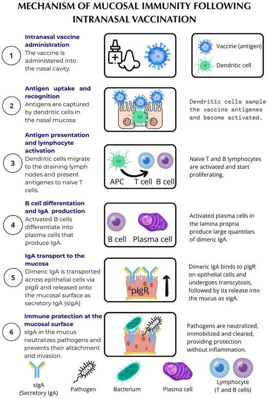
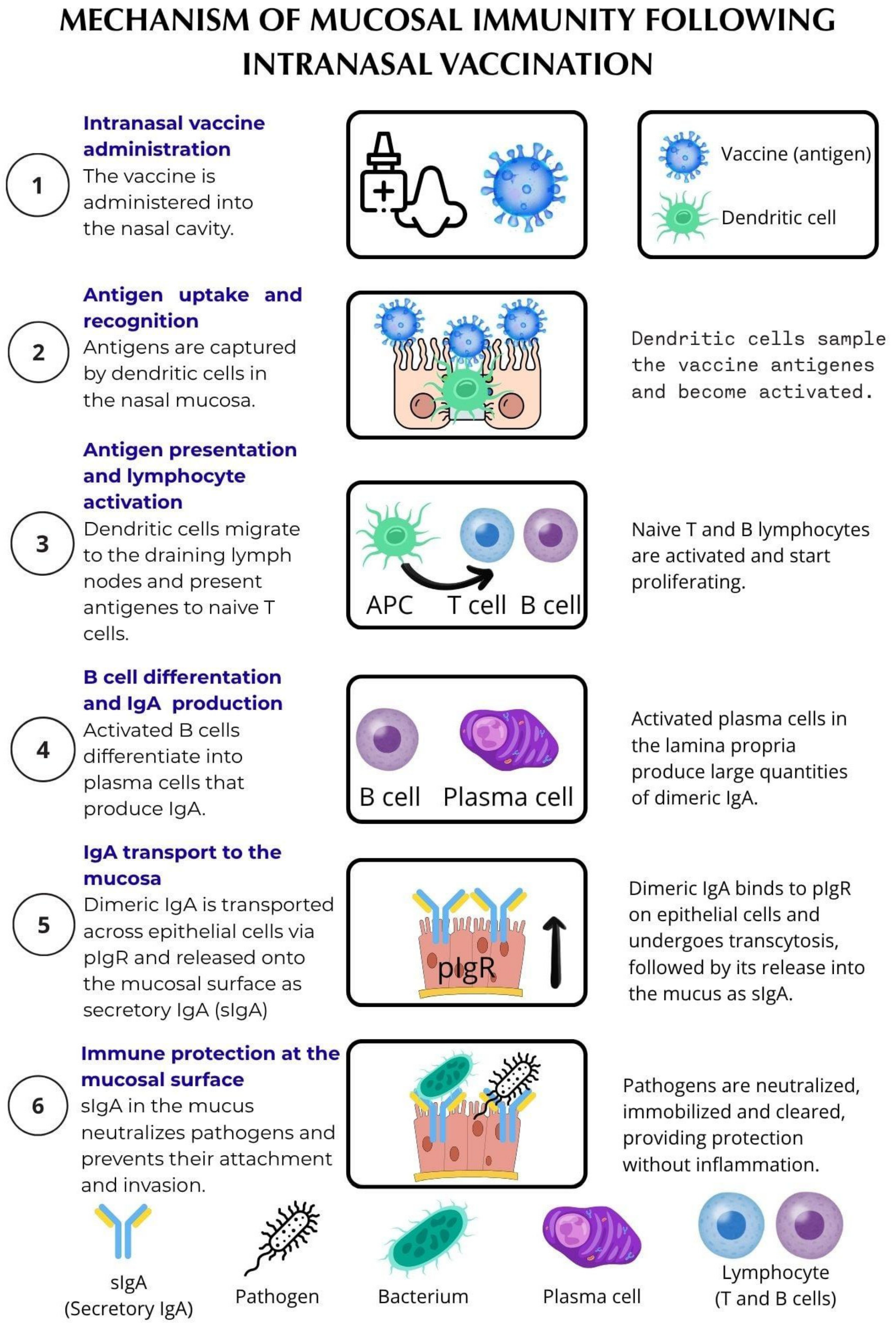
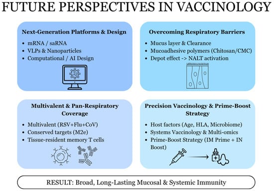
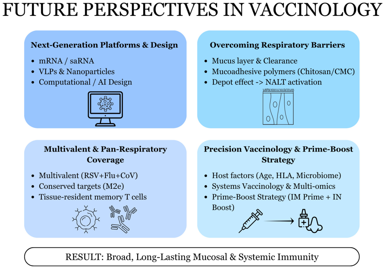

Disclaimer/Publisher’s Note: The statements, opinions and data contained in all publications are solely those of the individual author(s) and contributor(s) and not of MDPI and/or the editor(s). MDPI and/or the editor(s) disclaim responsibility for any injury to people or property resulting from any ideas, methods, instructions or products referred to in the content. |
© 2026 by the authors. Licensee MDPI, Basel, Switzerland. This article is an open access article distributed under the terms and conditions of the Creative Commons Attribution (CC BY) license.