


Abstract
Background/Objectives: Brucellosis remains a widespread zoonosis, yet no licensed human vaccine exists and the live attenuated vaccines used in animals are unsuitable for people because of residual virulence and the risk of accidental infection. Recombinant platforms are attractive alternatives, but direct head-to-head comparisons within a single study are scarce. We compared the safety, immunogenicity and protective efficacy of three recombinant candidates: the AddaS03-adjuvanted subunit vaccine BruVac-Pro, the capripoxvirus-vectored BruVac-CPX and the influenza virus-vectored BruVac-FLU. Methods: Inbred BALB/c mice (n = 355) received a prime-boost immunization, except for the single-dose Rev-1 reference group. A subgroup of 10 animals per group was monitored serologically from day 0 and subsequently challenged intraperitoneally with virulent Brucella melitensis 16M, and the remaining animals were assigned to safety, vector-detection and cellular-immunity read-outs. The timing and prospective nature of allocation to the challenge cohort could not be documented; selection bias in the efficacy endpoint therefore cannot be excluded. Safety was assessed from clinical observation, body weight and early serum clinical chemistry; antigen-specific IgG to Omp16, Omp19, L7/L12 and SodC was measured by indirect ELISA; and protection was quantified from splenic bacterial load (Y), the protective index (PI) and the proportion of animals with no splenic recovery (n = 10 per group). Immunogenicity was assessed as humoral (IgG) responses only; antigen-specific cellular responses were measured in separate subsets of the same experiment and are reported separately. Results: Mortality during the immunization phase was confined to the three sublingually dosed viral-vector and stabilizer groups (CPX/CPX 4/50, CCFLU/CCFLU 3/55, FLU/FLU 1/55), with no deaths in the Pro/Pro, AddaS03/AddaS03, Rev-1 or PBS/PBS groups (8/160 versus 0/195); surviving animals showed no overt clinical signs. Influenza-vector RNA was not detected in serum or spleens at days 3 and 6 by a qualitative assay of undetermined sensitivity. Seroconversion to Omp16 and Omp19 was confined to the Pro-containing group; no animal in any group seroconverted to SodC, and responses to L7/L12 were confined to isolated single animals without a group-level pattern. Splenic burden differed across groups (Kruskal–Wallis, p = 4 × 10⁻⁷). BruVac-Pro gave the strongest protection (Y 2.32; PI 2.62; 70.0% with no recovery; p = 0.028 for splenic burden and adjusted p = 0.015 for the proportion of animals with no recovery, both versus the phosphate-buffered saline (PBS) control). BruVac-CPX showed only a non-significant trend towards reduced burden (PI 1.54; p = 0.57 after correction for multiple comparisons) without increasing clearance (20.0%), and BruVac-FLU did not prevent colonization. Rev-1 confirmed model validity (PI 3.15; 80.0%). Conclusions: Under the non-equivalent, formulation-specific regimens tested, BruVac-Pro was the only experimental formulation with no mortality and no overt clinical signs, produced the most consistent antigen-specific IgG response, and was the only experimental candidate to reduce day-14 splenic burden significantly relative to the PBS and adjuvant-only controls. These findings support further dose, mechanism and durability studies of this formulation, but do not establish platform superiority, sterile protection or suitability for human use.
1. Introduction
Brucellosis remains an important zoonotic disease with substantial consequences for public health, livestock production and rural economies, particularly in endemic regions. Brucella spp. are facultative intracellular pathogens capable of surviving and replicating within host phagocytic cells, which promotes persistent infection and complicates both treatment and vaccine development. Among the zoonotic Brucella species, Brucella melitensis is considered particularly important because of its high infectivity for humans and its ability to cause severe systemic disease. Because most human infections originate from infected animals or contaminated animal products, sustainable control of brucellosis requires coordinated veterinary and public-health interventions within a One Health framework [1,2,3].
Vaccination of livestock remains one of the principal measures for reducing animal brucellosis and, consequently, human exposure. However, no licensed vaccine is currently available for the prevention of human brucellosis. The live attenuated vaccines used in veterinary practice, including B. melitensis Rev-1 and B. abortus S19 and RB51, provide effective protection in their respective target species but have several important limitations. These include residual virulence, the risk of abortion when administered under inappropriate conditions, interference with serological surveillance in the case of smooth vaccine strains, and the potential to cause infection or adverse reactions after accidental human exposure. These limitations preclude their use in humans and support the development of safer vaccines based on defined antigens and non-living delivery platforms [4,5,6].
Because Brucella is an intracellular pathogen, effective protection depends largely on cell-mediated immunity, particularly antigen-specific T-cell responses and IFN-gamma-mediated activation of infected macrophages. Humoral immunity may also contribute to protection, but antibody responses alone are generally insufficient to eliminate an established intracellular infection. Recombinant proteins, viral vectors, DNA, mRNA and nanoparticle-based delivery systems are therefore being investigated as alternatives to conventional live vaccines. These approaches avoid the administration of viable Brucella, allow selected antigens to be combined in a defined formulation, and provide opportunities to modulate the magnitude and type of immune response through the choice of vector, adjuvant, route of administration and prime-boost regimen [6,7]. Antigen-specific cellular responses to the candidates evaluated here are reported separately; the present study focuses on safety, humoral immunogenicity and protection.
Several Brucella proteins have shown potential as vaccine antigens, including the outer-membrane proteins Omp16 and Omp19, the ribosomal protein L7/L12, and Cu/Zn superoxide dismutase (SodC). Recombinant Omp16 and Omp19 have been shown to induce antigen-specific CD4+ and CD8+ T-cell responses and to protect mice against B. abortus challenge [8]. Recombinant Omp16 has also reduced bacterial colonization after challenge with either B. abortus or B. melitensis, providing direct evidence of activity against both species [9]. Similarly, combined immunization with Omp25 and L7/L12 conferred greater protection against B. abortus than either antigen administered individually, supporting the use of multicomponent formulations [10].
SodC has been evaluated using several delivery platforms. A plasmid DNA vaccine encoding B. abortus SodC induced antigen-specific antibodies, T-cell proliferation, IFN-gamma production and significant protection against virulent B. abortus 2308 [11]. Recombinant Semliki Forest virus particles expressing the same antigen also induced IFN-gamma production, cytotoxic T-cell activity and protection in BALB/c mice [12]. These studies provide a strong rationale for including SodC in recombinant vaccine constructs and demonstrate that its immunogenicity can be enhanced through nucleic-acid and viral-vector delivery. Nevertheless, because these experiments used B. abortus challenge models, their findings should be regarded primarily as platform-level evidence rather than direct demonstration of protection against B. melitensis.
Delivery systems based on nanoparticles and nucleic acids have further expanded the range of experimental brucellosis vaccines. Chitosan- and PLGA-based nanoparticles carrying Brucella antigens such as Omp19, Omp31 or oligopolysaccharide have induced systemic and mucosal responses and reduced bacterial colonization after challenge [13,14,15,16], and more recent nanoliposomal, niosomal and mRNA–lipid–nanoparticle formulations have similarly enhanced humoral and cellular responses and lowered splenic burdens in murine models [17,18,19]. Collectively, these findings underline that the delivery system, and not the antigen alone, is a key determinant of the immunogenicity and protective activity of recombinant Brucella antigens.
Viral-vector vaccines represent another promising approach. Influenza virus vectors expressing L7/L12, Omp16, Omp19 and SodC have induced antigen-specific cellular responses and protection against B. abortus in cattle and against B. melitensis in sheep, goats and guinea pigs [20,21,22,23,24]. Capripoxviruses are also being investigated as vaccine vectors because they can accommodate foreign antigen sequences and have a restricted host range [25]. In non-ruminant hosts they behave as replication-deficient vectors: recombinant capripoxvirus expresses the inserted antigen and elicits both antibody and cellular responses in mice and rabbits without producing infectious progeny, which combines antigen delivery with an intrinsic biological safety margin [26]. Recombinant sheeppox virus constructs carrying foreign genes at the thymidine kinase locus have been generated and characterized [27], and constructs expressing Brucella antigens, including Omp16, Omp19 and SodC, have since been produced and their scalable cultivation in Vero cells demonstrated [28]. However, the immunogenicity and protective efficacy of viral-vector vaccines may vary considerably according to the vector backbone, antigen composition, dose, administration route and host species.
Despite the large number of experimental brucellosis vaccines described to date, direct comparisons between different technological platforms remain limited. Most studies have evaluated individual candidates using different mouse strains, antigen doses, adjuvants, immunization schedules, challenge strains and definitions of protection. This methodological heterogeneity makes it difficult to determine whether differences in efficacy arise from the antigen itself, the delivery platform or the experimental design [29,30]. Comparative evaluation under controlled, head-to-head conditions is therefore needed to support the selection of candidates for further preclinical development. All three candidates evaluated here were developed at the Research Institute for Biological Safety Problems (RIBSP) and each has prior supporting evidence: influenza vectors expressing Brucella antigens have conferred protection in cattle, sheep, goats and guinea pigs [20,21,22,23,24]; recombinant capripoxvirus constructs carrying Brucella antigens have been generated and their scalable production established [27,28]; and the adjuvanted four-antigen subunit formulation builds on the protective activity reported for Omp16, Omp19, SodC and L7/L12 [8,9,10,11,12]. They were selected as the three most advanced in-house candidates: each had already been constructed, characterized and produced under the same institutional quality system, and each represents a distinct technological class—an adjuvanted purified-protein formulation, a DNA-virus vector and an RNA-virus vector so that a single controlled experiment could compare the three classes directly rather than across separate studies.
The present study compared the safety, immunogenicity and protective efficacy of three experimental recombinant vaccine formulations: the AddaS03-adjuvanted subunit vaccine BruVac-Pro, the recombinant capripoxvirus-vector vaccine BruVac-CPX, and the recombinant influenza virus-vector vaccine BruVac-FLU. The candidates were evaluated in inbred BALB/c mice using parallel prime-boost regimens within a single controlled study, followed by challenge with virulent B. melitensis 16M; the route of administration was that appropriate to each formulation (intramuscular for the subunit vaccine and sublingual for the viral-vector vaccines). Clinical observations, antigen-specific immune responses, splenic bacterial burdens and protection outcomes were assessed to determine the relative performance of the three formulations and to identify the candidate warranting further preclinical development as a potential vaccine against human brucellosis.
2. Materials and Methods
2.1. Ethics Statement and Regulatory Framework
This study was designed and performed as a controlled preclinical investigation under institutional study protocol LKB-B-02 (research program BR25293294), in accordance with the principles of Good Laboratory Practice (GLP) set out in the rules of the Eurasian Economic Union (Decision of the Council of the Eurasian Economic Commission No. 81 of 3 November 2016) and the Rules for Conducting Studies of Biological Medicinal Products of the Eurasian Economic Union (Decision No. 89 of 3 November 2016), together with the applicable regulatory acts of the Republic of Kazakhstan governing preclinical studies and good pharmaceutical practice. All experimental procedures involving animals were reviewed and approved by the Local Bioethics Commission of RIBSP, Republic of Kazakhstan (protocols No. 2 and No. 3, dated 30 March 2026).
Housing, immunization and clinical observation of uninfected animals were performed in dedicated laboratory-animal facilities. All work with virulent Brucella melitensis 16M preparation of the challenge inoculum, challenge, housing of infected animals, necropsy and processing of organs was performed in containment facilities corresponding to pathogen risk group II under the national classification of the Republic of Kazakhstan, in which groups are numbered inversely to the WHO and CDC/NIH scale, so that this corresponds to biosafety level 3 practices and facilities (BSL-3/ABSL-3) in that nomenclature. Access was controlled and class II type A2 biological safety cabinets were used (SOP-GMP-RIBSP-BSB-234-2020). Personal protective equipment comprised protective clothing, disposable gloves, respirators and eye or face protection. Surfaces, equipment, cages and instruments were decontaminated with approved disinfectants at working concentrations, and carcasses, organs, cultures, bedding and disposable materials were collected in labeled sealed containers and decontaminated before disposal (SOP-GMP-RIBSP-BSB-249-2020). A documented emergency procedure covered spills, bites, needlestick injuries and loss of PPE integrity. Ethical and biosafety oversight was provided by the Local Bioethics Commission of RIBSP (certificate No. LKB-SBO-BR25293294-01/2026; protocols No. 2 and No. 3 of 30 March 2026); the institute does not issue a separate institutional biosafety committee number.
This study was not subject to formal certification by an independent quality-assurance unit. All deviations from the study plan, incidents and unforeseen circumstances were documented with date, description, cause and corrective action; amendments to the protocol were issued in writing; and raw data together with interim and final reports signed by the principal investigator are archived at the institute with restricted access for not less than five years.
2.2. Vaccine Candidates and Control Preparations
Three experimental recombinant vaccine formulations, all developed at RIBSP, were evaluated.
BruVac-Pro (batch No. 0030326; certificate of analysis dated 28 March 2026) was a subunit vaccine presented as a homogeneous white emulsion. One dose contained the recombinant Brucella proteins Omp16 (10 µg), Omp19 (10 µg), SodC (10 µg) and L7/L12 (10 µg), i.e., 40 µg of total recombinant protein per dose. The proteins were expressed in Escherichia coli and purified at RIBSP; the purity of the active substance was not less than 95%, with residual E. coli genomic DNA below 100 pg per dose. Bacterial endotoxin was not determined for the protein bulk of this batch. The antigens were formulated with AddaS03™ (Cat. No. vac-as03-10, InvivoGen, San Diego, CA, USA), a preclinical-grade oil-in-water nanoemulsion of squalene, DL-α-tocopherol and polysorbate 80 described by the manufacturer as similar in formulation to the AS03 Adjuvant System, together with stabilizing excipients. AddaS03 is a research-grade product and is not manufactured by, affiliated with or endorsed by the manufacturer of AS03; it is referred to as AddaS03 throughout this manuscript, and no equivalence to the licensed AS03 Adjuvant System is implied. The antigen solution and AddaS03 were mixed at a 1:1 volume ratio according to the manufacturer’s instructions, so that each 0.1 mL dose contained 0.05 mL of the adjuvant emulsion together with 40 µg of total recombinant protein. No dose-ranging study was performed; the dose of 10 µg per antigen was selected on the basis of the published subunit-vaccine studies cited in Section 1. The formulation was stored at 2–8 °C and used within its 6-month shelf life (expiry 14 September 2026). It was given intramuscularly as a single injection of 0.1 mL per animal into the thigh musculature of a hind limb, using a sterile disposable insulin syringe with an integral fine-gauge needle, following the institutional procedure for intramuscular injection in mice (SOP-GMP-PR-813). The limb used, the exact needle gauge and whether the same limb was used at prime and boost were not recorded for individual animals. The corresponding adjuvant-only control group, designated AddaS03/AddaS03 throughout this manuscript, received the same volume of adjuvant without recombinant antigens.
BruVac-CPX was a recombinant capripoxvirus-vectored vaccine (clear to slightly opalescent, colorless liquid). The formulation contained the recombinant sheeppox virus (SPPV) constructs SPPV(TK-)-Omp19/SodC and SPPV(TK-)-Omp16, generated by insertion of the Brucella antigen genes into the thymidine kinase locus of the attenuated SPPV vaccine strain as described previously [27,28], together with mannitol (1.0%) and gelatin (0.1%) with an infectious titer of not less than 5.0 log10 TCID50/mL. Both constructs were present at a nominal titer of at least 5.0 log10 TCID50/mL, so the 30 µL sublingual dose delivered 3 × 10³ TCID50 of each. Residual host-cell DNA was 3 pg/mL, the batch met pharmacopoeial purity requirements, and insert identity was confirmed by PCR. Titers are release specifications; the preparations were not re-titrated before use.
BruVac-FLU was a recombinant influenza virus-vectored vaccine presented as a fine-pored lyophilized cake, grey to greyish-white in color. The formulation contained the recombinant influenza viruses Flu-NS1-80-Omp16, Flu-NS1-80-Cu-Zn-SOD (SodC), Flu-NS1-80-Omp19 and Flu-NS1-80-L7/L12, together with mannitol (3%), gelatin (0.3%) and chitosan (0.01%), with an infectious titer of not less than 4.50 log10 TCID50/mL. In each of the four constructs, the Brucella antigen was inserted into the truncated NS1 (NS1-80) gene of the influenza vector backbone. The four active substances, purified and concentrated preparations of the influenza vectors expressing Omp16, Omp19, L7/L12 and Cu-Zn-SOD, were each standardized to a biological activity of 6.0 log10 TCID50/mL and were combined with the stabilizing medium at a 1:1 ratio, giving the final concentrations of mannitol, gelatin and chitosan stated above. The titer of not less than 4.50 log10 TCID50/mL is the release specification; the batch used in this study was prepared to the activity given here. The vaccine was reconstituted before use and administered sublingually in a volume of 0.03 mL per animal, corresponding to a single dose of 3 × 10⁴ TCID50 per mouse.
The recombinant influenza vectors were generated by eight-plasmid reverse genetics on the pHW2000 backbone, with each Brucella antigen gene inserted in frame at nucleotide 80 of the NS1 gene, and were propagated in MDCK cells in serum-free medium. The virus was purified by a validated pilot-scale process comprising clarification, tangential-flow filtration, anion-exchange chromatography on Q-Sepharose Fast Flow and multimodal flow-through polishing on Capto Core 700, followed by sterile filtration through 0.22 µm membranes. Final preparations met the established release criteria for infectious titer, total protein, residual host-cell DNA (below 10 ng per dose) and bacterial endotoxin (not exceeding 0.15 IU/mL), and the identity and genetic stability of the inserts were confirmed by PCR and sequencing in every batch [31].
The recombinant sheeppox virus constructs were obtained by homologous recombination under transient dominant selection using integration plasmids carrying the Brucella antigen sequences; in SPPV(TK-)-Omp19/SodC, the Cu-Zn superoxide dismutase and Omp19 coding sequences were joined by overlap-extension PCR with an S2G4S2 linker. The viruses were propagated in Vero cells in serum-free medium, previously tested and confirmed free of mycoplasma [27,28].
The commercial live attenuated vaccine Brucella melitensis Rev-1, produced and characterized at RIBSP (marketing authorization No. RK-VP-1-5724-25 of 16 September 2025; series 1, control 1; manufactured 14 April 2026) [32,33], served as the model-qualification control. It was reconstituted in sterile phosphate-buffered saline (PBS) immediately before use and administered subcutaneously at 1 × 10⁵ CFU in 0.1 mL per animal, the dose recommended by the World Organisation for Animal Health (WOAH) for laboratory mice [34]; the actual viable count of the working dilution was verified by plating and colony counting. Sterile PBS served as the negative control. The cell-culture medium used to formulate the influenza vector (CCFLU) served as the corresponding formulation control for the BruVac-FLU platform. All preparations were prepared immediately before administration in accordance with the approved study protocol, and all manipulations were carried out under aseptic conditions and in compliance with biosafety requirements.
2.3. Experimental Animals and Study Design
Inbred BALB/c mice of both sexes, aged 6–8 weeks and weighing 18–20 g (n = 355), were used. Mice were specified pathogen-free and were supplied by Bereke LLP (Kazakhstan). They were held in quarantine on arrival and acclimatized for seven days before the study began. Animals were kept in polysulfone individually ventilated cages (model CAU-1) with a floor area of 800 cm2, at ten animals per cage, which exceeds the minimum floor area specified in Annex III of Directive 2010/63/EU for ten mice of this body weight, with males and females housed in separate cages throughout the study to prevent breeding, on a 12 h light–dark cycle, with feed given according to a defined regimen and water available at all times; the microclimate was checked daily and any departure from the set range was recorded. Purchase, quarantine and housing followed the institutional standard operating procedures RI-SMK-RIBSP-62-2019, SOP-GMP-RIBSP-BSB-209-2020 and SOP-SMK-RIBSP-BSB-778-2020.
Randomization was carried out before any procedure began. Animals were allocated to the experimental and control groups by simple randomization, with the groups subsequently checked for balance in sex, body weight and general clinical condition; the method used to generate the allocation sequence was not recorded in the study documentation. Each animal was entered on a randomization sheet recording identification number, group, sex, body weight and date of allocation. Each group comprised 45, 50 or 55 animals. Both sexes were represented in every group, with a female predominance: the groups of 50 animals (Pro/Pro, AddaS03/AddaS03, CPX/CPX, PBS/PBS) each comprised 28 females and 22 males, the FLU/FLU group of 55 comprised 33 females and 22 males, the CCFLU/CCFLU group of 55 comprised 30 females and 25 males, and the Rev-1 group of 45 comprised 28 females and 17 males (Table 1). In total, 203 females and 152 males were used. The procedure by which animals were assigned to terminal subgroups was not recorded; sex was not used as a stratification factor and was not recorded in the clinical-pathology worksheets, so the sex composition of individual terminal subgroups cannot be reconstructed retrospectively. Sex was not included as a factor in any analysis and the study was not powered to detect sex-dependent effects; this is stated as a limitation. Wherever technically possible, the laboratory and analytical stages were read blind to group allocation; immunization, allocation and daily clinical observation were not blinded. The individual mouse, not the cage, was the unit of analysis.
Table 1.
Design of the experimental groups: Vaccine formulation, prime/boost regimen, route of administration and number of animals per group.
Because most read-outs were terminal, each group was divided into designated subgroups, with a single animal intended to contribute to only one terminal endpoint, which is why group sizes of 45 to 55 animals were required to support the full panel of analyses. Ten animals per group formed the immunogenicity and protection cohort. These animals were identified at the start of the study and were bled by survival tail-vein sampling on days 0, 21, 28, 35, 42, 49 and 56, so that the ELISA data reflect within-animal kinetics, and the same animals were subsequently challenged and used for bacteriological examination and organ morphometry. Their assignment to the challenge was not documented as a prospective allocation, and the study records do not establish whether it was fixed before immunization; the possibility of selection bias in the efficacy endpoint therefore cannot be excluded, as discussed in Section 4. Ten further animals per group were euthanized early in the study and blood and spleens were collected at necropsy; in the BruVac-FLU group these comprised five animals on day 3 and five on day 6, and in the remaining groups ten animals on day 3. Material from five animals per group supplied the early clinical-chemistry panel (not performed in the Rev-1 group), and in the BruVac-FLU group the day-3 and day-6 animals supplied the samples for vector detection by reverse transcription polymerase chain reaction (RT-PCR). A further ten animals per group were euthanized between the day-42 and day-49 weighings for a later clinical-chemistry panel, the results of which were subsequently withdrawn as an analytical failure (Section 2.6). The remaining animals of each group were allocated under the study protocol to a separate laboratory, which carried out the antigen-specific T-cell analyses, necropsy and histopathology and reports them independently in a companion manuscript, currently in preparation. In the primary records, 138 animals carry an annotation assigning them to those analyses (97 females and 41 males; both the annotation and sex are held in the body-weight journal, which is why sex is available for these animals but not for the clinical-pathology, bacteriology or RT-PCR subgroups), between 19 and 20 per group; the annotation is not complete for every animal, and the animal-level record is provided as Supplementary Table. The overall design, immunization and sampling schedule are summarized in Table 1 and Figure 1.
Figure 1.
Study design and experimental timeline. BALB/c mice (n = 355) were allocated to seven groups. Prime-boost groups received primary immunization on day 0 (P) and a booster on day 21 (B), were challenged intraperitoneally with virulent Brucella melitensis 16M on day 42 (C) and euthanized on day 56 (14 days after challenge); the Rev-1 model-qualification group received a single subcutaneous immunization on study day 13 and was challenged on study day 42 in the same session as all other groups. Serum for ELISA was collected on days 0, 21, 28, 35, 42, 49 and 56 in the prime-boost groups and on days 13, 34, 42 and 56 in the Rev-1 group. Serum clinical chemistry is reported for day 3 (five animals per group) and was not performed in the Rev-1 group; a later panel (ten animals per group) was withdrawn as an analytical failure. In the BruVac-FLU group, five animals per time point were euthanized on days 3 and 6 for detection of vector RNA by RT-PCR. Body weight was recorded in all animals at seven-day intervals to day 63. Routes of administration: i.m., intramuscular; s.l., sublingual; s.c., subcutaneous. The terminal challenge subgroup comprised ten animals per group.
Sex was not used as a stratification factor when animals were allocated to terminal subgroups, and the sex of individual animals is not recorded in the clinical-pathology, bacteriology or RT-PCR worksheets. The sex composition of the individual terminal subgroups therefore cannot be reconstructed from the study records, and no such composition is claimed here. Sex was recorded for every animal in the body-weight records, which establishes the composition of each group as a whole (Table 1), and body weight is the only outcome for which the contribution of both sexes can be verified at the level of the individual animal. Because sex could not be attributed to the terminal read-outs, it was not included as a factor in any analysis and no sex-stratified comparison was attempted; the study was not powered for such a comparison. This limitation is acknowledged in Section 4. Animal-level allocation, including identification number, sex, group, the terminal endpoint as annotated in the primary record and the last recorded weighing day, is given in Supplementary Table group totals, sex totals and endpoint denominators in this manuscript follow that table. Cage assignment was not recorded for individual animals.
Beyond satisfactory clinical condition at allocation, no inclusion or exclusion criteria were set in advance. Eight animals were found dead during the immunization phase: four in the CPX/CPX group (two between days 14 and 21 and two between days 28 and 35), three in the CCFLU/CCFLU group (two between days 21 and 28 and one between days 28 and 35) and one in the FLU/FLU group (between days 21 and 28). No deaths occurred in the Pro/Pro, AddaS03/AddaS03, Rev-1 or PBS/PBS groups. These animals contributed body-weight measurements up to their last recorded weighing and are included in the denominators given beneath Table 2 up to that point; seven of the eight carry no terminal-endpoint annotation, and one animal of the CCFLU/CCFLU group (animal 28) carries a histopathology annotation in addition to the record of death; it is counted as an unscheduled death and not in the histopathology denominator (Supplementary Table). None of the eight is linked by annotation to the challenge, clinical-chemistry or RT-PCR subgroups reported here, although, as the challenge cohort cannot be identified in the allocation record, this cannot be verified from that record. No animal was removed from the study for any other reason. In one animal of the AddaS03/AddaS03 group, the primary spleen culture was overgrown by extraneous microflora; since every countable dilution in that group exceeded the upper limit of the countable range, this animal was retained and censored at that limit together with the other non-protected animals of the group. Allocated and analyzed numbers are otherwise identical.
Table 2.
Body-weight dynamics of experimental animals over the study period (g, M ± SD). Values up to day 42 are pre-challenge. From day 49 onward the number of animals per group decreases as the challenged subgroups are euthanized (day 56 in all groups) and the day-63 values refer to non-challenged animals only. Values are mean ± SD, with the number of animals contributing a weight on that day given in parentheses; n falls from day 7 onward as animals left the study at assigned terminal endpoints and through eight unscheduled deaths, and the animal-level record is provided as Supplementary Material. All means were recomputed from the primary body-weight records.
2.4. Immunization Schedule and Routes of Administration
Primary immunization was performed on day 0. A booster immunization was administered on day 21 to the groups assigned a two-dose (prime-boost) schedule in the study protocol. BruVac-Pro was administered intramuscularly, the viral-vectored vaccines BruVac-CPX and BruVac-FLU were administered sublingually, and the reference vaccine Brucella melitensis Rev-1 was administered subcutaneously. Animals in the negative-control group received 30 µL of PBS sublingually. Prime-boost groups were challenged with virulent Brucella melitensis 16M on day 42 (21 days after the booster) and euthanized on day 56 (14 days after challenge). Because the Rev-1 reference vaccine is administered as a single dose and requires no booster, this group was enrolled later so that its challenge would coincide with that of the prime-boost groups: it received a single subcutaneous immunization on study day 13 and was challenged on study day 42 together with all other groups, in the same challenge session and with the same inoculum stock, which corresponded to 29 days after its single immunization. It was euthanized on study day 56, i.e., 14 days after challenge, as in all other groups. Serum for ELISA was collected in this group on study days 13, 34, 42 and 56 (0, 21, 29 and 43 days after immunization). At euthanasia, biological material was collected for bacteriological examination of organs.
2.5. Clinical Monitoring and Body Weight
Mice were examined daily throughout the study, and more often in the days following each administration and after challenge. Findings were recorded on a structured score sheet covering behavior, activity, general appearance, food and water intake, body weight, respiratory status and any local reaction at the site of administration. The score sheet recorded the parameters listed above, and any animal showing a marked deterioration in general condition, sustained weight loss, inability to reach food or water, or a severe local reaction was to be withdrawn from the study and euthanized by cervical dislocation on the decision of the attending veterinarian, in line with EU Directive 2010/63/EU. Numerical thresholds for these criteria were not specified in advance in the study protocol. No animal was euthanized before its scheduled endpoint on welfare grounds. Eight animals were nevertheless found dead between day 14 and day 35, as detailed in Section 2.3. Each death was documented at the time in a separate institutional act recording the animal, the group and the date on which it was found dead. The animals were found dead rather than euthanized on welfare grounds, and no cause of death was established, so the events are reported as observed mortality; their distribution across groups is given in Section 3.1 and discussed in Section 4. Monitoring included assessment of general condition and behavior, response to external stimuli, condition of the hair coat, skin and visible mucous membranes, coordination of movement, and the character of respiration and urination. Body weight was measured at regular intervals (days 0, 7, 14, 21, 28, 35, 42, 49, 56 and 63) in all experimental and control groups as an integral indicator of safety and tolerability. Animals assigned to the challenge subgroup were euthanized on day 56 in all groups, including Rev-1. Group means from day 42 onward therefore include both challenged and non-challenged animals, and the day-63 values refer to the non-challenged animals only; the number of animals contributing to each time point decreases accordingly. In addition, eight unscheduled deaths occurred between day 14 and day 35 (Section 2.3). For these reasons, body weight up to day 14, before the first death, is used as the primary tolerability read-out, and all later time points are interpreted descriptively.
2.6. Clinical Chemistry
Animals assigned to the clinical-chemistry subgroups were euthanized on day 3 and, for the later panel, between the day-42 and day-49 weighings, and blood and spleens were collected at necropsy (n = 5 per group at day 3 and n = 10 per group at the later time point; the Rev-1 group was not assessed). The blood-collection technique was not recorded in the study documentation. The day-3 sample served as an early safety read-out after priming, and the later sample was obtained from a separate, non-challenged safety subgroup. Hematological parameters were recorded in the study documentation but are not reported here, because the tabulated values could not be reconciled with the primary analyzer records; this is acknowledged as a limitation in Section 4. Biochemical parameters, including alanine aminotransferase (ALT), aspartate aminotransferase (AST), glucose, total protein and bilirubin, were measured on an automated biochemistry analyzer (BioSystems A25, BioSystems S.A., Barcelona, Spain). Serum from the early time point was analyzed on 4–5 May 2026 and serum from the later time point on 22–23 May 2026; these are the dates on which stored sera were analyzed, not the dates of collection. The two runs were assessed separately against internal consistency criteria applied after the analyses had been completed, that is, as post hoc quality-control criteria rather than prespecified ones. The two runs comprised 70 and 90 samples respectively, which include animals from the other groups of the parent study analyzed in the same runs (Supplementary Table); only the six groups reported here are presented. In the early run, direct bilirubin was below total bilirubin in all 70 samples and total protein ranged from 56.7 to 68.2 g/L, consistent with correct performance of the panel; the direct-bilirubin channel read below the lower limit of the reference range stated in the analyzer manual (1.0 to 5.0 mg/dL) in every group, including the PBS control. This is a uniform, channel-level offset rather than a group effect, and that reference range has not been validated for mouse serum; the channel was therefore not interpreted against a reference and is not tabulated, but the values are held in the signed clinical-chemistry protocol provided with this submission. No internal constraint was violated in the early run. In the later run, direct bilirubin exceeded total bilirubin in 55 of 90 samples, which is arithmetically impossible because direct bilirubin is a fraction of the total, and total protein fell to group means of 25.1 to 55.8 g/L on the same instrument. These are failures at the level of the analytical run rather than of individual observations. The entire later biochemical panel was therefore withdrawn, including the transaminase and glucose channels, which cannot be regarded as verified while the run that produced them is not. Only the early panel is reported, in Table 3 Analytical balances (OHAUS Pioneer PA213C, OHAUS Corporation, USA) were used for weighing. Because the seven groups were started on different calendar dates (2, 3, 6 and 15 April 2026), the later blood sampling was performed for all groups in a single session on a common calendar date, which fell on different days after primary immunization in different groups. That panel has been withdrawn, as described above, and is not reported; the day-3 sampling was performed on day 3 after primary immunization in every group.
Table 3.
Serum biochemical parameters on day 3 after primary vaccination (M ± SD; n = 5 per group).
2.7. Detection of Influenza-Vector Persistence by One-Step RT-PCR
Dissemination of the influenza-vectored vaccine BruVac-FLU beyond the site of administration was assessed by detection of vector RNA in serum and spleens. Because BruVac-FLU is based on a replication-restricted RNA virus, detection of vector RNA in serum and in a distant lymphoid organ is the relevant safety read-out for this platform; the DNA-based capripoxvirus vector of BruVac-CPX cannot be detected by this RNA-based assay and its biodistribution was not assessed in the present study. Animals received BruVac-FLU sublingually at a dose of 30 µL; from the relevant group, a subsample of 5 animals per time point was analyzed, and samples were collected on days 3 and 6 after immunization.
Serum and spleens were used as the study material. After euthanasia, spleens were aseptically removed; tissue fragments (10 to 20 mg) were placed in ice-cold phosphate-buffered saline (PBS, Gibco, Thermo Fisher Scientific, Waltham, MA, USA) at a 1:10 ratio and homogenized to a uniform suspension. The homogenate was centrifuged at 3000 rpm for 10 min to remove cell debris, and the supernatant was used for RNA extraction. Blood samples were centrifuged at 3000 rpm for 10 min to obtain serum. All samples were stored at −20 °C until RNA extraction.
Viral RNA was extracted from serum and spleen supernatant using the innuPREP RNA Mini Kit (Analytik Jena, Jena, Germany) according to the manufacturer’s instructions; all steps were performed under RNase-free conditions to minimize RNA degradation. RNA yield ranged from 30 to 40 ng/µL for serum and 40 to 50 ng/µL for spleens, with A260/A280 ratios of 1.80 to 1.90, indicating suitable purity for downstream analysis.
Amplification was performed with the QIAGEN OneStep RT-PCR Kit (QIAGEN GmbH, Hilden, Germany) using the specific primers NS1 and NS834. These primers anneal to the NS gene segment of the influenza vector backbone and therefore detect all four recombinant constructs with a single primer pair, independently of the inserted Brucella antigen. The 25 µL reaction mixture contained 5 µL of 5× RT-PCR buffer, 1 µL of dNTP mix, 0.5 µL of primer NS1, 0.5 µL of primer NS834, 12 µL of RNase-free water, 1 µL of enzyme mix and 3 µL of RNA template. Thermal cycling consisted of a reverse-transcription step at 50 °C for 30 min, initial denaturation at 95 °C for 15 min, followed by 40 cycles of denaturation at 94 °C for 20 s, primer annealing at 52 °C for 30 s and elongation at 72 °C for 2 min, with a final elongation at 60 °C for 1 min.
Amplification products were analyzed by horizontal electrophoresis in a 1.5% agarose gel in TAE buffer at constant voltage for 40 min, and DNA fragments were visualized under a UV transilluminator. A GeneRuler 100 bp DNA Ladder (Fermentas, Vilnius, Lithuania) was used to estimate amplicon size. The presence of a specific band of the expected size (890 bp) was scored as a positive RT-PCR result. Each run included a positive control consisting of RNA from the recombinant influenza vector and a no-template negative control, and RNA integrity was verified before amplification (yield 30 to 50 ng/µL; A260/A280 1.80 to 1.90). The assay was qualitative, and its analytical limit of detection was not formally determined. A positive control spiked into spleens or serum matrix and an endogenous internal amplification control were not included, so matrix-related inhibition cannot be formally excluded. Samples from the site of administration, the respiratory tract, the oral cavity and the draining lymph nodes were not collected, and shedding was not assessed.
2.8. Assessment of the Humoral Immune Response (Indirect ELISA)
The antigen-specific humoral immune response was assessed by indirect enzyme-linked immunosorbent assay (ELISA). Serum samples were collected on days 0, 21, 28, 35, 42, 49 and 56 in the prime-boost groups. Because the Rev-1 reference group received a single immunization on study day 13, it followed a separate sampling schedule, with serum collected on study days 13, 34, 42 and 56. Specific IgG antibodies to the recombinant Brucella antigens SodC, Omp16, Omp19 and L7/L12 were determined.
Before testing, sera were centrifuged at 1500 rpm for 10 min and heat-inactivated at 56 °C for 30 min.
ELISA was performed in 96-well Nunc-Immuno PolySorp plates (Cat. No. 183519, Thermo Fisher Scientific, Waltham, MA, USA) sensitized with the recombinant antigens SodC, Omp16, Omp19 or L7/L12; bovine serum albumin (Capricorn Scientific, Ebsdorfergrund, Germany) was used as the blocking agent, and the washing and diluting solutions contained Tween 80 (Cat. No. L13315AP, Thermo Scientific). Each serum was tested in a single well per antigen, without technical replicates. For screening, 0.09 mL of 1× washing solution was added to each well, followed by 0.01 mL of negative control serum, positive control serum or test serum, corresponding to a 1:10 serum dilution. Plates were sealed and incubated for 60 min at 37 ± 1 °C, then washed five times with 0.3 mL of 1× washing solution per well.
Bound IgG was detected with recombinant Staphylococcus aureus protein A conjugated to horseradish peroxidase (Pierce Protein A, Peroxidase Conjugated, Thermo Scientific), diluted 1:40 in 1× solution. A volume of 0.1 mL conjugate was added to each well, followed by incubation for 60 min at 37 ± 1 °C. After five further washes, 0.1 mL of ABTS substrate solution (HiMedia, Mumbai, India) was added and plates were incubated for 25 ± 5 min at room temperature in the dark. The reaction was stopped by adding 0.1 mL stop solution per well. Optical density was measured at 405 nm using a Multiskan Plus microplate reader (Labsystems, Helsinki, Finland).
Positive (K+), negative (K−) and conjugate controls were included on each plate. For each sample, the sample-to-positive ratio was calculated as:
S/P(%) = (OD sample − OD negative)/(OD positive − OD negative) × 100
A sample was classified as positive when S/P was 40% or above and as negative when the S/P value was below 40% in accordance with the positivity threshold defined in the assay standard operating procedures (SOP-GMP-NR-858 to SOP-GMP-NR-861). Results were expressed as the number and percentage of seropositive animals per group and as individual S/P values. The coating concentration of the recombinant antigens is the one defined in those same standard operating procedures, which are provided with this submission, and was not re-optimized for this study. The 40% S/P positivity threshold is the one defined in those procedures; it was not independently validated against a panel of characterized positive and negative murine sera, and no coefficient of variation was determined, because each serum was tested once at a single 1:10 dilution without technical replicates. The threshold should therefore be regarded as operational rather than as a validated diagnostic cut-off for mouse sera.
2.9. Challenge Infection and Assessment of Protective Efficacy
Protective efficacy was evaluated in a model of experimental brucellosis induced by the virulent Brucella melitensis 16M strain, using a murine challenge model adapted from Chapter 3.1.4 (Brucellosis: Brucella abortus, B. melitensis and B. suis) of the WOAH Terrestrial Manual [34]. The model is not WOAH-compliant and was not intended as a potency test. It departs from the cited procedure in mouse strain (BALB/c rather than CD1), group size (ten rather than six animals), challenge strain (B. melitensis 16M rather than B. abortus 544), interval to euthanasia (14 rather than 15 days) and plating (a single plate per dilution rather than quadruplicate plating). Selected WOAH criteria, namely the validity thresholds for the unvaccinated control and the reference vaccine group and the procedure for differentiating the Rev.1 vaccine strain from the challenge strain, were applied as described below. Animals were challenged intraperitoneally with a nominal dose of 2 × 10⁵ CFU of Brucella melitensis 16M in 0.1 mL. The challenge inoculum was prepared from a single stock for the entire challenge session, and the working dilution was back-titrated by plating and colony counting on the day of challenge; the delivered dose determined by back-titration was 2.1 × 10⁵ CFU per animal. The challenge strain was cultured on Brucella Agar Base supplemented with glycerol (20 mL/L), glucose (10 g/L), L-cysteine and yeast extract, and incubated at 37 ± 1 °C before preparation of the challenge suspension. Fourteen days after challenge, animals were euthanized and spleens were aseptically removed and individually weighed.
Each spleen was weighed and sterile PBS was added to a fixed final volume of 5 mL, so that the whole organ was recovered in a constant volume irrespective of its mass. After homogenization, two further ten-fold dilutions of this primary suspension were prepared, and 0.2 mL of the primary suspension and of each dilution was plated, once per dilution and without replicate plates, onto solid medium based on Brucella Agar Base (Oxoid Ltd., Basingstoke, UK) supplemented with Brucella Selective Supplement (Oxoid Ltd., SR0083A, Basingstoke, UK) to suppress extraneous microflora. Plates were incubated at 37 °C and colonies were counted after 5 to 7 days of incubation. Isolates were identified by colony morphology and by staining of smears. Primary plating for all groups was performed on non-selective medium, and the counts recorded at this stage therefore represent total Brucella. For the Rev-1 group, colonies recovered from the spleen were subcultured and assigned to either the vaccine or challenge strain on the basis of their antibiotic-sensitivity profile, using Brucella Agar Base supplemented with benzylpenicillin (3 µg/mL) and with streptomycin (2.5 to 5.0 µg/mL): the Rev.1 vaccine strain does not grow in the presence of benzylpenicillin but grows in the presence of streptomycin, whereas virulent B. melitensis shows the opposite pattern. Differentiation was performed in accordance with Chapter 3.1.4 of the WOAH Terrestrial Manual [34]. No live vaccine strain was administered to any other group, so differentiation was not required elsewhere. Only isolates identified as the challenge strain 16M were counted towards the splenic bacterial load in this group. Animals from which the Rev-1 vaccine strain alone was recovered were therefore classified as having no detectable colonization by B. melitensis 16M, even though viable bacteria were present on the primary non-selective plate. Both the primary plating record and the strain-differentiation record for this group are provided to the editor and reviewers.
For enumeration, the least dilute plate with a countable colony number (fewer than 300 colonies) was used for each animal, since the plate bearing the largest countable number gives the most precise estimate. Where no plate in the series was countable, the load was censored at the upper limit of the countable range. Where growth was confluent at every dilution, the count was treated as censored at the upper limit of the assay. The bacterial load in the spleen (X) was calculated as:
where N is the number of colonies counted, D is the dilution factor relative to the primary suspension (D = 1, 10 and 100, respectively), and 25 is the conversion factor accounting for the 5 mL total volume of the homogenate and the 0.2 mL plated per dilution. Because the whole spleen was homogenized in a constant final volume, X expresses the total bacterial load per organ and is independent of spleen mass. Bacterial load was expressed in logarithmic form as:
X = N × D × 25
Y = log10(x)
In the absence of Brucella growth, the load was set to the limit of detection of the assay, i.e., 25 bacteria per spleen (Y = 1.40), corresponding to a single colony on the plate seeded with the primary suspension. Because only 0.2 mL of a 5 mL homogenate was plated per dilution, absence of growth indicates a load below this limit rather than complete sterility of the organ; this outcome is therefore reported as “no detectable splenic colonization”.
The challenge experiment was considered valid when the mean Y in the unvaccinated (PBS) control group was 4.5 or higher and the standard deviation of Y in that control group did not exceed 0.8, and when the mean Y in the Brucella melitensis Rev-1 reference group was 2.5 or lower; these validity criteria are adapted from Chapter 3.1.4 of the WOAH Terrestrial Manual [34], in which the consistency criterion (standard deviation of Y not exceeding 0.8) is applied to each group of six mice. In the present study the criterion was applied only to the unvaccinated challenge-control group, which is a deliberate departure from the cited procedure; in vaccinated groups a bimodal distribution of protected and non-protected animals was expected, so splenic burden was summarized as median with range in addition to mean, and analyzed by non-parametric tests (see Section 2.11). The protective index (PI) was calculated for each group against the unvaccinated PBS control as:
PI = Y(PBS) − Y(group)
For the subunit vaccine, the reduction relative to its adjuvant-only control (AddaS03/AddaS03) is also reported. Lower Y values and higher PI values indicated greater protective efficacy. Protection was additionally expressed as the proportion of animals from which Brucella could not be recovered from the spleen.
2.10. Necropsy and Macroscopic Evaluation
At necropsy, the anatomical position of internal organs, the condition of the serous membranes, and the shape, size, color, consistency and any visible pathological changes of the liver, spleen, lungs, kidneys and inguinal and cervical lymph nodes were assessed macroscopically. The liver and spleen were weighed, and the organ index was calculated as:
For each animal, absolute organ mass and organ indices were recorded and compared between groups; the injection site and organs were additionally inspected for macroscopic signs of local reactions or pathology.
2.11. Statistical Analysis
No a priori power calculation was performed. Group sizes were fixed in the study protocol (LKB-B-02) as the minimum number of animals considered sufficient for a reliable comparative assessment of protection while respecting the reduction principle of the 3Rs. The absence of a formal sample-size calculation is acknowledged as a limitation in Section 4.
Statistical analysis was performed using the actual number of animals included in the analysis at each stage of the study. Quantitative data are presented as mean ± standard deviation (M ± SD). The normality of the data distribution was assessed using the Shapiro–Wilk test before the selection of comparison methods.
Biochemical parameters are reported as group means ± SD, together with the relative difference from the PBS/PBS control shown in Figure 2. Each parameter was analyzed separately by one-way ANOVA with group as the factor, followed by Tukey post hoc comparisons against the PBS/PBS control. Only the day-3 panel is analyzed, the later panel having been withdrawn (Section 2.6). No correction was applied across the panel, so the p-values reported in Section 3.4 are nominal. With five parameters in six groups and five animals per group, these analyses have limited power to exclude small effects, and they are therefore interpreted as exploratory safety read-outs rather than as confirmatory tests. Organ masses and organ indices are reported as descriptive summary data and were not subjected to inferential testing. The splenic bacterial load (Y = log10(X)) was not normally distributed and, given the moderate group sizes and the bimodal distribution of protected and non-protected animals, is reported as mean ± SD together with the median and range, and was analyzed using the non-parametric Kruskal–Wallis test across all groups, followed by Dunn post hoc comparisons with Holm correction applied across all 21 pairwise comparisons between the seven groups. All pairwise p-values reported for splenic bacterial load are Holm-adjusted. In samples where colony counts exceeded 300 at every dilution, the exact load could not be determined and the value was censored at the upper limit of the countable range (300 colonies at the highest dilution plated). The data are censored at both ends: culture-negative animals were assigned the detection limit (Y = 1.40) and animals with confluent growth at every dilution the upper limit of the countable range (Y = 5.88). Splenic burden is therefore reported as median with range alongside the mean, and group means in the unprotected groups are conservative estimates. Because the analysis is rank-based, the result depends only on the rank of the censored observations and not on the values substituted for them; this was confirmed by repeating the analysis with half the detection limit assigned to culture-negative animals, which gave identical test statistics.
Figure 2.
Relative differences (%) of each group from the PBS/PBS control for serum biochemical parameters at day 3 after primary vaccination (n = 5 per group). Values are calculated from the group means in Table 3; positive values (red) indicate an increase and negative values (blue) a decrease relative to the control. Direct bilirubin is not displayed (Section 2.6).
Body-weight dynamics, based on repeated measurements of the same animals over time, were analyzed by two-way repeated-measures ANOVA (mixed-effects model) with group and time as factors, followed by Tukey post hoc comparisons. The mixed-effects analysis used all animals with at least one post-baseline weighing; animals leaving the study at an assigned terminal endpoint, or through unscheduled death, contributed measurements up to that point, and the model accommodated the resulting unbalanced design without imputation. Because eight unscheduled deaths occurred between day 14 and day 35 and were confined to three groups, missingness after day 14 is not independent of treatment; the model was therefore fitted to the weighings on days 0, 7 and 14 only, with group, time (as a categorical factor) and their interaction as fixed effects and a random intercept for each animal, and all later time points are summarized descriptively. The group × time interaction was tested by a likelihood-ratio test, and the individual gain in body weight from day 0 to day 14 was compared between groups by one-way ANOVA with Tukey post hoc comparisons. This model was fitted with the statsmodels package (version 0.15, Python 3) and the Tukey comparisons with the same package; all other analyses were performed in GraphPad Prism. The number of animals contributing at each time point is given beneath Table 2. Categorical outcomes were compared using the Fisher exact test, within two comparison families. The first comprised the six group-versus-PBS comparisons of the proportion of animals from which Brucella melitensis was not recovered; the second comprised the group-versus-PBS comparisons of seropositivity at day 42, the pre-challenge time point. Within each family, p-values were adjusted by the Holm method. For every comparison, this manuscript reports the raw p-value, the Holm-adjusted p-value, and the family and number of comparisons within which the adjustment was made; the first family comprised six comparisons and the second twelve. Seropositivity at the remaining time points is presented descriptively, because independent tests at successive bleeds do not account for the repeated sampling of the same animals. Differences were considered statistically significant at p < 0.05. Except for the day 0 to 14 body-weight model described above, all analyses were performed using GraphPad Prism version 10 (GraphPad Software, San Diego, CA, USA).
2.12. Use of Generative Artificial Intelligence
Generative artificial intelligence was not used to generate data, figures or scientific content, or to assist in study design, data collection, analysis or interpretation. Any use was limited to superficial language editing of the manuscript text.
3. Results
3.1. Clinical Observations and Body-Weight Dynamics
Among the animals surviving to their assigned endpoints, their general condition remained satisfactory throughout the study. Response to external stimuli was preserved, the hair coat, skin and visible mucous membranes showed no pathological changes, and no disturbances of movement coordination, respiration or urination were recorded. No local reactions were observed at the injection or administration sites. Mortality, however, was not evenly distributed. Eight animals were found dead between day 14 and day 35: four of 50 in the CPX/CPX group (8.0%), three of 55 in the CCFLU/CCFLU group (5.5%) and one of 55 in the FLU/FLU group (1.8%). No deaths occurred in the Pro/Pro, AddaS03/AddaS03, Rev-1 or PBS/PBS groups. Taken together, all eight deaths occurred among the 160 animals receiving a sublingual viral-vector preparation or its stabilizing medium, and none among the remaining 195 animals (Fisher exact p = 0.0015). Because cage identity was not recorded, animals housed together cannot be treated as independent units and this comparison is presented as an exploratory animal-level analysis rather than as evidence of a formulation-specific effect. Each death was documented in a separate institutional act, but no cause of death was established. Two features of this distribution are relevant to its interpretation: the PBS/PBS control was also administered sublingually and recorded no deaths, which argues against the route itself; and the CCFLU/CCFLU group, which received the stabilizing medium without any recombinant antigen, accounted for three of the eight deaths, which argues against an antigen-specific effect. The mortality is therefore more consistent with the viral-vector formulations and their stabilizing medium than with the route of administration or the recombinant antigens. No overt clinical toxicity was observed for BruVac-Pro under the limited conditions assessed, which comprised daily clinical observation, body weight, and serum clinical chemistry at day 3 in five animals per group. For BruVac-CPX and BruVac-FLU, no such conclusion can be drawn, since mortality occurred in those groups and in the formulation control and no cause of death was established. Six of the eight animals found dead were male and two were female, although males were the smaller sex in all three groups; with eight events and no post-mortem examination this cannot be distinguished from chance and is reported for completeness.
Body weight, an integral indicator of safety and tolerability, increased steadily in all groups over the observation period, following the physiological growth pattern expected for laboratory mice (Table 2, Figure 3a). Starting weights were comparable across groups (about 19.6 to 20.0 g), confirming the homogeneity of the experimental groups at baseline. Up to the day-42 challenge, weight gain was steady in every group, although the number of animals contributing to each weighing had already declined: ten animals per group were euthanized before the day-7 weighing, and the later clinical-chemistry subgroups between the day-42 and day-49 weighings. The number of animals contributing to each group at each weighing day is given beneath Table 2. Most of the reduction follows the assigned terminal endpoints, but eight animals in the CPX/CPX, FLU/FLU and CCFLU/CCFLU groups died between day 14 and day 35 and contributed no further weights, so the missingness is not independent of treatment. Inferential comparison of body-weight trajectories is accordingly restricted to the interval up to day 14, before the first death; weights from day 14 onward are presented as survivor-only descriptive data. Over the interval up to day 14, before the first death, mean body weight rose in every group (Pro/Pro from 19.88 ± 0.75 to 21.05 ± 1.09 g; AddaS03/AddaS03 19.61 ± 1.04 to 20.42 ± 1.18 g; CPX/CPX 19.90 ± 1.18 to 20.15 ± 1.11 g; FLU/FLU 19.95 ± 0.94 to 20.72 ± 0.70 g; CCFLU/CCFLU 19.91 ± 1.01 to 20.62 ± 1.02 g; Rev-1 19.69 ± 1.22 to 20.18 ± 1.10 g; PBS/PBS 19.64 ± 0.81 to 20.00 ± 0.92 g), and in the mixed-effects model fitted to days 0 to 14 (925 weighings from 355 animals) body weight increased with time in every group (time effect, p < 0.001) and no group showed a negative trend. The rate of gain differed between groups (group × time interaction, p < 0.001) because the Pro/Pro and FLU/FLU groups gained more than the PBS/PBS control over the first 14 days (mean gain 1.12 ± 1.19 g and 0.95 ± 0.93 g versus 0.25 ± 0.24 g; Tukey-adjusted p < 0.001 for both), not because any group lost weight: the smallest gain, in the CPX/CPX group (0.09 ± 0.40 g), did not differ from the control (Tukey-adjusted p = 0.92), and the AddaS03/AddaS03, CCFLU/CCFLU and Rev-1 groups did not differ from the control either (adjusted p = 0.08, 0.45 and 0.78). Over this interval, none of the preparations was associated with weight loss or growth retardation. The descriptive trajectories over the full pre-challenge period show the same ordering, with the largest gain in the Pro/Pro group (22.19 ± 1.47 g on day 42) and the smallest in the CPX/CPX group (20.27 ± 1.25 g), a difference of less than 2 g that lies within physiological variability for mice of this age. Later trajectories are descriptive only and are not interpreted as evidence of tolerability, because animals that died could contribute no further weights.
Figure 3.
Body weight and organ morphometry. (a) Body-weight dynamics over the study period, recomputed from the primary body-weight journal; dotted lines mark the boost (day 21) and challenge (day 42) time points. Values are group means ± SD, and the number of animals contributing at each time point is given beneath Table 2. Values up to day 42 are pre-challenge, and the day-63 values refer to non-challenged animals only. (b) Liver mass; (c) liver index; (d) spleen mass; (e) spleen index, all at day 14 after challenge; symbols are group means and bars are SD (n = 10 per group). Organ data are descriptive and were not subjected to inferential testing.
3.2. Detection of Influenza-Vector Genetic Material by RT-PCR
Possible systemic dissemination of the influenza-vectored vaccine BruVac-FLU was evaluated by one-step RT-PCR of serum and spleen samples collected on days 3 and 6 after immunization. Electrophoretic analysis of the amplification products showed no specific bands of the expected size in any serum or spleen sample from BruVac-FLU-immunized animals at either time point. In every run, the positive control produced a distinct 890 bp band, confirming correct performance of the reagents and of the NS1 and NS834 primers, whereas the no-template negative control gave no amplification, excluding contamination. Influenza-vector RNA was therefore not detected in serum or spleens at either time point under the conditions of this assay. Because the analytical limit of detection was not established and no tissue-matrix positive control was included, this result should be read as the absence of detectable vector RNA in the matrices and at the time points tested, rather than as evidence that dissemination did not occur.
3.3. Antigen-Specific Humoral Immune Response
Antigen-specific IgG antibodies to the recombinant Brucella proteins Omp16, Omp19, L7/L12 and SodC were measured by indirect ELISA on days 0, 21, 28, 35, 42, 49 and 56. No specific antibodies were detected on day 0 in any group, confirming the absence of a pre-existing seropositive background. Group-level seroconversion was confined to the Pro-containing group and was directed against the outer-membrane proteins Omp16 and Omp19. No animal in any group reached the positivity threshold for SodC at any time point. For L7/L12, isolated single animals exceeded the threshold at individual bleeds, including one Pro/Pro animal and one PBS/PBS animal at day 42, but no group showed a consistent response (Figure 4).
Figure 4.
Antigen-specific humoral immune response (S/P %) over time for (a) SodC, (b) L7/L12, (c) Omp16 and (d) Omp19. Points are group means from serial survival bleeds of the same ten animals at each time point, and the shaded band is the standard deviation. The horizontal reference line marks the S/P = 40% positivity threshold. The Rev-1 group received a single immunization on day 13 and was challenged on day 42 together with all other groups.
The response was most clearly expressed in the Pro/Pro group. Seropositivity to Omp16 appeared as early as day 21 and reached 100% from day 28, remaining at that level through day 42. A similar pattern was seen for Omp19, with all Pro/Pro animals becoming seropositive by day 35 and high responses persisting to day 42. In the groups without a Pro component, S/P values for Omp16 and Omp19 remained below the positivity threshold in all but four animals at the pre-challenge time point: one CPX/CPX animal for Omp16, and two Rev-1 animals and one PBS/PBS animal for Omp19. On the SodC-coated plates the positive control serum gave an optical density approximately an order of magnitude above the mean optical density of the test samples, confirming that the coating antigen, the conjugate and the substrate performed as expected. The consistently negative S/P values recorded for SodC across all groups, including the controls, reflect sample optical densities below that of the negative-control serum. A functioning positive control establishes that the reagents and the plate performed as intended on that run; it does not establish that the assay had sufficient sensitivity for every murine IgG subclass or for low-titer responses. These results are therefore reported as the absence of a signal above the predefined S/P threshold of 40%, and not as an absence of specific antibody. The pre-challenge (day 42) antigen-specific responses are summarized in Table 4.
Table 4.
Antigen-specific humoral immune responses at day 42, before challenge.
Statistical comparison of seropositivity frequencies against the PBS/PBS control (Fisher exact test) confirmed these observations. At the pre-challenge time point (day 42) the Pro/Pro group differed significantly from the PBS/PBS control for Omp16 (10/10 versus 0/10 seropositive; Fisher exact raw p = 1.1 × 10⁻⁵, Holm-adjusted p = 1.3 × 10⁻⁴) and for Omp19 (10/10 versus 1/10; raw p = 1.2 × 10⁻⁴, Holm-adjusted p = 1.3 × 10⁻³), within a family of twelve comparisons comprising six group-versus-PBS comparisons for each of the two antigens. Seropositivity at days 21, 28, 35, 49 and 56 is presented descriptively and was not tested inferentially. No other group differed significantly from the control for either antigen. The full time course of S/P values and seroconversion is shown in Figure 4.
3.4. Serum Clinical Chemistry
To assess any effect of the preparations on basic metabolic and hepatic function, serum was analyzed on day 3 after primary vaccination. For the parameters reported, group mean values fell within the reference intervals specified in the operating manual of the BioSystems A25 analyzer. Because group means cannot exclude abnormal values in individual animals, individual results are held in the study records and are available from the corresponding author on reasonable request.
At day 3, one-way ANOVA detected a difference between groups for ALT (F = 8.03, p < 0.001), whereas AST, total bilirubin, glucose and total protein did not differ significantly. In post hoc comparisons with the PBS/PBS control, ALT activity was lower in the vaccinated groups. This shift was not in the direction expected for hepatocellular injury (Table 3).
A second panel was obtained from a separate, non-challenged safety subgroup between the day-42 and day-49 weighings, but the analytical run that produced it failed internal consistency checks and the entire panel was withdrawn (Section 2.6). No conclusion is drawn from it. The relative differences of each group from the PBS/PBS control at day 3 are summarized in Figure 2.
3.5. Necropsy, Organ Indices and Macroscopic Findings
At necropsy on day 14 after challenge, macroscopic findings were assessed in the challenged subgroup (n = 10 per group). Splenomegaly, the principal macroscopic marker of active Brucella infection in this model, was pronounced in the unprotected groups. The AddaS03/AddaS03 and PBS/PBS groups had the highest spleen mass and spleen index (0.89 ± 0.18 g and 4.32 ± 0.80%, and 0.69 ± 0.18 g and 3.53 ± 0.90%, respectively), whereas the Pro/Pro (0.25 ± 0.09 g; 1.16 ± 0.38%) and Rev-1 (0.29 ± 0.06 g; 1.39 ± 0.27%) groups, which also showed the lowest splenic bacterial burdens, had low spleen indices consistent with the absence of active infection. Liver mass and liver index showed a similar but less marked trend.
The CPX/CPX (0.23 ± 0.13 g; 1.11 ± 0.66%) and FLU/FLU (0.25 ± 0.04 g; 1.18 ± 0.17%) groups also showed limited splenic enlargement, and in the case of BruVac-FLU this was not paralleled by a corresponding reduction in splenic bacterial burden (Section 3.6). Splenomegaly and bacterial burden therefore diverged for the influenza-vectored candidate, and the organ data are presented as descriptive observations that should not be read as an independent measure of protection (Table 5, Figure 3b–e).
Table 5.
Morphometric parameters and organ indices by study group at day 14 after challenge (M ± SD).
3.6. Protective Efficacy After Challenge
Protective efficacy was assessed after intraperitoneal challenge with virulent Brucella melitensis 16M, based on the splenic bacterial load (Y), the protective index (PI) and the proportion of animals with no Brucella recovery (Table 6, Figure 5). The challenge model met the validity criteria set out in Section 2.9: the unvaccinated PBS/PBS group had a high mean burden (Y = 4.94 ± 0.57), and the Rev-1 positive control showed strong protection against the challenge strain (Y = 1.79 ± 0.84; PI = 3.15; 8 of 10 animals with no recovery of B. melitensis 16M, 80.0%), confirming the adequacy of the challenge model.
Table 6.
Comparative protective efficacy after challenge with Brucella melitensis 16M.
Figure 5.
Protective efficacy after intraperitoneal challenge with virulent Brucella melitensis 16M. (a) Splenic bacterial burden (Y = log10 of the bacterial load per spleen); each dot is one animal, the horizontal bar is the group median, and the dotted line marks the limit of detection (Y = 1.40, assigned when no Brucella was recovered). (b) Proportion of animals with no detectable splenic colonization (no growth from the plated homogenate). Significance versus PBS/PBS: panel (a) Dunn post hoc with Holm correction; panel (b) Fisher exact test with Holm correction (Pro/Pro adjusted p = 0.015; Rev-1 adjusted p = 0.004; all other groups adjusted p = 1.00). * p < 0.05, ** p < 0.01.
Splenic bacterial burden differed significantly across groups (Kruskal–Wallis, p = 4.0 × 10⁻⁷). Among the experimental candidates, the subunit vaccine BruVac-Pro gave the clearest protection. The Pro/Pro group had the lowest bacterial burden (Y = 2.32 ± 1.50; median 1.40) and the greatest proportion of animals with no splenic recovery (7 of 10, 70.0%). This corresponded to a protective index of 2.62 relative to the unvaccinated PBS control, and of 3.42 relative to its own adjuvant-only control (AddaS03/AddaS03).
The reduction in burden was significant against both the adjuvant control (Dunn with Holm correction, p = 0.002) and the PBS control (p = 0.028), as was the higher proportion of protected animals (Fisher exact, raw p = 0.0031, Holm-adjusted p = 0.015 versus PBS, within a family of six group-versus-PBS comparisons).
The capripoxvirus-vectored vaccine BruVac-CPX produced an intermediate reduction in burden relative to PBS/PBS (Y = 3.40 ± 1.58; median 3.29; PI = 1.54); however, after correction for multiple comparisons, this reduction did not reach statistical significance (Dunn with Holm correction, p = 0.57), and the proportion of animals with no detectable splenic colonization (2 of 10, 20.0%) also did not differ from the control (Fisher exact, raw p = 0.47, Holm-adjusted p = 1.00). The apparent effect of BruVac-CPX should therefore be regarded as a non-significant trend. The influenza-vectored vaccine BruVac-FLU produced only a marginal, non-significant change in bacterial load (Y = 4.17 ± 1.07; median 3.83; p = 1.00 versus PBS) and did not prevent splenic colonization, with no animal free of detectable bacteria. The two formulation and adjuvant controls behaved like the negative control (CCFLU/CCFLU, Y = 5.16 ± 0.95; AddaS03/AddaS03, Y = 5.74 ± 0.37), indicating that the protection seen with BruVac-Pro was attributable to the protein preparation rather than to the adjuvant or stabilizer alone; whether it reflects the recombinant antigens specifically cannot be established from these data, because endotoxin was not measured in the protein bulk (Section 4). As expected, the Rev-1 reference produced the strongest protection (Dunn with Holm correction, p = 0.002; Fisher exact, raw p = 0.0007, Holm-adjusted p = 0.004 versus PBS).
Splenic burden and the proportion of animals with no detectable recovery derive from the same culture, and should therefore be read as two views of a single assay rather than as independent confirmations of protection.
4. Discussion
The development of a safe and effective vaccine against human brucellosis remains one of the outstanding problems of modern vaccinology. Despite considerable progress in understanding the biology of Brucella spp. and in controlling animal brucellosis, no licensed vaccine exists for specific prophylaxis in humans, and the live attenuated vaccines used in animals are unsuitable for people because of residual virulence and the risk of accidental infection [4,5,6]. Against this background, recombinant platforms that combine a high safety profile with the capacity to induce protective immunity are of particular interest [6,7]. Most published studies, however, have assessed single candidates under heterogeneous conditions, so that direct comparisons of different platforms within one experiment remain scarce [29,30]. In the present study, three experimental recombinant vaccine candidates, the subunit vaccine BruVac-Pro and the viral-vectored vaccines BruVac-CPX and BruVac-FLU, were compared head-to-head within a single preclinical experiment, each administered by the route appropriate to its platform, which is the principal strength of this work and addresses a recognized gap in the field.
Tolerability differed between the formulations. No deaths and no overt clinical signs were recorded for BruVac-Pro or its adjuvant-only control, whereas mortality occurred in the two sublingually dosed groups receiving the capripoxvirus-vectored vaccine and the stabilizing medium of the influenza-vectored vaccine, and in one animal of the BruVac-FLU group. Among the surviving animals no clinical signs of adverse effects were recorded, body-weight gain up to the first death followed the expected physiological pattern, and the early serum biochemical panel gave no indication of pathological change. The absence of detectable viral-vector RNA in serum and spleen by RT-PCR is consistent both with the design of the vector backbone and with previous work on this platform at our institute. Truncation of the NS1 open reading frame removes the C-terminal effector domain that antagonizes the type I interferon response, and the proteolytic cleavage site of the hemagglutinin has been removed, so that the recombinant viruses replicate poorly in interferon-competent tissues and are strongly attenuated in mammalian hosts [35,36,37]. In cattle vaccinated with the registered influenza-vectored Brucella abortus vaccine built on the same backbone, influenza A virus was not detected by RT-PCR in nasal swabs collected on days 1 to 5 and on day 21 after both prime and booster vaccination, nor in milk, and the vector could not be recovered by two consecutive passages in embryonated eggs [24]. The present observations in mice are in line with that record. They should nonetheless be read as the absence of detectable vector RNA in the matrices tested at days 3 and 6 under the conditions used: the limit of detection of the assay was not established, no tissue-matrix positive control was included, and the capripoxvirus vector was not examined. Together, these observations indicate that BruVac-Pro was the only experimental formulation with neither mortality nor observed clinical signs under the conditions assessed, whereas the sublingual viral-vector preparations and their stabilizing medium were associated with mortality that could not be explained by the route of administration or by the recombinant antigens, and that requires investigation with systematic necropsy before these platforms are taken further.
Assessment of the antibody response showed that the humoral immunogenicity of the individual antigens differed substantially. The strongest antibody responses were directed against the outer-membrane proteins Omp16 and Omp19, and the highest and most durable seroconversion was seen in the Pro-containing group, especially with the Pro/Pro regimen. This is consistent with earlier reports that Omp16 and Omp19 are strongly immunogenic and can drive both antibody and T-cell responses against Brucella [8,9,13]. The pronounced antibody response of the AddaS03-adjuvanted subunit formulation is consistent with the reported capacity of squalene and alpha-tocopherol oil-in-water emulsions of this type, including the licensed AS03 Adjuvant System that AddaS03 resembles, to enhance antigen uptake and presentation in the draining lymph nodes and to promote strong antigen-specific antibody responses [38,39]. By contrast, S/P values for SodC remained below the positivity threshold in every group and at every time point, and L7/L12 responses were confined to isolated single animals, so that no group-level seroconversion to either antigen was recorded. An important caveat is that only the humoral arm of the response was measured. Protection against Brucella is largely mediated by Th1-polarized cellular immunity and IFN-gamma-dependent macrophage activation [6,29,30], and viral-vectored vaccines in particular act mainly through T-cell responses [20,26]; a low antibody titer therefore does not necessarily indicate low overall immunogenicity for the viral-vectored candidates. The weaker antibody responses of BruVac-CPX and BruVac-FLU should be interpreted in this light, and the results are best read as a comparison of humoral immunogenicity rather than of total immunogenicity. The limited SodC seroconversion is likewise in line with the view that this antigen is better suited to nucleic-acid or viral-vector delivery designed to elicit cellular rather than antibody-mediated immunity [11,12].
The most decisive part of the study was the comparison of protective efficacy after challenge with virulent Brucella melitensis 16M. Among the three complete formulations tested, BruVac-Pro provided the strongest protection, with the lowest splenic bacterial burden, the highest protective index and the largest proportion of animals with no detectable splenic colonization; it was the only candidate significant by both the reduction in burden (p = 0.028) and the proportion of animals with no detectable splenic colonization (adjusted p = 0.015 versus PBS). These two outcomes derive from the same culture and are not independent confirmations. The capripoxvirus-vectored BruVac-CPX showed an intermediate reduction in splenic bacterial burden that did not reach significance after correction for multiple comparisons (p = 0.57), and showed no detectable splenic colonization in only 2 of 10 animals, whereas BruVac-FLU produced only a marginal, non-significant change and did not prevent splenic colonization. This gradient, a partial and statistically non-significant reduction in burden for the capripoxvirus vector and essentially no reduction for the influenza vector, neither accompanied by an increase in the proportion of animals with no detectable splenic colonization, is consistent with the wider literature. A systematic review of murine Brucella vaccine studies indicates that most experimental candidates achieve reductions in splenic colonization of approximately 1 to 3 log10 CFU without eliminating the organism [29], and defined subunit and polysaccharide formulations typically fall within this range [40,41]. Against this background, the reduction of 2.62 log10 obtained with BruVac-Pro lies within the upper part of the range reported for non-living Brucella vaccines, whereas the effects of the two vectored candidates fall below it. The pattern also echoes our earlier observations that vector-based Brucella vaccines can induce robust T-cell responses yet do not consistently prevent splenic colonization after virulent challenge [20,23,37]. The concordance between the bacteriological outcomes and the humoral results, with the most consistent Omp16 and Omp19 responses in the Pro/Pro group, strengthens the conclusion that BruVac-Pro is the most effective of the three candidates in this model. In a proportion of animals, particularly in the AddaS03/AddaS03 and CCFLU/CCFLU groups, colony counts exceeded the countable range at all dilutions, so their splenic burdens are lower bounds. This limits the precision of the mean values in the unprotected groups but does not affect the direction or the significance of the between-group comparisons, which are based on ranks.
Notably, the Rev-1 vaccine strain was still recoverable from the spleen of a proportion of animals at the time of necropsy, illustrating the persistence of live attenuated Brucella vaccines in the host. This property underlies the residual-virulence concerns that preclude their use in humans and is absent, by construction, from the non-replicating and replication-restricted platforms evaluated here.
It should be emphasized that the Rev-1 vaccine was included solely as a positive control to verify the adequacy of the challenge model, not as a comparator for the experimental platforms. As expected, it produced the highest level of protection and confirmed that the model performed correctly. The aim of the study was not to match a licensed veterinary vaccine but to identify which of three complete in-house formulations warrants further development towards a human vaccine [4,7]. Because the three formulations differ simultaneously in antigen composition, delivered dose, route, adjuvantation and regimen, the results cannot be attributed to the delivery platform as such. The defensible conclusion is that BruVac-Pro, as formulated and administered here, produced the strongest efficacy signal among the complete regimens tested, and that the efficacy of a recombinant vaccine depends on the formulation as a whole and not on the choice of antigens alone [29,30].
No group seroconverted to SodC or L7/L12, although both antigens were present in the BruVac-Pro and BruVac-FLU formulations; for L7/L12, only isolated single animals, including one in the unimmunized PBS/PBS control, exceeded the threshold, which is consistent with assay noise close to the cut-off rather than with an antigen-specific response. Several explanations are possible and are not mutually exclusive: the plate-bound recombinant antigen may not present the epitopes recognized in vivo; 10 µg per antigen may be below the threshold for a detectable IgG response to these two proteins; detection relied on protein A, which binds murine IgG2a and IgG2b efficiently but IgG1 only weakly, so an IgG1-dominated response to these two antigens would be under-detected; and screening at a single 1:10 dilution with a 40% S/P cut-off may not resolve low-titer responses. Absent IgG does not exclude a T-cell response to either antigen, and the cellular data reported separately will address this directly.
Why BruVac-Pro produced a stronger efficacy signal than the two vectored formulations cannot be resolved from this experiment. The three formulations differ simultaneously in antigen composition, adjuvantation, route and delivered dose, and any of these could account for the difference. Plausible contributors include the presence of four purified antigens at a defined 10 µg each, the strong antibody-promoting activity of the squalene and alpha-tocopherol emulsion, and systemic intramuscular delivery, as against priming with a low sublingual dose of replication-restricted vectors. These are hypotheses rather than demonstrated mechanisms; route-matched and dose-matched studies, together with the cellular data reported separately, would be needed to distinguish them.
This study has limitations. Protection against an intracellular pathogen such as Brucella depends heavily on Th1-polarized cellular immunity and IFN-gamma-mediated macrophage activation [6,30]. In the present report, protection was assessed from the humoral response and splenic bacterial burden; antigen-specific cellular (T-cell) responses and histopathology were investigated in separate animal subsets from the same experiment and were performed by a separate laboratory, which reports them independently in a companion manuscript currently in preparation; those data should be considered alongside the present findings when the overall immunogenicity of the formulations is evaluated. Vector detection relied on conventional gel-based RT-PCR without determination of the limit of detection and without a tissue-matrix positive control, so matrix-related inhibition cannot be formally excluded. The absence of amplification should therefore be interpreted as absence of detectable vector RNA under the conditions used, rather than as proof that no dissemination occurred. Vector detection was also confined to days 3 and 6 after immunization, the interval at which a replication-restricted influenza vector would be expected to be most abundant; later persistence or delayed dissemination would not have been captured by this sampling window, and additional later time points would be required to exclude it.
The three formulations also differ in the route of administration appropriate to each, so the comparison reflects the performance of complete vaccine products rather than of antigens delivered under identical conditions; route-matched studies would be required to separate the contribution of the delivery route from that of the platform itself. Even in the genetically uniform inbred BALB/c model, splenic burden showed a clearly bimodal distribution of fully protected and non-protected animals, which produced large within-group standard deviations; for this reason, the burden data were analyzed non-parametrically and are also reported as medians. The challenge subgroups were of moderate size (n = 10 per group); although this was sufficient to demonstrate significant burden reduction and a significantly higher proportion of animals without detectable colonization for BruVac-Pro, the intermediate effect of BruVac-CPX did not reach significance after correction for multiple comparisons and warrants confirmation in adequately powered studies. Finally, the study used a single Brucella melitensis challenge strain in mice; confirmation in additional models and evaluation of the durability of protection will be needed. Future work should therefore incorporate antigen-specific T-cell and cytokine read-outs (for example IFN-gamma ELISPOT or intracellular cytokine staining), larger group sizes and optimization of dose and schedule for the leading candidate.
Several further limitations should be noted. The ten animals per group forming the immunogenicity and protection cohort were identified at the start of the study and followed longitudinally, but their assignment to the challenge is not documented as a prospective allocation and the records do not establish when it was fixed; selection bias in the principal efficacy endpoint therefore cannot be excluded. No empty-vector control was included for BruVac-CPX, so the effect observed in that group cannot be attributed to the Brucella inserts rather than to non-specific effects of the capripoxvirus vector. Biodistribution was assessed only for the influenza vector; the DNA-based capripoxvirus vector requires a separate assay and was not examined. Serum clinical chemistry is reported for day 3 only, in five animals per group and not in the Rev-1 group, so the safety profile of the experimental formulations cannot be compared directly with that of the licensed live vaccine. The later panel was withdrawn because the analytical run that produced it failed internal consistency checks (Section 2.6), and no safety conclusion rests on it. Hematological parameters were recorded during the study but are not reported, because the tabulated values could not be reconciled with the primary analyzer records; the safety assessment presented here therefore rests on clinical observation, body weight and serum clinical chemistry, and a hematological evaluation will be required in confirmatory studies. Eight animals were found dead during the immunization phase, all of them in the three groups dosed sublingually with a viral-vector preparation or its stabilizing medium. Each death was documented in a separate institutional act, but no cause of death was established and no post-mortem examination was carried out, so the mechanism could not be determined. The imbalance (8/160 versus 0/195; exploratory animal-level Fisher exact p = 0.0015, which assumes independence of animals) is not explained by the sublingual route, since the PBS control was given by the same route without deaths; cage identity was not recorded, however, so clustered environmental or handling effects cannot be excluded and the comparison is exploratory; the contribution of the stabilizing medium, of the vector preparations themselves and of the sublingual dosing procedure, cannot be separated from these data. Systematic necropsy of animals found dead, and a stabilizer-only arm dosed in parallel, should be included in confirmatory studies before either vectored platform is advanced. The adjuvant used was AddaS03, a preclinical-grade AS03-like emulsion, not the licensed AS03 Adjuvant System; a GMP-grade equivalent would be required for any clinical formulation. Sex was recorded for every animal in the body-weight records but not in the clinical-pathology, bacteriology or RT-PCR worksheets, so the sex composition of the individual terminal subgroups cannot be reconstructed and no sex-stratified comparison was attempted. Bacterial endotoxin was not determined in the E. coli-expressed protein bulk used in BruVac-Pro. The difference in protection between BruVac-Pro and its adjuvant-only control therefore establishes that adding the protein preparation conferred protection, but does not exclude a contribution from residual microbial components of the E. coli-derived bulk alongside the antigen-specific response; endotoxin quantification and, ideally, comparison with an endotoxin-depleted preparation would be required to attribute protection exclusively to the recombinant antigens.
5. Conclusions
Three experimental recombinant Brucella vaccine formulations were compared within a single controlled experiment in BALB/c mice. BruVac-Pro was the only experimental formulation associated with neither mortality nor overt clinical signs, and showed no clinically important changes in general condition, body-weight dynamics or serum biochemical parameters. Mortality was confined to the sublingually dosed capripoxvirus-vectored and influenza-vectored preparations and to the stabilizing medium of the latter (8 of 160 animals, against none of 195 in the remaining groups). Antigen-specific IgG was detected at the group level only against Omp16 and Omp19; no group reached the positivity threshold for L7/L12 or SodC. Because the formulations are multivalent and no single-antigen or antigen-deletion groups were included, this identifies Omp16 and Omp19 as the antigens eliciting a detectable antibody response, rather than establishing them as the protective components. Among the experimental candidates, BruVac-Pro gave the greatest reduction in splenic bacterial burden, the highest protective index and the largest proportion of animals with no detectable splenic colonization; BruVac-CPX showed only a non-significant trend towards a lower burden, and BruVac-FLU did not reduce colonization. Rev-1 confirmed that the challenge model performed as expected. Because antigen composition, route, adjuvantation and delivered dose differ between the three formulations, these results compare complete vaccine products rather than delivery platforms under controlled conditions. Under the non-equivalent, formulation-specific regimens tested, BruVac-Pro reduced day-14 splenic Brucella melitensis 16M burden relative to the PBS and adjuvant-only controls in BALB/c mice. This identifies BruVac-Pro as the formulation warranting further optimization and confirmatory murine evaluation. The study examined one challenge strain, one mouse strain, one short post-challenge interval and splenic colonization only, and does not address durability, dissemination to other organs, histopathology, reproductive safety, dose optimization, toxicology or protection in a second model.
Supplementary Materials
The following supporting information can be downloaded at: https://www.mdpi.com/article/10.3390/vaccines14110944/s1. Supplementary Table: Animal-level record for all 355 animals (group, identification number, sex, the terminal-endpoint annotation as recorded in the primary body-weight journal, the last recorded weighing day and the number of weighings contributed), with group summaries, the per-day denominators underlying. A glossary and a group designation key. Day of euthanasia and cage identity are not held in the primary journal and are not included.
Author Contributions
Conceptualization, B.Y. (Bolat Yespembetov), O.C., K.Z. and S.A.; methodology, B.Y. (Bolat Yespembetov), S.A., M.S., Y.B., N.A. and S.R.; software, A.A. (Akbope Abdykalyk) and A.T.; validation, O.C., Z.K. (Zhumagali Koshemetov) and K.J.; formal analysis, A.A. (Akbope Abdykalyk), T.A. and Y.B.; investigation, S.A., M.S., N.S., A.A. (Azamat Abdimukhtar), K.T., A.T., A.A. (Aktoty Anarbekova), Y.S. (Yeldos Serikbay), B.Y. (Bekzat Yerzhigit), N.O., Y.S. (Yeraly Shayakhmetov), S.M., A.Z., K.S., A.I., A.S., Z.K. (Zhanat Kondibayeva), M.T., M.M. and A.A. (Alina Aliyeva); resources, K.Z., S.N. and Z.K. (Zhumagali Koshemetov); data curation, S.A., M.S., K.J., N.O. and N.S.; writing—original draft preparation, S.A., A.A. (Akbope Abdykalyk) and B.Y. (Bekzat Yerzhigit); writing—review and editing, O.C., N.A., S.R. and M.S.; visualization, A.A. (Akbope Abdykalyk); supervision, B.Y. (Bolat Yespembetov), O.C. and K.Z.; project administration, O.C.; funding acquisition, O.C. and K.Z. All authors have read and agreed to the published version of the manuscript.
Funding
This research was funded by the Ministry of Health of the Republic of Kazakhstan under the program-targeted funding for scientific and scientific-technical programs for 2024–2026 (Project No. IRN BR25293294, “Vaccine against brucellosis based on genomic technologies”), implemented by the Limited Liability Partnership Research Institute for Biological Safety Problems.
Institutional Review Board Statement
The animal study protocol was approved by the Local Bioethics Commission (Ethics Committee) of the Research Institute for Biological Safety Problems, Republic of Kazakhstan (protocol codes No. 2 and No. 3, both dated 30 March 2026).
Informed Consent Statement
Not applicable.
Data Availability Statement
Animal-level data underlying this study, comprising the allocation and randomization records, the body-weight records, the signed clinical-chemistry protocols, the ELISA optical densities and the colony counts, have been provided to the editor and reviewers for the purpose of peer review. For this revision the splenic colony counts, the ELISA seropositivity counts, the day-3 clinical-chemistry values and the body-weight means and denominators were re-derived from those primary protocols rather than from previously tabulated values. The challenge back-titration record and the statistical analysis files are held in the institutional study records and are available from the corresponding author on reasonable request. Animal-level data were not deposited in a public repository because the three candidates form part of an ongoing vaccine development program at the institute.
Acknowledgments
The authors sincerely thank the management of the Research Institute for Biological Safety Problems for creating the conditions for this study and for supporting the research. The authors are also grateful to the leaders and participants of the scientific and technical program within which the experimental vaccine platforms BruVac-Pro, BruVac-CPX and BruVac-FLU were developed, and to all staff who took part in preparing the experimental materials, conducting the preclinical studies and obtaining the results presented here.
Conflicts of Interest
All three vaccine candidates were developed at the Research Institute for Biological Safety Problems, which employs all authors and holds the institutional interest in their further development. No patents or patent applications relating to these candidates have been filed at the time of submission, and no author holds a personal financial or managerial interest in the products. The adjuvant AddaS03 was obtained commercially, and no material-transfer or supply agreement conferring any interest on the authors is in place. The funder had no role in the design of the study, in the collection, analysis or interpretation of data, or in the decision to publish the results.
Abbreviations
The following abbreviations are used in this manuscript:
| Abbreviation | Definition |
| ALT | Alanine aminotransferase |
| AddaS03 | Preclinical-grade AS03-like squalene and alpha-tocopherol oil-in-water emulsion (InvivoGen); not the licensed AS03 Adjuvant System |
| AST | Aspartate aminotransferase |
| CFU | Colony-forming unit |
| ELISA | Enzyme-linked immunosorbent assay |
| GLP | Good Laboratory Practice |
| IFN-γ | Interferon gamma |
| Omp | Outer-membrane protein |
| PBS | Phosphate-buffered saline |
| PI | Protective index |
| RT-PCR | Reverse-transcription polymerase chain reaction |
| SD | Standard deviation |
| SodC | Cu/Zn superoxide dismutase |
| SPPV | Sheeppox virus |
| S/P | Sample-to-positive ratio |
| TCID50 | 50% tissue culture infectious dose |
| WOAH | World Organisation for Animal Health |
References
- Pappas, G.; Papadimitriou, P.; Akritidis, N.; Christou, L.; Tsianos, E.V. The new global map of human brucellosis. Lancet Infect. Dis. 2006, 6, 91–99. [Google Scholar] [CrossRef] [Scilit] [PubMed]
- Moreno, E. Retrospective and prospective perspectives on zoonotic brucellosis. Front. Microbiol. 2014, 5, 213. [Google Scholar] [CrossRef] [Scilit] [PubMed]
- Godfroid, J.; Al Dahouk, S.; Pappas, G.; Roth, F.; Matope, G.; Muma, J.; Marcotty, T.; Pfeiffer, D.; Skjerve, E. A “One Health” surveillance and control of brucellosis in developing countries: Moving away from improvisation. Comp. Immunol. Microbiol. Infect. Dis. 2013, 36, 241–248. [Google Scholar] [CrossRef] [Scilit] [PubMed]
- Perkins, S.D.; Smither, S.J.; Atkins, H.S. Towards a Brucella vaccine for humans. FEMS Microbiol. Rev. 2010, 34, 379–394. [Google Scholar] [CrossRef] [Scilit] [PubMed]
- Vives-Soto, M.; Puerta-García, A.; Rodríguez-Sánchez, E.; Pereira, J.L.; Solera, J. What risk do Brucella vaccines pose to humans? A systematic review of the scientific literature on occupational exposure. PLoS Negl. Trop. Dis. 2024, 18, e0011889. [Google Scholar] [CrossRef] [Scilit] [PubMed]
- Heidary, M.; Dashtbin, S.; Ghanavati, R.; Mahdizade Ari, M.; Bostanghadiri, N.; Darbandi, A.; Navidifar, T.; Talebi, M. Evaluation of brucellosis vaccines: A comprehensive review. Front. Vet. Sci. 2022, 9, 925773. [Google Scholar] [CrossRef] [Scilit] [PubMed]
- Assanzhanova, N.; Zhugunissov, K.; Chervyakova, O.; Ryskeldinova, S.; Akmyrzayev, N.; Sagymbayeva, A.; Myrzakhmetov, Y.; Mailybayeva, A. Modern strategies for brucellosis vaccination: From traditional approaches to innovative platforms. Vaccines 2026, 14, 409. [Google Scholar] [CrossRef] [Scilit] [PubMed]
- Pasquevich, K.A.; Estein, S.M.; García Samartino, C.; Zwerdling, A.; Coria, L.M.; Barrionuevo, P.; Fossati, C.A.; Giambartolomei, G.H.; Cassataro, J. Immunization with recombinant Brucella species outer membrane protein Omp16 or Omp19 in adjuvant induces specific CD4+ and CD8+ T cells as well as systemic and oral protection against Brucella abortus infection. Infect. Immun. 2009, 77, 436–445, Correction in Infect. Immun. 2009, 77, 1719. [Google Scholar] [CrossRef] [Scilit] [PubMed]
- Alizadeh, H.; Dezfulian, M.; Rahnema, M.; Fallah, J.; Esmaeili, D. Protection of BALB/c mice against pathogenic Brucella abortus and Brucella melitensis by vaccination with recombinant Omp16. Iran. J. Basic Med. Sci. 2019, 22, 1302–1307. [Google Scholar] [CrossRef] [Scilit] [PubMed]
- Gupta, S.; Mohan, S.; Somani, V.K.; Aggarwal, S.; Bhatnagar, R. Simultaneous immunization with Omp25 and L7/L12 provides protection against brucellosis in mice. Pathogens 2020, 9, 152. [Google Scholar] [CrossRef] [Scilit] [PubMed]
- Oñate, A.A.; Céspedes, S.; Cabrera, A.; Rivers, R.; González, A.; Muñoz, C.; Folch, H.; Andrews, E. A DNA vaccine encoding Cu, Zn superoxide dismutase of Brucella abortus induces protective immunity in BALB/c mice. Infect. Immun. 2003, 71, 4857–4861. [Google Scholar] [CrossRef] [Scilit] [PubMed]
- Oñate, A.A.; Donoso, G.; Moraga-Cid, G.; Folch, H.; Céspedes, S.; Andrews, E. An RNA vaccine based on recombinant Semliki Forest virus particles expressing the Cu, Zn superoxide dismutase protein of Brucella abortus induces protective immunity in BALB/c mice. Infect. Immun. 2005, 73, 3294–3300. [Google Scholar] [CrossRef] [Scilit] [PubMed]
- Abkar, M.; Lotfi, A.S.; Amani, J.; Eskandari, K.; Fasihi Ramandi, M.; Salimian, J.; Nikbakht Brujeni, G.; Alamian, S.; Kamali, M.; Koushki, H. Survey of Omp19 immunogenicity against Brucella abortus and Brucella melitensis: Influence of nanoparticulation versus traditional immunization. Vet. Res. Commun. 2015, 39, 217–228. [Google Scholar] [CrossRef] [Scilit] [PubMed]
- Abkar, M.; Fasihi-Ramandi, M.; Kooshki, H.; Sahebghadam Lotfi, A. Oral immunization of mice with Omp31-loaded N-trimethyl chitosan nanoparticles induces high protection against Brucella melitensis infection. Int. J. Nanomed. 2017, 12, 8769–8778. [Google Scholar] [CrossRef] [Scilit] [PubMed]
- Maleki, M.; Salouti, M.; Shafiee Ardestani, M.; Talebzadeh, A. Preparation of a nanovaccine against Brucella melitensis M16 based on PLGA nanoparticles and oligopolysaccharide antigen. Artif. Cells Nanomed. Biotechnol. 2019, 47, 4248–4256. [Google Scholar] [CrossRef] [Scilit] [PubMed]
- Huang, J.; Wang, Y.; Wang, K.; Li, S.; Sun, P.; Guo, Y.; Liu, J.; Yang, R.; Zeng, M.; Pan, C.; et al. Biosynthesis and immunological evaluation of a dual-antigen nanoconjugate vaccine against Brucella melitensis. Engineering 2024, 33, 102–110. [Google Scholar] [CrossRef] [Scilit]
- Pirhajati, R.; Chiani, M.; Habibi, M.; Emami, T.; Shahrokhi, N.; Asadi Karam, M.R. Successful prophylactic against Brucella melitensis using a vaccine candidate consisting of recombinant antigens OMP19 and L7/L12 loaded in a nanoliposomal adjuvant system in a mouse model. Int. J. Pharm. 2026, 687, 126372. [Google Scholar] [CrossRef] [Scilit] [PubMed]
- Talebi, K.; Chiani, M.; Habibi, M.; Samadi, M.; Asadi Karam, M.R. Immune efficacy of a vaccine candidate based on binding protein 26 (BP26) from Brucella melitensis encapsulated in niosome nanoparticles against brucellosis. Pharm. Res. 2026, 43, 1487–1500. [Google Scholar] [CrossRef] [Scilit] [PubMed]
- Liu, X.; Xiu, C.; He, L.; Zhao, Y.; Li, B. Strong immune response and protection against Brucella abortus by Omp25 and BP26 mRNA vaccine candidates. Int. Immunopharmacol. 2025, 158, 114765. [Google Scholar] [CrossRef] [Scilit] [PubMed]
- Tabynov, K.; Kydyrbayev, Z.; Ryskeldinova, S.; Yespembetov, B.; Zinina, N.; Assanzhanova, N.; Kozhamkulov, Y.; Inkarbekov, D.; Gotskina, T.; Sansyzbay, A. Novel influenza virus vectors expressing Brucella L7/L12 or Omp16 proteins in cattle induced a strong T-cell immune response, as well as high protectiveness against B. abortus infection. Vaccine 2014, 32, 2034–2041. [Google Scholar] [CrossRef] [Scilit] [PubMed]
- Mailybayeva, A.; Yespembetov, B.; Ryskeldinova, S.; Zinina, N.; Sansyzbay, A.; Renukaradhya, G.J.; Petrovsky, N.; Tabynov, K. Improved influenza viral vector based Brucella abortus vaccine induces robust B and T-cell responses and protection against Brucella melitensis infection in pregnant sheep and goats. PLoS ONE 2017, 12, e0186484. [Google Scholar] [CrossRef] [Scilit] [PubMed]
- Mailybayeva, A.; Ryskeldinova, S.; Zinina, N.; Zhou, E.M.; Renukaradhya, G.J.; Tabynov, K. Evaluation of duration of immunogenicity and protective efficacy of improved influenza viral vector-based Brucella abortus vaccine against Brucella melitensis infection in sheep and goats. Front. Vet. Sci. 2020, 7, 58. [Google Scholar] [CrossRef] [Scilit] [PubMed]
- Bugybayeva, D.; Kydyrbayev, Z.; Zinina, N.; Assanzhanova, N.; Yespembetov, B.; Kozhamkulov, Y.; Zakarya, K.; Ryskeldinova, S.; Tabynov, K. A new candidate vaccine for human brucellosis based on influenza viral vectors: A preliminary investigation for the development of an immunization schedule in a guinea pig model. Infect. Dis. Poverty 2021, 10, 13. [Google Scholar] [CrossRef] [Scilit] [PubMed]
- Ryskeldinova, S.; Zinina, N.; Kydyrbayev, Z.; Yespembetov, B.; Kozhamkulov, Y.; Inkarbekov, D.; Assanzhanova, N.; Mailybayeva, A.; Bugybayeva, D.; Sarmykova, M.; et al. Registered influenza viral vector based Brucella abortus vaccine for cattle in Kazakhstan: Age-wise safety and efficacy studies. Front. Cell. Infect. Microbiol. 2021, 11, 669196. [Google Scholar] [CrossRef] [Scilit] [PubMed]
- Liu, F.; Zhang, H.; Liu, W. Construction of recombinant capripoxviruses as vaccine vectors for delivering foreign antigens: Methodology and application. Comp. Immunol. Microbiol. Infect. Dis. 2019, 65, 181–188. [Google Scholar] [CrossRef] [Scilit] [PubMed]
- Aspden, K.; Passmore, J.A.; Tiedt, F.; Williamson, A.L. Evaluation of lumpy skin disease virus, a capripoxvirus, as a replication-deficient vaccine vector. J. Gen. Virol. 2003, 84, 1985–1996. [Google Scholar] [CrossRef] [Scilit] [PubMed]
- Chervyakova, O.; Tailakova, E.; Kozhabergenov, N.; Sadikaliyeva, S.; Sultankulova, K.; Zakarya, K.; Maksyutov, R.A.; Strochkov, V.; Sandybayev, N. Engineering of recombinant sheep pox viruses expressing foreign antigens. Microorganisms 2021, 9, 1005. [Google Scholar] [CrossRef] [Scilit] [PubMed]
- Amanova, Z.; Sametova, Z.; Chervyakova, O.; Turyskeldi, S.; Kurmasheva, A.; Abitayev, R.; Ussembay, A.; Kondibayeva, Z.; Toktyrova, D.; Mazbayeva, D.; et al. BelloStage™-3000 bioreactor versus conventional cultivation of recombinant capripoxvirus expressing Brucella antigens in Vero cells: A step towards the development of a new human brucellosis vaccine. Cells 2025, 14, 1631. [Google Scholar] [CrossRef] [Scilit] [PubMed]
- Darbandi, A.; Zadeh Alamdary, S.; Koupaei, M.; Ghanavati, R.; Heidary, M.; Talebi, M. Evaluation of immune responses to Brucella vaccines in mouse models: A systematic review. Front. Vet. Sci. 2022, 9, 903890. [Google Scholar] [CrossRef] [Scilit] [PubMed]
- Zhang, Y.; Li, D.; Wang, B.; Li, X.; Qi, Y.; Ding, H. Brucellosis vaccine development: Immunological mechanisms, animal models, and future directions. Arch. Microbiol. 2026, 208, 271. [Google Scholar] [CrossRef] [Scilit] [PubMed]
- Assanzhanova, N.; Sagymbayeva, A.; Shynybekova, G.; Shorayeva, K.; Ryskeldinova, S.; Mailybayeva, A.; Myrzakhmetov, Y.; Yamanova, E.; Sidikhov, R.; Almezhanova, M.; et al. Pilot-scale downstream processing of recombinant influenza virus vectors expressing Brucella spp. antigens using an integrated membrane-chromatography purification platform. Vaccines 2026, 14, 626. [Google Scholar] [CrossRef] [Scilit] [PubMed]
- Alpysbayeva, S.E.; Yespembetov, B.A.; Zinina, N.N.; Serikbay, E.B.; Abdimukhtar, A.R.; Maulenbayeva, M.M.; Barakbayev, K.B.; Shorayeva, K.A.; Moldagulova, S. Commission testing of a conjunctival live dry vaccine against brucellosis of sheep and goats from the Brucella melitensis Rev-1 strain. Gylym Zhane Bilim (Sci. Educ.) 2024, 4, 306–318. [Google Scholar] [CrossRef] [Scilit]
- Alpysbayeva, S.; Yespembetov, B.; Abdykalyk, A.; Syrym, N.; Shorayeva, K.; Toleukhan, A.; Zinina, N.; Sarmykova, M.; Abdimukhtar, A.; Tileukhanov, K.; et al. Comparison of subcutaneous and conjunctival Brucella melitensis Rev.1 vaccination in small ruminants: Serological response and post-challenge bacterial dissemination. Small Rumin. Res. 2026, 264, 107871. [Google Scholar] [CrossRef] [Scilit]
- World Organisation for Animal Health (WOAH). Brucellosis (infection with B. abortus, B. melitensis and B. suis). In WOAH Terrestrial Manual; World Organisation for Animal Health: Paris, France, 2022; Chapter 3.1.4; Available online: https://www.woah.org/fileadmin/Home/eng/Health_standards/tahm/3.01.04_BRUCELLOSIS.pdf (accessed on 19 September 2026).
- Talon, J.; Salvatore, M.; O’Neill, R.E.; Nakaya, Y.; Zheng, H.; Muster, T.; García-Sastre, A.; Palese, P. Influenza A and B viruses expressing altered NS1 proteins: A vaccine approach. Proc. Natl. Acad. Sci. USA 2000, 97, 4309–4314. [Google Scholar] [CrossRef] [Scilit] [PubMed]
- Ferko, B.; Stasakova, J.; Romanova, J.; Kittel, C.; Sereinig, S.; Katinger, H.; Egorov, A. Immunogenicity and protection efficacy of replication-deficient influenza A viruses with altered NS1 genes. J. Virol. 2004, 78, 13037–13045. [Google Scholar] [CrossRef] [Scilit] [PubMed]
- Bugybayeva, D.; Ryskeldinova, S.; Zinina, N.; Sarmykova, M.; Assanzhanova, N.; Kydyrbayev, Z.; Tabynov, K. Development of human vectored brucellosis vaccine formulation: Assessment of safety and protectiveness of influenza viral vectors expressing Brucella immunodominant proteins in mice and guinea pigs. BioMed Res. Int. 2020, 2020, 1438928. [Google Scholar] [CrossRef] [Scilit] [PubMed]
- Garçon, N.; Vaughn, D.W.; Didierlaurent, A.M. Development and evaluation of AS03, an adjuvant system containing α-tocopherol and squalene in an oil-in-water emulsion. Expert Rev. Vaccines 2012, 11, 349–366. [Google Scholar] [CrossRef] [Scilit] [PubMed]
- Morel, S.; Didierlaurent, A.; Bourguignon, P.; Delhaye, S.; Baras, B.; Jacob, V.; Planty, C.; Elouahabi, A.; Harvengt, P.; Carlsen, H.; et al. Adjuvant System AS03 containing α-tocopherol modulates innate immune response and leads to improved adaptive immunity. Vaccine 2011, 29, 2461–2473. [Google Scholar] [CrossRef] [Scilit] [PubMed]
- Gupta, S.; Singh, D.; Gupta, M.; Bhatnagar, R. A combined subunit vaccine comprising BP26, Omp25 and L7/L12 against brucellosis. Pathog. Dis. 2019, 77, ftaa002. [Google Scholar] [CrossRef] [Scilit] [PubMed]
- Bhattacharjee, A.K.; Izadjoo, M.J.; Zollinger, W.D.; Nikolich, M.P.; Hoover, D.L. Comparison of protective efficacy of subcutaneous versus intranasal immunization of mice with a Brucella melitensis lipopolysaccharide subunit vaccine. Infect. Immun. 2006, 74, 5820–5825. [Google Scholar] [CrossRef] [Scilit] [PubMed]
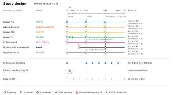
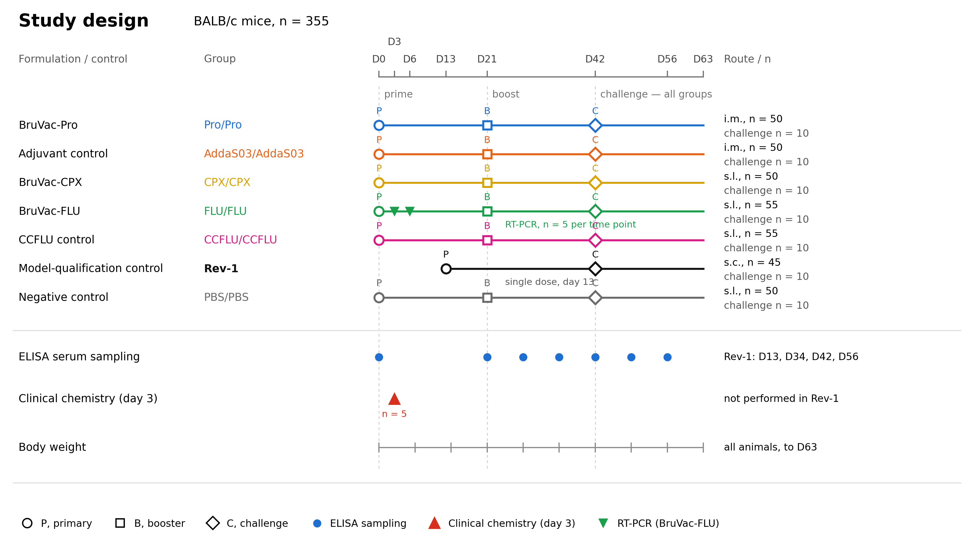
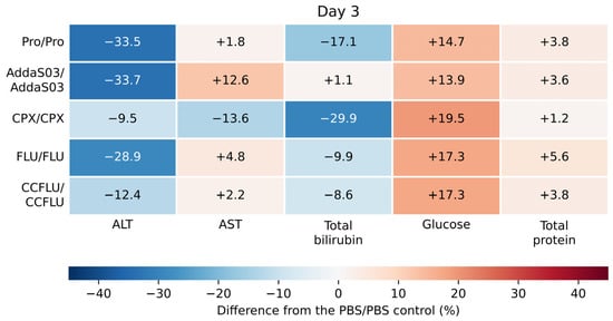
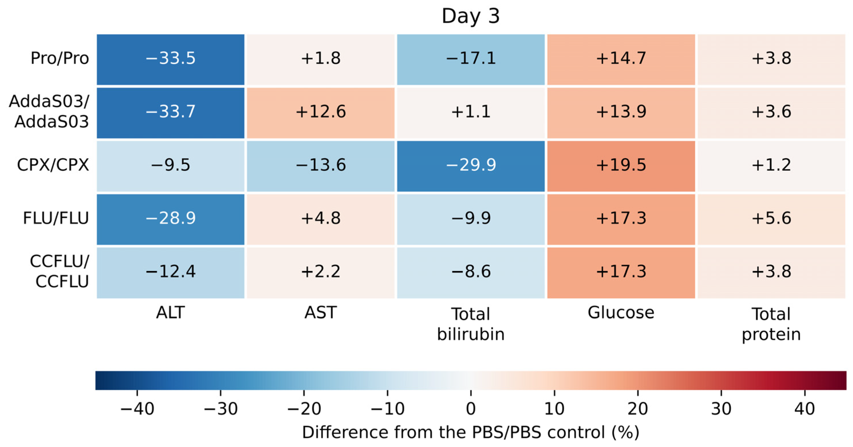
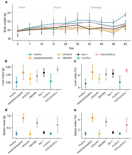
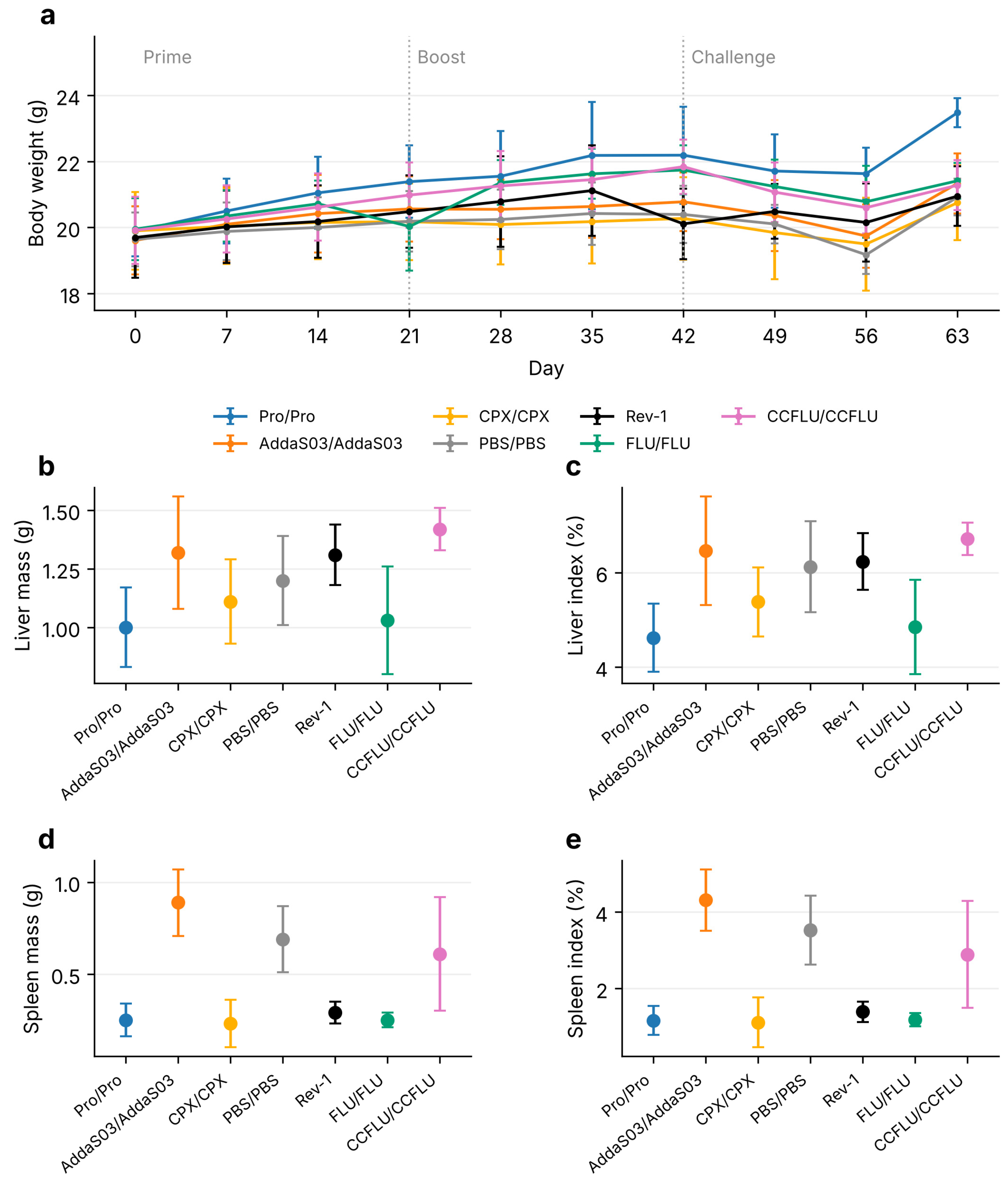
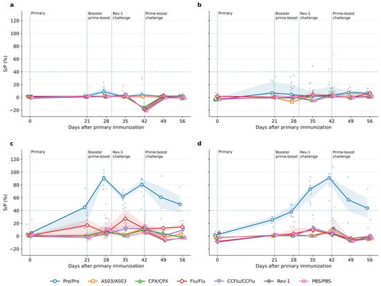
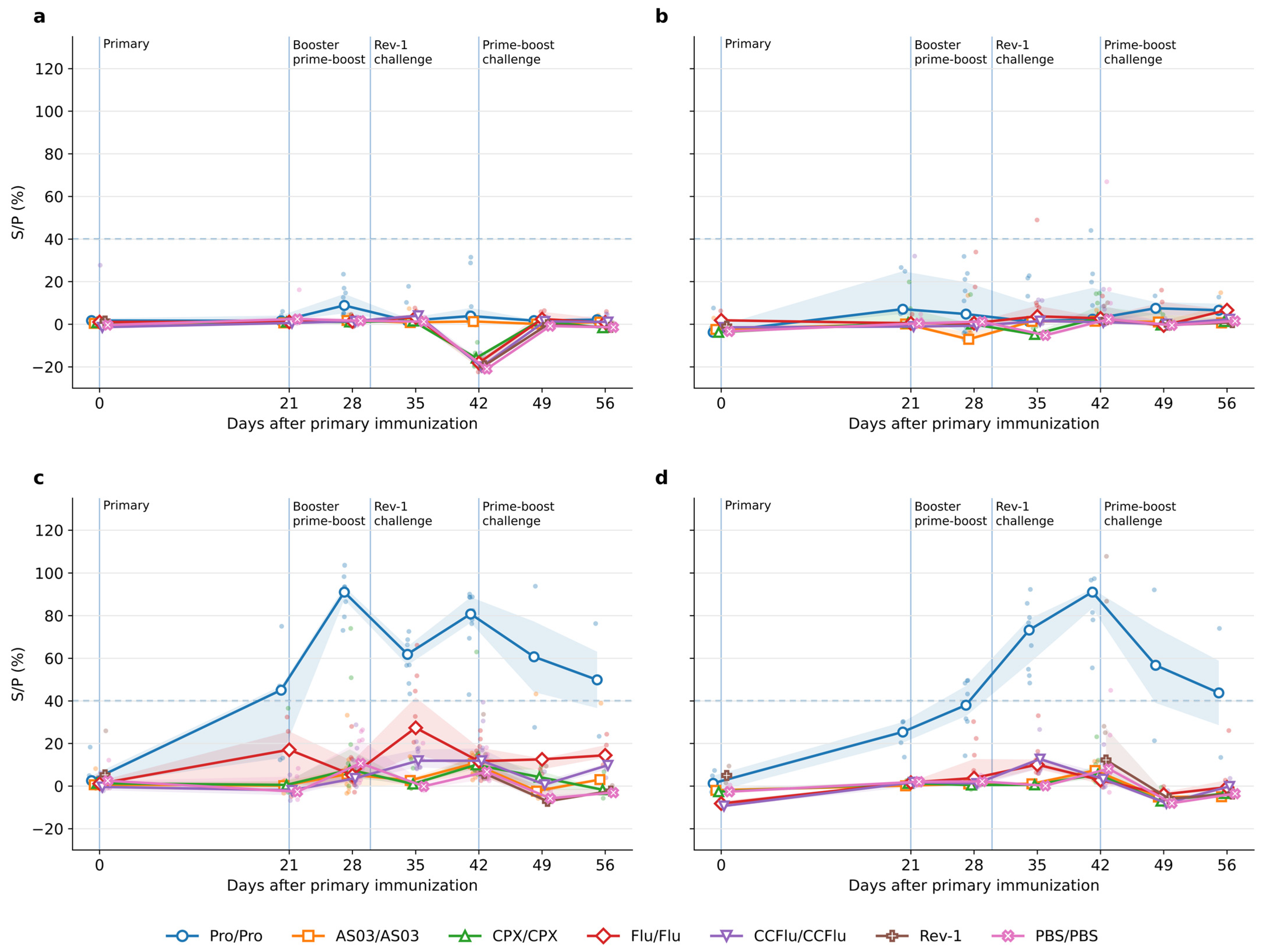
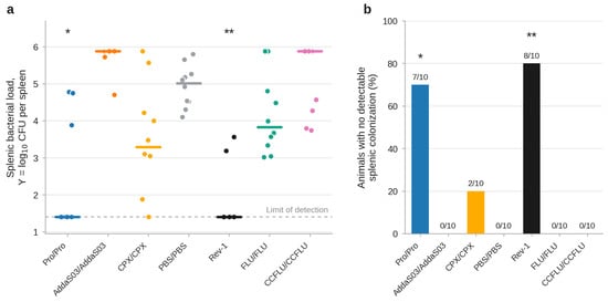
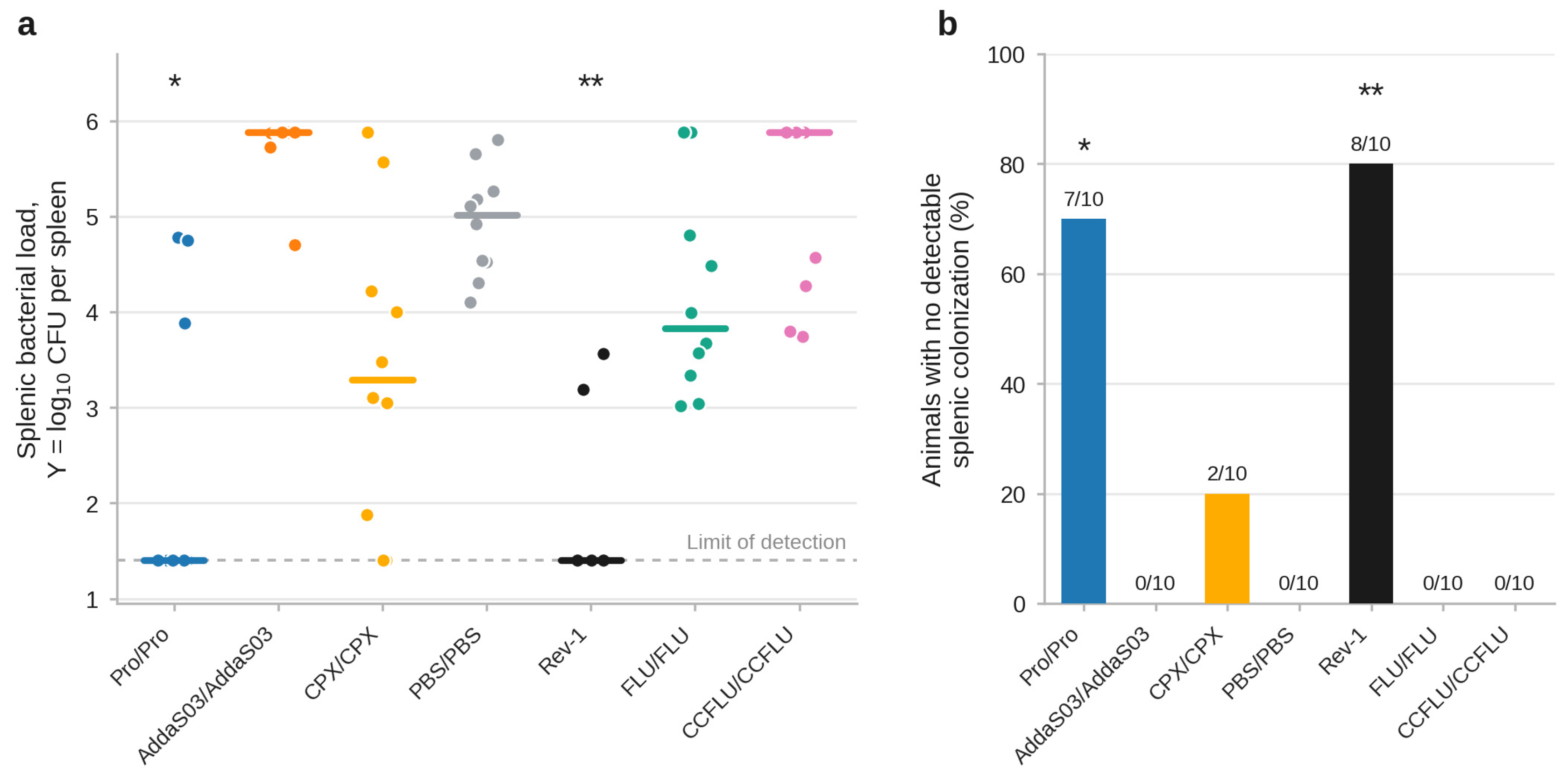

Disclaimer/Publisher’s Note: The statements, opinions and data contained in all publications are solely those of the individual author(s) and contributor(s) and not of MDPI and/or the editor(s). MDPI and/or the editor(s) disclaim responsibility for any injury to people or property resulting from any ideas, methods, instructions or products referred to in the content. |
© 2026 by the authors. Licensee MDPI, Basel, Switzerland. This article is an open access article distributed under the terms and conditions of the Creative Commons Attribution (CC BY) license.