


Abstract
The adaptive immune response against Mycobacterium tuberculosis is incompletely understood. A better comprehension of host immunity might contribute significantly to the development of an efficacious tuberculosis vaccine. Intensive research has characterized the basic components of protective immunity, such as the requirement for robust CD4 T cell responses. In recent years, important work has been published defining the antigenic targets of human M. tuberculosis–specific T and B cells, including the interrogation of the M. tuberculosis proteome by employing peptide libraries and protein microarrays to identify hitherto unknown antigens. Findings from these studies show that human immune responses during M. tuberculosis infection are narrowly focused against a few antigens that are enriched for secreted proteins. In this review we summarize the current knowledge on the M. tuberculosis immunoproteome, covering its composition as well as the methods of discovery. The identity and features of the antigens that typically dominate immune responses against M. tuberculosis are discussed, as are the implications of work in this area for tuberculosis vaccine design.
1. Introduction
Despite significant advances in understanding the immune response against Mycobacterium tuberculosis (Mtb), an effective vaccine to reduce its enormous global disease burden is still elusive. One issue accounting for the slow progress in this area is that, to date, the immune configuration that defines protective immunity, often referred to as “the correlates of protection”, is incompletely understood [1]. This pertains to the specific elements making up the protective immune response, such as specific antibody titers and various T cell phenotypes. Genomic tools, such as the use of targeted gene deletions, have enabled better understanding of the pathways involved in immune protection and control of Mtb infections [2]. Consequently, CD4 T cells have been identified as the central mediator of adaptive immunity against Mtb, with less prominent secondary roles for B cells and other T cell subsets [3]. Defining the precise targets of protective T and B cell responses to Mtb thus constitutes a major goal for research in this area.
Proteome-wide screening techniques have facilitated progress toward the discovery of the antigenic repertoire or “immunoproteome” of Mtb that constitutes specific protein targets of B and T cells, with particular focus on identification of targets recognized by CD4+ T cells and serum antibodies [4]. This has provided valuable insights into the breadth of the immune response as well as the categorization of some specific Mtb proteins according to their immunodominance, i.e., the hierarchy of immune recognition. Remarkably, studies defining the immunoproteome of Mtb have found that antigen-specificity in Mtb-infected hosts is narrowly focused against a small fraction of the >4000 translated gene products that comprise the complete Mtb proteome [5]. It is now understood that clinical TB manifests along a spectrum ranging from latent infection to subclinical and active disease [6]. Along this continuum, the T and B cell targets recognized by the immune system correlate strongly with the disease state [7,8]. This review summarizes the current knowledge on the specific antigens that constitute the immunoproteome of Mtb. We also hypothesize regarding potential mechanisms that may lead to immunodominance of some antigens while others remain unrecognized or “cryptic”. The relevance of these findings to ongoing attempts to develop more effective vaccines for prevention of TB is discussed.
2. TB Natural History and Immunity
The Mycobacterium tuberculosis complex (MTBC) comprises at least 10 currently recognized species of closely related acid-fast bacterial species, which are known to cause tuberculosis-like disease in mammals [9]. Of these, Mtb and Mycobacterium africanum are adapted to cause chronic infections manifesting as clinical tuberculosis (TB) in humans. These are obligate human pathogens, meaning that infection and transmission in natural settings are mainly restricted to the human host [10]. A critical early stage in the evolution of Mtb from an environmental microbe to a professional human pathogen was most likely the acquisition of the ability to survive inside protozoa such as free-living amoeba, providing some of the fundamental mechanisms that enable infection of and survival within mammalian phagocytic cells like macrophages [9]. The life cycle of Mtb is initiated when it enters the airways and infects the resident alveolar macrophages [11]. Here, dendritic cells take up and transport bacilli to the mediastinal lymph node to present to naïve T cells. Exogenous material, like bacteria, is principally processed and presented via the major histocompatibility complex (MHC) II pathway and thus presented to CD4 T cells. In animal models, CD8 T cells play a non-redundant role in control of Mtb infection, but such a function has not been substantiated in humans [12]. The current view is that Mtb primarily optimized its virulence by simultaneous genomic reduction and further expansion of its highly conserved core virulence systems [13]. This hypothesis is supported by the observation that Mtb possesses a smaller genome compared to ancestral non-tuberculous mycobacteria (NTMs), as well as a larger proportion of virulence-related genes compared to the lower frequency of such genes in NTMs [14,15]. Prominent among these are the so-called Type VII or ESX secretion systems, whose substrates, as discussed below, contribute prominently to the immunoproteome of Mtb [16]. A major rate-limiting step in the mounting of the adaptive TB immunity, observed both in humans and animal models, is a delay of several weeks before antigen-specific CD4 T cells arrive in the lung to activate macrophages via production of interferon-γ (IFN-γ) [2]. A granuloma forms consisting of a layer of lymphocytes surrounding Mtb-infected neutrophils and macrophages. As infection persists, there follows a continued buildup of bacterial antigens which are potential targets for CD4 T cells [17]. At length, chronically hyperinflammatory T cells drive lung tissue destruction and create cavities. The bacterial life cycle is completed when, through these cavities, Mtb escapes into the airways, is coughed out of the body, and infects a fresh host. Why some of these targets are strongly recognized by host adaptive immunity while others are only weakly or not at all represented in the Mtb immunoproteome remains a key question for current research that may have major implications in vaccine design.
3. Approaches to Defining the Mtb Immunoproteome
Through the use of high-throughput peptide and protein screening, a more comprehensive picture of the Mtb immunoproteome has begun to emerge. These methods have allowed for a relatively unbiased examination of the proteome to define T and B cell antigens. Hitherto, such antigen screening relied on the insertion of random Mtb genomic DNA fragments into phages to generate recombinant expression libraries in E.coli. In pioneering work by Young & Bloom, such a DNA expression library was constructed using λgt11 phages [18]. The proteins expressed by the antigen-bearing E.coli were transferred to nitrocellulose membranes and then probed with a number of monoclonal antibodies against Mtb antigens. In a subsequent study, the same method was adapted to assess reactivity to pooled serum of TB patients [19]. Positive plaques were recovered, amplified, and the DNA sequenced to identify the corresponding proteins. While the use of antibody screening of phage libraries is a powerful approach, several limitations are apparent. Such methods likely create a bias toward identification of highly expressed antigens, and limitations of library construction may not allow complete genome coverage. In addition, the use of antibodies for screening can only indirectly identify a subset of T cell antigens (i.e., those that stimulate helper T cell responses that drive antibody production), and assumes that T cell specificity matches the specificity of the antibodies produced by T cell-dependent B cell responses (i.e., linked recognition).
A significant improvement in the serologic approach was provided by a more exhaustive screening of antibody reactivity against microarrays carrying in vitro translated products from ~4000 open reading frames (ORFs) of the Mtb genome. As reported by Kunnath-Velayudhan et al., microarrays loaded with proteins representing 99% of expressed Mtb ORFs were tested against sera from confirmed TB patients, suspected TB cases and healthy controls [5]. Remarkably, using this method only ~10% of the Mtb proteome, corresponding to 484 proteins, was detected as reactive with serum antibodies, with a mere 0.5% of the proteome detected at high frequency as antibody targets during active disease. A follow-up study investigated the human antibody responses against a smaller set of 356 proteins, selected based on reactivity to at least 1 serum sample [20]. Here, 10 proteins were significantly associated with active TB disease. In accordance with the earlier work, these proteins were enriched for secreted and cell wall antigens [7]. Again, harnessing a proteome-wide microarray, the antibody specificities of individuals across TB disease states were mapped [7]. Querying the sera of donors with latent TB infection (LTBI) and active TB disease, 650 out of 3963 antigens tested scored above the binding threshold. There were 109 antigens that were differentially expressed between LTBI and active TB, and a minimal subset comprised of 6 antigens that delivered 90% accuracy in discriminating these two groups. In addition, comparing the antibody profiles of LTBI individuals and highly exposed persons testing negative for infection by IFN-γ release assay, known as “resisters”, a group of 13 antigens was sufficient to distinguish these two groups with high confidence.
Unlike antibodies, which can bind their targets directly, CD4 T cells require antigen processing and presentation by antigen-presenting cells (APCs). For that reason, probing the entire Mtb proteome by directly using T cells becomes cumbersome and is further complicated by the genetic variations in the MHC encoded antigen-presenting molecules. Thus, researchers tend to focus on pre-selected individual proteins or small sets of related antigens when querying T cell responses directly. In an early attempt to more comprehensively identify T cell epitopes, the Mtb peptidome was broadly screened based on predicted MHC class II binding affinity to reveal distinct potential CD4 T cell epitopes [21]. To accomplish this, the protein sequences encoded by 21 Mtb genomes were aligned, and all possible 15-mer peptides were identified, which was followed by in silico prediction of the binding strength of these peptides to 22 of the most prevalent HLA-DR, -DP and -DQ class II alleles. This yielded a total of 20,610 peptides with an average coverage of 5 peptides per ORF. These peptides were synthesized, arranged in pools of 20 peptides each, and tested for stimulation of IFN-γ production by peripheral blood mononuclear cells (PBMCs) from LTBI individuals. Interestingly, the CD4 T cell recognition of Mtb antigens was found to be concentrated against mostly epitopes belonging to the ESX and PE/PPE protein families. Thus, as seen with antibodies, CD4 T cell responses against the Mtb immunoproteome exhibit a similar narrow antigenic repertoire.
The narrowly focused range of T cell epitopes was further highlighted in a quantitative analysis evaluating the breadth of the epitope specificity of the CD4 T cell response in a South African cohort of 63 LTBI individuals [22]. Limited to an overlapping peptide library spanning 11 proteins previously identified as immunogenic, this study found the epitope coverage was broader than expected, with up to 125 unique epitopes (after removal of redundant epitopes) recognized by at least one study participant. On average, 24 distinct epitopes derived from up to 8 antigens were recognized by a single donor, and 86% of donors recognized epitopes from at least one antigen. However, no single antigen was recognized by all donors. The same group expanded their previously established peptide library of 20,610 peptides by 610 peptides to include peptides from three BCG strains as well as overlapping 15-mers of twelve TB vaccine candidates [8]. These were tested on PBMCs isolated from subjects with LTBI and TB patients undergoing drug treatment. TB patients recognized a total of 137 unique epitopes, with an average of 8 epitopes (range 1–27) recognized per individual. Here, they confirmed the antigenic islands observed earlier, but the breadth of the response targeting these antigens was narrower in the TB patients compared to the LTBI subjects. Importantly, some epitopes recognized by the TB patients map to antigens that were not identified for the LTBI group. The antigen rv1813c was only recognized by TB patients but not in LTBI, whereas EsxV, Mtb32A and PPE42 were reactive in the LTBI group but not in the TB patient group. This indicates that the transition from latent to active TB is not simply a function of the magnitude of T cell responses, but involves differential recognition of epitopes. Moreover, a pool of 116 peptides found to be reactive only in the TB patient group was designed that could reliably discriminate active disease from latent infection, adding potential clinical relevance to the findings. This bolsters existing evidence that Mtb modulates its gene expression across TB disease states [23].
In a different approach to directly identify presented peptides, Bettencourt et al. purified peptides from MHC-I and MHC-II complexes on BCG-infected macrophages [24]. Analysis by mass spectrometry showed that antigens presented on MHC-II were dominated by membrane-associated proteins. This may reflect the absence of the ESX-1 locus in BCG, which removes the immunodominant effects of its secreted antigens Early Secretory Antigenic Target 6 (ESAT-6) and Culture Filtrate Protein 10 (CFP-10). Indeed, peptides presented by MHC-II on Mtb-infected human dendritic cells were dominated by the secreted substrates of Mtb ESX secretion systems, including CFP-10 and various PE/PPE proteins [25]. Importantly, these antigens were immunogenic and could elicit T cell responses in LTBI individuals.
The scope of the above studies is severely restricted by their exclusive attention to the responses of T cells present in peripheral circulation. Since TB primarily and most prominently affects the lung, the antigen-specificity of resident T cells in that tissue may be different from that of the T cells circulating in the blood. Mucosal intranasal BCG vaccination in mice resulted in enhanced protection in the lungs compared to systemic intradermal vaccination [26]. Mucosal vaccination formed higher frequencies of antigen-specific tissue-resident CD4 T cells with superior proliferative capacity. Likewise, in mice, during Mtb infection, differences in CD4 T cell functionality between the lung vasculature and parenchyma have been shown, with CD4 T cells in the tissue being significantly better at restricting bacterial growth than those in the lung vasculature [27]. Thus, direct profiling of the immunoproteome recognized by T cells in the lung tissue is likely to be critical to further our understanding of the most relevant targets of protective immune responses in TB.
4. Distinctive Properties of Immunodominant TB Antigens
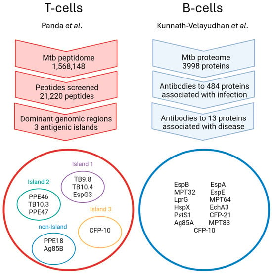
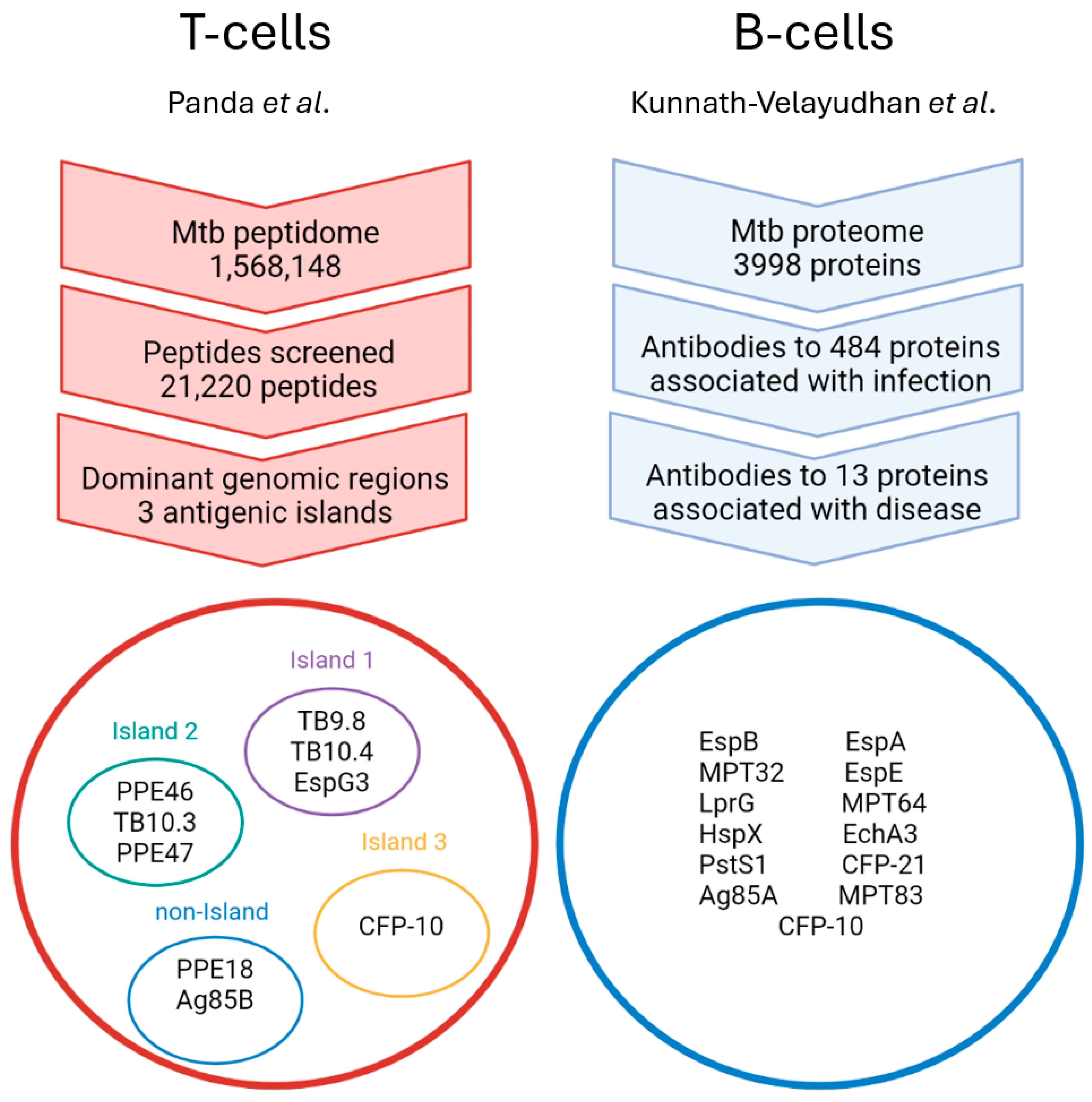
The characterization of the Mtb immunoproteome by the methods described above permits the classification of antigens according to their functional biological category and hierarchy in which they are recognized by the immune system [28]. Interestingly, both B and T cell specificity appear to be enriched for a small set of Mtb antigens, often referred to as immunodominant antigens (Figure 1). Whereas sera from patients with active TB reacted at detectable levels against approximately 10% of the total Mtb proteome, the bulk of the antibody responses appear to be focused against just 13 proteins, most of which are secreted antigens (Table 1) [5]. Prominent among these are the secreted substrates of ESX-1, including CFP-10 and EspB, as well as Antigen 85 (Ag85) A, secreted by the general Sec pathway. In the previously mentioned studies, Lindestam Arlehamn and colleagues showed that the CD4 T cell response during latent infection and active TB disease is centered on 3 genomic regions, referred to as “antigenic islands”, related to secreted antigens [8,21]. Strikingly, these regions constitute 0.55% of the Mtb ORFs but represent the antigenic targets of 42% of the total CD4 T cell response in LTBI donors [21]. The antigens contained within these regions in both studies include ESX-3 substrates such as TB9.8 and TB10.4 (island 1) as well as the ESX-1 substrates CFP-10 and ESAT-6 (island 3). Island 2 is not associated with an ESX system, but is believed to have arisen from a duplication of ESX-3. In addition, notable overrepresentation of the PE/PPE protein family was observed. A comparison of the most prominent B and T cell targets discovered in the above-mentioned two publications shows virtually no overlap between the antigen groups (Figure 1). Whether this accurately reflects the biology or if it is a function of methodological differences is not immediately obvious.
Figure 1.
Antigenic targets of T and B cells during active TB disease. A summary of the screens for antigens recognized by T and B cells. Mtb targets recognized by CD4 T cells of TB patients are concentrated in three antigenic islands (red circle) [8]. Island 1 contains 3 antigens secreted via the ESX-3 system, whereas island 3 encodes proteins secreted by the ESX-1 system. The 3 antigens in island 2 are not associated with an ESX system and are thought to have originated from a duplication of ESX-3. In addition, reactivity was detected, although at low frequencies, to antigens PPE18/Mtb39A (Rv1196) and Ag85B (Rv1886c) that do not comprise a discrete antigenic island. Similarly, the Mtb proteome interrogated with human sera yielded 13 antigens strongly associated with TB disease (blue circle) [5].
Table 1.
List of immunodominant B and T cell targets. An overview of all the Mtb antigens shown in Figure 1, listing their gene identification number (Rv number) in the H37Rv reference genome, associated secretion system, and known function (source: TB Genome Annotation Portal).
This fixation of the immune response on a select group of antigens is accompanied by a lack of antigenic variation exhibited by Mtb, which maintains a highly stable genome lacking the type of antigenic variation seen in other bacterial pathogens with established human transmission and persistence [29]. A cross-species analysis of genome-wide accumulation of mutations in different species of bacteria reported that the slowest rate of evolution was observed for Mtb [30]. Compared to the rapidly evolving Acinetobacter baumannii at a rate of 10−5 to 10−6 substitutions per site per year, the rate for Mtb was markedly lower at 10−8 substitutions per site per year. Furthermore, in a landmark paper, Comas et al. demonstrated that virtually all known T cell epitopes are hyperconserved in Mtb [31]. Interestingly, most of these epitopes are distributed across secreted immunodominant antigens such as CFP-10, ESAT-6, TB10.4, and the Ag85 complex. This finding indicates that unlike most pathogens that seek to escape immune pressure, the preservation of T cell epitopes may drive immune responses that are more advantageous to the bacterium than to the host. One possibility is that in the chronic phase of Mtb infection, the CD4 T cell response may be misdirected to cause hyperinflammation and tissue damage rather than bacterial containment or eradication.
Expanding upon this in a comparative analysis across 216 phylogenetically diverse strains of Mtb, genomic regions containing T cell epitopes were more conserved than surrounding non-epitope-containing regions [32]. Also, 78% of epitopes contained no amino acid substitution, while 19% of epitopes showed only 1 substitution. Therefore, in contrast to some pathogens, most notably human immunodeficiency viruses HIV-1 and HIV-2, which under immune pressure develop escape mutants, Mtb has consolidated its immunodominant antigenic repertoire with minimal to no variation [33]. This might be part of a decoy strategy whereby immunodominant antigens are specialized to elicit strong but suboptimal T cell responses favorable to Mtb survival. Although pre-clinical studies conducted mostly in mice have found that immunization with such antigens can yield modest reductions in bacterial load, superior protection relative to BCG has so far failed to materialize in human trials based on immunization with immunodominant antigens [34]. Therefore, robust immune recognition does not necessarily equate to significant protection.
The secretory aspect of many of the immunodominant antigens provides another piece of evidence contributing to the decoy hypothesis. TB lesions in immunocompetent humans are described as paucibacillary, often having few or no detectable bacilli despite the presence of extensive lesions [35]. Yet, mycobacterial antigens can be found to be abundantly present within lesions and in adjacent tissue. Disproportionate pathology relative to bacterial numbers might therefore be attributed to an excessive T cell response against an abundance of secreted antigens. Export of Ag85B out of Mtb-infected dendritic cells and into the extracellular space has been shown to be an active process in a murine model, which may have two important effects on the progress of infection and host response [36]. Firstly, this antigen export diverts proteins away from the antigen processing and presentation pathways in infected cells. Secondly, uninfected bystander cells can capture these exported antigens and efficiently present them to CD4 T cells. In this way, antigen secretion and export serve as an evasion strategy by not only misdirecting T cells toward uninfected cells but also by potentially driving destructive responses in distal healthy tissue.
The temporal regulation of secreted antigen expression also strongly suggests a bacterial immunoevasive strategy. For example, in Mtb-infected mice transcripts of ESAT-6 and Ag85B normalized to bacterial numbers peaked on day 15 post-infection, after which they sharply fell [37]. This coincided with a decline of antigen-specific lung T cells in the same period. Likewise, in a separate study, Ag85B concentrations dropped rapidly within 2 weeks post-infection [38]. This temporal regulation of immunodominant antigen expression implies that early secretion might skew the specificity of T cells toward these antigens, after which such T cells are rendered nearly obsolete by the paucity of their cognate antigens during infection. This concept might also explain how, despite the high frequencies of CD8 T cells specific for TB10.4 in Mtb-infected mice, these T cells were unable to recognize Mtb-infected macrophages [39]. Collectively, these experimental data imply the deployment of dominant Mtb antigens in a decoy mechanism to subvert host immunity. However, this has not been established mechanistically and will require further research.
5. Comparison of the Immunoproteome of Mtb to Other Bacterial Pathogens
The published literature describing the immunoproteome of bacterial pathogens other than Mtb is scarce, showing that this field is significantly understudied. The studies discussed here differ considerably in their methods and definitions and only aim to be a descriptive overview. The few available studies assessing serum antibody reactivity include both proteome-wide unbiased approaches as well as surveys of pre-selected sets of antigens (Table 2). For example, in the case of Streptococcus pneumoniae (S. pneumoniae), display libraries expressing 15–150 amino acid fragments of the full proteome were probed with serum antibodies. From the serum samples of exposed and convalescing individuals, it was found that approximately 140 proteins were reactive, corresponding to ~7% of the proteome [40]. Likewise, a genome-wide protein microarray of 2652 Staphylococcus aureus antigens showed 1086 antigens (41% of the proteome) exhibited IgG reactivity when probed with patient sera [41]. Nevertheless, strong IgG antibody reactivity in all samples was observed for only 104 antigens. Interestingly, this subset of 104 antigens was enriched for secreted proteins, such as the pore-forming toxin LukS-PV. Similarly, a protein microarray containing antigens of Salmonella typhi was screened with sera from acute typhoid patients and healthy controls [42]. From a total of 2724 antigens tested (63% of the proteome), 2442 antigens were IgG-reactive, and 809 were IgM-reactive, based on recognition by at least 1 patient sample. However, among at least 50% of all patient samples, only 38 and 36 antigens were IgG- and IgM-reactive, respectively. Furthermore, a subset of 127 IgG-reactive antigens was identified as serodominant, making up 4.7% of antigens tested and ~3% of the total proteome.
In a similar manner, the complete proteome of Chlamydia trachomatis, encompassing 908 ORFs, was tested against 99 patient sera taken from women urogenitally infected [43]. This revealed that 709 proteins (78% of the proteome) were recognized by at least one sample. However, applying more stringent criteria, only 27 antigens (~3% of the proteome) were recognized by at least 50% of the samples. Lastly, the immunoproteome of Burkholderia pseudomallei was evaluated by pre-selecting 1205 antigens from a total proteome of ~5900 proteins, based on various criteria such as surface expression and constituting components of the flagella [44]. A protein array was probed with 88 sera from melioidosis patients in Singapore, identifying 170 reactive antigens, constituting ~14% of antigens screened or ~3% of the total proteome. Following further testing of these 170 antigens against a larger collection of 747 patient sera, 49 antigens were identified that could discriminate between melioidosis patients and healthy people diagnosed with other bacterial infections. These data suggest that the size of the Mtb immunoproteome is well within the range of the bacteria discussed above, which also, by most criteria, seem to have relatively small sets of antigens making up the dominant targets of their specific immune recognition.
Table 2.
Immunoproteomes of bacterial pathogens. Description of bacterial immunoproteomes targeted by human serum antibodies. The number of proteins recognized in the various screens and their fraction of the proteome recognized by serum antibodies from infected hosts are stated. In addition, the number of dominant antigens and their fraction of the total proteome are listed.
Although the comparative immunoproteome data are still limited and far from complete, the information summarized above suggests some of the features of Mtb that distinguish it as such an intractable pathogen. We propose that Mtb is highly persistent in its host due to a combination of hyperconservation of immunodominant antigens and the lack of protection provided by immune responses elicited by immunodominant antigens. Admittedly, the quality and scope of data available on antigen conservation in other bacterial pathogens is variable and fragmentary, but a few examples stand out as worthy of consideration. A screen of the antibody repertoire induced by infection with S. pneumoniae revealed both polymorphic and nonpolymorphic antigens reactive to patient sera [40]. Importantly, the study rediscovered the known protective yet highly polymorphic surface antigens PspA and PspC. Subsequent immunization of mice with recombinant PspA showed a significant reduction in bacterial load in the lung following challenge. In contrast, Pizza et al. analyzed the genome of Neisseria meningitidis (N. meningitidis) in silico for ORFs predicted to be surface or exported proteins [45]. They identified 570 such ORFs, of which 350 proteins were successfully expressed as recombinant proteins. Mice were immunized with these proteins and sera collected to analyze antibody binding to the N. meningitidis surface as well as bactericidal activity. From this, seven strongly positive antigens were shown to induce complement-mediated bactericidal activity. Among these was GNA2132 (also known as NHBA), now a component of several licensed meningococcal vaccines. Interestingly, they reported a high level of sequence conservation (~83% amino acid identity) for NHBA across 80 genomes of N. meningitidis [45]. Taken together, the studies suggest a range of conservation of immunodominant antigens in common Gram-positive and Gram-negative bacterial pathogens that does not reach the extraordinarily high level observed in Mtb. Indeed, immunodominant antigens can provide lasting immune protection in some bacterial pathogens, which has generally not been found for Mtb. Therefore, vaccine design requires careful consideration, taking into account these findings, and may benefit from further studies characterizing the immunoproteomes of other successful and persistent bacterial pathogens.
6. Do Subdominant or Cryptic Antigenic Targets Have a Role in Vaccine Design?
Since the introduction of BCG over a hundred years ago, no new TB vaccine has been approved for clinical use outside of investigational studies [46]. BCG is efficacious against childhood TB but provides incomplete and limited protection against adult pulmonary TB, which is the major driver of the global epidemic. The vaccine pipeline has seen review of many candidates, with just a few passing the pre-clinical testing stage and advancing to human trials [47]. A disappointing although significant milestone was the phase 2b clinical trial of MVA85A, a vaccinia virus vector delivering the immunodominant Ag85A, in South African infants which showed no efficacy for prevention of TB beyond standard BCG vaccination [48]. Curiously, the trial only assessed T cell mediated immunity even though antibody recognition of Ag85A was found to be strongly associated with TB disease [5]. Another vaccine candidate that has entered clinical testing is the M72/AS01E protein subunit vaccine. A phase 2b trial concluded after 3 years and reported 54% efficacy against active pulmonary TB in LTBI adults, allowing M72/AS01E to proceed to phase 3 trials, currently ongoing [49]. This vaccine consists of a fusion of the antigens Mtb32A and Mtb39A, selected for their induction of robust T cell proliferation and IFN-γ responses in PBMCs from healthy PPD-positive individuals [50,51]. Consistent with this, Mtb39A, also known as PPE18, was recognized as a prominent target of CD4 T cells in LTBI humans [21]. In clinical settings and controlled trials in humans, TB vaccines are administered either intradermally or intramuscularly. However, as mentioned above, the route of immunization can have a significant impact on the induction of tissue-resident immunity. Most dramatically, intravenous (IV) BCG administration in macaques significantly increased tissue-resident T cells in addition to sterilized infection in most animals [52]. Thereafter, in a similar macaque model, protection conferred by IV BCG correlated with antigen-specific CD4 T cells in the airways. Hence, the method of vaccine delivery needs careful consideration [53].
In order to avoid a probable decoy effect of immunodominant antigens, immunization with subdominant antigens has been proposed as a vaccine strategy [54]. An argument against this method is the weak presentation of such antigens during natural infection. Moreover, immunodominance of antigens generated by natural infection might override the putative protective capacity of subdominant antigen immunization. Even so, recognition by memory T cells of their cognate antigens requires a lower activation threshold compared to naïve T cells undergoing priming [55]. Consequently, weakly presented antigens might still be able to stimulate recall responses of memory T cells. This is not to say that cryptic antigens are inherently more protective, but rather that they could possess qualities that have been overlooked or masked by the immunodominant antigens. Our group has previously shown that administration of the subdominant ribosomal protein antigen rplJ at 4 weeks following BCG immunization significantly reduced lung pathology from subsequent Mtb infection compared to BCG alone [56]. Although bacterial burden in the lungs was not affected, a significant reduction in the mediastinal lymph nodes was observed.
Other recent studies showed significant qualitative differences in the T cell responses elicited by dominant versus subdominant Mtb antigens [57]. Thus, a small group of seven Mtb proteins was selected based on sequence variability, indicating that they may serve as subdominant antigens that are subject to immune pressure [32]. Subsequently, in a Kenyan cohort of subjects with LTBI, four of these variable antigens (LldD2, RimJ, Rv0010c and Rv0012) were shown to stimulate relatively low IFN-γ-secreting CD4 T cell responses compared to classical dominant antigens (e.g., Ag85B, CFP-10 and ESAT-6), while eliciting IL-17-producing CD4 T cells in higher proportions [57]. Given the increased interest in IL-17 as possibly contributing to effective immune protection against Mtb, these variable antigens might be considered for incorporation in prospective TB vaccines [58].
It has also become clear that at least some of the classically defined immunodominant antigens of Mtb contain within their structure subdominant or cryptic epitopes that may be revealed under certain conditions. This has suggested that it may be useful to leverage the ubiquity of dominant antigens in protective vaccination by shifting responses to the subdominant epitopes therein. In a study to examine this hypothesis, deletion of the dominant CD4 T cell epitope from ESAT-6 elicited responses specific for cryptic ESAT-6 epitopes in BALB/c and CB6F1 mice, but not in C57BL/6 mice [59]. Immunization with the ESAT-6 mutant protein lacking the major immunodominant epitope prior to Mtb infection significantly reduced bacterial burden in the lung of the aforementioned two mouse strains. Importantly, cryptic epitope-specific CD4 T cells exhibited enhanced proliferative capacity as well as polyfunctionality based on secretion of IFN-γ, TNFα, and IL-2.
Thus, targeting of subdominant antigens might open hitherto unexplored avenues to significantly enhanced protective immune responses. Recognition of subdominant variable antigens by T cells of persons with LTBI is a crucial finding that warrants further evaluation in experimental animal models of Mtb infection [57].
7. Conclusions and Future Perspectives
In light of the severe loss in human capital brought on by the TB epidemic, there is an urgent need to produce an efficacious vaccine. The complex nature of host-pathogen interactions, coupled with the vast resources required for clinical trials, calls for a departure from conventional strategies in pursuit of original thinking. The identification of the antigens that comprise the Mtb immunoproteome marks important steps forward to better understand the TB host-pathogen interactions and might significantly contribute to rational vaccine design. We have presented the argument that the narrow focus on antigenic targets could be a decoy mechanism, particularly in view of the secretory nature of many of them. Therefore, excluding these putative decoy antigens from vaccines might prevent the raising of non-productive memory cells. Moreover, subdominant antigens deserve more attention as potentially valuable vaccine targets, as it was shown that some induce Th17 cells, which under certain conditions are associated with checking the progression to TB disease [60]. Finally, more work is needed to characterize the differences in antigen-specificity of B and T cells between LTBI and active TB disease. In particular, those that remain LTBI over long periods in high exposure environments are suitable donors for such future studies. This could potentially provide clues as to which antigens correlate with either protective or pathological responses. Ultimately, a viable TB vaccine would likely have to incorporate a number of mutually reinforcing strategies to be effective in real-world settings.
Author Contributions
Conceptualization: M.A., T.W.N. and S.A.P.; Writing—Original draft preparation: M.A.; Writing—Review and editing: M.A., T.W.N. and S.A.P.; Visualization: M.A., T.W.N. and S.A.P.; Supervision: S.A.P.; Project administration: S.A.P.; Funding acquisition: S.A.P. All authors have read and agreed to the published version of the manuscript.
Funding
The authors were supported by NIH grants 5R01AI045889, 5P01CA257885 and 5P30CA013330.
Institutional Review Board Statement
Not applicable.
Informed Consent Statement
Not applicable.
Data Availability Statement
No new data were created or analyzed in this study. Data sharing is not applicable to this article.
Conflicts of Interest
The authors declare no conflicts of interest.
References
- Wang, J.; Fan, X.Y.; Hu, Z. Immune Correlates of Protection as a Game Changer in Tuberculosis Vaccine Development. npj Vaccines 2024, 9, 208. [Google Scholar] [CrossRef] [Scilit] [PubMed]
- Flynn, J.A.L.; Chan, J. Immune Cell Interactions in Tuberculosis. Cell 2022, 185, 4682–4702. [Google Scholar] [CrossRef] [Scilit] [PubMed]
- Saraiva, M.; Branchett, W.J.; Rengarajan, J.; O’Garra, A. The Host Immune Response to Mycobacterium tuberculosis Determining Protection or Disease Progression. Nat. Immunol. 2026, 27, 1104–1118. [Google Scholar] [CrossRef] [Scilit] [PubMed]
- Kunnath-Velayudhan, S.; Porcelli, S.A. Recent Advances in Defining the Immunoproteome of Mycobacterium tuberculosis. Front. Immunol. 2013, 4, 64319. [Google Scholar] [CrossRef] [Scilit] [PubMed]
- Kunnath-Velayudhan, S.; Salamon, H.; Wang, H.Y.; Davidow, A.L.; Molina, D.M.; Huynh, V.T.; Cirillo, D.M.; Michel, G.; Talbot, E.A.; Perkins, M.D.; et al. Dynamic Antibody Responses to the Mycobacterium tuberculosis Proteome. Proc. Natl. Acad. Sci. USA 2010, 107, 14703–14708. [Google Scholar] [CrossRef] [Scilit] [PubMed]
- Richards, A.S.; Sossen, B.; Emery, J.C.; Horton, K.C.; Heinsohn, T.; Frascella, B.; Balzarini, F.; Oradini-Alacreu, A.; Häcker, B.; Odone, A.; et al. Quantifying Progression and Regression across the Spectrum of Pulmonary Tuberculosis: A Data Synthesis Study. Lancet Glob. Health 2023, 11, e684–e692. [Google Scholar] [CrossRef] [Scilit] [PubMed]
- Khan, M.Z.; Irvine, E.B.; Cizmeci, D.; Davies, L.R.L.; Randall, A.Z.; Pablo, J.; Vidal, S.J.; Lasrado, N.; Darrah, P.A.; Hsiao, J.C.; et al. A Proteome-Wide Atlas of Humoral Immunity to Mycobacterium tuberculosis across the Spectrum of Disease. Front. Immunol. 2026, 17, 1810894. [Google Scholar] [CrossRef] [Scilit] [PubMed]
- Panda, S.; Morgan, J.; Cheng, C.; Saito, M.; Gilman, R.H.; Ciobanu, N.; Crudu, V.; Catanzaro, D.G.; Catanzaro, A.; Rodwell, T.; et al. Identification of Differentially Recognized T Cell Epitopes in the Spectrum of Tuberculosis Infection. Nat. Commun. 2024, 15, 765. [Google Scholar] [CrossRef] [Scilit] [PubMed]
- Gagneux, S. Ecology and Evolution of Mycobacterium tuberculosis. Nat. Rev. Microbiol. 2018, 16, 202–213. [Google Scholar] [CrossRef] [Scilit] [PubMed]
- Brites, D.; Gagneux, S. Co-Evolution of Mycobacterium tuberculosis and Homo Sapiens. Immunol. Rev. 2015, 264, 6–24. [Google Scholar] [CrossRef] [Scilit] [PubMed]
- Cohen, S.B.; Gern, B.H.; Delahaye, J.L.; Adams, K.N.; Plumlee, C.R.; Winkler, J.K.; Sherman, D.R.; Gerner, M.Y.; Urdahl, K.B. Alveolar Macrophages Provide an Early Mycobacterium tuberculosis Niche and Initiate Dissemination. Cell Host Microbe 2018, 24, 439–446.e4. [Google Scholar] [CrossRef] [Scilit] [PubMed]
- Lin, P.L.; Flynn, J.A.L. CD8 T Cells and Mycobacterium tuberculosis Infection. Semin. Immunopathol. 2015, 37, 239–249. [Google Scholar] [CrossRef] [Scilit] [PubMed]
- Orgeur, M.; Brosch, R. Evolution of Virulence in the Mycobacterium tuberculosis Complex. Curr. Opin. Microbiol. 2018, 41, 68–75. [Google Scholar] [CrossRef] [Scilit] [PubMed]
- Brosch, R.; Gordon, S.V.; Marmiesse, M.; Brodin, P.; Buchrieser, C.; Eiglmeier, K.; Garnier, T.; Gutierrez, C.; Hewinson, G.; Kremer, K.; et al. A New Evolutionary Scenario for the Mycobacterium tuberculosis Complex. Proc. Natl. Acad. Sci. USA 2002, 99, 3684–3689. [Google Scholar] [CrossRef] [Scilit] [PubMed]
- Gey Van Pittius, N.C.; Sampson, S.L.; Lee, H.; Kim, Y.; Van Helden, P.D.; Warren, R.M. Evolution and Expansion of the Mycobacterium tuberculosis PE and PPE Multigene Families and Their Association with the Duplication of the ESAT-6 (Esx) Gene Cluster Regions. BMC Evol. Biol. 2006, 6, 95. [Google Scholar] [CrossRef] [Scilit] [PubMed]
- Hmama, Z.; Peña-Díaz, S.; Joseph, S.; Av-Gay, Y. Immunoevasion and Immunosuppression of the Macrophage by Mycobacterium tuberculosis. Immunol. Rev. 2015, 264, 220–232. [Google Scholar] [CrossRef] [Scilit] [PubMed]
- Hunter, R.L. Tuberculosis as a Three-Act Play: A New Paradigm for the Pathogenesis of Pulmonary Tuberculosis. Tuberculosis 2016, 97, 8–17. [Google Scholar] [CrossRef] [Scilit] [PubMed]
- Young, R.A.; Bloom, B.R.; Grosskinsky, C.M.; Ivanyi, J.; Thomas, D.; Davis, R.W. Dissection of Mycobacterium tuberculosis Antigens Using Recombinant DNA. Proc. Natl. Acad. Sci. USA 1985, 82, 2583–2587. [Google Scholar] [CrossRef] [Scilit] [PubMed]
- Lodes, M.J.; Dillon, D.C.; Mohamath, R.; Day, C.H.; Benson, D.R.; Reynolds, L.D.; McNeill, P.; Pedral Sampaio, D.; Skeiky, Y.A.W.; Badaro, R.; et al. Serological Expression Cloning and Immunological Evaluation of MTB48, a Novel Mycobacterium tuberculosis Antigen. J. Clin. Microbiol. 2001, 39, 2485–2493. [Google Scholar] [CrossRef] [Scilit] [PubMed]
- Kunnath-Velayudhan, S.; Davidow, A.L.; Wang, H.Y.; Molina, D.M.; Huynh, V.T.; Salamon, H.; Pine, R.; Michel, G.; Perkins, M.D.; Xiaowu, L.; et al. Proteome-Scale Antibody Responses and Outcome of Mycobacterium tuberculosis Infection in Nonhuman Primates and in Tuberculosis Patients. J. Infect. Dis. 2012, 206, 697–705. [Google Scholar] [CrossRef] [Scilit] [PubMed]
- Lindestam Arlehamn, C.S.; Gerasimova, A.; Mele, F.; Henderson, R.; Swann, J.; Greenbaum, J.A.; Kim, Y.; Sidney, J.; James, E.A.; Taplitz, R.; et al. Memory T Cells in Latent Mycobacterium tuberculosis Infection Are Directed against Three Antigenic Islands and Largely Contained in a CXCR3+CCR6+ Th1 Subset. PLoS Pathog. 2013, 9, e1003130. [Google Scholar] [CrossRef] [Scilit] [PubMed]
- Lindestam Arlehamn, C.S.; McKinney, D.M.; Carpenter, C.; Paul, S.; Rozot, V.; Makgotlho, E.; Gregg, Y.; van Rooyen, M.; Ernst, J.D.; Hatherill, M.; et al. A Quantitative Analysis of Complexity of Human Pathogen-Specific CD4 T Cell Responses in Healthy M. Tuberculosis Infected South Africans. PLoS Pathog. 2016, 12, e1005760. [Google Scholar] [CrossRef] [Scilit] [PubMed]
- Talaat, A.M.; Lyons, R.; Howard, S.T.; Johnston, S.A. The Temporal Expression Profile of Mycobacterium tuberculosis Infection in Mice. Proc. Natl. Acad. Sci. USA 2004, 101, 4602–4607. [Google Scholar] [CrossRef] [Scilit] [PubMed]
- Bettencourt, P.; Müller, J.; Nicastri, A.; Cantillon, D.; Madhavan, M.; Charles, P.D.; Fotso, C.B.; Wittenberg, R.; Bull, N.; Pinpathomrat, N.; et al. Identification of Antigens Presented by MHC for Vaccines against Tuberculosis. npj Vaccines 2020, 5, 2. [Google Scholar] [CrossRef] [Scilit] [PubMed]
- Leddy, O.; Ogongo, P.; Huffaker, J.; Gan, M.; Milligan, R.; Mahmud, S.; Ni, H.M.; Yuki, Y.; Bobosha, K.; Wassie, L.; et al. Immunopeptidomics Can Inform the Design of MRNA Vaccines for the Delivery of Mycobacterium tuberculosis MHC Class II Antigens. Sci. Transl. Med. 2025, 17, eadw9184. [Google Scholar] [CrossRef] [Scilit] [PubMed]
- Bull, N.C.; Stylianou, E.; Kaveh, D.A.; Pinpathomrat, N.; Pasricha, J.; Harrington-Kandt, R.; Garcia-Pelayo, M.C.; Hogarth, P.J.; McShane, H. Enhanced Protection Conferred by Mucosal BCG Vaccination Associates with Presence of Antigen-Specific Lung Tissue-Resident PD-1 + KLRG1 − CD4 + T Cells. Mucosal Immunol. 2019, 12, 555–564. [Google Scholar] [CrossRef] [Scilit] [PubMed]
- Sakai, S.; Kauffman, K.D.; Schenkel, J.M.; McBerry, C.C.; Mayer-Barber, K.D.; Masopust, D.; Barber, D.L. Cutting Edge: Control of Mycobacterium tuberculosis Infection by a Subset of Lung Parenchyma–Homing CD4 T Cells. J. Immunol. 2014, 192, 2965–2969. [Google Scholar] [CrossRef] [Scilit] [PubMed]
- Lindestam Arlehamn, C.S.; Lewinsohn, D.; Sette, A.; Lewinsohn, D. Antigens for CD4 and CD8 T Cells in Tuberculosis. Cold Spring Harb. Perspect. Med. 2014, 4, a018465. [Google Scholar] [CrossRef] [Scilit] [PubMed]
- Ernst, J.D. Antigenic Variation and Immune Escape in the MTBC. Adv. Exp. Med. Biol. 2017, 1019, 171. [Google Scholar] [CrossRef] [Scilit] [PubMed]
- Duchêne, S.; Holt, K.E.; Weill, F.X.; Le Hello, S.; Hawkey, J.; Edwards, D.J.; Fourment, M.; Holmes, E.C. Genome-Scale Rates of Evolutionary Change in Bacteria. Microb. Genom. 2016, 2, e000094. [Google Scholar] [CrossRef] [Scilit] [PubMed]
- Comas, Ĩ.; Chakravartti, J.; Small, P.M.; Galagan, J.; Niemann, S.; Kremer, K.; Ernst, J.D.; Gagneux, S. Human T Cell Epitopes of Mycobacterium tuberculosis Are Evolutionarily Hyperconserved. Nat. Genet. 2010, 42, 498–503. [Google Scholar] [CrossRef] [Scilit] [PubMed]
- Coscolla, M.; Copin, R.; Sutherland, J.; Gehre, F.; De Jong, B.; Owolabi, O.; Mbayo, G.; Giardina, F.; Ernst, J.D.; Gagneux, S.M. Tuberculosis T Cell Epitope Analysis Reveals Paucity of Antigenic Variation and Identifies Rare Variable TB Antigens. Cell Host Microbe 2015, 18, 538–548. [Google Scholar] [CrossRef] [Scilit] [PubMed]
- Cuevas, J.M.; Geller, R.; Garijo, R.; López-Aldeguer, J.; Sanjuán, R. Extremely High Mutation Rate of HIV-1 In Vivo. PLoS Biol. 2015, 13, e1002251. [Google Scholar] [CrossRef] [Scilit] [PubMed]
- Chugh, S.; Bahal, R.K.; Dhiman, R.; Singh, R. Antigen Identification Strategies and Preclinical Evaluation Models for Advancing Tuberculosis Vaccine Development. npj Vaccines 2024, 9, 57. [Google Scholar] [CrossRef] [Scilit] [PubMed]
- Hunter, R.L.; Actor, J.K.; Hwang, S.A.; Karev, V.; Jagannath, C. Pathogenesis of Post Primary Tuberculosis: Immunity and Hypersensitivity in the Development of Cavities. Ann. Clin. Lab. Sci. 2014, 44, 365–387. [Google Scholar] [PubMed]
- Srivastava, S.; Grace, P.S.; Ernst, J.D. Antigen Export Reduces Antigen Presentation and Limits T Cell Control of M. Tuberculosis. Cell Host Microbe 2016, 19, 44–54. [Google Scholar] [CrossRef] [Scilit] [PubMed]
- Rogerson, B.J.; Jung, Y.J.; LaCourse, R.; Ryan, L.; Enright, N.; North, R.J. Expression Levels of Mycobacterium tuberculosis Antigen-Encoding Genes versus Production Levels of Antigen-Specific T Cells during Stationary Level Lung Infection in Mice. Immunology 2006, 118, 195–201. [Google Scholar] [CrossRef] [Scilit] [PubMed]
- Ernst, J.D.; Cornelius, A.; Bolz, M. Dynamics of Mycobacterium tuberculosis Ag85B Revealed by a Sensitive Enzyme-Linked Immunosorbent Assay. mBio 2019, 10, e00611-19. [Google Scholar] [CrossRef] [Scilit] [PubMed]
- Yang, J.D.; Mott, D.; Sutiwisesak, R.; Lu, Y.J.; Raso, F.; Stowell, B.; Babunovic, G.H.; Lee, J.; Carpenter, S.M.; Way, S.S.; et al. Mycobacterium tuberculosis-Specific CD4+and CD8+T Cells Differ in Their Capacity to Recognize Infected Macrophages. PLoS Pathog. 2018, 14, e1007060. [Google Scholar] [CrossRef] [Scilit] [PubMed]
- Giefing, C.; Meinke, A.L.; Hanner, M.; Henics, T.; Minh, D.B.; Gelbmann, D.; Lundberg, U.; Senn, B.M.; Schunn, M.; Habel, A.; et al. Discovery of a Novel Class of Highly Conserved Vaccine Antigens Using Genomic Scale Antigenic Fingerprinting of Pneumococcus with Human Antibodies. J. Exp. Med. 2008, 205, 117–131. [Google Scholar] [CrossRef] [Scilit] [PubMed]
- Radke, E.E.; Brown, S.M.; Pelzek, A.J.; Fulmer, Y.; Hernandez, D.N.; Torres, V.J.; Thomsen, I.P.; Chiang, W.K.; Miller, A.O.; Shopsin, B.; et al. Hierarchy of Human IgG Recognition within the Staphylococcus Aureus Immunome. Sci. Rep. 2018, 8, 13296. [Google Scholar] [CrossRef] [Scilit] [PubMed]
- Liang, L.; Juarez, S.; Nga, T.V.T.; Dunstan, S.; Nakajima-Sasaki, R.; Huw Davies, D.; McSorley, S.; Baker, S.; Felgner, P.L. Immune Profiling with a Salmonella Typhi Antigen Microarray Identifies New Diagnostic Biomarkers of Human Typhoid. Sci. Rep. 2013, 3, 1043. [Google Scholar] [CrossRef] [Scilit] [PubMed]
- Wang, J.; Zhang, Y.; Lu, C.; Lei, L.; Yu, P.; Zhong, G. A Genome-Wide Profiling of the Humoral Immune Response to Chlamydia Trachomatis Infection Reveals Vaccine Candidate Antigens Expressed in Humans. J. Immunol. 2010, 185, 1670–1680. [Google Scholar] [CrossRef] [Scilit] [PubMed]
- Felgner, P.L.; Kayala, M.A.; Vigil, A.; Burk, C.; Nakajima-Sasaki, R.; Pablo, J.; Molina, D.M.; Hirst, S.; Chew, J.S.W.; Wang, D.; et al. A Burkholderia Pseudomallei Protein Microarray Reveals Serodiagnostic and Cross-Reactive Antigens. Proc. Natl. Acad. Sci. USA 2009, 106, 13499–13504. [Google Scholar] [CrossRef] [Scilit] [PubMed]
- Pizza, M.; Scarlato, V.; Masignani, V.; Giuliani, M.M.; Aricò, B.; Comanducci, M.; Jennings, G.T.; Baldi, L.; Bartolini, E.; Capecchi, B.; et al. Identification of Vaccine Candidates Against Serogroup B Meningococcus by Whole-Genome Sequencing. Science 2000, 287, 1816–1820. [Google Scholar] [CrossRef] [Scilit] [PubMed]
- Lange, C.; Aaby, P.; Behr, M.A.; Donald, P.R.; Kaufmann, S.H.E.; Netea, M.G.; Mandalakas, A.M. 100 Years of Mycobacterium Bovis Bacille Calmette-Guérin. Lancet Infect. Dis. 2021, 22, e2–e12. [Google Scholar] [CrossRef] [Scilit] [PubMed]
- Martín, C.; Gonzalo-Asensio, J.; Aguiló, N.; Arbués, A. Toward Tuberculosis Elimination: An Update on Tuberculosis Vaccines in Clinical Trials. Int. J. Infect. Dis. 2026, 167, 108513. [Google Scholar] [CrossRef] [Scilit] [PubMed]
- Tameris, M.D.; Hatherill, M.; Landry, B.S.; Scriba, T.J.; Snowden, M.A.; Lockhart, S.; Shea, J.E.; McClain, J.B.; Hussey, G.D.; Hanekom, W.A.; et al. Safety and Efficacy of MVA85A, a New Tuberculosis Vaccine, in Infants Previously Vaccinated with BCG: A Randomised, Placebo-Controlled Phase 2b Trial. Lancet 2013, 381, 1021–1028. [Google Scholar] [CrossRef] [Scilit] [PubMed]
- Tait, D.R.; Hatherill, M.; Van Der Meeren, O.; Ginsberg, A.M.; Van Brakel, E.; Salaun, B.; Scriba, T.J.; Akite, E.J.; Ayles, H.M.; Bollaerts, A.; et al. Final Analysis of a Trial of M72/AS01E Vaccine to Prevent Tuberculosis. N. Engl. J. Med. 2019, 381, 2429–2439. [Google Scholar] [CrossRef] [Scilit] [PubMed]
- Skeiky, Y.A.W.; Lodes, M.J.; Guderian, J.A.; Mohamath, R.; Bement, T.; Alderson, M.R.; Reed, S.G. Cloning, Expression, and Immunological Evaluation of Two Putative Secreted Serine Protease Antigens of Mycobacterium tuberculosis. Infect. Immun. 1999, 67, 3998–4007. [Google Scholar] [CrossRef] [Scilit] [PubMed]
- Dillon, D.C.; Alderson, M.R.; Day, C.H.; Lewinsohn, D.M.; Coler, R.; Bement, T.; Campos-Neto, A.; Skeiky, Y.A.W.; Orme, I.M.; Roberts, A.; et al. Molecular Characterization and Human T-Cell Responses to a Member of a Novel Mycobacterium tuberculosis Mtb39 Gene Family. Infect. Immun. 1999, 67, 2941–2950. [Google Scholar] [CrossRef] [Scilit] [PubMed]
- Darrah, P.A.; Zeppa, J.J.; Maiello, P.; Hackney, J.A.; Wadsworth, M.H.; Hughes, T.K.; Pokkali, S.; Swanson, P.A.; Grant, N.L.; Rodgers, M.A.; et al. Prevention of Tuberculosis in Macaques after Intravenous BCG Immunization. Nature 2020, 577, 95–102. [Google Scholar] [CrossRef] [Scilit] [PubMed]
- Darrah, P.A.; Zeppa, J.J.; Wang, C.; Irvine, E.B.; Bucsan, A.N.; Rodgers, M.A.; Pokkali, S.; Hackney, J.A.; Kamath, M.; White, A.G.; et al. Airway T Cells Are a Correlate of i.v. Bacille Calmette-Guerin-Mediated Protection against Tuberculosis in Rhesus Macaques. Cell Host Microbe 2023, 31, 962–977.e8. [Google Scholar] [CrossRef] [Scilit] [PubMed]
- Orr, M.T.; Ireton, G.C.; Beebe, E.A.; Huang, P.-W.D.; Reese, V.A.; Argilla, D.; Coler, R.N.; Reed, S.G. Immune Subdominant Antigens as Vaccine Candidates against Mycobacterium tuberculosis. J. Immunol. 2014, 193, 2911–2918. [Google Scholar] [CrossRef] [Scilit] [PubMed]
- Weng, N.P.; Araki, Y.; Subedi, K. The Molecular Basis of the Memory T Cell Response: Differential Gene Expression and Its Epigenetic Regulation. Nat. Rev. Immunol. 2012, 12, 306–315. [Google Scholar] [CrossRef] [Scilit] [PubMed]
- Kennedy, S.C.; Johnson, A.J.; Bharrhan, S.; Arlehamn, C.S.L.; Xu, J.; Garforth, S.J.; Chan, J.; Jacobs, W.R.; Sette, A.; Almo, S.C.; et al. Identification of Mycobacterial Ribosomal Proteins as Targets for CD4 + T Cells That Enhance Protective Immunity in Tuberculosis. Infect. Immun. 2018, 86, 10–1128. [Google Scholar] [CrossRef] [Scilit] [PubMed]
- Ogongo, P.; Wassie, L.; Tran, A.; Columbus, D.; Huffaker, J.; Sharling, L.; Ouma, G.; Ouma, S.G.; Bobosha, K.; Arlehamn, C.S.L.; et al. Rare Variable M. Tuberculosis Antigens Induce Predominant Th17 Responses in Human Infection. JCI Insight 2026, 11, 14. [Google Scholar] [CrossRef] [Scilit] [PubMed]
- Maseeme, M.; Tezera, L.B.; Noursadeghi, M.; Leslie, A.; Pollara, G. Context-Specific Roles for IL-17 in Tuberculosis. PLoS Pathog. 2026, 22, e1014054. [Google Scholar] [CrossRef] [Scilit] [PubMed]
- Woodworth, J.S.; Aagaard, C.S.; Hansen, P.R.; Cassidy, J.P.; Agger, E.M.; Andersen, P. Protective CD4 T Cells Targeting Cryptic Epitopes of Mycobacterium tuberculosis Resist Infection-Driven Terminal Differentiation. J. Immunol. 2014, 192, 3247–3258. [Google Scholar] [CrossRef] [Scilit] [PubMed]
- Sun, M.; Phan, J.M.; Kieswetter, N.S.; Huang, H.; Yu, K.K.Q.; Smith, M.T.; Liu, Y.E.; Wang, C.; Gupta, S.; Obermoser, G.; et al. Specific CD4+ T Cell Phenotypes Associate with Bacterial Control in People Who ‘Resist’ Infection with Mycobacterium tuberculosis. Nat. Immunol. 2024, 25, 1411–1421. [Google Scholar] [CrossRef] [Scilit] [PubMed]

Disclaimer/Publisher’s Note: The statements, opinions and data contained in all publications are solely those of the individual author(s) and contributor(s) and not of MDPI and/or the editor(s). MDPI and/or the editor(s) disclaim responsibility for any injury to people or property resulting from any ideas, methods, instructions or products referred to in the content. |
© 2026 by the authors. Licensee MDPI, Basel, Switzerland. This article is an open access article distributed under the terms and conditions of the Creative Commons Attribution (CC BY) license.