


Abstract
Objective: To construct a novel calcium ion–based squalene water-in-oil emulsion, termed calcium ion reverse micelle squalene emulsion (CISE), as a vaccine adjuvant delivery system by exploiting the high affinity of calcium ions for phosphate groups and biomacromolecules. Methods: CISE was prepared using an ultrasonic emulsification method and characterized by laser particle size analysis and transmission electron microscopy (TEM). Its adjuvant efficacy and biocompatibility were evaluated through both in vitro and in vivo experiments. Results: CISE has a particle size of approximately 124.7 nm and exhibits good dispersibility. In vitro experiments show that antigen presenting cells (APCs) efficiently internalize CISE primarily through caveolae mediated endocytosis. CISE promotes the maturation and activation of bone marrow derived dendritic cells (BMDCs), accompanied by elevated production of reactive oxygen species (ROS) and enhanced peroxisomal activity. Notably, CISE possesses prominent lysosomal escape capacity within APCs. After two rounds of subcutaneous immunization in mice, CISE induced high levels of IgG antibodies and elicited a relatively balanced Th1/Th2 immune response. Following pulmonary mucosal immunization, CISE not only triggered robust IgG production but also induced high levels of IgA antibodies. Conclusions: When loaded with a model antigen, CISE effectively induces humoral, cellular, and mucosal immunity, demonstrating significant potential as a vaccine adjuvant.
1. Introduction
Infectious diseases have long been a major threat to human health and social stability. Despite significant advances in anti-infective therapies and disease surveillance, a substantial number of individuals still die each year from infections caused by viruses, bacteria, and other pathogens [1]. The continuous emergence and re-emergence of infectious diseases, combined with pathogen mutation, rising antimicrobial resistance, and accelerated global population mobility, have further complicated efforts to prevent and control these diseases [2]. Infectious diseases not only cause high morbidity and mortality but also impose a persistent and substantial burden on global healthcare systems and socioeconomic development [3].
Vaccines are biological preparations derived from pathogenic microorganisms or their specific components, designed to induce antigen-specific immune protection in the host. Traditional vaccines mainly include live attenuated vaccines, inactivated vaccines, and subunit vaccines [4]. Live attenuated vaccines can mimic the natural infection process and typically elicit comprehensive immune responses; however, they carry a certain risk of residual virulence. Inactivated vaccines are safer than live attenuated vaccines but generally exhibit relatively lower immunogenicity [5]. Subunit vaccines, which contain only defined antigenic components, offer improved safety profiles but often require additional immunostimulatory strategies to achieve optimal immune efficacy [6].
Adjuvants are substances added to vaccine formulations to enhance the magnitude and duration of the host immune response to antigens [7]. Their main mechanisms of action include improving antigen presentation efficiency, activating innate immune signaling pathways [8], promoting cytokine secretion, prolonging antigen retention, and inducing immunological memory [9,10]. The rational use of adjuvants can not only reduce the required antigen dose and the number of immunizations but also improve antigen stability, thereby enhancing overall vaccine efficacy [11].
The most widely used adjuvants to date are aluminum-based compounds. Since their introduction in the early 20th century, aluminum adjuvants have been extensively applied in various human vaccines [12]. However, aluminum adjuvants exhibit limited ability to induce Th1-type cellular immunity, whereas protection against intracellular pathogens, such as Mycobacteria or certain viruses, typically relies on a robust Th1-type immune response [13]. In addition, aluminum salts can cause local inflammatory reactions at the injection site and carry a risk of IgE antibody induction, potentially increasing the likelihood of allergic responses [14]. MF59 has been extensively applied in clinical seasonal influenza vaccine vaccination [15,16]. Its core mechanism of action is to rapidly establish an immune microenvironment at the injection site, efficiently recruit and activate antigen presenting cells (APCs), and thereby improve antigen delivery efficiency as well as the cross protective capacity of vaccines [17,18]. Nevertheless, MF59 fails to mount effective immune defenses at the mucosal portals of pathogen invasion [19]. Calcium phosphate, a naturally occurring inorganic component in the human body, exhibits excellent biocompatibility and biodegradability [20]. Compared with aluminum-based adjuvants, calcium phosphate carries a lower risk of elevating IgE levels during immune stimulation, making it more suitable for populations requiring multiple immunizations or long-term protection [21]. Calcium phosphate particles can be phagocytosed by macrophages or dendritic cells and subsequently degraded in lysosomes into metabolizable products, thereby avoiding long-term retention in vivo [22].
Based on the above theoretical framework, this study employed squalene as the oil phase, incorporating soy phosphatidylcholine (SPC) and calcium chloride. In this system, SPC forms reverse micelles within the nonpolar environment, while Ca2+ binds to the negatively charged hydrophilic groups of the phospholipids and localizes within the aqueous core of the reverse micelles, thereby constructing a composite system with both delivery functionality and immunostimulatory potential. In vitro studies demonstrated that CISE could be internalized by APCs and exhibited lysosomal escape. Mice receiving two intrapulmonary doses of OVA-loaded CISE generated high levels of IgG and IgA and elicited a relatively balanced Th1/Th2 immune response. These findings indicate a synergistic adjuvant effect of calcium ions and squalene in the CISE formulation.
2. Materials and Methods
2.1. Materials and Mnimals
Chemicals and reagents: Ovalbumin, squalene (SQ, room temperature density 0.858 g/mL), polysorbate 80 (Tween 80, MW 1310), sorbitan trioleate (Span 85, MW 957.5), soy phosphorylcholine (SPC), calcein, coumarin 6, 3,3′,5,5′-tetramethylbenzidine (TMB), chlorpromazine hydrochloride (CPZ), methyl-β-cyclodextrin (MCD), and amiloride (AMI) were purchased from Sigma-Aldrich (Merck KGaA, Darmstadt, Germany). Cell culture and biochemical reagents: Recombinant mouse GM-CSF, a reactive oxygen species (ROS) detection kit, 4′,6-diamidino-2-phenylindole (DAPI), disodium hydrogen phosphate dodecahydrate (Na2HPO4·12H2O), anhydrous calcium chloride (CaCl2), TRIS, Hoechst 33342 staining solution, rhodamine 123, and LysoTracker Red were obtained from Beyotime (Shanghai, China). RPMI 1640 and DMEM cell culture media were purchased from Servicebio (Wuhan, China). Antibodies: Goat anti-mouse IgG-horseradish peroxidase (HRP) (Cat. No. 31430), IgG1-HRP (Cat. No. PA1-74421), IgG2a-HRP (Cat. No. A-10685), and IgA-HRP monoclonal antibodies (Cat. No. PA1-74397), as well as goat anti-mouse CD3-APC, CD4-FITC, CD8-PE, CD40-FITC, CD80-APC, and CD86-eFluor 450, were purchased from Thermo Fisher Scientific (Waltham, MA, USA). Purified anti-mouse IL-4 (Cat. No. 504101), purified anti-mouse IFN-γ (Cat. No. 517901), biotinylated anti-mouse IL-4 (Cat. No. 504201), and biotinylated anti-mouse IFN-γ (Cat. No. 505803) were purchased from BioLegend (San Diego, CA, USA). Fluorescent probes: Peroxi SPY555 was obtained from Spirochrome (Stein am Rhein, Switzerland).
Female Kunming (KM) mice, aged 8 weeks, were provided from the Experimental Animal Center of Anhui Medical University. All the animals were raised in specific pathogen-free conditions in the animal facility of Pharmacy School of AMU. All animal-related procedures and protocols were carried out according to the bioethical regularity, and performances were approved by the Animal Ethical Committee of AMU (Approval No. LLSC20-230287).
2.2. Preparation and Characterization of CISE
Preparation of CISE: A weighed quantity of soy phosphorylcholine (SPC) was dissolved in chloroform, and chloroform was completely removed by a rotary evaporator. Aqueous CaCl2 solution was added at a molar ratio of SPC:Ca = 2:1, and the resulting SPC film was fully hydrated under magnetic stirring. After complete hydration, water was removed by rotary evaporation. Squalene and Span 85 were added at a mass ratio of CaCl2:squalene:Span 85 = 1:100:10. The Ca/SPC polymer was fully dissolved in squalene under magnetic stirring to serve as the oil phase. 10 mM TBS solution (composed of 0.9% sodium chloride and Tris) was used as the aqueous phase. The oil phase, aqueous phase and Tween 80 were mixed, and probe ultrasonication (Power: 97.5 W, pulse mode: 5 s on/5 s off, total sonication time: 5 min) was performed in an ice-water bath to obtain CISE. The final CISE had a composition of squalene/CaCl2/SPC/Span 85/Tween 80/TBS (100:1:9:10:10:1000, w/w).
Preparation of SWE: SQ-in-water emulsions (SWE) is an oil-in-water emulsion adjuvant similar to MF59, differing from MF59 in that it is prepared by ultrasonic emulsification. Sodium citrate aqueous solution (10 nM), 5% (v/v) squalene, 0.5% (w/v) Span 85 and Tween 80 were mixed and emulsified by probe ultrasonication (Power: 97.5 W, pulse mode: 5 s on/5 s off, total sonication time: 5 min) in an ice-water bath to obtain an MF59 analogue designated as SWE.
Preparation of CaP: Calcium phosphate (CaP) was prepared by the following method. A mixture containing 12.5 mM calcium chloride, 12.5 mM disodium hydrogen phosphate and 15.6 mM sodium citrate was thoroughly stirred under magnetic stirring for 48 h, followed by ultrasonication for 30 min to obtain CaP, which was stored at 4 °C for further use.
Using ovalbumin (OVA) as the model antigen, each adjuvant formulation was mixed with OVA and magnetically stirred at room temperature for 30 min. The antigen-adjuvant complexes were designated OVA-CISE, OVA-SWE, and OVA-CaP.
Preparation of fluorescent-labeled adjuvants: Coumarin 6 (Cou6) and calcein (Cal) were used to label the squalene oil phase and calcium ions, respectively. Briefly, Cou6 was added into the squalene solution, and Cal was dissolved in the calcium chloride solution under magnetic stirring until complete dissolution. Fluorescently labeled adjuvants were then prepared following the aforementioned CISE fabrication procedure. The only modification was that the oil phase adopted coumarin 6-labeled squalene solution, and calcein-containing calcium chloride solution was used for hydration.
The particle size and zeta-potential of CISE were determined using a Zetasizer Nano ZS90 (Malvern, Worcestershire, UK). After negative staining with 2% (w/v) phosphotungstic acid, the morphology was observed under a transmission electron microscope (Talos L120C G2,Thermo Fisher Scientific, Waltham, MA, USA).
CISE was aliquoted into centrifuge tubes, sealed under nitrogen atmosphere, and stored at 4 °C, 25 °C and 37 °C for stability investigation. Samples were collected at predetermined time points. Dynamic light scattering (DLS) was employed to determine particle size, polydispersity index (PDI), and zeta-potential. Meanwhile, the macroscopic appearance of the emulsions was visually inspected, and physical destabilization phenomena such as phase separation and demulsification were recorded to comprehensively evaluate the storage stability.
2.3. In Vitro Assay of CISE Action on APC
2.3.1. Obtaining Mouse BMDCs
Healthy KM mice aged 6–8 weeks were selected. Bilateral femurs and tibias were dissected under a laminar-flow biosafety cabinet. The bone-marrow cavities were repeatedly flushed with RPMI 1640 medium, and the bone-marrow flush-through was filtered through a 100-mesh sterile cell sieve to obtain a single-cell suspension. Red blood cells were lysed by adding red-blood-cell lysis buffer. The harvested cells were then seeded into culture dishes and cultured in complete RPMI 1640 medium supplemented with 10% FBS and 1% penicillin-streptomycin, with 20 ng/mL GM-CSF added as the differentiation stimulus. Cells were incubated at 37 °C in a humidified incubator with 5% CO2. During the culture period, half of the medium was replaced every other day, and fresh GM-CSF was replenished accordingly [23]. On day 6, non-adherent and loosely adherent cells were collected for subsequent experiments.
2.3.2. Cytotoxicity of CISE by MTT Assay
The cytotoxicity of CISE toward cells was assessed using the MTT assay. RAW264.7 cells were adjusted to a density of 1 × 10⁵ cells/100 μL and seeded into 96-well plates. After 12 h of incubation, CISE with varying squalene concentrations (0–1200 μg/mL) was added for a 24-h treatment. Twenty microliters of MTT solution (5 mg/mL) were added to each well, followed by 4 h of incubation. Subsequently, 150 μL of dimethyl sulfoxide (DMSO) was added, and the plates were shaken for 10 min. The absorbance of each well was measured at a wavelength of 490 nm using a microplate reader (BioTek, Winooski, VT, USA). Cell viability (%) was calculated as follows (1):
where ODadjuvant is the absorbance of wells containing cells with adjuvant, ODcell is the absorbance of wells containing cells only, and ODblank is the absorbance of wells containing medium only.
2.3.3. Uptake of CISE by BMDCs and RAW264.7 Cells
RAW264.7 cells or BMDCs were seeded in 12-well plates at a density of 1 × 10⁵ cells per well and incubated overnight at 37 °C in a humidified atmosphere containing 5% CO2. Add fluorescent-labeled SWE, CaP or CISE and incubate for another 2 h. Cells were washed three times with sterile PBS, filtered through a cell strainer, and analyzed by flow cytometry (FC, CytoFLEX, Beckman Coulter, Brea, CA, USA) to determine the percentage of antigen-presenting cells that had taken up adjuvants as well as the mean fluorescence intensity (MFI). APC uptake of CISE and control groups was also visualized using a laser scanning confocal microscope (LSCM, LSM 800, Zeiss, Oberkochen, Germany). The cell seeding and treatment procedures were basically consistent with those for flow cytometry. After treatment, cells were fixed with 4% paraformaldehyde for 15 min, rinsed three times with PBS, and stained with DAPI for 10 min in the dark to fluorescently label cell nuclei. Following three additional PBS washes, cells were observed under LSCM.
2.3.4. Pathways of APC Uptake of CISE
To investigate the uptake mechanism of CISE by antigen-presenting cells (APCs) using the coumarin 6-labeled oil phase (Cou6-CISE), RAW264.7 cells or BMDCs were seeded in 12-well plates at a density of 1 × 10⁵ cells per well. The cells were incubated overnight at 37 °C in a humidified atmosphere containing 5% CO2. The culture medium was then removed, and cells were pretreated for 1 h with either 30 μM chlorpromazine (CPZ, clathrin-mediated endocytosis inhibitor), 5 mM methyl-β-cyclodextrin (MCD, caveolae-mediated endocytosis inhibitor), or 50 μM amiloride (AMI, macropinocytosis inhibitor). A low-temperature control group (incubated at 4 °C) and a normal control group (37 °C, without inhibitors) were additionally set up. Subsequently, Cou6-CISE was added to each well, and the cells were further incubated for 1 h. Cells were harvested by centrifugation at 1200 rpm, washed three times with PBS, resuspended and filtered through a cell strainer. The fluorescence intensity was measured by flow cytometry.
2.3.5. Tracking the Intracellular CISE
To evaluate lysosomal escape of CISE after cellular uptake, RAW264.7 cells were evenly seeded in laser confocal culture dishes at a density of 1 × 10⁵ cells per dish and incubated overnight. Different groups were treated with Cou6-CISE or Cal-CISE for 0, 4, 8, or 16 h. Following treatment, cells were washed three times with sterile PBS and incubated with LysoTracker Red (lysosomal probe) at 37 °C in the dark for 1 h. The nuclei were subsequently stained with Hoechst 33342 for 30 min. After three additional PBS washes, cells were observed using a laser scanning confocal microscope (LSCM). Confocal images were further analyzed with ImageJ 1.54p software for colocalization, and the Pearson correlation coefficient was calculated to assess the lysosomal escape efficiency of CISE.
2.3.6. CISE-Induced Upregulation of BMDC Co-Stimulatory Molecules
APC activation and maturation was measured through detection of CD40 and CD80 and CD86 on mouse BMDCs that had been stimulated with CISE or controls. BMDCs were seeded into 12-well plates at a density of 1 × 10⁵ cells per well and incubated overnight at 37 °C in a humidified atmosphere with 5% CO2. Cells were then treated with OVA-CISE or control formulations for 24 h. After incubation, cells were stained with CD40-FITC, CD80-APC, and CD86-eFluor 450 antibodies and incubated at 4 °C for 30 min. Following three washes with PBS, the cells were analyzed by flow cytometry to determine the mean fluorescence intensity (MFI).
2.3.7. CISE-Induced ROS Production in APCs
RAW264.7 cells were seeded into 12-well plates at a density of 1 × 10⁵ cells per well and incubated overnight at 37 °C in a humidified atmosphere with 5% CO2. Cells were then treated with CISE or control formulations for 4 h. Subsequently, 10 μM DCFH-DA probe was added, and cells were incubated at 37 °C in the dark for 30 min. After three washes with PBS, intracellular ROS levels were quantified by flow cytometry, and the mean fluorescence intensity (MFI) was recorded.
2.3.8. CISE-Induced Peroxisome Changes in APCs
RAW264.7 cells were seeded in 12-well plates at a density of 1 × 10⁵ cells per well. The cells were incubated overnight at 37 °C in a humidified atmosphere containing 5% CO2. Cells were then treated with CISE or control formulations for 4 h. Subsequently, SPY555 fluorescent probe was added, and cells were incubated at 37 °C in the dark for 1 h. After three washes with PBS, fluorescence intensity was measured by flow cytometry, and the MFI was recorded. Peroxisomal changes were also observed using LSCM. Cell seeding and treatment were performed as described above for flow cytometry. After incubation with the peroxisome probe, cell nuclei were stained with Hoechst 33342. Cells were washed three times with PBS and subsequently imaged using LSCM.
2.4. CISE Immunogenicity Study in Mice
2.4.1. Mouse Immunization
Six- to eight-week-old female KM mice were acclimated to the laboratory environment for one week before use and randomly divided into six groups (n = 6 per group): PBS, OVA, OVA-SWE, OVA-CaP, and OVA-CISE. All groups except the OVA-CISE group received subcutaneous immunization, while the OVA-CISE group was subjected to subcutaneous immunization and intratracheal instillation for follow-up experimental assessments. Each administration consisted of 50 μL containing 5 μg of OVA. A booster immunization was performed three weeks after the primary immunization. Intratracheal instillation: After mice were anesthetized, an indwelling needle was gently inserted into the trachea. The adjuvant was aspirated and slowly instilled through a flexible tube. Mice were closely monitored for respiratory status, general condition, and adverse reactions including cough, dyspnea, and abnormal oronasal secretions.
2.4.2. Mouse Sample Collection
Blood collection via retro-orbital plexus puncture: Blood samples were collected every seven days after the primary immunization by retro-orbital plexus puncture. Briefly, a sterile capillary tube was gently inserted along the medial wall of the mouse orbit, and blood was allowed to flow freely into a sterile centrifuge tube. Samples were kept at room temperature for 2 h to allow clotting, followed by centrifugation at 12,000 rpm for 10 min. The serum was harvested and stored at −20 °C until further analysis. Mucosal sample collection: Lavage fluids from different mucosal sites, including vaginal lavage fluid (VLF), nasal lavage fluid (NLF), oral lavage fluid (OLF), bronchoalveolar lavage fluid (BALF), and intestinal lavage fluid (ILF), were collected eight weeks after primary immunization. Pre-cooled sterile PBS was used to repeatedly flush the corresponding mucosal surfaces to obtain mucosal samples. All mucosal samples were centrifuged at 12,000× g for 10 min at 4 °C, and the supernatants were collected and stored at −20 °C.
2.4.3. In Vivo Safety Evaluation of Immunized Mice and Analysis of Antibodies and Cytokines
To assess the in vivo safety of CISE, mice were euthanized three days after immunization, and major organs including the heart, liver, spleen, lungs, and kidneys were collected. The tissues were fixed in 4% paraformaldehyde for 24 h, followed by dehydration with ethanol, clearing in xylene, and embedding in paraffin. Sections approximately 4 μm thick were prepared using a paraffin microtome (RM2255, Leica Biosystems, Nußloch, Germany) and stained with hematoxylin and eosin (H&E). Histological observations were performed using a slide scanner (Pannoramic SCAN, 3D Histech, Budapest, Hungary).
The levels of IgG and sIgA were measured using an ELISA. OVA was diluted to 10 μg/mL in coating buffer, and 50 μL was added to each well of a microplate and incubated overnight at 4 °C. The plates were washed three times with 200 μL PBST (PBS containing 0.05% Tween 20), followed by the addition of 150 μL blocking buffer (1% BSA) for 2 h. After discarding the blocking buffer and washing three times, 50 μL of diluted serum was added to each well and incubated for 2 h. For IgG titer detection, serum samples were subjected to two-fold serial dilution. Serum from the blank experimental group was used as the negative control. The highest dilution factor at which the OD value exceeded twice that of the negative control was defined as the antibody titer. The wells were washed five times, and 50 μL of appropriately diluted HRP-conjugated secondary antibodies (IgG, 1:1000; IgG2a, 1:1500; IgG1, 1:1000; IgA, 1:2500) was added to each well and incubated for 2 h. After washing five times, 50 μL TMB substrate was added to each well and incubated for 15 min for color development. The reaction was stopped with 2 M H2SO4, and absorbance was immediately measured using a microplate reader (BioTek, Winooski, VT, USA) [24].
Splenocyte preparation: Mice were euthanized three weeks after the final immunization, and spleens were aseptically collected and rinsed with pre-cooled sterile PBS. Each spleen was gently dissociated using the plunger of a sterile syringe to obtain a cell suspension. Following filtration, erythrocytes were lysed using red blood cell lysis buffer. The cells were then washed 2–3 times with sterile PBS and resuspended in complete RPMI 1640 medium.
Cytokine detection in splenocyte supernatants: Splenocytes were seeded in 24-well plates at a density of 1 × 10⁶ cells per well. OVA was added for stimulation to achieve a final concentration of 100 μg/mL. After gentle mixing, cells were incubated at 37 °C for 48 h. Upon completion of incubation, cells were harvested and centrifuged at 1200 rpm for 5 min, and the supernatants were collected for cytokine measurement. ELISA measurement of IL-4 and IFN-γ in splenocyte supernatants: Purified IFN-γ and IL-4 were diluted to 2 μg/mL in coating buffer. A 50 μL aliquot was added to each well of a 96-well microplate, followed by overnight coating at 4 °C. The coating solution was discarded, and wells were washed three times with 200 μL PBST (PBS containing 0.05% Tween-20). Plates were blocked with 150 μL blocking buffer (1% bovine serum albumin) for 2 h. After removal of blocking buffer and three washes, 10-fold diluted splenocyte supernatants were added to each well. Meanwhile, standards were serially diluted (starting concentrations: IFN-γ 2000 pg/mL, IL-4 250 pg/mL) to generate the standard curve, and incubated for 2 h. Sample and standard solutions were discarded, and plates were washed four times. Each well was supplemented with 50 μL biotin-conjugated detection antibody (2 μg/mL), and incubated for 2 h. After discarding the detection antibody solution and three washes, 50 μL streptavidin-HRP (1:500 dilution) was added to each well, followed by a 2-h incubation. Wells were emptied and washed six times, and 50 μL TMB substrate solution was added for 15 min color development. The reaction was terminated with 2 M sulfuric acid, and absorbance values were immediately read using a microplate reader.
2.4.4. Assay of Proliferation and T Cell Subtype of Splenocytes of Vaccinated Mice
Splenocyte proliferation: Splenocytes were seeded in 96-well plates at 1 × 10⁵ cells per well and stimulated with OVA at a final concentration of 100 μg/mL. Unstimulated splenocytes served as the negative control, while medium-only wells served as the blank control. After incubation at 37 °C for 48 h, cell proliferation was assessed using the MTT assay. Briefly, 20 μL of MTT solution (5 mg/mL) was added to each well and incubated for a further 4 h, followed by the addition of 150 μL DMSO to dissolve the formazan crystals. Absorbance was measured at 490 nm using a microplate reader. The splenocyte stimulation index (SI) was calculated as follows (2):
T cell subsets: The isolated splenocytes were adjusted to a concentration of 1 × 10⁶ cells/mL and transferred to brown centrifuge tubes. Each tube was supplemented with 1 μL of APC-conjugated anti-mouse CD3, FITC-conjugated anti-mouse CD4, and PE-conjugated anti-mouse CD8 antibodies. The samples were incubated at 4 °C in the dark for 2 h, centrifuged at 1200 rpm for 5 min, and the supernatants were discarded. After three washes with PBS, the cells were resuspended and filtered through a cell strainer. The proportions of CD3+, CD4+, and CD8+ T-lymphocytes were determined by flow cytometry.
2.5. Statistical Analysis
Data are expressed as mean ± standard deviation (Mean ± SD). Statistical analyses were conducted using GraphPad Prism 9.5. Group comparisons were performed by one-way or two-way ANOVA as appropriate. Significance was defined as follows: ns, not significant (p > 0.05); *, p < 0.05; **, p < 0.01; ***, p < 0.001.
3. Results
3.1. CISE Characteristics
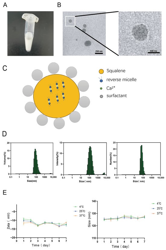
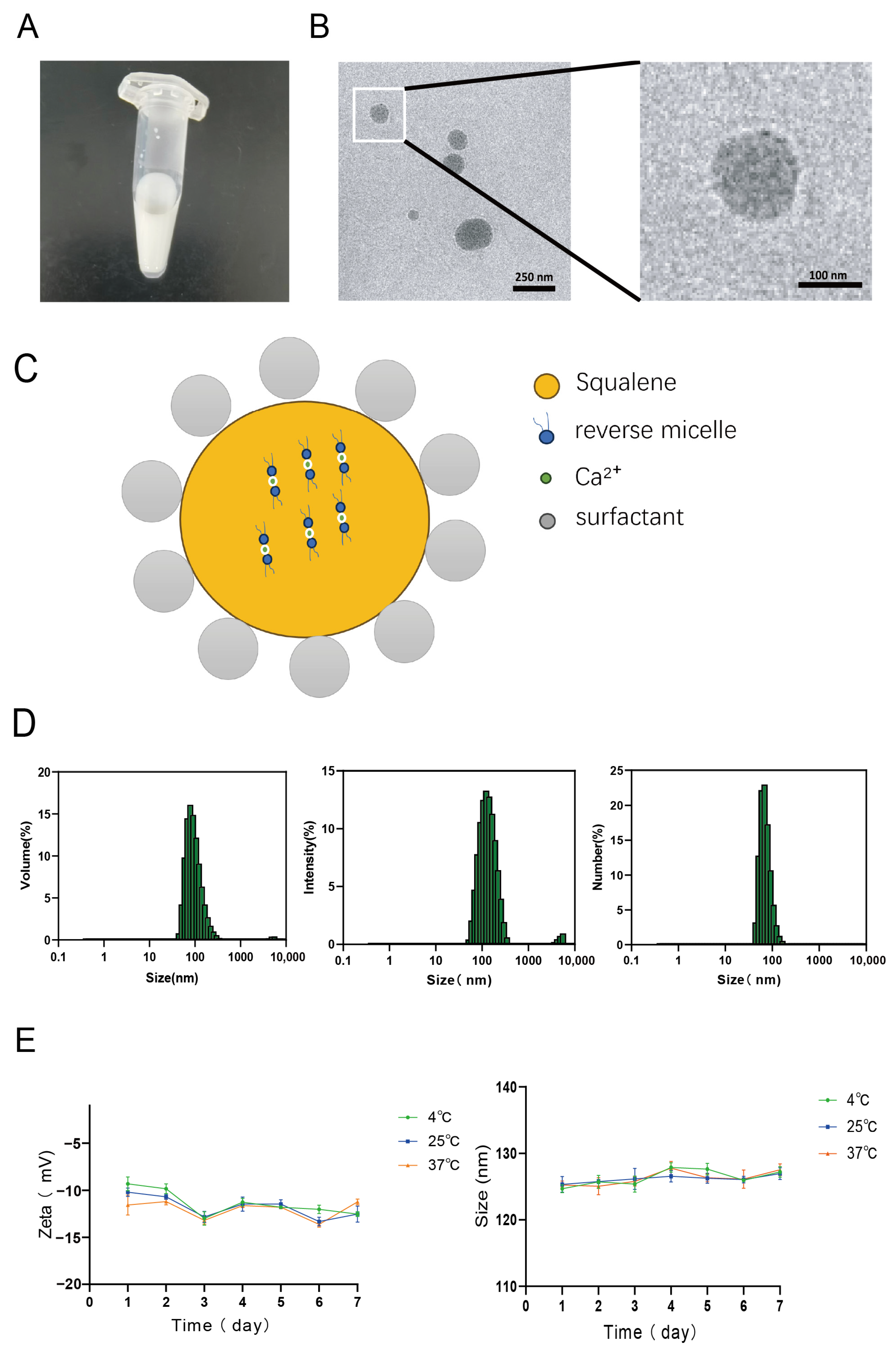
The particle size, polydispersity index (PDI), and zeta potential of CISE, measured by a laser particle size analyzer, are summarized in (Table 1). The average particle size was 124.7 ± 0.6 nm, with a PDI of 0.205 ± 0.03 and a zeta potential of −9.3 ± 0.73 mV. CISE appeared (Figure 1A) milky-white in appearance without phase separation. Transmission electron microscopy (TEM) images (Figure 1B) showed that emulsion droplets appeared as light circular regions with sizes ranging from 100 to 200 nm, consistent with dynamic light scattering (DLS) measurements (Figure 1D). Dark spots observed within the light circles are likely due to Ca2+ binding with soybean phosphatidylcholine, forming reverse micelles in the oil phase, with Ca2+ localized inside the micelles. The schematic illustration of CISE (Figure 1C) shows that Ca2+ binds to SPC to form reverse micelles within squalene, with calcium ions localized inside the micelles. Stability tests (Figure 1E) indicated that the particle size and zeta potential of CISE remained largely unchanged over seven days, suggesting short-term stability of the emulsion.
Table 1.
Characterization of particle size, PDI, and zeta potential of CISE (n = 3).
Figure 1.
Characterization of CISE (n = 3). (A) CISE exhibited a milky-white appearance with no observable phase separation. (B) TEM images showing Ca2+ located within reverse micelles in the squalene oil phase. (C) Schematic illustration of CISE. (D) Volume-, intensity-, and number-weighted size distributions obtained by dynamic light scattering (DLS). (E) Particle size and zeta potential changes after one-week storage at different temperatures, indicating short-term stability of the formulation.
3.2. In Vitro CISE Showed No Toxicity to APC Which Efficiently Took up CISE by Multiple Pathways
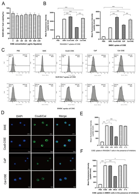
The cytotoxicity of CISE at different squalene concentrations in RAW264.7 cells was evaluated using the MTT assay. As shown in (Figure 2A), cell viability remained above 80% even at a squalene concentration of 1200 μg/mL, indicating low cytotoxicity and good biocompatibility of CISE.
Figure 2.
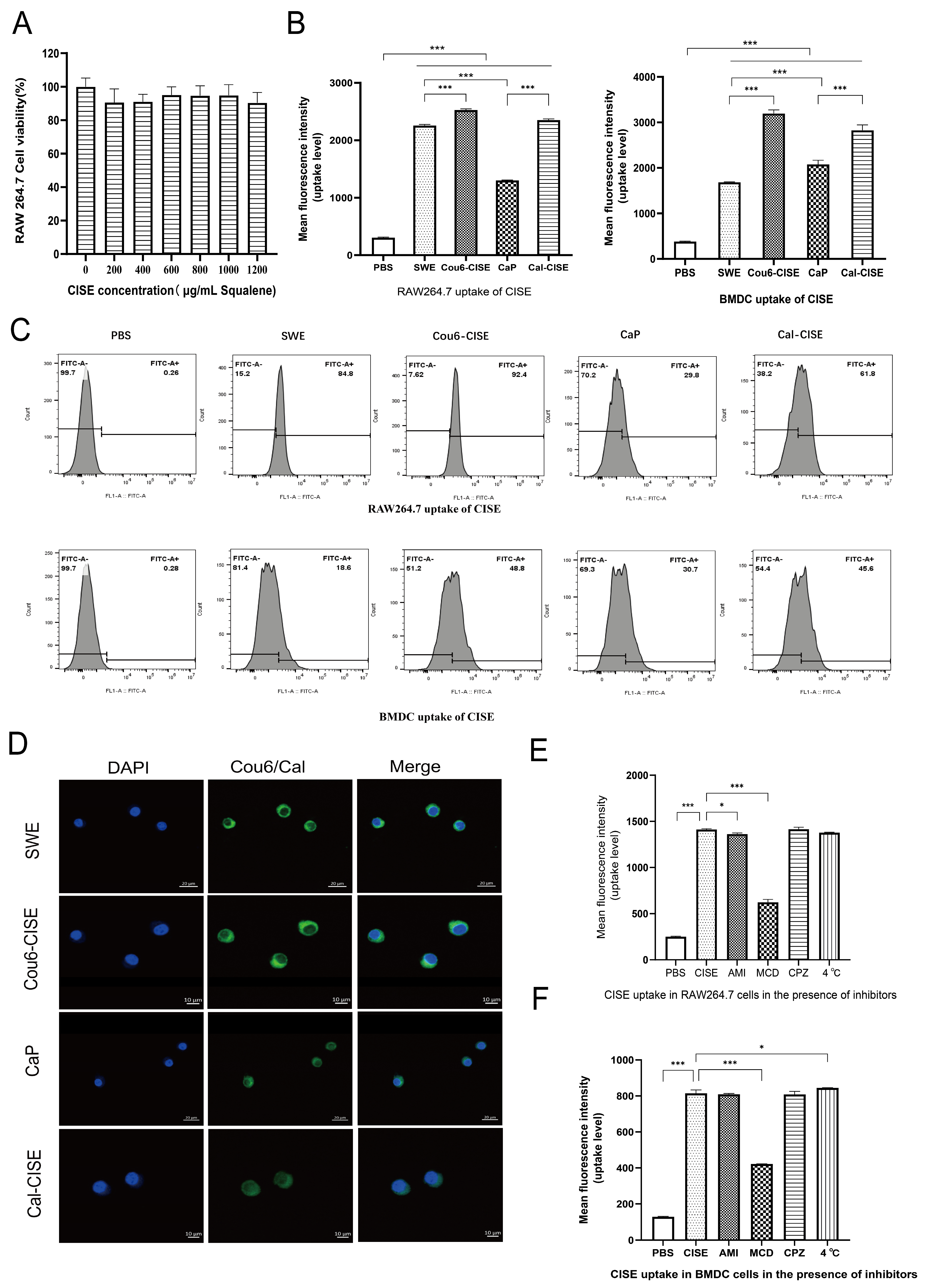
Toxicity and uptake of CISE by APCs and internalization pathways (n = 3). (A) MTT assay showing low cytotoxicity of CISE. (B) Flow-cytometric determination of mean fluorescence intensity (MFI) for the uptake of coumarin-6- and calcein-labeled CISE by APC. (C) Flow-cytometric detection of the uptake rate of CISE by antigen-presenting cells. (D) Confocal microscopy showing RAW264.7 cell nuclei in blue and CISE oil phase and Ca2+ in green. (E) Flow-cytometric analysis of CISE uptake in inhibitor-pretreated RAW264.7, showing decreased mean fluorescence intensity (MFI) upon MCD treatment and indicating predominant caveolin-mediated endocytosis. (F) Flow-cytometric analysis of CISE uptake in inhibitor-pretreated BMDCs, showing decreased mean fluorescence intensity (MFI) upon MCD treatment and indicating predominant caveolin-mediated endocytosis. Data are presented as mean ± SD. Statistical significance was determined using one-way or two-way ANOVA. Asterisks indicate * p < 0.05, *** p < 0.001.
The oil phase of each experimental group was labeled with coumarin-6, and calcium ions were labeled with calcein. RAW264.7 cells and BMDCs were incubated with each group of fluorescent-labeled adjuvants, respectively. Flow-cytometry results (Figure 2B,C) demonstrated that the phagocytic efficiency of CISE in both types of antigen-presenting cells was higher than that of SWE and CaP. Although BMDCs exhibited an uptake trend consistent with RAW264.7 cells, their overall uptake efficiency was lower, which was presumed to be attributable to the relatively low viability of the isolated BMDCs. Laser-scanning confocal microscopy images of RAW264.7 cells (Figure 2D) revealed distinct green fluorescence signals for both squalene and calcium ions, verifying the efficient internalization of CISE by RAW264.7 cells and further corroborating the flow-cytometry findings.
To investigate the mechanism of CISE uptake by antigen-presenting cells, RAW264.7 cells and BMDCs were pretreated with specific endocytic inhibitors. Chlorpromazine (CPZ) inhibits clathrin-mediated endocytosis, methyl-β-cyclodextrin (MCD) inhibits caveolae-mediated endocytosis, amiloride (AMI) inhibits macropinocytosis [25], and incubation at 4 °C blocks energy-dependent uptake [26]. Cells were then incubated with CISE for 2 h. Flow-cytometry results (Figure 2E,F) demonstrated that MCD treatment significantly reduced the mean fluorescence intensity in both RAW264.7 cells and BMDCs compared with the untreated control group, whereas no significant differences in mean fluorescence intensity were observed following treatment with AMI, CPZ, or incubation at 4 °C relative to the control group. These findings indicate that CISE enters cells primarily through the caveolin-mediated endocytosis pathway.
3.3. CISE Promotes the Expression of Co-Stimulatory Molecules in BMDCs and Induces Lysosomal Escape in RAW264.7 Cells
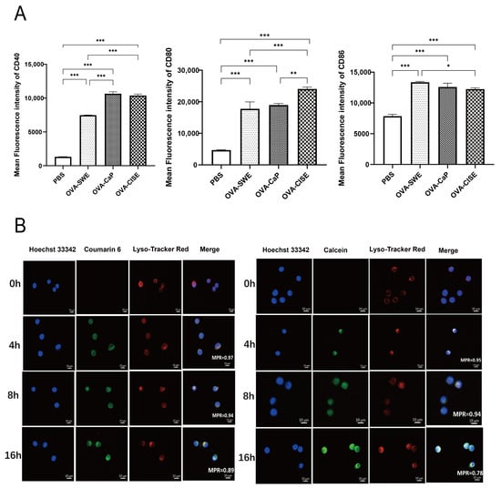
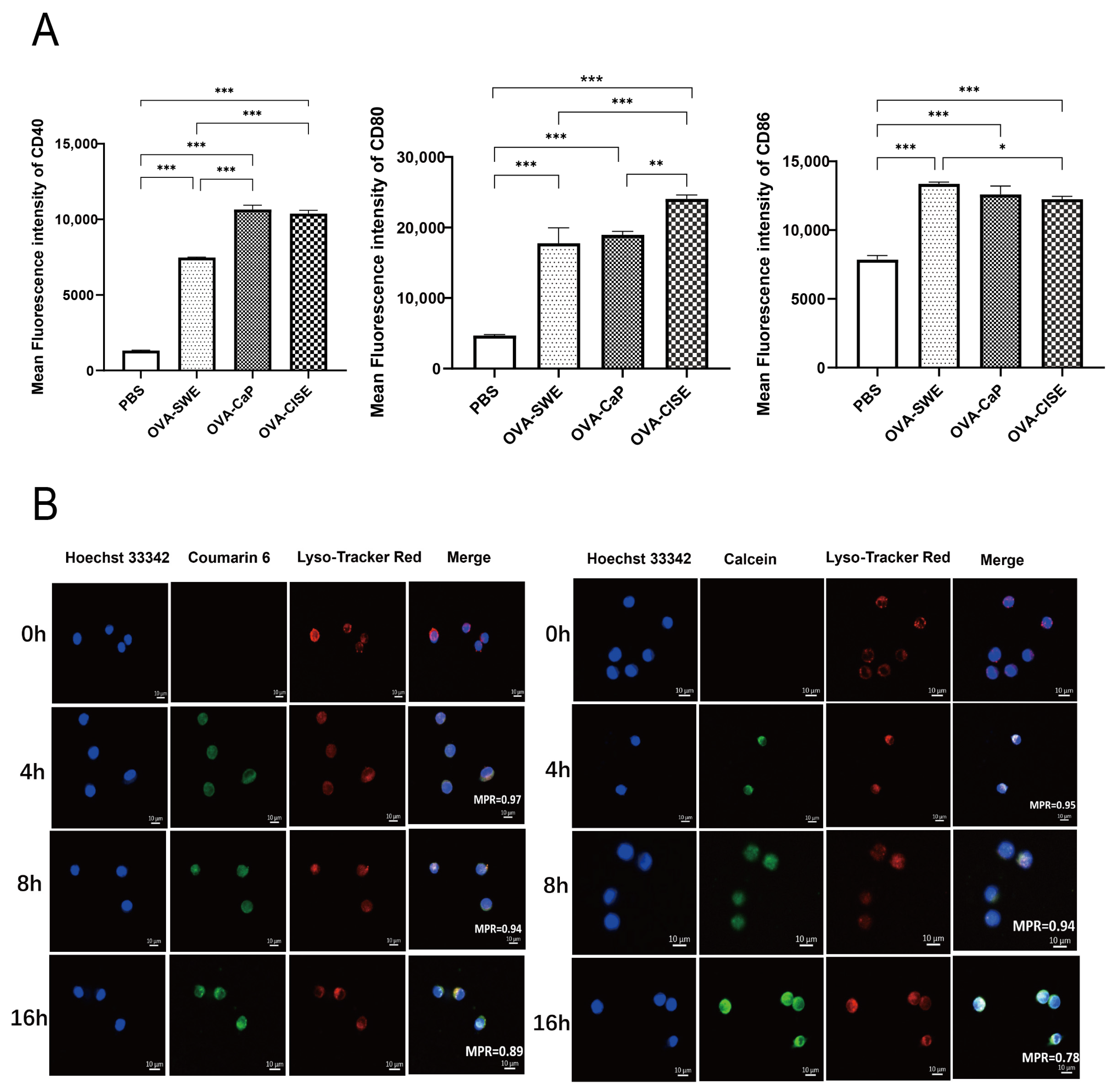
In vitro experiments (Figure 3A) demonstrated that OVA-CISE effectively promoted the maturation and activation of BMDCs. This treatment significantly enhanced T cell immune responses by upregulating the expression of the co-stimulatory molecules CD40, CD80, and CD86 [27]. However, CD83 expression and additional functional parameters were not assessed; therefore, further studies are needed to provide a more comprehensive characterization of CISE-induced BMDC maturation and function. Adjuvant delivery systems with efficient lysosomal escape can rapidly disrupt the endosomal membranes upon internalization, preventing antigen degradation by acidic environments and hydrolytic enzymes. This facilitates antigen entry into the cytosol, effectively activating dendritic cells and other antigen-presenting cells [28]. This promotes MHC class I-mediated cross-presentation and robust induction of both cellular and humoral immune responses [29]. Confocal laser scanning microscopy (Figure 3B) revealed that intracellular CISE did not completely colocalize with lysosomes, suggesting partial lysosomal escape. Further colocalization analysis of APC uptake images using ImageJ showed that Cou6-CISE and Cal-CISE exhibited high colocalization with lysosomes at 4 h and 8 h, but clear escape was observed at 16 h. Notably, the oil-phase component of CISE showed higher Pearson’s correlation with lysosomes compared to the calcium component, likely due to the lipophilic nature of squalene.
Figure 3.
CISE activates BMDCs and induces lysosomal escape in RAW264.7 cells. (A) Flow cytometry showing upregulation of CD40, CD80, and CD86 in BMDCs after CISE treatment. (B) Confocal microscopy showing Cou6-labeled oil phase and Cal-labeled Ca2+ (green) partially separated from LysoTracker-Red (red) in RAW264.7 cells, with Pearson coefficients of 0.89 and 0.78 at 16 h, indicating lysosomal escape. Data are presented as mean ± SD. Statistical significance was determined using one-way or two-way ANOVA. Asterisks indicate * p < 0.05, ** p < 0.01, *** p < 0.001.
3.4. CISE Induces Reactive Oxygen Species Production and Enhances Peroxisome Activity in APCs
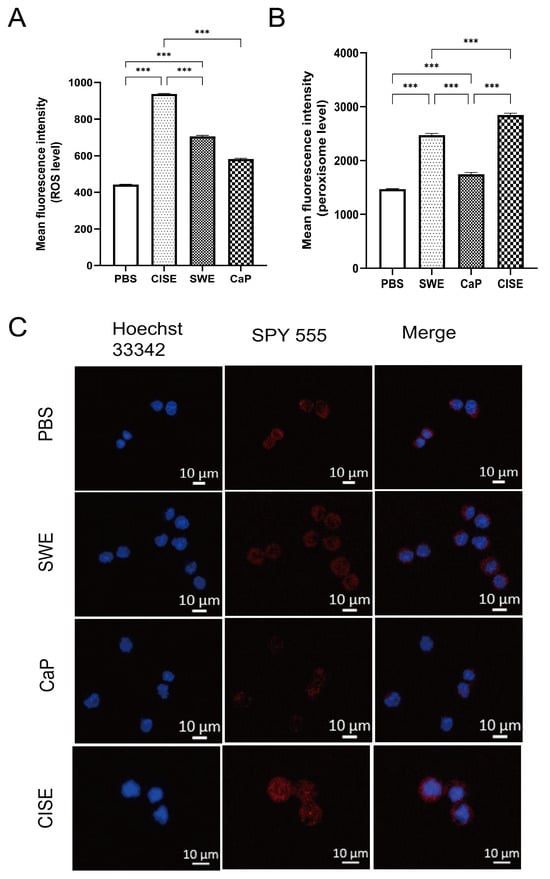
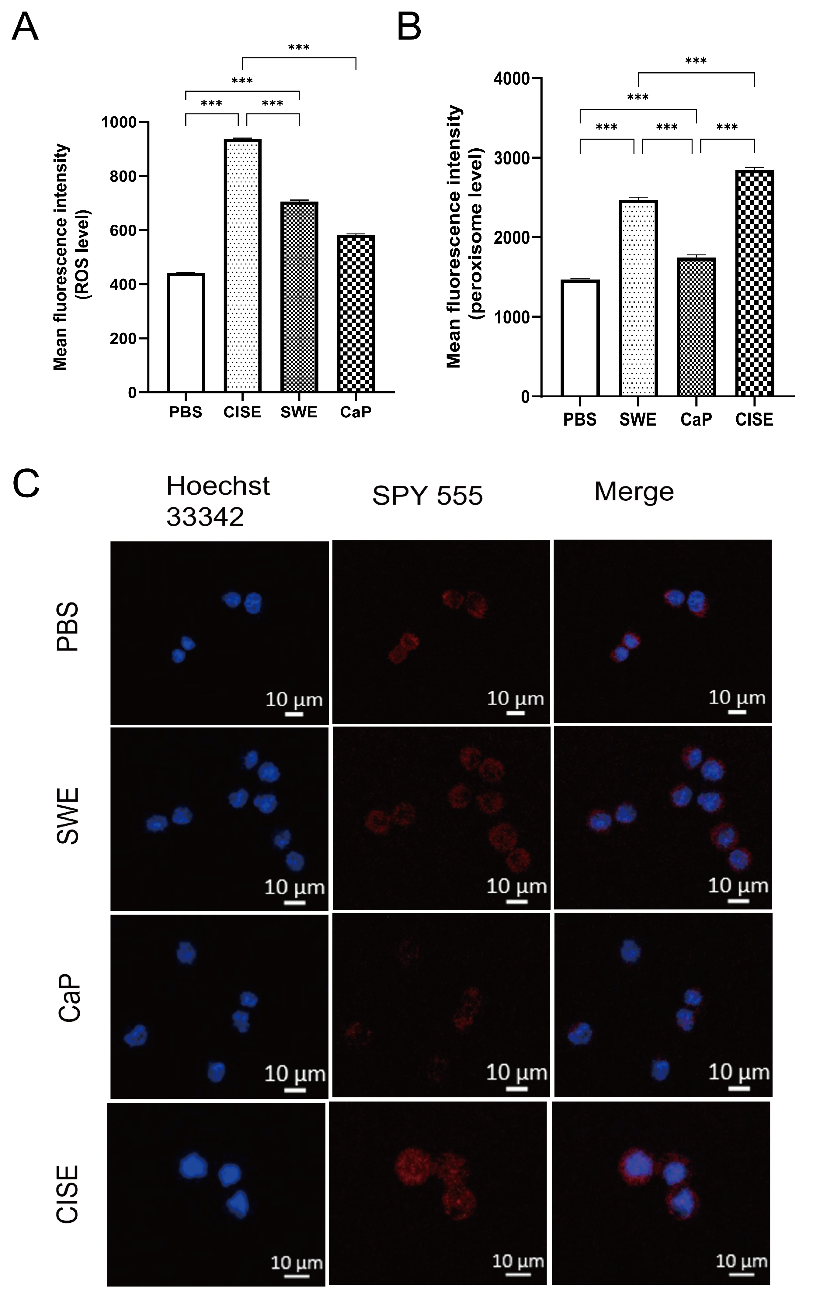
Excessively produced reactive oxygen species (ROS) can cause significant oxidative damage and adverse biological effects in organisms; therefore, ROS have traditionally been regarded as harmful by-products of cellular respiration. Nevertheless, studies have demonstrated that ROS, as important immune regulatory signaling molecules, play a critical role in adjuvant-mediated immune activation [30]. Adjuvants can induce intracellular ROS production through multiple pathways, thereby promoting the maturation and activation of antigen-presenting cells such as dendritic cells, and stimulating the secretion of pro-inflammatory cytokines including IL-12 and TNF-α. These events provide essential conditions for effectively activating naive T cells and initiating adaptive immune responses [31]. Meanwhile, ROS participate in the regulation of antigen processing and presentation, enhance the efficiency of antigen cross-presentation, and further elevate the magnitude of cellular immune responses [32]. In addition, ROS can induce immunogenic cell death to release damage-associated molecular patterns (DAMPs) such as calreticulin and HMGB1, which amplify local inflammation and immune-activating signals, thereby potentiating the immunostimulatory effect of adjuvants. Flow cytometry analysis (Figure 4A) showed that ROS levels induced by CISE were significantly higher than those in the control group, suggesting that CISE may exert its adjuvant effect by generating a controlled amount of ROS.
Figure 4.
CISE induces reactive oxygen species production and enhances peroxisome activity in antigen-presenting cells. (A) Mean fluorescence intensity of intracellular ROS in RAW264.7 cells after CISE treatment. (B) Mean fluorescence intensity (MFI) of intracellular peroxisomes in RAW264.7 cells following CISE treatment. (C) Laser scanning confocal microscopy images: Peroxisomes in RAW264.7 cells labeled with the SPY555 probe after treatment with CISE and control adjuvants, respectively. Data are presented as mean ± SD. Statistical significance was determined using one-way or two-way ANOVA. Asterisks indicate *** p < 0.001.
Peroxisomes are key organelles involved in cellular fatty acid metabolism and the clearance of reactive oxygen species (ROS), maintaining intracellular redox homeostasis through antioxidant enzyme systems such as catalase [33]. During adjuvant-induced immune activation and inflammatory responses, excessive ROS production is often observed, which may lead to oxidative stress and cellular damage. Under these conditions, peroxisomes serve as an important antioxidant defense system, alleviating oxidative damage and maintaining immune cell homeostasis through compensatory proliferation and functional enhancement [34]. Flow cytometry analysis (Figure 4B) showed that the mean fluorescence intensity (MFI) in all treated groups was significantly higher than that in the PBS group, indicating an increase in intracellular peroxisome levels accompanied by ROS production. Confocal laser scanning microscopy (Figure 4C) further demonstrated that the red fluorescence intensity of peroxisomes in the CISE group was markedly higher than that in other groups, corroborating the flow cytometry results. This further indicates that the ROS generation induced by CISE in APCs is tightly regulated and does not result in notable cellular damage.
3.5. CISE Elicits Strong Humoral and Cytokine Responses
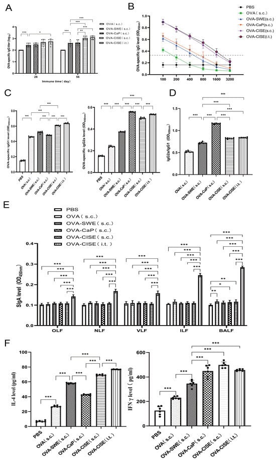
Immunoglobulin G (IgG) is the most abundant and functionally diverse antibody in serum, playing a central role in humoral immune responses by mediating anti-infective and immunoregulatory functions through multiple mechanisms [35]. Secretory immunoglobulin A (sIgA) is the predominant effector antibody in mucosal immunity, primarily present in exocrine secretions covering mucosal surfaces such as the gastrointestinal, respiratory, and reproductive tracts. It serves as the first line of defense against pathogen invasion at mucosal sites [36]. Following two immunizations (Figure 5A,B), OVA-CISE elicited robust IgG responses in mice. Compared with the control groups, OVA-CISE showed superior IgG induction. Notably, the IgG2a/IgG1 ratio in the OVA-CISE group was approximately 1 (Figure 5C,D), similar to that of the SWE group, whereas the IgG2a/IgG1 ratio in the OVA-CaP group was greater than 1, suggesting that squalene may play a dominant role in shaping the immune response. In the subcutaneously administered control groups (Figure 5E), sIgA levels in lavage fluids from various sites were either undetectable or very low. In contrast, pulmonary mucosal administration of CISE resulted in markedly elevated sIgA levels, highlighting a key advantage of lung mucosal immunization over traditional subcutaneous vaccination.
Figure 5.
In vivo evaluation of immune responses induced by CISE. (n = 6) (A) OVA-specific serum IgG titers following two immunizations (n = 6). (B) Serum dilution curves after the second immunization. The dashed line indicates the cutoff value. (C) OVA-specific serum IgG1 and IgG2a levels post-immunization. (n = 6) (D) The serum OVA-specific IgG2a-to-IgG1 ratio was calculated according to the IgG2a and IgG1 levels obtained from ELISA. (n = 6) (E) sIgA levels in lavage fluids from different mucosal sites after two immunizations (n = 6). (F) Cytokine levels in splenocyte supernatants after two immunizations. (n = 6) Data are presented as mean ± SD. Statistical significance was determined using one-way or two-way ANOVA. Asterisks indicate * p < 0.05, ** p < 0.01, *** p < 0.001.
Interferon-gamma (IFN-γ) is a potent immunoregulatory cytokine [37], whereas interleukin-4 (IL-4) primarily orchestrates the initiation and regulation of humoral immune responses [38]. To evaluate the immunological effects of CISE, cytokine levels in splenocyte supernatants were measured. As shown in (Figure 5F), CISE induced significantly higher levels of IL-4 and IFN-γ compared with the control groups, indicating that CISE elicits stronger humoral and cellular immune responses than SWE and CaP.
3.6. In Vivo Safety Evaluation of CISE Showed That OVA-CISE Induces Highly Proliferative Immune Cells and T Cell Responses, Contributing to the Establishment of Effective Immunity
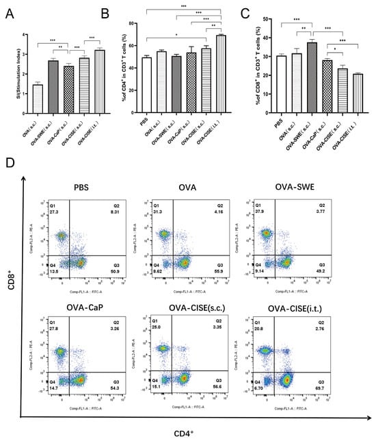
Splenocyte proliferation was assessed using the MTT assay. As shown in (Figure 6A), splenocytes from the CISE-treated group exhibited significantly higher proliferation compared with all control groups, indicating that CISE strongly stimulates splenocyte proliferation. Analysis of splenic lymphocyte subsets (Figure 6D) revealed that the proportion of CD4+ T cells in the CISE group was higher than in the SWE and CaP groups. Regarding CD8+ T cells, the SWE group showed higher levels than both the CaP and CISE groups. We also carried out quantitative analysis for CD4+ and CD8+ T-cell subsets (Figure 6B,C), which further corroborated the flow-cytometry data. No significant changes in body weight were observed following vaccination, and all mice survived during the observation period. Histological examination of the major organs (Figure S1), including the heart, liver, spleen, lungs, and kidneys, revealed no apparent pathological abnormalities at the assessed time point compared with the control group. These findings suggest that CISE was not associated with evident histopathological changes in the examined organs at three days after immunization. However, further studies with additional observation time points are needed to evaluate its longer-term safety.
Figure 6.
T cell subsets, splenocyte stimulation index (SI), and in vivo safety evaluation in immunized mice (n = 6). (A) Splenocyte proliferation stimulation index measured by MTT assay. (B) Quantitative analysis of the proportion of CD4+ T-cell subsets. (C) Quantitative analysis of the proportion of splenic CD8+ T-cell subsets. (D) Proportions of CD4+ and CD8+ T cell subsets in splenocytes after the second immunization. Data are presented as mean ± SD. Statistical significance was determined using one-way or two-way ANOVA. Asterisks indicate * p < 0.05, ** p < 0.01, *** p < 0.001.
4. Discussion
Aluminum-based adjuvants have been widely used in human vaccines for decades, yet they possess inherent drawbacks, such as poor capacity to induce Th1-biased cellular immunity, limited antigen-adsorbing capacity, and potential local inflammatory reactions at the injection site [13,14]. Calcium phosphate-based adjuvants exhibit excellent biocompatibility and low risk of allergic reactions; however, their immunostimulatory potency still needs to be improved for immunization purposes [20]. The efficacy of squalene emulsions as vaccine adjuvants has been well-documented. Therefore, combining squalene emulsion with calcium-based components represents a promising strategy for constructing synergistic adjuvants. SWE, a squalene emulsion analogue, can rapidly recruit antigen-presenting cells at the administration site and establish a pro-inflammatory immune microenvironment. Calcium ions can gradually dissociate and release inside cells, continuously regulating antigen processing and cross-presentation. The rational combination of these two components not only promotes the activation and maturation of antigen-presenting cells but also shapes an overall immune response with higher magnitude and relatively balanced phenotypes. The CISE system constructed in this study integrates the respective advantages of squalene emulsion and calcium ions in activating humoral and cellular immunity, so as to achieve synergistically enhanced immunostimulatory effects.
Naturally derived soy phosphatidylcholine (SPC) was selected as the amphiphilic component for CISE preparation. This molecule contains both positively charged choline head groups and negatively charged phosphate groups, and acts as an emulsifier distributed at the oil-water interface of squalene. Calcium ions bind to the phosphate groups of phospholipids via electrostatic interactions and are localized within the aqueous core of reverse micelles, forming a unique emulsion structure with calcium ions embedded inside reverse micelles. Dark spots inside emulsion droplets observed under transmission electron microscopy verify the existence of calcium-phospholipid reverse micelles, providing direct morphological evidence for the microstructure of CISE. The average particle size of the as-prepared CISE was 124.7 ± 0.6 nm. Nano-emulsions within this size range are readily recognized and taken up by antigen-presenting cells such as dendritic cells, ensuring effective delivery of adjuvant-antigen complexes to the immune system, which serves as the prerequisite for triggering potent immune responses. Stability tests demonstrated that CISE showed no obvious aggregation, demulsification or phase separation for at least 7 d under appropriate storage conditions. The storage stability of vaccine formulations is of great practical significance for vaccine application in resource-limited regions.
In terms of uptake by antigen-presenting cells, this study found that CISE mainly entered cells via the caveolin-mediated endocytic pathway, and the cellular uptake efficiency of CISE was superior to that of SWE and CaP control adjuvants. Results from lysosomal-escape experiments demonstrated that CISE possessed a certain capacity for lysosomal escape. After escaping from lysosomes, antigens can enter the MHC-I pathway and further activate CD8+ cytotoxic T-cell responses [29].
Immune-cell activation constitutes the foundation for exerting immune effects. Under homeostatic conditions, dendritic cells maintain an immature phenotype. Upon recognizing danger signals such as pathogens, they undergo maturation. By up-regulating antigen-presenting and co-stimulatory molecules as well as secreting pro-inflammatory cytokines, dendritic cells can efficiently activate naive T cells and initiate adaptive immunity, which forms a critical immune mechanism for host defense against exogenous invasion. This maturation process is characterized by the up-regulation of molecules including CD40, CD80 and CD86 [39]. In the present study, CISE significantly increased the expression levels of co-stimulatory molecules CD40, CD80 and CD86 on BMDCs, enhanced the output of co-stimulatory signals, and effectively triggered adaptive immune responses.
Adjuvants can induce intracellular ROS production through multiple pathways, thereby promoting the maturation and activation of antigen-presenting cells such as dendritic cells. Meanwhile, ROS can participate in regulating antigen processing and presentation, enhance the efficiency of antigen cross-presentation, and further boost the magnitude of cellular immune responses. In the present study, we observed that CISE could elevate intracellular ROS levels, which may represent one of the mechanisms underlying the immunoadjuvant effects of CISE. We also detected an increase in peroxisome abundance, which constitutes one of the cellular strategies to modulate ROS and keep ROS concentrations within a controllable range.
Cytokine responses and IgG subclass profiles are complementary and collectively reflect the characteristics of the immune response induced by CISE. IFN-γ is generally associated with Th1-type immunity and promotes IgG2a class switching, whereas IL-4 is linked to Th2-type immunity and facilitates IgG1 class switching [40]. Accordingly, the ratio of antigen-specific IgG2a to IgG1 can be used to characterize the type of immune response. In this study, elevated IFN-γ and IL-4 responses were detected in culture supernatants of OVA-restimulated splenocytes, indicating the potential activation of both Th1- and Th2-associated immune pathways. This observation is broadly consistent with the IgG subclass data: the IgG2a/IgG1 ratio in the CISE group was close to 1, with neither subclass showing marked predominance. Collectively, these results indicate that CISE induces a mixed Th1/Th2 immune response. Nevertheless, we acknowledge that although our data suggest concurrent activation of Th1- and Th2-associated pathways, they cannot confirm the existence of a stable Th1/Th2 balance. Further investigations focusing on antigen-specific T-cell subsets and their functional profiles, as well as the mechanisms governing B-cell class switching, are required to fully delineate the nature of the immune response elicited by CISE. The subcutaneous injection control groups only generated high serum antibody titers but barely detectable mucosal sIgA. In contrast, intratracheally instilled OVA-CISE triggered markedly elevated sIgA antibodies at multiple mucosal sites including the respiratory tract and intestine. Pulmonary administration could activate bronchus-associated lymphoid tissue (BALT). The activated lymphocytes migrated to diverse mucosal sites via the common mucosal homing mechanism and achieved multi-site mucosal immune responses, which represents a major advantage inaccessible to conventional subcutaneous vaccines. Splenocyte proliferation and T-cell subset assays revealed that CISE effectively promoted lymphocyte proliferation, increased the proportion of CD4+ T cells, and preserved the functional activity of CD8+ T cells, suggesting that CISE could potentiate both humoral and cellular immune responses simultaneously.
Safety is a prerequisite for the application of vaccines. In the in vivo safety evaluation, immunized mice maintained good mental status without abnormal body weight loss. H&E-stained pathological sections of major organs including the heart, liver, spleen, lungs and kidneys showed no obvious pathological injuries such as inflammatory infiltration or necrosis, which confirmed the low in vivo toxicity of CISE.
Despite these promising findings, several limitations of the present study should be acknowledged. Although elevated sIgA levels in mucosal lavage samples indicated enhanced mucosal antibody responses following pulmonary immunization with CISE, the underlying cellular mechanisms remain incompletely characterized. In particular, we did not directly investigate the phenotypic characteristics, activation status, or functional properties of tissue-resident immune cells in the respiratory mucosa. Therefore, the contribution of these cell populations to the mucosal immune responses induced by CISE remains to be elucidated. Future studies incorporating detailed immunophenotyping and functional analyses of mucosal tissue-resident immune cells will be important for further clarifying the mechanisms of CISE-mediated mucosal immunity.
An additional limitation of this study is that the memory phenotype of splenic T cells was not characterized. Although we analyzed the proportions of splenic CD4+ and CD8+ T cells, we did not distinguish central memory T cells (T_CM) from effector memory T cells (T_EM). These subsets exhibit distinct phenotypic and functional characteristics and may provide complementary information regarding the quality of vaccine-induced cellular immunity. Consequently, the effects of CISE on memory T-cell differentiation and the potential maintenance of long-term cellular immune responses remain unclear. Future studies incorporating additional phenotypic markers, such as CD44 and CD62L, together with functional assessments, will be necessary to characterize these memory T-cell populations and further elucidate the effects of CISE on vaccine-induced cellular immunity. In addition, we have not evaluated the protective efficacy of the CISE-adjuvanted formulation in a pathogen challenge model. Although our findings confirm the immunological effects of CISE, including its modulatory effects on antibody responses and selected cellular immune parameters, these measurements cannot directly demonstrate its protective effects against infection or disease. Further studies using an appropriate challenge model are required to determine whether the immune responses elicited by CISE translate into enhanced protective efficacy.
5. Conclusions
In this study, calcium-ion-entrapped squalene emulsion adjuvant (CISE) was successfully constructed using a simple and feasible preparation process. The formulation exhibits favorable physical storage stability, which facilitates the subsequent development of vaccine preparations. Animal experiments confirmed that intratracheal immunization with CISE could not only elicit high-level systemic OVA-specific IgG antibody responses, but also effectively trigger the production of sIgA antibodies at multiple mucosal sites. It demonstrates unique advantages in activating mucosal immunity compared with conventional subcutaneous vaccine adjuvants. In vitro cellular evaluation combined with in vivo histopathological examination of major organs collectively confirmed the low in vivo toxicity of CISE. In summary, CISE is capable of concurrently activating humoral, cellular and mucosal immune responses, and possesses important reference value for the research and development of mucosal vaccines against respiratory infectious diseases. Upon further evaluation through in-depth mechanistic exploration and pathogen-antigen challenge experiments, CISE holds great promise as a candidate adjuvant for mucosal vaccines, providing new insights and experimental evidence for the development of novel vaccine-delivery systems.
Supplementary Materials
The following supporting information can be downloaded at: https://www.mdpi.com/article/10.3390/vaccines14110924/s1, Figure S1: Histological analysis of heart, liver, spleen, lung, and kidney tissues after CISE or control treatment.
Author Contributions
The authors state that the author contributions to this work are listed as follows: Conceptualization, T.W.; methodology, T.Z., T.W. and Y.C.; software, T.Z.; validation, T.Z., T.W. and Y.C.; formal analysis, T.Z., T.W. and Y.C.; investigation, T.Z.; resources, T.W.; data curation, T.Z. and T.W.; Writing—Original Draft preparation, T.Z.; Writing—Review and Editing, T.W.; visualization, T.Z. and T.W.; supervision, T.W.; project administration, T.W.; funding acquisition, T.W. All authors have read and agreed to the published version of the manuscript.
Funding
This work was partially supported by the Higher Education Institutions Natural Research Planning Project from the Scientific Research Foundation of Education Department of Anhui Province of China (KJ2016SD28).
Institutional Review Board Statement
The animal experimental protocol was approved by the Animal Ethics Committee of Anhui Medical University (Approval No. LLSC20-230287, Approval Date: 20 February 2023).
Data Availability Statement
The original contributions presented in this study are included in the article. Further inquiries can be directed to the corresponding author.
Conflicts of Interest
The authors declare no conflicts of interest.
References
- Wang, S.; Li, W.; Wang, Z.; Yang, W.; Li, E.; Xia, X.; Yan, F.; Chiu, S. Emerging and reemerging infectious diseases: Global trends and new strategies for their prevention and control. Signal Transduct. Target. Ther. 2024, 9, 223. [Google Scholar] [CrossRef] [Scilit] [PubMed]
- De Gaetano, S.; Ponzo, E.; Midiri, A.; Mancuso, G.; Filippone, D.; Infortuna, G.; Zummo, S.; Biondo, C. Global Trends and Action Items for the Prevention and Control of Emerging and Re-Emerging Infectious Diseases. Hygiene 2025, 5, 18. [Google Scholar] [CrossRef] [Scilit]
- Taki, A.; Smooker, P. Small Wonders-The Use of Nanoparticles for Delivering Antigen. Vaccines 2015, 3, 638–661. [Google Scholar] [CrossRef] [Scilit] [PubMed]
- Mazur, N.I.; Higgins, D.; Nunes, M.C.; Melero, J.A.; Langedijk, A.C.; Horsley, N.; Buchholz, U.J.; Openshaw, P.J.; McLellan, J.S.; Englund, J.A.; et al. The respiratory syncytial virus vaccine landscape: Lessons from the graveyard and promising candidates. Lancet Infect. Dis. 2018, 18, e295–e311. [Google Scholar] [CrossRef] [Scilit] [PubMed]
- Liu, H.-H.; Xie, Y.; Yang, B.-P.; Wen, H.-Y.; Yang, P.-H.; Lu, J.-E.; Liu, Y.; Chen, X.; Qu, M.-M.; Zhang, Y.; et al. Safety, immunogenicity and protective effect of sequential vaccination with inactivated and recombinant protein COVID-19 vaccine in the elderly: A prospective longitudinal study. Signal Transduct. Target. Ther. 2024, 9, 129. [Google Scholar] [CrossRef] [Scilit] [PubMed]
- Orosco, F.; Nicdao, D. Next-generation subunit vaccine delivery systems: Design, applications, and prospects. J. Appl. Biol. Biotechnol. 2024, 12, 1–14. [Google Scholar] [CrossRef] [Scilit]
- Zhao, T.; Cai, Y.; Jiang, Y.; He, X.; Wei, Y.; Yu, Y.; Tian, X. Vaccine adjuvants: Mechanisms and platforms. Signal Transduct. Target. Ther. 2023, 8, 283. [Google Scholar] [CrossRef] [Scilit] [PubMed]
- Lavelle, E.C.; McEntee, C.P. Vaccine Adjuvants: Tailoring Innate Recognition to Send the Right Message. Immunity 2024, 57, 772–789. [Google Scholar] [CrossRef] [Scilit] [PubMed]
- Coffman, R.L.; Sher, A.; Seder, R.A. Vaccine Adjuvants: Putting Innate Immunity to Work. Immunity 2010, 33, 492–503. [Google Scholar] [CrossRef] [Scilit] [PubMed]
- Rapaka, R.R. How do adjuvants enhance immune responses? eLife 2024, 13, e101259. [Google Scholar] [CrossRef] [Scilit] [PubMed]
- Stertman, L.; Palm, A.E.; Zarnegar, B.; Carow, B.; Lunderius Andersson, C.; Magnusson, S.E.; Carnrot, C.; Shinde, V.; Smith, G.; Glenn, G.; et al. The Matrix-M™ adjuvant: A critical component of vaccines for the 21(st) century. Hum. Vaccin. Immunother. 2023, 19, 2189885. [Google Scholar] [CrossRef] [Scilit] [PubMed]
- Verma, S.K.; Mahajan, P.; Singh, N.K.; Gupta, A.; Aggarwal, R.; Rappuoli, R.; Johri, A.K. New-age vaccine adjuvants, their development, and future perspective. Front. Immunol. 2023, 14, 1043109. [Google Scholar] [CrossRef] [Scilit] [PubMed]
- Laera, D.; HogenEsch, H.; O’Hagan, D.T. Aluminum Adjuvants-‘Back to the Future’. Pharmaceutics 2023, 15, 1884. [Google Scholar] [CrossRef] [Scilit] [PubMed]
- Xing, J.; Zhao, X.; Li, X.; Fang, R.; Sun, M.; Zhang, Y.; Song, N. The recent advances in vaccine adjuvants. Front. Immunol. 2025, 16, 1557415. [Google Scholar] [CrossRef] [Scilit] [PubMed]
- Imran, M.; Puig-Barbera, J.; Ortiz, J.R.; Fischer, L.; O’Brien, D.; Bonafede, M.; Mansi, J.A.; Boikos, C. Relative Effectiveness of MF59 Adjuvanted Trivalent Influenza Vaccine vs Nonadjuvanted Vaccines During the 2019–2020 Influenza Season. Open Forum Infect. Dis. 2022, 9, ofac167. [Google Scholar] [CrossRef] [Scilit] [PubMed]
- Ko, E.J.; Kang, S.M. Immunology and efficacy of MF59-adjuvanted vaccines. Hum. Vaccines Immunother. 2018, 14, 3041–3045. [Google Scholar] [CrossRef] [Scilit] [PubMed]
- Jie, J.; Liu, G.; Feng, J.; Huo, D.; Wu, Y.; Yuan, H.; Tai, G.; Ni, W. MF59 Promoted the Combination of CpG ODN1826 and MUC1-MBP Vaccine-Induced Antitumor Activity Involved in the Enhancement of DC Maturation by Prolonging the Local Retention Time of Antigen and Down-Regulating of IL-6/STAT3. Int. J. Mol. Sci. 2022, 23, 10887. [Google Scholar] [CrossRef] [Scilit] [PubMed]
- O’Hagan, D.T.; Podda, A. MF59: A safe and potent oil in water emulsion adjuvant for influenza vaccines, which induces enhanced protection against virus challenge. In Influenza Vaccines for the Future; Rappuoli, R., Del Giudice, G., Eds.; Birkhäuser Basel: Basel, Switzerland, 2008; pp. 221–244. [Google Scholar]
- Firdaus, F.Z.; Skwarczynski, M.; Toth, I. Developments in Vaccine Adjuvants. Methods Mol. Biol. 2022, 2412, 145–178. [Google Scholar] [CrossRef] [Scilit] [PubMed]
- Lin, Y.; Wang, X.; Huang, X.; Zhang, J.; Xia, N.; Zhao, Q. Calcium phosphate nanoparticles as a new generation vaccine adjuvant. Expert. Rev. Vaccines 2017, 16, 895–906. [Google Scholar] [CrossRef] [Scilit] [PubMed]
- Johnson, L.; Duschl, A.; Himly, M. Nanotechnology-Based Vaccines for Allergen-Specific Immunotherapy: Potentials and Challenges of Conventional and Novel Adjuvants under Research. Vaccines 2020, 8, 237. [Google Scholar] [CrossRef] [Scilit] [PubMed]
- Teng, Z.; Li, J.; Ren, M.; Xie, Q.; Dong, H.; Zhou, J.; Mu, S.; Bai, M.; Sun, S.; Guo, H. Mechanistic investigation of calcium phosphate mineralization potentiating the cellular immune response of foot-and-mouth disease virus-like particle vaccines. Nanoscale 2025, 17, 15512–15522. [Google Scholar] [CrossRef] [Scilit] [PubMed]
- Lutz, M.B.; Kukutsch, N.; Ogilvie, A.L.J.; Rößner, S.; Koch, F.; Romani, N.; Schuler, G. An advanced culture method for generating large quantities of highly pure dendritic cells from mouse bone marrow. J. Immunol. Methods 1999, 223, 77–92. [Google Scholar] [CrossRef] [Scilit] [PubMed]
- Wang, N.; Wang, T.; Zhang, M.; Chen, R.; Deng, Y. Using procedure of emulsification–lyophilization to form lipid A-incorporating cochleates as an effective oral mucosal vaccine adjuvant-delivery system (VADS). Int. J. Pharm. 2014, 468, 39–49. [Google Scholar] [CrossRef] [Scilit] [PubMed]
- Salloum, G.; Bresnick, A.R.; Backer, J.M. Macropinocytosis: Mechanisms and regulation. Biochem. J. 2023, 480, 335–362. [Google Scholar] [CrossRef] [Scilit] [PubMed]
- Huth, U.S.; Schubert, R.; Peschka-Süss, R. Investigating the uptake and intracellular fate of pH-sensitive liposomes by flow cytometry and spectral bio-imaging. J. Control. Release 2006, 110, 490–504. [Google Scholar] [CrossRef] [Scilit] [PubMed]
- Mbongue, J.C.; Nieves, H.A.; Torrez, T.W.; Langridge, W.H. The Role of Dendritic Cell Maturation in the Induction of Insulin-Dependent Diabetes Mellitus. Front. Immunol. 2017, 8, 327. [Google Scholar] [CrossRef] [Scilit] [PubMed]
- Ten Broeke, T.; Wubbolts, R.; Stoorvogel, W. MHC class II antigen presentation by dendritic cells regulated through endosomal sorting. Cold Spring Harb. Perspect. Biol. 2013, 5, a016873. [Google Scholar] [CrossRef] [Scilit] [PubMed]
- Colbert, J.D.; Cruz, F.M.; Rock, K.L. Cross-presentation of exogenous antigens on MHC I molecules. Curr. Opin. Immunol. 2020, 64, 1–8. [Google Scholar] [CrossRef] [Scilit] [PubMed]
- Lin, W.; Shen, P.; Song, Y.; Huang, Y.; Tu, S. Reactive Oxygen Species in Autoimmune Cells: Function, Differentiation, and Metabolism. Front. Immunol. 2021, 12, 635021. [Google Scholar] [CrossRef] [Scilit] [PubMed]
- Malla, S.; Shahreen, N.; Saha, R. Immunometabolism at the Crossroads of Infection: Mechanistic and Systems-Level Perspectives from Host and Pathogen. Immunometabolism 2025, 7, e00069. [Google Scholar] [CrossRef] [Scilit] [PubMed]
- Ho, N.I.; Huis in ‘t Veld, L.G.M.; Raaijmakers, T.K.; Adema, G.J. Adjuvants Enhancing Cross-Presentation by Dendritic Cells: The Key to More Effective Vaccines? Front. Immunol. 2018, 9, 2874. [Google Scholar] [CrossRef] [Scilit] [PubMed]
- Terlecky, S.R.; Terlecky, L.J.; Giordano, C.R. Peroxisomes, oxidative stress, and inflammation. World J. Biol. Chem. 2012, 3, 93–97. [Google Scholar] [CrossRef] [Scilit] [PubMed]
- Pascual-Ahuir, A.; Manzanares-Estreder, S.; Proft, M. Pro- and Antioxidant Functions of the Peroxisome-Mitochondria Connection and Its Impact on Aging and Disease. Oxid. Med. Cell Longev. 2017, 2017, 9860841. [Google Scholar] [CrossRef] [Scilit] [PubMed]
- Tsaregorodtseva, T.S.; Gubaidullina, A.A.; Kayumova, B.R.; Shaimardanova, A.A.; Issa, S.S.; Solovyeva, V.V.; Sufianov, A.A.; Sufianova, G.Z.; Rizvanov, A.A. Neutralizing Antibodies: Role in Immune Response and Viral Vector Based Gene Therapy. Int. J. Mol. Sci. 2025, 26, 5224. [Google Scholar] [CrossRef] [Scilit] [PubMed]
- Mantis, N.J.; Rol, N.; Corthésy, B. Secretory IgA’s complex roles in immunity and mucosal homeostasis in the gut. Mucosal Immunol. 2011, 4, 603–611. [Google Scholar] [CrossRef] [Scilit] [PubMed]
- Kiritsy, M.C.; Ankley, L.M.; Trombley, J.; Huizinga, G.P.; Lord, A.E.; Orning, P.; Elling, R.; Fitzgerald, K.A.; Olive, A.J. A genetic screen in macrophages identifies new regulators of IFNγ-inducible MHCII that contribute to T cell activation. eLife 2021, 10, e65110. [Google Scholar] [CrossRef] [Scilit] [PubMed]
- Keegan, A.D.; Leonard, W.J.; Zhu, J. Recent advances in understanding the role of IL-4 signaling. Fac. Rev. 2021, 10, 71. [Google Scholar] [CrossRef] [Scilit] [PubMed]
- Hancock, W.W.; Sayegh, M.H.; Zheng, X.-G.; Peach, R.; Linsley, P.S.; Turka, L.A. Costimulatory function and expression of CD40 ligand, CD80, and CD86 in vascularized murine cardiac allograft rejection. Proc. Natl. Acad. Sci. USA 1996, 93, 13967–13972. [Google Scholar] [CrossRef] [Scilit] [PubMed]
- Grødeland, G.; Fossum, E.; Bogen, B. Polarizing T and B Cell Responses by APC-Targeted Subunit Vaccines. Front. Immunol. 2015, 6, 367. [Google Scholar] [CrossRef] [Scilit] [PubMed]

Disclaimer/Publisher’s Note: The statements, opinions and data contained in all publications are solely those of the individual author(s) and contributor(s) and not of MDPI and/or the editor(s). MDPI and/or the editor(s) disclaim responsibility for any injury to people or property resulting from any ideas, methods, instructions or products referred to in the content. |
© 2026 by the authors. Licensee MDPI, Basel, Switzerland. This article is an open access article distributed under the terms and conditions of the Creative Commons Attribution (CC BY) license.