


Abstract
Background: Mitochondria are the primary organelles that regulate cellular bioenergetic metabolism and maintain homeostasis, providing essential structural support for optimal cell survival. Nonetheless, advancing age leads to cumulative damage to mitochondrial structure and functional integrity, which is a defining characteristic of biological aging and is closely linked to the emergence and progression of numerous age-related diseases, including neurodegenerative disorders, cardiovascular diseases, and metabolic disorders. Scope of review: This article offers a thorough summary and review of mitochondrial quality control (MQC), emphasizing numerous critical processes, including mitochondrial biosynthesis, dynamic remodeling (fusion and fission), and mitophagy. We thoroughly elucidate the molecular pathways that regulate MQC and demonstrate how age-related dysregulation precipitates cellular senescence, highlighting the transition from physiological maintenance to pathological malfunction, which ultimately culminates in cellular aging. Conclusions and implications: This study systematically elaborates the pathophysiological mechanisms in the field, comprehensively evaluates the clinical translational potential of targeting the MQC pathway, highlights the key objectives of “restoring mitochondrial plasticity and removing dysfunctional mitochondria”, and explores novel intervention strategies. The restoration of normal mitochondrial function in cells throughout aging is a very promising path for precision medicine therapeutics with great translational potential, according to recent state-of-the-art research. The development of novel therapeutic approaches to improve functional healthy mitochondria can effectively delay aging and reduce the rising global burden of age-related diseases.
1. Introduction
The aging global population presents substantial challenges to existing healthcare systems, particularly highlighted by the rising prevalence of chronic conditions such as neurodegenerative disorders, cardiovascular failure, and sarcopenia. Increasing data indicate that mitochondrial dysfunction is not simply a passive result of aging but a fundamental factor that actively accelerates organismal aging [1,2]. Mitochondria, as the metabolic center of the cell, are crucial for energy conversion, redox signaling, and directly affecting cell fate. Mitochondrial homeostasis is essential for maintaining cellular health, a process carefully governed by mitochondrial quality control (MQC) processes [3].
In physiological conditions, MQC functions as an integrated, dynamic monitoring system. It regulates mitochondrial biogenesis and morphological remodeling (fusion/fission dynamics) through the peroxisome proliferator-activated receptor γ coactivator 1-alpha-sirtuin 1 (PGC1α-SIRT1) axis, while concurrently recognizing and removing damaged organelles via targeted mitophagy [4]. Nonetheless, the aging process is often associated with a comprehensive breakdown of this surveillance system. The efficacy of MQC declines with age, as evidenced by diminished bioenergetic signaling and altered mitophagic flux. This deterioration leads to the intracellular buildup of fragmented mitochondria, thus ensnaring cells in a self-sustaining cycle of oxidative stress and cellular senescence.
Contemporary research endeavors have primarily focused on molecular signaling pathways and therapeutic potentials in isolation or have been restricted to individual disease models. This work aims to elucidate the role of dysregulated MQC in the progression of several age-related diseases. Additionally, the potential impact of contemporary therapies, including mitochondrial transplantation and nicotinamide adenine dinucleotide (NAD+) supplementation, on the delay of the aging process is examined in this study. The objective is to implement comprehensive strategies that will alleviate the progressively severe adverse effects of age-related diseases.
2. Methods
To thoroughly clarify the mechanisms of MQC in aging across diverse diseases and investigate potential strategies for postponing aging via the targeted modulation of mitochondrial quality control, we performed a systematic literature review encompassing primary biomedical databases such as PubMed/Medline, Web of Science, Scopus, and the Cochrane Library. The research utilized a retrieval strategy that merged Boolean operators (AND, OR, NOT) with Medical Subject Headings (MeSH), encompassing three principal concepts: (1) fundamental MQC mechanisms (including mitochondrial synthesis, the mitotic chaperone system, dynamic mitochondrial regulation, mitochondrial autophagy, and mitochondrial translocation); (2) age-associated diseases (including neurodegenerative disorders, cardiovascular conditions, metabolic diseases, and cancers); and (3) translational medicine approaches (such as small-molecule inhibitors and biomarkers). Secondly, we conducted a manual assessment of the reference lists from the selected papers to confirm that no essential research was overlooked. The inclusion criteria for literature were tightly confined to original research articles, systematic reviews, and meta-analyses published in English and subjected to peer review.
3. Integrated Molecular Framework of the Mitochondrial Quality Control Network
The regulation and maintenance of mitochondrial homeostasis are governed by a complex, hierarchical interconnected network. This network regulates critical activities such as mitochondrial biosynthesis, dynamic remodeling, and autophagy, and adeptly translates metabolic information into mitochondrial adaptive responses via unique post-translational modifications and interorgan communication [5,6] (Figure 1).
Figure 1.
The fundamental mechanism of mitochondrial quality control (MQC) for preserving cellular homeostasis. This model outlines four coordinated methods of mitochondrial quality regulation. Cellular stress, such as physical exertion or starvation, increases the AMP/ATP ratio and activates the AMPK/SIRT1–PGC-1α signaling pathway, which works in conjunction with NRF1/2 to promote TFAM expression and augment mitochondrial biogenesis. Mitochondrial fusion and fission are regulated by OPA1, MFN1/2, and DRP1, enabling network reorganization and the separation of dysfunctional mitochondria. Mitochondrial stress activates UPRmt through ATF5 and NRF2/ARE signaling, resulting in the expression of protective genes. Defective mitochondria are removed through mitophagy via NIX/FUNDC1–LC3-mediated ubiquitin-independent pathways or PINK1/Parkin-mediated ubiquitin-dependent pathways. Moreover, dysfunctional mitochondria can be transported along microtubules and actin filaments by KIF5B and Myo19, and later eliminated via mitocytosis into migrasomes. These systems collectively maintain mitochondrial homeostasis and cellular life. Created in BioRender (2026). 18 June 2026. https://BioRender.com/dlmsfxh.
3.1. Mitochondrial Biogenesis
Mitochondrial biogenesis is a dynamic process involving the utilization of pre-existing mitochondria by cells to generate new ones. This mechanism requires synchronized regulation between the nuclear genome and the mitochondrial genome to maintain homeostasis in mitochondrial quantity, function, and cellular energy [7,8].
The regulation of mitochondrial biogenesis constitutes a multi-level cascade network, with its core primarily mediated by the transcription co-activator PGC-1α [9]. The activated PGC-1α enhances its transcriptional activity by binding to nuclear respiratory factors (NRF-1 and NRF-2) and further recruiting epigenetic enzymes, thereby inducing the transactivation of various nuclear-encoded mitochondrial structural and functional genes by the NRFs. Most critically, it upregulates the expression of mitochondrial transcription factor A (TFAM), which promotes mtDNA replication and the transcription of core subunits involved in oxidative phosphorylation, thereby facilitating mitochondrial biogenesis [8,10,11]. This metabolic process is closely linked to the cellular bioenergetic signaling pathway involving adenosine monophosphate (AMP)-dependent protein kinase (AMPK) and SIRT1. When cells enter an energy-deficient state due to movement, starvation, or metabolic stress, the elevated AMP/ATP ratio promotes the activation of AMPK, which directly facilitates the phosphorylation of PGC-1α, thereby enhancing its transcriptional activity, and AMPK increases mitochondrial biosynthesis by reducing the repressive influence of transcription factor EB (TFEB) via mechanistic target of rapamycin complex 1 (mTORC1) contact inhibition [12,13,14,15]. Recent studies demonstrate that AMPK directly phosphorylates five conserved serine residues in folliculin interacting protein 1 (FNIP1), thereby inhibiting the RagC GAP activity of the folliculin (FLCN)–FNIP1 complex. This alteration facilitates the detachment of mTORC1 from the lysosomal membrane, resulting in the nuclear translocation of TFEB, even in the presence of sufficient amino acids. Consequently, this signaling cascade enhances the expression of genes related to mitochondrial biogenesis [16]. Simultaneously, AMPK also enhances intracellular NAD+ levels by phosphorylating the NAMPT enzyme, thereby indirectly activating the deacetylase SIRT1, which enhances PGC-1α activity via deacetylation. This dual regulatory system satisfies the energy requirements of cellular metabolism by enhancing mitochondrial biosynthesis [17]. Furthermore, calcium ions (Ca2+) function as an essential reverse signaling molecule. A reduction in mitochondrial membrane potential (Δψm) diminishes the mitochondria’s capacity to sequester Ca2+, resulting in an elevation of cytoplasmic Ca2+ concentration. The elevated cytoplasmic Ca2+ concentration modulates the expression and transcriptional activity of PGC-1α via activating Ca2+-dependent kinases, such as CaMKII and PKC, as well as the phosphatase calmodulin kinase [18].
3.2. Mitochondrial Unfolded Protein Response, UPRmt
The mitochondrial unfolded protein response (UPRmt) is a retrograde signaling system activated by disturbances in mitochondrial proteostasis. When physiological or pathological stressors disrupt the balance between the synthesis of proteins encoded by mitochondrial DNA (mtDNA) and those encoded by nuclear DNA (nDNA), it leads to the abnormal accumulation of unassembled protein subunits within the mitochondrial matrix. Additionally, with aging, the excessive generation of ROS in mitochondria can cause oxidative damage to mitochondrial proteins. This damage can alter their original spatial conformation, induce protein denaturation, and ultimately contribute to the formation of various protein aggregates. Therefore, excessive accumulation of misfolded or not assembled proteins in the mitochondrial matrix caseinolytic protease P (CLPP) degrades them into peptide fragments, which are then transported to the intermembrane space by the inner membrane ABC transporter HAF-1 [19]. Consequently, these signals facilitate the nuclear translocation of the transcription factor activated transcription factor atfs-1 (ATFS-1) (or its human equivalent, ATF5), resulting in the transcriptional upregulation of nuclear genes that encode mitochondrial molecular chaperones, including HSP60 and HSP10, along with proteases [20]. Mitochondrial reactive oxygen species (mtROS) function as signaling molecules that trigger an antioxidant response mediated by NFE2L2 (NRF2). Activated NFE2L2 increases its expression and stimulates mitochondrial biosynthesis via attaching to the antioxidant response element (ARE) in the NRF1 promoter [21,22]. Furthermore, ATF4, CHOP, and C/EBPβ also participate in the regulation of this pathway; phosphorylation of eIF2α promotes the translation of CHOP, ATF4, and ATF5 mRNA, thereby amplifying the UPRmt signaling pathway [21,22].
3.3. Mitochondrial Dynamics
Mitochondria function as a highly dynamic regulatory network within cells. The equilibrium between the processes of mitochondrial fusion and fission determines the stability of this dynamic network. Recent study has concentrated on the mechanisms of mitochondrial fusion and fission as well as the regulatory networks controlling connections between various organelles.
Mitofusin 1/2 (MFN1/2) on the outer mitochondrial membrane mediates mitochondrial fusion; optic atrophy 1 (OPA1) on the inner membrane mediates inner membrane fusion [23,24]. Through their GTPase domains, MFN1 and MFN2 facilitate homotypic or heterotypic dimerization, which connects neighboring mitochondria and encourages the fusing of the outer mitochondrial membrane [25]. Thereafter, the proteolytic cleavage of OPA1 splice variants by the proteases OMA1 and YME1L produces the active forms required for the fusing of the inner mitochondrial membrane [26,27]. Mitochondrial fission is predominantly regulated by the cytosolic dynamin-related protein DRP1. DRP1 is attracted to the mitochondrial surface by receptor proteins such as MFF, MID49/51, and FIS1. Upon recruitment, it forms helical structures around the constriction sites and employs GTP hydrolysis to facilitate membrane scission, ultimately resulting in mitochondrial division [28,29]. The processes of mitochondrial fusion and division do not occur in isolation but are precisely regulated to maintain dynamic homeostasis.
The maintenance of mitochondrial dynamic equilibrium relies on the coordinated regulation of multiple molecular mechanisms. PINK1/Parkin enhances the proteasomal destruction of MFN2 during mitochondrial fusion by promoting its ubiquitination, therefore aiding in the removal of damaged mitochondria. The deubiquitinating enzyme USP30 counteracts Parkin’s actions by removing ubiquitin chains from MFN1 and MFN2, therefore preserving mitochondrial integrity [30,31]. DRP1, the principal regulator of mitochondrial fission, relies on a multi-site phosphorylation process for its action. Phosphorylation of DRP1 at Ser637 by PKA obstructs its translocation to the mitochondria, thereby impeding the fission process. Conversely, calcineurin-mediated dephosphorylation at this locus facilitates mitochondrial fission. Furthermore, during mitosis, CDK1 phosphorylates DRP1 at Ser616, promoting mitochondrial fragmentation [32,33,34]. Moreover, SUMOylation is essential for the regulation of DRP1 stability. Research has shown that SENP3-facilitated deSUMOylation improves the mitochondrial translocation efficiency of DRP1 [35]. The endoplasmic reticulum-mitochondrial contact sites (MAMs) serve as critical structural hubs for interorganelle communication. Within these microdomains, the endoplasmic reticulum pre-suction sites regulate actin polymerization through INF2, thereby facilitating the mitochondrial translocation of DRP1 [36]. Furthermore, Bcl-2 family proteins directly regulate mitochondrial fusion and fission dynamics. Bcl-xL selectively binds to the C-terminal domain of DRP1 via its BH4 domain, thereby diminishing DRP1′s GTPase activity and its translocation to the mitochondria. This activity opposes excessive mitochondrial fragmentation and maintains network integrity [37,38,39]. Conversely, the pro-apoptotic protein Bax has been shown to facilitate fission site selection by building complexes with DRP1 and MFN2, a process that is independent of Bax-induced mitochondrial pore development [40,41]. Additionally, Bcl-xL directly interacts with MFN1 and MFN2, augmenting their GTPase activity to facilitate outer mitochondrial membrane fusion [42]. Conversely, Mcl-1 associates with OPA1 to preserve inner mitochondrial membrane fusion and cristae structure; thus, Mcl-1 knockdown cells demonstrate abnormal OPA1 processing and cristae enlargement [43].
3.4. Mitophagy
When mitochondrial damage exceeds the reparative and fusion systems’ compensatory capacity, cells initiate mitophagy to remove severely damaged mitochondria. Mitophagy is driven synergistically by two complementary pathways—ubiquitin-dependent and ubiquitin-independent—to ensure the selective removal of damaged mitochondria.
Mitophagy mostly relies on the PINK1-Parkin pathway, which is dependent on ubiquitin. In functional mitochondria, PINK1 (PTEN-induced kinase 1) is translocated to the inner mitochondrial membrane through the TOM/TIM complex, where it is swiftly degraded after cleavage by the PARL protease [44]. Upon the loss of Δψm, PINK1 stabilizes on the outer mitochondrial membrane, accumulating at the TOM complex, where it undergoes autophosphorylation and activation [45]. Activated PINK1 phosphorylates ubiquitin (at Ser65) and the ubiquitin-like domain of Parkin (at Ser65), thereby synergistically recruiting and activating the cytosolic E3 ubiquitin ligase Parkin [46]. The activated Parkin protein promotes the assembly of polyubiquitin chains at lysine 63 (Lys63) and Lys48 residues on mitochondrial outer membrane proteins, including MFN1/2, VDAC, and TOM20 [47]. These ubiquitin chains are identified by autophagy receptors such as p62/SQSTM1, OPTN, NDP52, and TAX1BP1, which then associate with LC3 on the autophagosome membrane. This connection enables the localization of damaged mitochondria to autophagosomes, thus finalizing the mitophagy process [46,48]. Recent research reveals that OPTN and NDP52 function as principal downstream autophagy receptors for Parkin, with their activity further enhanced by TBK1-mediated phosphorylation, establishing a positive feedback loop that improves mitophagy efficiency [49,50]. The ubiquitin-independent process is directly facilitated by intrinsic mitochondrial membrane proteins. Proteins including NIX/BNIP3L, FUNDC1, and BCL2L13 engage with LC3 via their LIR motifs to commence the autophagic process, a mechanism additionally enhanced by HIF-1α under hypoxic conditions [51,52]. Furthermore, other proteins located in the inner membrane and cristae, including FKBP8, Prohibitin 2, and PHB2, have been identified as receptors that promote mitophagy [53].
AMPK has a crucial coordinating role in mitophagy. AMPK promotes the recruitment of DRP1 to the mitochondrial surface by directly phosphorylating mitochondrial fission factor (MFF) at Ser155 and Ser172, leading to mitochondrial fragmentation. This fragmentation generates substrates of appropriate dimensions for mitophagy and enables phosphorylated MFF to serve as a signaling platform for the recruitment of autophagy receptors [54,55]. Cellular energy deprivation, characterized by an increased AMP/ATP ratio, activates AMPK, which subsequently phosphorylates the activation site Ser555 of ULK1 kinase, hence facilitating autophagosome nucleation and particularly activating mitophagy [55]. Moreover, AMPK alleviates the inhibition of ULK1 by mTORC1 via the phosphorylation of Raptor (a subunit of mTORC1), so creating a positive feedback amplification loop along the AMPK-ULK1 pathway [56]. A recent study indicates that AMPK has a unique role in regulating several mitophagy activities. The primary manifestations are as follows. In the non-ubiquitination-dependent mitophagy pathway, AMPK recruits 14-3-3 proteins by phosphorylating the Ser556 and Ser694 residues of ULK1 to inhibit NiX-dependent mitophagy; conversely, in the ubiquitin-dependent mitophagy pathway, AMPK directly phosphorylates Parkin’s Ser108, thereby augmenting Parkin-dependent impaired mitophagy [57]. Additionally, acetyl-CoA is involved in regulating mitophagy. With enough nutrition, the LRR domain of the endogenous ligand of acetyl-CoA, the mitochondrial NOD-like receptor NLRX1, binds to acetyl-CoA, maintaining it in a self-inhibitory closed conformation and obscuring the LIR motif to obstruct mitophagy. During hunger, NLRX1 oligomerizes, hence diminishing its affinity for acetyl-coenzyme. This leads to LIR exposure, which subsequently attracts LC3 and initiates autophagy [57].
3.5. Mitocytosis
Mitocytosis, a distinctive method of mitochondrial secretion, was first identified by Yu Li’s team at Tsinghua University in 2021. It refers to the process by which migratory cells selectively eliminate defective mitochondria through migrasome formation. Mitocytosis primarily entails the selective transport of dysfunctional mitochondria and the necessary positioning of the cytoskeleton. The principal procedure is as follows. Damaged mitochondria are conveyed to the cell periphery by the microtubule motor protein KIF5B and are associated with the cortical actin network via myosin 19, situating them adjacent to the site of migration body formation. The mitochondrial tubular structures extending to the cell membrane are cleaved by DRP1, producing migrasomes that encapsulate fragmented mitochondria around 240 nanometers in diameter, which are then expelled from the cell and disseminated throughout the body [58,59]. This process contrasts with the intracellular autophagosome-lysosome degradation pathway reliant on mitochondrial autophagy; rather, it alleviates the accumulation of damaged mitochondria within cells via a coordinated mechanism of “migration structure-extracellular release”, thus preserving mitochondrial membrane potential, respiratory function, and cellular viability. It is especially appropriate for cell types exhibiting significant migratory activity, such as neutrophils [60,61].
The mitocytosis selection mechanism depends on the recognition and translocation of impaired mitochondria. Mitochondria with decreased membrane potential and increased ROS levels are more easily translocated to the cell periphery and integrated into migrasomes [58]. In this mechanism, DRP1 segregates injured mitochondria from the mitochondrial network; thereafter, kinesin family member 5B (KIF5B) conveys them to the cell membrane, where myosin 19 (Myo19) aids in their localization to cortex-rich areas. Consequently, the execution of mitocytosis necessitates synchronized interactions among mitochondrial dynamics, motor protein transport, and migrasome biogenesis [62].
The interaction between integrins and particular extracellular matrix dictates the localization and efficacy of migratory bodies. The interaction between integrin α5 and fibronectin is the most thoroughly defined model to date [63,64]. The transmembrane protein TSPAN4 and cholesterol are essential for the excretion of migratory bodies: TSPAN4 enhances the aggregation of membrane domains and preserves membrane structural integrity, while cholesterol aids in cell membrane fusion; collectively, they facilitate the formation of retracted fibers [61,65,66].
Recent studies have elucidated that the initiation of migratory bodies is meticulously regulated by lipid and calcium signaling: sphingolipid synthase 2 (SMS2) establishes a fixed focal point at the leading edge of migrating cells, facilitating migratory body growth by converting ceramide to sphingolipids; phosphatidylinositol-4-phosphate 5-kinase 1α (PIP5K1A) synthesizes phosphatidylinositol-4,5-diphosphate (PI(4,5)P2) at the formation site, which recruits Rab35, thereby creating a platform for subsequent tetraspanin-dependent expansion through interaction with integrin α5 [67]. The calcium ion (Ca2+) sensor synaptotagmin-1 localizes to the migratory body formation site prior to TSPAN4 recruitment, inducing unstable membrane bulging, which is subsequently stabilized in sequence by tetraspanins [68].
4. The Deterioration of Mitochondrial Quality Control: A Primary Catalyst of Aging
Aging signifies a progressive, systemic decline in physiological integrity, marked by a decrease in the organism’s intrinsic regenerative capacity and an increased vulnerability to degenerative diseases [69]. The systemic breakdown of the MQC network serves as a fundamental molecular mechanism influencing the processes of physiological and pathological aging [70,71]. The disruption of MQC triggers a multi-tiered cascade of failures, resulting in the accumulation of damaged mitochondria and the onset of cellular senescence (Figure 2).
Figure 2.
The disruption of the mitochondrial quality control network is a crucial biological mechanism that drives cellular senescence. With aging, mitochondrial homeostasis progressively deteriorates, intensifying the advancement of cellular senescence. Telomere erosion activates the p53 protein, which subsequently inhibits the expression or activity of PGC-1α. Following the functional decline of PGC-1α, its downstream targets NRF1 and TFAM exhibit diminished activity, thereby suppressing mtDNA replication and impairing mitochondrial biogenesis. Furthermore, aging disturbs the dynamic balance between mitochondrial fusion (MFN1/2, OPA1) and fission (DRP1, MFF, FIS1), resulting in the overproduction of ROS and facilitating the release of damage-associated molecular patterns (DAMPs), including free mtDNA and cardiolipin, into the cytoplasm, thereby hastening cellular senescence. Ultimately, compromised mitochondrial autophagy with aging leads to the abnormal accumulation of dysfunctional mitochondria; these inadequately removed damaged mitochondria subsequently initiate an inflammatory cascade that propels the advancement of cellular senescence. Created in BioRender (2026). 18 June 2026. https://BioRender.com/5t6ya9a.
4.1. The Deterioration of Biogenesis: A Systemic Failure in Nuclear-Mitochondrial Communication
The decline in mitochondrial biogenesis with aging is not merely due to a decrease in mitochondrial numbers, but rather results from the dysregulation of the mitochondrial renewal system, which includes impaired mitochondrial biogenesis, ineffective clearance of damaged mitochondria, and asynchrony in bidirectional nuclear-mitochondrial communication [72,73]. Under typical circumstances, cells regulate mitochondrial biogenesis by integrating nutritional status, energy stress, redox state, and mitochondrial gene expression via the AMPK–SIRT1/SIRT3–PGC-1α–NRF1/2–TFAM pathway. Nevertheless, certain variables associated with aging can interfere with this process. Research has shown that elevated production of NAD+-depleting enzymes such CD38 results in reduced NAD+ levels, thereby impairing SIRT1/SIRT3 action [74]. Moreover, DNA damage and telomere attrition associated with aging activate p53, thereby inhibiting PGC-1α production [75,76]. Decreased PGC-1α levels, as a fundamental element of mitochondrial biogenesis, expedite vascular aging and neural metabolic impairment [72,73,77]. Ultimately, chronic inflammation, dysregulated nutrition sensing, and sedentary behavior in aging hinder AMPK’s responsiveness to energy cues, hence further suppressing PGC-1α production. Caloric restriction successfully alleviates hepatic aging, whereas exercise partially reinstates the reduction in PGC-1α-mediated mitochondrial translation and rescues reduced respiratory chain activity in old skeletal muscles [78,79,80]. Consequently, diminished mitochondrial biogenesis with aging contributes to the aging process via many methods. The reduction in mitochondrial quantity results in insufficient ATP synthesis and inadequate respiratory reserves in cells, significantly compromising high-energy-demanding tissues such as skeletal muscle, heart, neurons, and renal tubules [77,81]. Conversely, compromised mitochondrial biogenesis can hinder the respiratory chain, resulting in electron leakage and the excessive production of reactive oxygen species, thereby further undermining the mitochondrial biogenesis process [82]. Mitochondrial biogenesis depends on autophagy for the prompt removal of damaged mitochondria. With aging, reduced autophagy and inadequate mitochondrial synthesis lead to the buildup of impaired mitochondria. Damaged mitochondria produce mtDNA and cardiolipin, subsequently activating pathways such cGAS-STING and inflammasomes, hence intensifying chronic inflammation in senescent cells [83,84].
4.2. Stagnant Dynamics: The Senescence-Associated Mitochondrial Dysfunction
The senescence-associated systemic dysregulation of mitochondrial dynamics, leads to the fragmentation of mitochondrial networks and the deterioration of cristae structure. This structural degradation ultimately leads to respiratory chain failure, hence intensifying cellular senescence and overall organismal decline [85,86,87].
Studies indicate that in senescent tissues caused by oxidative stress, calcium overload, inflammatory neurodegenerative disorders, and metabolic diseases, the expression levels of the outer mitochondrial membrane proteins MFN1 and MFN2, as well as the inner membrane protein OPA1, are significantly reduced, while the activity of DRP1 is significantly enhanced, leading to the transformation of mitochondria in cells from tubular structures into short, round fragmented forms. This change results in reduced efficiency of the mitochondrial respiratory chain, increased ROS levels, disruption of calcium homeostasis, and heightened sensitivity to cellular inflammatory responses [85,86,88]. Studies have demonstrated that decreased OPA1 expression levels lead to mitochondrial dynamics imbalance, exacerbating age-related hearing loss [89]. Moreover, OPA1 deficiency is associated with muscle loss; muscle-specific knockout of OPA1 in adult mice induces muscle atrophy, metabolic dysregulation, systemic inflammation, epithelial cell senescence, and a prolonged lifespan phenotype [90]. However, mitochondria in certain aged cells or tissues exhibit excessive fusion, impairing their efficient clearance and subsequently elevating ROS levels while promoting aging-related phenotypes [91]. Research further reveals that DRP1 deficiency in mouse hearts and brains results in increased mitochondrial connectivity, impaired mitochondrial respiration, heart failure, and neurodegenerative damage [92]. However, age-related disparities in mitochondrial fusion and fission do not occur in isolation. They are involved in other mitochondrial functions as well as create a highly synchronized, deleterious cycle characterized by impaired mitophagy, diminished biogenesis, and disturbed proteostasis. Research indicates that the co-deficiency of DRP1 and Parkin in the mouse heart and brain further reduces mitochondrial degradation, thereby exacerbating heart failure and neurodegenerative damage [92], and neurons derived from older donors have diminished membrane potential and altered mitochondrial dynamics. This arises mostly from impaired autolysosomal acidification and reduced dendritic lysosomal reserves, which hinder the removal of defective mitochondria. Pharmacologically enhancing autophagosome production substantially mitigates these hyperactive dynamics and improves mitochondrial health [93].
Disrupted mitochondrial dynamics influence numerous cellular activities associated with the aging process. Mitochondrial dynamic imbalance enhances electron transport chain leakage, raises ROS levels, and disrupts protein homeostasis. Research in Drosophila has shown that increasing DRP1 expression in middle-aged subjects enhances mitochondrial respiration and diminishes protein aggregation in old musculature [94]. Mitochondrial dynamic imbalance consistently triggers inflammatory pathways via the release of mtDNA and cardiolipin while simultaneously inhibiting mitochondrial autophagy, hence exacerbating cellular inflammatory responses [91]. Consequently, mitochondrial dynamic imbalance signifies not only a chemical modification but also an irreversible failure of the entire regulatory network.
4.3. Inhibition of Mitophagy: The Accumulation of Proteotoxic Waste and Inflammaging
Mitophagy demonstrates a significant functional decline throughout the aging period. This reduction signifies a dual pathology involving the ubiquitin-dependent system (PINK1-Parkin axis) and the ubiquitin-independent route (receptor-mediated pathway), governed by multi-tiered regulation that includes transcriptional regulation, metabolic signaling, and organelle interaction networks [95,96].
The effectiveness of mitophagy significantly declines with age. Investigations into the ubiquitin-dependent system (PINK1-Parkin axis) have revealed that the absence of PINK1 and Parkin in Drosophila intestinal stem cells leads to a marked increase in age-related markers (elevated ROS levels). The overexpression of PINK1 or Parkin can significantly reduce molecular and biochemical markers of aging, hence prolonging the lifespan of fruit flies. Moreover, in mammals, the expression levels of mRNA and protein for PINK1 and Parkin in the auditory cortex of aged mice were significantly diminished, and the expression level of Parkin in the atrophied muscles of elderly adults was significantly reduced [97,98,99,100]. The reduction in expression of the non-ubiquitin-dependent mitophagy receptor BNIP3L/NIX during human epidermal aging hinders mitophagy in terminally differentiated keratinocytes, leading to the accumulation of damaged mitochondria and diminished ATP-dependent mitochondrial respiratory function, which exacerbates and aggravates cellular senescence [101]. Impaired mitophagy with aging leads to the accumulation of dysfunctional mitochondria, ensnaring cells in a self-sustaining loop of mitochondrial stress, ROS bursts, and progressive senescence. The continuous emission of ROS from these depolarized organelles accelerates oxidative damage and mitochondrial DNA mutagenesis while also inducing the cytosolic release of mitochondrial DNA. This directly activates the cGAS-STING and NLRP3 inflammasome pathways, hence augmenting the senescence phenotype of cells [96]. This defective clearance process is consistently linked to systemic disorders, such as Aβ- and p-tau-induced neurotoxicity in Alzheimer’s disease, along with insulin resistance and lipid dysregulation in metabolic syndromes [96,102].
5. MQC Dysregulation: A Contributor to Age-Related Pathology
The dysfunction of the MQC regulatory network is a crucial factor contributing to the pathophysiology of age-related illnesses. The integrity of this network relies on the meticulous regulation of mitochondrial synthesis, homeostasis, and autophagy. Systemic dysregulation leads to the accumulation of dysfunctional mitochondria, exacerbation of oxidative stress, and disruptions in energy metabolism, which have been identified as a common pathological basis for neurodegenerative diseases, cardiovascular diseases, metabolic disorders, and cancer [103,104]. This section will elucidate the pathogenic pathways of mitochondrial dysfunction across various physiological states. Firstly, the mitochondrial dysfunction in senescent tissues leads to a bioenergetic deficit in high-energy metabolic tissues; secondly, there is systemic metabolic rigidity affecting glucose and skeletal homeostasis; additionally, malignant tumors exhibit a paradox wherein cancer cells aberrantly utilize the mitochondrial quality control mechanism in response to a deteriorating host environment, thereby promoting their unchecked proliferation. However, the majority of evidence connecting MQC dysfunction to this disease originates from cellular or animal models, and disease-specific biomarkers indicative of MQC activity in humans are inadequately verified.
5.1. The Bioenergetic Crisis: Neurodegeneration and Cardiovascular Dysfunction
The progressive deterioration of the MQC system serves as the primary biological mechanism linking cardiovascular and cerebrovascular aging to disease progression. Neurons and cardiomyocytes, as energy-demanding cells, fundamentally rely on the integrity of a multi-tiered mitochondrial quality control network for their survival and functional maintenance [85,105,106].
5.1.1. Neurodegenerative Diseases
PGC-1α, a major regulatory factor in mitochondrial biogenesis, is highly expressed in the central nervous system [107]. In neurodegenerative diseases associated with aging, the expression level of PGC-1α is markedly diminished, which subsequently impedes the transcription of downstream NRF1/2-TFAM and the production of oxidative phosphorylation subunits, thereby instigating a cascade of events such as mtDNA depletion, contributing to disease advancement [108]. The phenomena have been validated in several neurological disorders, including Alzheimer’s disease, Parkinson’s disease, Huntington’s disease, and amyotrophic lateral sclerosis [107].
In contrast to the mitochondrial fusion rate mediated by MFN1/2-OPA1, senescent neurons demonstrated a markedly increased rate of mitochondrial fission induced by DRP1. This leads to mitochondrial network fragmentation, structural impairment to the cristae, and dissociation of the respiratory chain [94,108]. Neuronal synapses, being specialized organelles, rely on mitochondria for energy supply and calcium regulation during synaptic transmission. Research indicates that the extent of mitochondrial damage in synapses is strongly inversely connected with aging, a conclusion corroborated in persons with Alzheimer’s disease, Parkinson’s disease, and Huntington’s disease [109]. Subsequent study indicates that senescent astrocytes exhibit mitochondria fragmentation, a reduction in mitochondrial membrane potential, and a decrease in the expression of genes related to mitochondrial biosynthesis (PGC-1α, TFB1, ATP5A1) [108].
Investigations into models of aging-associated neurodegenerative disorders have demonstrated that PINK1-Parkin-mediated ubiquitin-dependent mitophagy is suppressed. In Parkinson’s disease, dysfunctions in the PINK1/PARKIN signaling cascade result in insufficient tagging and the elimination of impaired mitochondria. The accumulation of depolarized mitochondria promotes the persistent production of ROS, ultimately leading to cellular dysfunction [110,111]. Studies on Alzheimer’s disease models reveal that the downregulation of mitophagy receptors (OPTN, NDP52) and lysosomal dysfunction lead to impaired autophagy flux, which intensifies mitochondrial damage caused by Aβ and phosphorylated tau, thereby hastening disease progression [110]. Recent studies indicate that NAD+ enhancers, such as NMN, augment activated mitophagy and unfolded protein response pathways in models of Parkinson’s disease [112]. These findings indicate that mitochondrial dysfunction is a significant causal element in the advancement of neurodegenerative disorders. Alleviating this functional anomaly may hinder the progression of various illnesses.
5.1.2. Cardiovascular Disorders
Dysregulation of mitochondrial quality control mechanisms plays a significant role in the pathophysiology of cardiovascular diseases, primarily marked by the release of excessive mtDNA and cardiolipin from impaired mitochondria. The released mtDNA and cardiolipin activate the cGAS-STING and NLRP3 inflammasomes, leading to aseptic inflammation within cells [113,114]. In the interim, mitochondrial dysfunction results in inadequate intracellular ATP production, which ultimately impairs cardiac contractile function [113]. Moreover, the clinical symptoms of cardiac disorders may be intensified by mitochondrial dysfunction, which can directly provoke myocardial cell hypertrophy, myocardial fibrosis, and ventricular remodeling [105].
The expression of PGC-1α, an essential protein in mitochondrial biogenesis, diminishes with aging in heart tissue. This results in a decrease in the amount of mitochondrial DNA (mtDNA) and impairs the production of oxidative phosphorylation proteins. Investigations utilizing PGC-1α-deficient animals to assess the impact of mitochondrial biogenesis on cardiac function revealed that these mice exhibited considerable myocardial dysfunction under conditions of stress overload [113]. Furthermore, the mitochondrial homeostatic imbalance associated with SIRT1/PGC-1α/Mfn2 plays a role in age-related vascular calcification [115]. Research demonstrates that in the cardiac tissue of 24-month-old mice, the expression levels of mitochondrial fusion-related genes (MFN1, MFN2, and OPA1) are significantly reduced, whereas the expression levels of mitochondrial fission-related genes (FIS1 and DRP1) are considerably increased [116]. In studies of a high-fat diet-induced obese cardiomyopathy model, researchers observed a significant increase of DRP1 (a mitochondrial fission protein) expression in cardiomyocytes, accompanied by a marked downregulation of MFN1/2 (proteins associated with mitochondrial fusion). These modifications result in impaired heart metabolic function [116]. Furthermore, studies have demonstrated that reduction or deficiency in OPA1 disrupts mitochondrial structure and impairs the respiratory chain function, thereby decreasing ATP production in cardiomyocytes and increasing the risk of cytochrome C release [117]. The aforementioned suggests that mitochondrial dynamic equilibrium influences cardiac cell senescence, with obesity exacerbating this effect. In aged cardiomyocytes, the expression levels of mitophagy receptors (OPTN, NDP52) are significantly reduced, leading to the accumulation of depolarized mitochondria, which further exacerbates cardiomyocyte senescence [118], while enhancing Parkin-mediated mitochondrial autophagy in the heart can improve mitochondrial health and delay cardiac aging [105]. The data indicate that mitochondrial dysfunction is a key component contributing to the progression of cardiac problems in aging persons. Maintaining MQC homeostasis may potentially delay the start and progression of cardiac diseases with aging.
5.2. Metabolic Disorders: Systemic Insulin Resistance and Skeletal Fragility
The dysregulation of MQC in essential metabolic organs, such as pancreatic β-cells, liver, skeletal muscle, and adipose tissue, can lead to impairments in insulin production and insulin resistance as a result of aging. This cascade leads to energy metabolism abnormalities, increased oxidative stress, and cellular senescence, ultimately promoting the progression of type 2 diabetes and skeletal system illnesses [119].
5.2.1. Type 2 Diabetes
The expression of PGC-1α, an essential protein in mitochondrial biogenesis, declines with age, leading to compromised oxidative phosphorylation and contributing to abnormal glucose metabolic responses in the body. Clinical research involving patients with type 2 diabetes mellitus (T2DM) and prediabetic individuals has demonstrated that PGC-1α expression is simultaneously reduced in skeletal muscle cells and pancreatic β-cells, resulting in disturbances in cellular energy metabolism and insufficient insulin production [120]. Furthermore, research on pancreatic β-cells, hepatocytes, and brown adipocytes has revealed that deficiencies in MQC trigger mitochondrial integrated stress responses through the activation of the mitochondrial-nuclear signaling pathway, leading to chromatin remodeling and the degradation of cellular identity characteristics [121].
The dynamic equilibrium of mitochondria is essential for the insulin release in pancreatic β-cells. Research reveals that the limitation of mitochondrial fission reduces glucose sensitivity in β-cells, leading to impaired glucose regulation. Investigations concerning high-glucose treatment of β-cells revealed a marked reduction in the expression of the mitochondrial fusion protein MFN2, while the expression of the mitochondrial fission protein DRP1 was considerably increased. This led to excessive mitochondrial fission, resulting in inadequate ATP generation in cells, which impaired insulin release by β-cells and altered glucose homeostasis in the body. Furthermore, research on β-cell-specific Drp1 knockout mice demonstrated increased mitochondrial fusion in β-cells, accompanied by disturbance in calcium signaling and impaired insulin synthesis. The results demonstrate that mitochondrial fission is essential for the physiological response to glucose [122]. Moreover, the PINK1-Parkin pathway-mediated ubiquitin-dependent mitophagy flux is significantly reduced in age-related T2DM, leading to the accumulation of depolarized mitochondria and continuous production of ROS. Elevated ROS induce concurrent oxidative stress and inflammation in pancreatic β-cells, ultimately resulting in diminished insulin synthesis [123]. Furthermore, dysfunctional mitochondria release increased quantities of mtDNA and cardiolipin into the cytoplasm. Mitochondrial DNA and cardiolipin within the cell activate the NLRP3 inflammasome, leading to aseptic inflammation and ultimately creating a harmful cycle of ‘mitochondrial–inflammation–diabetes’ [124]. Mitochondrial dysfunction is a primary factor in the failure of pancreatic β-cells throughout aging. Restoring mitochondrial homeostasis may mitigate the decline of pancreatic β-cell facticity associated with aging.
5.2.2. Skeletal System Diseases
The deterioration of the skeletal system mostly manifests as sarcopenia, osteoporosis, and osteoarthritis, which are the principal factors leading to functional impairment and diminished quality of life in the elderly population [125].
Research on sarcopenia reveals that affected individuals exhibit significantly reduced expression of PGC-1α in skeletal muscle cells, leading to compromised mitochondrial biosynthesis, which exacerbates muscle energy imbalance and accelerates the onset of muscle fiber atrophy symptoms [126]. Moreover, research on aged skeletal muscle cells indicates less mitochondrial fusion and increased mitochondrial fission. Simultaneously, the PINK1-Parkin-mediated ubiquitin-dependent mitophagy pathway is concurrently impaired, resulting in insufficient elimination of damaged mitochondria, which accelerates the senescence of skeletal muscle cells [126].
The mitochondrial quality of bone-associated cells is essential for maintaining the balance of bone remodeling in osteoporosis [127]. The osteogenic differentiation of bone marrow mesenchymal stem cells (BMMSCs) is profoundly affected by mitochondrial activity and mtDNA amount, as demonstrated by clinical research. The osteogenic differentiation potential of MSCs is compromised by diminished mitochondrial activity and mtDNA levels. Osteoclast-specific TFAM gene knockout mice were used in a study to illustrate that osteoclast activity escalates with age, leading to substantial bone loss in these mice (NCT05483738). The aforementioned findings indicate significant potential for addressing mitochondrial quality control to mitigate age-related osteoporosis. Recent research has confirmed that genistein can alleviate senescence characteristics in BMMSCs of ovariectomized mice by modulating mitochondrial biosynthesis and mitophagy activities [128].
Impairment of MQC in chondrocytes leads to chondrocyte senescence and worsens osteoarthritis symptoms. The expression level of MFN2 in chondrocytes is markedly diminished in osteoarthritis, as demonstrated by study findings [129]. The advancement of osteoarthritis can be mitigated by reinstating MFN2 expression levels, while the depletion of MFN2 hastens chondrocyte senescence via impairing calcium homeostasis in chondrocytes [130]. Mitophagy is crucial in osteoarthritis. The lack of PINK1 exacerbates degenerative changes in cartilage tissue, while PINK1 overexpression alleviates chondrocyte senescence via inhibiting the p38 MAPK/NF-κB signaling pathway [131]. The observations indicate that maintaining normal mitochondrial function in chondrocytes during aging may significantly mitigate the incidence of osteoarthritis. A recent study demonstrates that in an IL-1β-induced osteoarthritis chondrocyte model, ginsenoside Rh1 can restore mitophagy in chondrocytes via activating the AMPK/PINK1/Parkin pathway, hence reducing chondrocyte senescence and extracellular matrix degradation [132].
5.3. The Oncogenic Paradox: Maladaptive Exploitation of MQC for Survival
The disturbance of the MQC regulatory network plays a dual role in cancer. Unlike degenerative illnesses caused by MQC failure, tumor cells utilize the MQC route through adaptive reprogramming, transforming it into a mechanism for acquiring survival advantages and therapeutic resistance [103,133].
PGC-1α expression is significantly reduced in senescent tissues, whereas it exhibits an inverse trend in tumor cells. Studies demonstrate that increased PGC-1α levels in circulating tumor cells (CTCs) enhance tumor cell invasion and distant metastasis via promoting oxidative phosphorylation and mitochondrial biogenesis. Suppression of PGC-1α expression in cancer cells can reduce their invasive and metastatic potential [134]. Similarly, as age advances, the suppression of mitophagy in cancer cells has a dual role in tumor growth. In the initial phases of tumor development, inadequacies in mitophagy lead to the accumulation of impaired mitochondria and sustained generation of ROS, significantly increasing the likelihood of normal cells transforming into cancerous cells [135]. Furthermore, specific autophagy-related proteins have tumor-suppressive characteristics; for instance, the lack of BNIP3 enhances development and metastasis in breast cancer [136]. Conversely, several chemotherapy agents (e.g., doxorubicin and cisplatin) induce mitochondrial dysfunction and oxidative stress to eradicate cancer cells. Studies indicate that enhancing mitophagy by suppressing the BNIP3 gene in rectal cancer stem cells elevates their vulnerability to doxorubicin [137]. In transformed cells, the disruption of mitophagy may impede tumor growth by inducing cell cycle arrest and apoptosis [138,139]. However, KRAS inhibitor therapy can stimulate the NLRX1-mediated mitophagy pathway, enabling tumor cells to evade drug-induced cytotoxicity and acquire chemoresistance [140]. This underscores the complexity of mitophagy regulation within the tumor microenvironment.
6. Therapeutic Frontiers: Targeting MQC for Geroprotection and Disease Reversal
Recent research on aging has shifted from regarding mitochondrial failure as the exclusive marker of aging to recognizing MQC dysfunction as the primary driver of organismal aging. Consequently, methods for delaying aging have evolved from rudimentary antioxidant supplements to more targeted therapies. Concentrating on the regulatory mechanisms that oversee mitochondrial quality holds significant potential for anti-aging interventions. This section examines several methods to modify mitochondrial activity to decelerate aging, including lifestyle alterations and novel pharmaceuticals.
6.1. Lifestyle Interventions
Physical activity and dietary adjustments are universally recognized as crucial for prolonging a healthy lifespan; their effectiveness is attributed not only to metabolic enhancements, but also to the activation of the evolutionarily conserved MQC signaling pathway. A five-year longitudinal study including 500,000 individuals showed that adverse living conditions, lifestyles, and social pressures reduce mitochondrial quantity to varying degrees and increase mitochondrial susceptibility to aging [141]. Two further investigations including the elderly revealed that regardless of genetic predispositions linked to aging, a nutritious diet and regular physical activity could alleviate hereditary risks and significantly improve healthy life expectancy [142]. In excitatory tissues such as heart, skeletal muscle, and the central nervous system (CNS), these therapies enhance mitochondrial plasticity through stress signals, hence increasing resilience against protein toxicity and oxidative stress.
6.1.1. Exercise
Exercise, as a non-pharmacological method to restore MQC homeostasis in aging tissues, has demonstrated significant therapeutic efficacy. Research indicates that persons who engage in regular physical activity as they age maintain mitochondrial respiratory capacity in skeletal muscles, whereas sedentary individuals have a marked decline in this function. This discovery suggests that exercise can delay muscle aging by maintaining the muscular biosynthesis process [143,144], and a meta-analysis on exercise and skeletal muscle mitochondrial biogenesis found PGC-1α as the most frequently reported biomarker of mitochondrial biogenesis in exercise regimens [145]. Moreover, a 16-week aerobic exercise program in typically aged mice resulted in significant increases in the expression of genes and proteins associated with mitochondrial quality control, enhancing skeletal muscle strength through increased mitochondrial respiratory capacity [146]. Results from human skeletal muscle biopsies indicate that irrespective of age (young or elderly), persons participating in prolonged exercise display increased FIS1 expression in skeletal muscles, as well as heightened levels of fusion and oxidative phosphorylation proteins. Furthermore, aged adults who engage in exercise exhibit elevated levels of Parkin expression relative to their sedentary counterparts, suggesting that consistent physical activity aids in the preservation of mitochondrial quality control throughout life [147]. Subsequent clinical investigations have demonstrated that high-intensity regular exercise can mitigate age-associated reductions in mitochondrial capacity, mitochondrial protein synthesis, exercise performance, muscle function, and insulin sensitivity [81,148,149]. These advantages are chiefly ascribed to exercise’s modulation of systemic inflammatory conditions through circulating molecules (e.g., peptides, metabolites, and nucleic acids), consequently improving mitochondrial homeostasis. Clinical data studies indicate that long-term exercisers exhibit a biological age approximately 5.8 years younger than their peers [150,151,152].
6.1.2. Dietary Adjustments
Excessive feeding can expose cells to sustained conditions of elevated energy, sugar, and fat levels. This promotes the continuous stimulation of protein synthesis, lipid synthesis, and cell proliferation, while simultaneously inhibiting autophagy and damage clearance pathways, ultimately resulting in the failure of cellular nutrition sensing mechanisms. This pathogenic mechanism is crucial in regulating the development of age-related illnesses. The swift advancement of age-related illnesses is frequently due not to inadequate energy availability, but to functional impairments in energy consumption processes. The enduring nature of this clinical condition exacerbates chronic inflammatory responses, hence hastening the aging process. Furthermore, prolonged nutritional surplus leads to adipocyte hypertrophy, facilitates hypoxia, triggers cell death, and results in immune cell infiltration inside adipose tissue, while also steering macrophages toward a pro-inflammatory phenotype. Simultaneously, the body compensates by secreting extra insulin to preserve glucose homeostasis; the combined impact of heightened insulin levels and chronic inflammation throughout various organs can lead to insulin resistance. Chronic overstimulation adversely affects pancreatic β-cell function, hence hastening the onset and progression of type 2 diabetes. Moreover, under states of nutritional surplus, mitochondria experience elevated levels of fatty acids and glucose substrates, resulting in heightened generation of ROS. Failure of mitochondrial quality control initiates a cascade of inflammatory responses that expedite cellular aging and ultimately lead to severe chronic metabolic diseases. In conclusion, the significance of calorie restriction in postponing the advancement of age-related disorders is becoming increasingly apparent [70,153,154,155].
Caloric restriction (CR) involves a reduction in caloric intake by 20–40% without causing malnutrition and is currently considered one of the most effective interventions for delaying aging [156]. The conventional perspective posits that caloric restriction stimulates mitochondrial biogenesis through the activation of the SIRT1–PGC-1α–NRF1/2–TFAM pathway, which increases TFAM expression, augments mitochondrial protein synthesis and oxidative phosphorylation, and ultimately enhances mitochondrial renewal [157,158]. Preliminary human research has corroborated this conventional perspective. Following a six-month intervention involving 36 young, overweight yet non-obese adults, findings revealed enhanced expression of mitochondrial function-associated genes, including PGC-1α, TFAM, eNOS, SIRT1, and PARL in skeletal muscle among the CR and CR+ exercise cohorts; mitochondrial DNA (mtDNA) content increased by approximately 35% in the CR group and by about 21% in the CR+ exercise group; concurrently, muscle DNA damage was diminished [159]. Conversely, alternative research contends that caloric restriction does not directly stimulate mitochondrial synthesis; instead, it augments mitochondrial function by preserving existing mitochondrial elements, enhancing antioxidant clearance capabilities, and diminishing oxidative damage levels. This conclusion was subsequently corroborated in mice subjected to lifelong caloric restriction, where researchers discovered that such restriction preserved the oxidative capacity and efficiency of skeletal muscle mitochondria in elderly mice without augmenting mitochondrial quantity [160]. Secondly, caloric restriction can reestablish the dynamic equilibrium of mitochondria and the association with autophagy. Research has shown that caloric restriction diminishes increased concentrations of MFN2 and FIS1 in the gastrocnemius muscle of aged mice [160]. Recent studies demonstrate that mild fasting and other shortages decrease cytoplasmic acetyl-CoA levels, hence reducing its affinity for the LRR domain of mitochondrial NLRX1. This method facilitates the recruitment of LC3 protein and initiates the selective elimination of damaged mitochondria, hence diminishing free radical generation to maintain cellular viability [156]. Nevertheless, the majority of existing scientific evidence regarding heat restriction-induced mitochondrial autophagy is derived from animal and cellular studies, with scant data accessible from higher animals and clinical investigations [161]. Moreover, calorie restriction can diminish DNA damage in human muscle tissue [159] and reduce oxidative stress in mice, thereby alleviating both DNA damage and protein oxidative damage [162]. Recent human investigations indicate that a caloric restriction of around 14% sustained over two years can improve thymusogenesis [163], and that caloric restriction diminishes several indicators linked to cellular aging [164]. Consequently, caloric restriction preserves mitochondrial quality control not via a singular channel, but by enhancing mitochondrial function through various mechanisms. Moreover, alongside calorie restriction, dietary alterations can enhance mitochondrial quality control. Phenolic compounds enhance mitochondrial activity by neutralizing free radicals and reinstating the antioxidant enzyme system. Epidemiological evidence indicates that consistent adherence to a phenol-rich dietary pattern—characterized by a high consumption of fruits, vegetables, nuts, legumes, and unsaturated fatty acids—significantly correlates with healthy aging after 70, defined by the absence of 11 major chronic diseases and the maintenance of cognitive, physical, and mental functions [165].
6.2. Pharmacological Geroprotectors
A healthy lifestyle supports anti-aging; nonetheless, age-related anabolic resistance needs targeted medicinal interventions to rectify perceived physiological inadequacies. Next-generation therapeutic agents restore normal tissue function by reactivating mitochondrial quality control mechanisms that gradually become inactive with aging.
6.2.1. Pharmacological Manipulation of Mitochondrial Biogenesis to Mitigate Aging
Pharmaceuticals impede aging by augmenting PGC-1α expression to rejuvenate mitochondrial biosynthesis. Studies indicate that mesoporous silica nanoparticles (SP@NNPm) coated with a PGC-1α inducer (SP) and overexpressed natural killer cell 2 group member D (NKG2D) can effectively stimulate PGC-1α-mediated mitochondrial biosynthesis in aging models, thereby alleviating cellular senescence characteristics and postponing the degenerative progression of intervertebral discs [166]. Furthermore, metformin promotes mitochondrial biogenesis via the activation of the AMPK-PGC-1α pathway, hence augmenting energy metabolism. Consequently, metformin is seen as a promising medication with possible anti-aging effects [167].
6.2.2. Pharmacological Modulation of Mitochondrial Dynamics to Mitigate Aging
Mitochondria in senescent cells generally display excessive fission or irregular fusion, leading to collapse of the mitochondrial network and subsequent mitochondrial dysfunction. Pharmacological intervention targeting mitochondria to restore their dynamic equilibrium is an effective approach to delay tissue aging. The DRP1 inhibitor Mdivi-1 has demonstrated the ability to obstruct high-glucose-induced senescence in vascular endothelial cells by inhibiting the cGAS-STING pathway and subsequent generation of inflammatory cytokines in these cells [168]. Additionally, flavonoids and homocyanidin (Hom) derived from sage inhibit the activation of the cGAS-STING pathway by preventing the translocation of DRP1 into mitochondria and subsequent mitochondrial fission, hence alleviating inflammation-induced cellular senescence [168].
6.2.3. Pharmacological Modulation of Mitophagy to Prolong Longevity
Mitophagy is a crucial quality control mechanism for the elimination of impaired mitochondria, and its functional decline is the principal cause of mitochondrial damage accumulation with aging. Studies indicate that in aging models of high-fat diet-induced cochlear cells (HEI-OC1) and cochlear explants, the SIRT1 agonist SRT2104 safeguards mitochondrial function by upregulating the expression of mitophagy-related proteins (including PINK1, Parkin, BNIP3, and LC3-II), consequently reducing the levels of aging markers (p53, p21) and the number of aging-associated β-galactosidase (SA-β-gal) positive cells. The suppression of SIRT1 completely nullifies these protective effects, indicating that the control of mitophagy may alleviate aging [169]. SIRT3 is a NAD+-dependent deacetylase located in the mitochondrial matrix, crucial for maintaining MQC. In the realm of senescence and metabolic strain, terpenoids derived from common herbs (thymol and carvacrol) activate PINK1-dependent mitophagy through their interaction with SIRT3, thereby significantly reducing hepatic lipid accumulation in mice subjected to a high-fat diet, as well as alleviating the functional decline of skeletal muscle and epigenetic aging in SAMP8 mice displaying accelerated senescence [170]. Prior research suggests that pharmaceutical therapies targeting mitochondrial quality control demonstrate significant potential in delayingaging.
6.3. Bioengineering and Genetic Technologies for Modulating Mitochondrial Quality Control to Prolong Lifespan
Although pharmacological interventions can affect signaling pathways, they cannot physically replace absent organelles or correct accumulated somatic mutations. To overcome the inherent limitations of small-molecule medicines, organelle transplantation and precise genome editing can markedly improve mitochondrial activity, offering novel therapeutic strategies for aging mitigation.
6.3.1. Mitochondrial Transplantation
The mitochondrial transplantation approach entails the infusion of exogenous healthy mitochondria into cells or tissues exhibiting functional deficiencies, promoting the elimination of defective mitochondria in recipient cells through activation of the PINK1-Parkin-mediated mitophagy pathway. This method significantly enhances the quantity of mitochondria in damaged cells while simultaneously improving their functionality, hence preserving cell viability [171]. Studies indicate that intravenous injection of mitochondria from young mice into older specimens may markedly enhance mitochondrial function, reduce oxidative stress, and perhaps reverse cognitive and physical decline associated with aging [172]. In a 24-month-old aging mouse model, a single allogeneic mitochondrial transplantation into hindlimb muscles (quadriceps, tibialis anterior, and gastrocnemius) resulted in a 65% increase in basal ATP levels, a significant enhancement in cytochrome c oxidase and citrate synthase activities, a twofold increase in mitochondrial marker protein expression levels, and a notable improvement in exercise endurance in recipient mice [173]. The study demonstrated that transferring healthy mitochondria from adipose-derived mesenchymal stem cells (ASCs) of healthy donor horses to recipient ASCs with impaired metabolic efficiency could enhance their ability to alleviate inflammatory responses in synovial cells stimulated by LPA [174]. In a UVA radiation-induced skin photoaging model, mitochondrial transplantation reinstated the oxidative environment of fibroblasts, reduced dermal collagen deployment, increased collagen fiber density, and improved the senescent phenotype of tissue architecture in animal models [175]. The observations suggest that altering the mitochondrial quality control network in impaired cells and tissues through mitochondrial transplantation has considerable application potential for mitigating aging.
6.3.2. Mitochondrial Gene Editing
Mutations in mitochondrial DNA are significantly associated with aging and several degenerative diseases. Globally, around 1 in 5000 persons carries harmful mitochondrial DNA mutations, with 95% of them being point mutations [176]. Nonetheless, traditional nuclear gene editing methods present difficulties when used on mitochondria. Recently proposed DddA-derived cytosine base editors (DdCBEs) utilize the DddA deaminase from bacterial toxins, targeted to the mitochondrial matrix via mitochondrial targeting signals, to enable C•G to T•A conversion without inducing double-strand breaks [177]. Researchers have created mitochondrial adenine base editors (mitoABEs) that enable the conversion of A•T to G•C base pairs. The eTd-mtABE variant series, resulting from the guided evolution of TadA deaminase, has markedly enhanced mitochondrial gene editing efficiency [178,179]. The researchers, led by Li Dali, utilized DdCBE variants in a Leigh disease rat model with near-homoplasmic mutations to improve the recovery rate of wild-type mtDNA to 53%. This significantly improved motor dysfunction and cardiac impairment in the model organisms, restoring functional levels akin to those of wild-type rats [178,179]. Despite considerable progress in mitochondrial genome editing, its therapeutic use faces considerable challenges—specifically, off-target effects (including nuclear genome and mitochondrial bystander editing) require further refinement [177].
Nonetheless, both pharmaceutical therapies and genetic engineering strategies aimed at mitochondrial quality management to mitigate aging have thus far been validated solely at the cellular or animal level. Additional validation in primates and clinical environments is necessary prior to the clinical application of these techniques.
7. Conclusions and Future Perspectives
This review consolidates extensive evidence demonstrating that aging may essentially represent a biological energy crisis. Impairments in MQC serve as indicators of aging and as pathogenic catalysts for several age-associated illnesses, including neurological disorders and cardiovascular failure. Nevertheless, several obstacles persist until the successful treatment of aging-related disorders may be realized with focused MQC treatments.
7.1. Constraints of Bioavailability
Notwithstanding the significant potential of novel anti-aging drugs in delaying aging by targeting the MQC, their practical application faces tremendous obstacles. The limitations emerge from various systemic issues, including pharmacokinetics, efficacy, safety, and illness complexity.
The principal challenge in the therapeutic application of natural polyphenols is their bioavailability issue. Pharmacokinetic investigations in humans demonstrate that the parent chemical entering systemic circulation after oral treatment exhibits significantly low quantities. The majority of the substances rapidly convert into inactive glucuronides and sulfate conjugates in the liver and intestines. As a result, the dosages employed in in vitro pharmacodynamic investigations are nearly unachievable under physiological settings in humans. Furthermore, the clinically characterized circulating forms are primarily their metabolites, which exhibit significantly different biological action compared to the parent medication [180]. Furthermore, significant interspecific variability poses an additional critical hurdle to the clinical translation of anti-aging therapeutic medicines. NAD+ precursors demonstrated significant anti-aging benefits in preclinical studies; however, human clinical trials failed to replicate the profound phenotypic reversals observed in animal models. These inequities can be attributed to the complexities of metabolic regulatory networks, variations in drug target distribution among tissues, and inconsistencies in necessary threshold values [181,182]. In patients with confirmed monogenic premature aging syndromes, such as Werner syndrome, NAD+ precursor supplements have demonstrated a considerable improvement in arteriosclerosis and skin ulceration [183]. Significantly, contemporary clinical trials predominantly depend on surrogate endpoints (such as insulin sensitivity and inflammatory cytokine levels); the extent to which these metrics can be genuinely correlated with definitive endpoints (e.g., prolonged healthy lifespan and diminished disability rates) remains uncertain [184].
7.2. The Imperative of Precision Therapy
At present, no singular independent biomarker can universally detect all senescent cells, suggesting that the nature and extent of mitochondrial dysfunction may vary considerably among persons, tissues, and cells within the same tissue [185]. This heterogeneity is especially evident throughout the aging process [186]. Researchers have uncovered both sex-specific and common molecular fingerprints in frail elderly populations that converge on unique mitochondrial pathways. The development of biomarkers reflecting mitochondrial activity during aging offers a valuable means for the early detection of functional decline and therapeutic approaches.
7.3. The Imperative of Liquid Biopsies
Although artificial intelligence models can improve medication screening and precision medicine through predictive analytics, the clinical translation process still relies on invasive biopsies. Invasive biopsies not only cause patient discomfort but also significantly impede the conduct of clinical investigations. Therefore, the development of highly reproducible, cost-effective, and high-throughput diagnostic instruments is crucial for the effective use of a targeted mitochondrial anti-aging treatment. Peripheral blood, serving as a “liquid biopsy sample”, has excellent reproducibility, cost-efficiency, and significant throughput [187]. Contemporary research emphasizes classic indicators like the inducible cytokine GDF15 and the development of innovative detection techniques, including circulating free mitochondrial DNA (cf-mtDNA) and critical mitochondrial proteins found in extracellular vehicles (EVs). Recent research suggest that mitochondrial vesicles may provide a non-invasive method to assess the bioenergetic condition of the brain and heart, facilitating the real-time monitoring of therapy efficacy using simple blood samples [188].
Despite the numerous challenges related to the development of the aforementioned technologies, the combined application of these approaches may finally render mitochondrial-targeted anti-aging as a clinically feasible diagnostic and therapeutic tool (Figure 3).
Figure 3.
An illustration of the translational research and precision medicine on aging brought on by the dysregulation of MQC. The image portrays a specific therapeutic approach aimed at delaying the aging process. The procedure comprises four fundamental phases. (1) Non-invasive characteristic analysis: Collection of blood samples to measure circulating biomarkers for the evaluation of overall mitochondrial health. (2) Multi-omics analysis: Utilizing high-throughput multi-omics technologies to gather complex data for comprehensive analysis; (3) Classification of individuals into three risk categories (high, medium, or low) based on age-related mitochondrial characteristics; patient stratification employs sophisticated methodologies that integrate molecular pathways regulating mitochondrial quality control with associated risk levels. (4) Targeted intervention strategies utilizing these precise molecular classifications to guide therapeutic application. Created in BioRender (2026). 18 June 2026. https://BioRender.com/8o351it.
Author Contributions
J.C., X.L. and C.X. conceived the concept. J.C. and X.L. contributed to drafting and revising the manuscript. Y.S., F.W. and Q.F. contributed to producing the figures. All authors have read and agreed to the published version of the manuscript.
Funding
We are grateful to the National Natural Science Foundation of China [grant number 82401843, 82571399], the Natural Science Basic Research Program of Shaanxi Province [grant number 2024JC-YBQN-0846], the Shaanxi Province Key Research and Development Program General Project [grant number 2024SF-YBXM-046], the Shaanxi Province Innovation Capacity Support Program [grant number 2024RS-CXTD-84, 2023-CX-TD-74], the Science and Technology Incubation Fund and Talent Program Projects of Shaanxi Provincial People’s Hospital [grant number 2023BS-01, 2023-JY-22], and the Young Talent Fund of the University Association for Science and Technology in Shaanxi (grant number 20250323) for their support in this work.
Informed Consent Statement
Not applicable.
Data Availability Statement
No new data were created or analyzed in this study. Data sharing is not applicable to this article.
Acknowledgments
During the drafting of this manuscript, the authors utilized the Gemini 2.5 Pro and Grammarly Editor, web-based version tools for language polishing, grammatical correction, and proofreading. After incorporating all feedback received, the revised version was subjected to a second review by native English speakers, further enhancing the clarity and readability of the scientific presentation. All authors have meticulously reviewed and revised every text generated by these tools and assume full responsibility for the entire content of this publication. The figures in this article were supported by BioRender.
Conflicts of Interest
The authors declare that they have no competing interests.
Abbreviations
The following abbreviations are used in this manuscript:
| AMPK | Adenosine 5′-monophosphate (AMP)-activated protein kinase |
| ARE | Antioxidant response element |
| ASCs | Adipose-derived mesenchymal stem cells |
| ATFS-1 | Activated transcription factor atfs-1 |
| BMMSCs | Bone marrow mesenchymal stem cells |
| Ca2+ | Calcium ions |
| cf-mtDNA | Circulating free mitochondrial DNA |
| CLPP | Caseinolytic protease P |
| CNS | Central nervous system |
| CR | Caloric restriction |
| CTCs | Circulating tumor cells |
| DAMPs | Damage-associated molecular patterns |
| DdCBEs | DddA-derived cytosine base editors |
| EVs | Extracellular vehicles |
| FNIP1 | Folliculin interacting protein 1 |
| HEI-OC1 | High-fat diet-induced cochlear cells |
| Hom | Homocyanidin |
| KIF5B | Kinesin family member 5B |
| LONP1 | Lon peptidase 1 |
| Lys63 | Lysine 63 |
| MAMs | Endoplasmic reticulum-mitochondrial contact sites |
| MFF | Mitochondrial fission factor |
| MFN1/2 | Mitofusin 1/2 |
| mitoABEs | Mitochondrial adenine base editors |
| MQC | Mitochondrial quality control |
| MeSH | Medical Subject Headings |
| mtDNA | Mitochondrial DNA |
| mTORC1 | Mechanistic target of rapamycin complex 1 |
| mtROS | Mitochondrial reactive oxygen species |
| Myo19 | Myosin 19 |
| NAD+ | Nicotinamide adenine dinucleotide |
| NKG2D | Natural killer cell 2 group member D |
| NRF-1/2 | Nuclear respiratory factors 1/2 |
| OPA1 | Optic atrophy 1 |
| PGC1α-SIRT1 | Peroxisome proliferator-activated receptor γ coactivator 1-alpha-sirtuin 1 |
| PINK1 | PTEN-induced kinase 1 |
| PIP5K1A | Phosphatidylinositol-4-phosphate 5-kinase 1α |
| ROS | Reactive oxygen species |
| SAMP | Senescence-associated mitochondrial phenotype |
| SA-β-gal | Aging-associated β-galactosidase |
| SMS2 | Sphingolipid synthase 2 |
| T2DM | Type 2 diabetes mellitus |
| TFAM | Mitochondrial transcription factor A |
| TFEB | Transcription factor EB |
| UPRmt | Mitochondrial unfolded protein response |
| Δψm | Mitochondrial membrane potential |
References
- Picard, M.; Shirihai, O.S. Mitochondrial signal transduction. Cell Metab. 2022, 34, 1620–1653. [Google Scholar] [CrossRef] [Scilit] [PubMed]
- Guo, J.; Huang, X.; Dou, L.; Yan, M.; Shen, T.; Tang, W.; Li, J. Aging and aging-related diseases: From molecular mechanisms to interventions and treatments. Signal Transduct. Target. Ther. 2022, 7, 391. [Google Scholar] [CrossRef] [Scilit]
- Spinelli, J.B.; Haigis, M.C. The multifaceted contributions of mitochondria to cellular metabolism. Nat. Cell Biol. 2018, 20, 745–754. [Google Scholar] [CrossRef] [Scilit] [PubMed]
- Picard, M.; McEwen, B.S. Psychological Stress and Mitochondria: A Conceptual Framework. Psychosom. Med. 2018, 80, 126–140. [Google Scholar] [CrossRef] [Scilit] [PubMed]
- Ng, M.Y.W.; Wai, T.; Simonsen, A. Quality control of the mitochondrion. Dev. Cell 2021, 56, 881–905. [Google Scholar] [CrossRef] [Scilit] [PubMed]
- Wiedemann, N.; Pfanner, N. Mitochondrial Machineries for Protein Import and Assembly. Annu. Rev. Biochem. 2017, 86, 685–714. [Google Scholar] [CrossRef] [Scilit] [PubMed]
- Liu, L.; Li, Y.; Chen, G.; Chen, Q. Crosstalk between mitochondrial biogenesis and mitophagy to maintain mitochondrial homeostasis. J. Biomed. Sci. 2023, 30, 86. [Google Scholar] [CrossRef] [Scilit] [PubMed]
- Scarpulla, R.C.; Vega, R.B.; Kelly, D.P. Transcriptional integration of mitochondrial biogenesis. Trends Endocrinol. Metab. 2012, 23, 459–466. [Google Scholar] [CrossRef] [Scilit] [PubMed]
- Scarpulla, R.C. Transcriptional paradigms in mammalian mitochondrial biogenesis and function. Physiol. Rev. 2008, 88, 611–638. [Google Scholar] [CrossRef] [Scilit] [PubMed]
- Giguere, V. Transcriptional control of energy homeostasis by the estrogen-related receptors. Endocr. Rev. 2008, 29, 677–696. [Google Scholar] [CrossRef] [Scilit] [PubMed]
- Jamwal, S.; Blackburn, J.K.; Elsworth, J.D. PPARgamma/PGC1alpha signaling as a potential therapeutic target for mitochondrial biogenesis in neurodegenerative disorders. Pharmacol. Ther. 2021, 219, 107705. [Google Scholar] [CrossRef] [Scilit] [PubMed]
- Wu, Z.; Puigserver, P.; Andersson, U.; Zhang, C.; Adelmant, G.; Mootha, V.; Troy, A.; Cinti, S.; Lowell, B.; Scarpulla, R.C.; et al. Mechanisms controlling mitochondrial biogenesis and respiration through the thermogenic coactivator PGC-1. Cell 1999, 98, 115–124. [Google Scholar] [CrossRef] [Scilit] [PubMed]
- Young, N.P.; Kamireddy, A.; Van Nostrand, J.L.; Eichner, L.J.; Shokhirev, M.N.; Dayn, Y.; Shaw, R.J. AMPK governs lineage specification through Tfeb-dependent regulation of lysosomes. Genes Dev. 2016, 30, 535–552. [Google Scholar] [CrossRef] [Scilit] [PubMed]
- Settembre, C.; Di Malta, C.; Polito, V.A.; Garcia Arencibia, M.; Vetrini, F.; Erdin, S.; Erdin, S.U.; Huynh, T.; Medina, D.; Colella, P.; et al. TFEB links autophagy to lysosomal biogenesis. Science 2011, 332, 1429–1433. [Google Scholar] [CrossRef] [Scilit] [PubMed]
- Jager, S.; Handschin, C.; St-Pierre, J.; Spiegelman, B.M. AMP-activated protein kinase (AMPK) action in skeletal muscle via direct phosphorylation of PGC-1alpha. Proc. Natl. Acad. Sci. USA 2007, 104, 12017–12022. [Google Scholar] [CrossRef] [Scilit] [PubMed]
- Malik, N.; Ferreira, B.I.; Hollstein, P.E.; Curtis, S.D.; Trefts, E.; Weiser Novak, S.; Yu, J.; Gilson, R.; Hellberg, K.; Fang, L.; et al. Induction of lysosomal and mitochondrial biogenesis by AMPK phosphorylation of FNIP1. Science 2023, 380, eabj5559. [Google Scholar] [CrossRef] [Scilit] [PubMed]
- Panes, J.D.; Godoy, P.A.; Silva-Grecchi, T.; Celis, M.T.; Ramirez-Molina, O.; Gavilan, J.; Munoz-Montecino, C.; Castro, P.A.; Moraga-Cid, G.; Yevenes, G.E.; et al. Changes in PGC-1alpha/SIRT1 Signaling Impact on Mitochondrial Homeostasis in Amyloid-Beta Peptide Toxicity Model. Front. Pharmacol. 2020, 11, 709. [Google Scholar] [CrossRef] [Scilit] [PubMed]
- Wu, H.; Kanatous, S.B.; Thurmond, F.A.; Gallardo, T.; Isotani, E.; Bassel-Duby, R.; Williams, R.S. Regulation of mitochondrial biogenesis in skeletal muscle by CaMK. Science 2002, 296, 349–352. [Google Scholar] [CrossRef] [Scilit] [PubMed]
- Haynes, C.M.; Yang, Y.; Blais, S.P.; Neubert, T.A.; Ron, D. The matrix peptide exporter HAF-1 signals a mitochondrial UPR by activating the transcription factor ZC376.7 in C. elegans. Mol. Cell 2010, 37, 529–540. [Google Scholar] [CrossRef] [Scilit] [PubMed]
- Fiorese, C.J.; Schulz, A.M.; Lin, Y.F.; Rosin, N.; Pellegrino, M.W.; Haynes, C.M. The Transcription Factor ATF5 Mediates a Mammalian Mitochondrial UPR. Curr. Biol. 2016, 26, 2037–2043. [Google Scholar] [CrossRef] [Scilit] [PubMed]
- Piantadosi, C.A.; Suliman, H.B. Mitochondrial transcription factor A induction by redox activation of nuclear respiratory factor 1. J. Biol. Chem. 2006, 281, 324–333. [Google Scholar] [CrossRef] [Scilit] [PubMed]
- Merry, T.L.; Ristow, M. Nuclear factor erythroid-derived 2-like 2 (NFE2L2, Nrf2) mediates exercise-induced mitochondrial biogenesis and the anti-oxidant response in mice. J. Physiol. 2016, 594, 5195–5207. [Google Scholar] [CrossRef] [Scilit] [PubMed]
- Chen, H.; Detmer, S.A.; Ewald, A.J.; Griffin, E.E.; Fraser, S.E.; Chan, D.C. Mitofusins Mfn1 and Mfn2 coordinately regulate mitochondrial fusion and are essential for embryonic development. J. Cell Biol. 2003, 160, 189–200. [Google Scholar] [CrossRef] [Scilit] [PubMed]
- Cipolat, S.; Martins de Brito, O.; Dal Zilio, B.; Scorrano, L. OPA1 requires mitofusin 1 to promote mitochondrial fusion. Proc. Natl. Acad. Sci. USA 2004, 101, 15927–15932. [Google Scholar] [CrossRef] [Scilit] [PubMed]
- Koshiba, T.; Detmer, S.A.; Kaiser, J.T.; Chen, H.; McCaffery, J.M.; Chan, D.C. Structural basis of mitochondrial tethering by mitofusin complexes. Science 2004, 305, 858–862. [Google Scholar] [CrossRef] [Scilit] [PubMed]
- Anand, R.; Wai, T.; Baker, M.J.; Kladt, N.; Schauss, A.C.; Rugarli, E.; Langer, T. The i-AAA protease YME1L and OMA1 cleave OPA1 to balance mitochondrial fusion and fission. J. Cell Biol. 2014, 204, 919–929. [Google Scholar] [CrossRef] [Scilit] [PubMed]
- Yamada, T.; Ikeda, A.; Murata, D.; Wang, H.; Zhang, C.; Khare, P.; Adachi, Y.; Ito, F.; Quiros, P.M.; Blackshaw, S.; et al. Dual regulation of mitochondrial fusion by Parkin-PINK1 and OMA1. Nature 2025, 639, 776–783. [Google Scholar] [CrossRef] [Scilit] [PubMed]
- Kamerkar, S.C.; Liu, A.; Higgs, H.N. Mitochondrial fission—Changing perspectives for future progress. J. Cell Sci. 2025, 138, jcs263640. [Google Scholar] [CrossRef] [Scilit] [PubMed]
- Kalia, R.; Wang, R.Y.; Yusuf, A.; Thomas, P.V.; Agard, D.A.; Shaw, J.M.; Frost, A. Structural basis of mitochondrial receptor binding and constriction by DRP1. Nature 2018, 558, 401–405. [Google Scholar] [CrossRef] [Scilit] [PubMed]
- Chen, Y.; Dorn, G.W., 2nd. PINK1-phosphorylated mitofusin 2 is a Parkin receptor for culling damaged mitochondria. Science 2013, 340, 471–475. [Google Scholar] [CrossRef] [Scilit] [PubMed]
- Bingol, B.; Tea, J.S.; Phu, L.; Reichelt, M.; Bakalarski, C.E.; Song, Q.; Foreman, O.; Kirkpatrick, D.S.; Sheng, M. The mitochondrial deubiquitinase USP30 opposes parkin-mediated mitophagy. Nature 2014, 510, 370–375. [Google Scholar] [CrossRef] [Scilit] [PubMed]
- Cribbs, J.T.; Strack, S. Reversible phosphorylation of Drp1 by cyclic AMP-dependent protein kinase and calcineurin regulates mitochondrial fission and cell death. EMBO Rep. 2007, 8, 939–944. [Google Scholar] [CrossRef] [Scilit] [PubMed]
- Cereghetti, G.M.; Stangherlin, A.; Martins de Brito, O.; Chang, C.R.; Blackstone, C.; Bernardi, P.; Scorrano, L. Dephosphorylation by calcineurin regulates translocation of Drp1 to mitochondria. Proc. Natl. Acad. Sci. USA 2008, 105, 15803–15808. [Google Scholar] [CrossRef] [Scilit] [PubMed]
- Cho, B.; Cho, H.M.; Kim, H.J.; Jeong, J.; Park, S.K.; Hwang, E.M.; Park, J.Y.; Kim, W.R.; Kim, H.; Sun, W. CDK5-dependent inhibitory phosphorylation of Drp1 during neuronal maturation. Exp. Mol. Med. 2014, 46, e105. [Google Scholar] [CrossRef] [Scilit] [PubMed]
- Guo, C.; Wilkinson, K.A.; Evans, A.J.; Rubin, P.P.; Henley, J.M. SENP3-mediated deSUMOylation of Drp1 facilitates interaction with Mff to promote cell death. Sci. Rep. 2017, 7, 43811. [Google Scholar] [CrossRef] [Scilit] [PubMed]
- Lewis, S.C.; Uchiyama, L.F.; Nunnari, J. ER-mitochondria contacts couple mtDNA synthesis with mitochondrial division in human cells. Science 2016, 353, aaf5549. [Google Scholar] [CrossRef] [Scilit] [PubMed]
- Li, H.; Alavian, K.N.; Lazrove, E.; Mehta, N.; Jones, A.; Zhang, P.; Licznerski, P.; Graham, M.; Uo, T.; Guo, J.; et al. A Bcl-xL-Drp1 complex regulates synaptic vesicle membrane dynamics during endocytosis. Nat. Cell Biol. 2013, 15, 773–785. [Google Scholar] [CrossRef] [Scilit] [PubMed]
- Edlich, F.; Banerjee, S.; Suzuki, M.; Cleland, M.M.; Arnoult, D.; Wang, C.; Neutzner, A.; Tjandra, N.; Youle, R.J. Bcl-x(L) retrotranslocates Bax from the mitochondria into the cytosol. Cell 2011, 145, 104–116. [Google Scholar] [CrossRef] [Scilit] [PubMed]
- Czabotar, P.E.; Garcia-Saez, A.J. Mechanisms of BCL-2 family proteins in mitochondrial apoptosis. Nat. Rev. Mol. Cell Biol. 2023, 24, 732–748. [Google Scholar] [CrossRef] [Scilit] [PubMed]
- Whelan, R.S.; Konstantinidis, K.; Wei, A.C.; Chen, Y.; Reyna, D.E.; Jha, S.; Yang, Y.; Calvert, J.W.; Lindsten, T.; Thompson, C.B.; et al. Bax regulates primary necrosis through mitochondrial dynamics. Proc. Natl. Acad. Sci. USA 2012, 109, 6566–6571. [Google Scholar] [CrossRef] [Scilit] [PubMed]
- Renault, T.T.; Floros, K.V.; Elkholi, R.; Corrigan, K.A.; Kushnareva, Y.; Wieder, S.Y.; Lindtner, C.; Serasinghe, M.N.; Asciolla, J.J.; Buettner, C.; et al. Mitochondrial shape governs BAX-induced membrane permeabilization and apoptosis. Mol. Cell 2015, 57, 69–82. [Google Scholar] [CrossRef] [Scilit] [PubMed]
- Du, M.; Yu, S.; Su, W.; Zhao, M.; Yang, F.; Liu, Y.; Mai, Z.; Wang, Y.; Wang, X.; Chen, T. Mitofusin 2 but not mitofusin 1 mediates Bcl-XL-induced mitochondrial aggregation. J. Cell Sci. 2020, 133, jcs245001. [Google Scholar] [CrossRef] [Scilit] [PubMed]
- Perciavalle, R.M.; Stewart, D.P.; Koss, B.; Lynch, J.; Milasta, S.; Bathina, M.; Temirov, J.; Cleland, M.M.; Pelletier, S.; Schuetz, J.D.; et al. Anti-apoptotic MCL-1 localizes to the mitochondrial matrix and couples mitochondrial fusion to respiration. Nat. Cell Biol. 2012, 14, 575–583. [Google Scholar] [CrossRef] [Scilit] [PubMed]
- Jin, S.M.; Lazarou, M.; Wang, C.; Kane, L.A.; Narendra, D.P.; Youle, R.J. Mitochondrial membrane potential regulates PINK1 import and proteolytic destabilization by PARL. J. Cell Biol. 2010, 191, 933–942. [Google Scholar] [CrossRef] [Scilit] [PubMed]
- Narendra, D.P.; Jin, S.M.; Tanaka, A.; Suen, D.F.; Gautier, C.A.; Shen, J.; Cookson, M.R.; Youle, R.J. PINK1 is selectively stabilized on impaired mitochondria to activate Parkin. PLoS Biol. 2010, 8, e1000298. [Google Scholar] [CrossRef] [Scilit] [PubMed]
- Lazarou, M.; Sliter, D.A.; Kane, L.A.; Sarraf, S.A.; Wang, C.; Burman, J.L.; Sideris, D.P.; Fogel, A.I.; Youle, R.J. The ubiquitin kinase PINK1 recruits autophagy receptors to induce mitophagy. Nature 2015, 524, 309–314. [Google Scholar] [CrossRef] [Scilit] [PubMed]
- Teresak, P.; Lapao, A.; Subic, N.; Boya, P.; Elazar, Z.; Simonsen, A. Regulation of PRKN-independent mitophagy. Autophagy 2022, 18, 24–39. [Google Scholar] [CrossRef] [Scilit] [PubMed]
- Heo, J.M.; Ordureau, A.; Paulo, J.A.; Rinehart, J.; Harper, J.W. The PINK1-PARKIN Mitochondrial Ubiquitylation Pathway Drives a Program of OPTN/NDP52 Recruitment and TBK1 Activation to Promote Mitophagy. Mol. Cell 2015, 60, 7–20. [Google Scholar] [CrossRef] [Scilit] [PubMed]
- Richter, B.; Sliter, D.A.; Herhaus, L.; Stolz, A.; Wang, C.; Beli, P.; Zaffagnini, G.; Wild, P.; Martens, S.; Wagner, S.A.; et al. Phosphorylation of OPTN by TBK1 enhances its binding to Ub chains and promotes selective autophagy of damaged mitochondria. Proc. Natl. Acad. Sci. USA 2016, 113, 4039–4044. [Google Scholar] [CrossRef] [Scilit] [PubMed]
- Moore, A.S.; Holzbaur, E.L. Dynamic recruitment and activation of ALS-associated TBK1 with its target optineurin are required for efficient mitophagy. Proc. Natl. Acad. Sci. USA 2016, 113, E3349–E3358. [Google Scholar] [CrossRef] [Scilit] [PubMed]
- Quinsay, M.N.; Thomas, R.L.; Lee, Y.; Gustafsson, A.B. Bnip3-mediated mitochondrial autophagy is independent of the mitochondrial permeability transition pore. Autophagy 2010, 6, 855–862. [Google Scholar] [CrossRef] [Scilit] [PubMed]
- Schweers, R.L.; Zhang, J.; Randall, M.S.; Loyd, M.R.; Li, W.; Dorsey, F.C.; Kundu, M.; Opferman, J.T.; Cleveland, J.L.; Miller, J.L.; et al. NIX is required for programmed mitochondrial clearance during reticulocyte maturation. Proc. Natl. Acad. Sci. USA 2007, 104, 19500–19505. [Google Scholar] [CrossRef] [Scilit] [PubMed]
- Yao, R.Q.; Ren, C.; Xia, Z.F.; Yao, Y.M. Organelle-specific autophagy in inflammatory diseases: A potential therapeutic target underlying the quality control of multiple organelles. Autophagy 2021, 17, 385–401. [Google Scholar] [CrossRef] [Scilit] [PubMed]
- Mikhail, A.I.; Ng, S.Y.; Mattina, S.R.; Ljubicic, V. AMPK is mitochondrial medicine for neuromuscular disorders. Trends Mol. Med. 2023, 29, 512–529. [Google Scholar] [CrossRef] [Scilit] [PubMed]
- Herzig, S.; Shaw, R.J. AMPK: Guardian of metabolism and mitochondrial homeostasis. Nat. Rev. Mol. Cell Biol. 2018, 19, 121–135. [Google Scholar] [CrossRef] [Scilit] [PubMed]
- Wang, H.; Wang, G.; Yin, T.; Li, H.; Wang, H.; Shao, Y.; Li, Y.; Hua, R.; Li, J.; Zang, Y. AMPK maintains the activation of hepatic stellate cells through mitophagy-induced metabolic reprogramming. J. Mol. Cell Biol. 2026, 17, mjaf030. [Google Scholar] [CrossRef] [Scilit] [PubMed]
- Longo, M.; Bishnu, A.; Risiglione, P.; Montava-Garriga, L.; Cuenco, J.; Sakamoto, K.; MacKintosh, C.; Ganley, I.G. Opposing roles for AMPK in regulating distinct mitophagy pathways. Mol. Cell 2024, 84, 4350–4367.E9. [Google Scholar] [CrossRef] [Scilit] [PubMed]
- Mehra, C.; Pernas, L. Move it to lose it: Mitocytosis expels damaged mitochondria. Dev. Cell 2021, 56, 2014–2015. [Google Scholar] [CrossRef] [Scilit] [PubMed]
- Bartolak-Suki, E.; Imsirovic, J.; Nishibori, Y.; Krishnan, R.; Suki, B. Regulation of Mitochondrial Structure and Dynamics by the Cytoskeleton and Mechanical Factors. Int. J. Mol. Sci. 2017, 18, 1812. [Google Scholar] [CrossRef] [Scilit] [PubMed]
- Ross, R.A.; Walton, J.D.; Han, D.; Guo, H.F.; Cheung, N.K. A distinct gene expression signature characterizes human neuroblastoma cancer stem cells. Stem Cell Res. 2015, 15, 419–426. [Google Scholar] [CrossRef] [Scilit] [PubMed]
- Huang, Y.; Zucker, B.; Zhang, S.; Elias, S.; Zhu, Y.; Chen, H.; Ding, T.; Li, Y.; Sun, Y.; Lou, J.; et al. Migrasome formation is mediated by assembly of micron-scale tetraspanin macrodomains. Nat. Cell Biol. 2019, 21, 991–1002. [Google Scholar] [CrossRef] [Scilit] [PubMed]
- Fan, Q.; Maejima, Y.; Wei, L.; Nakagama, S.; Shiheido-Watanabe, Y.; Sasano, T. The Pathophysiological Significance of “Mitochondrial Ejection” from Cells. Biomolecules 2022, 12, 1770. [Google Scholar] [CrossRef] [Scilit] [PubMed]
- Liang, H.; Ma, X.; Zhang, Y.; Liu, Y.; Liu, N.; Zhang, W.; Chen, J.; Liu, B.; Du, W.; Liu, X.; et al. The formation of migrasomes is initiated by the assembly of sphingomyelin synthase 2 foci at the leading edge of migrating cells. Nat. Cell Biol. 2023, 25, 1173–1184. [Google Scholar] [CrossRef] [Scilit] [PubMed]
- Wu, D.; Xu, Y.; Ding, T.; Zu, Y.; Yang, C.; Yu, L. Pairing of integrins with ECM proteins determines migrasome formation. Cell Res. 2017, 27, 1397–1400. [Google Scholar] [CrossRef] [Scilit] [PubMed]
- Dharan, R.; Huang, Y.; Cheppali, S.K.; Goren, S.; Shendrik, P.; Wang, W.; Qiao, J.; Kozlov, M.M.; Yu, L.; Sorkin, R. Tetraspanin 4 stabilizes membrane swellings and facilitates their maturation into migrasomes. Nat. Commun. 2023, 14, 1037. [Google Scholar] [CrossRef] [Scilit] [PubMed]
- Wang, S.; Wang, W.; Qiao, J.; Ali, S.; Jiang, Z.; Jiang, D.; Ma, J.; Huang, Y. Palmitoylation of Tspan4 is essential for migrasome formation. J. Cell Biol. 2026, 225, e202507023. [Google Scholar] [CrossRef] [Scilit] [PubMed]
- Ding, T.; Ji, J.; Zhang, W.; Liu, Y.; Liu, B.; Han, Y.; Chen, C.; Yu, L. The phosphatidylinositol (4,5)-bisphosphate-Rab35 axis regulates migrasome formation. Cell Res. 2023, 33, 617–627. [Google Scholar] [CrossRef] [Scilit] [PubMed]
- Han, Y.; Yu, L. Calcium ions promote migrasome formation via Synaptotagmin-1. J. Cell Biol. 2024, 223, e202402060. [Google Scholar] [CrossRef] [Scilit] [PubMed]
- Zenin, A.; Tsepilov, Y.; Sharapov, S.; Getmantsev, E.; Menshikov, L.I.; Fedichev, P.O.; Aulchenko, Y. Identification of 12 genetic loci associated with human healthspan. Commun. Biol. 2019, 2, 41. [Google Scholar] [CrossRef] [Scilit] [PubMed]
- Xu, X.; Pang, Y.; Fan, X. Mitochondria in oxidative stress, inflammation and aging: From mechanisms to therapeutic advances. Signal Transduct. Target. Ther. 2025, 10, 190. [Google Scholar] [CrossRef] [Scilit] [PubMed]
- Budinger, G.R.S.; Chandel, N.S. Mitochondria dysfunction: Cause or consequence of physiologic aging? Genes Dev. 2025, 39, 917–919. [Google Scholar] [CrossRef] [Scilit] [PubMed]
- Picca, A.; Faitg, J.; Auwerx, J.; Ferrucci, L.; D’Amico, D. Mitophagy in human health, ageing and disease. Nat. Metab. 2023, 5, 2047–2061. [Google Scholar] [CrossRef] [Scilit] [PubMed]
- Chen, S.; Sun, D.; Zhang, S.; Xu, L.; Wang, N.; Li, H.; Xu, X.; Wei, F. TIN2 modulates FOXO1 mitochondrial shuttling to enhance oxidative stress-induced apoptosis in retinal pigment epithelium under hyperglycemia. Cell Death Differ. 2024, 31, 1487–1505. [Google Scholar] [CrossRef] [Scilit] [PubMed]
- Li, B.S.; Zhu, R.Z.; Lim, S.H.; Seo, J.H.; Choi, B.M. Apigenin Alleviates Oxidative Stress-Induced Cellular Senescence via Modulation of the SIRT1-NAD+-CD38 Axis. Am. J. Chin. Med. 2021, 49, 1235–1250. [Google Scholar] [CrossRef] [Scilit] [PubMed]
- Xiong, S.; Patrushev, N.; Forouzandeh, F.; Hilenski, L.; Alexander, R.W. PGC-1alpha Modulates Telomere Function and DNA Damage in Protecting against Aging-Related Chronic Diseases. Cell Rep. 2015, 12, 1391–1399. [Google Scholar] [CrossRef] [Scilit] [PubMed]
- Salazar, G. NADPH Oxidases and Mitochondria in Vascular Senescence. Int. J. Mol. Sci. 2018, 19, 1327. [Google Scholar] [CrossRef] [Scilit] [PubMed]
- Souder, D.C.; McGregor, E.R.; Clark, J.P.; Rhoads, T.W.; Porter, T.J.; Eliceiri, K.W.; Moore, D.L.; Puglielli, L.; Anderson, R.M. Neuron-specific isoform of PGC-1alpha regulates neuronal metabolism and brain aging. Nat. Commun. 2025, 16, 2053. [Google Scholar] [CrossRef] [Scilit] [PubMed]
- de Smalen, L.M.; Borsch, A.; Leuchtmann, A.B.; Gill, J.F.; Ritz, D.; Zavolan, M.; Handschin, C. Impaired age-associated mitochondrial translation is mitigated by exercise and PGC-1alpha. Proc. Natl. Acad. Sci. USA 2023, 120, e2302360120. [Google Scholar] [CrossRef] [Scilit] [PubMed]
- Picca, A.; Pesce, V.; Fracasso, F.; Joseph, A.M.; Leeuwenburgh, C.; Lezza, A.M. Aging and calorie restriction oppositely affect mitochondrial biogenesis through TFAM binding at both origins of mitochondrial DNA replication in rat liver. PLoS ONE 2013, 8, e74644. [Google Scholar] [CrossRef] [Scilit] [PubMed]
- Chimienti, G.; Picca, A.; Fracasso, F.; Russo, F.; Orlando, A.; Riezzo, G.; Leeuwenburgh, C.; Pesce, V.; Lezza, A.M.S. The Age-Sensitive Efficacy of Calorie Restriction on Mitochondrial Biogenesis and mtDNA Damage in Rat Liver. Int. J. Mol. Sci. 2021, 22, 1665. [Google Scholar] [CrossRef] [Scilit] [PubMed]
- Grevendonk, L.; Connell, N.J.; McCrum, C.; Fealy, C.E.; Bilet, L.; Bruls, Y.M.H.; Mevenkamp, J.; Schrauwen-Hinderling, V.B.; Jorgensen, J.A.; Moonen-Kornips, E.; et al. Impact of aging and exercise on skeletal muscle mitochondrial capacity, energy metabolism, and physical function. Nat. Commun. 2021, 12, 4773. [Google Scholar] [CrossRef] [Scilit] [PubMed]
- Miwa, S.; Jow, H.; Baty, K.; Johnson, A.; Czapiewski, R.; Saretzki, G.; Treumann, A.; von Zglinicki, T. Low abundance of the matrix arm of complex I in mitochondria predicts longevity in mice. Nat. Commun. 2014, 5, 3837. [Google Scholar] [CrossRef] [Scilit] [PubMed]
- Picca, A.; Lezza, A.M.S.; Leeuwenburgh, C.; Pesce, V.; Calvani, R.; Landi, F.; Bernabei, R.; Marzetti, E. Fueling Inflamm-Aging through Mitochondrial Dysfunction: Mechanisms and Molecular Targets. Int. J. Mol. Sci. 2017, 18, 933. [Google Scholar] [CrossRef] [Scilit] [PubMed]
- Palikaras, K.; Lionaki, E.; Tavernarakis, N. Coordination of mitophagy and mitochondrial biogenesis during ageing in C. elegans. Nature 2015, 521, 525–528. [Google Scholar] [CrossRef] [Scilit] [PubMed]
- Lateef, O.M.; Fakorede, S.; Harris, C.; Lateef, A.M.; Ajayi, A.F. Mitochondria at the crossroads of aging and cardiovascular disease. Life Sci. 2026, 385, 124127. [Google Scholar] [CrossRef] [Scilit] [PubMed]
- Liu, Y.J.; McIntyre, R.L.; Janssens, G.E.; Houtkooper, R.H. Mitochondrial fission and fusion: A dynamic role in aging and potential target for age-related disease. Mech. Ageing Dev. 2020, 186, 111212. [Google Scholar] [CrossRef] [Scilit] [PubMed]
- Nelson, G.; Kucheryavenko, O.; Wordsworth, J.; von Zglinicki, T. The senescent bystander effect is caused by ROS-activated NF-kappaB signalling. Mech. Ageing Dev. 2018, 170, 30–36. [Google Scholar] [CrossRef] [Scilit] [PubMed]
- Zanfardino, P.; Amati, A.; Perrone, M.; Petruzzella, V. The Balance of MFN2 and OPA1 in Mitochondrial Dynamics, Cellular Homeostasis, and Disease. Biomolecules 2025, 15, 433. [Google Scholar] [CrossRef] [Scilit] [PubMed]
- Zhang, A.; Pan, Y.; Wang, H.; Ding, R.; Zou, T.; Guo, D.; Shen, Y.; Ji, P.; Huang, W.; Wen, Q.; et al. Excessive processing and acetylation of OPA1 aggravate age-related hearing loss via the dysregulation of mitochondrial dynamics. Aging Cell 2024, 23, e14091. [Google Scholar] [CrossRef] [Scilit] [PubMed]
- Tezze, C.; Romanello, V.; Desbats, M.A.; Fadini, G.P.; Albiero, M.; Favaro, G.; Ciciliot, S.; Soriano, M.E.; Morbidoni, V.; Cerqua, C.; et al. Age-Associated Loss of OPA1 in Muscle Impacts Muscle Mass, Metabolic Homeostasis, Systemic Inflammation, and Epithelial Senescence. Cell Metab. 2017, 25, 1374–1389.E6. [Google Scholar] [CrossRef] [Scilit] [PubMed]
- Hruby, A.J.; Higuchi-Sanabria, R. Mitochondrial dysfunction in cellular senescence: A bridge to neurodegenerative disease. npj Aging 2025, 11, 99. [Google Scholar] [CrossRef] [Scilit] [PubMed]
- Kageyama, Y.; Hoshijima, M.; Seo, K.; Bedja, D.; Sysa-Shah, P.; Andrabi, S.A.; Chen, W.; Hoke, A.; Dawson, V.L.; Dawson, T.M.; et al. Parkin-independent mitophagy requires Drp1 and maintains the integrity of mammalian heart and brain. EMBO J. 2014, 33, 2798–2813. [Google Scholar] [CrossRef] [Scilit] [PubMed]
- Klinman, E.; Kwon, J.S.; Dolle, R.E.; Pak, S.C.; Silverman, G.A.; Perlmutter, D.H.; Yoo, A.S. Sequelae and reversal of age-dependent alterations in mitochondrial dynamics via autophagy enhancement in reprogrammed human neurons. bioRxiv 2025. [Google Scholar] [CrossRef] [Scilit] [PubMed]
- Rana, A.; Oliveira, M.P.; Khamoui, A.V.; Aparicio, R.; Rera, M.; Rossiter, H.B.; Walker, D.W. Promoting Drp1-mediated mitochondrial fission in midlife prolongs healthy lifespan of Drosophila melanogaster. Nat. Commun. 2017, 8, 448. [Google Scholar] [CrossRef] [Scilit] [PubMed]
- Bai, Y.; Ma, T.; Zhao, S.; Li, S.; Wang, X.; Li, J.; Sun, W.; Yang, Y.; Liu, F.; Shan, Q.; et al. Mitochondria-associated condensates maintain mitochondrial homeostasis and promote lifespan. Nat. Aging 2025, 5, 1983–2002. [Google Scholar] [CrossRef] [Scilit] [PubMed]
- Kataura, T.; Wilson, N.; Ma, G.; Korolchuk, V.I. Mitophagy as a guardian against cellular aging. Autophagy 2025, 21, 249–251. [Google Scholar] [CrossRef] [Scilit] [PubMed]
- Pan, W.; Wang, Y.; Bai, X.; Yin, Y.; Dai, L.; Zhou, H.; Wu, Q.; Wang, Y. Deubiquitinating enzyme USP30 negatively regulates mitophagy and accelerates myocardial cell senescence through antagonism of Parkin. Cell Death Discov. 2021, 7, 187. [Google Scholar] [CrossRef] [Scilit] [PubMed]
- Koehler, C.L.; Perkins, G.A.; Ellisman, M.H.; Jones, D.L. Pink1 and Parkin regulate Drosophila intestinal stem cell proliferation during stress and aging. J. Cell Biol. 2017, 216, 2315–2327. [Google Scholar] [CrossRef] [Scilit] [PubMed]
- Rana, A.; Rera, M.; Walker, D.W. Parkin overexpression during aging reduces proteotoxicity, alters mitochondrial dynamics, and extends lifespan. Proc. Natl. Acad. Sci. USA 2013, 110, 8638–8643. [Google Scholar] [CrossRef] [Scilit] [PubMed]
- Youn, C.K.; Jun, Y.; Jo, E.R.; Cho, S.I. Age-Related Hearing Loss in C57BL/6J Mice Is Associated with Mitophagy Impairment in the Central Auditory System. Int. J. Mol. Sci. 2020, 21, 7202. [Google Scholar] [CrossRef] [Scilit] [PubMed]
- Chevalier, F.P.; Rorteau, J.; Ferraro, S.; Martin, L.S.; Gonzalez-Torres, A.; Berthier, A.; El Kholti, N.; Lamartine, J. MiR-30a-5p Alters Epidermal Terminal Differentiation during Aging by Regulating BNIP3L/NIX-Dependent Mitophagy. Cells 2022, 11, 836. [Google Scholar] [CrossRef] [Scilit] [PubMed]
- Skawratananond, S.; Xiong, D.X.; Zhang, C.; Tonk, S.; Pinili, A.; Delacruz, B.; Pham, P.; Smith, S.C.; Navab, R.; Reddy, P.H. Mitophagy in Alzheimer’s disease and other metabolic disorders: A focus on mitochondrial-targeted therapeutics. Ageing Res. Rev. 2025, 108, 102732. [Google Scholar] [CrossRef] [Scilit] [PubMed]
- Dominiak, A.; Gawinek, E.; Banaszek, A.A.; Wilkaniec, A. Mitochondrial Quality Control in Neurodegeneration and Cancer: A Common Denominator, Distinct Therapeutic Challenges. Int. J. Mol. Sci. 2025, 26, 8693. [Google Scholar] [CrossRef] [Scilit] [PubMed]
- Wang, Q.; Sun, Y.; Li, T.Y.; Auwerx, J. Mitophagy in the pathogenesis and management of disease. Cell Res. 2026, 36, 11–37. [Google Scholar] [CrossRef] [Scilit] [PubMed]
- Ravindran, R.; Gustafsson, A.B. Mitochondrial quality control in cardiomyocytes: Safeguarding the heart against disease and ageing. Nat. Rev. Cardiol. 2025, 22, 798–813. [Google Scholar] [CrossRef] [Scilit] [PubMed]
- Zheng, Y.; Yang, J.; Li, X.; Qi, L.; Zheng, Z.; Kong, J.; Zhang, G.; Guo, Y. Mitochondria at the crossroads: Quality control mechanisms in neuronal senescence and neurodegeneration. Neurobiol. Dis. 2025, 208, 106862. [Google Scholar] [CrossRef] [Scilit] [PubMed]
- Tang, M.B.; Liu, Y.X.; Hu, Z.W.; Luo, H.Y.; Zhang, S.; Shi, C.H.; Xu, Y.M. Study insights in the role of PGC-1alpha in neurological diseases: Mechanisms and therapeutic potential. Front. Aging Neurosci. 2024, 16, 1454735. [Google Scholar] [CrossRef] [Scilit] [PubMed]
- Sen, M.K.; Dunville, K.; Miles, N.; Newbery, M.; Ng, N.S.; Ooi, L. Editorial: Recent advances in mitochondrial dysfunction and therapeutics for neurodegeneration and aging. Front. Cell. Neurosci. 2025, 19, 1650938. [Google Scholar] [CrossRef] [Scilit] [PubMed]
- Cicali, K.A.; Torres, A.K.; Tapia-Rojas, C. Synaptic mitochondria in aging and neurodegenerative diseases: Functional decline and vulnerability. Neural Regen. Res. 2026, 21, 2145–2152. [Google Scholar] [CrossRef] [Scilit] [PubMed]
- Quinn, P.M.J.; Moreira, P.I.; Ambrosio, A.F.; Alves, C.H. PINK1/PARKIN signalling in neurodegeneration and neuroinflammation. Acta Neuropathol. Commun. 2020, 8, 189. [Google Scholar] [CrossRef] [Scilit] [PubMed]
- Wadan, A.S.; Shaaban, A.H.; El-Sadek, M.Z.; Mostafa, S.A.; Moshref, A.S.; El-Hussein, A.; Ellakwa, D.E.; Mehanny, S.S. Mitochondrial-based therapies for neurodegenerative diseases: A review of the current literature. Naunyn Schmiedebergs Arch. Pharmacol. 2025, 398, 11357–11386. [Google Scholar] [CrossRef] [Scilit] [PubMed]
- Zhou, S.; Xiong, X.; Hou, J.; Duan, Q.; Zheng, Y.; Jiang, T.; Huang, J.; He, H.; Xu, J.; Chen, K.; et al. NAD+-Boosters Improve Mitochondria Quality Control In Parkinson’s Disease Models Via Mitochondrial UPR. Adv. Sci. 2025, 12, e08503. [Google Scholar] [CrossRef] [Scilit] [PubMed]
- Chen, L.; Qin, Y.; Liu, B.; Gao, M.; Li, A.; Li, X.; Gong, G. PGC-1alpha-Mediated Mitochondrial Quality Control: Molecular Mechanisms and Implications for Heart Failure. Front. Cell Dev. Biol. 2022, 10, 871357. [Google Scholar] [CrossRef] [Scilit] [PubMed]
- Krekora, J.; Derwich, M.; Drozdz, J.; Pawlowska, E.; Blasiak, J. Oxidative Stress, Mitochondrial Quality Control, Autophagy, and Sirtuins in Heart Failure. Int. J. Mol. Sci. 2025, 26, 9826. [Google Scholar] [CrossRef] [Scilit] [PubMed]
- Wu, C.; Wu, Z.; Hu, L.; Tang, L.; Hu, X.; Fang, C. SIRT1/PGC-1alpha/Mfn2 pathway regulates mitochondrial homeostasis in VSMC to attenuate aging-related vascular calcification. Sci. Rep. 2025, 15, 38045. [Google Scholar] [CrossRef] [Scilit] [PubMed]
- Maneechote, C.; Chattipakorn, S.C.; Chattipakorn, N. Future perspectives on the roles of mitochondrial dynamics in the heart in obesity and aging. Life Sci. 2024, 344, 122575. [Google Scholar] [CrossRef] [Scilit] [PubMed]
- Wai, T.; Garcia-Prieto, J.; Baker, M.J.; Merkwirth, C.; Benit, P.; Rustin, P.; Ruperez, F.J.; Barbas, C.; Ibanez, B.; Langer, T. Imbalanced OPA1 processing and mitochondrial fragmentation cause heart failure in mice. Science 2015, 350, aad0116. [Google Scholar] [CrossRef] [Scilit] [PubMed]
- Nager, M.; Calvoli, M.; Larsen, K.B.; Birgisdottir, A.B. The multifaceted role of autophagy and mitophagy in cardiovascular health and disease. Autophagy Rep. 2025, 4, 2572511. [Google Scholar] [CrossRef] [Scilit] [PubMed]
- Wang, K.; Gan, M.; Lei, Y.; Liao, T.; Li, J.; Niu, L.; Zhao, Y.; Chen, L.; Wang, Y.; Zhu, L.; et al. Perspectives on mitochondrial dysfunction in the regeneration of aging skeletal muscle. Cell. Mol. Biol. Lett. 2025, 30, 94. [Google Scholar] [CrossRef] [Scilit] [PubMed]
- Shuldiner, A.R.; McLenithan, J.C. Genes and pathophysiology of type 2 diabetes: More than just the Randle cycle all over again. J. Clin. Investig. 2004, 114, 1414–1417. [Google Scholar] [CrossRef] [Scilit]
- Walker, E.M.; Pearson, G.L.; Lawlor, N.; Stendahl, A.M.; Lietzke, A.; Sidarala, V.; Zhu, J.; Stromer, T.; Reck, E.C.; Li, J.; et al. Retrograde mitochondrial signaling governs the identity and maturity of metabolic tissues. Science 2025, 388, eadf2034. [Google Scholar] [CrossRef] [Scilit] [PubMed]
- Darwish, R.; Alcibahy, Y.; Abu-Sharia, G.; Butler, A.E. beta-Cell Mitochondrial Dysfunction: Underlying Mechanisms and Potential Therapeutic Strategies. Cells 2025, 14, 1861. [Google Scholar] [CrossRef] [Scilit] [PubMed]
- Manojlovic, M.; Zafirovic, S.; Tomic Naglic, D.; Stokic, E.; Rizzo, M.; Suri, J.S.; Isenovic, E. Mitochondrial dysfunction, reactive oxygen species, and diabetes mellitus—A triangular relationship: A review. Biomol. Biomed. 2025, 26, 547–558. [Google Scholar] [CrossRef] [Scilit] [PubMed]
- Zhang, Z.; He, X.; Sun, Y.; Li, J.; Sun, J. Type 2 Diabetes Mellitus: A Metabolic Model of Accelerated Aging—Multi-Organ Mechanisms and Intervention Approaches. Aging Dis. 2025, 17, 1399–1422. [Google Scholar] [CrossRef] [Scilit] [PubMed]
- Habiballa, L.; Salmonowicz, H.; Passos, J.F. Mitochondria and cellular senescence: Implications for musculoskeletal ageing. Free Radic. Biol. Med. 2019, 132, 3–10. [Google Scholar] [CrossRef] [Scilit] [PubMed]
- Huang, Y.; Wang, C.; Cui, H.; Sun, G.; Qi, X.; Yao, X. Mitochondrial dysfunction in age-related sarcopenia: Mechanistic insights, diagnostic advances, and therapeutic prospects. Front. Cell Dev. Biol. 2025, 13, 1590524. [Google Scholar] [CrossRef] [Scilit] [PubMed]
- Qin, Z.; Zhu, X.; Shen, Y.; Ling, H.; Ruan, N.; Ye, W.; Xu, Y.; Gan, X. An emerging role of mitochondrial quality control in bone metabolism: From molecular mechanisms to targeted therapeutic interventions. Cell. Mol. Life Sci. 2025, 82, 291. [Google Scholar] [CrossRef] [Scilit] [PubMed]
- Li, M.; Yu, Y.; Xue, K.; Li, J.; Son, G.; Wang, J.; Qian, W.; Wang, S.; Zheng, J.; Yang, C.; et al. Genistein mitigates senescence of bone marrow mesenchymal stem cells via ERRalpha-mediated mitochondrial biogenesis and mitophagy in ovariectomized rats. Redox Biol. 2023, 61, 102649. [Google Scholar] [CrossRef] [Scilit] [PubMed]
- Xu, L.; Wu, Z.; He, Y.; Chen, Z.; Xu, K.; Yu, W.; Fang, W.; Ma, C.; Moqbel, S.A.A.; Ran, J.; et al. MFN2 contributes to metabolic disorders and inflammation in the aging of rat chondrocytes and osteoarthritis. Osteoarthr. Cartil. 2020, 28, 1079–1091. [Google Scholar] [CrossRef] [Scilit] [PubMed]
- Hou, M.; Ma, Y.; Deng, Y.; Wu, Y.; Zhu, Y.; Liu, Y.; Li, X.; Yu, L.; He, Z.; Wang, Y.; et al. Therapeutic restoration of mitochondria-endoplasmic reticulum cross talk for osteoarthritis. Proc. Natl. Acad. Sci. USA 2025, 122, e2426992122. [Google Scholar] [CrossRef] [Scilit] [PubMed]
- Jie, L.; Zhang, Y.; Liu, J.; Fu, H.; Zhu, Z.; Huang, Z.; Shi, X.; Wang, P.; Yin, S. PINK1 overexpression suppresses p38 MAPK/NF-kappaB signaling to attenuate chondrocyte senescence in osteoarthritis. Int. J. Mol. Med. 2025, 56, 224. [Google Scholar] [CrossRef] [Scilit] [PubMed]
- Chen, H.; Zhao, D.; Liu, S.; Zhong, Y.; Wen, Y.; Chen, L. Ginsenoside Rh1 attenuates chondrocyte senescence and osteoarthritis via AMPK/PINK1/Parkin-mediated mitophagy. Int. Immunopharmacol. 2025, 159, 114911. [Google Scholar] [CrossRef] [Scilit] [PubMed]
- Park, J.H.; Kang, H.J.; Lee, Y.K.; Kang, H.; Kim, J.; Chung, J.H.; Chang, J.S.; McPherron, A.C.; Lee, S.B. Inactivation of EWS reduces PGC-1alpha protein stability and mitochondrial homeostasis. Proc. Natl. Acad. Sci. USA 2015, 112, 6074–6079. [Google Scholar] [CrossRef] [Scilit] [PubMed]
- Dan, L.; Wang, C.; Ma, P.; Yu, Q.; Gu, M.; Dong, L.; Jiang, W.; Pan, S.; Xie, C.; Han, J.; et al. PGC1alpha promotes cholangiocarcinoma metastasis by upregulating PDHA1 and MPC1 expression to reverse the Warburg effect. Cell Death Dis. 2018, 9, 466. [Google Scholar] [CrossRef] [Scilit] [PubMed]
- Panigrahi, D.P.; Praharaj, P.P.; Bhol, C.S.; Mahapatra, K.K.; Patra, S.; Behera, B.P.; Mishra, S.R.; Bhutia, S.K. The emerging, multifaceted role of mitophagy in cancer and cancer therapeutics. Semin. Cancer Biol. 2020, 66, 45–58. [Google Scholar] [CrossRef] [Scilit] [PubMed]
- Cao, L.; Yu, M.; Zhang, G.; Xu, L.; Shi, J.; Gao, H. BNIP3 is a key driver of breast cancer progression and a novel glycolysis-regulating biomarker. Sci. Rep. 2025, 15, 43548. [Google Scholar] [CrossRef] [Scilit] [PubMed]
- Yan, C.; Luo, L.; Guo, C.Y.; Goto, S.; Urata, Y.; Shao, J.H.; Li, T.S. Doxorubicin-induced mitophagy contributes to drug resistance in cancer stem cells from HCT8 human colorectal cancer cells. Cancer Lett. 2017, 388, 34–42. [Google Scholar] [CrossRef] [Scilit] [PubMed]
- Wei, P.; Zhang, X.; Yan, C.; Sun, S.; Chen, Z.; Lin, F. Mitochondrial dysfunction and aging: Multidimensional mechanisms and therapeutic strategies. Biogerontology 2025, 26, 142. [Google Scholar] [CrossRef] [Scilit] [PubMed]
- Jeong, D.; Park, S.H.; Kim, J.; Kim, H.; Jang, Y.; Koh, J.; Jeon, Y.K.; Tasaki, T.; Kwon, Y.T.; Han, D.; et al. Tumor-promoting UBR4 coordinates impaired mitophagy-associated senescence and lung adenocarcinoma pathogenesis. Proc. Natl. Acad. Sci. USA 2025, 122, e2425015122. [Google Scholar] [CrossRef] [Scilit] [PubMed]
- Yan, X.; Ding, H.; Ren, M.; Zang, L. Mitophagy in the mechanisms of treatment resistance in solid tumors. Oncol. Rev. 2025, 19, 1607983. [Google Scholar] [CrossRef] [Scilit] [PubMed]
- Kopp, W. Aging and “Age-Related” Diseases—What Is the Relation? Aging Dis. 2024, 16, 1316–1346. [Google Scholar] [CrossRef] [Scilit] [PubMed]
- Ren, L.; Hu, F.; Walsh, S.; Jin, X.; Hu, Y.; Li, S.; Jiang, Y.; Yu, M.; Wu, Y.; Zang, G.Y.; et al. Healthy lifestyle factors outweigh influence of APOE genetic risk on extending cognitively healthy life expectancy among Chinese older adults: Evidence from a nationwide cohort study. Alzheimers Dement. 2025, 21, e70090. [Google Scholar] [CrossRef] [Scilit] [PubMed]
- Cefis, M.; Marcangeli, V.; Hammad, R.; Granet, J.; Leduc-Gaudet, J.P.; Gaudreau, P.; Trumpff, C.; Huang, Q.; Picard, M.; Aubertin-Leheudre, M.; et al. Impact of physical activity on physical function, mitochondrial energetics, ROS production, and Ca2+ handling across the adult lifespan in men. Cell Rep. Med. 2025, 6, 101968. [Google Scholar] [CrossRef] [Scilit] [PubMed]
- Zhou, L.; Mozaffaritabar, S.; Koltai, E.; Giannopoulou, S.; Kolonics, A.; Gu, Y.; Pinho, R.A.; Miklossy, I.; Boldogh, I.; Radak, Z. Consecutive skeletal muscle PGC-1alpha overexpression: A double-edged sword for mitochondrial health in the aging brain. Biochim. Biophys. Acta Mol. Basis Dis. 2025, 1871, 167851. [Google Scholar] [CrossRef] [Scilit] [PubMed]
- Abrego-Guandique, D.M.; Aguilera Rojas, N.M.; Chiari, A.; Luciani, F.; Cione, E.; Cannataro, R. The impact of exercise on mitochondrial biogenesis in skeletal muscle: A systematic review and meta-analysis of randomized trials. Biomol. Concepts 2025, 16, 20250055. [Google Scholar] [CrossRef] [Scilit] [PubMed]
- Zhu, D.; Wang, L.; Gao, H.; Wang, Z.; Li, K.; Ma, X.; Zhao, L.; Xiao, W. Aerobic Exercise Delays Age-Related Sarcopenia in Mice via Alleviating Imbalance in Mitochondrial Quality Control. Metabolites 2025, 15, 472. [Google Scholar] [CrossRef] [Scilit] [PubMed]
- Balan, E.; Schwalm, C.; Naslain, D.; Nielens, H.; Francaux, M.; Deldicque, L. Regular Endurance Exercise Promotes Fission, Mitophagy, and Oxidative Phosphorylation in Human Skeletal Muscle Independently of Age. Front. Physiol. 2019, 10, 1088. [Google Scholar] [CrossRef] [Scilit] [PubMed]
- Robinson, M.M.; Dasari, S.; Konopka, A.R.; Johnson, M.L.; Manjunatha, S.; Esponda, R.R.; Carter, R.E.; Lanza, I.R.; Nair, K.S. Enhanced Protein Translation Underlies Improved Metabolic and Physical Adaptations to Different Exercise Training Modes in Young and Old Humans. Cell Metab. 2017, 25, 581–592. [Google Scholar] [CrossRef] [Scilit] [PubMed]
- Von Ruff, Z.D.; Miller, M.J.; Moro, T.; Reidy, P.T.; Ebert, S.M.; Volpi, E.; Adams, C.M.; Rasmussen, B.B. Resistance exercise training in older men reduces ATF4-activated and senescence-associated mRNAs in skeletal muscle. Geroscience 2025, 47, 4601–4622. [Google Scholar] [CrossRef] [Scilit] [PubMed]
- Zhang, J.; Zhu, W.W.; Huang, Y.Y.; Tang, C.H. Dietary phenolics and exercise complementation to delay aging at its source: A comprehensive review highlighting mitochondrial function. Front. Aging 2025, 6, 1693043. [Google Scholar] [CrossRef] [Scilit] [PubMed]
- Botella, J.; Perri, E.; Caruana, N.J.; Lopez-Calcerrada, S.; Brischigliaro, M.; Jamnick, N.A.; Oorschot, V.; Saner, N.J.; Diaz-Lara, J.; Taylor, D.F.; et al. Sprint interval exercise disrupts mitochondrial ultrastructure driving a unique mitochondrial stress response and remodelling in men. Nat. Commun. 2025, 17, 71. [Google Scholar] [CrossRef] [Scilit] [PubMed]
- Argentieri, M.A.; Amin, N.; Nevado-Holgado, A.J.; Sproviero, W.; Collister, J.A.; Keestra, S.M.; Kuilman, M.M.; Ginos, B.N.R.; Ghanbari, M.; Doherty, A.; et al. Integrating the environmental and genetic architectures of aging and mortality. Nat. Med. 2025, 31, 1016–1025. [Google Scholar] [CrossRef] [Scilit] [PubMed]
- Fernandes, S.A.; Demetriades, C. The Multifaceted Role of Nutrient Sensing and mTORC1 Signaling in Physiology and Aging. Front. Aging 2021, 2, 707372. [Google Scholar] [CrossRef] [Scilit] [PubMed]
- Zatterale, F.; Longo, M.; Naderi, J.; Raciti, G.A.; Desiderio, A.; Miele, C.; Beguinot, F. Chronic Adipose Tissue Inflammation Linking Obesity to Insulin Resistance and Type 2 Diabetes. Front. Physiol. 2019, 10, 1607. [Google Scholar] [CrossRef] [Scilit] [PubMed]
- Rohm, T.V.; Meier, D.T.; Olefsky, J.M.; Donath, M.Y. Inflammation in obesity, diabetes, and related disorders. Immunity 2022, 55, 31–55. [Google Scholar] [CrossRef] [Scilit] [PubMed]
- Zhang, Y.; Shen, X.; Shen, Y.; Wang, C.; Yu, C.; Han, J.; Cao, S.; Qian, L.; Ma, M.; Huang, S.; et al. Cytosolic acetyl-coenzyme A is a signalling metabolite to control mitophagy. Nature 2026, 649, 1022–1031. [Google Scholar] [CrossRef] [Scilit] [PubMed]
- Chen, J.; Liu, B.; Yao, X.; Yang, X.; Sun, J.; Yi, J.; Xue, F.; Zhang, J.; Shen, Y.; Chen, B.; et al. AMPK/SIRT1/PGC-1alpha Signaling Pathway: Molecular Mechanisms and Targeted Strategies From Energy Homeostasis Regulation to Disease Therapy. CNS Neurosci. Ther. 2025, 31, e70657. [Google Scholar] [CrossRef] [Scilit] [PubMed]
- Kapahi, P.; Kaeberlein, M.; Hansen, M. Dietary restriction and lifespan: Lessons from invertebrate models. Ageing Res. Rev. 2017, 39, 3–14. [Google Scholar] [CrossRef] [Scilit] [PubMed]
- Civitarese, A.E.; Carling, S.; Heilbronn, L.K.; Hulver, M.H.; Ukropcova, B.; Deutsch, W.A.; Smith, S.R.; Ravussin, E. Calorie restriction increases muscle mitochondrial biogenesis in healthy humans. PLoS Med. 2007, 4, e76. [Google Scholar] [CrossRef] [Scilit] [PubMed]
- Faitg, J.; Leduc-Gaudet, J.P.; Reynaud, O.; Ferland, G.; Gaudreau, P.; Gouspillou, G. Effects of Aging and Caloric Restriction on Fiber Type Composition, Mitochondrial Morphology and Dynamics in Rat Oxidative and Glycolytic Muscles. Front. Physiol. 2019, 10, 420. [Google Scholar] [CrossRef] [Scilit] [PubMed]
- Mehrabani, S.; Bagherniya, M.; Askari, G.; Read, M.I.; Sahebkar, A. The effect of fasting or calorie restriction on mitophagy induction: A literature review. J. Cachexia Sarcopenia Muscle 2020, 11, 1447–1458. [Google Scholar] [CrossRef] [Scilit] [PubMed]
- Lanza, I.R.; Zabielski, P.; Klaus, K.A.; Morse, D.M.; Heppelmann, C.J.; Bergen, H.R., 3rd; Dasari, S.; Walrand, S.; Short, K.R.; Johnson, M.L.; et al. Chronic caloric restriction preserves mitochondrial function in senescence without increasing mitochondrial biogenesis. Cell Metab. 2012, 16, 777–788. [Google Scholar] [CrossRef] [Scilit] [PubMed]
- Spadaro, O.; Youm, Y.; Shchukina, I.; Ryu, S.; Sidorov, S.; Ravussin, A.; Nguyen, K.; Aladyeva, E.; Predeus, A.N.; Smith, S.R.; et al. Caloric restriction in humans reveals immunometabolic regulators of health span. Science 2022, 375, 671–677. [Google Scholar] [CrossRef] [Scilit] [PubMed]
- Aversa, Z.; White, T.A.; Heeren, A.A.; Hulshizer, C.A.; Saul, D.; Zhang, X.; Molina, A.J.A.; Redman, L.M.; Martin, C.K.; Racette, S.B.; et al. Calorie restriction reduces biomarkers of cellular senescence in humans. Aging Cell 2024, 23, e14038. [Google Scholar] [CrossRef] [Scilit] [PubMed]
- Lin, Q.R.; Jia, L.Q.; Lei, M.; Gao, D.; Zhang, N.; Sha, L.; Liu, X.H.; Liu, Y.D. Natural products as pharmacological modulators of mitochondrial dysfunctions for the treatment of diabetes and its complications: An update since 2010. Pharmacol. Res. 2024, 200, 107054. [Google Scholar] [CrossRef] [Scilit] [PubMed]
- Liu, S.; Li, K.; He, Y.; Chen, S.; Yang, W.; Chen, X.; Feng, S.; Xiong, L.; Peng, Y.; Shao, Z. PGC1alpha-Inducing Senomorphic Nanotherapeutics Functionalized with NKG2D-Overexpressing Cell Membranes for Intervertebral Disc Degeneration. Adv. Sci. 2024, 11, 2400749. [Google Scholar] [CrossRef] [Scilit] [PubMed]
- Feng, Y.; Lu, Y. The nuclear-mitochondrial crosstalk in aging: From mechanisms to therapeutics. Free Radic. Biol. Med. 2025, 232, 391–397. [Google Scholar] [CrossRef] [Scilit] [PubMed]
- Wang, L.; Zhang, X.; Huang, X.; Sha, X.; Li, X.; Zheng, J.; Li, S.; Wei, Z.; Wu, F. Homoplantaginin alleviates high glucose-induced vascular endothelial senescence by inhibiting mtDNA-cGAS-STING pathway via blunting DRP1-mitochondrial fission-VDAC1 axis. FASEB J. Off. Publ. Fed. Am. Soc. Exp. Biol. 2024, 38, e70127. [Google Scholar] [CrossRef] [Scilit] [PubMed]
- Cho, S.I.; Jo, E.R.; Jang, H.S. SIRT1 activation by SRT2104 enhances mitophagy and reduces senescence in auditory cells. Sci. Rep. 2026, 16, 6404. [Google Scholar] [CrossRef] [Scilit] [PubMed]
- Civiletto, G.; Brunetti, D.; Lizzo, G.; Muller, K.; Jacot, G.E.; Daskalaki, I.; Sizzano, F.; Huh, M.; Di Meo, I.; Colombo, M.N.; et al. Herbal terpenoids activate autophagy and mitophagy through modulation of bioenergetics and protect from metabolic stress, sarcopenia and epigenetic aging. Nat. Aging 2025, 5, 2003–2021. [Google Scholar] [CrossRef] [Scilit] [PubMed]
- Lin, R.Z.; Im, G.B.; Luo, A.C.; Zhu, Y.; Hong, X.; Neumeyer, J.; Tang, H.W.; Perrimon, N.; Melero-Martin, J.M. Mitochondrial transfer mediates endothelial cell engraftment through mitophagy. Nature 2024, 629, 660–668. [Google Scholar] [CrossRef] [Scilit] [PubMed]
- Zhao, Z.; Yu, Z.; Hou, Y.; Zhang, L.; Fu, A. Improvement of cognitive and motor performance with mitotherapy in aged mice. Int. J. Biol. Sci. 2020, 16, 849–858. [Google Scholar] [CrossRef] [Scilit] [PubMed]
- Arroum, T.; Hish, G.A.; Burghardt, K.J.; McCully, J.D.; Huttemann, M.; Malek, M.H. Mitochondrial Transplantation’s Role in Rodent Skeletal Muscle Bioenergetics: Recharging the Engine of Aging. Biomolecules 2024, 14, 493. [Google Scholar] [CrossRef] [Scilit] [PubMed]
- Bourebaba, L.; Bourebaba, N.; Galuppo, L.; Marycz, K. Artificial mitochondrial transplantation (AMT) reverses aging of mesenchymal stromal cells and improves their immunomodulatory properties in LPS-induced synoviocytes inflammation. Biochim. Biophys. Acta Mol. Cell Res. 2024, 1871, 119806. [Google Scholar] [CrossRef] [Scilit] [PubMed]
- Lu, Z.; Zhang, W.; Wu, S.; Qi, K.; Zhu, S.; Zhang, X.; Chen, Y.; Chen, X.; Li, Y.; Liu, F.; et al. Mitochondrial transplantation reconstructs the oxidative microenvironment within fibroblasts to reverse photoaging. Biochem. Biophys. Res. Commun. 2025, 745, 151214. [Google Scholar] [CrossRef] [Scilit] [PubMed]
- Lim, K. Mitochondrial genome editing: Strategies, challenges, and applications. BMB Rep. 2024, 57, 19–29. [Google Scholar] [CrossRef] [Scilit] [PubMed]
- Joore, I.P.; Shehata, S.; Muffels, I.; Castro-Alpizar, J.; Jimenez-Curiel, E.; Nagyova, E.; Levy, N.; Tang, Z.; Smit, K.; Vermeij, W.P.; et al. Correction of pathogenic mitochondrial DNA in patient-derived disease models using mitochondrial base editors. PLoS Biol. 2025, 23, e3003207. [Google Scholar] [CrossRef] [Scilit] [PubMed]
- Chen, L.; Hong, M.; Luan, C.; Yuan, M.; Wang, Y.; Guo, X.; Fang, Y.; Huang, H.; Dong, X.; Gao, H.; et al. Efficient mitochondrial A-to-G base editors for the generation of mitochondrial disease models. Nat. Biotechnol. 2025, 44, 418–429. [Google Scholar] [CrossRef] [Scilit] [PubMed]
- Chen, L.; Luan, C.; Hong, M.; Yuan, M.; Huang, H.; Gao, D.; Guo, X.; Chen, Z.; Li, Y.; Yang, L.; et al. A mitochondrial disease model is generated and corrected using engineered base editors in rat zygotes. Nat. Biotechnol. 2025, 44, 382–386. [Google Scholar] [CrossRef] [Scilit] [PubMed]
- Novelle, M.G.; Wahl, D.; Dieguez, C.; Bernier, M.; de Cabo, R. Resveratrol supplementation: Where are we now and where should we go? Ageing Res. Rev. 2015, 21, 1–15. [Google Scholar] [CrossRef] [Scilit] [PubMed]
- Zhang, J.; Wang, H.L.; Lautrup, S.; Nilsen, H.L.; Treebak, J.T.; Watne, L.O.; Selbaek, G.; Wu, L.E.; Omland, T.; Pirinen, E.; et al. Emerging strategies, applications and challenges of targeting NAD+ in the clinic. Nat. Aging 2025, 5, 1704–1731. [Google Scholar] [CrossRef] [Scilit] [PubMed]
- Burdusel, D.; Coman, C.; Ancuta, D.L.; Hermann, D.M.; Doeppner, T.R.; Gresita, A.; Popa-Wagner, A. Translatability of life-extending pharmacological treatments between different species. Aging Cell 2024, 23, e14208. [Google Scholar] [CrossRef] [Scilit] [PubMed]
- Shoji, M.; Kato, H.; Koshizaka, M.; Kaneko, H.; Baba, Y.; Ishikawa, T.; Teramoto, N.; Kinoshita, D.; Yamaguchi, A.; Maeda, Y.; et al. Nicotinamide Riboside Supplementation Benefits in Patients With Werner Syndrome: A Double-Blind Randomized Crossover Placebo-Controlled Trial. Aging Cell 2025, 24, e70093. [Google Scholar] [CrossRef] [Scilit] [PubMed]
- Yu, D.; Zeng, X.; Barzilai, D.; Thor, D.; Lyu, Y.X. Bridging expectations and science: A roadmap for the future of longevity interventions. Biogerontology 2025, 26, 138. [Google Scholar] [CrossRef] [Scilit] [PubMed]
- Marzetti, E.; Di Lorenzo, R.; Picca, A. The mitochondrial side of frailty. Curr. Opin. Clin. Nutr. Metab. Care 2025, 29, 224–230. [Google Scholar] [CrossRef] [Scilit] [PubMed]
- Gropman, A.L.; Uittenbogaard, M.N.; Chiaramello, A.E. Challenges and opportunities to bridge translational to clinical research for personalized mitochondrial medicine. Neurotherapeutics 2024, 21, e00311. [Google Scholar] [CrossRef] [Scilit] [PubMed]
- Wilkinson, M.S.; Dunham-Snary, K.J. Blood-based bioenergetics: A liquid biopsy of mitochondrial dysfunction in disease. Trends Endocrinol. Metab. 2023, 34, 554–570. [Google Scholar] [CrossRef] [Scilit] [PubMed]
- Trumpff, C.; Huang, Q.; Michelson, J.; Liu, C.C.; Shire, D.; Habeck, C.G.; Stern, Y.; Picard, M. Blood mitochondrial health markers cf-mtDNA and GDF15 in human aging. bioRxiv 2025. [Google Scholar] [CrossRef] [Scilit] [PubMed]
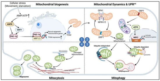
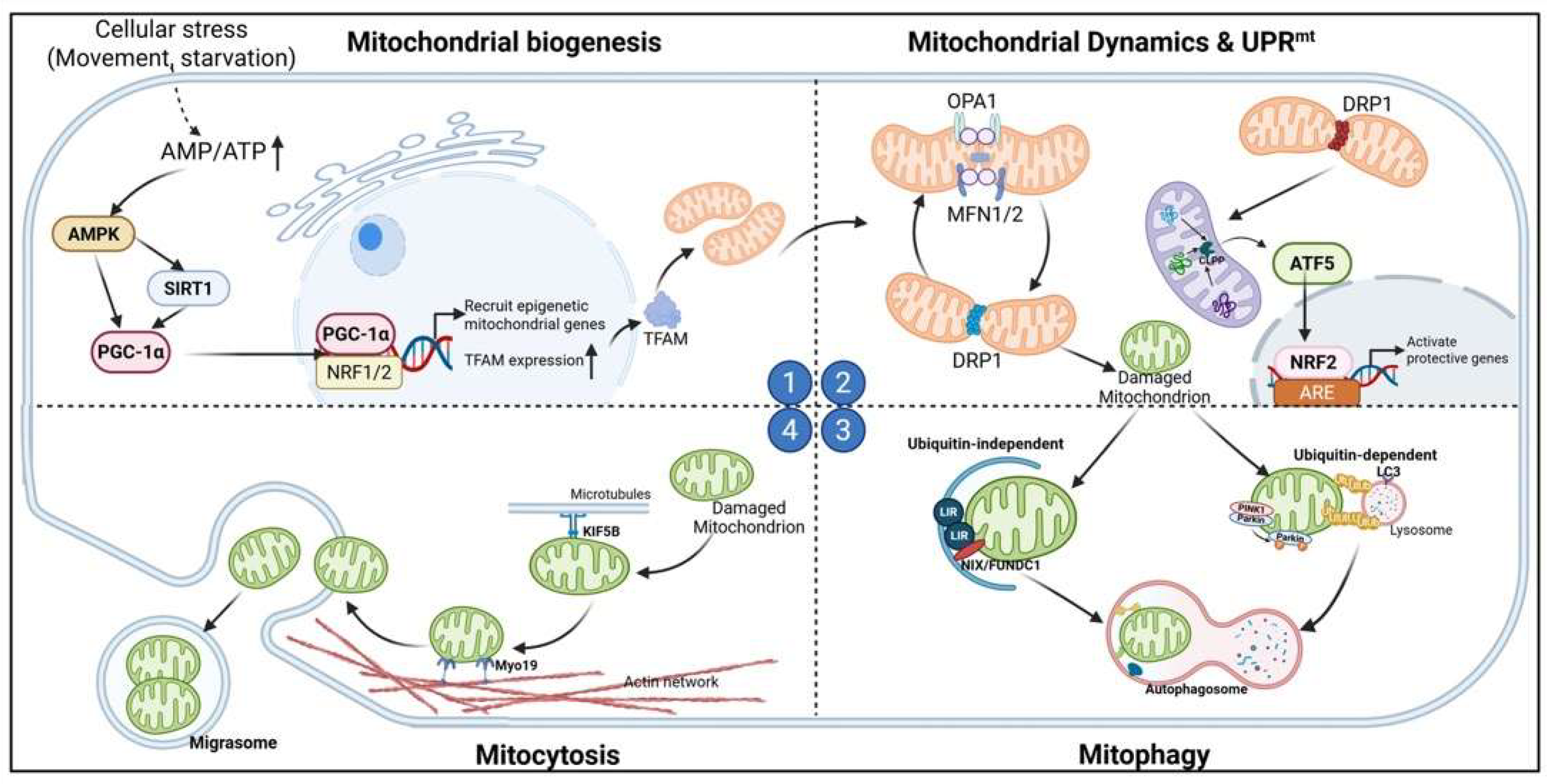
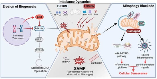
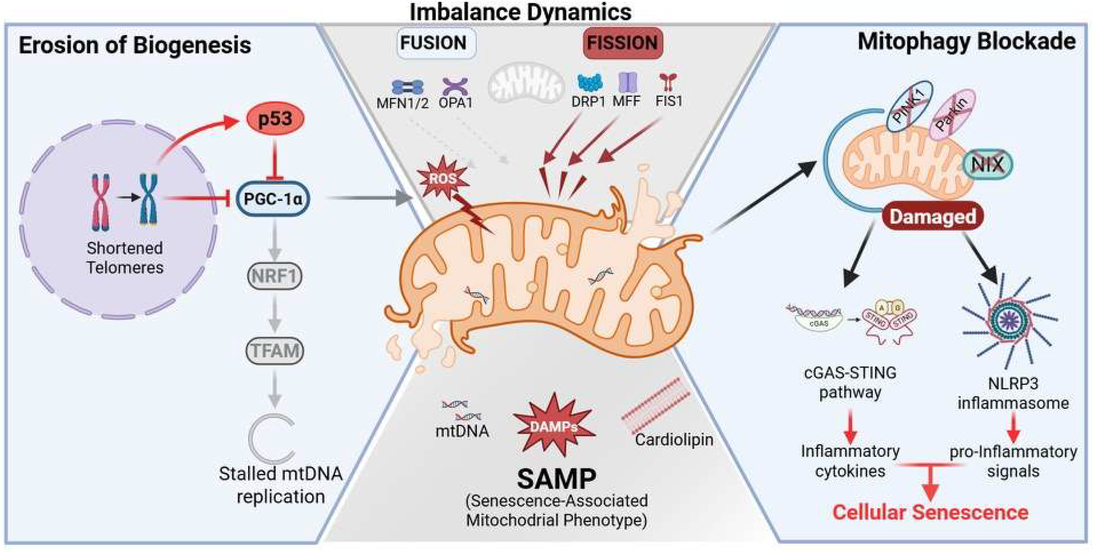
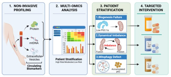
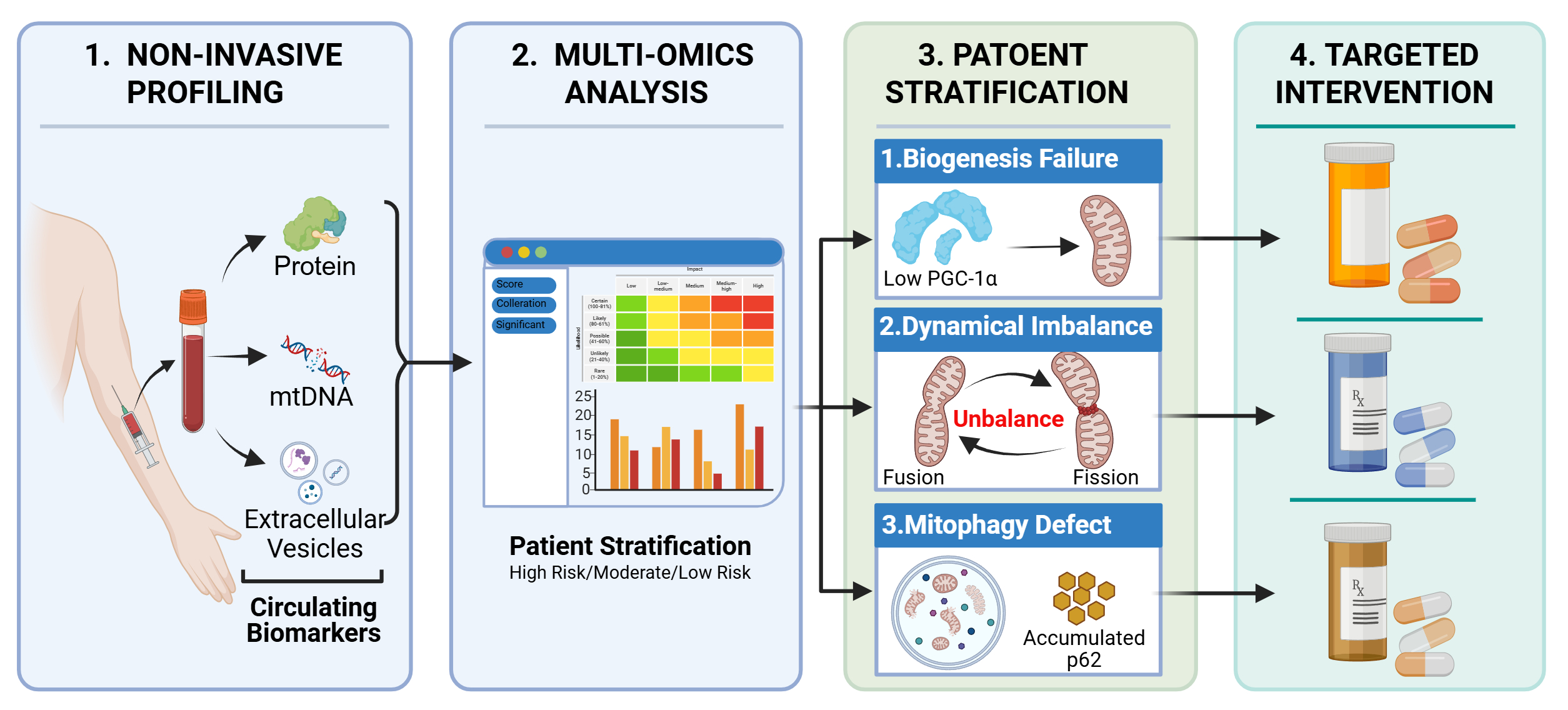
Disclaimer/Publisher’s Note: The statements, opinions and data contained in all publications are solely those of the individual author(s) and contributor(s) and not of MDPI and/or the editor(s). MDPI and/or the editor(s) disclaim responsibility for any injury to people or property resulting from any ideas, methods, instructions or products referred to in the content. |
© 2026 by the authors. Licensee MDPI, Basel, Switzerland. This article is an open access article distributed under the terms and conditions of the Creative Commons Attribution (CC BY) license.