


Abstract
Rosa roxburghii is a distinctive medicinal and edible plant resource. In an effort to uncover structurally novel secondary metabolites from this plant and explore their pharmacological potential, a phytochemical investigation of the fresh fruits of R. roxburghii led to the isolation of two new dihydroavicine alkaloids, roxburghcids D (1) and E (2), and a new triterpenoid glycoside, roxburghcid C (6), together with five known compounds (3–5, 7–8). The structures of the new compounds were unambiguously elucidated through extensive spectroscopic analyses (including 1D and 2D NMR and HRESIMS), quantum chemical calculations, and single-crystal X-ray diffraction. In bioassay evaluations, compounds 2 and 3 exhibited significant ABTS radical scavenging activities, with IC50 values of 39.59 and 17.38 μM, respectively. Additionally, compounds 2, 6, and 8 exhibited modest α-glucosidase inhibitory activities, with inhibition rates of 33.13%, 38.61%, and 36.85%, respectively, at a concentration of 100 μM. Furthermore, compounds 2 and 4 showed significant cytotoxicity against the A549 human cancer cell lines, with IC50 values of 7.23 and 8.36 μM, respectively. This study represents the first report of alkaloids isolated from R. roxburghii, enriching its phytochemical profile and providing valuable chemotaxonomic insights for this species.
1. Introduction
Oxygen is the foundational electron acceptor for aerobic life; nevertheless, the partial reduction of molecular oxygen during cellular respiration and normal metabolism inevitably yields highly reactive intermediate metabolites, predominantly reactive oxygen species (ROS) and reactive nitrogen species (RNS) [1,2]. Under physiological conditions, low-to-moderate concentrations of ROS/RNS function as crucial secondary messengers, playing indispensable roles in cellular signal transduction, host immune defense, and the maintenance of intracellular homeostasis [3]. However, when the overproduction of these highly reactive molecules overwhelms the scavenging capacity of endogenous antioxidant defense systems—comprising enzymes such as superoxide dismutase (SOD) and catalase (CAT), alongside the glutathione (GSH) network—the pro-oxidant/antioxidant equilibrium is disrupted, culminating in “oxidative stress” [4,5]. Extensive clinical and epidemiological evidence has substantiated that prolonged oxidative stress is not only a primary driver of the biological aging process [6] but is also deeply implicated in the pathogenesis and progression of cardiovascular diseases, neurodegenerative disorders, metabolic syndromes, and multiple malignancies [7]. In recent years, plant-derived natural products have garnered considerable attention as promising interventional candidates due to their extraordinary antioxidant capacities. Bioactive phytochemicals, including gallic acid, resveratrol, lycopene, and β-carotene, as well as vitamins C and E, have demonstrated potent efficacy in mitigating oxidative stress and neutralizing radical species in vivo [8]. Consequently, the continuous exploration of targeted therapeutic strategies capable of efficiently scavenging excessive reactive molecules or upregulating intrinsic antioxidant defense mechanisms holds profound clinical significance and translational medical value for the prevention and management of oxidative stress-related pathologies.
Rosa roxburghii Tratt. (Rosaceae) is a distinctive medicinal and edible plant resource native to Guizhou Province, China [9]. R. roxburghii has been designated as one of the twelve key characteristic and advantageous industries in Guizhou Province and is protected as a geographical indication product. By the end of 2022, the cultivation area of R. roxburghii in Guizhou had reached approximately 140,000 hectares, with an annual fresh fruit yield of 300,000 tons and a total industrial output value exceeding 15 billion RMB [10]. A systematic elucidation of the phytochemical basis of R. roxburghii is therefore a critical prerequisite for the development and expansion of its novel functional applications. R. roxburghii is traditionally utilized to invigorate the spleen, promote digestion, and act as an astringent agent [11]. Prior phytochemical studies on R. roxburghii have led to the isolation of various secondary metabolites, including triterpenoids [12], flavonoids [13], and phenolics [14]. In parallel, pharmacological evaluations have revealed that extracts or constituents from this plant possess diverse bioactivities, such as antidepressant [15], sedative–hypnotic [16], antitumor [17], and anti-aging [18] effects.
Driven by the objective to isolate novel secondary metabolites from this plant and elucidate their biomedical relevance, in the present study, A phytochemical investigation of the fruits of R. roxburghii led to the isolation and characterization of five alkaloids and three triterpenoid glycosides, including two new dihydroavicine-type alkaloids and a new oleanane-type triterpenoid glycoside. The chemical structures and absolute configurations of the new compounds were unambiguously elucidated through a comprehensive combination of extensive spectroscopic analyses, quantum chemical calculations, and single-crystal X-ray diffraction. Furthermore, selected isolated compounds were evaluated for their antioxidant capacities, α-glucosidase inhibitory potentials, and cytotoxic activities.
2. Materials and Methods
2.1. General Instrumentation and Reagents
Optical rotation values were determined at 25 °C using an Anton Paar MCP 500 digital polarimeter (Anton Paar GmbH, Graz, Austria). UV and ECD spectral analyses were performed on a Shimadzu-2600 spectrophotometer (Shimadzu Corporation, Kyoto, Japan) and an Applied Photophysics Chirascan spectrometer (Applied Photophysics Ltd., Leatherhead, UK), respectively. Infrared (IR) scans were conducted using KBr pellets on a Shimadzu IRAffinity-1 unit (Shimadzu Corporation, Kyoto, Japan). For 1D and 2D NMR experiments, Bruker AVANCE III-600 instruments (Bruker Corporation, Rheinstetten, Germany) were utilized, referencing TMS as the internal standard. HRESIMS measurements were taken on a Thermo Fisher Scientific Q Exactive Focus mass spectrometer (Thermo Fisher Scientific, Waltham, MA, USA). Semi-preparative HPLC separations were executed using a Shimadzu LC-20A system (Shimadzu Corporation, Kyoto, Japan) fitted with an SPD-20A UV detector and a Shim-pack GIS C18 column (10 × 250 mm, 5 μm). Silica gel (200–300 mesh; Qingdao Marine Chemical Group Co., Ltd., Qingdao, China) was used as the stationary phase for column chromatography.
Deuterated chloroform and methanol were purchased from Tenglong Weibo Technology (Qingdao, Shandong, China). Na2CO3, sodium dihydrogen phosphate, and disodium hydrogen phosphate were obtained from Shanghai Titan Scientific (Shanghai, China). Industrial-grade EtOH, petroleum ether, EtOAc, DCM, and MeOH were sourced from Chengdu Jinshan Chemical Reagent Co., Ltd. (Chengdu, Sichuan, China), while HPLC-grade methanol was provided by TEDIA (Fairfield, OH, USA). Biological reagents, including DMEM (Thermo Fisher Scientific, Waltham, MA, USA), penicillin–streptomycin (Suzhou Xinsaimei Biotechnology Co., Ltd., Suzhou, Jiangsu, China), Trypsin–EDTA Solution A (Shanghai Xiaopeng Biotechnology Co., Ltd., Shanghai, China), and fetal bovine serum (Beijing Lanjieke Technology Co., Ltd., Beijing, China), were used as received. The α-glucosidase, pNPG, and ABTS and DPPH assay kits were acquired from Shanghai Yuanye Bio-Technology Co., Ltd. (Shanghai, China).
2.2. Plant Material
In September 2023, fruit samples of Rosa roxburghii Tratt. were obtained from Longli County, Guizhou Province, P. R. China. Dr. Jingzhong Chen (Guizhou University of Traditional Chinese Medicine) was responsible for authenticating the botanical material. The voucher specimen (No. GZLL20230903) was lodged at the Natural Products Research Center of Guizhou Province.
2.3. Extraction and Isolation
Fresh fruits of Rosa roxburghii (150 kg) were sliced and extracted three times with 95% EtOH under reflux. The solvent was removed in vacuo to yield a crude extract (11.68 kg), which was suspended in H2O (20 L) and partitioned sequentially with petroleum ether (PE) and EtOAc. This process afforded a PE fraction (Fr. A, 64.97 g) and an EtOAc fraction (Fr. B, 914.50 g).
Fraction A was subjected to chromatography over silica gel (200–300 mesh) using a gradient elution of PE–EtOAc (1:0 to 1:1, v/v). The eluates were pooled based on TLC analysis to give nine subfractions (A1–A9). Further fractionation of A7 via silica gel CC (PE–EtOAc, 10:1 to 4:1, v/v) yielded subfractions A7-1–A7-3. Subfraction A7-3 was purified by silica gel CC with isocratic elution using dichloromethane (DCM) to afford compounds 1 (60.3 mg) and 2 (10.5 mg).
Fraction B was separated by silica gel CC (DCM–MeOH, 1:0 to 1:1, v/v) to yield six fractions (B1–B6). Fraction B2 was subjected to silica gel CC (DCM–MeOH, 30:1 to 1:1, v/v) to give subfractions B2-1–B2-5. Subsequent chromatography of B2-3 (DCM–MeOH, 80:1 to 1:1, v/v) resulted in subfractions B2-3-1–B2-3-4. Subfraction B2-3-2 was purified by silica gel CC (DCM–MeOH, 50:1, v/v) followed by semipreparative HPLC (60% MeOH–H2O, 2 mL/min) to yield 3 (80.1 mg, tR = 25 min). Similarly, purification of B2-3-4 via silica gel CC (DCM–MeOH, 80:1, v/v) afforded 4 (52.1 mg). Fraction B2-4 was fractionated over silica gel (DCM–MeOH, 50:1 to 1:1, v/v) to produce subfractions B2-4-1–B2-4-4. Subfraction B2-4-1 was passed through a Sephadex LH-20 column (DCM–MeOH, 1:1, v/v) to give B2-4-1-1 and B2-4-1-2. Fr. B2-4-1-2 was purified by semipreparative HPLC (70% MeOH–H2O, 2 mL/min) to yield 5 (13.0 mg, tR = 26 min).
Fraction B5 was subjected to medium-pressure liquid chromatography (MPLC) over reversed-phase C18 silica gel, eluting with a gradient of MeOH/H2O (10:90 to 60:40, v/v) to afford six subfractions (Fr. B5-1 to B5-6). Fraction B5-3 was first chromatographed over a silica gel column eluted with a gradient of CH2Cl2/MeOH (10:1 to 0:1, v/v), followed by size-exclusion chromatography on Sephadex LH-20 (CH2Cl2/MeOH, 1:1, v/v) to yield three subfractions (Fr. B5-3-1 to B5-3-3). Subsequently, subfraction B5-3-1-1 was purified by silica gel column chromatography (CH2Cl2/MeOH, 10:1, v/v) to give compound 8 (10 mg). Subfraction B5-3-1-2 was separated over a silica gel column (CH2Cl2/MeOH, 6:1, v/v) to afford additional compound 7 (7 mg). Subfraction B5-3-1-3 was purified by semi-preparative HPLC (isocratic 60% MeOH/H2O, 2.0 mL/min) to yield compound 6 (9.7 mg, tR = 85 min).
2.3.1. Roxburghcid D (1)
White powder; −0.51 (c 0.23, MeOH); UV (MeOH) λmax (log ε): 203 (3.93), 229 (3.70), 280 (3.58), 320 (3.58) nm; ECD (MeOH) λ (Δε) 217 (−0.84), 261 (−0.26) nm; IR (KBr) υmax 3385, 2960, 2902, 1469, 1228, 1033, 941, 850, 806 cm–1; for 1H and 13C NMR data, see Table 1; HR-ESI-MS m/z 378.1333 [M + H]+ (calcd for C22H20O5N, 378.1336).
2.3.2. Roxburghcid E (2)
White powder; −1.05 (c 0.13, MeOH); UV (MeOH) λmax (log ε): 204 (3.89), 231 (4.22), 281 (4.06), 317 (3.87) nm; ECD (MeOH) λ (Δε) 230 (4.28), 281 (0.80) nm; IR (KBr) υmax 3566, 1481, 1465, 1247, 1037, 937, 864 cm–1; for 1H and 13C NMR data, see Table 1; HR-ESI-MS m/z 378.1331 [M + H]+ (calcd for C22H20O5N, 378.1336).
2.3.3. Aegelbine-A (3)
Yellow needle crystals. 1H NMR (600 MHz, CD3OD) δ: 7.90 (1H, d, J = 9.5 Hz, H-5), 7.73 (1H, d, J = 2.2 Hz, H-2), 7.26 (1H, s, H-4), 6.81 (1H, d, J = 2.2 Hz, H-3), 6.29 (1H, d, J = 9.5 Hz, H-6); 13C NMR (150 MHz, CD3OD) δ: 148.3 (C-2), 107.9 (C-3), 111.2 (C-4), 147.1 (C-5), 114.6 (C-6), 141.1 (C-7), 147.1 (C-8), 163.0 (C-9), 127.3 (C-10), 117.8 (C-11), 131.7 (C-12).
2.3.4. Canthine-6-one (4)
Yellow needle crystals. 1H NMR (600 MHz, CDCl3) δ: 8.80 (1H, d, J = 5.0 Hz, H-2), 8.63 (1H, d, J = 8.2 Hz, H-8), 8.07 (1H, d, J = 7.7 Hz, H-11), 8.00 (1H, dd, J = 9.8, 1.0 Hz, H-4), 7.93 (1H, d, J = 4.9 Hz, H-1), 7.68 (1H, t, J = 7.8 Hz, H-9), 7.50 (1H, t, J = 7.6 Hz, H-10), 6.96 (1H, d, J = 9.7 Hz, H-5); 13C NMR (150 MHz, CDCl3) δ: 116.5 (C-1), 145.9 (C-2), 139.7 (C-4), 159.6 (C-6), 117.4 (C-8), 131.0 (C-9), 125.8 (C-10), 122.8 (C-11), 124.5 (C-12), 139.5 (C-13), 130.4 (C-14), 132.1 (C-15), 136.3 (C-16).
2.3.5. (R)-5-(1-Hydroxyethyl)-canthine-6-one (5)
Yellow needle crystals. 1H NMR (600 MHz, CD3OD) δ: 8.77 (1H, d, J = 5.0 Hz, H-2), 8.58 (1H, d, J = 8.2 Hz, H-8), 8.21 (2H, m, H-4, H-11), 8.11 (1H, d, J = 5.0 Hz, H-1), 7.74 (1H, m, H-9), 7.57 (1H, t, J = 7.6 Hz, H-10), 5.20 (1H, m, H-17), 1.62 (3H, d, J = 6.5 Hz, H-18); 13C NMR (150 MHz, CD3OD) δ: 117.3 (C-1), 146.5 (C-2), 133.3 (C-4), 147.5 (C-5), 160.0 (C-6), 117.8 (C-8), 131.8 (C-9), 126.9 (C-10), 124.1 (C-11), 125.8 (C-12), 140.5 (C-13), 131.5 (C-14), 132.2 (C-15), 136.8 (C-16), 65.9 (C-17), 23.4 (C-18).
2.3.6. Roxburghcid C (6)
Needle crystals; −36.7 (c 0.15, MeOH); UV (MeOH) λmax (log ε): 203 (3.42), 255 (3.75) nm; IR (KBr) υmax 3360, 2926, 2868, 1749, 1556, 1454, 1446, 1377, 1247, 1035, 1006, 871, 785, 596, 540 cm–1, for 1H and 13C NMR data, see Table 2; HR-ESI-MS m/z 671.3749 [M + H]+ (calcd for C36H56O10Na, 671.3766).
2.3.7. 2α,3β,19α,23-Tetrahydroxylurs-12-en-28-oic Acid-β-D-glucopyranosyl Ester (7)
1H NMR (CD3OD, 600 MHz) δH: 5.30 (2H, d, J = 8.2 Hz, H-12, H-1′), 3.35–3.78 (5H, H2′–6′), 3.66 (1H, m, H-2), 3.48 (1H, d, J = 11.1 Hz, H-23b), 3.25 (1H, d, J = 11.1 Hz, H-23a), 2.50 (1H, s, H-18), 1.32 (3H, s, H-27), 1.19 (3H, s, H-29), 1.02 (3H, s, H-25), 0.91 (3H, d, J = 7.0 Hz, H-30), 0.76 (3H, s, H-26), 0.68 (3H, s, H-24). 13C NMR (CD3OD, 500 MHz) δC: 48.0 (C-1), 69.7 (C-2), 78.3 (C-3), 44.1 (C-4), 48.5 (C-5), 19.2 (C-6), 33.5 (C-7), 40.9 (C-8), 48.2 (C-9), 39.0 (C-10), 24.8 (C-11), 129.5 (C-12), 139.8 (C-13), 42.8 (C-14), 29.6 (C-15), 26.5 (C-16), 48.3 (C-17), 55.0 (C-18), 73.6 (C-19), 42.9 (C-20), 27.2 (C-21), 38.3 (C-22), 66.3 (C-23), 13.9 (C-24), 17.7 (C-25), 17.6 (C-26), 24.7 (C-27), 178.5 (C-28), 27.1 (C-29), 16.6 (C-30), 95.8 (C-1′), 73.8 (C-2′), 78.6 (C-3′), 71.1 (C-4′), 78.2 (C-5′), 62.4 (C-6′).
2.3.8. 2α,3α,19α-Trihydroxy-olean-12-en-28-oic Acid-β-D-glucopyranosyl Ester (8)
White powder. 1H NMR (600 MHz, CD3OD) δ: 5.35 (1H, d, J = 8.1 Hz, H-1′), 5.32 (1H, t, J = 3.7 Hz, H-12), 1.29 (3H, s, H-27), 0.97 (3H, s, H-23), 0.97 (3H, s, H-29), 0.93 (3H, s, H-26), 0.92 (3H, s, H-25), 0.85 (3H, s, H-30), 0.72 (3H, s, H-24). 13C NMR (150 MHz, CD3OD) δ: 42.4 (C-1), 67.1 (C-2), 80.1 (C-3), 39.5 (C-4), 49.8 (C-5), 19.3 (C-6), 33.8 (C-7), 41.1 (C-8), 49.4 (C-9), 39.5 (C-10), 24.9 (C-11), 124.9 (C-12), 144.4 (C-13), 42.7 (C-14), 29.5 (C-15), 28.4 (C-16), 47.1 (C-17), 45.1 (C-18), 82.4 (C-19), 36.0 (C-20), 29.4 (C-21), 33.2 (C-22), 29.2 (C-23), 22.4 (C-24), 16.9 (C-25), 17.8 (C-26), 25.2 (C-27), 178.6 (C-28), 28.6 (C-29), 25.1 (C-30), 95.8 (C-1′), 73.9 (C-2′), 78.7 (C-3′), 71.1 (C-4′), 78.3 (C-5′), 62.4 (C-6′).
2.3.9. Crystal Data of Roxburghcid C (6)
C36H56O10, M = 648.80 g/mol, monoclinic, space group P21 (no. 4), a = 15.5231(4) Å, b = 6.41190(10) Å, c = 16.9954(4) Å, α = 90°, β = 91.272(2), γ = 90°, V = 1691.18(6) Å3, Z = 2, T = 100.01(10) K, μ (Cu Kα) = 0.747 mm−1, Dcalc = 1.274 g/cm3, 10,601 reflections measured (5.2° ≤ 2Θ ≤ 148.004°), 4867 unique (Rint = 0.0498, Rsigma = 0.0689) which were used in all calculations. The final R1 was 0.0603 (I > 2σ(I)), and wR2 was 0.1601 (all data). Flack parameter = 0.3 (3). CCDC number: 2539246.
Table 1.
NMR data of compounds 1–2.
Table 2.
NMR data of compound 6.
2.4. NMR and ECD Calculations
Conformational analyses for compounds 1 and 2 were initiated using the Spartan14 software. The resulting conformers were geometrically optimized at the B3LYP/6-31G(d) level in the gas phase using the Gaussian 09 program package [19]. Stable conformers with relative energies within a specific window were selected for further calculations, and their room-temperature equilibrium populations were determined according to the Boltzmann distribution law. For NMR calculations, Gauge-Independent Atomic Orbital (GIAO) shielding tensors were calculated at the mPW1PW91/6-311G(d,p) level using the PCM solvation model (DMSO) [20]. The calculated 13C and 1H shielding constants were subjected to DP4+ probability analysis to determine the most likely configuration [21]. ECD calculations were performed using Time-Dependent Density Functional Theory (TDDFT) [22] at the wB97XD/6-311G(d,p) level in methanol with the PCM solvation model. The final ECD spectra were simulated by Boltzmann weighting of the dominant conformers using a Gaussian function with a bandwidth (sigma/gamma) of 0.3 eV.
2.5. α-Glucosidase Inhibitory and Antioxidant Assay
The α-glucosidase inhibition activity was evaluated following a previously described protocol [23]. Briefly, sample solutions (25 μL) at varying concentrations (100 μM) were mixed with α-glucosidase (50 μL, 0.2 U/mL) in 96-well plates and pre-incubated at 37 °C for 10 min. The reaction was initiated by the addition of 5 mM p-nitrophenyl-α-D-glucopyranoside (pNPG, 25 μL), and after incubation at 37 °C for 5 min, the reaction was quenched with 100 μL of 0.1 M Na2CO3. Absorbance was subsequently recorded at 405 nm using a microplate reader. All experiments were performed in triplicate and independently repeated three times.
The ABTS and DPPH radical scavenging capacities were assessed using commercial kits according to the manufacturer’s instructions. For the ABTS assay, a working solution was prepared by diluting the stock ABTS radical cation with anhydrous EtOH. A 10 μL aliquot of the sample (100, 50, 25, 12.5, and 6.25 μM) was mixed with 190 μL of the ABTS working solution and incubated at 25 °C for 6 min in the dark. Absorbance was measured at 734 nm. For the DPPH assay, 50 μL of the sample (100 μM) was combined with 225 μL of EtOH and 225 μL of DPPH solution. Following incubation at 25 °C for 30 min in the dark, a 300 μL aliquot was transferred to a 96-well plate, and absorbance was recorded at 517 nm [24]. All experiments were performed in triplicate and independently repeated three times.
ABTS/DPPH radical scavenging rate (%) = [1 − (A1 − A2)/A0] × 100%
In this equation, A0, A1, and A2 correspond to the absorbance values recorded for the blank control, the experimental sample, and the sample control.
2.6. Cytotoxicity Assay
The in vitro antiproliferative activities of compounds 1–6 against three human cancer cell lines (A549, HepG2, and MCF-7) were evaluated using a Cell Counting Kit-8 (CCK-8) assay. Cells were cultured in Dulbecco’s Modified Eagle Medium (DMEM) supplemented with 10% fetal bovine serum (FBS) at 37 °C in a humidified atmosphere of 5% CO2. Briefly, A549 cells were seeded into 96-well plates at a density of 5 × 10³ cells/well, while HepG2 and MCF-7 cells were seeded at 1 × 10⁴ cells/well. After a 24 h incubation to allow for cell adherence, the cells were treated with the tested compounds at a concentration of 20 μM. Following an additional 24 h incubation, CCK-8 reagent was added to each well. After a 2 h colorimetric reaction period, the absorbance was measured at 450 nm using a Multiskan FC microplate reader (Thermo Fisher Scientific, Waltham, MA, USA). Cisplatin was employed as a positive control [25]. All experiments were performed in triplicate and independently repeated three times.
2.7. Statistical Analysis
Statistical analyses were performed using SPSS version 27.0. Data are presented as the mean ± standard deviation (SD). Comparisons were conducted using one-way analysis of variance (ANOVA), followed by the Least Significant Difference (LSD) and Tukey’s post hoc tests. A p-value < 0.05 or <0.01 was considered statistically significant. All experiments were performed in triplicate and independently repeated three times.
3. Results
3.1. Identification of New Compounds
Roxburghcid D (1) was isolated as a white amorphous powder. Its molecular formula was determined to be C22H19O5N by HRESIMS (m/z 378.1333 [M + H]+; calcd for C22H20O5N, 378.1336), requiring 13 indices of hydrogen deficiency (IHDs). The IR spectrum displayed absorption bands characteristic of hydroxyl (3385 cm−1), benzene ring (1469 cm−1), and methylenedioxy (941 cm−1) functionalities. The 1H NMR data for 1 (Table 1) revealed six aromatic protons [δH 7.83 (1H, s, H-4), 7.77 (1H, d, J = 8.6 Hz, H-11), 7.53 (1H, d, J = 8.6 Hz, H-12), 7.50 (1H, s, H-10), 7.30 (1H, s, H-1), and 6.98 (1H, s, H-7)]; two methylenedioxy groups [δH 6.14 (2H, dd, J = 4.6, 1.0 Hz, H-OCH2O-2/3), and 6.07 (1H, dd, J = 6.8, 1.2 Hz, H-OCH2O-8/9)]; an oxygenated methine [δH 3.07 (1H, m, H-1′)]; and two methyl resonances [δH 0.89 (3H, d, J = 6.2 Hz, H-2′) and an N-methyl δH 2.61 (3H, s)]. The 13C NMR and HSQC spectra revealed 22 carbon resonances, including two methyls [δC (19.5, C-2′) and (42.4, N-Me)]; two methylenedioxy carbons [δC 101.2 and 101.0]; eight methine carbons [an N-bearing carbon δC (69.9, C-6), an oxygenated carbon δC (66.1, C-1′), six aromatic carbons (104.1, C-1), (100.4, C-4), (109.4, C-7), (103.4, C-10), (119.9, C-11), and (123.6, C-12)]; and ten quaternary aromatic carbons [δC (147.2, C-2), (147.9, C-3), (126.2, C-4a), (139.0, C-4b), (125.6, C-6a), (146.6, C-8), (147.5, C-9), (124.8, C-10a), (123.3, C-10b), and (130.6, C-12a)].
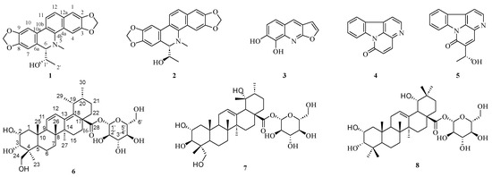
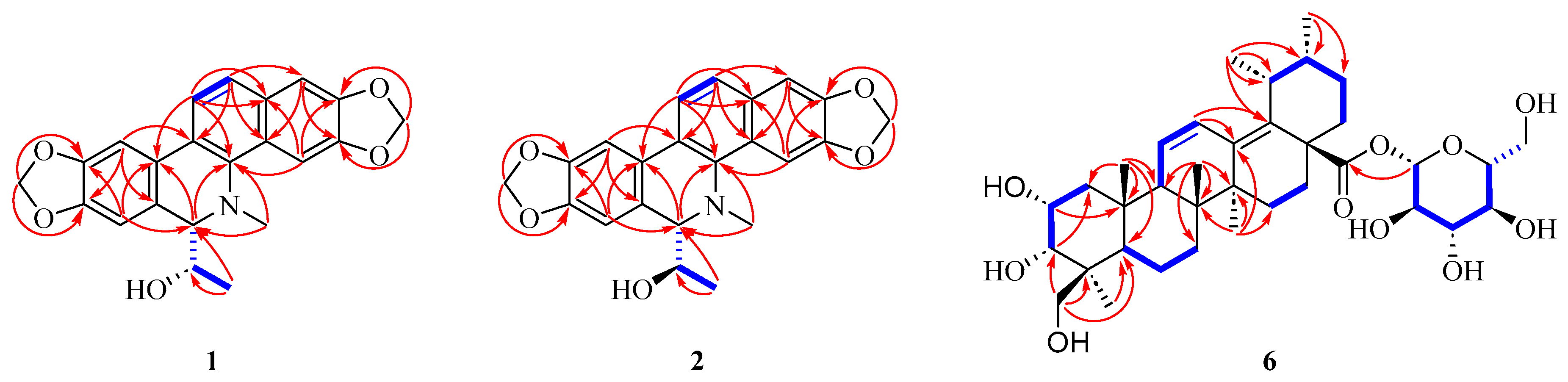
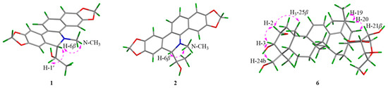
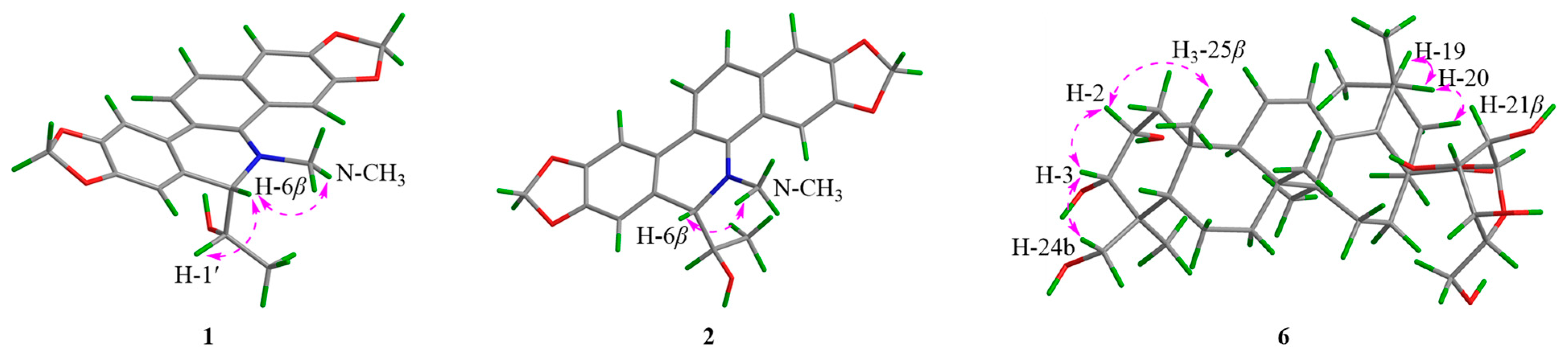
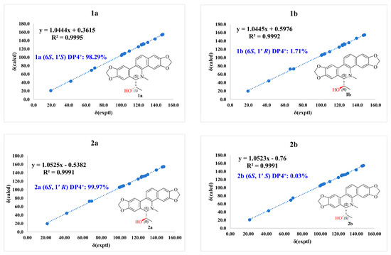
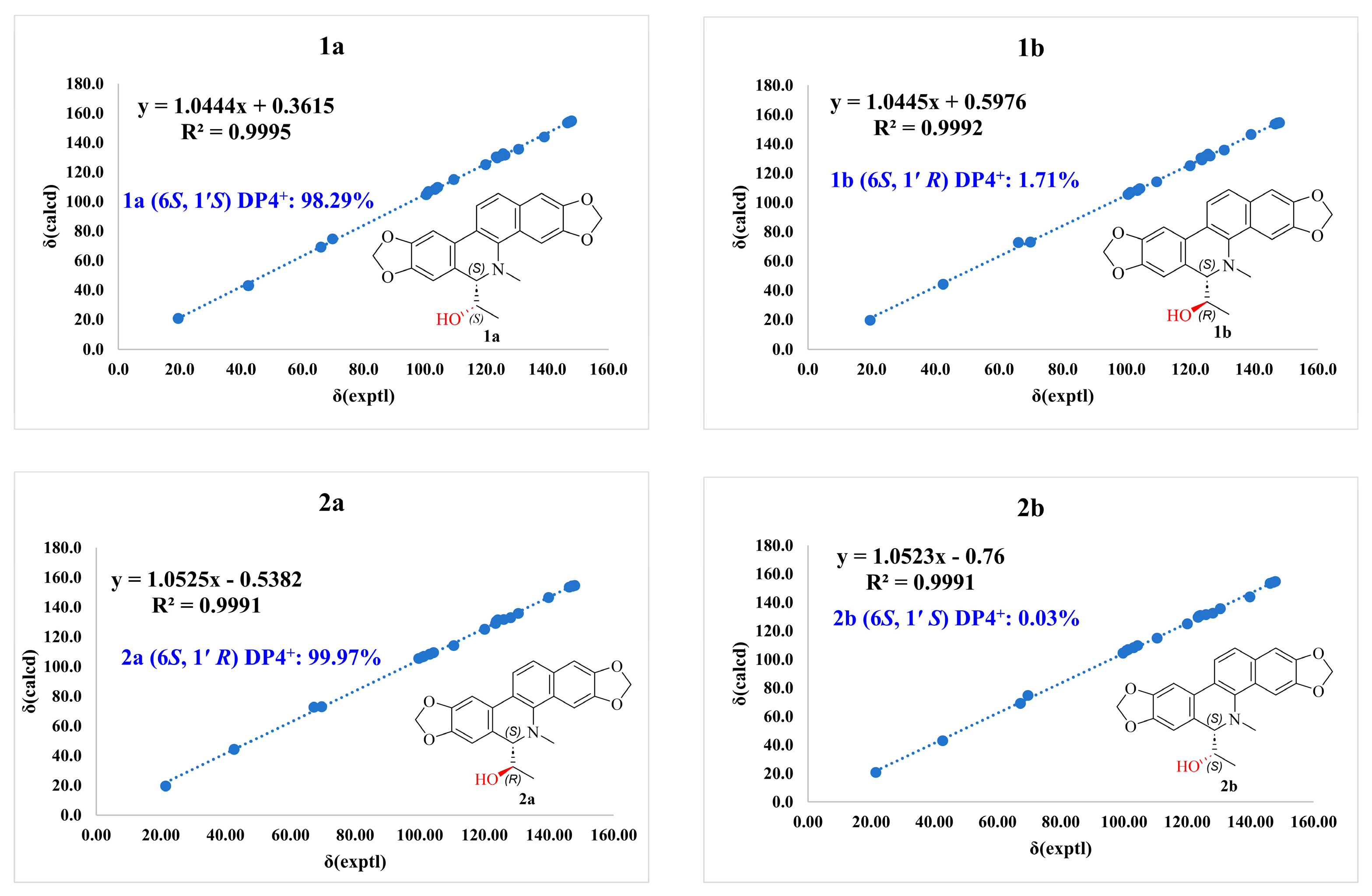
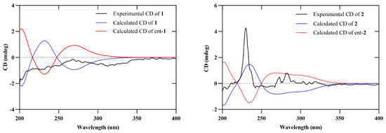
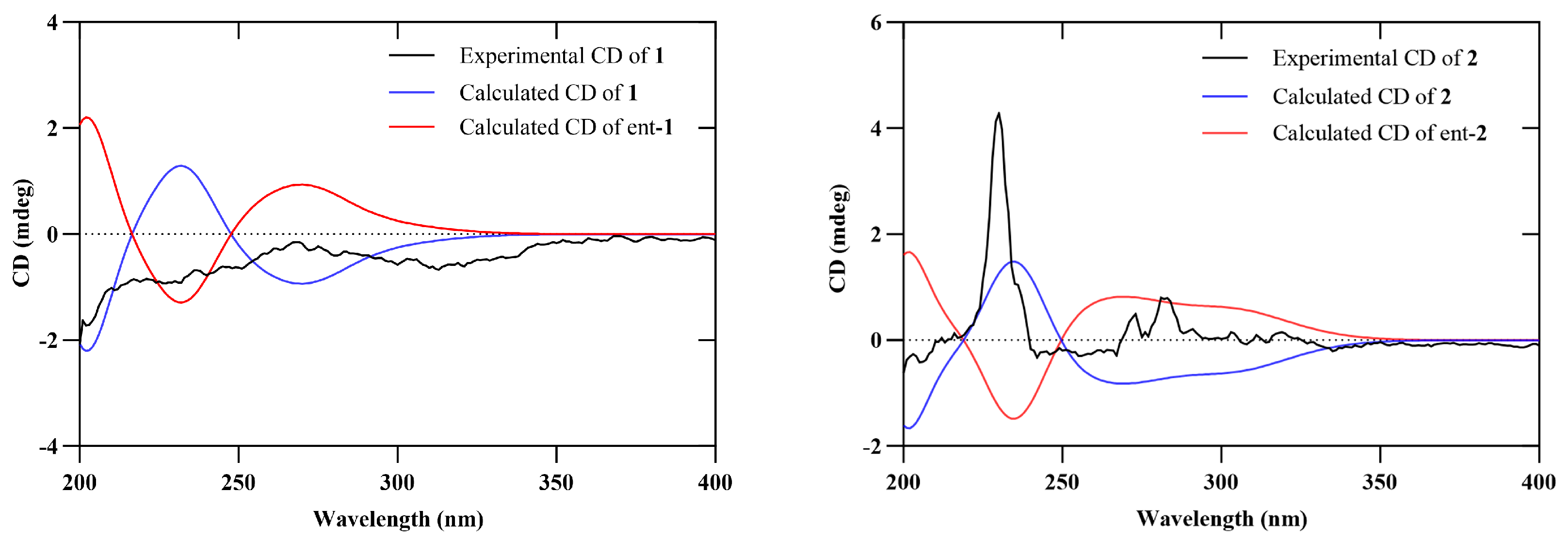
Detailed analysis of the 1H and 13C NMR data suggested that compound 1 possesses a dihydroavicine alkaloid skeleton, structurally distinct from the known analogue 6-acetonyldihydroavicine [26] only by the substitution at C-6. Specifically, the acetonyl group in the analogue is replaced by a 1-hydroxyethyl moiety in 1 (Figure 1). This assignment was substantiated by key HMBC correlations (Figure 2) from H-2′ (δH 0.89) to C-1′ (δC 66.1) and C-6 (δC 69.9). The NOESY spectrum corroborated this relative configuration, with key correlations among N-CH3, H-6, and H-1′ indicating their co-facial β-orientation (Figure 3). By combining DP4+ probability analysis (Figure 4) and ECD calculations (Figure 5), the absolute configuration was determined to be 6S, 1′S, and named roxburghcid D.
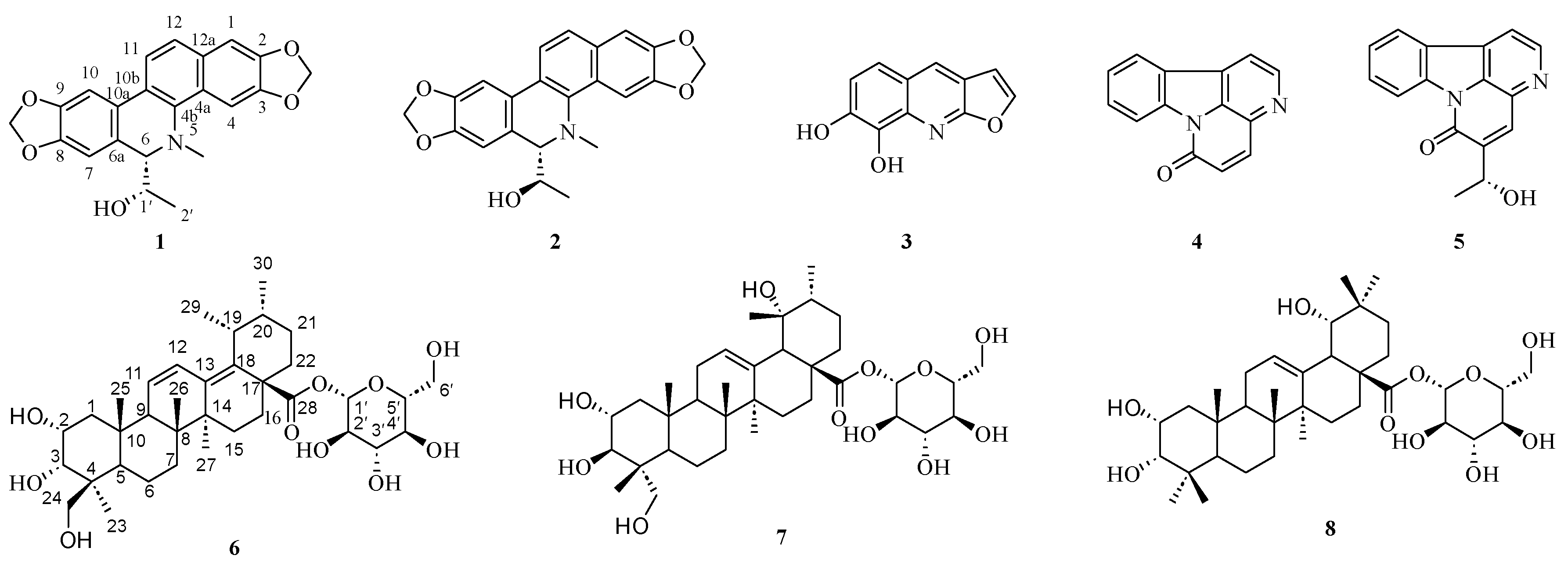
Figure 1.
The structures of compounds 1–8.
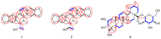
Figure 2.
The key 1H-1H COSY (blue bold) and HMBC (red arrows) correlations of 1–2, 6.
Figure 3.
Key NOESY (purple arrows) correlations of 1–2, 6.
Figure 4.
DP4+ probability analysis of compounds 1 and 2.
Figure 5.
Experimental and calculated ECD spectra of compounds 1 and 2.
Roxburghcid E (2) was obtained as a white amorphous powder. The molecular formula C22H19O5N was established by HR-ESI-MS ([M + H]+ 378.1331, C22H20O5N, calcd for 378.1336). The IR spectrum showed absorption bands for hydroxy (3566 cm−1), aromatic (1465 cm−1), and methylenedioxy (937 cm−1) functionalities. The 1H NMR and 13C NMR data of 2 (Table 1) closely resembled those of 1, suggesting that they share the same planar constitution. However, minor differences in chemical shifts were observed, particularly around the chiral centers. Detailed analysis of 2D NMR correlations confirmed that 2 is the C-1′ epimer of 1. The NOESY spectrum corroborated this relative configuration. Key correlations between the N-CH3 and H-6 protons indicated a co-facial relationship, establishing their β-orientations. In contrast, the lack of observable NOE cross-peaks between H-1′ and either the N-CH3/H-7 protons suggested they reside on opposite faces, permitting the assignment of H-1′ as α-oriented (Figure 3). The absolute configuration was established as (6S, 1′R) via DP4+ probability analysis (Figure 4) and ECD calculations (Figure 5).
Roxburghcid C (6) was isolated as colorless needles. Its molecular formula was established as C36H56O10 on the basis of the HRESIMS pseudomolecular ion peak at m/z 671.3749 [M + Na]+ (calcd for C36H56O10Na, 671.3766), requiring 9 IHDs. The 1H NMR data (Table 2) revealed the presence of six methyls [δH 0.95 (3H, s, H-23), 0.88 (3H, s, H-25), 0.76 (3H, s, H-26), 0.88 (3H, s, H-27), 0.92 (3H, d, J = 7.0 Hz, H-29), and 0.81 (3H, d, J = 6.6 Hz, H-30)], an oxygenated methylene [δH 3.25 (1H, dd, J = 11.1, 4.6 Hz, H-24a) and 3.42 (1H, dd, J = 11.2, 5.3 Hz, H-24b)], and two oxygenated methines [δH 3.79 (1H, m, H-2) and 3.06 (1H, q, J = 8.4, 7.7 Hz, H-3)]. Additionally, a set of proton signals indicative of a β-glucopyranosyl moiety was observed [δH 5.30 (1H, d, J = 8.2 Hz, H-1′), 3.57 (1H, m, H-2′), 3.20 (1H, m, H-3′), 3.06 (1H, q, J = 8.4, 7.7 Hz, H-4′), 3.20 (1H, m, H-5′), 3.42 (1H, dd, J = 11.2, 5.3 Hz, H-6′a), and 3.65 (1H, dd, J = 11.4, 4.9 Hz, H-6′b)]. The 13C NMR (Table 2) and HSQC spectra resolved 36 carbon resonances, which were classified into six methyls [δC 22.9 (C-23), 19.2 (C-25), 16.2 (C-26), 20.9 (C-27), 16.1 (C-29), and 19.2 (C-30)], nine methylenes [δC 41.7 (C-1), 18.0 (C-6), 32.3 (C-7), 24.6 (C-15), 31.7 (C-16), 24.2 (C-21), 31.1 (C-22), and two oxygenated carbons δC 63.4 (C-24) and 61.0 (C-6′)], thirteen methines [δC 47.8 (C-5), 54.0 (C-9), 33.6 (C-19), 26.9 (C-20), and seven oxygenated δC 64.5 (C-2), 72.4 (C-3), 94.6 (C-1′), 72.7 (C-2′), 76.6 (C-3′), 69.8 (C-4′), and 77.7 (C-5′)], and eight quaternary carbons [δC 43.9 (C-4), 40.2 (C-8), 37.3 (C-10), 41.6 (C-14), 46.2 (C-17), two olefinic carbons δC 134.5 (C-13) and 137.4 (C-18), and one ester carbonyl δC 175.3 (C-28)].
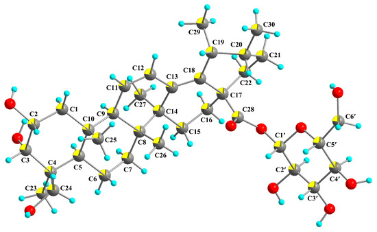
Analysis of the NMR data revealed that the structure of compound 6 is closely related to that of rubuside H [27], with the principal differences residing in their stereochemical configurations. In compound 6, the hydroxyl groups at C-2 and C-3, as well as the methyl groups at C-4, C-19, and C-20, were assigned the α-orientation. This relative configuration was further corroborated by the NOESY spectrum. Key NOE correlations among H-24b/H-3/H-2/H3-25 indicated that these protons are co-facial, thereby establishing their β-orientation. Similarly, NOE cross-peaks observed among H-19/H-20/H-21β suggested a co-facial relationship, confirming the β-orientation of H-19 and H-20. Ultimately, a single-crystal X-ray diffraction analysis unambiguously established the absolute configuration of 6 as 2R, 3S, 4S, 5R, 8R, 9R, 10S, 14S, 17S, 19R, and 20R (CCDC No. 2539246) (Figure 6), and it was named roxburghcid C.
Figure 6.
X-ray of compound 6.
The known compounds were identified as aegelbine-A (3) [28], canthine-6-one (4) [29], (R)-5-(1-hydroxyethyl)-canthine-6-one (5) [30], 2α,3β,19α,23-tetrahydroxylurs-12-en-28-oic acid-β-D-glucopyranosyl ester (7) [31], and 2α,3α,19α-trihydroxy-olean-12-en-28-oic acid-β-D-glucopyranosyl ester (8) [32].
3.2. α-Glucosidase Inhibitory and Antioxidant Activities
The isolates were assessed for their α-glucosidase inhibitory and antioxidant properties in vitro. At a tested concentration of 100 μM, compounds 2, 6, and 8 showed moderate α-glucosidase inhibition (33.13%, 38.61%, and 36.85%, respectively). To assess variations in α-glucosidase inhibitory activity among the evaluated compounds, Tukey’s multiple-comparison test was applied. The results demonstrated comparable inhibitory effects among certain subsets of compounds; specifically, no statistically significant differences were observed between compound 8 and compounds 2 or 6, nor between compound 5 and compounds 1, 3, or 4.
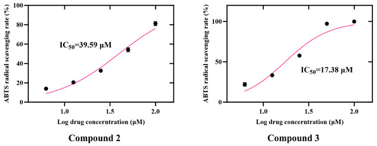
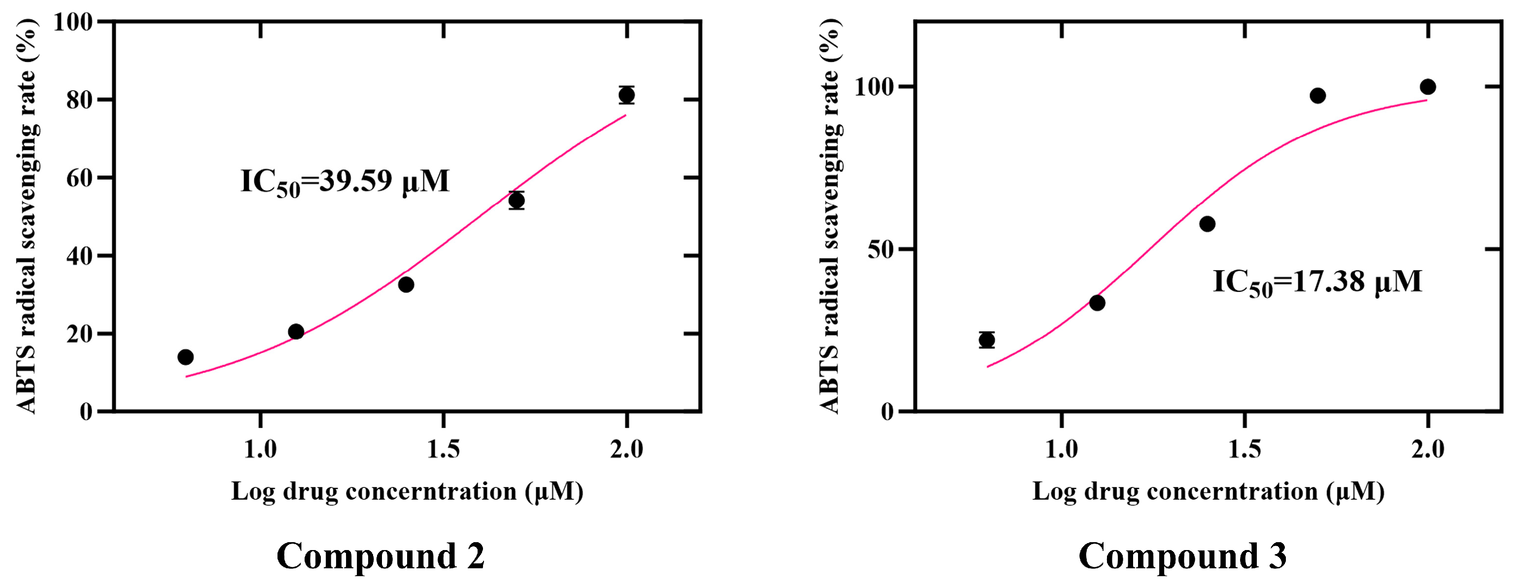
In the ABTS radical scavenging assay, compounds 2 and 3 exhibited significant activities with respective inhibition rates of 81.18% and 99.97% at 100 μM. According to Tukey’s multiple-comparison test, compounds 2 and 3 demonstrated the highest ABTS radical scavenging potential, with a significant difference noted between the two. Furthermore, no significant differences in scavenging capacity were detected between compounds 1 and 6 or between compound 4 and compounds 5, 6, 7, and 8. Concentration-dependency evaluations further confirmed that compounds 2 and 3 exerted substantial dose-dependent effects across a concentration range of 6.25–100 μM, with calculated IC50 values of 39.59 μM and 17.38 μM, respectively (Figure 7).
Figure 7.
Concentration-dependent ABTS radical scavenging activity of compounds 2 and 3.
Furthermore, compound 3 displayed moderate DPPH radical scavenging activity, achieving 25.94% at a concentration of 100 μM. According to Tukey’s post hoc analysis, compound 3 possessed the highest scavenging potential within the tested group, showing statistically significant differences compared to compounds 1, 2, and 4–8. Moreover, no significant variations in scavenging efficacy were observed between compound 8 and compounds 1, 2, and 4–7. All corresponding bioactivity results are detailed in Table 3.
Table 3.
α-glucosidase inhibitory and radical scavenging activities of compounds 1–8 (100 μM).
3.3. In Vitro Cytotoxicity Assay of Compounds 1–6
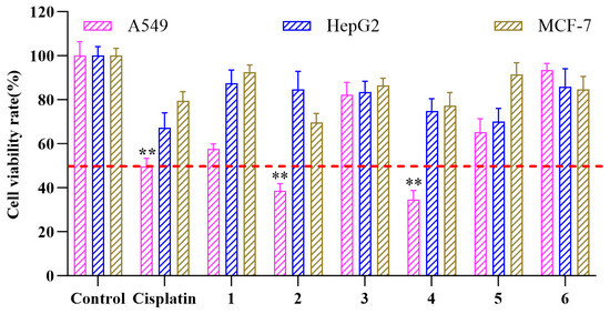
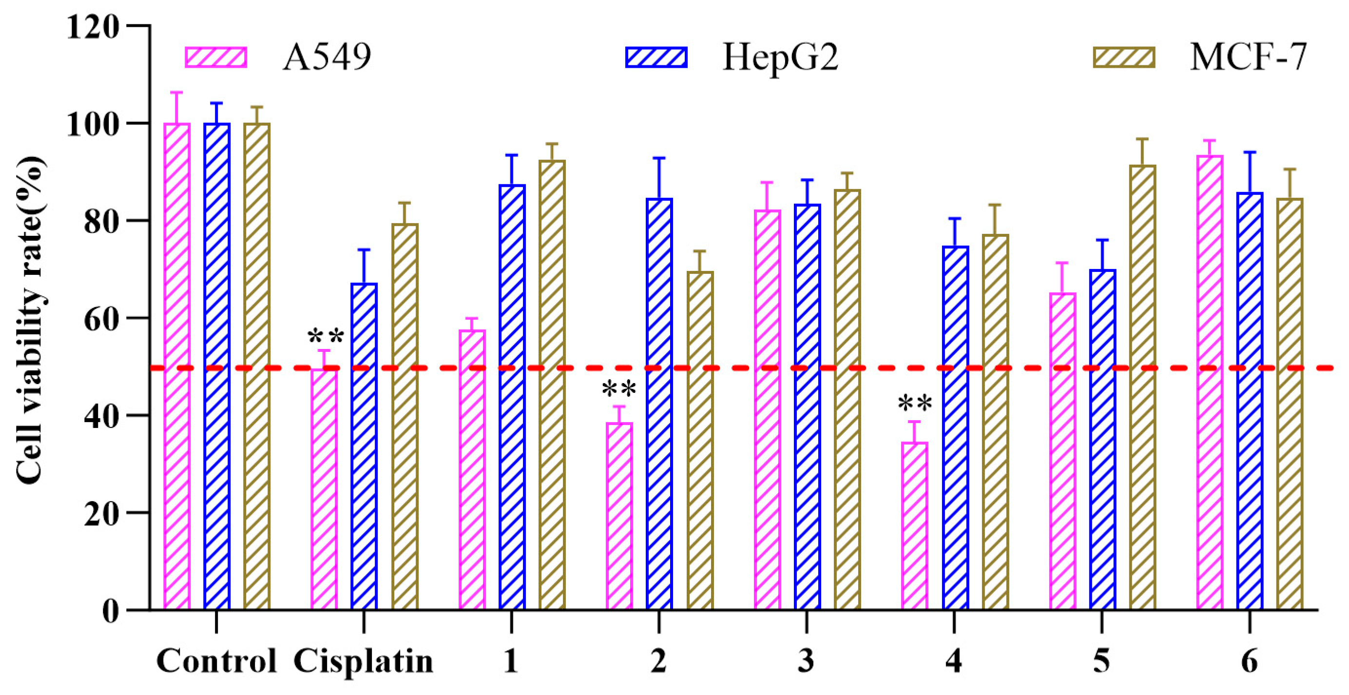
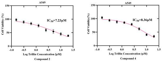
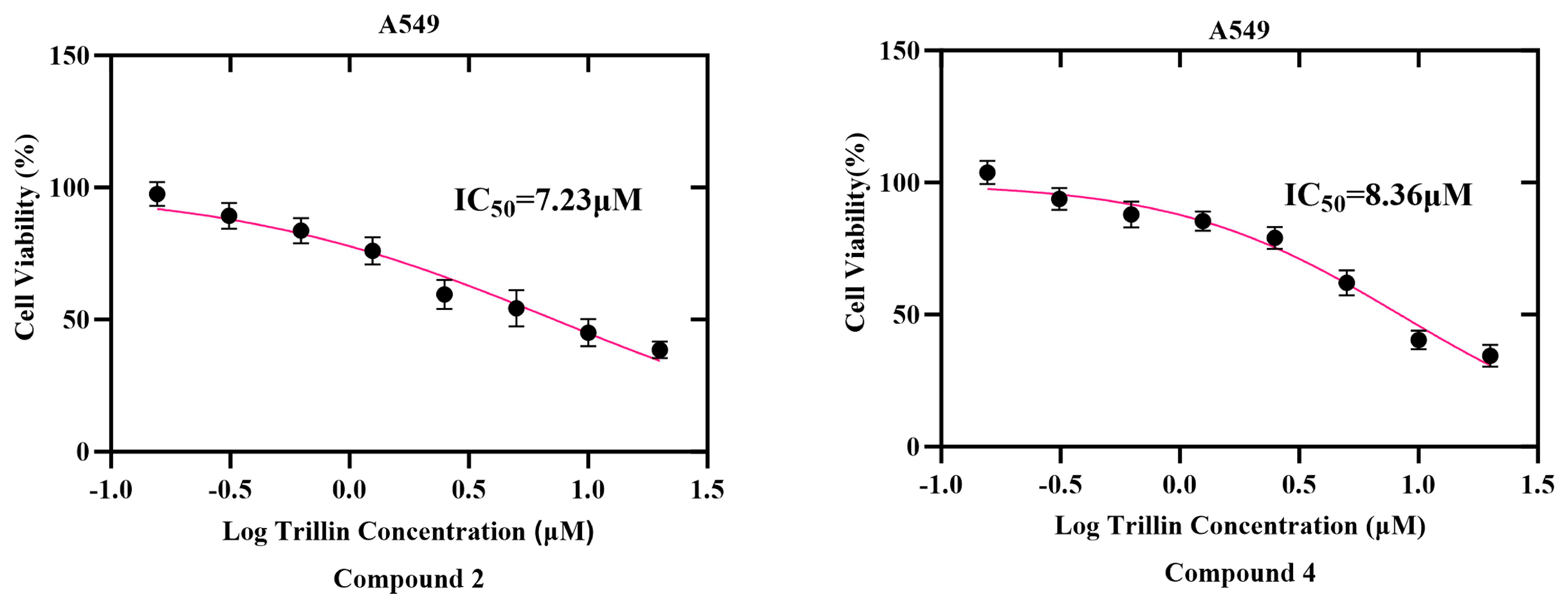
The in vitro antiproliferative activities of compounds 1–6 were evaluated against a panel of three human cancer cell lines: A549 (lung), MCF-7 (breast), and HepG2 (liver). In an initial screening at 20 μM, compounds 2 and 4 exhibited potent growth inhibition against A549 cells, reducing cell viability to 38.60% and 34.50%, respectively (Figure 8). Subsequent concentration–response analyses determined the IC50 values of compounds 2 and 4 to be 7.23 and 8.36 μM, respectively (Figure 9).
Figure 8.
Cell viability of A549, HepG2, and MCF-7 cells following treatment with compounds 1–6 (20 µM). Data are expressed as mean ± SD (n = 3). ** p < 0.05 compared with the control.
Figure 9.
Dose-dependent inhibitory effects and IC50 values of compounds 2 and 4 against A549 cells.
4. Discussion
R. roxburghii has garnered considerable scientific attention due to its complex phytochemical profile and potent health-promoting properties. Renowned as a robust botanical source of dietary antioxidants, this species harbors a dynamic repository of enzymatic antioxidants—particularly superoxide dismutase (SOD)—and essential micronutrients. These components act synergistically with a diverse array of secondary metabolites, including triterpenoids, sesquiterpenoids, flavonoids, phenolic acids, organic acids, and polysaccharides. To date, phytochemical investigations have elucidated over 50 distinct compounds from R. roxburghii. Among these, pentacyclic triterpenoids emerge as the predominant class, comprising more than 33 identified structures characterized primarily as polyhydroxylated ursane- and oleanane-type scaffolds alongside their corresponding glycosides [12,13,33]. Additionally, approximately 15 sesquiterpenoids, largely monocyclic variants and their glycosidic derivatives, have been documented [33]. The application of high-resolution analytical techniques, such as LC-MS/MS, has further delineated the rich landscape of flavonoids and polyphenolics [34,35], solidifying the plant’s reputation as a premier source of natural antioxidants. Building upon this established phytochemical foundation, the present study provides a targeted exploration of the secondary metabolome in fresh R. roxburghii fruits. This endeavor yielded the successful isolation and structural elucidation of five alkaloids and three triterpenoid glycosides. Most notably, this study reports the first-ever discovery of alkaloidal constituents within R. roxburghii, specifically designated as roxburghcids D (1) and E (2). Furthermore, a new triterpenoid glycoside, roxburghcid C (6), was identified, featuring a relatively rare α-configuration of the methyl group at the C-29 position. The discovery of these structurally diverse and previously undescribed constituents substantially broadens the recognized phytochemical spectrum of R. roxburghii, providing novel molecular scaffolds for future pharmacological and antioxidant bioactivity investigations.
Previous studies have established that R. roxburghii extracts effectively mitigate oxidative stress by modulating endogenous cellular defense systems. For instance, in an H2O2-challenged HepG2 cell model, these extracts confer robust cytoprotection via a dual mechanism: directly attenuating the intracellular accumulation of reactive oxygen species (ROS) and indirectly upregulating critical antioxidant enzymes, such as superoxide dismutase (SOD) and glutathione peroxidase (GSH-Px), thereby restoring redox homeostasis [36]. Expanding upon these extract-level observations, the present study evaluates the intrinsic antioxidant potential of the specifically isolated constituents. Compounds 2 and 3 demonstrated significant ABTS radical scavenging activity, achieving inhibition rates between 81.18% and 99.97% at a screening concentration of 100 μM. Subsequent assays confirmed a distinct dose-dependent response across a concentration gradient (6.25–100 μM), yielding IC50 values of 39.59 and 17.38 μM, respectively. From a structure–activity relationship (SAR) perspective, the alkaloids exhibited superior ABTS radical scavenging activity compared to the triterpene glycosides. Notably, the two new alkaloids, compounds 1 and 2, are epimers differing exclusively in the stereochemical configuration of the hydroxyl group at C-1. The isomer possessing the S-configuration (α-hydroxyl group) displayed a markedly lower ABTS scavenging capacity than its counterpart with the R-configuration (β-hydroxyl group). Classical reference antioxidants—such as resveratrol, vitamin E, and lycopene—typically exhibit potent in vitro scavenging capacities with IC50 values well below 10 μM [37]. In contrast, the individual compounds evaluated herein display relatively modest intrinsic efficacy (IC50 > 10 μM). Consequently, we postulate that rather than acting as singular, highly potent radical scavengers, these secondary metabolites function as integral co-contributors. They likely augment the overall antioxidant efficacy of R. roxburghii through additive or synergistic interactions within its complex phytochemical matrix.
Beyond its well-established antioxidant properties, R. roxburghii exhibits significant systemic efficacy in modulating glucolipid metabolism. In vitro studies have demonstrated that polyphenolic fractions enriched from R. roxburghii leaves exert robust inhibitory effects against α-glucosidase. Concurrently, in vivo evidence from diabetic animal models (e.g., db/db mice) reveals that these polyphenols promote hepatic glycogen synthesis and ameliorate insulin resistance via the upregulation of the PI3K/Akt signaling cascade. The integration of this systemic metabolic regulation with the localized inhibition of intestinal α-glucosidase establishes a potent, multi-target synergistic network for glycemic control [38]. Previous literature reports underscore that specific triterpenoids within R. roxburghii, such as rosamultic acid, roxbuterpene B, euscaphic acid, pomolic acid, and 2α,3α,24-trihydroxy-urs-12,18-diene-28-oic acid, possess potent α-glucosidase inhibitory capabilities (IC50 = 0.9–13.6 μM) [12]. Although the compounds screened in the present study exhibited relatively weak α-glucosidase inhibition, their combined intrinsic antioxidant capacities and metabolic regulatory potential suggest they play an indispensable role within the plant’s broader phytotherapeutic matrix. We propose that through dynamic synergy with highly active molecular scaffolds, these newly identified components contribute to a collective pharmacological interplay, thereby maintaining and amplifying the macroscopic efficacy of R. roxburghii against metabolic syndrome.
The dual role of oxidative stress in modulating tumor cell apoptosis remains a central paradigm in contemporary oncology research. In the context of hepatocellular carcinoma (HCC), previous studies have demonstrated that triterpenoid acid extracts from R. roxburghii (TAR) inhibit HepG2 cell proliferation and migration in a time- and dose-dependent manner. Mechanistically, these extracts induce a robust accumulation of intracellular reactive oxygen species (ROS). This supraphysiological ROS surge activates the c-Jun N-terminal kinase (JNK) signaling cascade, culminating in irreversible G2/M cell cycle arrest. Concurrently, the pronounced ROS generation triggers the dissipation of the mitochondrial membrane potential, facilitating the cytosolic translocation of cytochrome c. This process activates the caspase cascade, ultimately driving mitochondria-dependent apoptosis [39]. Building upon the chemical diversity elucidated earlier in this study, our preliminary in vitro screening identified promising antitumor properties among the newly isolated constituents. Notably, we report for the first time that roxburghcid E (2) and canthine-6-one (4)—alkaloids characterized from R. roxburghii—exhibit potent cytotoxicity against human non-small cell lung cancer A549 cells, yielding IC50 values of 7.23 and 8.36 μM, respectively. Structurally, alkaloids 2 and 4 exhibited significant inhibitory activity against A549 cells. Furthermore, the new alkaloids 1 and 2 differ solely in the stereochemical configuration of the hydroxyl group at the C-1′ position; notably, the epimer with the α-hydroxyl at C-1 displayed a substantially lower A549 inhibitory potency than that with the β-hydroxyl. These findings successfully correlate the structural diversity of the plant with its pharmacological efficacy. They not only significantly expand the known spectrum of bioactive compounds in R. roxburghii but also provide a preliminary basis for future lung cancer therapies targeting these specific alkaloids.
5. Conclusions
In this study, a phytochemical investigation of R. roxburghii fruits led to the isolation of two new dihydroavicine-type alkaloids, roxburghcids D (1) and E (2), and a new triterpenoid glycoside, roxburghcid C (6), alongside five known analogues. Structurally, compound 6 is notable as a rare ursane-type triterpenoid glycoside featuring an α-oriented C-29 methyl group. In in vitro biological evaluations, compounds 2 and 3 displayed significant ABTS radical scavenging capacities, while compounds 2, 6, and 8 exhibited moderate α-glucosidase inhibitory activities. Furthermore, compounds 2 and 4 demonstrated significant cytotoxicity against the A549 human lung cancer cell line. Importantly, this study constitutes the first report of alkaloids derived from R. roxburghii. These findings not only expand the known phytochemical space of this species but also provide a preliminary reference for its development and utilization as a medicinal and functional food resource.
Supplementary Materials
The following supporting information can be downloaded at: https://www.mdpi.com/article/10.3390/antiox15060680/s1.
Author Contributions
Data curation, writing—review and editing, L.Z.; data curation, Y.-J.Z.; Resources, W.-X.D.; Investigation, F.-J.C.; Methodology, X.P.; Supervision, Y.W.; Formal analysis, L.-S.Y.; Software, Q.-J.L.; Funding acquisition, Project administration, X.-S.Y. All authors have read and agreed to the published version of the manuscript.
Funding
This research was funded by the Guizhou Provincial Basic Research Program (Nos. QKHJC-QN [2025]-434; QKHJC-ZD [2025]-026); the National Natural Science Foundation of China (No. 32560109); the Guizhou Provincial Science and Technology Major Project (Nos. QKHZD [2025]049; QKHZD [2025]050); the Guizhou Provincial Special Fund for the Construction of Innovation Capability of Scientific Research Institutions (No. QJHFQ2024-005); the Establishment of an Innovation Base for the Creation and Pilot-Scale Verification of Novel Cili Products in Guizhou (No. QKHZYD [2025] 014); the Guizhou Provincial Technology Innovation Center for the Cili Industry (No. QKHPTJSZX [2025] 006); and the Guizhou Provincial Basic Research Program (No. QKHJC [2023] 429).
Institutional Review Board Statement
Not applicable.
Informed Consent Statement
Not applicable.
Data Availability Statement
The original contributions presented in this study are included in the article and Supplementary Materials. Further inquiries can be directed to the corresponding authors.
Conflicts of Interest
The authors declare no conflicts of interest.
References
- Sies, H. Oxidative eustress: On constant alert for redox homeostasis. Redox Biol. 2021, 41, 101867. [Google Scholar] [CrossRef] [Scilit]
- Murphy, M.P.; Bayir, H.; Belousov, V.; Chang, C.J.; Davies, K.J.A.; Davies, M.J.; Dick, T.P.; Finkel, T.; Forman, H.J.; Janssen-Heininger, Y.; et al. Guidelines for measuring reactive oxygen species and oxidative damage in cells and in vivo. Nat. Metab. 2022, 4, 651–662. [Google Scholar] [CrossRef] [Scilit] [PubMed]
- Hong, Y.; Boiti, A.; Vallone, D.; Foulkes, N.-S. Reactive oxygen species signaling and oxidative stress: Transcriptional regulation and evolution. Antioxidants 2024, 13, 312. [Google Scholar] [CrossRef] [Scilit]
- Forman, H.J.; Zhang, H. Targeting oxidative stress in disease: Promise and limitations of antioxidant therapy. Nat. Rev. Drug Discov. 2021, 20, 689–709. [Google Scholar] [CrossRef] [Scilit]
- Akter, S.; Madhuvilakku, R.; Kar, A.-K.; Nila, I.-S.; Liu, P.-D.; Inuzuka, H.; Wei, W.-Y.; Hong, Y. Reactive oxygen species (ROS) in cancer: From mechanism to therapeutic implications. Sig. Transduct. Target. Ther. 2026, 11, 111. [Google Scholar] [CrossRef] [Scilit]
- Fang, S.-H.; Jiang, M.-J.; Jiao, J.; Zhao, H.Y.; Liu, D.-Z.; Gao, D.-N.; Wang, T.-E.; Yang, Z.; Yuan, H.-P. Unraveling the ROS-inflammation-immune balance: A new perspective on aging and disease. Aging Dis. 2026, 17, 305–325. [Google Scholar] [CrossRef] [Scilit] [PubMed]
- Bhat, A.-H.; Dar, K.-B.; Anees, S.; Zargar, M.-A.; Masood, A.; Sofi, M.-A.; Ganie, S.-A. Oxidative stress, mitochondrial dysfunction and neurodegenerative diseases; a mechanistic insight. Biomed. Pharmacother. 2015, 74, 101–110. [Google Scholar] [CrossRef] [Scilit]
- Kim, M.; Jang, H.; Kim, W.; Kim, D.; Park, J.-H. Therapeutic applications of plant-derived extracellular vesicles as antioxidants for oxidative stress-related diseases. Antioxidants 2023, 12, 1286. [Google Scholar] [CrossRef] [Scilit] [PubMed]
- Editorial Committee of Flora of China, Chinese Academy of Sciences. Flora of China; Science Press: Beijing, China, 1985; Volume 37, p. 452. [Google Scholar]
- Wang, L.; Wei, T.-T.; Zheng, L.; Jiang, F.-F.; Ma, W.-T.; Lu, M.; Wu, X.-M.; An, H.-M. Recent advances on main active ingredients, pharmacological activities of Rosa roxburghii and its development and utilization. Foods 2023, 12, 1051. [Google Scholar] [CrossRef] [Scilit]
- Guizhou Provincial Medical Products Administration. Quality Standards of Traditional Chinese Medicines and Ethnic Medicines in Guizhou Province; China Medical Science Press: Beijing, China, 2019; Volume 2, p. 160.
- Zhang, S.; Xiang, L.-J.; Long, X.-X.; Guo, L.-J.; Wei, X.; Zhou, Y.-Q.; Feng, T.-T.; Zhou, Y.; Yin, X. Anti-inflammatory and α-glucosidase inhibitory triterpenoid with diverse carbon skeletons from the fruits of Rosa roxburghii. J. Agric. Food Chem. 2024, 72, 11503–11514. [Google Scholar] [CrossRef] [Scilit] [PubMed]
- Yu, Y.; Wu, J.; Bao, M.-F.; Yang, L.; Cai, Z.-L.; Schinnerl, J.; Cai, X.-H. Fruits from Rosa roxburghii: A valuable bioresource of potent radical scavengers and novel ursane-type triterpenoids. ACS Omega 2024, 9, 38023−38031. [Google Scholar] [CrossRef] [Scilit]
- Zhou, X.-L.; Qin, Y.-M.; Wang, Y.-L.; Wang, Y.-P.; Qin, Z.-C. Phytochemical profile and antioxidant characteristics of bound and free phenolics from Rosa roxburghii Tratt. Food Biosci. 2024, 57, 103576. [Google Scholar] [CrossRef] [Scilit]
- Chen, F.-J.; Li, L.-Q.; Huang, M.-Y.; Wang, Y.-K.; Wang, L.; Jin, F.-L.; Yang, L.-S.; Gao, M.; Li, L.-L.; Wang, Y.; et al. Natural product Kaji-ichigoside F1 exhibits rapid antidepression via activating the AMPA–BDNF–mTOR pathway and inhibiting the NMDAR–CaMKIIα pathway. Phytomedicine 2024, 126, 155452. [Google Scholar] [CrossRef] [Scilit]
- He, F.-J.; Yan, Y.-F.; Peng, M.; Gao, M.; Zhou, L.; Chen, F.-J.; Yang, L.-S.; Li, L.-Q.; Yang, X.-S. Therapeutic potential of Rosa roxburghii folium extract in insomnia treatment: A comprehensive evaluation of behavioral and neurochemical effects in a PCPA-induced mouse model. J. Sci. Food Agr. 2025, 105, 1044–1056. [Google Scholar] [CrossRef] [Scilit]
- Jiao, X.; Zhang, M.-Y.; Zhang, M.; Hao, L.-Y.; Wu, C.-D. Ultrasound-assisted enzymatic extraction, structural characterization, and anticancer activity of polysaccharides from Rosa roxburghii Tratt fruit. Int. J. Biol. Macromol. 2024, 259, 127926. [Google Scholar] [CrossRef] [Scilit]
- Chen, Z.; Zhu, Y.-P.; Lu, M.-T.; Yu, L.; Tan, S.-M.; Ren, T.-Y. Effects of Rosa roxburghii Tratt glycosides and quercetin on D-galactose-induced aging mice model. J. Food Biochem. 2022, 46, e14425. [Google Scholar] [CrossRef] [Scilit]
- Frisch, M.-J.; Trucks, G.-W.; Schlegel, H.-B.; Scuseria, G.E.; Robb, M.A.; Cheeseman, J.R.; Scalmani, G.; Barone, V.; Petersson, G.A.; Nakatsuji, H. Gaussian 09, Revision E.01; Gaussian, Inc.: Wallingford, CT, USA, 2013. [Google Scholar]
- Lodewyk, M.-W.; Siebert, M.-R.; Tantillo, D.-J. Computational prediction of 1H and 13C chemical shifts: A useful tool for natural product, mechanistic, and synthetic organic chemistry. Chem. Rev. 2012, 112, 1839–1862. [Google Scholar] [CrossRef] [Scilit]
- Zanardi, M.-M.; Sarotti, A.-M. Sensitivity analysis of DP4+ with the probability distribution terms: Development of a universal and customizable method. J. Org. Chem. 2021, 86, 8544–8548. [Google Scholar] [CrossRef] [Scilit]
- Srebro-Hooper, M.; Autschbach, J. Calculating natural optical activity of molecules from first principles. Annu. Rev. Phys. Chem. 2017, 68, 399–420. [Google Scholar] [CrossRef] [Scilit]
- Gou, X.-S.; Tian, D.-M.; Wei, J.-H.; Ma, Y.-H.; Zhang, Y.-X.; Chen, M.; Ding, W.-J.; Wu, B.; Tang, J.-S. New drimane sesquiterpenes and polyketides from marine-derived fungus penicillium sp. TW58-16 and their anti-inflammatory and α-glucosidase inhibitory effects. Mar. Drugs 2021, 19, 416. [Google Scholar] [CrossRef] [Scilit]
- Ren, Q.-X.; Wang, R.; Mu, Q.-R.; Chen, L.; Chen, M.; Wang, L.-J.; Li, P.; Yang, H.; Gao, W. Molecular networking and paterno-büchi reaction guided glycerides characterization and antioxidant activity assessment of Ganoderma lucidum spore oil. Food Chem. 2025, 468, 142500. [Google Scholar] [CrossRef] [Scilit]
- Song, M.; Chan, G.; Lin, L.-G.; Li, D.-R.; Zhang, K.-H.; Zhang, X.-Q.; Ye, W.-C.; Li, N.; Zhang, Q.-W. Triterpenoids from the fruits of Melia azedarach L. and their cytotoxic activities. Phytochemistry 2022, 201, 113280. [Google Scholar] [CrossRef] [Scilit] [PubMed]
- Fernandes, C.-C.; Vieira, P.-C.; Silva, V.-C.; Dall’Oglio, E.-L.; Silva, L.-E.; Sousa, P.-T., Jr. 6-acetonyl-N-methyl-dihydrodecarine, a new alkaloid from Zanthoxylum riedelianum. J. Braz. Chem. Soc. 2009, 20, 379–382. [Google Scholar] [CrossRef] [Scilit]
- Li, W.; Fu, H.-W.; Bai, H.; Sasaki, T.; Kato, H.; Koike, K. Triterpenoid saponins from Rubus ellipticus var. obcordatus. J. Nat. Prod. 2009, 72, 1755–1760. [Google Scholar] [CrossRef] [Scilit]
- Yang, C.-S.; Zhou, T.; Han, S.-Q.; Wang, X.; Dong, X.-Y.; Bo, P. Alkaloids and terpenoids from Trigonostemon lutescens and their potential antiproliferative activity. Chem. Nat. Compd. 2020, 56, 763–766. [Google Scholar] [CrossRef] [Scilit]
- Soriano-Agatón, F.; Lagoutte, D.; Poupon, E.; Roblot, F.; Fournet, A.; Gantier, J.-C.; Hocqemiller, R. Extraction, hemisynthesis, and synthesis of canthin-6-one analogues. evaluation of their antifungal activities. J. Nat. Prod. 2005, 68, 1581–1587. [Google Scholar] [CrossRef] [Scilit]
- Kim, H.-M.; Lee, J.-S.; Sezirahiga, J.; Kwon, J.-Y.; Jeong, M.; Lee, D.-H.; Choi, J.-H.; Jang, D.-S. A new canthinone-type alkaloid isolated from Ailanthus altissima swingle. Molecules 2016, 21, 642. [Google Scholar] [CrossRef] [Scilit]
- Shahani, S.; Monsef-Esfahani, H.-R.; Saeidnia, S.; Saniee, P.; Siavoshi, F.; Foroumadi, A.; Samadi, N.; Gohari, A.-R. Anti-Helicobacter pylori activity of the methanolic extract of Geum iranicum and its main compounds. Z. Naturforsch. 2012, 67, 172–180. [Google Scholar] [CrossRef] [Scilit]
- Huang, B.; Fu, H.-Z.; Chen, W.-K.; Luo, Y.-H.; Ma, S.-C. Hepatoprotective triterpenoid saponins from Callicarpa nudiflora. Chem. Pharm. Bull. 2014, 62, 695–699. [Google Scholar] [CrossRef] [Scilit]
- Yin, X.; Zhang, J.; Xiang, L.-J.; Lin, C.; Wang, Q.-M.; Long, K.-X.; Long, Q.-Y.; Long, X.-X.; Wei, Y.-J.; Zhou, Y.; et al. Discovery of diverse skeletons triterpenoid and sesquiterpenoid with anti-inflammatory and PTP1B inhibitory activities from the fruits of Rosa roxburghii. J. Mol. Struct. 2026, 1362, 145760. [Google Scholar] [CrossRef] [Scilit]
- Liu, M.-H.; Zhang, Q.; Zhang, Y.-H.; Lu, X.-Y.; Fu, W.-M.; He, J.-Y. Chemical analysis of dietary constituents in Rosa roxburghii and Rosa sterilis Fruits. Molecules 2016, 21, 1204. [Google Scholar] [CrossRef] [Scilit]
- Wang, R.-M.; He, R.-P.; Li, Z.-H.; Lin, X.; Wang, L. HPLC-Q-Orbitrap-MS/MS phenolic profiles and biological activities of extracts from roxburgh rose (Rosa roxburghii Tratt.) leaves. Arab. J. Chem. 2021, 14, 103257. [Google Scholar] [CrossRef] [Scilit]
- Mao, Y.; Sha, R.; Sun, Y.; Wang, Z.; Huang, J. Antioxidative and cytoprotective effects of Rosa roxburghii and metabolite changes in oxidative stress-induced HepG2 cells following Rosa roxburghii intervention. Foods 2024, 13, 3520. [Google Scholar] [CrossRef] [Scilit]
- Mukherjee, S.; Chopra, H.; Goyal, R.; Jin, S.; Dong, Z.-Z.; Das, T.; Bhattacharya, T. Therapeutic effect of targeted antioxidant natural products. Discov. Nano 2024, 19, 144. [Google Scholar] [CrossRef] [Scilit] [PubMed]
- Chen, C.; Tan, S.; Ren, T.; Wang, H.; Dai, X.; Wang, H. Polyphenol from Rosa roxburghii Tratt fruit ameliorates the symptoms of diabetes by activating the P13K/AKT insulin pathway in db/db mice. Foods 2022, 11, 636. [Google Scholar] [CrossRef] [Scilit]
- Zhai, B.-W.; Zhao, H.; Zhu, H.-L.; Huang, H.; Zhang, M.-Y.; Fu, Y.-J. Triterpene acids from Rosa roxburghii Tratt fruits exert anti-hepatocellular carcinoma activity via ROS/JNK signaling pathway-mediated cell cycle arrest and mitochondrial apoptosis. Phytomedicine 2023, 119, 154960. [Google Scholar] [CrossRef] [Scilit] [PubMed]

Disclaimer/Publisher’s Note: The statements, opinions and data contained in all publications are solely those of the individual author(s) and contributor(s) and not of MDPI and/or the editor(s). MDPI and/or the editor(s) disclaim responsibility for any injury to people or property resulting from any ideas, methods, instructions or products referred to in the content. |
© 2026 by the authors. Licensee MDPI, Basel, Switzerland. This article is an open access article distributed under the terms and conditions of the Creative Commons Attribution (CC BY) license.