


Abstract
Although the molecular pathogenesis of bilirubin-induced neuronal cell injury is not completely understood, certain recurrent themes resonate in the literature on this topic and include the generally untoward effects of high unconjugated bilirubin (UCB) concentrations on membranes (plasma, mitochondrial, and endoplasmic reticulum (ER)), cellular bioenergetics, and intracellular calcium homeostasis. Only in the last decade, ER was discovered as an early target of bilirubin neurotoxicity. We will review the main features of bilirubin neurotoxicity from the point of view of ER and bilirubin-induced ER stress. Neuronal excitotoxicity, mitochondrial energy failure, and increased intracellular calcium concentration are three phenomena linked spatially and temporally in the pathogenesis of bilirubin-induced neurotoxicity. ER, being the main intracellular calcium storage organelle, is involved in the increase in the universal second messenger, calcium. This event leads to the activation of proteolytic enzymes, apoptotic pathways, and necrosis, the occurrence of which is likely a function of the degree and duration of bilirubin exposure.
1. Introduction
Unconjugated bilirubin (UCB) is the end product of heme catabolism, that exhibits cytoprotective and cytotoxic properties. At physiological concentrations, UCB functions as a potent endogenous direct antioxidant; however, when present at elevated levels—especially in cases of sever neonatal hyperbilirubinemia—it becomes neurotoxic [1]. Prolonged and severe hyperbilirubinemia is marked by elevated concentrations of UCB, which promote its accumulation [2] within the CNS [3], ultimately leading to a severe form of chronic bilirubin encephalopathy known as kernicterus. Kernicterus is associated with irreversible neurological sequelae, including motor abnormalities, auditory deficits, and learning impairments [4,5,6]. These central nervous system (CNS) manifestations reflect the regional distribution of bilirubin-induced neuropathology, which primarily affects the globus pallidus, subthalamic nucleus, brainstem nuclei, hippocampal CA2 neurons, and cerebellar Purkinje cells [7,8,9].
Among the emerging mechanisms, endoplasmic reticulum (ER) stress has gained attention in the last decade as an early and pivotal event of UCB-induced neuronal injury [10,11,12]. The ER represents an integrated molecular web that plays a central role in protein synthesis, calcium homeostasis, and glucose and lipid metabolism [13]. Perturbation of ER homeostasis leads to activation of the unfolded protein response (UPR), an orchestrated signaling that involves the activation of three main stress sensors (PERK, IRE1, and ATF6) [14]. While transient UPR activation promotes adaptive response and cellular survival, sustained or prolonged ER-stress shifts signaling toward pro-apoptotic pathways, notably via CHOP induction, inflammatory cascade, and calcium signaling [15]. Importantly, ER stress functions as a central node rather than an isolated event. ER has a specialized environment that makes it very sensitive to oxidative stress [16]. Simultaneously, disruption of ER calcium homeostasis triggers cytosolic calcium overload, activation of calpain and caspase pathways [17], and mitochondrial impairment [18]. Moreover, ER-stress sensors interface with inflammatory mediators such as NF-κB and JNK, triggering neuroinflammation [19].
Several studies demonstrated the important role of oxidative stress, calcium dysregulation, mitochondrial dysfunction, and inflammatory signaling in UCB-induced neurotoxicity [20]. However, the complex molecular mechanisms of UCB-induced neurotoxicity that lie on ER stress need further investigations. This review will first recapitulate the main known mechanisms of UCB neurotoxicity, then detail the molecular basis of ER stress, and focus on demonstrating how ER stress acts as a central hub interacting with pathways such as oxidative stress, calcium homeostasis, mitochondrial function, inflammation, and autophagy, ultimately determining neuronal fate. By framing ER stress as a systems-level regulator rather than a single linear pathway, we propose a conceptual model in which neuronal fate is determined by the dynamic balance between pro-survival and pro-apoptotic signaling. We hope this integrative perspective will provide new insights for future therapeutic strategies.
2. Molecular Mechanisms and Cellular Target of UCB-Induced Neurotoxicity
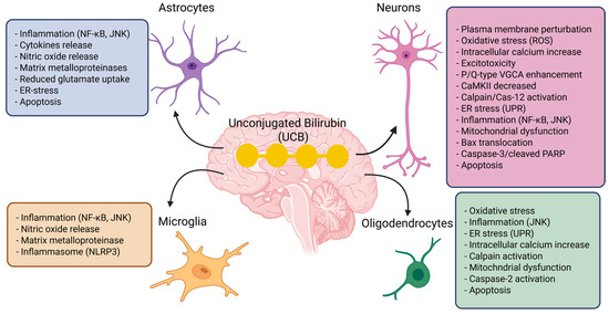
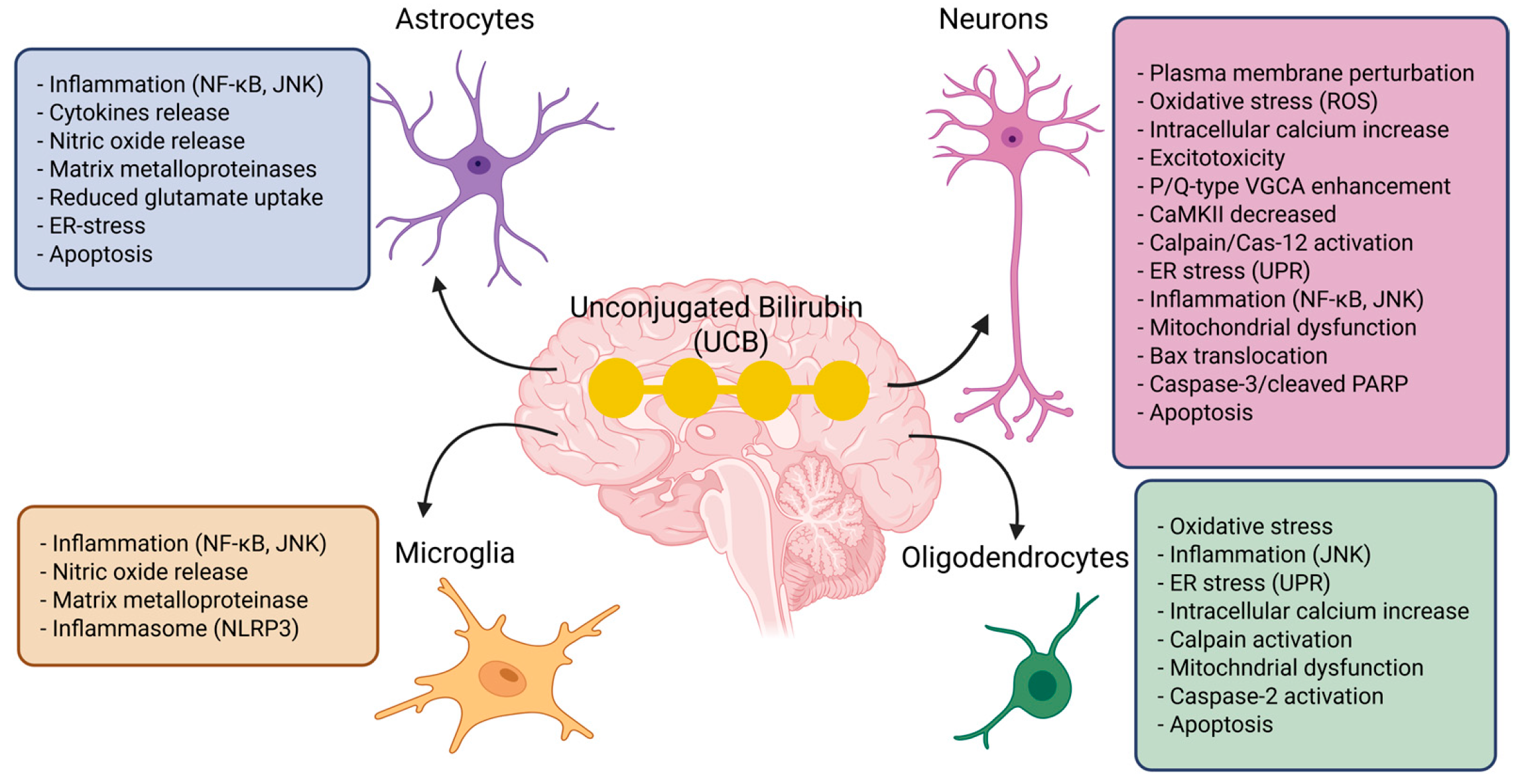
UCB is highly lipophilic and binds strongly to cell membranes, particularly myelin-rich membranes, making neurons the main targets of bilirubin toxicity. Non-neuronal cells in the CNS are also affected by UCB and may modulate bilirubin-induced neurotoxicity [21]. Figure 1 shows how UCB affect neurons, astrocytes, microglia, and oligodendrocytes. In vitro, neuronal exposure to UCB leads to reduced dendritic and axonal arborization, impaired neurite growth and branching, decreased proliferation, and increased apoptosis [22,23]. In neuroblastoma cells, bilirubin delays S-phase progression and induces cell-cycle arrest [22], a mechanism that may contribute to the cerebellar hypoplasia observed in murine models of kernicterus Bilirubin, and also causes major biochemical disturbances, including protein oxidation, lipid peroxidation, glutathione depletion, increased lactate dehydrogenase release, and nitric oxide production through NMDA receptor-mediated activation of neuronal nitric oxide synthase [24]. These events are closely associated with oxidative stress and mitochondrial dysfunction, which appear to be central mechanisms of neuronal injury. Astrocytes respond to toxic bilirubin levels by releasing inflammatory mediators such as IL-1β, TNF-α, and IL-6, as well as glutamate, and may ultimately undergo apoptosis, although they are generally less vulnerable than neurons [25]. Microglia, directly activated by UCB, acquire a phagocytic phenotype, secrete pro-inflammatory cytokines, and increase matrix metalloproteinase activity, supporting the idea that bilirubin encephalopathy involves a strong neuroinflammatory component [26]. Oligodendrocytes are likewise susceptible, showing mitochondrial impairment, increased reactive oxygen species, and caspase-3-mediated apoptosis; this may contribute to altered myelin production and axonal dysfunction in brain regions typically damaged in kernicterus [27].
Figure 1.
UCB exerts toxic effects on neurons, astrocytes, microglia, and oligodendrocytes. In neurons, UCB disrupts the plasma membrane, induces oxidative stress (ROS), increases intracellular calcium, promotes excitotoxicity, enhances P/Q-type voltage-gated calcium channels, decreases CaMKII, activates calpain and caspase-12, triggers endoplasmic reticulum stress (UPR), stimulates inflammatory pathways such as NF-κB and JNK, impairs mitochondrial function, causes Bax translocation, caspase-3 activation, and PARP cleavage, ultimately leading to apoptosis. In astrocytes, UCB promotes inflammation through NF-κB and JNK, with release of cytokines and nitric oxide, increased matrix metalloproteinases, reduced glutamate uptake, ER stress, and, under severe conditions, apoptosis. Microglia respond with rapid inflammatory activation, characterized by NF-κB/JNK signaling, release of nitric oxide, increased matrix metalloproteinases, and activation of the NLRP3 inflammasome, thereby amplifying neuroinflammation. Oligodendrocytes are also highly vulnerable: UCB induces oxidative stress, JNK-mediated inflammation, ER stress (UPR), intracellular calcium increase, calpain activation, mitochondrial dysfunction, caspase-2 activation, and finally apoptosis. Created in BioRender. Models, I. (2026) https://BioRender.com/gzshrzf (accessed on 25 March 2026).
Although neurons are the most sensitive cell type, non-neuronal cells also actively contribute to the progression of injury. Overall, the neurotoxicity of unconjugated bilirubin results from the combined effects of oxidative stress, calcium imbalance, inflammation, and altered glutamatergic transmission, which will be analyzed in detail below.
2.1. Oxidative Stress: A Central and Early Event
Oxidative stress represents a pivotal mechanism in bilirubin neurotoxicity and occurs when reactive oxygen species (ROS) generation overwhelms endogenous antioxidant defenses. Bilirubin displays a well-recognized “Janus-like” behavior: at low intracellular concentrations it exerts antioxidant effects, whereas at higher levels it becomes pro-oxidant [20]. In vitro studies in hepatic, endothelial, renal, and neuronal cell lines demonstrated that bilirubin acts as an antioxidant at approximately 7 ng/mg protein intracellularly, while concentrations exceeding 25 ng/mg protein shift its activity toward pro-oxidant effects [28].
At toxic concentrations, bilirubin enhances ROS production, promotes lipid and protein oxidation, and disrupts glutathione metabolism [29]. Oxidative imbalance contributes to the activation of mitochondrial apoptotic pathways [30,31]. Furthermore, UCB-induced oxidative stress affects DNA integrity by promoting oxidative DNA damage. In primary culture of neurons, UCB-induced ROS has been directly linked to Tet1 (ten-eleven translocation 1) protein inhibition and increased DNA methylation [32]. Cells attempt to counteract oxidative stress by upregulating compensatory mechanisms, including enhanced glutathione synthesis [22], activation of the redox regulator APE1/Ref-1 (apurinic/apyrimidinic endonuclease 1/redox effector factor 1) [33], activation of the stress-response adaptive protein DJ-1 (Parkinson disease protein 7) [22], and the activation of Nrf2-dependent antioxidant gene programs [34]. However, when these adaptive responses are insufficient, oxidative damage becomes a major driver of neuronal dysfunction.
2.2. Membrane Perturbation and Calcium Dysregulation
An enduring hypothesis [7] proposes that neuronal injury begins at the membrane level, where bilirubin perturbs membrane structure and dynamics. Disruption of plasma, mitochondrial, and endoplasmic reticulum (ER) membranes contributes to intracellular calcium overload. The resulting elevation in the universal second messenger Ca2+ activates downstream cascades, including protease activation, mitochondrial dysfunction, apoptotic signaling, and necrosis [30,35].
In parallel, bilirubin induces ER stress [10,36], with gene signatures resembling thapsigargin exposure. ER calcium release leads to sustained calcium elevations—more pronounced in neurons than in glia—and activation of ER stress-, calpain-, and calcium-dependent apoptotic pathways [37]. One possible scenario is that ER-derived Ca2+ release can be transferred to mitochondria through ER–mitochondria contact sites (MAMs), promoting mitochondrial Ca2+ overload, bioenergetics failure, and amplification of apoptotic signaling. Autophagy is also activated as an early protective response [38]. Collectively, these findings identify calcium dysregulation and ER stress as central and potentially targetable mechanisms.
2.3. Inflammation and Glial Contribution
Beyond oxidative stress, inflammation represents the most extensively studied pathogenic mechanism of bilirubin toxicity. Neurons exposed to bilirubin activate pro-inflammatory and pro-apoptotic pathways—including IL8, NOS signaling, and caspase cascades—with exacerbated injury in the presence of TNFα and IL1β, and impaired neurite outgrowth and synaptogenesis [11,39,40]. Although neurons are particularly vulnerable, glial cells actively amplify injury. Microglia and astrocytes release cytokines, nitric oxide, and matrix metalloproteinases, thereby sustaining neuroinflammation [25,26,41]. Conversely, blood–brain barrier interfaces display limited cytokine modulation in vivo and may regulate immune cell infiltration.
2.4. Glutamatergic Dysfunction and Excitotoxicity
Glutamate-mediated excitotoxicity provides a mechanistic link between cellular injury and regional brain vulnerability. Bilirubin-stimulated glial cells release glutamate [42,43], while both neurons and astrocytes exhibit reduced glutamate uptake [44]. The consequent extracellular glutamate accumulation results in NMDA receptor overactivation, calcium influx, and excitotoxicity [45,46].
Transcriptomic alterations in glutamatergic synapse-related genes in susceptible brain regions [47] parallel neuroimaging findings in affected neonates [48]. Glutamatergic dysfunction and excitotoxicity have therefore been proposed as unifying mechanisms linking regional susceptibility to the clinical topography of bilirubin-induced brain injury [20].
3. Molecular Basis of ER-Stress
The endoplasmic reticulum (ER) has a specialized oxidized environment that allows proper protein folding, making the ER extremely sensitive to alteration in cellular redox states and calcium homeostasis [49]. In addition to its role in protein quality control, the ER is also considered as a “nutrient-sensing apparatus” through its ability to control energy, glucose, and lipid metabolism [50]. ER stress is triggered by the changing of cellular redox state, calcium disruption, or the accumulation of misfolded proteins, which collectively initiates a cascade of signaling pathways known as the unfolded protein response (UPR) [51]. The UPR involves three main membrane-associated proteins: PERK (PKR-like endoplasmic reticulum kinase of the eukaryotic initiation factor 2α), IRE1 (inositol-requiring enzyme 1), and ATF6 (activating transcription factor-6). Under basal conditions, these three proteins are kept inactive through their interaction with the chaperone BiP (or GRP78), which dissociates by the misfolded proteins under ER-stress conditions.
Upon activation, PERK phosphorylates eukaryotic initiation factor 2 (elF2α), leading to the attenuation of protein synthesis and selective upregulation of ATF4. ATF4 induces the upregulation of CHOP that contributes to cell-cycle arrest by decrease in cyclin D1. Activated IRE1 mediates the selective splicing of XBP1 mRNA, which results in a stable, active transcription factor. ATF6 translocates to the Golgi apparatus where it undergoes proteolytic cleavage, generating a cleaved-active transcription factor [52]. All three UPR branches induce transcription factors (e.g., ATF4, CHOP, sXBP1, and ATF6) that reprogram the cellular expression profile by upregulating genes involved in stress response, inflammation, antioxidants, and apoptosis [14,19,53].
4. ER-Stress as a Central Integrator of UCB Toxicity In Vitro and In Vivo
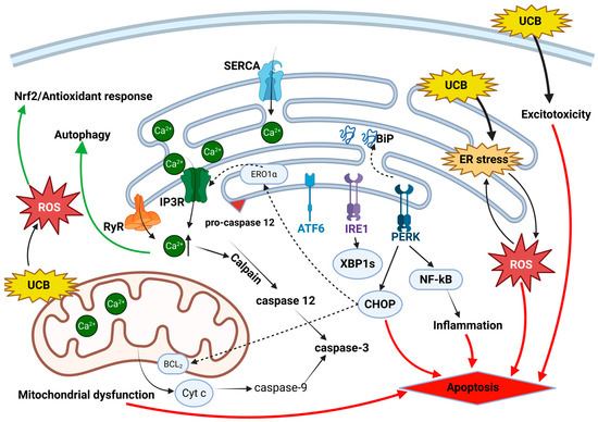
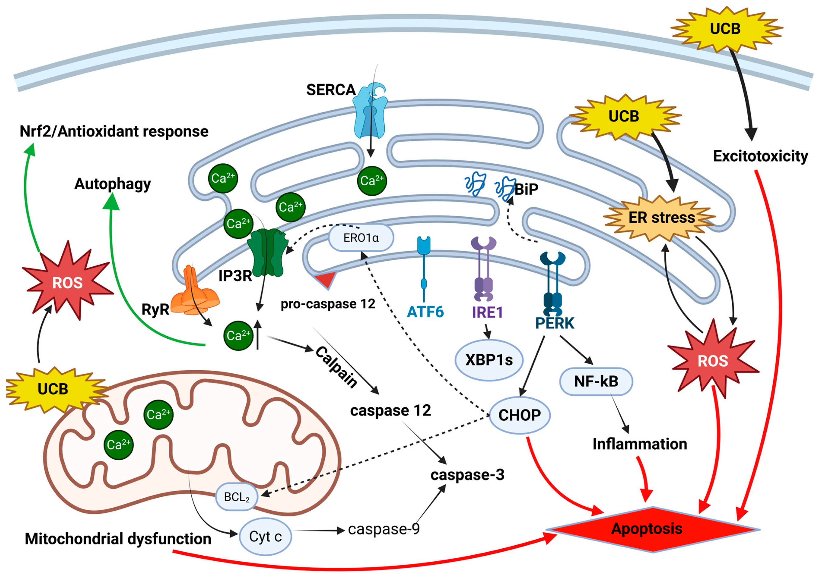
Cells must maintain bilirubin at very low, non-toxic intracellular levels through multiple protective mechanisms. As shown in Figure 2, after entering the cell, unconjugated bilirubin (UCB), due to its poor aqueous solubility, binds to intracellular ligandins and is shuttled toward the endoplasmic reticulum (ER), which acts as a “sink” for excess UCB [54]. Direct accumulation of UCB within the ER may represent a primary trigger of ER stress, as UCB can interact with hydrophobic regions of proteins, thereby interfering with proper protein folding and ER homeostasis. This proposed mechanism largely depends on the capacity of the cell, particularly the ER, to detoxify UCB.
Figure 2.
Molecular pathways dysregulated in bilirubin-induced neurotoxicity. Unconjugated bilirubin (UCB) triggers a coordinated pro-apoptotic signaling network integrating ER stress, calcium dysregulation, reactive oxygen species (ROS) generation, inflammation, excitotoxicity, and mitochondrial dysfunction. UCB disrupts cellular redox state and induces the activation of ER stress. The chaperone BiP dissociates from PERK and IRE1 and binds to the misfolded proteins, initiating the unfolded protein response. PERK phosphorylates eIF2α, with subsequent activation of the ATF4/CHOP axis and downstream inflammatory signal including NF-κB. IRE1 and ATF6 activate a transcriptional program that amplifies stress signals. UCB-induced ER stress perturbs calcium homeostasis, promoting Ca2+ release from the ER and activation of calpain and caspase-12, thereby further driving apoptotic signaling. Pro-survival pathways are concurrently activated to counteract UCB toxicity, including the Nrf2-mediated antioxidant response and autophagy. Cellular fate is determined by the intensity and duration of UCB exposure, reflecting the balance between adaptive stress responses and pro-apoptotic signaling. Pro-apoptotic signaling flows are indicated in red, while pro-survival signaling flows in green. Created in BioRender. Models, I. (2026) https://BioRender.com/gzshrzf (accessed on 25 March 2026).
Unlike hepatocytes, most cell types lack an efficient conjugation system and therefore rely on alternative pathways for UCB elimination [55]. One such pathway involves UCB oxidation by cytochrome P450 enzymes, which are abundant in the ER [56,57,58]. Cytochrome P450 enzymes are expressed in a tissue- and cell type-specific manner and are localized to different subcellular compartments, reflecting their diverse physiological and pathological roles [59]. Under conditions of elevated UCB, the activity or expression levels of specific cytochrome P450 isoforms may determine the susceptibility of individual cell types to UCB-induced ER stress. Further studies are needed to clarify how UCB affects ER protein-folding processes, and to determine whether variability in ER-stress sensitivity is directly related to differences in cytochrome P450 expression and activity across cell types.
ER-stress induction following UCB exposure has been reported in several in vitro models. Neuronal cell lines including SH-SY5Y and GI-ME-N show upregulation of ATF4, CHOP, ATF6, and BiP after 4 h of exposure [11,36]. Similarly, a primary culture of mouse hippocampal neurons exhibits increased protein content of CHOP, cleaved-ATF6, and BiP upon exposure to UCB for 24 h [37]. In rat oligodendrocyte precursor cultures, UCB exposure induces BiP at 1 h followed by an-upregulation of IRE-1α and ATF-6 at 2 h [60]. Microarray analysis performed on Hepa 1c1c7 cells exposed to UCB revealed a phosphorylation of PERK and elF2α after 1 h and an upregulation of CHOP, ATF3, and BiP [61]. Recent in vivo studies further support these findings. In the UGT1A1 knockout mouse model, it showed that the exposure of a developing cerebellum to sustained UCB levels induces the activation of ER-stress markers (ATF3 and CHOP) at the early stages of disease onset [12]. Moreover, activation of UPR-related genes, such as ATF3, CHOP, DDIT4, XBP1, and Fos has been reported in a mouse model of acute UCB-induced neurotoxicity [10]. Collectively, these studies support the concept that ER stress represents an early and central event of UCB toxicity.
4.1. Crosstalk Between UCB-Induced ER-Stress and Oxidative Stress
Accumulating evidence indicates that ER stress and oxidative stress are closely interconnected processes [49]. Unlike other organelles, the ER possesses a unique oxidizing environment that supports proper protein folding and disulfide bond formation. This characteristic makes the ER highly sensitive to changes in the cellular redox state. Under physiological conditions, the ER generates relatively high levels of reactive oxygen species (ROS) due to the presence of numerous oxidoreductase enzymes, such as ERO1, NOX4, and cytochrome P450, which participate in electron transfer reactions and ROS production [62]. When the activity of these enzymes becomes dysregulated, or when cells are exposed to conditions that alter the intracellular redox balance (e.g., changes in the GSH/GSSG ratio), misfolded proteins accumulate and the unfolded protein response (UPR) is activated as an adaptive mechanism [49]. ROS also modulates the activity of stress-responsive kinases, including PKC, PI3K, MEK/ERK1, and p38α, which contribute to cell survival or death. Although multiple kinases can be activated by ROS, the specific signaling response depends on the cell type and the nature of the inducing stimulus [63].
Several studies have shown that oxidative stress and depletion of intracellular glutathione (GSH) are hallmarks of UCB-induced neurotoxicity [12,22,29,33,64]. In response, cells activate the antioxidant mechanisms, where the Nrf2/HO-1 pathway, a master regulator of the antioxidant response machinery has been getting significant attention [34]. In addition, under ER-stress conditions, Nrf2 can be directly activated by PERK, contributing to adaptive responses [53].
Although numerous studies demonstrate increased oxidative stress following UCB exposure, it remains unclear whether oxidative stress is a primary trigger of ER stress or a secondary event downstream of ER dysfunction. Our previous studies in SH-SY5Y neuronal cells showed that the induction of antioxidant genes occurs later than the activation of ER stress-related genes [34], suggesting that oxidative stress is a secondary event in UCB toxicity. Consistent with this observation, treatment of SH-SY5Y cells with the antioxidant N-acetylcysteine (NAC) did not affect the expression of ER-stress markers, whereas treatment with the ER-stress inhibitor 4-phenylbutyrate (4-PBA) reduced the expression of the Nrf2-dependant HO-1 mRNA gene, a marker of oxidative stress (unpublished data).
4.2. UCB-Induced ER-Stress and Calcium Disruption
Cells tightly control intracellular calcium (Ca2+) levels through an integrated network of channels and transport systems located in the plasma membrane, mitochondria, and endoplasmic reticulum (ER) [65]. The ER functions as a dynamic Ca2+ reservoir and maintains Ca2+ concentrations that are several orders of magnitude higher than those in the cytosol, a condition essential for proper protein folding and ER function. Calcium release from the ER is primarily mediated by inositol 1,4,5-trisphosphate receptors (IP3Rs) and ryanodine receptors (RyRs), which are activated by IP3 and cytosolic Ca2+, respectively. Excess cytosolic Ca2+ is actively transported back into the ER by the sarco-/endoplasmic reticulum Ca2+-ATPase (SERCA) pumps [66]. Multiple isoforms of IP3Rs and RyRs are expressed in the brain, where they play critical roles in maintaining neuronal survival; however, their dysregulation can also contribute to apoptotic cell death [67].
Alterations in intracellular Ca2+ homeostasis can act both as a trigger for ER stress and as a consequence of it. Depletion of ER Ca2+ stores activates the unfolded protein response (UPR), and when the stress is severe or prolonged, ER stress promotes cell death [65]. Several ER-stress inducers, such as thapsigargin and EGTA, cause profound ER Ca2+ depletion, whereas others, including tunicamycin and dithiothreitol (DTT), activate the UPR without significantly reducing ER Ca2+ levels. These observations indicate that ER Ca2+ depletion is not an obligatory requirement for UPR activation [68].
One mechanism contributing to ER Ca2+ depletion involves the CHOP induction. During ER stress, CHOP upregulates ER oxidase 1α (ERO1α), which enhances IP3R activity and promotes Ca2+ release from the ER [69]. The resulting elevation in cytosolic Ca2+ activates the calpain/caspase-12 pathway, contributing to ER stress-mediated apoptosis [17].
An increase in intracellular Ca2+ levels following exposure to UCB has been reported in several in vitro studies, including in SH-SY5Y cells [38] and primary cultures of neurons [37,70]. Additionally, an ex vivo model of hippocampal slices treated with UCB (acute model) or derived from the Gunn rat (endogenous model) showed a strong Ca2+ oscilation in pyramidal cells that involved both membrane Ca2+ influx and intracellular sources [71]. Brain slices prepared from the ventral cochlear nucleus show that UCB treatment increases calcium entry into developing auditory neurons by enhancing P/Q-type voltage-gated calcium channel activity, potentially contributing to neurotoxicity [72]. Consistent with these observations, an in vivo study demonstrated that 24 h after UCB administration, intracellular free Ca2+ concentrations were significantly elevated, accompanied by increased caspase-3 activation and neuronal apoptosis [73]. Direct evidence for the involvement of ER stress in UCB-induced Ca2+ dysregulation has been demonstrated in mouse hippocampal neurons. UCB exposure induced large intracellular Ca2+ oscillations that occurred independently of action potential activity. Pharmacological inhibition of ER Ca2+ reuptake further confirmed the ER as a major source of Ca2+ release, indicating that UCB disrupts ER Ca2+ homeostasis and depletes ER Ca2+ stores. This ER Ca2+ imbalance was associated with the activation of the calpain/caspase-12 pathway [37]. Furthermore, UCB interferes with neuronal intracellular calcium homeostasis by altering the function and expression of calcium/calmodullin-dependent kinase II (CaMKII). UCB inhibits CaMKII and causes selective decreases in the protein content in the susceptible area, increasing the neuronal intracellular calcium [74]. While UCB acts as an inhibitor of CaMKII, it has also been shown to involve calmodulin (CaM) in augmenting VGCC (voltage-gated calcium channel) currents, contributing to a “calcium load” that triggers toxic effects in developing neurons [72].However, further studies are needed to determine whether Ca2+ overload results primarily from enhanced extracellular Ca2+ influx, increased release from intracellular stores, or a combination of both.
4.3. UCB-Induced ER Stress Shows Possible Link to Mitochondrial Dysfunction
During chronic ER stress, the major contributor of cell death results from crosstalk between the ER and mitochondria occurring at multiple levels [75]. First, ER stress promotes calcium release, which can trigger mitochondrial permeability transition pore (mPTP) opening, ATP depletion, increased mitochondrial ROS production, caspases activation and apoptosis [76]. Second, ER stress-induced CHOP upregulates ERO1α activity, thereby enhancing ROS generation and promoting cell death [69]. Third, CHOP downregulates anti-apoptotic BCL-2 proteins while upregulating pro-apoptotic members (PUMA, BIM, and NOXA), leading to increased oxidative stress, mitochondrial dysfunction, and apoptosis [77]. Forth, ER stress-associated ROS promotes physical interactions between the ER and mitochondria through the formation of mitochondria-associated membranes (MAMs), further amplifying stress and cell death signaling [78].
Direct evidence further supports mitochondrial injury following UCB exposure. UCB inhibits cytochrome c oxidase activity in isolated mitochondria [79,80] and immature cortical neurons [81]. Hepa 1c1c7 cells treated with UCB exhibits mitochondrial membrane depolarization, cytochrome c release, apoptosome formation and caspase-9/-3 activation [31]. UCB also induces mitochondrial depolarization and Bax translocation, mediating the mitochondrial pathway of apoptosis in primary rat neurons [40]. UCB exerts significant, concentration- and cell type-dependent effects on cellular metabolism by inhibiting respiration, ATP production, and disrupting the TCA cycle, thereby promoting ROS production and metabolic dysfunction [82]. In neurons which are highly energy-demanding and rely predominantly on oxidative phosphorylation (OXPHOS), this energy deficit directly impairs cellular function and promotes apoptosis. In glial cells, mitochondrial inhibition can trigger a compensatory shift toward the glycolytic pathway that influences glial activation states, potentially promoting pro-inflammatory signaling [83].
Oligodendrocyte precursor cells exposed to UCB undergo apoptosis, showing early signals of ER-stress markers, loss of mitochondrial membrane potential, and caspase-2 activation. The activation of caspase-2 is often considered an apical initiator that links ER stress to mitochondrial apoptosis [60]. While mechanistic links between ER–mitochondrial crosstalk in UCB-induced neurotoxicity have been proposed, direct evidence regarding the temporal sequence and cell-specific outcomes is lacking, highlighting the need for further research in this area.
4.4. UCB-Induced ER Stress Triggers Neuroinflammation
Persistent ER stress is known to elicit inflammatory responses and cell injury through multiple mechanisms [84]. The three major UPR branches contribute to inflammation, primarily through activation of the JNK and NF-κB signaling pathways [50,84]. Activated IRE1α recruits tumor necrosis factor receptor-associated factor 2 (TRAF2), and this complex interacts with and activates JNK and IκB kinase (IKK) while the XBP1s promote the production of pro-inflammatory cytokines such as IL-6 and IL-8 [85]. PERK-mediated translational attenuation leads to the release of NF-κB from its inhibitor IκB, thereby inducing the expression of multiple genes involved in inflammatory pathways [86]. In addition, the PERK/ATF4/CHOP axis contributes to the induction of inflammatory cytokines [87]. The induction of inflammation by specific arm(s) of UPR is probably dependent on cell type and inducer. Besides the direct role of ER stress in mediating inflammation, other factors—including reactive oxygen species (ROS) and nitric oxide (NO) production, disturbed Ca2+ homeostasis, and mitochondrial dysfunction—act synergistically to amplify the inflammatory response [85].
In vitro studies using primary neuronal cultures have demonstrated activation of NF-κB and increased expression of TNF-α, IL-1β, and IL-6 following UCB exposure [88,89]. Primary rat cortical microglia and astrocytes also exhibit NF-κB and JNK activation, leading to the upregulation of TNF-α, IL-1β, and IL-6 in response to UCB [25]. In our previous study, we demonstrated a link between ER stress and inflammation. UCB-induced IL-8 expression via NF-κB in SH-SY5Y cells was significantly reduced by the ER-stress inhibitor (4-PBA) and PERK silencing, confirming the contribution of ER stress to neuroinflammation [11]. In vivo, cerebellar mRNA levels of NF-κB, IL-18, and TNF-α are increased in UGT1−/− mice compared with controls [12]. Yueh et al. further demonstrated that the UGT1−/− mouse model exhibits activation of NF-κB and JNK and increased expression of TNF-α, IL-1β, and IL-6, creating a pro-inflammatory CNS environment via TLR2 signaling [90]. UCB induces inflammasome (NLR family pyrin domain containing three (NLRP3)) in N9 microglial cells and microglia of neonatal mice [91]. Emerging evidence indicates that Toll-like receptor (TLR) signaling and ER-stress pathways interact synergistically to amplify pro-inflammatory responses. TRAF-related proteins, such as TRAF2, can be recruited by both TLR2/4 and IRE1α, providing a potential mechanistic link between TLR signaling, ER stress, and NF-κB/JNK activation [85,92,93]. However, the contribution of ER stress to UCB-induced neuroinflammation still requires further investigation.
4.5. UCB-Induced ER Stress Activates Autophagy
Autophagy can be potently activated by ER stress [94]. ER stress induces autophagy as an adaptive mechanism to eliminate misfolded proteins and damaged organelles during the recovery phase following cellular stress. Ogata et al. demonstrated that ER stress-induced autophagy is regulated through IRE1α/TRAF2/JNK activation [95]. Margariti et al. reported that XBP1 mRNA splicing promotes autophagy through transcriptional activation of Beclin-1 [96]. Activation of the PERK arm of the unfolded protein response has also been shown to induce autophagy through ATF4-mediated transcriptional regulation of autophagy-related (ATG) genes [97].
Only a limited number of studies have investigated the role of UCB in autophagy induction. Upregulation of autophagy-related genes in human neuroblastoma SH-SY5Y cells following UCB exposure has been previously reported [36]. High levels of UCB also induce autophagy as a late response, as evidenced by the conversion of LC3-I to LC3-II in human brain microvascular endothelial cells [98]. Autophagy activation was confirmed in the cerebellum of Ugt1−/− mice as a delayed event triggered by UCB exposure [12]. A link between ER stress and autophagy activation was demonstrated in SH-SY5Y cells exposed to UCB. Pharmacological inhibition of ER stress significantly reduced UCB-induced autophagy, through ER-stress/mTOR/Ca2+ signaling. Importantly, our findings revealed that autophagy activation in neuronal cells plays a protective role against UCB toxicity by reducing HO-1 expression (oxidative stress marker), inflammation, and CHOP expression [38]. Autophagy facilitates the removal of damaged mitochondria (mitophagy), other oxidant-generating organelles, and oxidized macromolecules, thereby limiting ROS accumulation in neurons and glial cells [99]. Autophagy suppresses the activation of pro-inflammatory cytokines and their receptors, including NF-κB signaling, and regulates inflammasome by mediating the degradation of NLRP3 [100]. Furthermore, autophagy enhances the clearance of the misfolded proteins and damaged ER components, reducing ER-stress/CHOP apoptotic signaling [101]. Further studies are needed to determine whether pre-activation of autophagy in vivo could mitigate UCB-induced neurotoxicity in the central nervous system.
Another important degradation pathway downstream of ER stress, is the ER-associated degradation system (ERAD), which is a key component of protein quality control that guarantees ER homeostasis by facilitating protein refolding and degradation, thereby protecting cells from misfolded protein-induced toxicity [102]. ERAD-related genes are regulated by PERK/ATF4, IRE1/XBP1s, and ATF6, and function to eliminate the misfolded proteins arised from ER stress through retrotranslocation activity across the ER membrane into the cytosol, where ubiquitin-conjugating enzymes target them for 26S proteosomal degradation [103]. Notably, impairment of the ERAD system is common among the broad spectrum of neuropathology disorders [104]. To our knowledge, one study has demonstrated the induction of several ERAD genes by UCB, including: HERP (Homocysteine-induced ER Protein), EDEM1 (ER Degradation Enhancer Mannosidase alpha-like 1), SEL1L (SEL1L sdaptor subunit of SYVN1 ubiquitin ligase), and DERL1 (Derlin membrane-like domain family member 2) [36]. Future studies using additional in vitro and in vivo models are required to further elucidate the mechanism of the UCB-induced ERAD system and its relation to neurotoxicity in selective brain regions.
4.6. ER Stress, a Molecular Switch of Adaptive Versus Apoptotic Responses to UCB
ER stress-activated pathways are complex, and when cells fail to restore homeostasis, a rapid transition toward apoptotic signaling occurs [105]. The most extensively studied mechanism of ER stress-induced cell death involves CHOP induction. CHOP promotes apoptosis through multiple mechanisms: it downregulates the anti-apoptotic protein Bcl-2, upregulates pro-apoptotic genes (e.g., PUMA, NOXA, BAX, and BAK), induces ERO1α, thereby increasing oxidative stress, and stimulates GADD34 expression, which restores protein translation and further exacerbates ER stress [77]. Another key mediator of ER stress-induced apoptosis is intracellular Ca2+ signaling. ER calcium release activates calpain (Ca2+-dependent proteases), which subsequently trigger the caspase-12/caspase-9 pathway. In addition, Ca2+ released from the ER is taken up by mitochondria, leading to mitochondrial Ca2+ overload, cytochrome c release, and activation of the caspase cascade [76]. The ER and mitochondria are structurally and functionally connected through mitochondrial-associated membranes (MAMs), which contain protein complexes such as IP3R, GRP75, MFN2, and VDAC [75]. Under ER stress conditions, enhanced ER–mitochondria coupling can lead to sustained. During chronic ER stress, PERK has been shown to localize at MAM sites, facilitating ER–mitochondria tethering and promoting rapid transfer of reactive oxygen species (ROS) from the ER to mitochondria. This process contributes to cytochrome c release and may also promote the redistribution of Bcl-2 family proteins to mitochondria [78]. The IRE1α branch also contributes to apoptosis during prolonged ER stress through activation of the TRAF2–ASK1–JNK pathway, resulting in Bcl-2 inhibition and caspase-12 activation [14].
We previously demonstrated that ER stress plays a central role in UCB-induced neuronal apoptosis, with increased CHOP expression and caspase-3 activation. CHOP KO reduces UCB-induced neurotoxicity while pharmacological reduction in ER stress by 4-PBA significantly attenuated UCB-induced neuronal cell death [11]. CHOP induction has also been reported in Hepa 1c1c7 cells exposed to UCB, accompanied by caspase-12 activation [31], in the cerebellum of UGT1A1 knockout mice [12]. Similarly, oligodendrocyte precursor cells exposed to UCB undergo apoptosis associated with ER stress, calpain activation, and JNK signaling [60].
As shown in Figure 2, one possible explanation for cell type-specific susceptibility to UCB is the differential ability to maintain adaptive ER-stress responses. More resistant cells, such as astrocytes, may remain in a pro-survival phase through activation of Nrf2 and autophagy, whereas vulnerable cells, such as neurons, may undergo unresolved ER stress, leading to apoptosis driven by ROS accumulation, CHOP induction, Ca2+ dysregulation, and neuroinflammation.
5. Conclusions
Significant progress has been made in recent years toward understanding the mechanisms underlying UCB-induced neurotoxicity. Emerging evidence in the last decade indicates that the endoplasmic reticulum (ER) stress pathway represents a central integrative mechanism linking oxidative stress, inflammation, autophagy, Ca2+ dysregulation, mitochondrial dysfunction, and apoptosis. However, further in vivo studies are needed to clarify how ER stress modulates these interconnected signaling networks and how disturbances within this pathway determine the balance between cell survival and cell death.
Understanding the pathological conditions under which these pathways are activated, and the mechanisms through which they ultimately influence cell fate, will facilitate the identification of key molecular targets and support the development of targeted therapeutic strategies aimed at preventing or reducing UCB-induced neurotoxicity.
Author Contributions
Conceptualization, M.Q. and C.B.; resources, C.B. and C.T.; writing—original draft preparation, M.Q.; writing—review and editing, C.T. and C.B.; funding acquisition, C.B. and C.T. All authors have read and agreed to the published version of the manuscript.
Funding
This research received no external funding.
Institutional Review Board Statement
Not applicable.
Informed Consent Statement
Not applicable.
Data Availability Statement
No new data were created or analyzed in this study. Data sharing is not applicable to this article.
Acknowledgments
ChatGPT 5.3 (OpenAI, San Francisco, CA, USA) was used exclusively for language editing purposes in the preparation of this manuscript. The authors carefully reviewed and edited the output and take full responsibility for the content of the publication.
Conflicts of Interest
The authors declare no conflicts of interest.
Abbreviations
The following abbreviations are used in this manuscript:
| UCB | Unconjugated bilirubin |
| ATF4 | Activating transcription factor 4 |
| ATF6 | Activating transcription factor 6 |
| CHOP | C/EBP homologous protein |
| BiP | Binding immunoglobulin protein (GRP78) |
| eIF2α | Eukaryotic initiation factor 2 alpha |
| GRP78 | Glucose-regulated protein 78 |
| IRE1α | Inositol-requiring enzyme 1 alpha |
| PERK | PKR-like endoplasmic reticulum kinase |
| UPR | Unfolded protein response |
| sXBP1 | Spliced X-box binding protein 1 |
| DDIT4 | DNA damage-inducible transcript 4 |
| ERO1α | Endoplasmic reticulum oxidase 1 alpha |
| HO-1 | Heme oxygenase 1 |
| ATG | Autophagy-related gene |
| Bak | Bcl-2 homologous antagonist/killer |
| Bax | Bcl-2–associated X protein |
| Bcl-2 | B-cell lymphoma 2 |
| ASK1 | Apoptosis signal–regulating kinase 1 |
| GADD34 | Growth arrest and DNA damage-inducible protein 34 |
| NF-κB | Nuclear factor kappa B |
| IKK | IκB kinase |
| IL | Interleukin |
| IP3R | Inositol 1,4,5-trisphosphate receptor |
| JNK | c-Jun N-terminal kinase |
| LC3 | Microtubule-associated protein 1 light chain 3 |
| MAM | Mitochondria-associated membrane |
| mPTP | Mitochondrial permeability transition pore |
| mTOR | Mechanistic target of rapamycin |
| NAC | N-acetylcysteine |
| NO | Nitric oxide |
| NOXA | Phorbol-12-myristate-13-acetate-induced protein 1 |
| Nrf2 | Nuclear factor erythroid 2–related factor 2 |
| PUMA | p53 upregulated modulator of apoptosis |
| ROS | Reactive oxygen species |
| RyR | Ryanodine receptor |
| SERCA | Sarco/endoplasmic reticulum Ca2+-ATPase |
| TLR | Toll-like receptor |
| TRAF2 | TNF receptor-associated factor 2 |
| UGT1A1 | UDP-glucuronosyltransferase 1A1 |
| CaMKII | Calcium/calmodullin-dependent kinase II |
| NLRP3 | NLR family pyrin domain containing 3 |
| ERAD | ER-associated degradation |
| HERP | Homocysteine-induced ER Protein |
| EDEM1 | ER Degradation Enhancer Mannosidase alpha-like 1 |
| SEL1L | SEL1L sdaptor subunit of SYVN1 ubiquitin ligase |
| DERL1 | Derlin membrane-like domain family member 2 |
| Tet1 | Ten-eleven translocation 1 |
| APE1/Ref-1 | Apurinic/apyrimidinic endonuclease 1/redox effector factor 1 |
| DJ-1 | Parkinson disease protein 7 |
References
- Kaur, A.; Rohit; Aran, K.R. Unraveling the Dual Role of Bilirubin in Neurological Diseases: A Comprehensive Exploration of Its Neuroprotective and Neurotoxic Effects. Brain Res. 2025, 1851, 149472. [Google Scholar] [CrossRef] [Scilit]
- Zucker, S.D.; Goessling, W.; Hoppin, A.G. Unconjugated Bilirubin Exhibits Spontaneous Diffusion through Model Lipid Bilayers and Native Hepatocyte Membranes. J. Biol. Chem. 1999, 274, 10852–10862. [Google Scholar] [CrossRef] [Scilit]
- Gazzin, S.; Strazielle, N.; Tiribelli, C.; Ghersi-Egea, J.-F. Transport and Metabolism at Blood–Brain Interfaces and in Neural Cells: Relevance to Bilirubin-Induced Encephalopathy. Front. Pharmacol. 2012, 3, 89. [Google Scholar] [CrossRef] [Scilit]
- Bhutani, V.K.; Johnson, L.H.; Shapiro, S.M. Kernicterus in Sick and Preterm Infants (1999–2002): A Need for an Effective Preventive Approach. Semin. Perinatol. 2004, 28, 319–325. [Google Scholar] [CrossRef] [Scilit]
- Wusthoff, C.J.; Loe, I.M. Impact of Bilirubin-Induced Neurologic Dysfunction on Neurodevelopmental Outcomes. Semin. Fetal. Neonatal Med. 2015, 20, 52–57. [Google Scholar] [CrossRef] [Scilit] [PubMed]
- Amin, S.B.; Smith, T.; Timler, G. Developmental Influence of Unconjugated Hyperbilirubinemia and Neurobehavioral Disorders. Pediatr. Res. 2019, 85, 191–197. [Google Scholar] [CrossRef] [Scilit] [PubMed]
- Watchko, J.F. Kernicterus and the Molecular Mechanisms of Bilirubin-Induced CNS Injury in Newborns. Neuromol. Med. 2006, 8, 513–529. [Google Scholar] [CrossRef] [Scilit]
- Maisels, M.J.; Watchko, J.F. (Eds.) Neonatal Jaundice; Harwood Academic Publishers: Amsterdam, The Netherlands, 2000; ISBN 978-90-5702-626-3. [Google Scholar]
- Stevenson, D.K.; Maisels, M.J.; Watchko, J.F. Care of the Jaundiced Neonate; McGraw Hill Professional: New York, NY, USA, 2012; ISBN 978-0-07-176675-3. [Google Scholar]
- Schiavon, E.; Smalley, J.L.; Newton, S.; Greig, N.H.; Forsythe, I.D. Neuroinflammation and ER-Stress Are Key Mechanisms of Acute Bilirubin Toxicity and Hearing Loss in a Mouse Model. PLoS ONE 2018, 13, e0201022. [Google Scholar] [CrossRef] [Scilit]
- Qaisiya, M.; Brischetto, C.; Jašprová, J.; Vitek, L.; Tiribelli, C.; Bellarosa, C. Bilirubin-Induced ER Stress Contributes to the Inflammatory Response and Apoptosis in Neuronal Cells. Arch. Toxicol. 2017, 91, 1847–1858. [Google Scholar] [CrossRef] [Scilit] [PubMed]
- Vodret, S.; Bortolussi, G.; Jašprová, J.; Vitek, L.; Muro, A.F. Inflammatory Signature of Cerebellar Neurodegeneration during Neonatal Hyperbilirubinemia in Ugt1−/− Mouse Model. J. Neuroinflamm. 2017, 14, 64. [Google Scholar] [CrossRef] [Scilit]
- Almanza, A.; Carlesso, A.; Chintha, C.; Creedican, S.; Doultsinos, D.; Leuzzi, B.; Luís, A.; McCarthy, N.; Montibeller, L.; More, S.; et al. Endoplasmic Reticulum Stress Signalling—From Basic Mechanisms to Clinical Applications. FEBS J. 2019, 286, 241–278. [Google Scholar] [CrossRef] [Scilit] [PubMed]
- Merighi, A.; Lossi, L. Endoplasmic Reticulum Stress Signaling and Neuronal Cell Death. Int. J. Mol. Sci. 2022, 23, 15186. [Google Scholar] [CrossRef] [Scilit]
- Hetz, C. The Unfolded Protein Response: Controlling Cell Fate Decisions under ER Stress and Beyond. Nat. Rev. Mol. Cell Biol. 2012, 13, 89–102. [Google Scholar] [CrossRef] [Scilit] [PubMed]
- Ong, G.; Logue, S.E. Unfolding the Interactions between Endoplasmic Reticulum Stress and Oxidative Stress. Antioxidants 2023, 12, 981. [Google Scholar] [CrossRef] [Scilit]
- Bahar, E.; Kim, H.; Yoon, H. ER Stress-Mediated Signaling: Action Potential and Ca2+ as Key Players. Int. J. Mol. Sci. 2016, 17, 1558. [Google Scholar] [CrossRef] [Scilit]
- Bhattarai, K.R.; Riaz, T.A.; Kim, H.-R.; Chae, H.-J. The Aftermath of the Interplay between the Endoplasmic Reticulum Stress Response and Redox Signaling. Exp. Mol. Med. 2021, 53, 151–167. [Google Scholar] [CrossRef] [Scilit]
- Sprenkle, N.T.; Sims, S.G.; Sánchez, C.L.; Meares, G.P. Endoplasmic Reticulum Stress and Inflammation in the Central Nervous System. Mol. Neurodegener. 2017, 12, 42. [Google Scholar] [CrossRef] [Scilit]
- Gazzin, S.; Bellarosa, C.; Tiribelli, C. Molecular Events in Brain Bilirubin Toxicity Revisited. Pediatr. Res. 2024, 95, 1734–1740. [Google Scholar] [CrossRef] [Scilit]
- Watchko, J.F.; Tiribelli, C. Bilirubin-Induced Neurologic Damage—Mechanisms and Management Approaches. N. Engl. J. Med. 2013, 369, 2021–2030. [Google Scholar] [CrossRef] [Scilit]
- Deganuto, M.; Cesaratto, L.; Bellarosa, C.; Calligaris, R.; Vilotti, S.; Renzone, G.; Foti, R.; Scaloni, A.; Gustincich, S.; Quadrifoglio, F.; et al. A Proteomic Approach to the Bilirubin-Induced Toxicity in Neuronal Cells Reveals a Protective Function of DJ-1 Protein. Proteomics 2010, 10, 1645–1657. [Google Scholar] [CrossRef] [Scilit] [PubMed]
- Fernandes, A.; Falcão, A.S.; Abranches, E.; Bekman, E.; Henrique, D.; Lanier, L.M.; Brites, D. Bilirubin as a Determinant for Altered Neurogenesis, Neuritogenesis, and Synaptogenesis. Dev. Neurobiol. 2009, 69, 568–582. [Google Scholar] [CrossRef] [Scilit]
- Brito, M.A.; Vaz, A.R.; Silva, S.L.; Falcão, A.S.; Fernandes, A.; Silva, R.F.; Brites, D. N-Methyl-d-Aspartate Receptor and Neuronal Nitric Oxide Synthase Activation Mediate Bilirubin-Induced Neurotoxicity. Mol. Med. 2010, 16, 372–380. [Google Scholar] [CrossRef] [Scilit]
- Brites, D. The Evolving Landscape of Neurotoxicity by Unconjugated Bilirubin: Role of Glial Cells and Inflammation. Front. Pharmacol. 2012, 3, 88. [Google Scholar] [CrossRef] [Scilit] [PubMed]
- Silva, S.L.; Vaz, A.R.; Barateiro, A.; Falcão, A.S.; Fernandes, A.; Brito, M.A.; Silva, R.F.M.; Brites, D. Features of Bilirubin-Induced Reactive Microglia: From Phagocytosis to Inflammation. Neurobiol. Dis. 2010, 40, 663–675. [Google Scholar] [CrossRef] [Scilit] [PubMed]
- Brites, D. Bilirubin Injury to Neurons and Glial Cells: New Players, Novel Targets, and Newer Insights. Semin. Perinatol. 2011, 35, 114–120. [Google Scholar] [CrossRef] [Scilit]
- Bianco, A.; Dvořák, A.; Capková, N.; Gironde, C.; Tiribelli, C.; Furger, C.; Vitek, L.; Bellarosa, C. The Extent of Intracellular Accumulation of Bilirubin Determines Its Anti- or Pro-Oxidant Effect. Int. J. Mol. Sci. 2020, 21, 8101. [Google Scholar] [CrossRef] [Scilit]
- Brito, M.A.; Lima, S.; Fernandes, A.; Falcão, A.S.; Silva, R.F.M.; Butterfield, D.A.; Brites, D. Bilirubin Injury to Neurons: Contribution of Oxidative Stress and Rescue by Glycoursodeoxycholic Acid. Neurotoxicology 2008, 29, 259–269. [Google Scholar] [CrossRef] [Scilit]
- Silva, R.F.M.; Rodrigues, C.M.P.; Brites, D. Bilirubin-Induced Apoptosis in Cultured Rat Neural Cells Is Aggravated by Chenodeoxycholic Acid but Prevented by Ursodeoxycholic Acid. J. Hepatol. 2001, 34, 402–408. [Google Scholar] [CrossRef] [Scilit] [PubMed]
- Oakes, G.H.; Bend, J.R. Early Steps in Bilirubin-Mediated Apoptosis in Murine Hepatoma (Hepa 1c1c7) Cells Are Characterized by Aryl Hydrocarbon Receptor-Independent Oxidative Stress and Activation of the Mitochondrial Pathway. J. Biochem. Mol. Toxicol. 2005, 19, 244–255. [Google Scholar] [CrossRef] [Scilit]
- Gou, P.; Qi, X.; Yuan, R.; Li, H.; Gao, X.; Wang, J.; Zhang, B. Tet1-Mediated DNA Demethylation Involves in Neuron Damage Induced by Bilirubin In Vitro. Toxicol. Mech. Methods 2018, 28, 55–61. [Google Scholar] [CrossRef] [Scilit]
- Cesaratto, L.; Calligaris, S.D.; Vascotto, C.; Deganuto, M.; Bellarosa, C.; Quadrifoglio, F.; Ostrow, J.D.; Tiribelli, C.; Tell, G. Bilirubin-Induced Cell Toxicity Involves PTEN Activation through an APE1/Ref-1-Dependent Pathway. J. Mol. Med. 2007, 85, 1099–1112. [Google Scholar] [CrossRef] [Scilit]
- Qaisiya, M.; Coda Zabetta, C.D.; Bellarosa, C.; Tiribelli, C. Bilirubin Mediated Oxidative Stress Involves Antioxidant Response Activation via Nrf2 Pathway. Cell. Signal. 2014, 26, 512–520. [Google Scholar] [CrossRef] [Scilit]
- Hankø, E.; Hansen, T.W.R.; Almaas, R.; Lindstad, J.; Rootwelt, T. Bilirubin Induces Apoptosis and Necrosis in Human NT2-N Neurons. Pediatr. Res. 2005, 57, 179–184. [Google Scholar] [CrossRef] [Scilit]
- Calligaris, R.; Bellarosa, C.; Foti, R.; Roncaglia, P.; Giraudi, P.; Krmac, H.; Tiribelli, C.; Gustincich, S. A Transcriptome Analysis Identifies Molecular Effectors of Unconjugated Bilirubin in Human Neuroblastoma SH-SY5Y Cells. BMC Genom. 2009, 10, 543. [Google Scholar] [CrossRef] [Scilit]
- Rauti, R.; Qaisiya, M.; Tiribelli, C.; Ballerini, L.; Bellarosa, C. Bilirubin Disrupts Calcium Homeostasis in Neonatal Hippocampal Neurons: A New Pathway of Neurotoxicity. Arch. Toxicol. 2020, 94, 845–855. [Google Scholar] [CrossRef] [Scilit]
- Qaisiya, M.; Mardešić, P.; Pastore, B.; Tiribelli, C.; Bellarosa, C. The Activation of Autophagy Protects Neurons and Astrocytes against Bilirubin-Induced Cytotoxicity. Neurosci. Lett. 2017, 661, 96–103. [Google Scholar] [CrossRef] [Scilit] [PubMed]
- Vaz, A.R.; Silva, S.L.; Barateiro, A.; Fernandes, A.; Falcão, A.S.; Brito, M.A.; Brites, D. Pro-Inflammatory Cytokines Intensify the Activation of NO/NOS, JNK1/2 and Caspase Cascades in Immature Neurons Exposed to Elevated Levels of Unconjugated Bilirubin. Exp. Neurol. 2011, 229, 381–390. [Google Scholar] [CrossRef] [Scilit] [PubMed]
- Rodrigues, C.M.P.; Solá, S.; Brites, D. Bilirubin Induces Apoptosis via the Mitochondrial Pathway in Developing Rat Brain Neurons. Hepatology 2002, 35, 1186–1195. [Google Scholar] [CrossRef] [Scilit]
- Vaz, A.R.; Falcão, A.S.; Scarpa, E.; Semproni, C.; Brites, D. Microglia Susceptibility to Free Bilirubin Is Age-Dependent. Front. Pharmacol. 2020, 11, 1012. [Google Scholar] [CrossRef] [Scilit]
- Falcão, A.S.; Fernandes, A.; Brito, M.A.; Silva, R.F.M.; Brites, D. Bilirubin-Induced Inflammatory Response, Glutamate Release, and Cell Death in Rat Cortical Astrocytes Are Enhanced in Younger Cells. Neurobiol. Dis. 2005, 20, 199–206. [Google Scholar] [CrossRef] [Scilit] [PubMed]
- Fernandes, A.; Silva, R.F.M.; Falcão, A.S.; Brito, M.A.; Brites, D. Cytokine Production, Glutamate Release and Cell Death in Rat Cultured Astrocytes Treated with Unconjugated Bilirubin and LPS. J. Neuroimmunol. 2004, 153, 64–75. [Google Scholar] [CrossRef] [Scilit]
- Falcão, A.S.; Silva, R.F.M.; Pancadas, S.; Fernandes, A.; Brito, M.A.; Brites, D. Apoptosis and Impairment of Neurite Network by Short Exposure of Immature Rat Cortical Neurons to Unconjugated Bilirubin Increase with Cell Differentiation and Are Additionally Enhanced by an Inflammatory Stimulus. J. Neurosci. Res. 2007, 85, 1229–1239. [Google Scholar] [CrossRef] [Scilit]
- Granzotto, A.; Canzoniero, L.M.T.; Sensi, S.L. A Neurotoxic Ménage-à-Trois: Glutamate, Calcium, and Zinc in the Excitotoxic Cascade. Front. Mol. Neurosci. 2020, 13, 600089. [Google Scholar] [CrossRef] [Scilit]
- Grojean, S.; Lievre, V.; Koziel, V.; Vert, P.; Daval, J.L. Bilirubin Exerts Additional Toxic Effects in Hypoxic Cultured Neurons from the Developing Rat Brain by the Recruitment of Glutamate Neurotoxicity. Pediatr. Res. 2001, 49, 507–513. [Google Scholar] [CrossRef] [Scilit]
- Llido, J.P.; Fioriti, E.; Pascut, D.; Giuffrè, M.; Bottin, C.; Zanconati, F.; Tiribelli, C.; Gazzin, S. Bilirubin-Induced Transcriptomic Imprinting in Neonatal Hyperbilirubinemia. Biology 2023, 12, 834. [Google Scholar] [CrossRef] [Scilit] [PubMed]
- Wisnowski, J.L.; Panigrahy, A.; Painter, M.J.; Watchko, J.F. Magnetic Resonance Imaging of Bilirubin Encephalopathy: Current Limitations and Future Promise. Semin. Perinatol. 2014, 38, 422–428. [Google Scholar] [CrossRef] [Scilit]
- Cao, S.S.; Kaufman, R.J. Endoplasmic Reticulum Stress and Oxidative Stress in Cell Fate Decision and Human Disease. Antioxid. Redox Signal. 2014, 21, 396–413. [Google Scholar] [CrossRef] [Scilit] [PubMed]
- Hotamisligil, G.S. Endoplasmic Reticulum Stress and the Inflammatory Basis of Metabolic Disease. Cell 2010, 140, 900–917. [Google Scholar] [CrossRef] [Scilit]
- Hetz, C.; Papa, F.R. The Unfolded Protein Response and Cell Fate Control. Mol. Cell 2018, 69, 169–181. [Google Scholar] [CrossRef] [Scilit] [PubMed]
- Liu, Y.; Xu, C.; Gu, R.; Han, R.; Li, Z.; Xu, X. Endoplasmic Reticulum Stress in Diseases. MedComm 2024, 5, e701. [Google Scholar] [CrossRef] [Scilit]
- He, F.; Ru, X.; Wen, T. NRF2, a Transcription Factor for Stress Response and Beyond. Int. J. Mol. Sci. 2020, 21, 4777. [Google Scholar] [CrossRef] [Scilit]
- Sticova, E.; Jirsa, M. New Insights in Bilirubin Metabolism and Their Clinical Implications. World J. Gastroenterol. 2013, 19, 6398–6407. [Google Scholar] [CrossRef] [Scilit]
- Ostrow, J.D.; Tiribelli, C. Bilirubin, a Curse and a Boon. Gut 2003, 52, 1668–1670. [Google Scholar] [CrossRef] [Scilit] [PubMed]
- Abu-Bakar, A.; Moore, M.R.; Lang, M.A. Evidence for Induced Microsomal Bilirubin Degradation by Cytochrome P450 2A5. Biochem. Pharmacol. 2005, 70, 1527–1535. [Google Scholar] [CrossRef] [Scilit] [PubMed]
- Gazzin, S.; Zelenka, J.; Zdrahalova, L.; Konickova, R.; Zabetta, C.C.; Giraudi, P.J.; Berengeno, A.L.; Raseni, A.; Robert, M.C.; Vitek, L.; et al. Bilirubin Accumulation and Cyp mRNA Expression in Selected Brain Regions of Jaundiced Gunn Rat Pups. Pediatr. Res. 2012, 71, 653–660. [Google Scholar] [CrossRef] [Scilit]
- Gambaro, S.E.; Robert, M.C.; Tiribelli, C.; Gazzin, S. Role of Brain Cytochrome P450 Mono-Oxygenases in Bilirubin Oxidation-Specific Induction and Activity. Arch. Toxicol. 2016, 90, 279–290. [Google Scholar] [CrossRef] [Scilit] [PubMed]
- Pikuleva, I.A.; Waterman, M.R. Cytochromes P450: Roles in Diseases. J. Biol. Chem. 2013, 288, 17091–17098. [Google Scholar] [CrossRef] [Scilit]
- Barateiro, A.; Vaz, A.R.; Silva, S.L.; Fernandes, A.; Brites, D. ER Stress, Mitochondrial Dysfunction and Calpain/JNK Activation Are Involved in Oligodendrocyte Precursor Cell Death by Unconjugated Bilirubin. Neuromol. Med. 2012, 14, 285–302. [Google Scholar] [CrossRef] [Scilit]
- Oakes, G.H.; Bend, J.R. Global Changes in Gene Regulation Demonstrate That Unconjugated Bilirubin Is Able to Upregulate and Activate Select Components of the Endoplasmic Reticulum Stress Response Pathway. J. Biochem. Mol. Toxicol. 2010, 24, 73–88. [Google Scholar] [CrossRef] [Scilit]
- Zeeshan, H.M.A.; Lee, G.H.; Kim, H.-R.; Chae, H.-J. Endoplasmic Reticulum Stress and Associated ROS. Int. J. Mol. Sci. 2016, 17, 327. [Google Scholar] [CrossRef] [Scilit]
- Liu, N.; Liu, Y.; Wang, Y.; Feng, C.; Piao, M.; Liu, M. Oxidative Cell Death in the Central Nervous System: Mechanisms and Therapeutic Strategies. Front. Cell Dev. Biol. 2025, 13, 1562344. [Google Scholar] [CrossRef] [Scilit]
- Giraudi, P.J.; Bellarosa, C.; Coda-Zabetta, C.D.; Peruzzo, P.; Tiribelli, C. Functional Induction of the Cystine-Glutamate Exchanger System Xc—Activity in SH-SY5Y Cells by Unconjugated Bilirubin. PLoS ONE 2011, 6, e29078. [Google Scholar] [CrossRef] [Scilit] [PubMed]
- Ranzato, E.; Martinotti, S. The Interplay Between Ca2+ Homeostasis, Endoplasmic Reticulum Stress, and the Unfolded Protein Response in Human Diseases. Cells 2026, 15, 352. [Google Scholar] [CrossRef] [Scilit] [PubMed]
- Mekahli, D.; Bultynck, G.; Parys, J.B.; De Smedt, H.; Missiaen, L. Endoplasmic-Reticulum Calcium Depletion and Disease. Cold Spring Harb. Perspect. Biol. 2011, 3, a004317. [Google Scholar] [CrossRef] [Scilit]
- Ruiz, A.; Matute, C.; Alberdi, E. Endoplasmic Reticulum Ca2+ Release through Ryanodine and IP3 Receptors Contributes to Neuronal Excitotoxicity. Cell Calcium 2009, 46, 273–281. [Google Scholar] [CrossRef] [Scilit]
- Caspersen, C.; Pedersen, P.S.; Treiman, M. The Sarco/Endoplasmic Reticulum Calcium-ATPase 2b Is an Endoplasmic Reticulum Stress-Inducible Protein. J. Biol. Chem. 2000, 275, 22363–22372. [Google Scholar] [CrossRef] [Scilit]
- Li, G.; Mongillo, M.; Chin, K.-T.; Harding, H.; Ron, D.; Marks, A.R.; Tabas, I. Role of ERO1-Alpha-Mediated Stimulation of Inositol 1,4,5-Triphosphate Receptor Activity in Endoplasmic Reticulum Stress-Induced Apoptosis. J. Cell Biol. 2009, 186, 783–792. [Google Scholar] [CrossRef] [Scilit]
- Zhang, B.; Yang, X.; Gao, X. Taurine Protects against Bilirubin-Induced Neurotoxicity In Vitro. Brain Res. 2010, 1320, 159–167. [Google Scholar] [CrossRef] [Scilit]
- Cellot, G.; Di Mauro, G.; Ricci, C.; Tiribelli, C.; Bellarosa, C.; Ballerini, L. Bilirubin Triggers Calcium Elevations and Dysregulates Giant Depolarizing Potentials During Rat Hippocampus Maturation. Cells 2025, 14, 172. [Google Scholar] [CrossRef] [Scilit]
- Liang, M.; Yin, X.-L.; Shi, H.-B.; Li, C.-Y.; Li, X.-Y.; Song, N.-Y.; Shi, H.-S.; Zhao, Y.; Wang, L.-Y.; Yin, S.-K. Bilirubin Augments Ca2+ Load of Developing Bushy Neurons by Targeting Specific Subtype of Voltage-Gated Calcium Channels. Sci. Rep. 2017, 7, 431. [Google Scholar] [CrossRef] [Scilit]
- Gao, X.; Yang, X.; Zhang, B. Neuroprotection of Taurine against Bilirubin-Induced Elevation of Apoptosis and Intracellular Free Calcium Ion In Vivo. Toxicol. Mech. Methods 2011, 21, 383–387. [Google Scholar] [CrossRef] [Scilit] [PubMed]
- Bhutani, V.K.; Wong, R.J. Bilirubin Neurotoxicity in Preterm Infants: Risk and Prevention. J. Clin. Neonatol. 2013, 2, 61–69. [Google Scholar] [CrossRef] [Scilit]
- Carreras-Sureda, A.; Kroemer, G.; Cardenas, J.C.; Hetz, C. Balancing Energy and Protein Homeostasis at ER-Mitochondria Contact Sites. Sci. Signal. 2022, 15, eabm7524. [Google Scholar] [CrossRef] [Scilit]
- Marchi, S.; Patergnani, S.; Missiroli, S.; Morciano, G.; Rimessi, A.; Wieckowski, M.R.; Giorgi, C.; Pinton, P. Mitochondrial and Endoplasmic Reticulum Calcium Homeostasis and Cell Death. Cell Calcium 2018, 69, 62–72. [Google Scholar] [CrossRef] [Scilit]
- Li, Y.; Guo, Y.; Tang, J.; Jiang, J.; Chen, Z. New Insights into the Roles of CHOP-Induced Apoptosis in ER Stress. Acta Biochim. Biophys. Sin. 2014, 46, 629–640. [Google Scholar] [CrossRef] [Scilit]
- Pihán, P.; Ellerby, L.M.; Hetz, C. ER–Mitochondria Contact Sites: A Refuge for Mitochondrial mRNAs under ER Stress. Trends Cell Biol. 2025, 35, 541–543. [Google Scholar] [CrossRef] [Scilit]
- Malik, S.G.; Irwanto, K.A.; Ostrow, J.D.; Tiribelli, C. Effect of Bilirubin on Cytochrome c Oxidase Activity of Mitochondria from Mouse Brain and Liver. BMC Res. Notes 2010, 3, 162. [Google Scholar] [CrossRef] [Scilit]
- Rodrigues, C.M.P.; Solá, S.; Brito, M.A.; Brites, D.; Moura, J.J.G. Bilirubin Directly Disrupts Membrane Lipid Polarity and Fluidity, Protein Order, and Redox Status in Rat Mitochondria. J. Hepatol. 2002, 36, 335–341. [Google Scholar] [CrossRef] [Scilit]
- Vaz, A.R.; Delgado-Esteban, M.; Brito, M.A.; Bolaños, J.P.; Brites, D.; Almeida, A. Bilirubin Selectively Inhibits Cytochrome c Oxidase Activity and Induces Apoptosis in Immature Cortical Neurons: Assessment of the Protective Effects of Glycoursodeoxycholic Acid. J. Neurochem. 2010, 112, 56–65. [Google Scholar] [CrossRef] [Scilit] [PubMed]
- Dvořák, A.; Pospíšilová, K.; Žížalová, K.; Capková, N.; Muchová, L.; Vecka, M.; Vrzáčková, N.; Křížová, J.; Zelenka, J.; Vítek, L. The Effects of Bilirubin and Lumirubin on Metabolic and Oxidative Stress Markers. Front. Pharmacol. 2021, 12, 567001. [Google Scholar] [CrossRef] [Scilit] [PubMed]
- Afridi, R.; Kim, J.-H.; Rahman, M.H.; Suk, K. Metabolic Regulation of Glial Phenotypes: Implications in Neuron–Glia Interactions and Neurological Disorders. Front. Cell. Neurosci. 2020, 14, 20. [Google Scholar] [CrossRef] [Scilit]
- Garg, A.D.; Kaczmarek, A.; Krysko, O.; Vandenabeele, P.; Krysko, D.V.; Agostinis, P. ER Stress-Induced Inflammation: Does It Aid or Impede Disease Progression? Trends Mol. Med. 2012, 18, 589–598. [Google Scholar] [CrossRef] [Scilit]
- Chaudhari, N.; Talwar, P.; Parimisetty, A.; Lefebvre d’Hellencourt, C.; Ravanan, P. A Molecular Web: Endoplasmic Reticulum Stress, Inflammation, and Oxidative Stress. Front. Cell. Neurosci. 2014, 8, 213. [Google Scholar] [CrossRef] [Scilit] [PubMed]
- Tam, A.B.; Mercado, E.L.; Hoffmann, A.; Niwa, M. ER Stress Activates NF-κB by Integrating Functions of Basal IKK Activity, IRE1 and PERK. PLoS ONE 2012, 7, e45078. [Google Scholar] [CrossRef] [Scilit] [PubMed]
- Nishitoh, H. CHOP Is a Multifunctional Transcription Factor in the ER Stress Response. J. Biochem. 2012, 151, 217–219. [Google Scholar] [CrossRef] [Scilit]
- Fernandes, A.; Brites, D. Contribution of Inflammatory Processes to Nerve Cell Toxicity by Bilirubin and Efficacy of Potential Therapeutic Agents. Curr. Pharm. Des. 2009, 15, 2915–2926. [Google Scholar] [CrossRef] [Scilit]
- Falcão, A.S.; Fernandes, A.; Brito, M.A.; Silva, R.F.M.; Brites, D. Bilirubin-Induced Immunostimulant Effects and Toxicity Vary with Neural Cell Type and Maturation State. Acta Neuropathol. 2006, 112, 95–105. [Google Scholar] [CrossRef] [Scilit]
- Yueh, M.-F.; Chen, S.; Nguyen, N.; Tukey, R.H. Developmental Onset of Bilirubin-Induced Neurotoxicity Involves Toll-like Receptor 2-Dependent Signaling in Humanized UDP-Glucuronosyltransferase1 Mice. J. Biol. Chem. 2014, 289, 4699–4709. [Google Scholar] [CrossRef] [Scilit]
- Ercan, I.; Cilaker Micili, S.; Soy, S.; Engur, D.; Tufekci, K.U.; Kumral, A.; Genc, S. Bilirubin Induces Microglial NLRP3 Inflammasome Activation In Vitro and In Vivo. Mol. Cell. Neurosci. 2023, 125, 103850. [Google Scholar] [CrossRef] [Scilit]
- Timberlake, M.; Prall, K.; Roy, B.; Dwivedi, Y. Unfolded Protein Response and Associated Alterations in Toll-like Receptor Expression and Interaction in the Hippocampus of Restraint Rats. Psychoneuroendocrinology 2018, 89, 185–193. [Google Scholar] [CrossRef] [Scilit]
- Gotoh, T.; Endo, M.; Oike, Y. Endoplasmic Reticulum Stress-Related Inflammation and Cardiovascular Diseases. Int. J. Inflamm. 2011, 2011, 259462. [Google Scholar] [CrossRef] [Scilit]
- Kwon, J.; Kim, J.; Kim, K.I. Crosstalk between Endoplasmic Reticulum Stress Response and Autophagy in Human Diseases. Anim. Cells Syst. 2023, 27, 29–37. [Google Scholar] [CrossRef] [Scilit]
- Ogata, M.; Hino, S.; Saito, A.; Morikawa, K.; Kondo, S.; Kanemoto, S.; Murakami, T.; Taniguchi, M.; Tanii, I.; Yoshinaga, K.; et al. Autophagy Is Activated for Cell Survival after Endoplasmic Reticulum Stress. Mol. Cell. Biol. 2006, 26, 9220–9231. [Google Scholar] [CrossRef] [Scilit]
- Margariti, A.; Li, H.; Chen, T.; Martin, D.; Vizcay-Barrena, G.; Alam, S.; Karamariti, E.; Xiao, Q.; Zampetaki, A.; Zhang, Z.; et al. XBP1 mRNA Splicing Triggers an Autophagic Response in Endothelial Cells through BECLIN-1 Transcriptional Activation. J. Biol. Chem. 2013, 288, 859–872. [Google Scholar] [CrossRef] [Scilit]
- Harding, H.P.; Zhang, Y.; Bertolotti, A.; Zeng, H.; Ron, D. Perk Is Essential for Translational Regulation and Cell Survival during the Unfolded Protein Response. Mol. Cell 2000, 5, 897–904. [Google Scholar] [CrossRef] [Scilit]
- Palmela, I.; Sasaki, H.; Cardoso, F.L.; Moutinho, M.; Kim, K.S.; Brites, D.; Brito, M.A. Time-Dependent Dual Effects of High Levels of Unconjugated Bilirubin on the Human Blood-Brain Barrier Lining. Front. Cell. Neurosci. 2012, 6, 22. [Google Scholar] [CrossRef] [Scilit]
- Yun, H.R.; Jo, Y.H.; Kim, J.; Shin, Y.; Kim, S.S.; Choi, T.G. Roles of Autophagy in Oxidative Stress. Int. J. Mol. Sci. 2020, 21, 3289. [Google Scholar] [CrossRef] [Scilit]
- Chen, Y.; Chen, J.; Xing, Z.; Peng, C.; Li, D. Autophagy in Neuroinflammation: A Focus on Epigenetic Regulation. Aging Dis. 2024, 15, 739–754. [Google Scholar] [CrossRef] [Scilit]
- Kapuy, O. Mechanism of Decision Making between Autophagy and Apoptosis Induction upon Endoplasmic Reticulum Stress. Int. J. Mol. Sci. 2024, 25, 4368. [Google Scholar] [CrossRef] [Scilit] [PubMed]
- Chen, X.; Shi, C.; He, M.; Xiong, S.; Xia, X. Endoplasmic Reticulum Stress: Molecular Mechanism and Therapeutic Targets. Signal Transduct. Target. Ther. 2023, 8, 352. [Google Scholar] [CrossRef] [Scilit] [PubMed]
- Jeon, Y.J.; Ronai, Z.A. The Role of ER-Associated Degradation and ER-Phagy in Health and Disease. Signal Transduct. Target. Ther. 2026, 11, 7. [Google Scholar] [CrossRef] [Scilit]
- Shi, M.; Chai, Y.; Zhang, J.; Chen, X. Endoplasmic Reticulum Stress-Associated Neuronal Death and Innate Immune Response in Neurological Diseases. Front. Immunol. 2022, 12, 794580. [Google Scholar] [CrossRef] [Scilit]
- Urra, H.; Dufey, E.; Lisbona, F.; Rojas-Rivera, D.; Hetz, C. When ER Stress Reaches a Dead End. Biochim. Biophys. Acta 2013, 1833, 3507–3517. [Google Scholar] [CrossRef] [Scilit] [PubMed]
Disclaimer/Publisher’s Note: The statements, opinions and data contained in all publications are solely those of the individual author(s) and contributor(s) and not of MDPI and/or the editor(s). MDPI and/or the editor(s) disclaim responsibility for any injury to people or property resulting from any ideas, methods, instructions or products referred to in the content. |
© 2026 by the authors. Licensee MDPI, Basel, Switzerland. This article is an open access article distributed under the terms and conditions of the Creative Commons Attribution (CC BY) license.