


Abstract
Helicobacter pylori (H. pylori) infection is one of the most common bacterial infections worldwide. Its role in infection-associated cancers, such as gastric cancer and mucosa-associated lymphoid tissue (MALT) lymphoma is well known. However, mitochondrial alterations in these malignancies are less documented. Mitochondria are key organelles, maintaining cellular homeostasis under normal and pathological conditions. They regulate complex cellular processes and play a key role in carcinogenesis and cancer progression in H. pylori-associated malignancies. This review summarizes the role of mitochondrial stress in H. pylori infection, gastric cancer, and MALT lymphoma.
1. General Aspects of Helicobacter pylori-Associated Malignancies
Helicobacter pylori (H. pylori) is a spiral shaped bacterium, first identified by Robin Warren and Barry Marshall in 1982 [1]. It is recognized as one of the most common bacterial infections worldwide, affecting over 50% of the global population [2]. This gram-negative bacterium is able to colonize the gastric mucosa, as it is highly adapted to survive in acidic environment [3]. Recent comprehensive analyses showed that the global prevalence of H. pylori infection slightly decreased in the adult population (from ~52.6% to ~43.9% between 2015 and 2022) due to advances in hygiene in the developing countries. In contrary, the prevalence of H. pylori infections remained unchanged in children and adolescents in all World Health Organizations-monitored regions [4]. Its role in gastrointestinal diseases and cancer makes it a significant public health concern; long-term infection with H. pylori is a well-established risk factor for gastric cancer and for mucosa-associated lymphoid tissue (MALT) lymphoma. Clinically, H. pylori infection can be asymptomatic, particularly in the early stages of the infection, although it can mimic peptic ulcer disease. In advanced stages, patients may present with dysphagia, weight loss, vomiting, and anemia. The gold standard for diagnosis of these diseases remained the microscopic examination of gastric biopsy specimens [3,5].
Gastric cancer remains the third most common cause of cancer-related mortality worldwide, with an estimated 1 million new cases each year [6] despite the decrease in its incidence. Importantly, about 75% of distal gastric adenocarcinomas, arising from the gastric epithelium, are attributed to H. pylori infection. MALT lymphoma, on the other hand, develop from gastric lymphocytes. It has a lower incidence rate than gastric cancer, and H. pylori eradication therapy leads to disease remission in 70% of early-stage cases [7].
H. pylori infection leads to chronic gastric mucosal inflammation, which has a pivotal role in the progression of these malignancies. H. pylori infection modifies parietal cell function, leading to decreased gastric acid production that facilitates bacterial colonization and mucosal injury [8]. The inflammatory environment subsequently promotes genetic and epigenetic changes in gastric epithelial cells, contributing to neoplastic transformation. Additionally, H. pylori virulence factors, such as cytotoxin-associated gene A (CagA) and vacuolating cytotoxin A (VacA) disrupt normal cellular processes, further enhancing carcinogenesis. Besides affecting epithelial cells, H. pylori exert various effects on immune cells, such as neutrophils, macrophages, dendritic cells, and lymphocytes, leading to the release of pro-inflammatory cytokines and chemokines that recruit and further activate immune cells, amplifying the inflammatory response in the gastric mucosa. This augmented activation and proliferation of mucosal B cells can lead to DNA mutations, eventually leading to lymphomatous transformation [9].
Recently, mitochondrial homeostasis pathway targeting emerged as a promising therapeutic strategy for both of these H. pylori-associated malignancies [10]. Restoration of balanced mitochondrial dynamics, mitophagy induction, or disruption of cancer-specific mitochondrial metabolism have shown potential in preclinical studies, improving treatment efficacy. This review summarizes our current understanding of mitochondrial alterations in H. pylori-induced gastric adenocarcinoma and MALT lymphoma.
2. H. pylori Infection and Mitochondrial Stress
The maintenance of mitochondrial homeostasis is ensured by tightly regulated mechanisms. Their major role is ATP production via oxidative phosphorylation (OXPHOS) via the mitochondrial electron transport chain. Beyond energy production, mitochondria regulate several vital cellular processes, including ROS production, calcium homeostasis, synthesis of key molecules, and regulation of apoptosis. Mitochondria have their own circular DNA (mtDNA) and they can replicate independently within the cell. To maintain mitochondrial quality control, mtDNA repairing enzymes (mtBER, mismatch repair, POLG) and mitophagy aid repairing or getting rid of dysfunctional mitochondria. Furthermore, the balance between mitochondrial fission and fusion is crucial for mitochondrial homeostasis, cell stability and survival. Fusion is important in repairing damaged mitochondria when they fuse with a healthy mitochondrion. On the other hand, fission facilitates the damaged mitochondria to be engulfed via mitophagy. Additionally, mitochondrial fission and fusion are crucial in cellular energy production adaptation, in regulating apoptosis, and controlling oxidative stress [11].
Disruption of mitochondrial homeostasis by H. pylori infection plays a central role in cancer cell metabolic reprogramming, apoptosis evasion, enhanced proliferative capacity, and increased metastatic potential [12]. The infection induces shifts from OXPHOS toward glycolysis, supporting the high proliferative and metastatic potential of gastric epithelial cells. H. pylori is also responsible for causing mutations in mtDNA, which was shown to impair mitochondrial replication and transcription, exacerbate mitochondrial dysfunction, increase ROS production, and contribute to its tumorigenic potential [13].
Plus, alterations in mitochondrial dynamics is a hallmark of H. pylori infection, characterized by increased fission and reduced fusion, and associated with decreased apoptosis [14]. Furthermore, impairment of mitophagy leads to the accumulation of damaged mitochondria, promoting oxidative stress and increase inflammatory signaling [15] (Figure 1).
Figure 1.
H. pylori infection induces multifaceted pathological changes in gastric epithelial cells, many of which are mediated via mitochondrial-related pathways. H. pylori mediates most of its pathogenic effects through its virulence factors, with CagA and VacA being the most crucial. VacA forms an anion-selective channel in the inner mitochondrial membrane, resulting in dissipation of the mitochondrial membrane potential (Δψm). In addition, H. pylori upregulates spermine oxidase (SMOX) generating H2O2 and acrolein as a byproduct. Reactive oxygen species (ROS) coming from multiple pathways from the cytoplasm together with excessive mitochondrial ROS (mtROS) production, triggering ROS-induced ROS release (RIRR), amplifying oxidative stress. Eventually, mtROS leads to mtDNA mutations, enhancing the needs for DNA repair mechanisms, such as BER or mismatch repair. However, key enzymes of DNA repair (APE-1, YB-1 and POLG) are altered upon H. pylori infection, which exacerbates mitochondrial DNA instability. These insults activate mitochondrial quality control pathways, including mitochondrial dynamics (fusion and fission) and mitophagy. The persistent infection and chronic inflammation will eventually favor mitochondrial fission and induce mitophagy. In later stages of the infection, these pathways will be not sufficient, and dysfunctional mitochondria will be accumulated. In addition, mitochondrial dysfunction promotes NLRP3 inflammasome activation and release of inflammatory cytokines (such as IL 1β, IL 8, and TNF α), sustaining chronic inflammation in both epithelial and immune (B-) cells. Furthermore, CagA induces iNOS expression in immune cells (such as macrophages), leading to increased production of nitric oxide (NO) that also form other reactive nitrogen species (RNS), which in turn activate downstream inflammatory signaling pathways. These mitochondrial and inflammatory perturbations converge to drive cellular transformation and metaplastic progression. In gastric epithelial cells, multiple mitochondrial quality control pathways are initially activated in response to H. pylori infection; however, when ROS production exceeds these compensatory defenses, mitochondria typically trigger apoptosis, often associated with excessive mitochondrial fission. At the same time, H. pylori can dampen NLRP3 inflammasome activation to support cell survival, while promoting a shift from OXPHOS to glycolysis, and fostering the accumulation of mtDNA mutations. Taken together, these interconnected processes steer gastric epithelial cells toward metaplasia and dysplasia, ultimately facilitating malignant transformation during persistent H. pylori infection. The image shows the earliest effect of mitochondria-associated changes in the relation to the different stages of gastric cancer development. Note that most of those changes are present in the later stages of this process (arrows only showing the initial impacts). Although nuclear DNA alterations are not shown in the gastric epithelial cell on this image, it is crucial in the development of gastric cancer, and it is summarized in a vignette on the right side of the image. Solid standard arrow: induction/activation; solid blunt/T-bar arrow: blocking/inhibition; dashed line: translocation. Created in BioRender. Wappler-Guzzetta, EA. (2025) https://app.biorender.com/illustrations/6933475960be5c067831d4c8?slideId=7af0b0df-ca50-421a-88a5-9c1f3fa1cd5b (accessed on 12 December 2025).
2.1. H. pylori Infection on the Mitochondrial Functions of Gastric Epithelial Cells
H. pylori colonizes the mucosal surface via its virulence factors, leading to disrupted epithelial cell barrier function. The impairment of cellular integrity is primarily mediated through tight junction and adherens junction damage, which eventually results in decreased transepithelial electrical resistance (TEER), reflecting increased epithelial permeability [16]. Disruption of the epithelial barrier compromises its protective function, facilitating the translocation of luminal contents, such as acid, pepsin, and microbial products, as well as pathogens, such as H. pylori, into the lamina propria. These further exacerbate mucosal inflammation and injury [17]. Moreover, bacterial penetration damages the extracellular matrix in the lamina propria, which impairs epithelial cell regeneration and further disrupts tissue architecture. These contribute to chronic gastritis, and increase the risk of ulcer development and neoplastic transformation [18]. These microenvironmental alterations trigger various intracellular signaling changes, leading to mitochondrial dysfunction that predispose the cells to neoplastic transformations (Figure 1).
2.2. Molecular Mechanisms of Virulence Factors
H. pylori is a highly adapted bacterium that possesses a wide repertoire of virulence factors essential for its survival, colonization, and pathogenesis. These virulence factors include 1. urease, which neutralizes gastric acid; 2. flagella and associated motility proteins (FlaA, FlaB), which facilitate bacterial penetration through the mucus layer; 3. adhesins (such as BabA, SabA, OipA, and HopQ), which mediate attachment to the gastric epithelial cells; 4. The neutrophil-activating protein (Hp-NAP) that modulates immune responses; 5. Lipopolysaccharides (LPS), which mimic host molecules; 6. The cag pathogenicity island (cagPAI) encoding the type IV secretion system (T4SS) and its effector protein, CagA; 7. The vacuolating cytotoxin A (VacA); 8. γ-glutamyl transpeptidase (GGT); and 9. various enzymes (such as phospholipases) and antioxidants (such as catalase and superoxide dismutase) [19,20,21] (Table 1).
Among these, VacA and CagA stand out as the most critical virulence factors for inducing mitochondrial dysfunction due to their direct translocation into host cells. Virtually all H. pylori strains produce VacA, a secreted pore-forming toxin that permeabilizes mitochondrial membranes, whereas CagA is expressed in only ~60–70% of strains harboring the cagPAI, and is injected via T4SS. CagA has been described to modulate mitochondrial dynamics and mitophagy [22,23,24].
Table 1.
The role of H. pylori virulence factors in infection and in mitochondrial damage.
2.2.1. Vacuolating Cytotoxin A (VacA)
The vacuolating cytotoxin A (VacA) of H. pylori is a key factor responsible for inducing pronounced vacuolization in host cells. Upon binding to the cell surface, VacA is internalized and causes the formation of large intracellular vacuoles, which display characteristics of both late endosomes and early lysosomes. This vacuolization results primarily from VacA forming anion-selective channels in the vesicle membranes, leading to chloride ion accumulation and subsequent osmotic swelling. Although severe vacuolization is present in in vitro experiments, it is less prominent in human gastric tissue [25]. Besides its vacuole formation, VacA exerts direct effects on mitochondrial function, contributing to cellular injury and apoptosis [26].
VacA is synthesized as a ~140 kDa protoxin, proteolytically processed into p33 (N-terminal) and p55 (C-terminal) subunits, which remain non-covalently associated. The N-terminal hydrophobic domain of p33 appears critical for its mitochondrial targeting and membrane insertion. It likely allows the protein to engage with the outer mitochondrial membrane protein translocase complex (Translocase of the Outer Membrane [TOM]), and subsequently with the inner mitochondrial membrane protein translocase complex (Translocase of the Inner Membrane [TIM]), analogous to canonical presequence- or carrier-dependent import pathways [27,28,29].
The p55 domain is primarily responsible for host cell receptor binding, interacting with RPTPβ/α, lipid rafts, and sphingomyelin, and facilitating endocytosis. Previous studies suggested that the p33 subunit, but not the p55 subunit of VacA, could enter mitochondria to modulate organelle function [30]. Furthermore, crystallography studies revealed that both subunits are required for a physiologically stable pore and p55 mediates initial oligomerization between adjacent subunits. They can form flower-like hexadecameric or dodecameric assemblies upon acidification, which is essential for membrane insertion [31]. Reconstitution experiments demonstrated that independently expressed recombinant p33 and p55 can reassemble into functional oligomers, restoring vacuolating and channel-forming activities, confirming their interdependent roles [32].
VacA exhibits significant allelic polymorphism that profoundly influences its toxicity, cellular tropism, and disease association [33]. The vacA gene is mosaic-structured and categorized into three main variable regions: the signal sequence (s1/s2), mid-region (m1/m2), and intermediate region (i1/i2). The s1/m1 (especially s1/i1/m1) combination is recognized as the most pathogenic, strongly correlating with peptic ulceration, gastric atrophy, and adenocarcinoma risk (up to 5-fold higher than other strains) [34,35]. The s1/m1 strains produce higher levels of vacuolating activity and exhibit enhanced cellular tropism compared to s2 or m2 variants [36]. In contrast, s2 or m2 variants show reduced toxicity: the s2 allele encodes an N-terminal hydrophilic extension that impairs channel formation, secretion, and membrane insertion, while m2 strains produce a truncated toxin with limited cellular tropism and vacuolating activity [37,38]. These polymorphisms explain geographic disease variation—s1/m1 strains predominate in high-GC regions like East Asia—and synergize with cagA-positive strains to amplify mitochondrial stress and carcinogenesis [37].
Once localized to mitochondria, VacA assembles into oligomeric anion-selective channels within the inner membrane, primarily permeable to chloride ions. It was calculated that even a single channel is capable of dissipating the mitochondrial membrane potential (Δψm), which is essential for ATP synthesis and mitochondrial homeostasis. This membrane potential dissipation triggers the release of pro-apoptotic factors, such as cytochrome c, activates Bax and Bcl-2 family proteins and caspase-9/3, culminating in intrinsic apoptosis [30,39].
2.2.2. Cag Pathogenicity Island (cagPAI) and Cytotoxin-Associated Gene A (CagA)
Most H. pylori strains contain a ~40 kb gene cluster known as the cag pathogenicity island (cag PAI). These cag PAI- containing strains show enhanced potential to induce mtROS and inflammation, correlating with increased virulence and gastric disease severity [22,40].
The cag PAI contains about 30 to 31 genes, including cagA and multiple genes essential for the assembly and function of the type IV secretion system (T4SS), which is required for delivery of CagA into host cells [20,41]. CagA comprises an N-terminal secretion domain (for T4SS recognition); three conserved arginine-rich regions (ARR1-3) that facilitate plasma membrane binding to phosphatidylserine; a central multidomain region interacting with host effectors; and a variable C-terminal region harboring 1–10 Glu-Pro-Ile-Tyr-Ala (EPIYA) motifs [22]. Once internalized, CagA is phosphorylated at its glutamate-proline-isoleucine-tyrosine-alanine (EPIYA) motifs by host kinases c-Src and c-Abl [28]. It enables its interaction with the SRC homology 2 domain (SH2)-containing tyrosine phosphatase SHP-2 [42,43]. The SH2 domain-containing inositol 5-phosphatase 2 (SHIP2, INPPL1) is a lipid phosphatase that hydrolyzes phosphatidylinositol 3,4,5-trisphosphate [PI(3,4,5)P3] to phosphatidylinositol 3,4-bisphosphate [PI(3,4)P2]. Locally increased PI(3,4)P2 levels activate the RAS-ERK/MAPK pathway, modulate actin cytoskeleton dynamics, cell migration, and adhesion [44]. Moreover, it leads to cytoskeletal rearrangements, increased inflammation, enhanced permeability, disrupted epithelial barrier, and it promotes uncontrolled cell proliferation.
This phosphatidylinositol alteration strengthens integrin-mediated H. pylori-host cell attachment (via CagL-α5β1) and stabilizes bacterial microcolonies, thereby potentiating subsequent T4SS-mediated CagA delivery [44]. By facilitating greater CagA influx, SHIP2 indirectly promotes downstream oncogenic signaling through CagA-SHP2 complexes, Nuclear Factor of Activated T cells (NFAT) activation, and β-catenin stabilization, contributing to gastric carcinogenesis. In addition, CagA directly inhibits apoptosis [45].
The key domain of CagA phosphorylation, the EPIYA motifs are classified as A, B, C, and D type [43]. While A and B are ubiquitously expressed, H. pylori frequently contains the C type motif in strains from Western countries, and D type motif is often seen in East Asian strains [44]. The “Western-type” CagA (EPIYA-C) binds SHIP2 more avidly than the “East Asian-type” CagA (EPIYA-D), though both subtypes engage the SH2 domain of SHIP2 following Src/Abl kinase-mediated phosphorylation [45]. The EPIYA-D motif exhibits higher binding affinity, which leads to more pronounced pathogenetic effects [29]. Interestingly, strains carrying EPIYA-D are associated with higher risk of gastric cancer development due to their enhanced SHP-2 binding affinity [46]. They also demonstrate a more prominent effect on intracellular signaling and ROS production. However, the EPIYA-C motif can also induce host gene transcription involved in gastric cancer progression, such as erbB2, HGF-R, FGFR4 and TGF-ß [47].
Additionally, CagA effect can be enhanced by its multimerization: multimerization motifs within the C-terminal region of the CagA protein, known as CagA-multimerization (CM) motifs or CM-like sequences, are critical for its ability to self-associate (oligomerize) within the host cells. These motifs consist of approximately 16 amino acid residues located immediately downstream of the last EPIYA phosphorylation segment. The CM motifs enable CagA to form dimers or higher-order oligomers independently of its phosphorylation status, which markedly enhances its biological activity [22,48]. Structurally, CM motifs serve as interaction interfaces facilitating head-to-tail binding between CagA molecules, thereby stabilizing multimeric complexes. This multimerization significantly increases the strength of CagA’s interactions with host signaling proteins, especially the SH2 domain-containing phosphatase SHP2, a critical mediator of CagA-driven oncogenic signaling pathways [48,49]. CM motif repeats exponentially increase CagA binding affinity for SHP2, which amplifies its downstream effects, such as aberrant cell morphology (the “hummingbird phenotype”, which is the dramatic actin-dependent elongation of gastric epithelial cells), disrupted cell polarity, and enhanced proliferative signaling [48,49] (Figure 2). The hummingbird phenotype is linked to increased cellular motility and scattering, crucial for H. pylori-induced pathogenesis [50].
Figure 2.
The pathogenetic pathways of H. pylori CagA toxin in gastric epithelial cells. Phosphorylation of the EPIYA motifs leads to SHIP2 activation, which increases local PIP2 level. This activates signal transduction pathways, leading to gastric carcinogenesis and actin cytoskeleton modulation. Created in BioRender. Wappler-Guzzetta, EA. (2025) https://app.biorender.com/illustrations/6965087a2f45a20605d8a70a?slideId=7af0b0df-ca50-421a-88a5-9c1f3fa1cd5b (accessed on 30 January 2026).
2.2.3. Interactions Between VacA and CagA
The interaction between VacA and CagA consists of multilevel functional antagonistic and synergistic effects. This “yin-yang” regulation sustains chronic, non-lethal inflammation. VacA can clear ”competitors”, as it is a multifunctional toxin that creates membrane channels, induces vacuoles, and impairs autophagy. Also, it helps H. pylori to survive by suppressing T-cell activation and proliferation, disabling the host’s immune response against the bacteria [51]. While CagA drives transformation (via SHP2/β-catenin pathway), VacA sustains CagA for persistence.
On the other hand, CagA expression can inhibit VacA endocytosis and modify its cellular trafficking [52]. Additionally, co-infection with cagPAI-positive strains alters VacA subcellular localization, potentially enhancing its mitochondrial targeting and efficiency through T4SS-mediated crosstalk or shared receptor utilization, thereby amplifying mitochondrial stress [26,53].
Interestingly, CagA mediated PI3K/Akt activation and NFAT nuclear translocation counteract VacA-induced changes of mitochondrial membrane potential, cytochrome c release, and apoptosis. Also, CagA and VacA have shown to inhibit each other’s effects on epithelial cells in the morphological level: CagA reduces VacA -associated vacuolation, whereas VacA reduces hummingbird formation [50]. On the other hand, CagA-induced mitophagy clears VacA-damaged mitochondria, and balances ROS to prevent apoptosis while sustaining chronic stress. Plus, VacA can stabilize CagA levels by inhibiting CagA degradation. In VacA negative H. pylori strains, therefore, host mechanisms degrade 70% of injected CagA within 6 h.
Further studies on VacA–CagA interaction showed that VacA’s lipid raft disruption impairs lysosomal targeting of CagA. Moreover, VacA reduces glutathione, sensitizing cells to H2O2, which is a byproduct of CagA induced SMOX activity [54]. They also share NF-κB signaling pathways, where CagA induces canonical (p65/RelA), and VacA induces the non-canonical (RelB/p52) pathways, counterbalancing inflammation [55] (also see Table 2). CagA+-only strains have predominantly pro-inflammatory properties, causing excessive activation of the canonical p65/RelA pathway. This leads to cell cycle arrest and robust inflammation, potentially hindering the long-term survival of the bacteria. Conversely, VacA+ -only strains have pro-apoptotic properties via the NF-κB non-canonical pathway activation [56], which leads to increased pro-inflammatory cytokine secretion (such as IL-1β, TNF-α, IL-6, and IL-8), increased level of pro-apoptotic proteins (such as Bax) and decreased levels of anti-apoptotic proteins (such as Bcl-xl) [57]. Infection with a CagA+VacA+ (high virulence) strain induces a balanced level of total NF-κB activity, promoting cell proliferation and survival, allowing for a persistent H. pylori infection and long-term colonization. Furthermore, the amplified toxicity by the CagA+/s1m1 VacA+ (most virulent VacA) strains are associated with higher gastric cancer/MALT lymphoma prevalence [58], (Table 2).
Table 2.
Interactions between CagA and VacA, and their role in carcinogenesis [56]. *: level of activation. ↑: increased expression. ↓: decreased expression.
The coordinated mitochondrial targeting by VacA and CagA underscores H. pylori’s sophisticated manipulation of host cell metabolism, fostering persistence and disease progression (see Figure 1 and Figure 2). In Table 3, we summarize the most important points of interaction between VacA and CagA on the mitochondria.
Table 3.
The most important points of interaction between VacA and CagA on mitochondrial level.
2.2.4. The Role of VacA and CagA in ROS/RNS-Induced Oxidative Cascade and H. pylori- Induced Carcinogenesis
In addition to the previously mentioned antagonistic effect between VacA and CagA, these two virulence factors also have synergistic effects in the host cell between each other, such as the increased ROS production and subsequent oxidative damage. ROS generation is associated with the dysregulation a numerous signaling pathways, such as PI3K/AKT/mTOR, JAK/STAT3, NF-κB/MAPK, MAP/ERK/JNK, and the NRLP3 inflammasome pathway, which adds to the complexity of it downstream effects, resulting in the picture seen with the oxidative damage [61] (See Figure 3 for further details).
Figure 3.
H. pylori induces oxidative stress via its main virulence factors, VacA and CagA. Mitochondrial ROS (mtROS)-induced oxidative damage plays a central role in H. pylori-associated gastric cell damage, leading to chronic inflammation and/or carcinogenesis. Created in BioRender. Wappler-Guzzetta, EA. (2025) https://app.biorender.com/illustrations/6965086ddea12a99b008f915?slideId=7af0b0df-ca50-421a-88a5-9c1f3fa1cd5b (accessed on 30 January 2026).
The effect VacA and CagA on mitochondrial ROS is well described. The channel formation of VacA directly precipitates mitochondrial ROS (mtROS) overproduction through electron transport chain (ETC) leakage and impaired glutathione metabolism following ΔΨm dissipation [61,66,67]. Furthermore, VacA initiates ROS-induced ROS release (RIRR), a propagating mechanism wherein mtROS from one mitochondrion diffuses to adjacent organelles, eliciting synchronized Ca2+ waves and amplified ROS bursts that facilitate inter-mitochondrial communication [58].
In contrast, CagA prominently induces the expression and activity of spermine oxidase (SMOX), a key enzyme involved in polyamine metabolism that catalyzes the conversion of spermine into spermidine, producing hydrogen peroxide (H2O2) as a reactive oxygen species byproduct [68]. Studies have demonstrated that the generation of H2O2 contributes to oxidative DNA damage, mucosal inflammation, and drives gastric epithelial cell apoptosis, thus playing a pivotal role in the pathogenesis of H. pylori-associated gastric cancer. A pilot study also confirmed the increased level of SMOX expression in gastric cancer patients with the history of H. pylori infection [69]. Genetic deletion or pharmacological inhibition of SMOX in animal models significantly reduces inflammation, oxidative DNA damage, and tumorigenic signaling, including suppression of β-catenin pathway activation, a key driver of gastric oncogenesis [70]. CagA also promotes iNOS expression in epithelial/immune cells via the NF-κB/STAT3 pathway, generating NO- that forms peroxynitrite (ONOO−) with superoxide [71]. This chronic oxidative/nitrosative stress overwhelms the antioxidant defense systems (e.g., SOD2, glutathione, arginase) [67,72], causing mtDNA damage, lipid peroxidation, protein carbonylation, and nitrotyrosine formation, leading to carcinogenesis [73].
2.2.5. mtROS as Novel and Emerging Therapeutic Target Against H. pylori
H. pylori-induced mitochondrial ROS formation is a central player in epithelial injury, inflammation, and cancer progression, making mtROS a compelling therapeutic target. In vitro and animal studies support the usefulness of antioxidant strategies that preserve host mitochondrial function, alongside ROS-amplifying nanodrugs or small molecules that selectively overwhelm bacterial redox systems. These approaches are promising, but there is no clinical data on its safety and efficacy in clinical studies for these diseases. In Table 4, we summarize cell protective antioxidants targeting mitochondrial ROS.
Table 4.
Host protective strategies targeting mtROS by using antioxidants. AA2G: ascorbyl glucoside. ↑: increased expression.
The success of H. pylori eradication strategies continuously decline due to increased antibiotic resistance in the past decade. Several ROS-centered, largely antibiotic-free strategies are emerging for H. pylori, especially nanogenerators and nanozymes, which either boost bactericidal ROS locally or dampen harmful host oxidative stress. Reviews on mitochondrial ROS in infection and on mitochondria-targeted ROS nanomedicine in cancer highlight two translatable ideas: (1) to amplify ROS within pathogen or tumor mitochondria to kill the target cell, and (2) to protect or reprogram host mitochondria by modulating mtROS and antioxidant defenses [79,80,81].
Current H. pylori nanoplatforms primarily focus on localized extracellular/intra-bacterial ROS. A future direction is to incorporate explicit mitochondrial-targeting motifs (e.g., TPP ligands) to fine-tune mtROS in host cells, or to exploit bacteria-like energy machinery, building on cancer nanomedicine designs. In sum, ROS-centered nanogenerators (sonodynamic/chemodynamic, cascade nanozymes, MOFs) and redox-cycling antibiotics are leading emerging strategies, aiming to target multidrug-resistant H. pylori strains in the future. Their transition to human use will, however, require further studies [79,80,81] (See more details in Table 5).
Table 5.
Comparison of nanoparticle-based treatment strategies against H. pylori infection. Fe-HMME@DHA@MPN: pH-responsive reactive oxygen species (ROS) nanogenerator composed of acid-responsive metal polyphenol network (MPN) shell and mesoporous metal-organic nanostructure core [Fe-HMME (hematoporphyrin monomethyl ether, sonosensitizer)] loaded with dihydroartemisinin (DHA); PtCo@G@H2A: dual-targeted cascade catalytic nanozyme PtCo@Graphene@Hemin-2(L-arginine); Bi-MOF@CS-Se: selenized chitosan (CS-Se) modified bismuth-based metal-organic framework (Bi-MOF@CS-Se) nanodrug; ICG@FCS: chitosan-conjugated fucose loaded with indocyanine green sonodynamic nanoplatform.
2.3. H. pylori Induces Mitochondrial Dynamics Imbalance
H. pylori infection significantly disrupts mitochondrial dynamics, contributing to gastric epithelial cell damage and pathogenesis. Presumably, both VacA and CagA toxins can mediate mitochondrial fragmentation by recruiting dynamin-related protein 1 (Drp1), a key mediator of mitochondrial fission, to the mitochondrial surface. Fission protein 1 (FIS1) is also necessary for mitochondrial fission as it serves as binding sites for Drp1 recruitment. CagA can increase the expression of both Drp1 and FIS1 in a human gastric cancer (AGS) cell line [63]. Mitofusins (MFN1 and MFN2) are GTPases, located in the outer mitochondrial membrane, and they are essential for mitochondrial fusion. They facilitate the formation of connections between adjacent mitochondria, while the optic atrophy 1 (OPA1) protein fuses the inner mitochondrial membrane. Fusion is necessary for maintaining the integrity of the mitochondrial network, for mixing mitochondrial contents (e.g., mtDNA and proteins), and for the functional complementation of damaged organelles. CagA was shown to decrease the expression of both MFN1 and MFN2, resulting in decreased fusion in gastric epithelial cells [63]. This fragmentation disrupts mitochondrial dynamics, compromising mitochondrial network integrity and leads to decreased respiratory capacity and ATP production. Additionally, mitochondrial fission/fusion imbalance causes accumulation of damaged mitochondria, triggering mitophagy that is a quality control mechanism to remove dysfunctional organelles [63]. Inhibition of Drp1 activity in VacA-exposed cells prevents activation of the pro-apoptotic protein Bax, mitochondrial outer membrane permeabilization (MOMP), and subsequent cell death [64]. Moreover, VacA-induced mitochondrial damage induces metabolic stress pathways, including the AMPK pathway, which interplay with mitochondrial fission and autophagy to regulate toxin clearance and cell viability [53,63]. These coordinated alterations in mitochondrial dynamics and quality control mechanisms help the bacterium to maintain a favorable environment for chronic infection while driving pathogenesis through mitochondrial dysfunction [63] (See more details in Figure 1).
2.4. Autophagy–Mitophagy
H. pylori infection triggers both autophagy and mitophagy as crucial host defense mechanisms to remove damaged mitochondria and dysfunctional cellular components. VacA is a key inducer of both autophagy and mitophagy, promoting mitochondrial depolarization and stabilizing PINK1 on the outer mitochondrial membrane. PINK1 subsequently recruits Parkin for ubiquitination and LC3-mediated mitophagy [86,87]. On the other hand, VacA impairs this quality control by sustaining mitochondrial membrane potential (Δψm) perturbations, which eventually overwhelms the host cell’s mitophagic capacity, triggering apoptosis [27,63].
In contrast to VacA, CagA has a complex regulatory role in mitochondrial quality control and inflammation during H. pylori infection. It promotes selective mitophagy via the PINK1/Parkin pathway, which leads to the removal of damaged mitochondria. This action attenuates excessive inflammation by preventing the over-activation of the NLRP3 inflammasome, thereby balancing inflammatory responses and helping the bacterium to evade host immune clearance. By maintaining a controlled level of mtROS, CagA supports chronic infection and increases the survival and viability of the infected gastric epithelial cells [63,66].
Besides, CagA activates NADPH oxidase (NOX) through ERK/NF-κB signaling, which enhances ROS production and promotes inflammation [88], while simultaneously inhibiting general autophagy through pathways such as c-Met-PI3K/Akt/mTOR pathway, promoting cell stress [66,87] (Figure 1).
Chronic infection also suppresses autophagy via downregulation of key regulators like SIRT1 and RUNX3, contributing to impaired cellular clearance and promoting inflammation [27,88].
Overall, the interplay between H. pylori virulence factors and host autophagy pathways represents a dynamic balance where autophagy/mitophagy attempts to mitigate mitochondrial damage and control inflammation, but H. pylori can influence these pathways in order to survive, however it may also lead to gastric disease progression.
2.5. Apoptosis
H. pylori promotes gastric carcinogenesis through a complex balance between epithelial apoptosis and survival, heavily modulated by its virulence factors VacA and CagA. Their interaction determines whether cells die, survive with DNA damage, or enter a hyperproliferative state, favoring cancer.
In cases when mitophagy is insufficient to remove damaged mitochondria, both intrinsic and extrinsic apoptotic pathways can be activated. Additionally, VacA plays a major role in the initiation of apoptosis, while CagA can modulate, and in some extend counteract its effect.
VacA primarily activates the intrinsic (mitochondrial) apoptotic pathway by generating ROS, and causing subsequent DNA damage. This upregulates BH3-only proteins such as Bim and PUMA, activating BAX and BAK, which, together with VacA, permeabilize the mitochondrial outer membrane, dissipate Δψm, and trigger cytochrome c release. This cytochrome c release leads to caspase-9 and caspase-3 activation, causing apoptosis [89,90]. VacA can also intersect with the non-canonical forms of programmed cell death. In some gastric cell models, VacA-induced mitochondrial dysfunction, ER stress, and ATP depletion drive a mixed phenotype cell death, including apoptosis and autophagic cell death [91], or programmed necrosis [92,93].
As previously mentioned, CagA has an opposite role in apoptosis induction. VacA activates PI3K signaling pathways that dampens apoptosis, required for H. pylori-induced cell proliferation [59]. This PI3K-driven pro-survival signaling cooperates with MAPK pathways. In gastric cancer cells, inhibition of any of the three major MAPK branches—ERK (via MEK1/2), p38, or JNK—enhances apoptosis following H. pylori infection, indicating that MAPKs normally provide partial protection from cell death during infection [94]. Consistent with this, ERK1/2 and p38 activation is more pronounced in cagA+ strains and that this activation protects against epithelial apoptosis at least in part by upregulating the anti-apoptotic protein Bcl-2 [95]. Beyond kinase pathways, CagA can directly interfere with apoptotic control by reducing the expression of tumor-suppressive E3 ubiquitin ligases such as SIVA1 and ULF in SNU1 gastric cancer cells, a mechanism proposed to support gastric tumor development [96]. Additionally, CagA can also protect epithelial cells from VacA-induced mitochondrial apoptosis by preventing VacA trafficking to the mitochondria [52]. These findings support a model in which VacA drives early mitochondrial apoptosis and tissue damage, whereas CagA affects apoptotic regulators, limiting excessive cell loss, and promoting a chronic, pro-carcinogenic infection. In Table 5, we summarized the interplay between vacA and cagA in apoptosis regulation and their role in the development in gastric cancer and MALT lymphoma: VacA both antagonizes CagA phenotypes (through vacuolation vs. hummingbird morphology) and promotes CagA accumulation via autophagy disruption, potentially amplifying CagA oncogenic signaling. Meanwhile, CagA limits VacA entry and blocks VacA-induced apoptosis, helping to maintain long-term colonization and chronic inflammation, favoring carcinogenesis. Interestingly, while different VacA alleles carry a different risk of carcinogenesis. Furthermore, amino acid polymorphisms of both VacA and CagA have also been associated with GC or MALT lymphoma development [97] (see Table 6).
Table 6.
The role of VacA and CagA in apoptosis and carcinogenesis (gastric cancer and MALT lymphoma).
Other virulence factors can also influence the apoptotic pathways. For example, gamma-glutamyl transpeptidase (GGT) inhibits apoptosis and induces gastric epithelial cell proliferation through the induction of cyclooxygenase-2, epidermal growth factor-related peptides, and interleukin-8 [103].
H. pylori can also activate the extrinsic, death-receptor-mediated apoptotic pathway, which is tightly regulated and is often counterbalanced, especially during chronic infection. In gastric epithelial cells, infection upregulates Fas and sensitizes cells to TNF-related apoptosis-inducing ligand (TRAIL) and TNF-α, promoting engagement of death receptors such as Fas, TRAIL-R1/R2, and TNFR1. This receptor ligation leads to assembly of the death-inducing signaling complex (DISC), recruitment of FADD and procaspase-8, promote caspase-8 activation, and cleavage of BID, which links the extrinsic pathway to mitochondrial permeabilization and intrinsic apoptosis [104,105]. Furthermore, H. pylori can suppress apoptosis by inducing factors, such as FRA-1 [106], and by differentially affecting cell in the acute vs. chronic settings. While acute exposure accelerates epithelial apoptosis, prolonged infection reduces H. pylori-induced apoptosis, helping infected cells to survive [107,108]. Collectively, these data support the view that an imbalance between apoptosis and proliferation-induced by H. pylori virulence factors contributes to gastric carcinogenesis, and that eradication of H. pylori can at least partially normalize these processes [109].
2.6. mtDNA Mutations
H. pylori infection promotes genomic instability in gastric epithelial cells, affecting both the nuclear and the mtDNA. While the mutagenic impact on nuclear DNA partly stems from suppressed expression and activity of key DNA repair pathways, H. pylori similarly impairs mtDNA integrity, leading to increased mutation frequency, reduced mtDNA copy number, and compromised respiratory function [110]. In gastric adenocarcinoma cell lines (e.g., AGS cells), H. pylori exposure triggers mtDNA mutations predominantly in the hypervariable D-loop region—a non-coding regulatory element critical for mtDNA replication and transcription—along with rare heteroplasmic point mutations in protein-coding genes like cytochrome b (Cytb). The primary driver of H. pylori-induced mtDNA mutations is oxidative stress from mtROS overproduction, fueled by virulence factors, such as VacA. This oxidative burden causes base lesions, single and double-strand breaks, adducts formation and base mismatch and depletion of mtDNA content, as observed in both in vitro models and gastritis patient biopsies [67,111].
Mitochondrial base excision repair (mtBER) is a primary mechanism for correcting oxidative mtDNA damage with its key enzyme called apurinic/apyrimidinic endonuclease 1 (APE-1). Y-box binding protein 1 (YB-1) is implicated in mismatch repair, while DNA polymerase gamma (POLG), and DNA polymerase beta (POLB) would fill DNA gaps [13]. DNA ligase 3 is also a key element of the BER pathway, both in the nucleus and in the mitochondria. It interacts with tyrosyl-DNA phosphodiesterase 1 (TDP1), with NEIL1/2 glycosylases, and with POLG [112]. During H. pylori infection, oxidative stress leads to accumulation of mtDNA lesions, which triggers activation of BER pathways. YB-1 has been identified as participating in mitochondrial DNA repair, cooperating with APE-1 to maintain mitochondrial genome integrity under infection-induced stress [113]. Experimental knockdown of mitochondrial APE-1 and YB-1, heightens H. pylori-induced mtDNA mutation loads. These findings confirm that APE-1 and YB-1 actively protect mtDNA during infection, with multiple repair pathways converging to mitigate oxidative damage. However, persistent infection overwhelms these repair systems, leading to mitochondrial dysfunction and increased susceptibility to carcinogenesis [113].
The mitochondrial protein import complex, translocase of outer membrane (TOM), plays a crucial role in importing nuclear-encoded proteins required for mtDNA maintenance [28]. During early stages of infection, VacA transiently enhances mitochondrial import machinery and key factors for mtDNA replication and transcription such as POLG and TFAM; at later stages, however, VacA’s effect on these processes diminishes [13].
MtDNA damage can lead to leakage of mtDNA fragments into the cytoplasm, caused by mitochondrial dysfunction and oxidative stress. Once in the cytoplasm, mtDNA acts as a damage-associated molecular pattern (DAMP), and it is sensed by the cytoplasmic cyclic GMP-AMP synthase (cGAS), which produces cyclic GMP-AMP (cGAMP). cGAMP then activates stimulator of interferon genes (STING), triggering a downstream signaling cascade that induces type I interferon production and pro-inflammatory cytokines, integral to antiviral and inflammatory responses. Persistent activation of this cGAS-STING pathway by mtDNA from damaged mitochondria sustains chronic inflammation, contributing to gastric pathology associated with H. pylori infection [114].
Mitochondria has other ways to maintain quality control and mtDNA integrity, such as by mitochondrial fusion, which allows the compensation of damaged mtDNA with normal mtDNA (maintaining the mutation rate below 80%) [115], or by enabling the removal of damaged mtDNA with mitophagy [116]. As previously mentioned, both options are highly affected during H. pylori infection, leading to mutagenesis.
Furthermore, previous studies have investigated the association between mtDNA copy number in peripheral blood and GC risk. Some studies showed that higher mtDNA copy number is a risk factor to develop GC [117] or associated with poor prognosis with reduced survival period [118]. However, a recent article with an extensive meta-analysis found no discernible causal relationship between peripheral blood mtDNA copy number and GC [119].
In conclusion, H. pylori-mediated oxidative stress induces mtDNA damage that disrupts mitochondrial function. During early infection it promotes the increase of mitochondrial translocases and some mtDNA repair factors, such as POLG. However, during later stages of the disease, the repair mechanisms are overwhelmed, and DNA mutations are accumulating. In the nucleus, well-known oncogenes are induced, while in the mitochondria, genetic instability activates innate immune responses through the cGAS-STING pathway, promoting chronic inflammation and carcinogenesis in the gastric mucosa.
2.7. Mitochondrial Bioenergetics During H. pylori Infection
H. pylori infection induces profound alterations in mitochondrial bioenergetics, which contribute to the metabolic reprogramming underlying gastric pathogenesis and carcinogenesis. As previously mentioned, VacA toxin targets mitochondria and impairs their function by disrupting the mitochondrial membrane potential, decreasing ATP production, and inducing metabolic stress within infected gastric epithelial cells. This mitochondrial dysfunction caused by VacA leads to reduced oxidative phosphorylation efficiency and triggers compensatory metabolic shifts towards glycolysis, a hallmark of cancer metabolism known as the “Warburg effect” [120].
Despite initial mitochondrial damage, host cells activate adaptive mechanisms for mitochondrial restoration. Sensing cellular energy deficits via AMP-activated protein kinase (AMPK) leads to enhanced mitochondrial fission and turnover, which facilitates clearance of mitochondrial toxins and helps recover mitochondrial function and ATP levels in a time-dependent manner. Mitochondrial fission and autophagy work coordinately to maintain metabolic homeostasis even under continued VacA exposure [121].
Additionally, ATP-dependent Lon protease, a mitochondrial matrix protease crucial for degradation of damaged proteins and maintenance of mitochondrial proteostasis, is upregulated during H. pylori infection. The Lon protease protects mitochondrial function by eliminating proteins that are damaged by oxidative stress, preserving respiratory chain integrity and preventing excessive ROS production. However, persistent infection and prolonged stress overwhelm this protease system, resulting in progressive mitochondrial dysfunction and increased susceptibility to malignant transformation [122].
Collectively, these studies demonstrate that H. pylori-induced mitochondrial disruption leads to impaired oxidative phosphorylation and energy production, rerouting cellular metabolism towards glycolysis. At the same time, mitochondrial quality control mechanisms such as AMPK-mediated fission and Lon protease activation attempt to restore bioenergetic balance. Failure of these compensatory responses contributes to mitochondrial dysfunction, oxidative stress, and gastric carcinogenesis, highlighting mitochondrial bioenergetics as a critical axis in H. pylori pathobiology and a potential therapeutic target.
2.8. H. pylori-Induced Dysregulation of Immune Cells
H. pylori exerts a variety of effects on different immune cell types through its virulence factors and metabolic products, modulating both innate and adaptive immunity to promote persistent infection.
The bacterium induces production of cytokines and chemokines from both gastric epithelial cells and from immune cells [123,124,125]. The multiple cytokines in the gastric mucosa (including TNF, IFN-γ, IL-1β, IL-6, IL-8, and IL-18) are predicted to have pro-inflammatory effects, whereas IL-10 and TGF-β are cytokines that may limit the inflammatory response. The impaired integrity and basal membrane damage thus create a microenvironment favorable for persistent infection, chronic inflammation, and carcinogenesis. It is partially caused by mtDNA mutations and impaired repair mechanisms, leading to genomic instability and prolonged mitochondrial dysfunction [13]. Specifically, H. pylori activates NF-κB and MAPKs pathways in gastric epithelial cells, leading to secretion of IL-8, IL-6, IL-1β, and TNF-α. Members of the innate immune system (monocytes/macrophages and dendritic cells) will produce high levels of IL-1β, IL-6, TNF-α, IL-10 and IL-12 upon H. pylori infection. Kranzer et al. showed that human dendritic cells exposed to H. pylori produce IL-6, IL-8, IL-10, IL-12, and TNFa and they undergo maturation, however, DC activation and maturation are independent of the cagPAI and VacA status of H. pylori [126] (Figure 1).
Dendritic cells (DCs) also have the capacity to induce effector T cells at the mucosal sites. Khamri et al. demonstrated that H. pylori-stimulated dendritic cells can promote IL-1β/IL-23-dependent IL-17 production by CD4+ T cells [126]. D4+ T cells, also known as Th cells are divided into Th1, Th2, Th17, Th22, Th9, regulatory T cells (Tregs), and follicular helper T cells (Tfh). H. pylori exert a complex CD4+ T-cell response involving Th1, Th17, and regulatory T cells (Treg), each defined by characteristic cytokines. H. pylori stimulate dendritic cells to produce IL-12 to promote the differentiation to the Th1 type. Th1 cells mainly produce interferon-γ (IFN-γ), IL-2, and IL-12. IFN-γ can activate macrophages and strengthen their ability to phagocytose and kill H. pylori. Th17 cells differentiate from naïve CD4+ T cells in the presence of cytokines such as TGF-β, IL-6, IL-21 and IL-23. Th17 cells secrete IL-17, IL-21, IL-22 and IL-23, which recruit and activate neutrophils, induce epithelial antimicrobial peptides, chemokines, and matrix-metalloproteinases, and contribute to chronic inflammatory damage [127].To limit excessive immune response and permit bacterial persistence, H. pylori expands regulatory T cells in the gastric mucosa. TGF-β plays a key role in the differentiation of induced Tregs. These Treg cells typically produce high levels of IL-10 and TGF-β, which suppress Th1/Th17 effector functions, dampen local inflammation, and impair bacterial clearance [128,129]. H. pylori in general inhibits T-cell proliferation and activation, partly through the action of VacA which interferes with calcium signaling and mitochondrial function in T cells, leading to cell cycle arrest or apoptosis. Additionally, H. pylori skews T helper cell differentiation towards Th1 and Th17 and Treg, which are balancing the level of inflammation while fail an effective bacterial clearance [128,130].
IFN-γ produced by Th1 or gastric cells cannot only induce phagocytosis by the macrophages, but also to produce bactericidal substances such as nitric oxide and reactive oxygen species, enhancing inflammation [131]. One of the host’s defense strategies against H. pylori involves macrophage production of nitric oxide (NO) via inducible nitric oxide synthase (iNOS). In vitro studies have demonstrated that macrophages co-cultured with H. pylori can restrict bacterial growth through NO-dependent mechanisms. However, this bactericidal activity is often insufficient because the availability of the iNOS substrate L-arginine is limited. H. pylori induces arginase activity in macrophages, which competes for and depletes L-arginine, thereby reducing NO synthesis and enabling bacterial survival [131,132].
In macrophages, SMOX activity reduces intracellular spermine levels, an inhibitor of inducible nitric oxide synthase (iNOS), thereby promoting iNOS protein expression and nitric oxide (NO) production that enhances antimicrobial defense against H. pylori. Conversely, inhibition or knockdown of SMOX worsens bacterial clearance by impairing NO-mediated killing [72]. However, elevated NO can contribute to the buildup of reactive nitrogen species (RNS). Together with another byproduct of SMOX catalyzed reaction-H2O2 can enhance inflammatory pathways, mitochondrial dysfunction and mtROS production. Furthermore, SMOX-generated 3-aminopropanaldehyde is converted into the highly reactive electrophile acrolein. In gastric epithelial cells of H.p infected mouse models, acrolein forms stable adducts with DNA and proteins, causing genotoxic stress, DNA strand breaks, and mutations. Pharmacological scavengers of acrolein, such as 2-hydroxybenzylamine (2-HOBA), mitigate DNA damage and suppress gastric carcinogenesis in experimental models, highlighting the therapeutic potential of targeting SMOX-acrolein axis [68].
Neutrophils belong to one of the earliest and most abundant immune cells recruited upon H. pylori infection to the gastric mucosa. It is navigated by several factors, including the neutrophil-activating protein NapA, urease, and chemokines (notably IL-8) [133]. These factors can mediate the release l-selectin (CD62L) expressed on the cellular surface, with a subsequent upregulation of the β2-integrins CD11b and CD11c, which are essential for transendothelial migration of neutrophils to areas of inflammation [134].Once neutrophils are activated they will be the major source of reactive oxygen species (ROS), myeloperoxidase, and pro-inflammatory cytokines including IL-8, IL-1β, TNF-α, and IL-10, thereby amplifying local inflammation and recruiting additional leukocytes [135]. This persistent ROS release and sustained inflammation contributes to epithelial injury, DNA damage, and, over the long term, gastric carcinogenesis.
Chronic inflammation with elevated cytokine level drives persistent mtROS production triggering activation of the NLRP3 inflammasome complex. The activation mechanism involves mtROS facilitating the release of mitochondrial damage-associated molecular patterns (DAMPs), including oxidized mitochondrial DNA, which promote NLRP3 inflammasome assembly. This leads to caspase-1 activation, processing, and release of pro-inflammatory cytokines IL-1β and IL-18, amplifying gastric mucosal inflammation [20,63]. MtROS also directly activates NLRP3 by thioredoxin-interacting protein (TXNIP) dissociation from thioredoxin, an event that triggers inflammasome assembly independent of potassium (K+) efflux. However, H. pylori limits full caspase-1 activation, enabling bacterial persistence despite inflammasome priming [136,137]. Early during infection (around 6 h), H. pylori upregulates NLRP3 mRNA expression via activities of cag pathogenicity island (cagPAI) and VacA toxins, but by 24 h, NLRP3 protein levels are suppressed through induction of microRNA miR-223-3p and anti-inflammatory cytokine IL-10. This regulation results in accumulation of pro-IL-1β but limits production of the mature cytokine IL-1β, modulating the inflammasome output and preventing excessive inflammatory damage. However, external stimulation by microbial or environmental activators readily induce NLRP3 inflammasome formation and secretion of high amounts of mature IL-1beta cytokines in humans [136]. This fine-tuned control allows mitochondrial stress to prime the inflammasome without triggering full activation and pyroptotic cell death, thereby maintaining a state of chronic gastritis conducive to bacterial persistence and sustained host inflammation [138].
H. pylori infection could facilitate the persistence of follicles on which continuous follicular helper T-cell activation could lead to uncontrolled follicular B-cell proliferation. Gastric mucosa B cells (which are antigen-presenting cells; APCs) internalize bacterial antigens (for example urease, flagellin, outer membrane proteins), presenting them to T helper cells. Upon activation, antigen-specific T helper cells will express CD40L which interact with CD40 on the B cells and drives their entry into S phase; simultaneously, Th1 cytokines (IL-2, IFN-γ) and Th2 cytokines (IL-4, IL-5, IL-6, IL-10, IL-13) promote clonal expansion, antibody secretion, and isotype switching from IgM to IgG, ultimately leading to the differentiation into plasma cells and memory B cells [139]. B cells produce antibodies, predominantly IgA at the mucosal surface and IgG systemically, which can neutralize bacterial factors and promote clearance, although this humoral response alone rarely eradicates the infection.
Chronic stimulation by H. pylori also drives expansion of regulatory B cells (Bregs), typically IL-10, producing CD24+CD38+ subsets, which dampen Th1/Th17 responses and contribute to bacterial persistence by suppressing excessive inflammation. Over years, sustained B-cell activation within acquired gastric mucosa-associated lymphoid tissue creates a setting in which genetic lesions such as BCR3–MALT1 and other translocations can arise, leading to monoclonal proliferation and gastric MALT lymphoma [140].
H. pylori also inhibit the STING and RIG-I signaling via downregulation of IRF3 activation, suppressing type I interferon responses [141]. This immune modulation reduces bacterial clearance while promoting a tolerogenic environment facilitating chronic infection.
In summary, H. pylori orchestrates a multifaceted disruption involving epithelial and immune cells, modulating acid secretion, promoting mitochondrial ROS generation, and sustaining low level of chronic inflammation. The immune-modulatory strategy that simultaneously activates inflammatory responses and impairs immune clearance, contributing to persistent infection and inflammation-associated gastric malignant transformation.
3. The Role of Mitochondrial Stress in H. pylori-Linked Gastric Cancer
3.1. Different Haplotypes Polymorphism and Strains of H. pylori and Their Pathogenic Features
Although around 50% of people are infected with H. pylori, only a very small percentage will develop cancer. The likelihood of developing malignancies reflects an interaction between bacterial strain diversity, host genetic polymorphisms, and population background. Certain H. pylori haplogroups, such as the East Asian strains carry highly virulent constellations of virulence factors. On the host side, functional polymorphisms in the genes of pro- and anti-inflammatory cytokines (for example IL-1ß, TNFα, IL-10) and innate immune receptors such as TLR4 can modify the inflammatory response to the infection. Combinations of high-risk host genotypes and high-risk bacterial strains can amplify gastric cancer odds up to ten-fold relative to low-risk constellations.
A meta-analysis using data from BioBank Japan found that Germline pathogenic variants in nine genes (APC, ATM, BRCA1, BRCA2, CDH1, MLH1, MSH2, MSH6, and PALB2) were associated with the risk of gastric cancer [142].
H. pylori has many strains, which exert very different virulence and malignant potential. CagA is widely recognized as the first identified bacterial oncoprotein, classified as a class 1 oncoprotein by the WHO, and CagA-positive strains elevate GC odds ratios 2-5-fold. The risk of GC is further increased when individuals are infected by strains expressing the CagA containing a high number of repeats its C’-terminal variable region or the D type of this EPIYA motif [143]. VacA s1/m1 alleles synergize by exacerbating epithelial damage. Infection induces hypochlorhydria via parietal cell loss, promoting bacterial overgrowth and carcinogenic nitrite formation.
Additionally, CM motif polymorphisms—variations in sequence and copy number—differentiate Western-type and East Asian-type CagA. Western strains typically harbor two CM motifs with moderate SHP2-binding affinity, while East Asian CagA possesses a single but higher-affinity CM motif variant, correlating with increased virulence and gastric cancer risk in East Asian populations [48].
Overall, the CM motif-mediated oligomerization of CagA serves as a molecular amplifier, potentiating CagA-SHP2 interactions and oncogenic signaling cascades critical for H. pylori pathogenesis.
3.2. Gastric Cancer-Specific Mitochondrial Alterations Beyond Acute Infection
H. pylori-induced mitochondrial stress in GC manifests as a shift toward the Warburg effect, with upregulated glycolysis and suppressed oxidative phosphorylation (OXPHOS) supporting proliferation under hypoxia. Complex I deficiency from mitochondrial DNA (mtDNA) mutations reduces ATP production, downregulating peroxisome proliferator-activated receptor gamma coactivator 1-alpha (PGC1α) while upregulating glutaminase (GLS1)-driven glutaminolysis. Phosphorylated (Ser616) Drp1 hyperactivation coupled with Mfn1 and OPA1 suppression generates fragmented mitochondria that enhance invasion and metastasis [113].
Mitophagy is first induced in order to remove damaged mitochondria. CagA is helping this process, which favors pro-survival signaling without triggering apoptosis. However, later the damage will overwhelm the clearance, and damaged mitochondria will be accumulated. The impaired PTEN-induced kinase 1/Parkin (PINK1/Parkin)/microtubule-associated protein 1A/1B-light chain 3 (LC3-II) favors apoptosis resistance through Bcl-2 upregulation/Bax downregulation. Transcription factor A, mitochondrial (TFAM) downregulation and DNA polymerase gamma (POLG) upregulation fail to compensate for mtDNA loss, perpetuating genomic instability characteristic of GC progression [113].
3.3. mtROS-Mediated Apoptosis Evasion and Tumorigenesis
H. pylori infection-associated mtROS play a complex role in gastric cancer, contributing both to apoptotic cell death and to tumorigenesis by facilitating apoptosis evasion. Elevated mtROS levels can induce mitochondrial membrane depolarization, cytochrome c release, and activation of caspase-dependent apoptosis, serving as a tumor-suppressive mechanism to eliminate damaged cells. Several anticancer drugs exploit this by enhancing mtROS production to trigger apoptosis and cell cycle arrest in gastric cancer cell lines [144].
Conversely, chronic mtROS generation can promote gastric tumor progression by activating oncogenic signaling pathways such as β-catenin/Wnt, MAPK, and STAT3, and by inducing DNA mutations and genomic instability [141]. Tumor cells often develop mechanisms to evade mtROS-induced apoptosis, including upregulation of antioxidant defenses and alteration of mitochondrial dynamics, allowing them to tolerate oxidative stress and sustain proliferation.
Thus, mtROS create a dual effect in gastric cancer biology: low to moderate ROS levels act as signaling molecules promoting tumor growth and survival, while excessive ROS trigger apoptosis.
The mtROS-induced mtDNA mutations [113] accumulate through the Correa cascade and correlate with Lauren histotypes; intestinal-type GC shows mtDNA stability, while diffuse-type exhibits hypermutation. The mtROS-NF-κB loop sustains STAT3/YAP/β-catenin signaling, promoting cadherin 1 (CDH1) loss—a diffuse GC hallmark—and peritoneal metastasis.
3.4. Mitochondria-Targeted Therapeutic Strategies for H. pylori-Associated GC
H. pylori eradication remains the cornerstone (30–50% GC risk reduction), though antibiotic resistance limits efficacy. Chemotherapy is the major treatment for gastric cancer especially in advanced cancer stages, however the combination of two or even three agents are preferable due to their synergism and lower side effects. Targeted therapy in gastric cancer is currently directed towards cell surface and signaling molecules (HER2, VEGF/VEGFR, PD-1/PD-L1, CLDN18.2, FGFR, etc.) and not towards mitochondria. However, mitochondrial metabolism and mitochondrial stress responses are increasingly recognized as key determinants of response and resistance to these agents and to chemotherapy in general [145,146].
In the treatment of gastric and gastro-oesophageal cancers, HER2-directed antibodies (trastuzumab, trastuzumab deruxtecan), VEGFR2 blockade (ramucirumab), PD-1 inhibitors (pembrolizumab, nivolumab), and other biomarker-guided agents in trials (FGFR2, CLDN18.2, MET, EGFR) are mainly used. These remain largely non-mitochondrial targets [147,148].
Mitochondria are central to energy, redox control, apoptosis, and oncometabolite production; stress-adaptive changes in these functions support survival under therapy-induced stress. Resistant tumor cells often shift toward high OXPHOS and enhanced mitochondrial stress responses (UPRmt, integrated stress response, chaperones such as HSP90/TRAP1), which buffer oxidative and proteotoxic stress, and promote chemoresistance [149,150].
Targeting these adaptations (OXPHOS inhibitors, mtDNA replication inhibitors, mitochondrial HSP90/TRAP1 inhibitors, metformin, CPI-613, “mitocan” chemotherapeutics) is under active investigation to resensitize tumors to standard therapies [151,152]. Across standard drugs, small-molecule inhibitors, phytochemicals, nanomedicines, and mitochondrial transplantation, a common theme is pushing mitochondrial ROS and dysfunction beyond the adaptive capacity of gastric cancer cells, often to overcome chemoresistance or cancer stemness, while exploiting cancer-specific redox vulnerabilities: nanoparticle-delivered mitochondrial antioxidants overcome resistance, potentiating cisplatin efficacy. Clinical trials of using Drp1 inhibitors after H. pylori eradication also show potential therapeutic benefit [113,153]. In Table 7, we summarize mitochondrial stress and mtROS related aspects of gastric cancer treatment: how key, currently studied or emerging gastric cancer therapies intersect with mitochondrial stress/ROS.
Table 7.
Mitochondrial stress and mitochondrial ROS-related aspects of gastric cancer treatment.
4. Mitochondrial Stress in Helicobacter pylori-Associated MALT Lymphoma
4.1. Helicobacter pylori Infection in MALT Lymphoma: Pathogenic Significance
The prevalence of H. pylori infection is very high in patients diagnosed with gastric MALT lymphoma, highlighting the role of H. pylori infection in its pathogenesis [168]. The unique ability of H. pylori to manipulate host immune responses, including the downregulation of T-cell cytotoxicity and the induction of tolerogenic dendritic cell phenotypes, supports the persistence of lymphoma.
Unlike gastric cancer (cagA+ vacA s1/m1 dominant), MALT lymphoma associates with less virulent H. pylori strains, optimized for chronic immune evasion [169].
The pathogenesis of MALT lymphoma involves a longer antigenic stimulation without the apoptosis of the host cells. The CagA negative are dominantly the responsible strains for the pathogenicity. The genotyping of the strain can predict eradication success; cagA negative, without t(11;18) translocation are the easiest to cure with rituximab treatment.
This longer immunological stimulation induces lymphoid follicles in the gastric mucosa, then evolve to the polyclonal lymphoid hyperplasia and further toward the generation of oligoclonal then monoclonal B-cell population [6]. The reasons behind this progression, however, are not fully understood.
The progress of the disease can be linked directly and indirectly to H. pylori infection. The indirect route is through H. pylori- specific T helper cells connecting to the B cells via CD40L/CD40, triggering various downstream intracellular pathways. Additionally, FOXP3+ regulatory T-cells release cytokines, chemokines, and costimulatory molecules participate in the evolution and maintenance of the neoplastic B-cell population [170]. On the other hand, CagA can directly phosphorylate SHP-2, and activate the downstream signaling, either through ERK- p38MAPK, or via Bcl-XL/Bcl-2 to induce proliferation and to inhibit apoptosis.
At the molecular level, chronic stimulation favors the acquisition of characteristic chromosomal translocations such as t(11;18)(q21;q21) BCR3–MALT1, t(1;14)(p22;q32) BCL10-IGH, and t(14;18)(q32;q21) IGH-MALT1. BCR3-MALT1 translocation (t(11;18)) in 20–30% confers H. pylori-independent signaling through the canonical NF-κB pathway [171]. By contrast, the t(1;14)(p22;q32) translocation drives overexpression of BCL10, often with alterations in its CARD (caspase recruitment domain), which likewise leads to constitutive NF-κB activation. Collectively, these genetic lesions have established aberrant activation of the classical NF-κB pathway, which is a central mechanism in the pathogenesis of MALT lymphoma (Figure 4).
Figure 4.
In gastric MALT lymphoma, chronic Helicobacter pylori-driven signaling integrates H. pylori virulence factors with B-cell receptor (BCR) and T-cell-dependent pathways to promote antigen-driven proliferation and cell survival, and later antigen-independent tumor cell proliferation. CagA, delivered by the bacterial type IV secretion system, can engage SH2-domain-containing host proteins, contributing to the activation of ERK and p38 MAPK. This reinforces a pro-survival, pro-proliferative environment that favors the expansion of antigen-stimulated B cells. Our knowledge of the role of mitochondria in MALT lymphoma development and progression is limited, with mitochondrial reactive oxygen species (mtROS) production, mtDNA alterations, and NF-κB effect on mtDNA transcription being the main involvement according to current literature. NF-κB, a central molecule in MALT-lymphoma development and progression, can translocate to the mitochondria, binding to the mtDNA promoter regions to upregulate TFAM and CYTB, sustaining OXPHOS and mtROS-dependent survival signaling. In parallel, persistent cytokine and receptor signaling converges on NF-κB, which becomes constitutively active once characteristic chromosomal translocations arise, such as BCR3–MALT1 or BCL10/IGH rearrangements, locking in survival and growth signals even in the absence of ongoing antigen presence. Within the lymphoma clone, chronic BCR engagement and CD40 ligation by H. pylori-specific T-helper cells are central drivers of altered intracellular signaling. BCR downstream signaling provides Ca2+ flux and mitochondrial ROS that leads to sustained NFAT2 activation, while NF-κB both cooperates with NFAT2 on target genes and can upregulate Nfatc1 itself. This NFAT2–NF-κB–mitochondria axis helps in the maintenance of cell survival, ongoing proliferation, and resistance to apoptosis in the marginal-zone B-cell clone, creating a permissive background in which NF-κB-activating chromosomal translocations (such as BCR3–MALT1/BCL10) can induce antigen-independent cell proliferation and cell growth. Furthermore, both NF- κB and NFAT2 upregulate the expression of anti-apoptotic molecules, such as Bcl-2/Bcl-Xl, further shifting the cell survival balance towards an anti-apoptotic state. In addition, the complex cytokine signals present in the H. pylori- infected gastric mucosa further promotes B-cell survival, proliferation, and apoptosis inhibition. These cytokines include the pro-inflammatory IL-4, IFNγ, IL-13, IL-6, IL-12, TNFα, CCL-17, and CCL22; and the anti-inflammatory IL-10 and TGFβ. The constellation of inflammatory cells and their cytokine expression pattern results in an immunosuppressive environment and decreased CD4+ cell cytotoxic effect in the H. pylori-infected gastric mucosa, fostering the development of MALT lymphoma. Over time, the combination of sustained NF-κB signaling and H. pylori-conditioned cytokine milieu facilitates the selection of B-cell clones harboring NF-κB-activating chromosomal translocations, completing the transition from reactive lymphoid hyperplasia to antigen-independent MALT lymphoma. The most common genetic alterations are listed in the vignette in the right bottom part of the image. Solid standard arrow: induction/activation; Solid blunt/T-bar arrow: blocking/inhibition; Dashed line: translocation. Created in BioRender. Wappler-Guzzetta, EA. (2025) https://app.biorender.com/illustrations/6939ad8b8e22a2d07bb5d84e?slideId=8a60beb1-e4f2-42f8-a428-920db0d7648d (accessed on 12 December 2025).
4.2. Mitochondrial Stress in MALT Lymphoma
Emerging evidence positions NF-kB signaling as a key player in MALT lymphoma pathogenesis. In early, H. pylori-dependent MALT lymphoma, NF-κB activation is primarily extrinsically driven by chronic BCR and CD40 signaling, with modulation by CagA-induced ERK/p38 MAPK pathways and cooperation from NFAT2. This stage remains reversible with H. pylori eradication. However, the continuous antigen stimulation leads to chromosomal translocations, such as t(11;18)(q21;q21), t(1;14)(p22;q32), and t(14;18)(q32;q21). In this stage NF-κB becomes intrinsically and constitutively active, locking in B-cell survival and proliferation regardless of ongoing infection. NF-kB then exert many anti-apoptotic effects and promote proliferation; for example, upregulates genes such as Bcl-2, Bcl-Xl, and various inhibitors of apoptosis, and also induces cell cycle regulators. The latter mechanism drives antigen-independent proliferation, explaining why these lymphomas often fail to regress after H. pylori eradication. In addition, NF-κB upregulates chemokines and cytokines that attract and polarize T cells and other immune cells, helping maintain the CD40L- and cytokine-rich niche that further stimulates BCR/CD40 and NF-κB itself. NF-κB activation can even enhance the expression of BCR3–MALT1 fusion gene, creating a self-reinforcing loop. This NF-κB–centered signaling architecture is the core molecular engine that drives the transition from reactive lymphoid hyperplasia to overt, H. pylori-independent MALT lymphoma [172].
In the case of the gastric epithelial cells, VacA-induced mitochondrial depolarization and outer membrane permeabilization can trigger cytochrome c release and apoptosis. Therefore, we can hypothesize a similar mechanism in B cells, where chronic inflammation can lead to a persistent, sublethal level of mtROS production. We assume that mtROS can further enhance NF-kB activation via three mechanisms: One, mtROS generated at complexes I and III can oxidatively activate redox-sensitive kinases such as c-Src and MAP3Ks, which lie upstream of the IKK complex. Activated IKK phosphorylates IκBα, leading to its degradation and the release of NF-κB (typically p65/p50) to translocate into the nucleus and drive transcription of target genes. Two, ROS promote tyrosine phosphorylation (rather than the classic serine phosphorylation and degradation) of IκBα, which can also permit NF-κB activation without complete IκBα proteolysis. This mechanism has been shown in hypoxia and TNF-α models. Three, NF-κB induces expression of mitochondrial antioxidants like MnSOD (SOD2) and other redox regulators. This would create a feedback loop where mtROS activate NF-κB, and NF-κB then partially restrains mtROS to keep them at a “signaling” rather than a “toxic” level [173].
It is important to mention that while low level mtROS typically enhance NF-κB activity and favor survival, inflammation, and pro-survival gene expression, high mtROS or oxidative burst would inhibit IKK or damage NF-κB components, or push the cell toward apoptosis instead of adaptive signaling. In case of B cells, the low-grade mtROS output from stressed mitochondria is entirely consistent with sustained NF-κB activation and survival signaling, even though the exact quantitative relationship in MALT has not been tested experimentally.
Evidence from biochemical fractionation and imaging shows that NF-κB subunits such as RelA and IκBα can be found in mitochondrial fractions and can bind mitochondrial DNA regulatory regions, where they modulate transcription of some mitochondrial genes (for example COX subunits and Cyt b) [172]. Import of IκBα into mitochondria is stimulus-dependent (e.g., TNFα, hypoxia) and may involve components of the outer-membrane import machinery such as TOM40 [146]. During early hypoxia, studies showed that a RelA/IκBα complex transiently accumulates in mitochondria and it was dependent on STAT3 phosphorylation. Cooperation between STAT3 and the RelA/IκBα complex has been described to preserve mitochondrial function and cell viability [174,175].
Mitochondrial RelA/IκBα has been implicated in fine-tuning OXPHOS, mtROS production, and apoptosis sensitivity by adjusting expression of selected mtDNA-encoded respiratory chain components. Therefore mitochondrial NF-κB signaling helps tumor cells to survive by downregulating apoptosis and regulating mitochondrial stress (Figure 2).
4.3. Mitochondrial Stress-Related Aspects of MALT Lymphoma Therapy
In MALT lymphoma, the current treatment guideline is based on H. pylori eradication therapy, and conventional lymphoma modalities. No mitochondrial or mtROS-targeted drugs are used. Oxidative stress and ROS, however, are important in the pathogenesis of MALT lymphoma, with mitochondria-targeted therapies remaining experimental concepts without current routine application in this disease [176].
5. Conclusions and Future Perspectives
Besides its common and widely known pathological and microbiological effects, H. pylori infection is responsible for mitochondrial stress-driven GC and MALT lymphoma tumorigenesis and cancer progression. Our current knowledge in this field, however, is limited. Future preclinical and clinical studies investigating the role of H. pylori- associated effects on mitochondrial stress, mtROS, mitochondrial metabolism, and mitochondrial dynamics may lead to new therapeutic targets in these malignancies.
In H. pylori infection, therefore in prevention of neoplasia, gastric-localized ROS nanogenerators that eradicate H. pylori (especially multiresistant strains for overcoming the widespread problem if antibiotic resistance) while sparing microbiota and limiting mucosal mtROS injury can be a promising future direction in developing new drugs. Further discovery of long-term mtROS-modulating nutraceuticals (similar compounds like astaxanthin, ginseng, Nrf2 activators) as chemopreventive adjuncts, if possible, treatment optimized by monitoring biomarkers of oxidative stress can prevent H. pylori caused oxidative damage on host cells.
In gastric cancer, mitochondria-targeted pro-oxidant nanomedicines can potentially selectively trigger mtROS-driven apoptosis and overcome chemoresistance. Furthermore, combination of mtROS-modulating agents with immunotherapy could reprogram the immunosuppressive tumor microenvironment, enhancing antitumor immunity. Precision targeting of redox defenses (Prx2/Prx3, NOX, antioxidant enzymes) and mtROS downstream pathways, such as JAK2/STAT3 or TLR4–Akt–NF-κB, are further future anticancer targets.
Although there is no mtROS data regarding MALT lymphoma; extrapolation from H. pylori-driven ROS biology suggests to explore mtROS-sparing eradication regimens and redox-modulating agents to prevent MALT lymphoma progression.
The mtROS sits at the nexus of inflammation, cancerous transformation, and therapy response in H. pylori infection and gastric cancer. Promising future treatments therefore include mitochondria-targeted pro-oxidant nanoplatforms, and combinations of mtROS modulators with immunotherapy or eradication regimens in gastric cancer. In MALT lymphoma, translation of these concepts remains an important research gap.
Author Contributions
Conceptualization, V.V., A.G., E.M., I.C.; methodology, V.V., E.M.; writing—original draft preparation, V.V., A.G., E.M., B.B., E.A.W.-G., I.C.; writing—review and editing, V.V., A.G., E.M., B.B., E.A.W.-G., I.C.; visualization, E.A.W.-G., V.V.; supervision, I.C., E.M., E.A.W.-G. All authors have read and agreed to the published version of the manuscript.
Funding
The APC was funded by Semmelweis University (50%).
Institutional Review Board Statement
Not applicable.
Informed Consent Statement
Not applicable.
Data Availability Statement
No new data were created or analyzed in this study. Data sharing is not applicable to this article.
Conflicts of Interest
The authors declare no conflicts of interest.
Abbreviations
Abbreviations are detailed in the text at their first appearance and listed here:
| 8-OHdG | 8-hydroxy-2′-deoxyguanosine |
| AGS | human gastric cancer cell line |
| Akt | Ak strain transforming protein kinase B |
| AMPK | AMP-activated protein kinase |
| APC | adenomatosus polyposis coli gene/protein |
| APE-1 | Apurinic/apyrimidinic endonuclease 1 |
| APE-1 | apurinic/apyrimidinic endonuclease 1(APE-1) |
| ARR 1-3 | conserved arginin rich regions og CagPAI |
| ATM | Ataxia-Telangiectasia Mutated |
| ATP | adenosin triphosphate |
| BabA | Blood group antigen-binding adhesin |
| BAK | Bcl-2 antagonist/killer |
| Bax | Bcl-2-associated X protein |
| Bcl2 | B-cell lymphoma 2 gene/protein |
| Bcl-xl | B-cell lymphoma-extra large |
| BCR | B-cell receptor |
| BH3-only proteins | |
| BID | BH3 interacting-domain death agonist |
| Bim | Bcl-2 interacting mediator of cell death |
| Bi-MOF@CS-Se | selenized chitosan (CS-Se) modified bismuth-based metal-organic framework nanodrug |
| BRCA1 | breast cancer gene 1 |
| BRCA2 | breast cancer gene 2 |
| Breg | regulatory B cells |
| CagA | cytotoxin-associated gene A |
| CagL-α5β1 | integrin responsible for adhesion of CAgA |
| cagPAI | Cag pathogenicity island |
| CARD | caspase reccruitment domain |
| CD11b, CD11c | β2 integrin |
| CD40/CD40L | Cluster of differentiation 40 surface antigen/ligand |
| CD62L | release 1 selectin |
| CDH1 | cadherin 1 |
| cGAS | cyclic GMP-AMP synthase |
| cGAS-STING pathway | cyclic GMP-AMP synthase-stimulator of interferon genes- pathway |
| cGMP | cyclic GMP |
| CLDN18.2 | claudiin 18 isoform 2 |
| CM motifs | CagA-multimerization motifs, CM-like sequences |
| COX | cyclooxygenase |
| CPI-613 | experimental developmental code for devimistat |
| CYP11A1 | Cytochrome P450 Family 11 Subfamily A Member 1 |
| CYT997 | lexibulin dihydrochloride |
| CytB | cytochrome B |
| DAMP | damage-associated molecular pattern |
| DISC | death-inducing signaling complex |
| DNA | deoxyribonucleic acid |
| Drp1 | dynamin-related protein 1 |
| DS18561882 | MTHFD2 inhibitor |
| EGFR | epidermal growth factor receptor |
| EPIYA | glutamate-proline-isoleucine-tyrosine-alanin motif |
| ER | endoplasmic reticulum |
| ERBB2 | erythroblastic oncogene B tyrosin kinase B |
| ERK | extracellular signal-regulated kinase |
| FADD | Fas associated death domain |
| Fas | FS-7 associated surface antigen |
| FGFR | fibroblast growth factor receptor |
| FGFR2 | fibroblast growth factor receptor 2 |
| FGFR4 | fibroblast growth factor receptor 4 |
| Fis1 | mitochondrial fission protein 1 |
| FlaA | flagella associated motility protein A |
| FlaB | flagella associated motility protein B |
| FRA-1 | Fos-related antigen 1 |
| GGT | γ-glutamil transpeptidase |
| GSH | reduced glutathion |
| H. pylori | Helicobacter pylori |
| HER2 | human epidermal growth factor receptor (similar to ErbB2) |
| HGF-R | hepatocyte growth factor |
| HMME@DHA@MPN | pH-responsive reactive oxygen species (ROS) nanogenerator composed of acid-responsive metal polyphenol network (MPN) shell and mesoporous metal-organic nanostructure core [Fe-HMME (hematoporphyrin monomethyl ether, sonosensitizer)] loaded with dihydroartemisinin (DHA) |
| HMON@CUS/Gd=CuS | modified hollow mesoporous organosilica nanoparticles |
| HopQ | Helicobacter outer membrane protein Q |
| Hp-NAP | H. pylori neutrophil-activating protein |
| HSP90/TRAP1 | heat shock protein 90/Tumor Necrosis Factor Receptor-Associated Protein 1 |
| ICG@FCS | chitosan-conjugated fucose loaded with indocyanine green sonodynamic nanoplatform |
| IFNγ | interferon γ |
| IgG | immunglobuline G |
| IgM | immunglobuline M |
| IKK | Inhibitor of kappaB kinase |
| IL 10 | interleukin 10 |
| IL 12 | interleukin 12 |
| IL 17 | interleukin 17 |
| IL 18 | interleukin 18 |
| IL 1β | interleukin 1β |
| IL 23 | interleuki 23 |
| IL 4 | interleukin 4 |
| IL 5 | interleukin 5 |
| IL 8 | interleukin 8 |
| IL-1β | interleukin 1β |
| iNOS | inducible nitric oxide synthase |
| INPPL1 | Inositol Polyphosphate Phosphatase-Like 1 gene |
| JAK1/2 | Janus kinase 1/2 |
| JNK | c-Jun N-terminal kinase |
| KRAS | Kirsten rat sarcoma viral oncogene homolog |
| LPS | lipopolysaccharides |
| MALT | mucosa-associated lymphoid tissue |
| MAPK | Mitogen-Activated Protein Kinase |
| MEK1/2 | Mitogen-activated protein kinase kinase 1 and 2 |
| MET | MET proto-oncogene |
| Mfn1/2 | mitofusin 1/2 |
| MLH | mutL homolog 1 |
| MLH1 | MutL Protein Homolog 1 |
| MnSOD | Manganese Superoxide Dismutase |
| MOMP | mitochondrial outer membrane permeabilization |
| mPTP | mitochondrial permeability transition pore |
| mRNA | messenger ribonucleic acid |
| MSH1 | mutS homolog 1 |
| MSH2 | mutS homolog 2 |
| MSH6 | mutS homolog 6 |
| MSI | microsatellite instability |
| mtBER | mitochondrial base excision repair |
| mtDNA | mitochondrial deoxyribonucleic acid |
| MTHFD2 | Methylenetetrahydrofolate dehydrogenase (NADP+ dependent) 2 |
| mtROS | mitochondrial reactive oxygen species |
| NADPH | Nicotinamide Adenine Dinucleotide Phosphate |
| NapA | neutrophil-activating protein |
| NEIL1/2 | Nei-like DNA glycosylase 1 |
| NFAT | nuclear factor of activated T cells |
| NFAT2 | Nuclear Factor of Activated T cells 2 |
| NFκB | nuclear factor kappaB |
| NLRP3 | Nod-like receptor family pyrin domain-containing 3 |
| NOX | NADPH oxidase |
| Nrf2 | Nuclear factor erythroid 2-related factor 2. |
| NRLP3 | NLR family pyrin domain-containing 3 |
| ONOO- | peroxynitrite |
| OpA1 | Optic Atrophy 1 protein |
| p33 | p33 Mitogen-Activated Protein Kinase |
| p38 | p38 Mitogen-Activated Protein Kinase |
| p55 | p55 Mitogen-Activated Protein Kinase |
| p65/RelA | v-rel avian reticuloendotheliosis viral oncogene homolog A protein |
| PALB2 | Partner and Localizer of BRCA2 |
| PD-1 | Programmed Cell Death Protein 1 |
| PDL-1 | Programmed Death-Ligand 1 |
| PI(3,4)P | phosphatidilinositol 3,4 bisphosphate |
| PI(3,4,5)P | phosphatidilinositil 3,4,5 trisphosphate |
| PIK3CA | Phosphatidylinositol-4,5-Bisphosphate 3-Kinase Catalytic Subunit Alpha |
| PINK1 | PTEN-induced kinase 1 |
| POLB | polymerase beta |
| POLG | DNA polymerase gamma |
| PPARγ | Peroxisome Proliferator-Activated Receptor gamma |
| Prx/Trx | Peroxiredoxin/Thioredoxin antioxidant system |
| Prx2/Prx3 | peroxiredoxin 2/peroxiredoxin 3 |
| PtCo@G@H2A | dual-targeted cascade catalytic nanozyme PtCo@Graphene@Hemin-2(L-arginine) |
| PUMA 9 | P53 Upregulated Modulator of Apoptosis |
| RAS/ERK/MAPK | Rat sarcoma/Mitogen-Activated Protein Kinase/Extracellular Signal-Regulated Kinase |
| RelA/IκBα | v-rel avian reticuloendotheliosis viral oncogene homolog A/Inhibitor of κB alpha |
| RelB/p52 | v-rel avian reticuloendotheliosis viral oncogene homolog B/p52 |
| RIRR | ROS-Induced ROS release |
| ROS | reactive oxygen species |
| RPTP β/α | Receptor-type Protein Tyrosine Phosphatase beta/alpha |
| RUNX3 | Runt-related transcription factor 3 |
| SabA | Sialic acid-binding adhesin |
| SHIP2 | SRC homology 2 containing tyrosine phosphatase |
| SIRT1 | Sirtuin 1 |
| SIVA1 | Apoptosis Regulatory Protein Siva-1 |
| SMOX | spermine oxidase |
| SNU1 | gastric cancer cell line |
| STAT3 | Signal Transducer and Activator of Transcription 3 |
| STAT3/YAP/β-catenin signaling | Signal Transducer and Activator of Transcription 3/Yes-associated protein/beta catenin signaling |
| STING | stimulator of interferon genes |
| t(1;14)(p22;q32) BCL10-IGH | B-cell lymphoma 10-immunglubuline heavy chain |
| t(11;18)(q21;q21) BCR3–MALT1 | B-cell receptor 3- Mucosa-Associated Lymphoid Tissue Lymphoma Translocation Protein 1 |
| t(14;18)(q32;q21) IGH-MALT1 | immunglubuline heavy chain- Mucosa-Associated Lymphoid Tissue Lymphoma Translocation Protein 1 |
| T4SS | type 4 secretion system |
| TDP1 | tyrosyl-DNA ohosphodiesterase 1 |
| TEER | transepithelial electrical resistance |
| TFAM | mitochondrial transcriptional factor A |
| TGF β | Transforming Growth Factor-beta |
| Th cells | T helper cells |
| TIM | translocase of the mitochondrial inner membrane |
| TLR4 | toll like receptor 4 |
| TNF α | tumor necrosis factor alpha |
| TNFR1 | Tumor Necrosis Factor Receptor 1 |
| TOM | translocase of the mitochondrial outer membrane |
| TP53 | tumor protein 53 |
| TRAIL | TNF-related apoptosis-inducing ligand |
| TRAIL-R1/R2 | TNF-related apoptosis-inducing ligand receptor 1/2 |
| Treg | regulatory T cells |
| ULF | E3 ubiquitn ligase |
| UPRmt | mitochondrial unfolded protein response |
| USP36-SOD2 | Ubiquitin-specific Peptidase 36- Superoxide Dismutase 2 |
| VacA | vacuolating cytotoxin A |
| VEGF | vascular endothelial growth factor |
| VEGFR | vascular endothelial growth factor receptor |
| YB-1 | Y-box binding protein |
| YB-1 | Y-box binding protein 1 |
References
- Warren, J.R.; Marshall, B. Unidentified Curved Bacilli on Gastric Epithelium in Active Chronic Gastritis. Lancet 1983, 1, 1273–1275. [Google Scholar] [CrossRef] [Scilit] [PubMed]
- Li, Y.; Hahn, A.I.; Laszkowska, M.; Jiang, F.; Zauber, A.G.; Leung, W.K. Global Burden of Young-Onset Gastric Cancer: A Systematic Trend Analysis of the Global Burden of Disease Study 2019. Gastric Cancer 2024, 27, 684–700. [Google Scholar] [CrossRef] [Scilit]
- Ali, A.; AlHussaini, K.I. Helicobacter Pylori: A Contemporary Perspective on Pathogenesis, Diagnosis and Treatment Strategies. Microorganisms 2024, 12, 222. [Google Scholar] [CrossRef] [Scilit]
- Chen, Y.C.; Malfertheiner, P.; Yu, H.T.; Kuo, C.L.; Chang, Y.Y.; Meng, F.T.; Wu, Y.X.; Hsiao, J.L.; Chen, M.J.; Lin, K.P.; et al. Global Prevalence of Helicobacter Pylori Infection and Incidence of Gastric Cancer Between 1980 and 2022. Gastroenterology 2024, 166, 605–619. [Google Scholar] [CrossRef] [Scilit]
- Lordick, F.; Carneiro, F.; Cascinu, S.; Fleitas, T.; Haustermans, K.; Piessen, G.; Vogel, A.; Smyth, E.C. Gastric Cancer: ESMO Clinical Practice Guideline for Diagnosis, Treatment and Follow-Up. Ann. Oncol. 2022, 33, 1005–1020. [Google Scholar] [CrossRef] [Scilit]
- Lin, J.L.; Lin, J.X.; Lin, G.T.; Huang, C.M.; Zheng, C.H.; Xie, J.W.; Wang, J.B.; Lu, J.; Chen, Q.Y.; Li, P. Global Incidence and Mortality Trends of Gastric Cancer and Predicted Mortality of Gastric Cancer by 2035. BMC Public Health 2024, 24, 1763. [Google Scholar] [CrossRef] [Scilit]
- Grgov, S.; Katic, V.; Krstic, M.; Nagorni, A.; Radovanovic-Dinic, B.; Tasic, T. Treatment of Low-Grade Gastric MALT Lymphoma Using Helicobacter Pylori Eradication. Vojn. Pregl. 2015, 72, 431–436. [Google Scholar] [CrossRef] [Scilit]
- Lim, N.R.; Chung, W.C. Helicobacter Pylori-Associated Chronic Atrophic Gastritis and Progression of Gastric Carcinogenesis. Korean J. Gastroenterol. 2023, 82, 171–179. [Google Scholar] [CrossRef] [Scilit]
- Du, M.Q. Molecular Biology of Gastric MALT Lymphoma: Application in Clinical Management. Hematology 2002, 7, 339–344. [Google Scholar] [CrossRef] [Scilit]
- Dorflinger, B.; Badr, M.T.; Haimovici, A.; Fischer, L.; Vier, J.; Metz, A.; Eisele, B.; Bronsert, P.; Aumann, K.; Hoppner, J.; et al. Mitochondria Supply Sub-Lethal Signals for Cytokine Secretion and DNA-Damage in H. Pylori Infection. Cell Death Differ. 2022, 29, 2218–2232. [Google Scholar] [CrossRef] [Scilit]
- Liu, B.-H.; Xu, C.-Z.; Liu, Y.; Lu, Z.-L.; Fu, T.-L.; Li, G.-R.; Deng, Y.; Luo, G.-Q.; Ding, S.; Li, N.; et al. Mitochondrial Quality Control in Human Health and Disease. Mil. Med. Res. 2024, 11, 32. [Google Scholar] [CrossRef] [Scilit]
- Arjunan, A.; Venkatraman, G.; Joseph, L.D.; Perumalsamy, L.R. Mitochondrial Homeostasis and Their Impact on Gastric Carcinoma. Int. J. Biochem. Cell Biol. 2025, 186, 106827. [Google Scholar] [CrossRef] [Scilit]
- Shahi, A.; Kidane, D. Decoding Mitochondrial DNA Damage and Repair Associated with H. Pylori Infection. Front. Cell. Infect. Microbiol. 2024, 14, 1529441. [Google Scholar] [CrossRef] [Scilit]
- Yang, H.; Li, Y.; Hu, B. Potential Role of Mitochondria in Gastric Cancer Detection: Fission and Glycolysis. Oncol. Lett. 2021, 21, 439. [Google Scholar] [CrossRef] [Scilit]
- Han, L.; Shu, X.; Wang, J. Helicobacter Pylori-Mediated Oxidative Stress and Gastric Diseases: A Review. Front. Microbiol. 2022, 13, 811258. [Google Scholar] [CrossRef] [Scilit] [PubMed]
- Fiorentino, M.; Ding, H.; Blanchard, T.G.; Czinn, S.J.; Sztein, M.B.; Fasano, A. Helicobacter Pylori-Induced Disruption of Monolayer Permeability and Proinflammatory Cytokine Secretion in Polarized Human Gastric Epithelial Cells. Infect. Immun. 2013, 81, 876–883. [Google Scholar] [CrossRef] [Scilit]
- Wang, J.; Fan, X.; Lindholm, C.; Bennett, M.; O’Connoll, J.; Shanahan, F.; Brooks, E.G.; Reyes, V.E.; Ernst, P.B. Helicobacter Pylori Modulates Lymphoepithelial Cell Interactions Leading to Epithelial Cell Damage through Fas/Fas Ligand Interactions. Infect. Immun. 2000, 68, 4303–4311. [Google Scholar] [CrossRef] [Scilit]
- Alzahrani, S.; Lina, T.T.; Gonzalez, J.; Pinchuk, I.V.; Beswick, E.J.; Reyes, V.E. Effect of Helicobacter Pylori on Gastric Epithelial Cells. World J. Gastroenterol. 2014, 20, 12767–12780. [Google Scholar] [CrossRef] [Scilit]
- Baj, J.; Forma, A.; Sitarz, M.; Portincasa, P.; Garruti, G.; Krasowska, D.; Maciejewski, R. Helicobacter Pylori Virulence Factors—Mechanisms of Bacterial Pathogenicity in the Gastric Microenvironment. Cells 2020, 10, 27. [Google Scholar] [CrossRef] [Scilit]
- Kumar, S.; Dhiman, M. Inflammasome Activation and Regulation during Helicobacter Pylori Pathogenesis. Microb. Pathog. 2018, 125, 468–474. [Google Scholar] [CrossRef] [Scilit]
- Zhang, J.; Qian, J.; Zhang, X.; Zou, Q. Outer Membrane Inflammatory Protein A, a New Virulence Factor Involved in the Pathogenesis of Helicobacter Pylori. Mol. Biol. Rep. 2014, 41, 7807–7814. [Google Scholar] [CrossRef] [Scilit]
- Hatakeyama, M. Structure and Function of Helicobacter Pylori CagA, the First-Identified Bacterial Protein Involved in Human Cancer. Proc. Jpn. Acad. Ser. B Phys. Biol. Sci. 2017, 93, 196–219. [Google Scholar] [CrossRef] [Scilit] [PubMed]
- Cover, T.L.; Blanke, S.R. Helicobacter Pylori VacA, a Paradigm for Toxin Multifunctionality. Nat. Rev. Microbiol. 2005, 3, 320–332. [Google Scholar] [CrossRef] [Scilit]
- Tegtmeyer, N.; Harrer, A.; Rottner, K.; Backert, S. Helicobacter Pylori CagA Induces Cortactin Y-470 Phosphorylation-Dependent Gastric Epithelial Cell Scattering via Abl, Vav2 and Rac1 Activation. Cancers 2021, 13, 4241. [Google Scholar] [CrossRef] [Scilit]
- Necchi, V.; Sommi, P.; Vanoli, A.; Fiocca, R.; Ricci, V.; Solcia, E. Natural History of Helicobacter Pylori VacA Toxin in Human Gastric Epithelium in Vivo: Vacuoles and Beyond. Sci. Rep. 2017, 7, 14526. [Google Scholar] [CrossRef] [Scilit]
- Palframan, S.L.; Kwok, T.; Gabriel, K. Vacuolating Cytotoxin A (VacA), a Key Toxin for Helicobacter Pylori Pathogenesis. Front. Cell. Infect. Microbiol. 2012, 2, 92. [Google Scholar] [CrossRef] [Scilit] [PubMed]
- Wang, L.; Yi, J.; Yin, X.Y.; Hou, J.X.; Chen, J.; Xie, B.; Chen, G.; Wang, Q.F.; Wang, L.N.; Wang, X.Y.; et al. Vacuolating Cytotoxin A Triggers Mitophagy in Helicobacter Pylori-Infected Human Gastric Epithelium Cells. Front. Oncol. 2022, 12, 881829. [Google Scholar] [CrossRef] [Scilit]
- Chatre, L.; Fernandes, J.; Michel, V.; Fiette, L.; Ave, P.; Arena, G.; Jain, U.; Haas, R.; Wang, T.C.; Ricchetti, M.; et al. Helicobacter Pylori Targets Mitochondrial Import and Components of Mitochondrial DNA Replication Machinery through an Alternative VacA-Dependent and a VacA-Independent Mechanisms. Sci. Rep. 2017, 7, 15901. [Google Scholar] [CrossRef] [Scilit]
- Foo, J.H.; Culvenor, J.G.; Ferrero, R.L.; Kwok, T.; Lithgow, T.; Gabriel, K. Both the P33 and P55 Subunits of the Helicobacter Pylori VacA Toxin Are Targeted to Mammalian Mitochondria. J. Mol. Biol. 2010, 401, 792–798. [Google Scholar] [CrossRef] [Scilit]
- Galmiche, A.; Rassow, J.; Doye, A.; Cagnol, S.; Chambard, J.C.; Contamin, S.; de Thillot, V.; Just, I.; Ricci, V.; Solcia, E.; et al. The N-Terminal 34 kDa Fragment of Helicobacter Pylori Vacuolating Cytotoxin Targets Mitochondria and Induces Cytochrome c Release. EMBO J. 2000, 19, 6361–6370. [Google Scholar] [CrossRef] [Scilit] [PubMed]
- Zhang, K.; Zhang, H.; Li, S.; Pintilie, G.D.; Mou, T.C.; Gao, Y.; Zhang, Q.; van den Bedem, H.; Schmid, M.F.; Au, S.W.N.; et al. Cryo-EM Structures of Helicobacter Pylori Vacuolating Cytotoxin A Oligomeric Assemblies at near-Atomic Resolution. Proc. Natl. Acad. Sci. USA 2019, 116, 6800–6805. [Google Scholar] [CrossRef] [Scilit]
- Gonzalez-Rivera, C.; Gangwer, K.A.; McClain, M.S.; Eli, I.M.; Chambers, M.G.; Ohi, M.D.; Lacy, D.B.; Cover, T.L. Reconstitution of Helicobacter Pylori VacA Toxin from Purified Components. Biochemistry 2010, 49, 5743–5752. [Google Scholar] [CrossRef] [Scilit] [PubMed]
- Atherton, J.C.; Peek, R.M.; Tham, K.T.; Cover, T.L.; Blaser, M.J. Clinical and Pathological Importance of Heterogeneity in vacA, the Vacuolating Cytotoxin Gene of Helicobacter Pylori. Gastroenterology 1997, 112, 92–99. [Google Scholar] [CrossRef] [Scilit]
- Atherton, J.C.; Cao, P.; Peek, R.M.; Tummuru, M.K.; Blaser, M.J.; Cover, T.L. Mosaicism in Vacuolating Cytotoxin Alleles of Helicobacter Pylori. Association of Specific vacA Types with Cytotoxin Production and Peptic Ulceration. J. Biol. Chem. 1995, 270, 17771–17777. [Google Scholar] [CrossRef] [Scilit]
- Gerhard, M.; Lehn, N.; Neumayer, N.; Boren, T.; Rad, R.; Schepp, W.; Miehlke, S.; Classen, M.; Prinz, C. Clinical Relevance of the Helicobacter Pylori Gene for Blood-Group Antigen-Binding Adhesin. Proc. Natl. Acad. Sci. USA 1999, 96, 12778–12783. [Google Scholar] [CrossRef] [Scilit]
- McClain, M.S.; Cao, P.; Iwamoto, H.; Vinion-Dubiel, A.D.; Szabo, G.; Shao, Z.; Cover, T.L. A 12-Amino-Acid Segment, Present in Type S2 but Not Type S1 Helicobacter Pylori VacA Proteins, Abolishes Cytotoxin Activity and Alters Membrane Channel Formation. J. Bacteriol. 2001, 183, 6499–6508. [Google Scholar] [CrossRef] [Scilit]
- Jones, K.R.; Jang, S.; Chang, J.Y.; Kim, J.; Chung, I.S.; Olsen, C.H.; Merrell, D.S.; Cha, J.H. Polymorphisms in the Intermediate Region of VacA Impact Helicobacter Pylori-Induced Disease Development. J. Clin. Microbiol. 2011, 49, 101–110. [Google Scholar] [CrossRef] [Scilit]
- Liu, X.; He, B.; Cho, W.C.; Pan, Y.; Chen, J.; Ying, H.; Wang, F.; Lin, K.; Peng, H.; Wang, S. A Systematic Review on the Association between the Helicobacter Pylori vacA i Genotype and Gastric Disease. FEBS Open Biol. 2016, 6, 409–417. [Google Scholar] [CrossRef] [Scilit]
- Kimura, M.; Goto, S.; Wada, A.; Yahiro, K.; Niidome, T.; Hatakeyama, T.; Aoyagi, H.; Hirayama, T.; Kondo, T. Vacuolating Cytotoxin Purified from Helicobacter Pylori Causes Mitochondrial Damage in Human Gastric Cells. Microb. Pathog. 1999, 26, 45–52. [Google Scholar] [CrossRef] [Scilit] [PubMed]
- Tran, S.C.; Bryant, K.N.; Cover, T.L. The Helicobacter Pylori Cag Pathogenicity Island as a Determinant of Gastric Cancer Risk. Gut Microbes 2024, 16, 2314201. [Google Scholar] [CrossRef] [Scilit] [PubMed]
- Cover, T.L.; Lacy, D.B.; Ohi, M.D. The Helicobacter Pylori Cag Type IV Secretion System. Trends Microbiol. 2020, 28, 682–695. [Google Scholar] [CrossRef] [Scilit]
- Padda, J.; Khalid, K.; Cooper, A.C.; Jean-Charles, G. Association Between Helicobacter Pylori and Gastric Carcinoma. Cureus 2021, 13, e15165. [Google Scholar] [CrossRef] [Scilit]
- Alipour, M. Molecular Mechanism of Helicobacter Pylori-Induced Gastric Cancer. J. Gastrointest. Cancer 2021, 52, 23–30. [Google Scholar] [CrossRef] [Scilit]
- Fujii, Y.; Murata-Kamiya, N.; Hatakeyama, M. Helicobacter Pylori CagA Oncoprotein Interacts with SHIP2 to Increase Its Delivery into Gastric Epithelial Cells. Cancer Sci. 2020, 111, 1596–1606. [Google Scholar] [CrossRef] [Scilit]
- Ji, X.; Wu, Q.; Cao, X.; Liu, S.; Zhang, J.; Chen, S.; Shan, J.; Zhang, Y.; Li, B.; Zhao, H. Helicobacter Pylori East Asian Type CagA Hijacks More SHIP2 by Its EPIYA-D Motif to Potentiate the Oncogenicity. Virulence 2024, 15, 2375549. [Google Scholar] [CrossRef] [Scilit]
- Li, Q.; Liu, J.; Gong, Y.; Yuan, Y. Association of CagA EPIYA-D or EPIYA-C Phosphorylation Sites with Peptic Ulcer and Gastric Cancer Risks: A Meta-Analysis. Med. Baltim. 2017, 96, e6620. [Google Scholar] [CrossRef] [Scilit]
- Keikha, M.; Karbalaei, M. EPIYA Motifs of Helicobacter Pylori cagA Genotypes and Gastrointestinal Diseases in the Iranian Population: A Systematic Review and Meta-Analysis. New Microbes New Infect. 2021, 41, 100865. [Google Scholar] [CrossRef] [Scilit]
- Saadat, I.; Higashi, H.; Obuse, C.; Umeda, M.; Murata-Kamiya, N.; Saito, Y.; Lu, H.; Ohnishi, N.; Azuma, T.; Suzuki, A.; et al. Helicobacter Pylori CagA Targets PAR1/MARK Kinase to Disrupt Epithelial Cell Polarity. Nature 2007, 447, 330–333. [Google Scholar] [CrossRef] [Scilit]
- Nishikawa, H.; Hayashi, T.; Arisaka, F.; Senda, T.; Hatakeyama, M. Impact of Structural Polymorphism for the Helicobacter Pylori CagA Oncoprotein on Binding to Polarity-Regulating Kinase PAR1b. Sci. Rep. 2016, 6, 30031. [Google Scholar] [CrossRef] [Scilit]
- Argent, R.H.; Thomas, R.J.; Letley, D.P.; Rittig, M.G.; Hardie, K.R.; Atherton, J.C. Functional Association between the Helicobacter Pylori Virulence Factors VacA and CagA. J. Med. Microbiol. 2008, 57, 145–150. [Google Scholar] [CrossRef] [Scilit]
- Abdullah, M.; Greenfield, L.K.; Bronte-Tinkew, D.; Capurro, M.I.; Rizzuti, D.; Jones, N.L. VacA Promotes CagA Accumulation in Gastric Epithelial Cells during Helicobacter Pylori Infection. Sci. Rep. 2019, 9, 38. [Google Scholar] [CrossRef] [Scilit]
- Oldani, A.; Cormont, M.; Hofman, V.; Chiozzi, V.; Oregioni, O.; Canonici, A.; Sciullo, A.; Sommi, P.; Fabbri, A.; Ricci, V.; et al. Helicobacter Pylori Counteracts the Apoptotic Action of Its VacA Toxin by Injecting the CagA Protein into Gastric Epithelial Cells. PLoS Pathog. 2009, 5, e1000603. [Google Scholar] [CrossRef] [Scilit]
- Kim, I.J.; Blanke, S.R. Remodeling the Host Environment: Modulation of the Gastric Epithelium by the Helicobacter Pylori Vacuolating Toxin (VacA). Front. Cell. Infect. Microbiol. 2012, 2, 37. [Google Scholar] [CrossRef] [Scilit]
- Xu, H.; Chaturvedi, R.; Cheng, Y.; Bussiere, F.I.; Asim, M.; Yao, M.D.; Potosky, D.; Meltzer, S.J.; Rhee, J.G.; Kim, S.S.; et al. Spermine Oxidation Induced by Helicobacter Pylori Results in Apoptosis and DNA Damage. Cancer Res. 2004, 64, 8521–8525. [Google Scholar] [CrossRef] [Scilit]
- Lamb, A.; Yang, X.; Tsang, Y.N.; Li, J.; Higashi, H.; Hatakeyama, M.; Peek, R.M.; Blanke, S.R.; Chen, L. Helicobacter Pylori CagA Activates NF-κB by Targeting TAK1 for TRAF6-mediated Lys 63 Ubiquitination. EMBO Rep. 2009, 10, 1242–1249. [Google Scholar] [CrossRef] [Scilit]
- Wang, H.; Zhao, M.; Shi, F.; Zheng, S.; Xiong, L.; Zheng, L. A Review of Signal Pathway Induced by Virulent Protein CagA of Helicobacter Pylori. Front. Cell. Infect. Microbiol. 2023, 13, 1062803. [Google Scholar] [CrossRef] [Scilit]
- Yuan, L.; Yao, S.; Qu, N.; Li, X.; Yang, X.; Deng, A.; Zhang, M.; Cai, T.; Liang, L.; Liu, Z.; et al. Helicobacter Pylori VacA Modulates TRAF1-Mediated 4-1BB/NF-kappaB Axis to Induce Host Apoptosis and Chronic Inflammatory Damage. Mol. Med. 2025, 31, 317. [Google Scholar] [CrossRef] [Scilit]
- Ghanbarian, M.; Bakhtiari, R.; Mirbagheri, S.Z.; Rezaei, F.; Rahimi Foroushani, A.; Alebouyeh, M. Transcriptional Alteration of NF-kappaB-Associated Long Noncoding RNAs in the Stomach of Helicobacter Pylori-Infected and Non-Infected Patients. J. Infect. Dev. Ctries. 2023, 17, 1556–1565. [Google Scholar] [CrossRef] [Scilit]
- Kim, J.M.; Kim, J.S.; Lee, J.Y.; Sim, Y.S.; Kim, Y.J.; Oh, Y.K.; Yoon, H.J.; Kang, J.S.; Youn, J.; Kim, N.; et al. Dual Effects of Helicobacter Pylori Vacuolating Cytotoxin on Human Eosinophil Apoptosis in Early and Late Periods of Stimulation. Eur. J. Immunol. 2010, 40, 1651–1662. [Google Scholar] [CrossRef] [Scilit]
- Gobert, A.P.; Hawkins, C.V.; McNamara, K.M.; Wilson, K.T. Alterations in Polyamine Metabolism Induced by the Pathogen Helicobacter Pylori: Implications for Gastric Inflammation and Carcinogenesis. Methods Enzym. 2025, 715, 137–153. [Google Scholar] [CrossRef] [Scilit]
- Sah, D.K.; Arjunan, A.; Lee, B.; Jung, Y.D. Reactive Oxygen Species and H. Pylori Infection: A Comprehensive Review of Their Roles in Gastric Cancer Development. Antioxidants 2023, 12, 1712. [Google Scholar] [CrossRef] [Scilit]
- Lee, Y.G.; Park, D.H.; Chae, Y.C. Role of Mitochondrial Stress Response in Cancer Progression. Cells 2022, 11, 771. [Google Scholar] [CrossRef] [Scilit]
- Chen, D.; Wu, L.; Liu, X.; Wang, Q.; Gui, S.; Bao, L.; Wang, Z.; He, X.; Zhao, Y.; Zhou, J.; et al. Helicobacter Pylori CagA Mediated Mitophagy to Attenuate the NLRP3 Inflammasome Activation and Enhance the Survival of Infected Cells. Sci. Rep. 2024, 14, 21648. [Google Scholar] [CrossRef] [Scilit]
- Jain, P.; Luo, Z.Q.; Blanke, S.R. Helicobacter Pylori Vacuolating Cytotoxin A (VacA) Engages the Mitochondrial Fission Machinery to Induce Host Cell Death. Proc. Natl. Acad. Sci. USA 2011, 108, 16032–16037. [Google Scholar] [CrossRef] [Scilit]
- Zhang, Z.; Huang, H.; Chen, Z.; Yan, M.; Lu, C.; Xu, Z.; Li, Z. Helicobacter Pylori Promotes Gastric Cancer through CagA-Mediated Mitochondrial Cholesterol Accumulation by Targeting CYP11A1 Redistribution. Int. J. Biol. Sci. 2024, 20, 4007–4028. [Google Scholar] [CrossRef] [Scilit]
- Kim, I.J.; Lee, J.; Oh, S.J.; Yoon, M.S.; Jang, S.S.; Holland, R.L.; Reno, M.L.; Hamad, M.N.; Maeda, T.; Chung, H.J.; et al. Helicobacter Pylori Infection Modulates Host Cell Metabolism through VacA-Dependent Inhibition of mTORC1. Cell Host Microbe 2018, 23, 583–593 e8. [Google Scholar] [CrossRef] [Scilit]
- Huang, X.W.; Luo, R.H.; Zhao, Q.; Shen, Z.Z.; Huang, L.L.; An, X.Y.; Zhao, L.J.; Wang, J.; Huang, Y.Z. Helicobacter Pylori Induces Mitochondrial DNA Mutation and Reactive Oxygen Species Level in AGS Cells. Int. J. Med. Sci. 2011, 8, 56–67. [Google Scholar] [CrossRef] [Scilit]
- Sierra, J.C.; Piazuelo, M.B.; Luis, P.B.; Barry, D.P.; Allaman, M.M.; Asim, M.; Sebrell, T.A.; Finley, J.L.; Rose, K.L.; Hill, S.; et al. Spermine Oxidase Mediates Helicobacter Pylori-Induced Gastric Inflammation, DNA Damage, and Carcinogenic Signaling. Oncogene 2020, 39, 4465–4474. [Google Scholar] [CrossRef] [Scilit]
- Dzikowiec, M.; Galant, S.; Lik, P.; Goralska, K.; Nejc, D.; Piekarski, J.; Majos, A.; Brzezianska-Lasota, E.; Pastuszak-Lewandoska, D. Analysis of Spermine Oxidase Gene and Proinflammatory Cytokines Expression in Gastric Cancer Patients with and without Helicobacter Pylori Infection—A Pilot Study in Polish Population. Adv. Med. Sci. 2024, 69, 443–450. [Google Scholar] [CrossRef] [Scilit]
- McNamara, K.M.; Sierra, J.C.; Latour, Y.L.; Hawkins, C.V.; Asim, M.; Williams, K.J.; Barry, D.P.; Allaman, M.M.; Zagol-Ikapitte, I.; Luis, P.B.; et al. Spermine Oxidase Promotes Helicobacter Pylori-Mediated Gastric Carcinogenesis through Acrolein Production. Oncogene 2025, 44, 296–306. [Google Scholar] [CrossRef] [Scilit]
- Cherdantseva, L.A.; Potapova, O.V.; Sharkova, T.V.; Belyaeva, Y.Y.; Shkurupiy, V.A. Association of Helicobacter Pylori and iNOS Production by Macrophages and Lymphocytes in the Gastric Mucosa in Chronic Gastritis. J. Immunol. Res. 2014, 2014, 762514. [Google Scholar] [CrossRef] [Scilit]
- Chaturvedi, R.; Asim, M.; Barry, D.P.; Frye, J.W.; Casero, R.A.; Wilson, K.T. Spermine Oxidase Is a Regulator of Macrophage Host Response to Helicobacter Pylori: Enhancement of Antimicrobial Nitric Oxide Generation by Depletion of Spermine. Amino Acids 2014, 46, 531–542. [Google Scholar] [CrossRef] [Scilit]
- Ding, S.Z.; Minohara, Y.; Fan, X.J.; Wang, J.; Reyes, V.E.; Patel, J.; Dirden-Kramer, B.; Boldogh, I.; Ernst, P.B.; Crowe, S.E. Helicobacter Pylori Infection Induces Oxidative Stress and Programmed Cell Death in Human Gastric Epithelial Cells. Infect. Immun. 2007, 75, 4030–4039. [Google Scholar] [CrossRef] [Scilit]
- Kim, S.H.; Lim, J.W.; Kim, H. Astaxanthin Inhibits Mitochondrial Dysfunction and Interleukin-8 Expression in Helicobacter Pylori-Infected Gastric Epithelial Cells. Nutrients 2018, 10, 1320. [Google Scholar] [CrossRef] [Scilit]
- Kim, H.S.; Lim, J.W.; Kim, H. Korean Red Ginseng Extract Inhibits IL-8 Expression via Nrf2 Activation in Helicobacter Pylori-Infected Gastric Epithelial Cells. Nutrients 2022, 14, 1044. [Google Scholar] [CrossRef] [Scilit]
- Xie, C.; Yi, J.; Lu, J.; Nie, M.; Huang, M.; Rong, J.; Zhu, Z.; Chen, J.; Zhou, X.; Li, B.; et al. N -Acetylcysteine Reduces ROS-Mediated Oxidative DNA Damage and PI3K/Akt Pathway Activation Induced by Helicobacter Pylori Infection. Oxidative Med. Cell. Longev. 2018, 2018, 1874985. [Google Scholar] [CrossRef] [Scilit]
- Chen, X.; Liu, R.; Liu, X.; Xu, C.; Wang, X. L-Ascorbic Acid-2-Glucoside Inhibits Helicobacter Pylori-Induced Apoptosis through Mitochondrial Pathway in Gastric Epithelial Cells. Biomed. Pharmacother. 2018, 97, 75–81. [Google Scholar] [CrossRef] [Scilit]
- Calvino-Fernández, M.; Benito-Martínez, S.; Parra-Cid, T. Oxidative Stress by Helicobacter Pylori Causes Apoptosis through Mitochondrial Pathway in Gastric Epithelial Cells. Apoptosis 2008, 13, 1267–1280. [Google Scholar] [CrossRef] [Scilit]
- Mukherjee, A.; Ghosh, K.K.; Chakrabortty, S.; Gulyás, B.; Padmanabhan, P.; Ball, W.B. Mitochondrial Reactive Oxygen Species in Infection and Immunity. Biomolecules 2024, 14, 670. [Google Scholar] [CrossRef] [Scilit]
- Peng, H.; Yao, F.; Zhao, J.; Zhang, W.; Chen, L.; Wang, X.; Yang, P.; Tang, J.; Chi, Y. Unraveling Mitochondria-targeting Reactive Oxygen Species Modulation and Their Implementations in Cancer Therapy by Nanomaterials. Exploration 2023, 3, 20220115. [Google Scholar] [CrossRef] [Scilit]
- Teng, Z.; Liu, C.; Du, X.; Guo, Y.; Huang, Z.; Gu, J.; Chen, X.; Xiao, M.; Chen, S.; Hao, X.-B.; et al. Acid-Activated Mitochondria-Targeting Nanoprodrug for Reactive Oxygen Species (ROS)-Tolerant Cancer Therapy via Antioxidant Defense Inhibition. J. Control. Release 2025, 383, 113843. [Google Scholar] [CrossRef] [Scilit]
- Yu, J.; Guo, Z.; Yan, J.; Bu, C.; Peng, C.; Li, C.; Mao, R.; Zhang, J.; Wang, Z.; Chen, S.; et al. Gastric Acid-Responsive ROS Nanogenerators for Effective Treatment of Helicobacter Pylori Infection without Disrupting Homeostasis of Intestinal Flora. Adv. Sci. 2023, 10, 2206957. [Google Scholar] [CrossRef] [Scilit]
- Deng, H.; Zhang, Y.; Cai, X.; Yin, Z.; Yang, Y.; Dong, Q.; Qiu, Y.; Chen, Z. Dual-Targeted Graphitic Cascade Nanozymes for Recognition and Treatment of Helicobacter Pylori. Small 2024, 20, 2306155. [Google Scholar] [CrossRef] [Scilit]
- Zhou, Y.; Zhang, W.; He, C.; Shu, C.; Xu, X.; Wang, H.; Fei, X.; Li, N.; Hu, Y.; Xie, C.; et al. Metal-Organic Framework Based Mucoadhesive Nanodrugs for Multifunction Helicobacter Pylori Targeted Eradication, Inflammation Regulation and Gut Flora Protection. Small 2024, 20, 2308286. [Google Scholar] [CrossRef] [Scilit]
- Yin, X.; Lai, Y.; Zhang, X.; Zhang, T.; Tian, J.; Du, Y.; Li, Z.; Gao, J. Targeted Sonodynamic Therapy Platform for Holistic Integrative Helicobacter Pylori Therapy. Adv. Sci. 2025, 12, 2408583. [Google Scholar] [CrossRef] [Scilit]
- Luo, J.; Bai, L.; Tao, J.; Wen, Y.; Li, M.; Zhu, Y.; Luo, S.; Pu, G.; Ma, L. Autophagy Induced by H. Pylori VacA Regulated the Survival Mechanism of the SGC7901 Human Gastric Cancer Cell Line. Genes Genom. 2021, 43, 1223–1230. [Google Scholar] [CrossRef] [Scilit]
- Tsugawa, H.; Suzuki, H.; Saya, H.; Hatakeyama, M.; Hirayama, T.; Hirata, K.; Nagano, O.; Matsuzaki, J.; Hibi, T. Reactive Oxygen Species-Induced Autophagic Degradation of Helicobacter Pylori CagA Is Specifically Suppressed in Cancer Stem-like Cells. Cell Host Microbe 2012, 12, 764–777. [Google Scholar] [CrossRef] [Scilit]
- Suzuki, H.; Hirai, M. Helicobacter Pylori Infection and Oxidative Stress. J. Clin. Biochem. Nutr. 2024, 75, 178–182. [Google Scholar] [CrossRef] [Scilit]
- Dang, Y.; Zhang, Y.; Xu, L.; Zhou, X.; Gu, Y.; Yu, J.; Jin, S.; Ji, H.; Shu, Y.; Zhang, G.; et al. PUMA-Mediated Epithelial Cell Apoptosis Promotes Helicobacter Pylori Infection-Mediated Gastritis. Cell Death Dis. 2020, 11, 139. [Google Scholar] [CrossRef] [Scilit]
- Lin, Y.; Liu, K.; Lu, F.; Zhai, C.; Cheng, F. Programmed Cell Death in Helicobacter Pylori Infection and Related Gastric Cancer. Front. Cell. Infect. Microbiol. 2024, 14, 1416819. [Google Scholar] [CrossRef] [Scilit]
- Yahiro, K.; Akazawa, Y.; Nakano, M.; Suzuki, H.; Hisatune, J.; Isomoto, H.; Sap, J.; Noda, M.; Moss, J.; Hirayama, T. Helicobacter Pylori VacA Induces Apoptosis by Accumulation of Connexin 43 in Autophagic Vesicles via a Rac1/ERK-Dependent Pathway. Cell Death Discov. 2015, 1, 15035. [Google Scholar] [CrossRef] [Scilit]
- Radin, J.N.; Gonzalez-Rivera, C.; Ivie, S.E.; McClain, M.S.; Cover, T.L. Helicobacter Pylori VacA Induces Programmed Necrosis in Gastric Epithelial Cells. Infect. Immun. 2011, 79, 2535–2543. [Google Scholar] [CrossRef] [Scilit]
- Zhu, P.; Xue, J.; Zhang, Z.J.; Jia, Y.P.; Tong, Y.N.; Han, D.; Li, Q.; Xiang, Y.; Mao, X.H.; Tang, B. Helicobacter Pylori VacA Induces Autophagic Cell Death in Gastric Epithelial Cells via the Endoplasmic Reticulum Stress Pathway. Cell Death Dis. 2017, 8, 3207. [Google Scholar] [CrossRef] [Scilit]
- Ding, S.Z.; Smith, M.F.; Goldberg, J.B. Helicobacter Pylori and Mitogen-Activated Protein Kinases Regulate the Cell Cycle, Proliferation and Apoptosis in Gastric Epithelial Cells. J. Gastroenterol. Hepatol. 2008, 23, e67–e78. [Google Scholar] [CrossRef] [Scilit]
- Choi, I.J.; Kim, J.S.; Kim, J.M.; Jung, H.C.; Song, I.S. Effect of Inhibition of Extracellular Signal-Regulated Kinase 1 and 2 Pathway on Apoptosis and Bcl-2 Expression in Helicobacter Pylori-Infected AGS Cells. Infect. Immun. 2003, 71, 830–837. [Google Scholar] [CrossRef] [Scilit]
- Palrasu, M.; Zaika, E.; Paulrasu, K.; Caspa Gokulan, R.; Suarez, G.; Que, J.; El-Rifai, W.; Peek, R.M.; Garcia-Buitrago, M.; Zaika, A.I. Helicobacter Pylori Pathogen Inhibits Cellular Responses to Oncogenic Stress and Apoptosis. PLoS Pathog. 2022, 18, e1010628. [Google Scholar] [CrossRef] [Scilit]
- Hashinaga, M.; Suzuki, R.; Akada, J.; Matsumoto, T.; Kido, Y.; Okimoto, T.; Kodama, M.; Murakami, K.; Yamaoka, Y. Differences in Amino Acid Frequency in CagA and VacA Sequences of Helicobacter Pylori Distinguish Gastric Cancer from Gastric MALT Lymphoma. Gut Pathog. 2016, 8, 54. [Google Scholar] [CrossRef] [Scilit]
- Winter, J.A.; Letley, D.P.; Cook, K.W.; Rhead, J.L.; Zaitoun, A.A.M.; Ingram, R.J.M.; Amilon, K.R.; Croxall, N.J.; Kaye, P.V.; Robinson, K.; et al. A Role for the Vacuolating Cytotoxin, VacA, in Colonization and Helicobacter Pylori–Induced Metaplasia in the Stomach. J. Infect. Dis. 2014, 210, 954–963. [Google Scholar] [CrossRef] [Scilit]
- El Khadir, M.; Boukhris Alaoui, S.; Benajah, D.; Ibrahimi, S.A.; Chbani, L.; El Abkari, M.; Bennani, B. VACA Genotypes and CAGA-EPIYA-C Motifs of Helicobacter Pylori and Gastric Histopathological Lesions. Int. J. Cancer 2020, 147, 3206–3214. [Google Scholar] [CrossRef] [Scilit]
- Franco, A.T.; Johnston, E.; Krishna, U.; Yamaoka, Y.; Israel, D.A.; Nagy, T.A.; Wroblewski, L.E.; Piazuelo, M.B.; Correa, P.; Peek, R.M. Regulation of Gastric Carcinogenesis by Helicobacter Pylori Virulence Factors. Cancer Res. 2008, 68, 379–387. [Google Scholar] [CrossRef] [Scilit]
- Peek, R.M.; Moss, S.F.; Wang, S.; Holt, P.R.; Tham, K.T.; Blaser, M.J.; Perez-Perez, G.I.; Miller, G.G.; Atherton, J.C. Helicobacter Pylori cagA+ Strains and Dissociation of Gastric Epithelial Cell Proliferation From Apoptosis. JNCI J. Natl. Cancer Inst. 1997, 89, 863–868. [Google Scholar] [CrossRef] [Scilit]
- Keikha, M.; Sahebkar, A.; Yamaoka, Y.; Karbalaei, M. Helicobacter Pylori cagA Status and Gastric Mucosa-Associated Lymphoid Tissue Lymphoma: A Systematic Review and Meta-Analysis. J. Health Popul. Nutr. 2022, 41, 2. [Google Scholar] [CrossRef] [Scilit]
- Kim, K.M.; Lee, S.G.; Kim, J.M.; Kim, D.S.; Song, J.Y.; Kang, H.L.; Lee, W.K.; Cho, M.J.; Rhee, K.H.; Youn, H.S.; et al. Helicobacter Pylori Gamma-Glutamyltranspeptidase Induces Cell Cycle Arrest at the G1-S Phase Transition. J. Microbiol. 2010, 48, 372–377. [Google Scholar] [CrossRef] [Scilit]
- Czabotar, P.E.; Garcia-Saez, A.J. Mechanisms of BCL-2 Family Proteins in Mitochondrial Apoptosis. Nat. Rev. Mol. Cell Biol. 2023, 24, 732–748. [Google Scholar] [CrossRef] [Scilit]
- Lin, W.C.; Tsai, H.F.; Liao, H.J.; Tang, C.H.; Wu, Y.Y.; Hsu, P.I.; Cheng, A.L.; Hsu, P.N. Helicobacter Pylori Sensitizes TNF-Related Apoptosis-Inducing Ligand (TRAIL)-Mediated Apoptosis in Human Gastric Epithelial Cells through Regulation of FLIP. Cell Death Dis. 2014, 5, e1109. [Google Scholar] [CrossRef] [Scilit]
- Yang, Y.; You, B.; Dong, S.; Zhou, C. FRA-1 Suppresses Apoptosis of Helicobacter Pylori Infected MGC-803 Cells. Mol. Biol. Rep. 2021, 48, 611–621. [Google Scholar] [CrossRef] [Scilit]
- Liu, J.F.; Guo, D.; Kang, E.M.; Wang, Y.S.; Gao, X.Z.; Cong, H.Y.; Liu, P.; Zhang, N.Q.; Wang, M.Y. Acute and Chronic Infection of H. Pylori Caused the Difference in Apoptosis of Gastric Epithelial Cells. Microb. Pathog. 2021, 150, 104717. [Google Scholar] [CrossRef] [Scilit]
- Zhang, Y.; Sun, H.; Zhao, H.; Chen, X.; Li, J.; Li, B. Early Apoptosis of Monocytes Induced by Helicobacter Pylori Infection through Multiple Pathways. Dev. Comp. Immunol. 2017, 73, 46–51. [Google Scholar] [CrossRef] [Scilit]
- Martin, J.H.; Potthoff, A.; Ledig, S.; Cornberg, M.; Jandl, O.; Manns, M.P.; Kubicka, S.; Flemming, P.; Athmann, C.; Beil, W.; et al. Effect of H. Pylori on the Expression of TRAIL, FasL and Their Receptor Subtypes in Human Gastric Epithelial Cells and Their Role in Apoptosis. Helicobacter 2004, 9, 371–386. [Google Scholar] [CrossRef] [Scilit] [PubMed]
- Lee, S.; Shin, M.G.; Jo, W.H.; Kim, M.J.; Kim, H.R.; Lee, W.S.; Park, D.H.; Won, J.H.; Shin, J.H.; Suh, S.P.; et al. Association between Helicobacter Pylori-Related Peptic Ulcer Tissue and Somatic Mitochondrial DNA Mutations. Clin. Chem. 2007, 53, 1390–1392. [Google Scholar] [CrossRef] [Scilit] [PubMed]
- Babbar, M.; Basu, S.; Yang, B.; Croteau, D.L.; Bohr, V.A. Mitophagy and DNA Damage Signaling in Human Aging. Mech. Ageing Dev. 2020, 186, 111207. [Google Scholar] [CrossRef] [Scilit]
- Simsek, D.; Jasin, M. DNA Ligase III: A Spotty Presence in Eukaryotes, but an Essential Function Where Tested. Cell Cycle 2011, 10, 3636–3644. [Google Scholar] [CrossRef] [Scilit]
- Machado, A.M.; Desler, C.; Boggild, S.; Strickertsson, J.A.; Friis-Hansen, L.; Figueiredo, C.; Seruca, R.; Rasmussen, L.J. Helicobacter Pylori Infection Affects Mitochondrial Function and DNA Repair, Thus, Mediating Genetic Instability in Gastric Cells. Mech. Ageing Dev. 2013, 134, 460–466. [Google Scholar] [CrossRef] [Scilit]
- Shen, Q.Y.; Zhou, Y.Y.; Wang, Y.L.; Qi, Y.; Lian, D.B.; Li, Z.H.; Chen, M.F.; Zhang, X.L.; Zhang, Y.; Song, J.; et al. Blocking Dopamine Receptor 2 Decreases Gastrin Levels in H. Pylori-Infected Mice through Increasing Gastric Somatostatin Content. Am. J. Physiol. Cell Physiol. 2025, 328, C1409–C1422. [Google Scholar] [CrossRef] [Scilit]
- Ono, T.; Isobe, K.; Nakada, K.; Hayashi, J.-I. Human Cells Are Protected from Mitochondrial Dysfunction by Complementation of DNA Products in Fused Mitochondria. Nat. Genet. 2001, 28, 272–275. [Google Scholar] [CrossRef] [Scilit]
- Okuyama, K.; Kitajima, Y.; Egawa, N.; Kitagawa, H.; Ito, K.; Aishima, S.; Yanagihara, K.; Tanaka, T.; Noshiro, H. Mieap-Induced Accumulation of Lysosomes within Mitochondria (MALM) Regulates Gastric Cancer Cell Invasion under Hypoxia by Suppressing Reactive Oxygen Species Accumulation. Sci. Rep. 2019, 9, 2822. [Google Scholar] [CrossRef] [Scilit]
- Alikhani, M.; Saberi, S.; Esmaeili, M.; Michel, V.; Tashakoripour, M.; Abdirad, A.; Aghakhani, A.; Eybpoosh, S.; Vosough, M.; Mohagheghi, M.A.; et al. Mitochondrial DNA Copy Number Variations and Serum Pepsinogen Levels for Risk Assessment in Gastric Cancer. Iran Biomed. J. 2021, 25, 323–333. [Google Scholar] [CrossRef] [PubMed]
- Yang, Q.; Zhao, T.; Han, Y.; Ji, Z.; Ye, H. Dynamics of Mitochondrial DNA Copy Number Regulation in Relation to Gastric Cancer Survival. Discov. Oncol. 2025, 16, 1090. [Google Scholar] [CrossRef] [Scilit]
- Zhou, J.; Shangguan, T.; Xu, Y.; Chen, C.; Wang, K. Unveiling the Enigma: Investigating the Controversy Surrounding Mitochondrial DNA Copy Number and Gastric Cancer Using Mendelian Randomization Analysis. Med. Baltim. 2025, 104, e43916. [Google Scholar] [CrossRef] [Scilit] [PubMed]
- Liu, Y.; Zhang, Z.; Wang, J.; Chen, C.; Tang, X.; Zhu, J.; Liu, J. Metabolic Reprogramming Results in Abnormal Glycolysis in Gastric Cancer: A Review. Onco Targets Ther. 2019, 12, 1195–1204. [Google Scholar] [CrossRef] [Scilit]
- Seeger, A.Y.; Zaidi, F.; Alhayek, S.; Jones, R.M.; Zohair, H.; Holland, R.L.; Kim, I.J.; Blanke, S.R. Host Cell Sensing and Restoration of Mitochondrial Function and Metabolism within Helicobacter Pylori VacA Intoxicated Cells. mBio 2023, 14, e0211723. [Google Scholar] [CrossRef] [Scilit]
- Luo, B.; Wang, M.; Hou, N.; Hu, X.; Jia, G.; Qin, X.; Zuo, X.; Liu, Y.; Luo, K.; Song, W.; et al. ATP-Dependent Lon Protease Contributes to Helicobacter Pylori-Induced Gastric Carcinogenesis. Neoplasia 2016, 18, 242–252. [Google Scholar] [CrossRef] [Scilit]
- Duan, Y.; Xu, Y.; Dou, Y.; Xu, D. Helicobacter Pylori and Gastric Cancer: Mechanisms and New Perspectives. J. Hematol. Oncol. 2025, 18, 10. [Google Scholar] [CrossRef] [Scilit]
- Li, Y.; Liu, G.; Zhou, L.; Wang, Y.; Sun, Y.; Chen, Y.; Chen, L.; Xiao, J. Helicobacter Pylori-Induced Apoptosis in Gastric Diseases: Mechanisms, Implications, and Diagnostic Applications. Int. J. Gen. Med. 2025, 18, 2995–3009. [Google Scholar] [CrossRef] [Scilit] [PubMed]
- Dooyema, S.D.R.; Noto, J.M.; Wroblewski, L.E.; Piazuelo, M.B.; Krishna, U.; Suarez, G.; Romero-Gallo, J.; Delgado, A.G.; Peek, R.M. Helicobacter Pylori Actively Suppresses Innate Immune Nucleic Acid Receptors. Gut Microbes 2022, 14, 2105102. [Google Scholar] [CrossRef] [Scilit] [PubMed]
- Kranzer, K.; Sollner, L.; Aigner, M.; Lehn, N.; Deml, L.; Rehli, M.; Schneider-Brachert, W. Impact of Helicobacter Pylori Virulence Factors and Compounds on Activation and Maturation of Human Dendritic Cells. Infect. Immun. 2005, 73, 4180–4189. [Google Scholar] [CrossRef] [Scilit]
- Park, H.; Li, Z.; Yang, X.O.; Chang, S.H.; Nurieva, R.; Wang, Y.H.; Wang, Y.; Hood, L.; Zhu, Z.; Tian, Q.; et al. A Distinct Lineage of CD4 T Cells Regulates Tissue Inflammation by Producing Interleukin 17. Nat. Immunol. 2005, 6, 1133–1141. [Google Scholar] [CrossRef] [Scilit] [PubMed]
- Cadamuro, A.C.; Rossi, A.F.; Maniezzo, N.M.; Silva, A.E. Helicobacter Pylori Infection: Host Immune Response, Implications on Gene Expression and microRNAs. World J. Gastroenterol. 2014, 20, 1424–1437. [Google Scholar] [CrossRef] [Scilit]
- Jang, T.J. The Number of Foxp3-Positive Regulatory T Cells Is Increased in Helicobacter Pylori Gastritis and Gastric Cancer. Pathol. Res. Pract. 2010, 206, 34–38. [Google Scholar] [CrossRef] [Scilit]
- Moyat, M.; Velin, D. Immune Responses to Helicobacter Pylori Infection. World J. Gastroenterol. 2014, 20, 5583–5593. [Google Scholar] [CrossRef] [Scilit]
- Lewis, N.D.; Asim, M.; Barry, D.P.; Singh, K.; de Sablet, T.; Boucher, J.L.; Gobert, A.P.; Chaturvedi, R.; Wilson, K.T. Arginase II Restricts Host Defense to Helicobacter Pylori by Attenuating Inducible Nitric Oxide Synthase Translation in Macrophages. J. Immunol. 2010, 184, 2572–2582. [Google Scholar] [CrossRef] [Scilit]
- Gobert, A.P.; Cheng, Y.; Wang, J.Y.; Boucher, J.L.; Iyer, R.K.; Cederbaum, S.D.; Casero, R.A.; Newton, J.C.; Wilson, K.T. Helicobacter Pylori Induces Macrophage Apoptosis by Activation of Arginase II. J. Immunol. 2002, 168, 4692–4700. [Google Scholar] [CrossRef] [Scilit]
- Fu, H.W.; Lai, Y.C. The Role of Helicobacter Pylori Neutrophil-Activating Protein in the Pathogenesis of H. Pylori and Beyond: From a Virulence Factor to Therapeutic Targets and Therapeutic Agents. Int. J. Mol. Sci. 2022, 24, 91. [Google Scholar] [CrossRef] [Scilit]
- Hansen, P.S.; Madsen, P.H.; Petersen, S.B.; Nielsen, H. Inflammatory Activation of Neutrophils by Helicobacter Pylori; a Mechanism Insensitive to Pertussis Toxin. Clin. Exp. Immunol. 2001, 123, 73–80. [Google Scholar] [CrossRef] [Scilit] [PubMed]
- Shimoyama, T.; Fukuda, S.; Liu, Q.; Nakaji, S.; Fukuda, Y.; Sugawara, K. Production of Chemokines and Reactive Oxygen Species by Human Neutrophils Stimulated by Helicobacter Pylori. Helicobacter 2002, 7, 170–174. [Google Scholar] [CrossRef] [Scilit]
- Pachathundikandi, S.K.; Blaser, N.; Bruns, H.; Backert, S. Helicobacter Pylori Avoids the Critical Activation of NLRP3 Inflammasome-Mediated Production of Oncogenic Mature IL-1beta in Human Immune Cells. Cancers 2020, 12, 803. [Google Scholar] [CrossRef] [Scilit]
- Koch, K.N.; Muller, A. Helicobacter Pylori Activates the TLR2/NLRP3/Caspase-1/IL-18 Axis to Induce Regulatory T-Cells, Establish Persistent Infection and Promote Tolerance to Allergens. Gut Microbes 2015, 6, 382–387. [Google Scholar] [CrossRef] [Scilit] [PubMed]
- Kim, J.K.; Sapkota, A.; Roh, T.; Jo, E.K. The Intricate Interactions between Inflammasomes and Bacterial Pathogens: Roles, Mechanisms, and Therapeutic Potentials. Pharmacol. Ther. 2025, 265, 108756. [Google Scholar] [CrossRef] [Scilit] [PubMed]
- Reyes, V.E.; Peniche, A.G. Helicobacter Pylori Deregulates T and B Cell Signaling to Trigger Immune Evasion. Curr. Top Microbiol. Immunol. 2019, 421, 229–265. [Google Scholar] [CrossRef] [Scilit]
- Violeta Filip, P.; Cuciureanu, D.; Sorina Diaconu, L.; Maria Vladareanu, A.; Pop, C.S. MALT Lymphoma: Epidemiology, Clinical Diagnosis and Treatment. J. Med. Life 2018, 11, 187–193. [Google Scholar] [CrossRef] [Scilit]
- Wang, Y.; Xu, J.; Fu, Z.; Zhang, R.; Zhu, W.; Zhao, Q.; Wang, P.; Hu, C.; Cheng, X. The Role of Reactive Oxygen Species in Gastric Cancer. Cancer Biol. Med. 2024, 21, 740–753. [Google Scholar] [CrossRef] [Scilit] [PubMed]
- Usui, Y.; Taniyama, Y.; Endo, M.; Koyanagi, Y.N.; Kasugai, Y.; Oze, I.; Ito, H.; Imoto, I.; Tanaka, T.; Tajika, M.; et al. Helicobacter Pylori, Homologous-Recombination Genes, and Gastric Cancer. N. Engl. J. Med. 2023, 388, 1181–1190. [Google Scholar] [CrossRef] [Scilit] [PubMed]
- Figura, N.; Marano, L.; Moretti, E.; Ponzetto, A. Helicobacter Pylori Infection and Gastric Carcinoma: Not All the Strains and Patients Are Alike. World J. Gastrointest. Oncol. 2016, 8, 40–54. [Google Scholar] [CrossRef] [Scilit] [PubMed]
- Zou, P.; Chen, M.; Ji, J.; Chen, W.; Chen, X.; Ying, S.; Zhang, J.; Zhang, Z.; Liu, Z.; Yang, S.; et al. Auranofin Induces Apoptosis by ROS-Mediated ER Stress and Mitochondrial Dysfunction and Displayed Synergistic Lethality with Piperlongumine in Gastric Cancer. Oncotarget 2015, 6, 36505–36521. [Google Scholar] [CrossRef] [Scilit]
- Nakamura, Y.; Kawazoe, A.; Lordick, F.; Janjigian, Y.Y.; Shitara, K. Biomarker-Targeted Therapies for Advanced-Stage Gastric and Gastro-Oesophageal Junction Cancers: An Emerging Paradigm. Nat. Rev. Clin. Oncol. 2021, 18, 473–487. [Google Scholar] [CrossRef] [Scilit]
- Ye, M.; Xiu, L.-J.; Ji, Q.-Q.; Zhang, Y.-C.; Sun, Y.-W.; Zhao, Y.; Wang, D.; Li, Y.-J.; Wang, X.-W.; Yue, X.-Q.; et al. Research Progress in Targeted Therapies for Gastric Cancer. Int. J. Clin. Pharmacol. Ther. 2022, 60, 509–514. [Google Scholar] [CrossRef] [Scilit]
- Patel, T.H.; Cecchini, M. Targeted Therapies in Advanced Gastric Cancer. Curr. Treat. Options Oncol. 2020, 21, 70. [Google Scholar] [CrossRef] [Scilit]
- Zeng, Y.; Jin, R.U. Molecular Pathogenesis, Targeted Therapies, and Future Perspectives for Gastric Cancer. Semin. Cancer Biol. 2022, 86, 566–582. [Google Scholar] [CrossRef] [Scilit]
- Du, H.; Xu, T.; Yu, S.; Wu, S.; Zhang, J. Mitochondrial Metabolism and Cancer Therapeutic Innovation. Signal Transduct. Target. Ther. 2025, 10, 245. [Google Scholar] [CrossRef] [Scilit]
- Jin, P.; Jiang, J.; Zhou, L.; Huang, Z.; Nice, E.C.; Huang, C.; Fu, L. Mitochondrial Adaptation in Cancer Drug Resistance: Prevalence, Mechanisms, and Management. J. Hematol. Oncol. J. Hematol. Oncol. 2022, 15, 97. [Google Scholar] [CrossRef] [Scilit]
- Wang, S.-F.; Tseng, L.-M.; Lee, H.-C. Role of Mitochondrial Alterations in Human Cancer Progression and Cancer Immunity. J. Biomed. Sci. 2023, 30, 61. [Google Scholar] [CrossRef] [Scilit]
- Avolio, R.; Matassa, D.S.; Criscuolo, D.; Landriscina, M.; Esposito, F. Modulation of Mitochondrial Metabolic Reprogramming and Oxidative Stress to Overcome Chemoresistance in Cancer. Biomolecules 2020, 10, 135. [Google Scholar] [CrossRef] [Scilit]
- Ji, Z.; Liu, L.; Chen, J.; Zhu, W.; Zhao, Y.; Wu, H. TRAP1 Induced Cisplatin Resistance in Gastric Cancer Cells by Regulating Oxidative Stress. Front. Mol. Biosci. 2025, 12, 1676811. [Google Scholar] [CrossRef] [Scilit]
- Chen, Z.; Fan, J.; Chen, X.; Yang, K.; Wang, K. Oxidative Stress and Redox Signaling in Gastric Cancer: From Mechanisms to Therapeutic Implications. Antioxidants 2025, 14, 258. [Google Scholar] [CrossRef] [Scilit]
- Tanprasert, P.; Limpakan Yamada, S.; Chattipakorn, S.C.; Chattipakorn, N.; Shinlapawittayatorn, K. Targeting Mitochondria as a Therapeutic Anti-Gastric Cancer Approach. Apoptosis 2022, 27, 163–183. [Google Scholar] [CrossRef] [Scilit]
- Cao, Y.; Wang, J.; Tian, H.; Fu, G.H. Mitochondrial ROS Accumulation Inhibiting JAK2/STAT3 Pathway Is a Critical Modulator of CYT997-Induced Autophagy and Apoptosis in Gastric Cancer. J. Exp. Clin. Cancer Res. 2020, 39, 119. [Google Scholar] [CrossRef] [Scilit] [PubMed]
- Hayes, J.D.; Dinkova-Kostova, A.T.; Tew, K.D. Oxidative Stress in Cancer. Cancer Cell 2020, 38, 167–197. [Google Scholar] [CrossRef] [Scilit] [PubMed]
- Yao, Y.; Zhou, J.; Song, J.; Chen, C. Ruxolitinib Enhances Gastric Cancer to Chemotherapy by Suppressing JAK/STAT3 and Inducing Mitochondrial Dysfunction and Oxidative Stress. Immunopharmacol. Immunotoxicol. 2025, 47, 263–271. [Google Scholar] [CrossRef] [Scilit]
- Mo, H.-Y.; Wang, R.-B.; Ma, M.-Y.; Zhang, Y.; Li, X.-Y.; Wen, W.-R.; Han, Y.; Tian, T. MTHFD2-Mediated Redox Homeostasis Promotes Gastric Cancer Progression under Hypoxic Conditions. Redox Rep. 2024, 29, 2345455. [Google Scholar] [CrossRef] [Scilit] [PubMed]
- Zhao, X.; Lu, S.; Yan, M.; Zhu, Z.-G.; Dong, F.; Yan, C. Resveratrol Targets Mitochondrial USP36–SOD2 to Induce Autophagy-Ferroptosis and Inhibit Gastric Cancer Progression. Gastric Cancer 2025, 28, 1067–1084. [Google Scholar] [CrossRef] [Scilit]
- Zhao, M.; Yang, Y.; Nian, Q.; Shen, C.; Xiao, X.; Liao, W.; Zheng, Q.; Zhang, G.; Chen, N.; Gong, D.; et al. Phytochemicals and Mitochondria: Therapeutic Allies against Gastric Cancer. Phytomedicine 2023, 110, 154608. [Google Scholar] [CrossRef] [Scilit]
- Guo, W.; Chen, Z.; Chen, J.; Feng, X.; Yang, Y.; Huang, H.; Liang, Y.; Shen, G.; Liang, Y.; Peng, C.; et al. Biodegradable Hollow Mesoporous Organosilica Nanotheranostics (HMON) for Multi-Mode Imaging and Mild Photo-Therapeutic-Induced Mitochondrial Damage on Gastric Cancer. J. Nanobiotechnol. 2020, 18, 99. [Google Scholar] [CrossRef] [Scilit] [PubMed]
- Li, Z.; Lu, Y.; Wang, L.; Shi, L.; Wang, T. Reactive Oxygen Species-Dependent Nanomedicine Therapeutic Modalities for Gastric Cancer. Nanoscale Adv. 2025, 7, 3210–3227. [Google Scholar] [CrossRef] [Scilit]
- Tsai, H.-Y.; Tsai, K.-J.; Wu, D.-C.; Huang, Y.-B.; Lin, M.-W. Transplantation of Gastric Epithelial Mitochondria into Human Gastric Cancer Cells Inhibits Tumor Growth and Enhances Chemosensitivity by Reducing Cancer Stemness and Modulating Gastric Cancer Metabolism. Stem Cell Res. Ther. 2025, 16, 87. [Google Scholar] [CrossRef] [Scilit] [PubMed]
- Ghosh, S.; Ghatak, D.; Dutta, R.; Goswami, D.; De, R. PINK1 Insufficiency Can Be Exploited as a Specific Target for Drug Combinations Inducing Mitochondrial Pathology-Mediated Cell Death in Gastric Adenocarcinoma. Arch. Biochem. Biophys. 2024, 759, 110110. [Google Scholar] [CrossRef] [Scilit]
- Liu, Y.; Shi, Y.; Han, R.; Liu, C.; Qin, X.; Li, P.; Gu, R. Signaling Pathways of Oxidative Stress Response: The Potential Therapeutic Targets in Gastric Cancer. Front. Immunol. 2023, 14, 1139589. [Google Scholar] [CrossRef] [Scilit]
- Choi, H.-J.; Jhe, Y.-L.; Kim, J.; Lim, J.Y.; Lee, J.E.; Shin, M.-K.; Cheong, J.-H. FoxM1-Dependent and Fatty Acid Oxidation-Mediated ROS Modulation Is a Cell-Intrinsic Drug Resistance Mechanism in Cancer Stem-like Cells. Redox Biol. 2020, 36, 101589. [Google Scholar] [CrossRef] [Scilit]
- Gu, S.X.; Siddon, A.J.; Huntington, S.F.; Jain, D. Helicobacter Pylori–Negative Mucosa-Associated Lymphoid Tissue (MALT) Lymphoma of the Stomach: A Clinicopathologic Analysis. Am. J. Clin. Pathol. 2023, 160, 612–619. [Google Scholar] [CrossRef] [Scilit]
- Floch, P.; Mégraud, F.; Lehours, P. Helicobacter Pylori Strains and Gastric MALT Lymphoma. Toxins 2017, 9, 132. [Google Scholar] [CrossRef] [Scilit] [PubMed]
- Troppan, K.; Wenzl, K.; Neumeister, P.; Deutsch, A. Molecular Pathogenesis of MALT Lymphoma. Gastroenterol. Res. Pract. 2015, 2015, 102656. [Google Scholar] [CrossRef] [Scilit]
- Pereira, M.I.; Medeiros, J.A. Role of Helicobacter Pylori in Gastric Mucosa-Associated Lymphoid Tissue Lymphomas. World J. Gastroenterol. 2014, 20, 684–698. [Google Scholar] [CrossRef] [Scilit] [PubMed]
- Cogswell, P.C.; Kashatus, D.F.; Keifer, J.A.; Guttridge, D.C.; Reuther, J.Y.; Bristow, C.; Roy, S.; Nicholson, D.W.; Baldwin, A.S. NF-Kappa B and I Kappa B Alpha Are Found in the Mitochondria. Evidence for Regulation of Mitochondrial Gene Expression by NF-Kappa B. J. Biol. Chem. 2003, 278, 2963–2968. [Google Scholar] [CrossRef] [Scilit] [PubMed]
- Carra, G.; Avalle, L.; Secli, L.; Brancaccio, M.; Morotti, A. Shedding Light on NF-kappaB Functions in Cellular Organelles. Front. Cell Dev. Biol. 2022, 10, 841646. [Google Scholar] [CrossRef] [Scilit] [PubMed]
- Avalle, L.; Poli, V. Nucleus, Mitochondrion, or Reticulum? STAT3 à La Carte. Int. J. Mol. Sci. 2018, 19, 2820. [Google Scholar] [CrossRef] [Scilit]
- Ivanova, I.G.; Perkins, N.D. Hypoxia Induces Rapid, STAT3 and ROS Dependent, Mitochondrial Translocation of RelA(P65) and IκBα. Biosci. Rep. 2019, 39, BSR20192101. [Google Scholar] [CrossRef] [Scilit]
- Marini, C.; Cossu, V.; Bauckneht, M.; Carta, S.; Lanfranchi, F.; D’Amico, F.; Ravera, S.; Orengo, A.M.; Ghiggi, C.; Ballerini, F.; et al. Mitochondrial Generated Redox Stress Differently Affects the Endoplasmic Reticulum of Circulating Lymphocytes and Monocytes in Treatment-Naïve Hodgkin’s Lymphoma. Antioxidants 2022, 11, 762. [Google Scholar] [CrossRef] [Scilit]
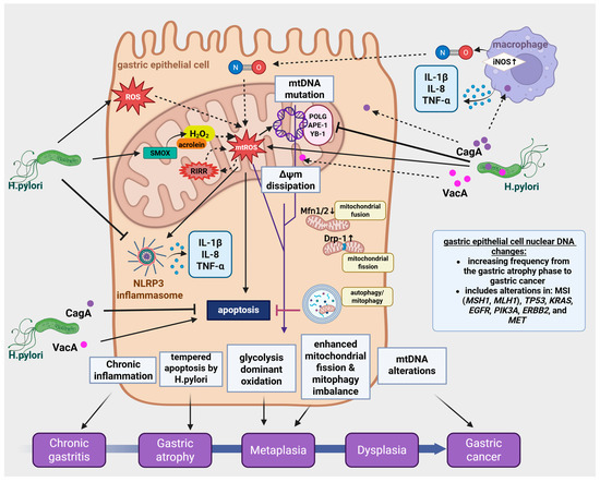
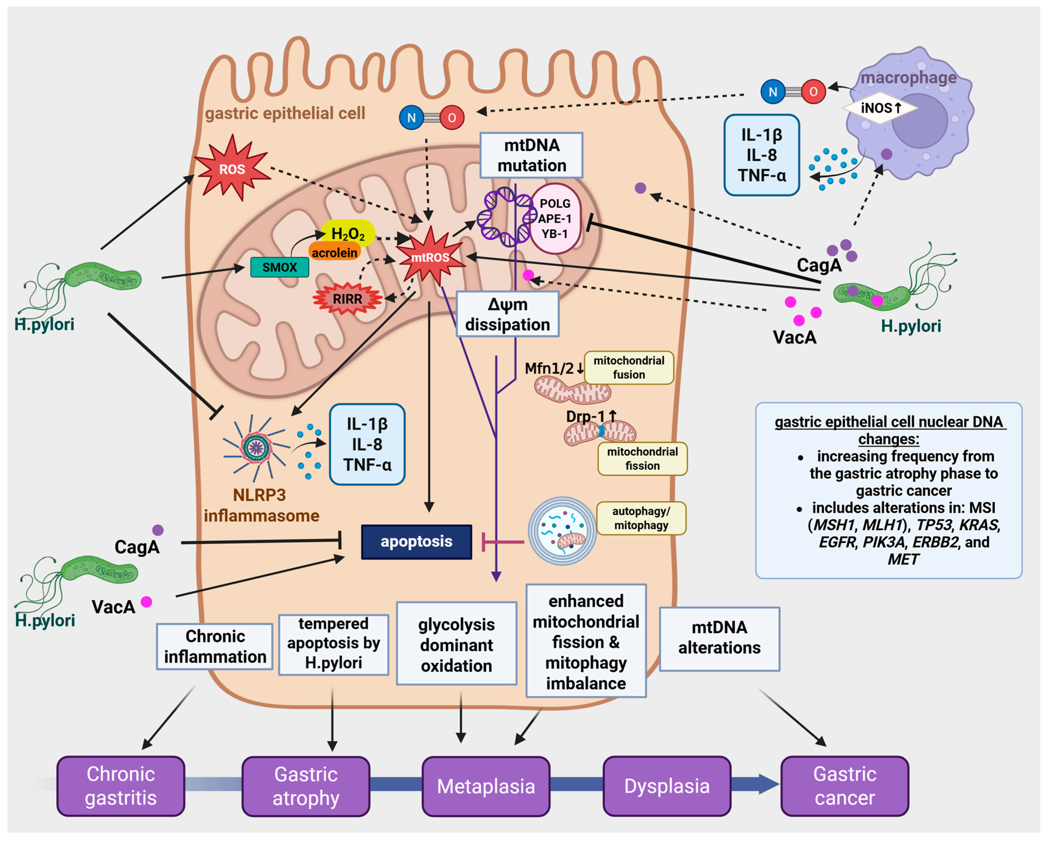
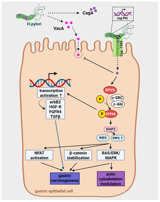
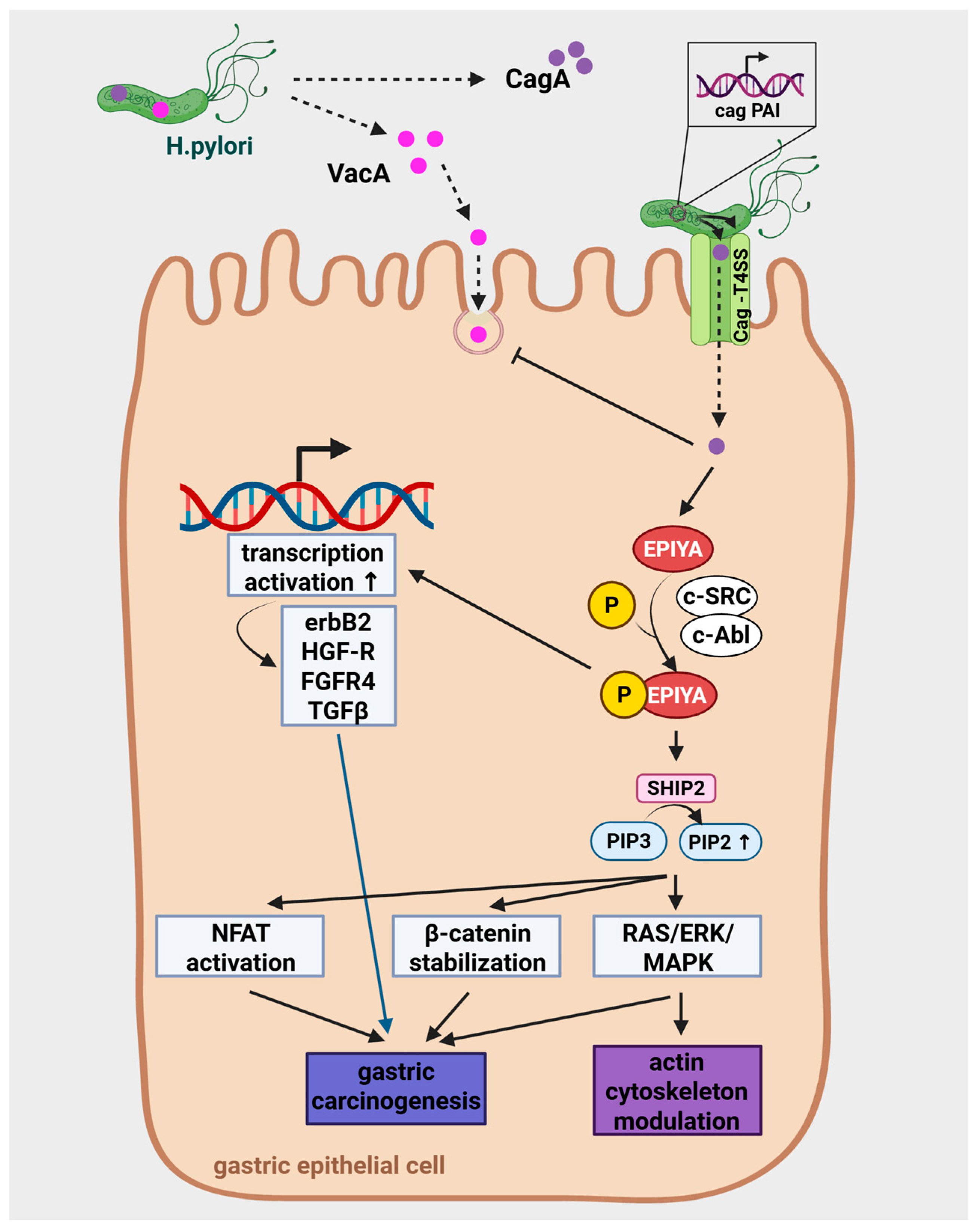
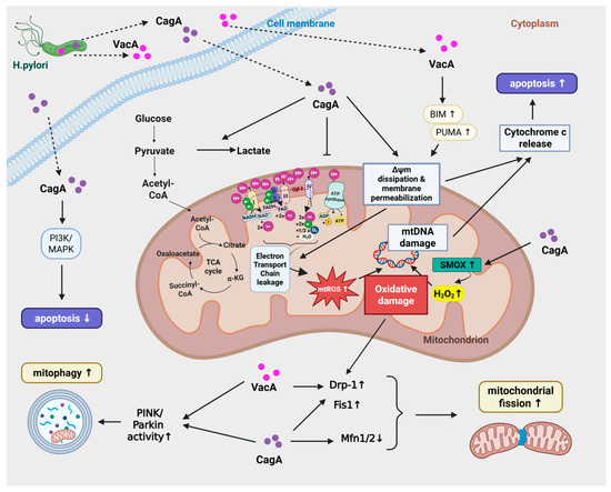
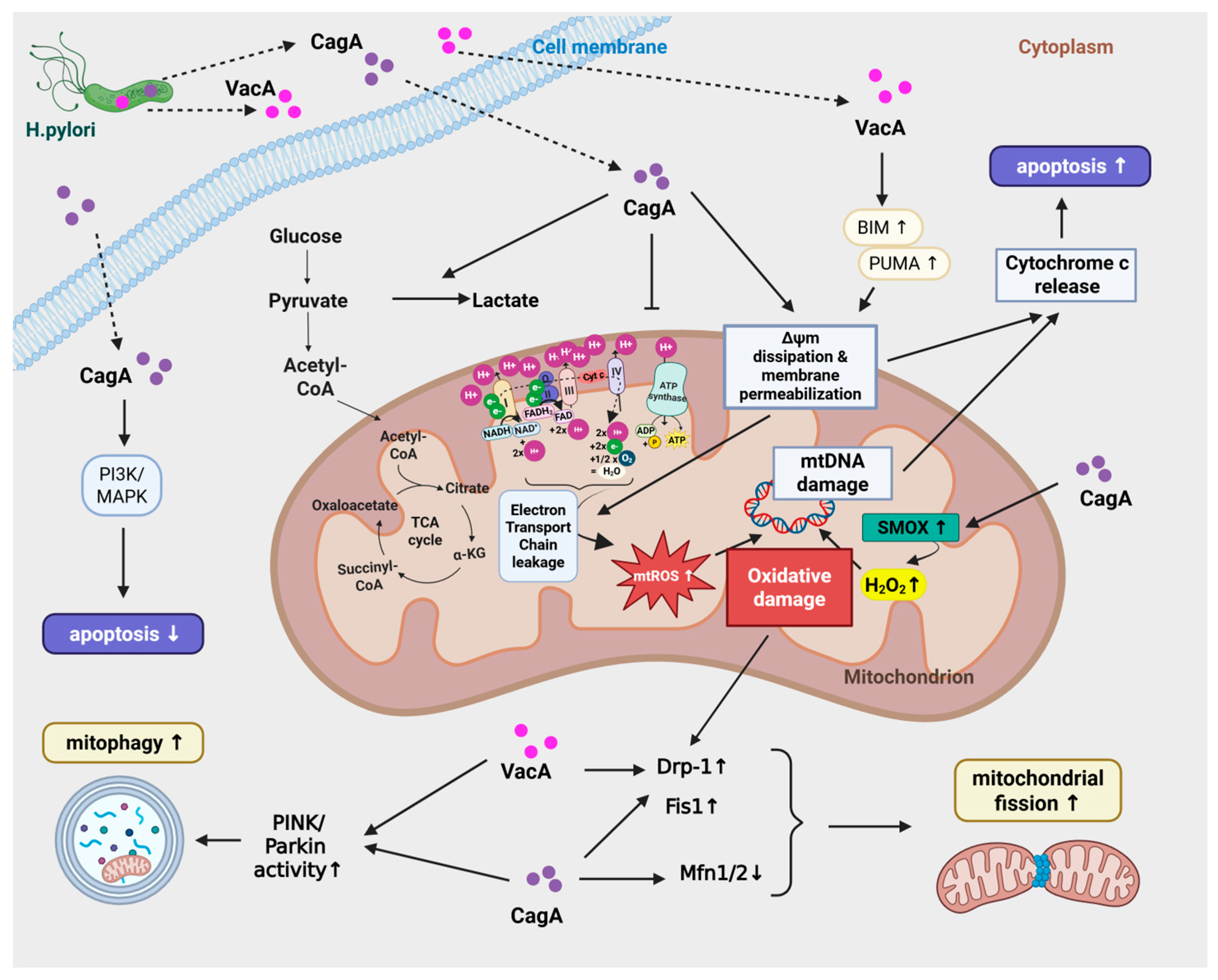
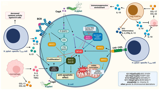
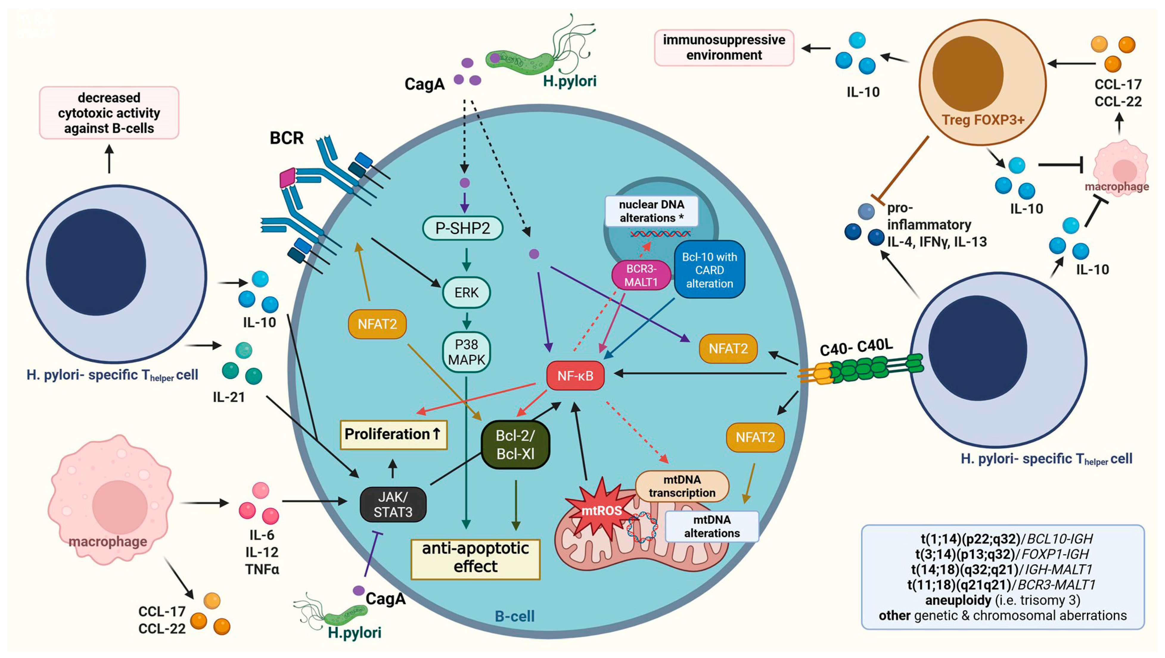

Disclaimer/Publisher’s Note: The statements, opinions and data contained in all publications are solely those of the individual author(s) and contributor(s) and not of MDPI and/or the editor(s). MDPI and/or the editor(s) disclaim responsibility for any injury to people or property resulting from any ideas, methods, instructions or products referred to in the content. |
© 2026 by the authors. Licensee MDPI, Basel, Switzerland. This article is an open access article distributed under the terms and conditions of the Creative Commons Attribution (CC BY) license.