


Abstract
Cardiac remodeling consists of genetic, molecular, cellular, and interstitial changes that clinically manifest as alterations in cardiac size, mass, geometry, and function following cardiac injury. This process is associated with a poorer prognosis, mainly due to the development and progression of ventricular dysfunction. In this review, we discuss the concepts, diagnosis, and pathophysiological mechanisms underlying cardiac remodeling, including cell death, mitochondrial dysfunction and energy metabolism, oxidative stress, inflammation, the cardiac lymphatic system, collagen, contractile proteins, calcium handling, ventricular geometry, neurohormonal activation, and microRNAs. Finally, the article addresses the clinical implications of cardiac remodeling and discusses pharmacological strategies aimed at attenuating or reversing this process, as well as future perspectives.
1. Introduction
One of the earliest reports of the use of the term “remodeling” in cardiology dates back to 1982, in a study of experimentally induced myocardial infarction by Hockman and Bulkley [1]. In their study, the authors used the term to describe the process by which infarcted tissue was replaced by scar tissue [1]. In 1985, J. Pfeffer first used the term “cardiac remodeling” to describe the progressive increase in left ventricular cavity size in an experimental rat model of myocardial infarction [2]. A few years later, Pfeffer and Braunwald further developed this concept and used the term “remodeling” to describe the structural adaptation of the left ventricle following myocardial infarction, particularly ventricular cavity enlargement, which was associated with ventricular dysfunction and a poor prognosis [2]. In this context, the term “cardiac remodeling” was adopted to characterize the changes occurring in the left ventricle after myocardial infarction [3].
In subsequent years, the term “remodeling” was used in various clinical scenarios, including cardiac hypertrophy and fibrosis. In 2000, the International Forum on Cardiac Remodeling published a consensus document that broadened and standardized the concept of cardiac remodeling, defining it as a process involving genetic, molecular, cellular, and interstitial changes that are clinically manifested by alterations in cardiac size, geometry, and function [4]. The Forum also recognized two types of cardiac remodeling: physiological (adaptive) and pathological (maladaptive). Subsequent studies have retained this definition, with a primary focus on pathological remodeling and, consequently, emphasizing that cardiac remodeling represents a dynamic trajectory that may progress toward adverse remodeling or improve through reverse remodeling [5,6].
In 2016, our research group published a comprehensive review addressing the concepts, pathophysiological mechanisms, clinical implications, and therapeutic strategies related to cardiac remodeling, contributing to the consolidation and systematization of knowledge in this field [7]. Given the advances made over the past decade, a reassessment and update of this framework based on the most recent evidence is warranted.
2. Diagnosis
Diagnosis of cardiac remodeling is based on the assessment of morphofunctional changes, including alterations in cardiac size, shape, and mass, as well as systolic and diastolic parameters [4]. These parameters can be assessed using noninvasive imaging techniques and circulating biomarkers.
The most widely used imaging modalities are two-dimensional and three-dimensional echocardiography and cardiac magnetic resonance imaging [8,9]. Strain echocardiography is an emerging technique that enables the detection of early alterations in cardiac function [8]. Cardiac magnetic resonance imaging can be used for both functional and structural assessment of the myocardium and is particularly valuable for detecting myocardial inflammation and fibrosis [9].
Although not widely used in clinical practice, another diagnostic approach is based on the assessment of biomarkers. Cardiac remodeling involves multiple cell types (cardiomyocytes, fibroblasts, endothelial cells, and immune cells) and biological processes, and circulating biomarkers may reflect the specific pathophysiological pathways in which they are involved. Thus, heart-type fatty acid-binding protein (hFABP), ischemia-modified albumin (IMA), and cardiac myosin-binding protein C (cMyC) are mainly associated with acute cardiomyocyte injury; tumor necrosis factor alpha (TNF-α), interleukin (IL)-1, IL-6, and growth differentiation factor-15 (GDF-15) reflect inflammatory activation; B-type natriuretic peptide (BNP) and N-terminal pro-B-type natriuretic peptide (NT-proBNP) reflect the endocrine response of the heart to hemodynamic stress; and galectin-3, osteoglycin (mimecan), collagen-derived peptides, matrix metalloproteinases (MMP) and tissue inhibitors of metalloproteinases (TIMP) are more closely associated with fibrosis and extracellular matrix remodeling [8,10,11]. In addition, several microRNAs (miR-1, miR-133a/b, miR-208b, and miR-499) regulate processes involved in cell growth, differentiation, proliferation, and death and are implicated in cardiac remodeling, with potential therapeutic applications [8].
3. Pathophysiological Mechanisms
The mechanisms involved in the development of pathological cardiac remodeling are complex, highly interconnected, and not yet fully understood. Nevertheless, the most extensively studied mechanisms are summarized in Table 1 and discussed below.
Table 1.
Pathophysiology of ventricular dysfunction in cardiac remodeling.
3.1. Cell Death
Cardiomyocyte death encompasses multiple, integrated forms of cell death that may occur independently or interact through complex molecular pathways. For example, necroptosis shares features with both apoptosis and necrosis. The major forms of cardiomyocyte death include apoptosis, or programmed cell death, necrosis, autophagy-associated cell death, pyroptosis, ferroptosis, and cuproptosis [12,13] (Table 2).
Table 2.
Types of cardiomyocyte death.
Different cell types and molecular mediators act as regulators and effectors of cell death, although some of these pathways remain incompletely understood. Caspase-8 is an important molecular link between necroptosis, pyroptosis, and ferroptosis; however, other mediators, including Receptor-interacting serine/threonine-protein kinase (RIPK) 1 and 3, and the inflammasome, are also involved [13,14]. Apoptosis is a form of programmed cell death that differs from the other forms described above in that it generally does not induce an inflammatory response. The intrinsic apoptotic pathway is regulated by members of the BCL-2 family, including BAX, and leads to apoptosome formation, caspase activation, and degradation of cellular components. The extrinsic pathway is activated by the interaction of death receptors, such as tumor necrosis factor receptor (TNFR) and Fas receptor (FasR), with their respective extracellular ligands [13,15,16]. These regulated cell death pathways contribute to cardiomyocyte loss and, depending on the pathway involved, to the activation of inflammatory responses, thereby promoting structural and functional alterations in the myocardium and the progression of adverse remodeling.
In recent years, special attention has been given to autophagy because of its dual role in cardiac remodeling. Autophagy is an intracellular process characterized by the degradation and removal of unnecessary or dysfunctional cellular components, particularly damaged organelles and proteins [17]. This process is tightly regulated, primarily through the AMPK/mTOR/ULK1 axis. When appropriately activated, autophagy may exert a protective effect by facilitating the elimination of damaged organelles and misfolded proteins, thereby preventing proteotoxicity and limiting the activation of other cell death pathways [18,19].
However, dysregulated autophagy may contribute to adverse cardiac remodeling through distinct mechanisms. Insufficient autophagic activity impairs the clearance of damaged proteins and dysfunctional organelles, leading to their accumulation, increased cellular stress, and cardiomyocyte dysfunction. Conversely, excessive autophagic activation may result in the inappropriate degradation of essential cellular components, ultimately contributing to cardiomyocyte dysfunction and cell death. These alterations may ultimately contribute to pathological cardiac remodeling, including cardiomyocyte hypertrophy and myocardial fibrosis [18,19].
Importantly, in pathological cardiac remodeling, multiple forms of cardiomyocyte death contribute to the loss of contractile tissue, the development of fibrosis, and the progression to heart failure.
3.2. Mitochondrial Dysfunction and Energy Metabolism
The heart has a high and continuous demand for ATP to sustain its essential functions. Under physiological conditions, cardiac ATP is generated predominantly through mitochondrial oxidative phosphorylation, with fatty acids serving as the primary energy substrate, while glucose, lactate, and ketone bodies serve as complementary substrates [20].
During cardiac remodeling and heart failure, reductions in the availability of energy substrates, decreased activity of the citric acid cycle, and impaired electron transport chain activity have been observed. This mitochondrial dysfunction impairs ATP production, increases oxidative stress, disrupts Ca2+ homeostasis, and activates inflammatory and cell death pathways, thereby contributing to the progression of ventricular dysfunction [21,22,23].
In addition to mitochondrial dysfunction, cardiac remodeling involves alterations in substrate utilization and a loss of metabolic flexibility [7]. In heart failure, there is a decrease in fatty acid oxidation associated with increased reliance on glycolysis. A reduction in β-oxidation may result in the accumulation of triglycerides and lipotoxicity, leading to mitochondrial structural alterations and further impairment of mitochondrial function [21,22].
In this context, ketone bodies have received increasing attention as energy substrates for the myocardium, particularly in heart failure [22,24]. Their increased utilization may represent an adaptive response to alterations in the metabolism of other substrates, although their role in cardiac remodeling has not yet been fully established.
3.3. Oxidative Stress
Oxidative stress is characterized by a disruption of cellular redox homeostasis in which the production and/or accumulation of reactive oxygen species (ROS) exceeds the capacity of redox regulatory systems to maintain physiological redox signaling and control. This imbalance results in oxidative modifications of cellular components and may ultimately lead to cellular dysfunction and damage [25,26,27].
ROS can be generated from several sources in the heart, including the mitochondrial electron transport chain, catecholamine metabolism, lipoxygenases, cyclooxygenases, the NADPH oxidase system, cytochrome P450, xanthine oxidase, and glucose oxidase [25,26,27].
Under physiological conditions, ROS play a central role in several cellular processes, including the regulation of cardiac contractility, cardiomyocyte proliferation and differentiation, apoptosis, and signaling pathways involved in both physiological and pathological cardiac hypertrophy [28]. However, under pathological conditions, excessive ROS production can trigger multiple processes that exert detrimental effects on the heart. ROS can induce lipid peroxidation of cellular membranes and damage nuclear and mitochondrial DNA, thereby contributing to mitochondrial dysfunction and energy deficits. Importantly, ROS exhibit a dose-dependent effect: at low levels, they participate in physiological signaling, whereas excessive ROS accumulation is associated with pathological conditions and cellular damage [25,26,27].
Oxidative stress is also an important contributor to alterations in cytoskeletal proteins associated with ventricular remodeling, including titin, desmin, and tubulin. These alterations may impair force transmission during systole and increase myocardial stiffness during diastole [25,26,27].
In addition, ROS can activate kinase signaling pathways involved in cardiac hypertrophy, inflammation, and apoptosis. These effects involve several signaling molecules and pathways, including extracellular signal-regulated kinase 1/2 (ERK1/2), c-Jun N-terminal kinase (JNK), p38 mitogen-activated protein kinase (p38 MAPK), protein kinase B (Akt), and transforming growth factor-beta (TGF-β) [25,26,27]. These pathways interact extensively and may contribute to different aspects of cardiac remodeling, including hypertrophy, inflammation, fibrosis, and apoptosis.
Oxidative stress is also an important modulator of the extracellular matrix. In different models of cardiac injury, increased ROS production has been associated with the activation of MMP and collagen accumulation, thereby contributing to extracellular matrix remodeling and myocardial fibrosis [25,26,27].
Another detrimental effect of excessive ROS production involves proteins that regulate cardiac calcium handling. ROS can increase ryanodine receptor activity and inhibit sarcoplasmic reticulum Ca2+-ATPase 2 (SERCA2) activity, resulting in intracellular calcium overload and reduced myofilament calcium sensitivity. These alterations may contribute to impaired contractile function during systole and impaired relaxation during diastole. In addition, calcium overload can activate proteolytic enzymes, such as calpains, further contributing to myocardial injury [25,26,27].
Recently, several biomarkers associated with oxidative stress, including myeloperoxidase (MPO), growth differentiation factor-15 (GDF-15), and indoxyl sulfate, have received increasing attention. These molecules have also been investigated as potential targets or mediators of therapeutic strategies aimed at modulating oxidative stress-related pathways, including those involving sodium–glucose cotransporter 2 (SGLT2) inhibitors, metformin, glutathione, and soluble guanylate cyclase stimulators such as vericiguat [26]. However, further evidence is needed to establish whether oxidative stress can be considered a specific and effective therapeutic target in cardiac remodeling.
3.4. Inflammation
Cardiac remodeling is associated with the activation of inflammatory pathways, resulting in the upregulation of nuclear factor kappa B (NF-κB), activation of inflammasome pathways, and increased expression of cytokines such as interleukin (IL)-6, TNF-α, IL-18, and IL-33. In addition, the immune system, involving dendritic cells, macrophages, and B and T lymphocytes, can also modulate the cardiac remodeling process, depending on the nature of the cardiac stimulus [29,30]. Although acute and chronic inflammation are physiological mechanisms involved in tissue repair and host defense following an insult, persistent and dysregulated inflammation actively contributes to the progression of pathological cardiac remodeling through crosstalk among innate and adaptive immune cells, fibroblasts, cardiomyocytes, and endothelial cells [29,30].
Cardiomyocyte injury leads to the release of damage-associated molecular patterns (DAMPs), which activate Toll-like receptors (TLR2 and TLR4), initiating the priming phase through MyD88, IRAK1/4, and NF-κB signaling. NF-κB promotes the upregulation of NLRP3 and pro-IL-1β expression. Upon activation, NLRP3 associates with ASC (apoptosis-associated speck-like protein containing a CARD), which recruits pro-caspase-1, thereby assembling the inflammasome complex. Caspase-1 promotes the maturation and release of pro-inflammatory cytokines, amplifying the inflammatory response. Additionally, caspase-1-mediated cleavage of gasdermin D (GSDMD) leads to the formation of pores in the plasma membrane, allowing the release of intracellular contents and compromising membrane integrity. This process may ultimately result in inflammation-mediated cell death, known as pyroptosis [30,31,32].
Activation of inflammatory pathways promotes the recruitment of immune cells. Neutrophils are the first immune cells to arrive at the site of injury, where they contribute to cellular phagocytosis, the release of proteases, cytokines, and chemokines, and the exacerbation of redox imbalance [30]. Thus, oxidative stress may act as an initial trigger of inflammation, whereas inflammation, in turn, increases ROS production, establishing a positive feedback loop that amplifies myocardial injury [26,30]. In models of pressure overload, neutrophils may also obstruct small blood vessels, thereby exacerbating myocardial hypoxia [30].
Following neutrophil recruitment, monocytes are extensively recruited and subsequently differentiate into macrophages. Pro-inflammatory macrophages produce mediators that amplify the inflammatory response, particularly TNF-α, IL-1β, and IL-6 [29,33].
Adaptive immunity also contributes to the progression of cardiac remodeling. Dendritic cells, macrophages, and B lymphocytes can present antigens to T lymphocytes, promoting their activation and differentiation. An imbalance between regulatory T cells (Tregs) and T helper 1 (Th1) cells may influence the regulation of inflammation and immune responses or contribute to the progression of myocardial injury. While Tregs exert immunoregulatory functions by limiting excessive inflammatory responses and promoting tissue repair, Th1 cells not only enhance inflammatory responses but also contribute to cardiac fibrosis by stimulating fibroblast activation and the production of pro-fibrotic mediators [29,34].
Thus, persistent and dysregulated inflammatory activation can induce several detrimental changes, including excessive fibroblast activation, increased oxidative stress, cell death, neurohormonal activation, altered calcium handling, and endothelial dysfunction, among others [25,26].
3.5. Cardiac Lymphatic System
Recent studies have investigated the role of the lymphatic system in the inflammatory cascade underlying cardiac remodeling. Following myocardial infarction, compensatory lymphangiogenesis enhances immune cell clearance, reduces myocardial edema, and attenuates inflammatory and fibrotic responses [35,36]. In addition, lymphatic endothelial cells secrete reelin, which may directly promote cardiomyocyte proliferation and survival [33,36]. However, when lymphangiogenesis is insufficient or maladaptive, as observed in advanced ischemic heart disease and end-stage heart failure, lymphatic dysfunction may have opposing effects by delaying the resolution of the immune response and promoting chronic interstitial edema, inflammation, and fibrosis [36].
3.6. Collagen
The heart contains a highly organized and dynamic extracellular matrix (ECM), in which collagen, predominantly types I and III, provides structural and mechanical support. The ECM is regulated by the balance between collagen synthesis and degradation, particularly through the interplay between MMP and TIMP [37]. Beyond its structural role, the extracellular matrix (ECM) also constitutes a biologically active microenvironment involved in several processes, including cell proliferation, migration, and differentiation [37].
MMPs are synthesized as inactive enzymatic precursors (pro-MMPs), and their activity is regulated by their interaction with TIMPs, which bind to MMPs and inhibit their proteolytic activity, as well as by zinc-dependent activation mechanisms. Furthermore, these enzymes exhibit different substrate specificities and can degrade not only collagen but also other ECM components, such as fibronectin [38]. Another important aspect of collagen dynamics is its organization and stabilization through the formation of cross-links, a process primarily regulated by lysyl oxidase (LOX) [39]. Thus, the extent of collagen degradation or deposition, as well as its degree of organization and stabilization, can influence myocardial structure and function [38,39].
During cardiac remodeling, the balance between collagen synthesis and degradation becomes disrupted [40]. Following myocardial infarction, increased MMP activity contributes to early ECM degradation and infarct expansion, followed by fibroblast activation and collagen deposition during scar formation [41].
Conversely, excessive collagen deposition and disorganized ECM remodeling may lead to myocardial fibrosis and increased ventricular stiffness, contributing to diastolic dysfunction and electrical instability. Fibrotic remodeling is driven by the interplay of mechanical, neurohormonal, metabolic, and inflammatory stimuli, with important contributions from angiotensin II, endothelin-1, and transforming growth factor-beta (TGF-β) signaling, which promote cardiac fibroblast activation and the synthesis of type I and III collagen [42].
Myocardial fibrosis can occur in different patterns, including replacement fibrosis, characterized by collagen deposition in areas of cardiomyocyte loss, as occurs following myocardial infarction; interstitial fibrosis, characterized by collagen accumulation in the interstitial space between cardiomyocytes; and perivascular fibrosis, characterized by matrix deposition around blood vessels, which may impair oxygen and substrate delivery [39].
Thus, alterations in collagen turnover and organization are important determinants of cardiac remodeling. Importantly, in different models of cardiac remodeling, fibrosis has been associated with increased myocardial stiffness, diastolic dysfunction, impaired contractility, reduced coronary flow, and malignant arrhythmias. In addition, fibrosis has been identified as a predictor of mortality in patients with cardiac dysfunction [38,39].
3.7. Contractile Protein Alteration
Alterations in sarcomeric proteins constitute an important component of cardiac remodeling and may contribute to contractile dysfunction [7]. Myosin, the primary motor protein of the sarcomere, exists in isoforms with distinct kinetic and energetic properties.
In the adult human heart, β-myosin predominates, whereas α-myosin accounts for a smaller proportion. α-Myosin exhibits a higher rate of force development, whereas β-myosin has greater energetic efficiency. Although a shift from α- to β-myosin has been observed in models of pressure overload, its magnitude and relevance in the human heart remain controversial [43]. In addition to changes in isoform expression, cardiac remodeling is associated with post-translational modifications of sarcomeric proteins, including alterations in the phosphorylation of troponin I and cardiac myosin-binding protein C (cMyBP-C) [44]. These modifications may alter myofilament Ca2+ sensitivity, force generation, and length-dependent activation of the sarcomere. In experimental models of heart failure, alterations in the phosphorylation and oxidation of these proteins have been associated with changes in myofilament function and contractile capacity [45].
Thus, contractile protein dysfunction during cardiac remodeling results from integrated alterations in sarcomeric composition and regulation, which may impair force generation and myocardial adaptation to mechanical load. Collectively, these changes contribute to the progression of ventricular dysfunction and represent potential targets for therapeutic strategies aimed at modulating sarcomeric function [44].
3.8. Calcium Transport Alteration
Excitation–contraction coupling in cardiomyocytes depends on the precise regulation of intracellular Ca2+. Membrane depolarization promotes Ca2+ influx through L-type calcium channels, which activates ryanodine receptors (RyR2) and triggers Ca2+ release from the sarcoplasmic reticulum. Ca2+ then binds to troponin C, promoting actin–myosin interaction and contraction [46]. During relaxation, most cytosolic Ca2+ is taken up by the sarcoplasmic reticulum via SERCA2a, which is regulated by phospholamban, whereas a smaller fraction is extruded from the cell through the Na+/Ca2+ exchanger [7,47].
In cardiac remodeling and heart failure, this system becomes dysregulated, primarily characterized by reduced SERCA2a activity and alterations in RyR2 regulation. Impaired Ca2+ reuptake by the sarcoplasmic reticulum, together with increased diastolic Ca2+ leak through RyR2, reduces sarcoplasmic reticulum Ca2+ content and impairs systolic Ca2+ release, thereby contributing to contractile dysfunction. Alterations in phospholamban phosphorylation, calcium/calmodulin-dependent protein kinase II (CaMKII) activity, and L-type calcium channel function may also contribute to impaired intracellular Ca2+ handling [48,49].
In addition, T-tubule remodeling may disrupt the spatial organization and functional coupling of L-type calcium channels and RyR2, thereby impairing excitation–contraction coupling [46]. Increased cytosolic Ca2+ during diastole may also promote abnormal electrical activity and arrhythmias. Thus, alterations in the expression, activity, and organization of Ca2+-handling proteins contribute to reduced contractility, impaired relaxation, and electrical instability of the heart [46,49].
3.9. Neurohormonal Activation
Neurohormonal activation also contributes to the progression of cardiac remodeling. Initially compensatory, persistent neurohormonal activation contributes to ventricular deterioration and the progression to heart failure [27]. This process primarily involves the sympathetic nervous system (SNS) and the renin–angiotensin–aldosterone system (RAAS) [8]. Hyperactivation of the RAAS increases angiotensin II (Ang II) production, promoting cardiomyocyte hypertrophy, fibroblast proliferation, and fibrosis. Aldosterone also stimulates collagen deposition, apoptosis, and endothelial dysfunction, whereas Ang II activates NADPH oxidases, thereby increasing ROS production and oxidative stress [8,27].
In parallel, SNS hyperactivation occurs early after cardiac injury, increasing norepinephrine levels and, when persistent, myocardial oxygen consumption, arrhythmogenesis, and apoptosis [27]. The bidirectional interaction between the SNS and RAAS perpetuates this response. As a counterregulatory mechanism, cardiac stretch stimulates the release of atrial natriuretic peptide (ANP) and B-type natriuretic peptide (BNP), which exert vasodilatory, natriuretic, antifibrotic, and antihypertrophic effects. However, degradation of these peptides by neprilysin limits their activity, making neprilysin inhibition a therapeutic strategy to enhance their effects and promote reverse remodeling [8,27].
In addition to classical neurohormonal mechanisms, visceral and epicardial adipose tissues actively participate in this process through the secretion of endocrine and paracrine mediators [50,51]. Leptin and resistin exert pro-inflammatory and profibrotic effects, whereas adiponectin and omentin exert antihypertrophic and vasculoprotective effects, in part through AMP-activated protein kinase (AMPK) signaling. Dysfunction of epicardial adipose tissue promotes the local release of these mediators, thereby contributing to myocardial fibrosis and diastolic stiffness [51].
In addition to these mechanisms, endothelin-1, which is stimulated by hypoxia and Ang II, promotes vasoconstriction, inflammation through the induction of TNF-α, IL-6, and IL-1β, and macrophage activation [8]. These signals converge on redox-sensitive pathways, such as NF-κB and MAPK, which regulate the expression of genes involved in cardiac remodeling. Thus, the interplay between neurohormonal and endocrine systems shapes the cardiac response. Persistent activation of these pathways promotes oxidative stress, inflammation, fibrosis, and myocardial dysfunction, whereas their modulation may contribute to reverse remodeling [8,25].
3.10. microRNAs (miRNA)
miRNAs are small, non-coding RNA molecules that regulate gene expression at the post-transcriptional level, primarily by binding to messenger RNAs and thereby inhibiting their translation into proteins.
In recent years, the role of miRNAs in various clinical and pathophysiological contexts has become increasingly recognized. Thus, miRNAs have been implicated in cardiac hypertrophy (miR-22, miR-133a), contractile function and kinetics (miR-208a), collagen accumulation and fibrosis (miR-21, miR-29), electrical conduction (miR-1), inflammation (miR-146a, miR-155, miR-223), oxidative stress (miR-210, miR-181c, miR-16-5p), and apoptosis (miR-24, miR-21) [52,53].
miR-16-5p has been investigated for its involvement in the interplay between endoplasmic reticulum (ER) stress and redox imbalance. Under conditions of ER stress, the unfolded protein response (UPR) is activated through three major signaling pathways, one of which is mediated by activating transcription factor 6 (ATF6). Toro et al. demonstrated that miR-16-5p directly targets ATF6, a transcription factor involved in the expression of antioxidant components [54]. In human ventricular AC16 cells exposed to ER stress induced by tunicamycin, miR-16-5p overexpression increased oxidative damage, reduced the expression of antioxidant enzymes, including catalase, superoxide dismutase 1, and glutathione peroxidase, and impaired mitochondrial respiration. Conversely, miR-16-5p inhibition restored ATF6 expression, increased the expression of antioxidant genes, and attenuated inflammatory and apoptotic responses. These findings suggest that miR-16-5p may contribute to oxidative stress by impairing ATF6-mediated cytoprotective signaling [54]. Collectively, these findings indicate that modulation of miR-16-5p may promote cardiomyocyte protection under cellular stress, highlighting miRNAs as potential therapeutic targets to be explored in future strategies for cardiac remodeling.
Therefore, alterations in miRNA expression, which can influence multiple relevant pathophysiological mechanisms, may represent important modulators of the cardiac remodeling process.
4. Clinical Implications
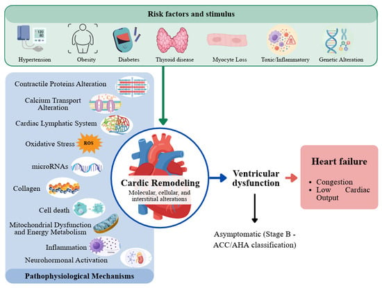
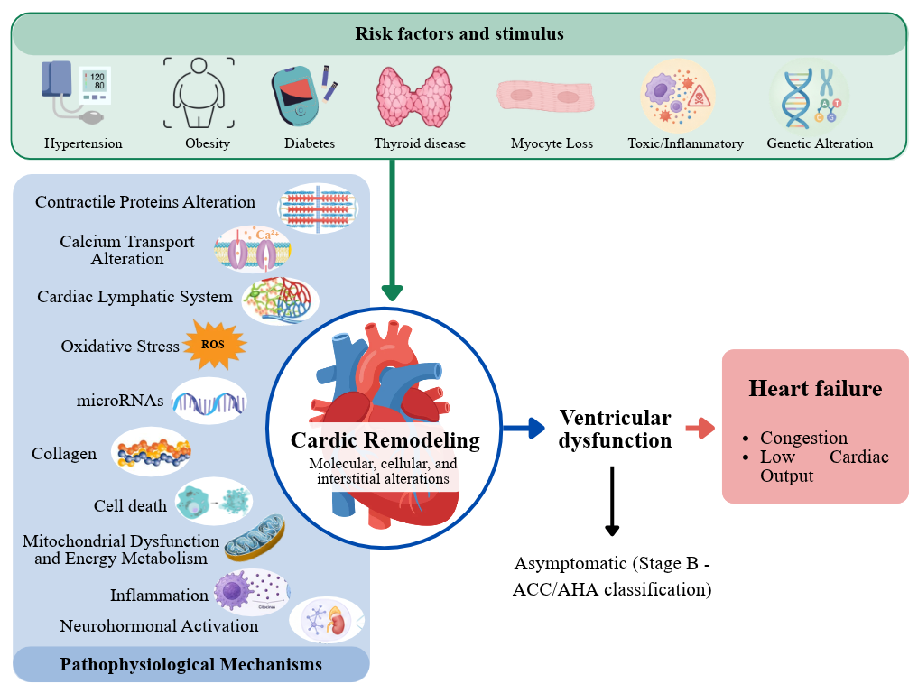
Cardiac dysfunction remains the principal consequence of cardiac remodeling. The initial cardiac insult (ischemia, pressure or volume overload, hypertension, or neurohormonal activation) triggers cellular and molecular alterations that ultimately lead to changes in cardiac architecture and geometry. The combination of these factors is responsible for the initiation and progression of ventricular dysfunction, which may initially be asymptomatic but can gradually progress to clinical manifestations characterized by the signs and symptoms of heart failure and/or sudden death [8,55] (Figure 1).
Figure 1.
Natural history of cardiac remodeling.
Heart failure prevalence is estimated at approximately 3.4% among adults and 8.3% among older individuals, with approximately 50% of patients presenting with heart failure with preserved ejection fraction (HFpEF) [56]. Despite advances in therapy, heart failure remains associated with a substantial clinical burden, with one-year rehospitalization and mortality rates of approximately 35.7% and 23.3%, respectively [56,57]. These outcomes reflect the progressive nature of the disease and the complexity of the underlying mechanisms involved in its development [56].
Additionally, specific clinical scenarios exhibit unique features regarding the remodeling process, such as after acute myocardial infarction. Acute ischemia results in the disintegration of collagen fibers responsible for maintaining cardiac architecture. Without this supporting tissue, necrotic areas may undergo myocyte sliding and realignment along the infarcted wall, resulting in thinning of the affected area and cavity dilation. This acute ventricular dilation, characterized by infarcted wall thinning and distension, is termed infarction expansion. This, in turn, predisposes the heart to ventricular rupture and provides the anatomical substrate for future aneurysm formation. Importantly, this combination of changes increases overall ventricular wall stress, serving as a hemodynamic stimulus for remodeling in the non-infarcted portion of the left ventricle and leading to cardiac dysfunction in the chronic phase post-infarction [58].
5. Clinical Approach
Thus, targeting cardiac remodeling effectively is a priority and should be implemented in stages, particularly in high-risk patients.
The first step to consider is the control of risk factors, as shown in Figure 1. This involves the proper management of high blood pressure, obesity, diabetes, and thyroid disease, for example. Additionally, it includes regular monitoring of valvular heart disease, smoking cessation, and avoidance of excessive alcohol consumption. Similarly, controlling risk factors for myocardial infarction, as well as measures to limit infarct size, such as early and effective revascularization therapies combined with strategies to maintain coronary patency, is essential. This approach prevents the remodeling phenomenon and, consequently, cardiac dysfunction.
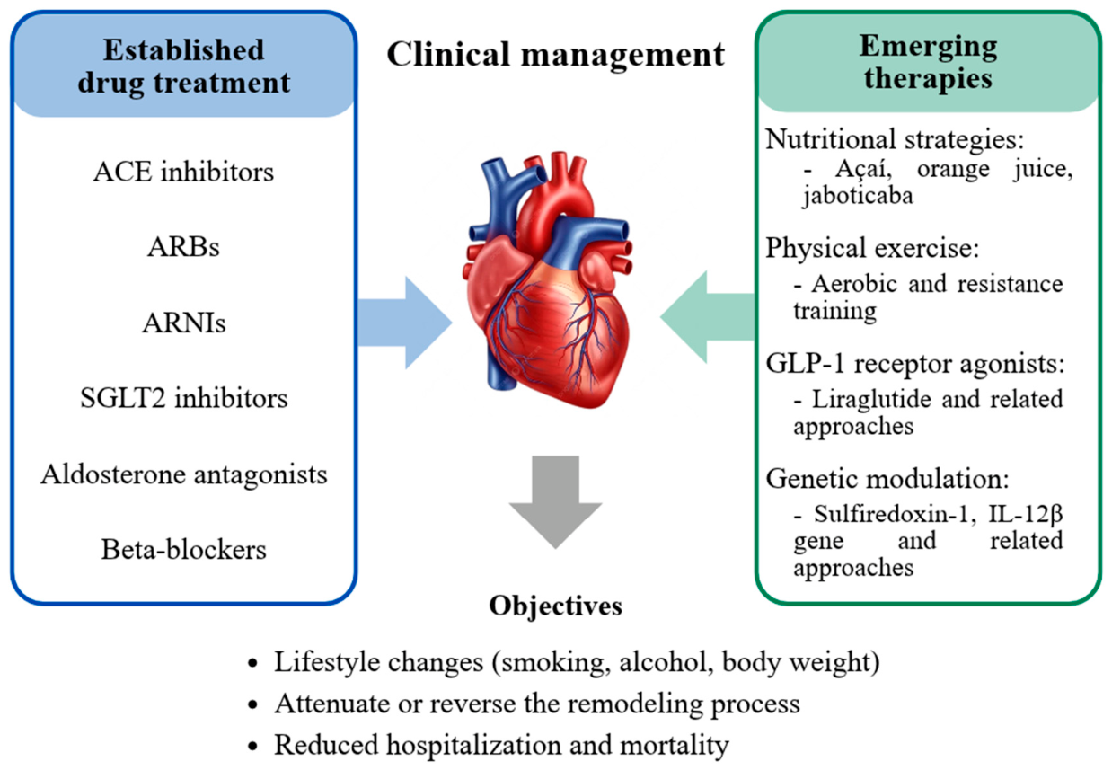
In the second step, when heart failure is established, it is not surprising that established treatments that consistently reduce mortality are those that attenuate or reverse the remodeling process. These include angiotensin-converting enzyme inhibitors or angiotensin II receptor blockers or angiotensin receptor–neprilysin inhibitors, SGLT2 inhibitors, aldosterone antagonists, and beta-blockers. These medications exert their beneficial effects by attenuating the different pathophysiological mechanisms described above [59,60,61,62]. Therefore, optimizing these treatments is a priority in the management of these patients.
The effectiveness of these strategies depends not only on their prescription but also on their appropriate implementation and long-term maintenance. Regular clinical follow-up allows therapeutic responses to be assessed, potential adverse effects to be monitored, and doses to be adjusted accordingly. Despite the robust evidence supporting pharmacological therapy, challenges remain regarding its implementation in clinical practice. A population-based modeling study estimated that only 18.2% of eligible patients with heart failure with reduced ejection fraction (HFrEF) in the United States received all four pharmacological classes concomitantly (renin–angiotensin system inhibitors, beta-blockers, mineralocorticoid receptor antagonists, and SGLT2 inhibitors) [62]. The authors highlighted poor adherence, polypharmacy, medication costs, and limited access to healthcare services as major barriers to therapeutic optimization [62].
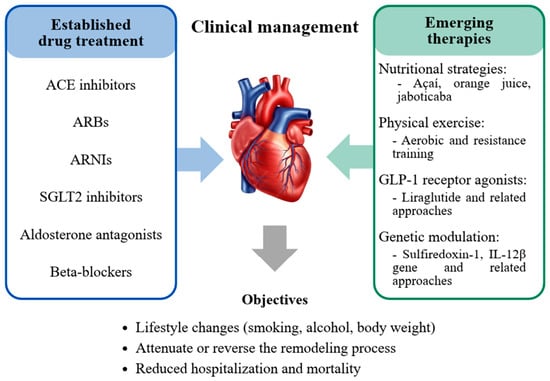
The clinical management of cardiac remodeling involves combining established pharmacological therapies with lifestyle modifications to prevent and control risk factors. In parallel, novel therapeutic approaches are being investigated, broadening the perspectives for patient management (Figure 2).
Figure 2.
Clinical management and emerging therapeutic strategies for cardiac remodeling. Established pharmacological therapies and emerging therapeutic strategies under investigation in experimental and clinical studies. The central panel illustrates an integrated clinical management approach focused on lifestyle modification, attenuation or reversal of cardiac remodeling, and improvement of clinical outcomes. ACE: angiotensin-converting enzyme; ARBs: angiotensin II receptor blockers; ARNIs: angiotensin receptor–neprilysin inhibitors; GLP-1: Glucagon-like peptide-1; SGLT2: sodium–glucose cotransporter 2.
6. Emerging Therapies
Understanding the pathophysiological mechanisms and consequences of cardiac remodeling enables the development of studies aimed at identifying novel therapeutic strategies. In recent years, there has been growing interest in targeting oxidative stress and inflammation to mitigate remodeling. However, despite promising results in experimental studies, clinical trials have mostly yielded negative results. To address this, recent research has evaluated the effects of foods such as açaí and orange juice in attenuating cardiac dysfunction, showing promising results [63].
Furthermore, in an experimental model of myocardial infarction, rats receiving jaboticaba supplementation showed improved diastolic function and reduced fibrosis [64]. Beyond nutritional strategies, the cardioprotective effects of physical exercise have also been widely investigated. Aerobic and resistance training stimulate the secretion of exosomes containing microRNAs capable of acting on different cardiac cell types. Exercise-induced increases in miR-133a, miR-133b, and miR-1 are associated with the attenuation of pathological hypertrophy, whereas increases in miR-206 and miR-342-5p contribute to the inhibition of apoptotic pathways [65]. In infarcted rats, resistance training reduced oxidative stress and preserved key enzymes involved in cardiac metabolism, including creatine kinase and lactate dehydrogenase, although chamber dilation and left ventricular systolic dysfunction—as assessed by echocardiography—remained unchanged [66].
Another strategy receiving increasing attention is the use of glucagon-like peptide-1 (GLP-1) receptor agonists. The effects of a 12-day liraglutide pretreatment were evaluated in a rat model of acute doxorubicin-induced cardiotoxicity [67]. In this study, pretreatment did not attenuate doxorubicin-induced myocardial injury. Conversely, other studies using chronic models of doxorubicin-induced cardiotoxicity have reported cardioprotective effects with GLP-1 receptor agonists, suggesting that the response may depend on the experimental model and the context of cardiac injury [67].
More recently, genetic modulation has emerged as a potential strategy to directly target pathways involved in cardiac remodeling. Overexpression of sulfiredoxin-1 (Srxn1) in mice directly suppressed the Toll-like receptor (TLR)-4/NF-κB signaling pathway, preventing fibrotic, hypertrophic, and inflammatory processes [68]. Similarly, inhibition of the IL-12β gene in mice subjected to a transverse aortic constriction model of pressure overload was associated with preserved systolic function and reduced hypertrophy, fibrosis, and myocardial leukocyte infiltration [69].
Altogether, these studies demonstrate that different strategies can target distinct mechanisms involved in cardiac remodeling. However, further studies are needed to determine the clinical applicability of these emerging approaches.
7. Conclusions
Cardiac remodeling is a complex process that contributes to the development and progression of ventricular dysfunction. Despite substantial advances in therapeutic strategies, morbidity and mortality associated with cardiac remodeling and ventricular dysfunction remain high. Therefore, a comprehensive understanding of the pathophysiological mechanisms underlying cardiac remodeling is essential, as are the development and evaluation of novel therapeutic approaches.
Author Contributions
E.M.D.M.Z., L.N.D.P.C., C.R.T. and M.F.M. drafted the manuscript; S.A.R.D.P., B.F.P. and L.Z. revised the manuscript. All authors have read and agreed to the published version of the manuscript.
Funding
This research was funded by the National Council for Scientific and Technological Development, CNPq (process number 300598/2026-2 and 302905/2025-1).
Institutional Review Board Statement
Not applicable.
Informed Consent Statement
Not applicable.
Data Availability Statement
No new data were created or analyzed in this study. Data sharing is not applicable.
Conflicts of Interest
The authors declare no conflicts of interest.
References
- Hochman, J.S.; Bulkley, B.H. Expansion of Acute Myocardial Infarction: An Experimental Study. Circulation 1982, 65, 1446–1450. [Google Scholar] [CrossRef] [Scilit] [PubMed]
- Pfeffer, J.M.; Pfeffer, M.A.; Braunwald, E. Influence of Chronic Captopril Therapy on the Infarcted Left Ventricle of the Rat. Circ. Res. 1985, 57, 84–95. [Google Scholar] [CrossRef] [Scilit] [PubMed]
- Pfeffer, M.A.; Braunwald, E. Ventricular Remodeling after Myocardial Infarction. Experimental Observations and Clinical Implications. Circulation 1990, 81, 1161–1172. [Google Scholar] [CrossRef] [Scilit] [PubMed]
- Cohn, J.N.; Ferrari, R.; Sharpe, N. Cardiac Remodeling-Concepts and Clinical Implications: A Consensus Paper from an International Forum on Cardiac Remodeling. J. Am. Coll. Cardiol. 2000, 35, 569–582. [Google Scholar] [CrossRef] [Scilit] [PubMed]
- Aimo, A.; Gaggin, H.K.; Barison, A.; Emdin, M.; Januzzi, J.L. Imaging, Biomarker, and Clinical Predictors of Cardiac Remodeling in Heart Failure With Reduced Ejection Fraction. JACC Heart Fail. 2019, 7, 782–794. [Google Scholar] [CrossRef] [Scilit] [PubMed]
- Bhatt, A.S.; Ambrosy, A.P.; Velazquez, E.J. Adverse Remodeling and Reverse Remodeling After Myocardial Infarction. Curr. Cardiol. Rep. 2017, 19, 71. [Google Scholar] [CrossRef] [Scilit] [PubMed]
- Azevedo, P.S.; Polegato, B.F.; Minicucci, M.F.; Paiva, S.A.R.; Zornoff, L.A.M. Cardiac Remodeling: Concepts, Clinical Impact, Pathophysiological Mechanisms and Pharmacologic Treatment. Arq. Bras. Cardiol. 2016, 106, 62–69. [Google Scholar] [CrossRef] [Scilit] [PubMed]
- Leancă, S.A.; Crișu, D.; Petriș, A.O.; Afrăsânie, I.; Genes, A.; Costache, A.D.; Tesloianu, D.N.; Costache, I.I. Left Ventricular Remodeling after Myocardial Infarction: From Physiopathology to Treatment. Life 2022, 12, 1111. [Google Scholar] [CrossRef] [Scilit] [PubMed]
- Gissler, M.C.; Antiochos, P.; Ge, Y.; Heydari, B.; Grani, C.; Kwong, R.Y. Cardiac Magnetic Resonance Evaluation of LV Remodeling Post-Myocardial Infarction: Prognosis, Monitoring and Trial Endpoints. JACC Cardiovasc. Imaging 2024, 17, 1366–1380. [Google Scholar] [CrossRef] [Scilit] [PubMed]
- Goetze, J.P.; Bruneau, B.G.; Ramos, H.R.; Ogawa, T.; de Bold, M.K.; de Bold, A.J. Cardiac Natriuretic Peptides. Nat. Rev. Cardiol. 2020, 17, 698–717. [Google Scholar] [CrossRef] [Scilit] [PubMed]
- González, A.; Richards, A.M.; de Boer, R.A.; Thum, T.; Arfsten, H.; Hülsmann, M.; Falcao-Pires, I.; Díez, J.; Foo, R.S.Y.; Chan, M.Y.; et al. Cardiac Remodelling—Part 1: From Cells and Tissues to Circulating Biomarkers. A Review from the Study Group on Biomarkers of the Heart Failure Association of the European Society of Cardiology. Eur. J. Heart Fail. 2022, 24, 927–943. [Google Scholar] [CrossRef] [Scilit] [PubMed]
- Qi, L.; Yi, J.; Ji, Y.; Jiang, H.; Sun, J.; Shen, Y.; Sun, H. Network of Cell Death in Myocardial Ischemia-Reperfusion Injury: Mechanisms and Targeted Therapeutic Strategies. Biochem. Pharmacol. 2026, 250, 117999. [Google Scholar] [CrossRef] [Scilit] [PubMed]
- Sheng, S.Y.; Li, J.M.; Hu, X.Y.; Wang, Y. Regulated Cell Death Pathways in Cardiomyopathy. Acta Pharmacol. Sin. 2023, 44, 1521–1535. [Google Scholar] [CrossRef] [Scilit] [PubMed]
- Xiang, Q.; Yi, X.; Zhu, X.-H.; Wei, X.; Jiang, D.-S. Regulated Cell Death in Myocardial Ischemia–Reperfusion Injury. Trends Endocrinol. Metab. 2024, 35, 219–234. [Google Scholar] [CrossRef] [Scilit] [PubMed]
- Teringova, E.; Tousek, P. Apoptosis in Ischemic Heart Disease. J. Transl. Med. 2017, 15, 87. [Google Scholar] [CrossRef] [Scilit] [PubMed]
- Bertheloot, D.; Latz, E.; Franklin, B.S. Necroptosis, Pyroptosis and Apoptosis: An Intricate Game of Cell Death. Cell. Mol. Immunol. 2021, 18, 1106–1121. [Google Scholar] [CrossRef] [Scilit] [PubMed]
- Ghosh, R.; Pattison, J.S. Macroautophagy and Chaperone-Mediated Autophagy in Heart Failure: The Known and the Unknown. Oxid. Med. Cell. Longev. 2018, 2572, 8602041. [Google Scholar] [CrossRef] [Scilit] [PubMed]
- Tian, R.-Z.; Zhuang, D.-L.; Vong, C.T.; He, X.; Ouyang, Q.; Liang, J.-H.; Guo, Y.-P.; Wang, Y.-H.; Zhao, S.; Yuan, H.; et al. Role of Autophagy in Myocardial Remodeling After Myocardial Infarction. J. Cardiovasc Pharmacol. 2025, 85, 1–11. [Google Scholar] [CrossRef] [Scilit] [PubMed]
- Dan, L.-X.; Xie, S.-P. Autophagy in Cardiac Pathophysiology: Navigating the Complex Roles and Therapeutic Potential in Cardiac Fibrosis. Life Sci. 2025, 380, 123761. [Google Scholar] [CrossRef] [Scilit] [PubMed]
- Sun, Q.; Karwi, Q.G.; Wong, N.; Lopaschuk, G.D. Advances in Myocardial Energy Metabolism: Metabolic Remodelling in Heart Failure and Beyond. Cardiovasc. Res. 2024, 120, 1996–2016. [Google Scholar] [CrossRef] [Scilit] [PubMed]
- Yang, H.-M. Mitochondrial Dysfunction in Cardiovascular Diseases. Int. J. Mol. Sci. 2025, 26, 1917. [Google Scholar] [CrossRef] [Scilit] [PubMed]
- Pavlović, N.; Todorović, P.; Maglica, M.; Kumrić, M.; Vukojević, K.; Pogorelić, Z.; Božić, J. Mitochondrial Dysfunction in Cardiomyopathy and Heart Failure: From Energetic Collapse to Therapeutic Opportunity. Biomolecules 2025, 15, 1572. [Google Scholar] [CrossRef] [Scilit] [PubMed]
- Bhullar, S.K.; Dhalla, N.S. Status of Mitochondrial Oxidative Phosphorylation during the Development of Heart Failure. Antioxidants 2023, 12, 1941. [Google Scholar] [CrossRef] [Scilit] [PubMed]
- Saucedo-Orozco, H.; Voorrips, S.N.; Yurista, S.R.; de Boer, R.A.; Westenbrink, B.D. SGLT2 Inhibitors and Ketone Metabolism in Heart Failure. J. Lipid Atheroscler. 2022, 11, 1. [Google Scholar] [CrossRef] [Scilit] [PubMed]
- Capece, U.; Nilo, D.; Morciano, C.; Nilo, R.; Spiezia, S.; Sircana, M.C.; Russo, V.; Perrone, M.A.; Bonfrate, L.; Acierno, C.; et al. Oxidative Stress in Diabetic Cardiomyopathy: Molecular Mechanisms and Emerging Therapeutic Targets. Biomolecules 2026, 16, 470. [Google Scholar] [CrossRef] [Scilit] [PubMed]
- Martins, D.; Garcia, L.R.; Queiroz, D.A.R.; Lazzarin, T.; Tonon, C.R.; Balin, P.d.S.; Polegato, B.F.; de Paiva, S.A.R.; Azevedo, P.S.; Minicucci, M.F.; et al. Oxidative Stress as a Therapeutic Target of Cardiac Remodeling. Antioxidants 2022, 11, 2371. [Google Scholar] [CrossRef] [Scilit] [PubMed]
- D’Amato, A.; Cestiè, C.; Ferranti, F.; Segato, C.; Prosperi, S.; Germanò, R.; Myftari, V.; Bartimoccia, S.; Castellani, V.; Badagliacca, R.; et al. Implications of Oxidative Stress in the Pathophysiological Pathways of Heart Failure. Int. J. Mol. Sci. 2025, 26, 5165. [Google Scholar] [CrossRef] [Scilit] [PubMed]
- D’Oria, R.; Schipani, R.; Leonardini, A.; Natalicchio, A.; Perrini, S.; Cignarelli, A.; Laviola, L.; Giorgino, F. The Role of Oxidative Stress in Cardiac Disease: From Physiological Response to Injury Factor. Oxid. Med. Cell. Longev. 2020, 2020, 5732956. [Google Scholar] [CrossRef] [Scilit] [PubMed]
- Nazaruk, J.; Bilnik, B.; Niewiadomski, M.; Pawlak, W.; Gajewski, P. Immunometabolism in Cardiac Remodeling: Mechanisms and Therapeutic Perspectives. Int. J. Mol. Sci. 2026, 27, 3906. [Google Scholar] [CrossRef] [Scilit] [PubMed]
- Han, H.; Xu, J.; Zhou, Z.; Zhong, C.; Li, H.; Liu, A.; Cui, J. Inflammatory Mechanisms and Targeted Drugs in Heart Failure. Clin. Exp. Hypertens. 2026, 48, 2608225. [Google Scholar] [CrossRef] [Scilit] [PubMed]
- Toldo, S.; Abbate, A. The Role of the NLRP3 Inflammasome and Pyroptosis in Cardiovascular Diseases. Nat. Rev. Cardiol. 2024, 21, 219–237. [Google Scholar] [CrossRef] [Scilit] [PubMed]
- Yan, Z.; Qi, Z.; Yang, X.; Ji, N.; Wang, Y.; Shi, Q.; Li, M.; Zhang, J.; Zhu, Y. The NLRP3 Inflammasome: Multiple Activation Pathways and Its Role in Primary Cells during Ventricular Remodeling. J. Cell. Physiol. 2021, 236, 5547–5563. [Google Scholar] [CrossRef] [Scilit] [PubMed]
- Alcaide, P.; Kallikourdis, M.; Emig, R.; Prabhu, S.D. Myocardial Inflammation in Heart Failure With Reduced and Preserved Ejection Fraction. Circ. Res. 2024, 134, 1752–1766. [Google Scholar] [CrossRef] [Scilit] [PubMed]
- Peikert, A.; Vacca, A.; Norata, G.D.; Schiattarella, G.G.; Osto, E. Cellular Interactions and Immunometabolic Mechanisms in Heart Failure With Preserved Ejection Fraction: From Molecular Mechanisms to Clinical Evidence. Circ. Heart Fail. 2026, 19, 371–385. [Google Scholar] [CrossRef] [Scilit] [PubMed]
- Epelde, F. Cardiac Lymphatic Dysfunction in Heart Failure: A New Paradigm for Congestion, Inflammation, and Therapy. Med. Sci. 2026, 14, 266. [Google Scholar] [CrossRef] [Scilit] [PubMed]
- Huang, T.; Qi, T.; Yao, L.; Zhu, Z.; Li, C.; Tang, P.; Meng, Z.; Wen, Z.; Wang, T.; Liu, S.; et al. The Role of the Cardiac Lymphatic System in Heart Failure “Reverse Remodeling”: From Developmental Signals to Druggable Targets. Front. Immunol. 2026, 17, 1795814. [Google Scholar] [CrossRef] [Scilit] [PubMed]
- Silva, A.C.; Pereira, C.; Fonseca, A.C.R.G.; Pinto-do-Ó, P.; Nascimento, D.S. Bearing My Heart: The Role of Extracellular Matrix on Cardiac Development, Homeostasis, and Injury Response. Front. Cell Dev. Biol. 2021, 8, 621644. [Google Scholar] [CrossRef] [Scilit] [PubMed]
- Spinale, F.G. Myocardial Matrix Remodeling and the Matrix Metalloproteinases: Influence on Cardiac Form and Function. Physiol. Rev. 2007, 87, 1285–1342. [Google Scholar] [CrossRef] [Scilit] [PubMed]
- Ghazal, R.; Wang, M.; Liu, D.; Tschumperlin, D.J.; Pereira, N.L. Cardiac Fibrosis in the Multi-Omics Era: Implications for Heart Failure. Circ. Res. 2025, 136, 773–802. [Google Scholar] [CrossRef] [Scilit] [PubMed]
- Vieira, N.M.; Cunha, L.N.d.P.; Tonon, C.R.; Monte, M.G.; Ballin, P.d.S.; Ferreira, N.F.; de Campos, D.H.S.; Correa, C.R.; Murata, G.M.; Ribolla, P.E.M.; et al. Septic Cardiac Remodeling: A New Concept in Cardiac Dysfunction Induced by Experimental Sepsis. Antioxidants 2026, 15, 630. [Google Scholar] [CrossRef] [Scilit] [PubMed]
- Marcos-Garcés, V.; Bertolín-Boronat, C.; Merenciano-González, H.; Martínez Mas, M.L.; Climent Alberola, J.I.; López-Bueno, L.; Payá Rubio, A.; Pérez-Solé, N.; Ríos-Navarro, C.; de Dios, E.; et al. Left Ventricular Remodeling After Myocardial Infarction—Pathophysiology, Diagnostic Approach and Management During Cardiac Rehabilitation. Int. J. Mol. Sci. 2025, 26, 10964. [Google Scholar] [CrossRef] [Scilit] [PubMed]
- Xu, Z.; Zhao, Y. Advances in Targeting Myocardial Fibrosis: Integrating Mechanisms and Therapeutics. Front. Cardiovasc. Med. 2026, 13, 1769016. [Google Scholar] [CrossRef] [Scilit] [PubMed]
- Walklate, J.; Ferrantini, C.; Johnson, C.A.; Tesi, C.; Poggesi, C.; Geeves, M.A. Alpha and Beta Myosin Isoforms and Human Atrial and Ventricular Contraction. Cell Mol. Life Sci. 2021, 78, 7309–7337. [Google Scholar] [CrossRef] [Scilit] [PubMed]
- Greenman, A.C.; de Lange, W.; Ralphe, J.C. Post-Translational Modifications of Cardiac Myosin-Binding Protein-C: Mechanisms behind Fine-Tuning the Sarcomere. J. General. Physiol. 2026, 158, e202513889. [Google Scholar] [CrossRef] [Scilit] [PubMed]
- Choi, J.; Wood, P.T.; Holmes, J.B.; Dominic, K.L.; dos Remedios, C.G.; Campbell, K.S.; Stelzer, J.E. Differential Effects of Myosin Activators on Myocardial Contractile Function in Nonfailing and Failing Human Hearts. Am. J. Physiol. Heart Circ. Physiol. 2025, 328, H161–H173. [Google Scholar] [CrossRef] [Scilit] [PubMed]
- Loh, Y.H.; Lv, J.; Goh, Y.; Sun, X.; Zhu, X.; Muheyati, M.; Luan, Y. Remodelling of T-Tubules and Associated Calcium Handling Dysfunction in Heart Failure: Mechanisms and Therapeutic Insights. Can. J. Cardiol. 2024, 40, 2569–2588. [Google Scholar] [CrossRef] [Scilit] [PubMed]
- Setterberg, I.E.; Le, C.; Frisk, M.; Perdreau-Dahl, H.; Li, J.; Louch, W.E. Corrigendum: The Physiology and Pathophysiology of T-Tubules in the Heart. Front. Physiol. 2021, 12, 790227. [Google Scholar] [CrossRef] [Scilit] [PubMed]
- Benitah, J.-P.; Perrier, R.; Mercadier, J.-J.; Pereira, L.; Gómez, A.M. RyR2 and Calcium Release in Heart Failure. Front. Physiol. 2021, 12, 734210. [Google Scholar] [CrossRef] [Scilit] [PubMed]
- Marks, A.R. Calcium Cycling Proteins and Heart Failure: Mechanisms and Therapeutics. J. Clin. Investig. 2013, 123, 46–52. [Google Scholar] [CrossRef] [Scilit] [PubMed]
- Komić, L.; Komić, J.; Pavlović, N.; Kumrić, M.; Bukić, J.; Jerončić Tomić, I.; Božić, J. The Adipokine Axis in Heart Failure: Linking Obesity, Sarcopenia and Cardiac Dysfunction in HFpEF. Int. J. Mol. Sci. 2026, 27, 612. [Google Scholar] [CrossRef] [Scilit] [PubMed]
- Karmazyn, M.; Gan, X.T. Beyond Leptin and Adiponectin: The Diverse Roles of Adipokines in the Myocardial Hypertrophic Process and Heart Failure and Their Potential Contribution in Obesity. Int. J. Mol. Sci. 2026, 27, 3. [Google Scholar] [CrossRef] [Scilit] [PubMed]
- Mkhize, S.A.; Gunter, S.; Manilall, A.; Mokotedi, L.; Mosoma, K.L.; Nthlane, R.; Millen, A.M.E.; Michel, F.S. Involvement of microRNA-146a-5p, but Not -155-5p and -29b-5p, in Left Ventricular Remodeling and Dysfunction in Spontaneously Hypertensive Rats. Am. J. Physiol.-Heart Circ. Physiol. 2025, 328, H885–H899. [Google Scholar] [CrossRef] [Scilit] [PubMed]
- Barwari, T.; Joshi, A.; Mayr, M. MicroRNAs in Cardiovascular Disease. J. Am. Coll. Cardiol. 2016, 68, 2577–2584. [Google Scholar] [CrossRef] [Scilit] [PubMed]
- Toro, R.; Pérez-Serra, A.; Mangas, A.; Campuzano, O.; Sarquella-Brugada, G.; Quezada-Feijoo, M.; Ramos, M.; Alcalá, M.; Carrera, E.; García-Padilla, C.; et al. MiR-16-5p Suppression Protects Human Cardiomyocytes against Endoplasmic Reticulum and Oxidative Stress-Induced Injury. Int. J. Mol. Sci. 2022, 23, 1036. [Google Scholar] [CrossRef] [Scilit] [PubMed]
- Pitoulis, F.G.; Terracciano, C.M. Heart Plasticity in Response to Pressure- and Volume-Overload: A Review of Findings in Compensated and Decompensated Phenotypes. Front. Physiol. 2020, 11, 92. [Google Scholar] [CrossRef] [Scilit] [PubMed]
- Fonseka, O.; Gare, S.R.; Chen, X.; Zhang, J.; Alatawi, N.H.; Ross, C.; Liu, W. Molecular Mechanisms Underlying Heart Failure and Their Therapeutic Potential. Cells 2025, 14, 324. [Google Scholar] [CrossRef] [Scilit] [PubMed]
- Foroutan, F.; Rayner, D.G.; Ross, H.J.; Ehler, T.; Srivastava, A.; Shin, S.; Malik, A.; Benipal, H.; Yu, C.; Alexander Lau, T.H.; et al. Global Comparison of Readmission Rates for Patients With Heart Failure. JACC 2023, 82, 430–444. [Google Scholar] [CrossRef] [Scilit] [PubMed]
- Zornoff, L.A.M.; Paiva, S.A.R.; Duarte, D.R.; Spadaro, J. Ventricular Remodeling after Myocardial Infarction: Concepts and Clinical Implications. Arq. Bras. Cardiol. 2009, 92, 150–156. [Google Scholar] [CrossRef] [Scilit] [PubMed]
- Sperry, B.W.; Abdel Jawad, M.; Bhatt, D.L.; Mullens, W.; Russo, A.M.; Starling, R.C. Refining the Role of Beta-Blockers in Cardiovascular Medicine. JACC 2026, 88, 1503–1524. [Google Scholar] [CrossRef] [Scilit] [PubMed]
- Zornoff, L.A.M.; Matsubara, B.B.; Matsubara, L.S.; Paiva, S.A.R.; Spadaro, J. Early Rather than Delayed Administration of Lisinopril Protects the Heart after Myocardial Infarction in Rats. Basic Res. Cardiol. 2000, 95, 208–214. [Google Scholar] [CrossRef] [Scilit] [PubMed]
- Cannata, A.; Crespo-Leiro, M.G.; Bromage, D.I.; Ruschitzka, F.; McDonagh, T.A. Heart Failure with Reduced Ejection Fraction. Lancet 2026, 407, 529–542. [Google Scholar] [CrossRef] [Scilit] [PubMed]
- Keykhaei, M.; Sandhu, A.T.; Hsue, P.Y.; Greene, S.J.; Ziaeian, B.; Kittleson, M.; Villarreal-Gonzalez, M.; Hallaj, S.; Ramsis, M.; Agarwal, M.A.; et al. National Impact of Optimal Implementation of Guideline-Directed Medical Therapy in Heart Failure With Reduced Ejection Fraction. JACC Heart Fail. 2026, 103295, 103295. [Google Scholar] [CrossRef] [Scilit] [PubMed]
- Lazzarin, T.; Garcia, L.R.; Martins, D.; Queiroz, D.A.R.; Tonon, C.R.; Balin, P.d.S.; Polegato, B.F.; de Paiva, S.A.R.; Azevedo, P.S.; Minicucci, M.; et al. Role of Nutrients and Foods in Attenuation of Cardiac Remodeling through Oxidative Stress Pathways. Antioxidants 2022, 11, 2064. [Google Scholar] [CrossRef] [Scilit] [PubMed]
- da Silva, R.C.; Polegato, B.F.; Azevedo, P.S.; Fernandes, A.A.; Okoshi, K.; de Paiva, S.A.R.; Minicucci, M.F.; Zornoff, L.A.M. Jaboticaba (Myrciaria Jaboticaba) Attenuates Ventricular Remodeling after Myocardial Infarction in Rats. Antioxidants 2022, 11, 249. [Google Scholar] [CrossRef] [Scilit] [PubMed]
- Yu, P.; Lv, F.; Wang, W.; Yuan, S. Exercise-Induced Muscle Exosomes: MicroRNA Cargo as Regulators of Cardiovascular Remodeling and Disease Progression. J. Transl. Med. 2026, 24, 669. [Google Scholar] [CrossRef] [Scilit] [PubMed]
- Rodrigues, E.; Lima, A.; Gomes, M.; Souza, L.; Pontes, T.; Pagan, L.; Murata, G.; Damatto, F.; Carvalho Depra, I.; Rego, A.; et al. Influence of Isolated Resistance Exercise on Cardiac Remodeling, Myocardial Oxidative Stress, and Metabolism in Infarcted Rats. Antioxidants 2023, 12, 896. [Google Scholar] [CrossRef] [Scilit] [PubMed]
- Tonon, C.R.; Monte, M.G.; Balin, P.S.; Fujimori, A.S.S.; Ribeiro, A.P.D.; Ferreira, N.F.; Vieira, N.M.; Cabral, R.P.; Okoshi, M.P.; Okoshi, K.; et al. Liraglutide Pretreatment Does Not Improve Acute Doxorubicin-Induced Cardiotoxicity in Rats. Int. J. Mol. Sci. 2024, 25, 5833. [Google Scholar] [CrossRef] [Scilit] [PubMed]
- Wang, H.; Yang, Y.; Ye, Y.; Wei, X.; Chen, S.; Cheng, B.; Lv, Y. Srxn1 Overexpression Protect Against Cardiac Remodelling by Inhibiting Oxidative Stress and Inflammation. J. Cell. Mol. Med. 2025, 29, e70432. [Google Scholar] [CrossRef] [Scilit] [PubMed]
- Bhattarai, U.; Niu, Z.; Pan, L.; He, X.; Wang, D.; Orman, T.C.; Wang, H.; Zeng, H.; Chen, J.-X.; Xu, X.; et al. Genetic Inhibition of IL-12β Suppresses Systolic Overload-Induced Cardiac Oxidative Stress, Inflammation, and Heart Failure Development. Redox Biol. 2026, 91, 104082. [Google Scholar] [CrossRef] [Scilit] [PubMed]

Disclaimer/Publisher’s Note: The statements, opinions and data contained in all publications are solely those of the individual author(s) and contributor(s) and not of MDPI and/or the editor(s). MDPI and/or the editor(s) disclaim responsibility for any injury to people or property resulting from any ideas, methods, instructions or products referred to in the content. |
© 2026 by the authors. Licensee MDPI, Basel, Switzerland. This article is an open access article distributed under the terms and conditions of the Creative Commons Attribution (CC BY) license.