


Abstract
To integrate complementary pharmacological properties relevant to cutaneous wound repair, we designed and synthesized six novel molecular hybrids combining a JAK1-targeting 7H-pyrrolo[2,3-d]pyrimidine scaffold with selected hydrogen sulfide (H2S)-releasing moieties. The compounds were characterized for their H2S-releasing properties and evaluated in human HaCaT keratinocytes, using abrocitinib, a selective JAK1 inhibitor, as a reference compound. Compounds II, III, and V emerged as the most promising derivatives, displaying marked pro-migratory activity in both scratch wound and Transwell assays, with effects exceeding those of the corresponding H2S donors and abrocitinib under the tested conditions. Notably, compounds II, III, and V exhibited distinct cysteine-dependent H2S-release profiles, characterized by a progressive increase followed by a relatively stable plateau during the 30-min amperometric assay performed at 100 µM. Molecular docking studies supported the ability of the selected hybrids to retain key interactions within the JAK1 binding pocket, while highlighting linker-dependent differences in ligand orientation. Further biological characterization showed that compounds II and V modulated the expression of the extracellular matrix-related genes COL1A1, MMP2, and MMP9, whereas compounds II, III, and V attenuated IFN-γ/TNF-α-induced STAT1 phosphorylation with effects comparable to abrocitinib. Under inflammatory conditions, compounds II and V also counteracted the cytokine-induced reduction in COL1A1 expression. Overall, these findings identify compounds II, III, and V as promising multifunctional H2S-releasing molecular hybrids and support the development of H2S-based hybrid pharmacological strategies combining H2S-releasing properties with enhanced keratinocyte migration, modulation of extracellular matrix-related responses, and attenuation of cytokine-induced STAT1 activation.
1. Introduction
Chronic wounds represent a major clinical challenge due to their prolonged healing time, high risk of complications, and considerable economic burden on healthcare systems. Cutaneous wound healing is a highly coordinated process involving four overlapping phases: hemostasis, inflammation, proliferation, and remodeling, and disruption of their temporal and spatial coordination may result in impaired healing and chronic wound formation [1,2]. Chronic wounds are frequently associated with underlying conditions such as diabetes, obesity, malnutrition, and vascular disorders, as well as with pharmacological treatments and lifestyle-related factors [2,3,4]. At the tissue level, they are characterized by a persistent inflammatory environment, increased levels of pro-inflammatory cytokines, reactive oxygen species (ROS), and proteolytic enzymes, cellular senescence, and susceptibility to chronic infection [4]. In particular, failure to resolve inflammation can interfere with the subsequent proliferative and remodeling phases, ultimately compromising re-epithelialization and tissue repair. Among the signaling pathways involved in inflammatory responses, the Janus kinase/signal transducer and activator of transcription (JAK/STAT) pathway represents a key intracellular signaling pathway through which numerous cytokines regulate immune and tissue responses. The JAK family comprises four members—JAK1, JAK2, JAK3, and TYK2—whose activation promotes the phosphorylation of seven STAT proteins (STAT1–STAT6, including STAT5A and STAT5B). Upon cytokine binding to their cognate receptors, activated JAKs promote STAT recruitment and phosphorylation, followed by STAT dimerization and nuclear translocation, where the resulting dimers regulate the transcription of target genes [5,6]. Dysregulated JAK/STAT signaling contributes to several inflammatory skin disorders, and pharmacological inhibition of this pathway has therefore emerged as an effective therapeutic strategy in dermatology [7]. JAK inhibitors have shown clinical activity in inflammatory and immune-mediated cutaneous diseases, including pyoderma gangrenosus [8,9], atopic dermatitis [10,11], alopecia areata [12], vitiligo [13], and psoriasis [14]. These observations provide a rationale for investigating JAK/STAT modulation in conditions characterized by persistent cutaneous inflammation, including the inflammatory environment associated with impaired wound repair. In parallel, hydrogen sulfide (H2S), an endogenous gasotransmitter, has emerged as an important modulator of cellular redox homeostasis and inflammatory signaling and may influence cutaneous repair through its pleiotropic biological activities, including anti-inflammatory, antioxidant, cytoprotective, and pro-angiogenic effects [15]. Endogenous H2S is mainly generated through the activity of cystathionine-γ-lyase (CSE), cystathionine-β-synthase (CBS), and 3-mercaptopyruvate sulfurtransferase (3-MST) [16,17]. Growing evidence indicates that H2S can influence multiple processes involved in wound repair. Experimental studies have shown that exogenous H2S can accelerate wound closure, reduce inflammation and oxidative stress, promote angiogenesis, and modulate tissue remodeling [18,19,20]. More recently, H2S supplementation was shown to directly enhance keratinocyte migration and improve diabetic skin wound healing, further supporting a role for H2S signaling in re-epithelialization and cutaneous repair [21]. On this basis, we hypothesized that combining a JAK1-targeting scaffold with H2S-releasing moieties within a single molecular entity could provide controlled H2S-releasing properties while integrating complementary pharmacological activities relevant to cutaneous repair. Abrocitinib is a selective JAK1 inhibitor approved for the treatment of moderate-to-severe atopic dermatitis [22,23]. Its interaction with JAK1 is primarily mediated by the pyrrolo[2,3-d]pyrimidine scaffold, which establishes key interactions within the ATP-binding site, while the cyclobutane-containing region contributes to the overall ligand orientation within the binding pocket [24]. This structural arrangement provided a suitable opportunity for molecular modification, enabling the incorporation of H2S-releasing groups while retaining the JAK1-interacting pharmacophore. Accordingly, two series of molecular hybrids were designed based on a common 7H-pyrrolo [2,3-d]pyrimidine scaffold linked to three different H2S-releasing moieties: 4-hydroxybenzothioamide (TBZ), ethyl 4-hydroxybenzodithioate (HBTA), and 5-(4-hydroxyphenyl)-3H-1,2-dithiole-3-thione (ADT-OH). The cyclobutane moiety of abrocitinib was replaced by H2S-releasing functionalities introduced through two distinct linking strategies, affording derivatives bearing either an N-acetamide linker or an ether bridge (Figure 1A and Figure 1B, respectively). The resulting compounds were characterized for their H2S-releasing properties and evaluated in human HaCaT keratinocytes for cytotoxicity and pro-migratory activity. The most active derivatives were subsequently compared with the corresponding H2S donors and abrocitinib and further characterized for their effects on extracellular matrix-related gene expression and cytokine-induced STAT1 activation. Molecular docking studies were performed to investigate how the structural modifications introduced by the hybridization strategy affected the binding mode of the selected compounds within JAK1.
Figure 1.
Design rationale of the target compounds. The cyclobutane moiety of abrocitinib was replaced by different H2S-releasing moieties introduced through two linking strategies: (A) N-acetamide derivatives (I–III); (B) ether derivatives (IV–VI).
2. Materials and Methods
2.1. Synthesis
All reagents, solvents, and other chemicals were commercial products obtained from Merck Life Science s.r.l. (Milan, Italy). Reaction progress was monitored by thin-layer chromatography (TLC) on Merck silica gel 60 F254 plates and visualized under UV light at 254 nm. Preparative chromatographic purifications were performed by column chromatography on silica gel (Kieselgel 60). Solvents were removed under reduced pressure using a Büchi R-114 rotary evaporator (Flawil, Switzerland). Melting points were determined using a Büchi Melting Point B-540 instrument and are uncorrected; values refer to recrystallized or chromatographically purified materials. 1H and 13C NMR spectra were recorded on a Bruker Avance 400 MHz spectrometer (San Jose, CA, USA) in DMSO-d6. Chemical shifts (δ) are reported in parts per million (ppm). The following abbreviations were used to describe signal multiplicities: s (singlet), d (doublet), t (triplet), q (quartet), p (pentet), m (multiplet), dd (doublet of doublets), and bs (broad singlet). Mass spectra of the final products were recorded on an LTQ-XL (Thermo Fisher Scientific, Waltham, MA, USA) mass spectrometer equipped with an ESI ion source. MeOH was used as a solvent for compound infusion into the LTQ Orbitrap source. The purity of final compounds was ≥95%, as determined by high-performance liquid chromatography with UV–vis detection (LC–UV).
HPLC chromatograms, mass spectra and 1H and 13C NMR spectra of compounds I–VI are provided in the Supporting Information.
2.1.1. Synthesis of Molecular Hybrids
Tert-butyl (3-((7H-pyrrolo[2,3-d]pyrimidin-4-yl)amino)propyl)carbamate (2):
4-Chloro-7H-pyrrolo[2,3-d]pyrimidine (1.0 equiv, 6.53 mmol) was dissolved in iPrOH (15 mL). After 30 min, tert-butyl (3-aminopropyl)carbamate (1.0 equiv, 6.53 mmol) and DIPEA (2.0 equiv, 13.06 mmol) were added, and the reaction mixture was stirred at 80 °C for 48 h. The reaction mixture was then concentrated under reduced pressure, and the crude residue was dissolved in DCM and extracted with water and brine. The desired product was isolated by column chromatography using DCM/MeOH (9.5:0.5, v/v) as the eluent to afford compound 2 as a white solid (75% yield).
1H NMR (400 MHz, DMSO-d6) δ 11.46 (s, 1H), 8.08 (s, 1H), 7.32 (t, J = 5.8 Hz, 1H), 7.08–7.03 (m, 1H), 6.86 (t, J = 5.8 Hz, 1H), 6.51 (dd, J = 3.5, 1.9 Hz, 1H), 3.44 (q, J = 6.6 Hz, 2H), 3.00 (q, J = 6.8 Hz, 2H), 1.70 (p, J = 7.1 Hz, 2H), 1.37 (s, 9H). 13C NMR (100 MHz, DMSO-d6) δ 156.10, 155.62, 151.41, 150.04, 120.66, 102.45, 98.49, 77.45, 37.86, 37.63, 29.74, 28.26. ESI-MS m/z: calcd for C14H22N5O2 [M+H]+ 292.36; found 292.2.
N1-(7H-pyrrolo[2,3-d]pyrimidin-4-yl)propane-1,3-diamine (3) Tert-butyl (3-((7H-pyrrolo [2,3-d]pyrimidin-4-yl)amino)propyl)carbamate (1.0 equiv, 3.61 mmol) was dissolved in DCM (7 mL) and stirred at room temperature for 10 min. The reaction mixture was cooled to 0 °C in an ice bath, and trifluoroacetic acid (4 mL) was added. The mixture was then allowed to warm to room temperature and stirred for 4 h, until complete consumption of the starting material, as monitored by TLC (DCM/MeOH 8.5:1.5, v/v). After solvent removal under reduced pressure, the resulting trifluoroacetate salt was dissolved in 6 M aqueous NaOH to liberate the corresponding free amine. The mixture was extracted with DCM, and the combined organic layers were dried over anhydrous sodium sulfate, filtered, and concentrated under reduced pressure to afford compound 3 as a pale-yellow oil (100% yield).
1H NMR (400 MHz, DMSO-d6) δ 11.46 (s, 1H), 8.08 (s, 1H), 7.05 (d, J = 3.4 Hz, 1H), 6.53 (d, J = 3.5 Hz, 1H), 3.57–3.39 (m, 2H), 2.72 (t, J = 6.9 Hz, 2H), 1.79–1.70 (m, 2H). 13C NMR (100 MHz, DMSO-d6) δ 156.19, 151.45, 150.00, 120.75, 102.47, 98.59, 38.13, 37.27, 30.71. ESI-MS m/z calcd for C9H13N5 [M+H]+ 192.24; found 192.2.
2-(4-carbamothioylphenoxy)acetic acid (5A)
4-Hydroxybenzothioamide (1.0 equiv, 3.26 mmol) was dissolved in DMF (7 mL). The reaction mixture was cooled to 0 °C in an ice bath, and sodium hydride (1.1 equiv, 3.60 mmol) was added. After 1 h, tert-butyl 2-bromoacetate (1.3 equiv, 4.24 mmol) was added, and the reaction mixture was allowed to warm to room temperature and stirred for 24 h. The solvent was then removed under reduced pressure. The crude residue was dissolved in DCM and extracted with water and brine. The combined organic layers were dried over anhydrous sodium sulfate, filtered, and concentrated under reduced pressure. The crude product was dissolved in DCM (6 mL) without further purification and stirred at room temperature for 10 min. The reaction mixture was then cooled to 0 °C in an ice bath, and trifluoroacetic acid (4 mL) was added. The mixture was allowed to warm to room temperature and stirred for 4 h, until complete consumption of the starting material, as monitored by TLC (DCM/MeOH 9:1, v/v). After solvent removal under reduced pressure, the desired product was crystallized from diethyl ether/n-hexane (1:1, v/v) to afford 5A as a yellow solid (100% yield). 1H NMR (400 MHz, DMSO-d6) δ 9.66 (s, 1H), 9.33 (s, 1H), 7.92 (d, J = 8.9 Hz, 2H), 6.93 (d, J = 8.9 Hz, 2H), 4.75 (s, 2H). 13C NMR (100 MHz, DMSO-d6) δ 199.09, 170.32, 160.79, 132.38, 129.80, 114.01, 64.96. ESI-MS m/z calcd for C9H9NO3S [M−H]− 210.23; found 210.0.
2-(4-(3-thioxo-3H-1,2-dithiol-5-yl)phenoxy)acetic acid (5B)
5-(4-Hydroxyphenyl)-3H-1,2-dithiole-3-thione (1.0 equiv, 2.21 mmol) was dissolved in DMF (7 mL). The reaction mixture was cooled to 0 °C in an ice bath, and sodium hydride (1.1 equiv, 2.43 mmol) was added. After 1 h, tert-butyl 2-bromoacetate (1.3 equiv, 2.87 mmol) was added, and the reaction mixture was allowed to warm to room temperature and stirred for 24 h. The solvent was then removed under reduced pressure. The crude residue was dissolved in DCM and extracted with water and brine. The combined organic layers were dried over anhydrous sodium sulfate, filtered, and concentrated under reduced pressure. The crude product was dissolved in DCM (6 mL) without further purification and stirred at room temperature for 10 min. The reaction mixture was then cooled to 0 °C in an ice bath, and trifluoroacetic acid (4 mL) was added. The mixture was allowed to warm to room temperature and stirred for 4 h, until complete consumption of the starting material, as monitored by TLC (DCM/MeOH 9.5:0.5, v/v). After solvent removal under reduced pressure, the desired product was crystallized from diethyl ether/n-hexane (1:1, v/v) to afford 5B as a dark orange solid (100% yield).
1H NMR (400 MHz, DMSO-d6) δ 7.87 (d, J = 8.9 Hz, 2H), 7.76 (s, 1H), 7.06 (d, J = 8.9 Hz, 2H), 4.82 (s, 2H). 13C NMR (100 MHz, DMSO-d6) δ 214.87, 173.69, 169.70, 161.15, 134.33, 128.94, 124.10, 115.54, 64.59. ESI-MS m/z calcd for C11H8O3S3 [M−H]− 283.35; found 282.9.
2-(4-((ethylthio)carbonothioyl)phenoxy)acetic acid (5C)
Ethyl 4-hydroxybenzodithioate (1.0 equiv, 2.52 mmol) was dissolved in DMF (7 mL). The reaction mixture was cooled to 0 °C in an ice bath, and sodium hydride (1.1 equiv, 2.77 mmol) was added. After 1 h, tert-butyl 2-bromoacetate (1.3 equiv, 3.27 mmol) was added, and the reaction mixture was allowed to warm to room temperature and stirred for 24 h. The solvent was then removed under reduced pressure. The crude residue was dissolved in DCM and extracted with water and brine. The combined organic layers were dried over anhydrous sodium sulfate, filtered, and concentrated under reduced pressure. The crude product was dissolved in DCM (6 mL) without further purification and stirred at room temperature for 10 min. The reaction mixture was then cooled to 0 °C in an ice bath, and trifluoroacetic acid (4 mL) was added. The mixture was allowed to warm to room temperature and stirred for 4 h, until complete consumption of the starting material, as monitored by TLC (DCM/MeOH 9:1, v/v). After solvent removal under reduced pressure, the desired product was crystallized from diethyl ether/n-hexane (1:1, v/v) to afford 5C as a pink solid (100% yield). 1H NMR (400 MHz, DMSO-d6) δ 8.01 (d, J = 8.9 Hz, 2H), 7.00 (d, J = 8.9 Hz, 2H), 4.79 (s, 2H), 3.36 (q, J = 7.4 Hz, 2H), 1.33 (t, J = 7.4 Hz, 3H). 13C NMR (100 MHz, DMSO-d6) δ 169.68, 162.04, 137.62, 128.49, 114.40, 64.69, 30.67, 12.34. ESI-MS m/z calcd for C11H12O3S2 [M−H]− 255.32; found 255.0.
N-(3-((7H-pyrrolo[2,3-d]pyrimidin-4-yl)amino)propyl)-2-(4-carbamothioylphenoxy) acetamide (I)
2-(4-Carbamothioylphenoxy)acetic acid (1.0 equiv, 1.66 mmol) and DIPEA (2.0 equiv, 3.32 mmol) were dissolved in DMF (10 mL). HOBt (1.2 equiv, 1.99 mmol) and TBTU (1.2 equiv, 1.99 mmol) were added, and the reaction mixture was stirred at room temperature for 30 min. N1-(7H-pyrrolo [2,3-d]pyrimidin-4-yl)propane-1,3-diamine (1.0 equiv, 1.66 mmol) was then added, and the reaction mixture was stirred at room temperature for 24 h. The mixture was concentrated under reduced pressure, and the crude residue was extracted with DCM, water, and brine. The desired product was isolated by column chromatography using DCM/MeOH (9:1, v/v) as the eluent to afford compound I as a yellow solid (35% yield).
1H NMR (400 MHz, DMSO-d6) δ 11.48 (bs, 1H), 9.67 (s, 1H), 9.34 (s, 1H), 8.30 (t, J = 5.8 Hz, 1H), 8.08 (s, 1H), 7.94 (d, J = 8.9 Hz, 2H), 7.40 (s, 1H), 6.98 (d, J = 8.9 Hz, 2H), 6.51 (dd, J = 3.3, 1.9 Hz, 1H), 4.56 (s, 2H), 3.46 (q, J = 6.6 Hz, 2H), 3.21 (dd, J = 12.9, 6.6 Hz, 2H), 1.75 (p, J = 6.8 Hz, 2H). 13C NMR (100 MHz, DMSO-d6) δ 199.04, 167.63, 160.75, 156.58, 151.77, 150.44, 132.50, 129.84, 121.23, 114.26, 102.91, 98.99, 67.55, 37.95, 36.63, 29.96. ESI-MS m/z calcd for C18H20N6O2S [M+H]+ 385.46; found 385.2. mp 170–172 °C.
N-(3-((7H-pyrrolo[2,3-d]pyrimidin-4-yl)amino)propyl)-2-(4-(3-thioxo-3H-1,2-dithiol-5-yl)phenoxy)acetamide (II)
2-(4-(3-Thioxo-3H-1,2-dithiol-5-yl)phenoxy)acetic acid (1.0 equiv, 0.703 mmol) and DIPEA (2.0 equiv, 1.41 mmol) were dissolved in DMF (10 mL). HOBt (1.2 equiv, 0.840 mmol) and TBTU (1.2 equiv, 0.840 mmol) were added, and the reaction mixture was stirred at room temperature for 30 min. N1-(7H-pyrrolo [2,3-d]pyrimidin-4-yl)propane-1,3-diamine (1.0 equiv, 0.703 mmol) was then added, and the reaction mixture was stirred at room temperature for 24 h. The mixture was concentrated under reduced pressure, and the crude residue was extracted with DCM, water, and brine. The desired product was isolated by column chromatography using DCM/MeOH (8.5:1.5, v/v) as the eluent to afford compound II as an orange solid (27% yield, 0.189 mmol, 86.6 mg). 1H NMR (400 MHz, DMSO-d6) δ 11.53 (bs, 1H), 8.31 (t, J = 5.6 Hz, 1H), 8.09 (s, 1H), 7.89 (d, J = 8.8 Hz, 2H), 7.76 (s, 1H), 7.48 (s, 1H), 7.10 (d, J = 8.8 Hz, 2H), 7.08 (m, 1H), 6.53 (s, 1H), 4.62 (s, 2H), 3.46 (dd, J = 12.7, 6.5 Hz, 2H), 3.22 (dd, J = 12.7, 6.5 Hz, 2H), 1.85–1.68 (m, 2H). 13C NMR (101 MHz, DMSO-d6) δ 215.48, 174.27, 167.61, 161.72, 156.44, 151.54, 150.42, 134.99, 129.61, 124.88, 121.58, 116.40, 103.02, 99.29, 67.71, 38.23, 36.85, 30.04. ESI-MS m/z calcd for C20H19N5O2S3 [M+H]+ 458.58; found 458.2. mp 215–217 °C.
Ethyl-4-(2-((3-((7H-pyrrolo[2,3-d]pyrimidin-4-yl)amino)propyl)amino)-2-oxoethoxy)benzo-dithioate (III)
2-(4-((Ethylthio)carbonothioyl)phenoxy)acetic acid (1.0 equiv, 0.390 mmol) was dissolved in DMF (10 mL). HOBt (1.2 equiv, 0.468 mmol), TBTU (1.2 equiv, 0.468 mmol), and DIPEA (2.0 equiv, 0.780 mmol) were added, and the reaction mixture was stirred at room temperature for 30 min. N1-(7H-pyrrolo [2,3-d]pyrimidin-4-yl)propane-1,3-diamine (1.0 equiv, 0.390 mmol) was then added, and the reaction mixture was stirred at room temperature for 24 h. The mixture was concentrated under reduced pressure, and the crude residue was extracted with DCM, water, and brine. The desired product was isolated by column chromatography using DCM/MeOH (9:1, v/v) as the eluent to afford compound III as a pink solid (31% yield). 1H NMR (400 MHz, DMSO-d6) δ 11.46 (bs, 1H), 8.31 (t, J = 5.7 Hz, 1H), 8.08 (s, 1H), 8.03 (d, J = 8.9 Hz, 2H), 7.36 (t, J = 5.5 Hz, 1H), 7.05 (s, 1H), 7.04 (d, J = 8.9 Hz, 2H), 6.55–6.46 (m, J = 3.2, 1.8 Hz, 1H), 4.60 (s, 2H), 3.46 (dd, J = 12.7, 6.5 Hz, 2H), 3.39–3.33 (m, 2H), 3.22 (dd, J = 12.8, 6.6 Hz, 2H), 1.75 (p, J = 6.8 Hz, 2H), 1.32 (t, J = 7.4 Hz, 3H). 13C NMR (101 MHz, DMSO-d6) δ 167.41, 162.41, 156.62, 151.85, 150.49, 138.22, 129.00, 121.20, 115.10, 102.31, 98.94, 67.58, 37.97, 36.68, 31.15, 29.95, 12.80. ESI-MS m/z calcd for C20H23N5O2S2 [M+H]+ 430.56; found 430.2. mp 160–163 °C.
4-(3-aminopropoxy)benzothioamide (6A)
4-Hydroxybenzothioamide (1.0 equiv, 3.26 mmol) was dissolved in DMF (7 mL). The reaction mixture was cooled to 0 °C in an ice bath, and sodium hydride (1.1 equiv, 3.59 mmol) was added. After 1 h, tert-butyl (3-bromopropyl)carbamate (1.3 equiv, 4.24 mmol) was added, and the reaction mixture was allowed to warm to room temperature and stirred for 24 h. The solvent was then removed under reduced pressure. The crude residue was dissolved in DCM and extracted with water, brine, and 1 M NaOH. The combined organic layers were dried over anhydrous sodium sulfate, filtered, and concentrated under reduced pressure. The crude product was dissolved in DCM (6 mL) without further purification and stirred at room temperature for 10 min. The reaction mixture was then cooled to 0 °C in an ice bath, and trifluoroacetic acid (4 mL) was added. The mixture was allowed to warm to room temperature and stirred for 4 h, until complete consumption of the starting material, as monitored by TLC (DCM/MeOH 9:1, v/v). After solvent removal under reduced pressure, the desired product was crystallized from diethyl ether/n-hexane (1:1, v/v) to afford 6A as a yellow solid (100% yield).
1H NMR (400 MHz, DMSO-d6): 9.65 (s, 1H), 9.34 (s, 1H), 7.96 (d, J = 4.6 Hz, 2H), 7.87 (s, 3H) 6.95 (d, J = 9.0 Hz, 2H), 4.12 (t, J = 6.1 Hz, 2H), 2.97 (t, J = 7.4 Hz, 2H), 2.07 − 1.98 (m, 2H). 13C NMR (100 MHz, DMSO-d6): δ 198.49, 160.92, 131.48, 129.45, 113.52, 64.86, 36.26, 26.75. ESI-MS m/z calcd for C10H14N2OS [M+H]+ 211.30; found 211.1.
5-(4-(3-aminopropoxy)phenyl)-3H-1,2-dithiole-3-thione (6B)
5-(4-Hydroxyphenyl)-3H-1,2-dithiole-3-thione (1.0 equiv, 2.21 mmol) was dissolved in DMF (7 mL). The reaction mixture was cooled to 0 °C in an ice bath, and sodium hydride (1.1 equiv, 2.43 mmol) was added. After 1 h, tert-butyl (3-bromopropyl)carbamate (1.3 equiv, 2.87 mmol) was added, and the reaction mixture was allowed to warm to room temperature and stirred for 24 h. The solvent was then removed under reduced pressure. The crude residue was dissolved in DCM and extracted with water, brine, and 1 M NaOH. The combined organic layers were dried over anhydrous sodium sulfate, filtered, and concentrated under reduced pressure. The crude product was dissolved in DCM (6 mL) without further purification and stirred at room temperature for 10 min. The reaction mixture was then cooled to 0 °C in an ice bath, and trifluoroacetic acid (4 mL) was added. The mixture was allowed to warm to room temperature and stirred for 4 h, until complete consumption of the starting material, as monitored by TLC (DCM/MeOH 9:1, v/v). After solvent removal under reduced pressure, the desired product was crystallized from diethyl ether/n-hexane (1:1, v/v) to afford 6B as an orange solid (100% yield). 1H NMR (400 MHz, DMSO-d6) δ 7.90 (d, J = 8.9 Hz, 2H), 7.85 (bs, 3H, overlapping signal), 7.77 (s, 1H), 7.09 (d, J = 8.8 Hz, 2H), 4.16 (t, J = 6.1 Hz, 2H), 2.98 (t, J = 5.8 Hz, 2H), 2.03 (p, J = 6.4 Hz, 2H). 13C NMR (100 MHz, DMSO-d6): δ 214.84, 173.74, 161.74, 134.23, 129.02, 123.84, 115.54, 65.11, 36.25, 26.68. ESI-MS m/z calcd for C12H13NOS3 [M+H]+ 284.43; found 284.1.
ethyl 4-(3-aminopropoxy)benzodithioate (6C)
Ethyl 4-hydroxybenzodithioate (1.0 equiv, 2.52 mmol) was dissolved in DMF (7 mL). The reaction mixture was cooled to 0 °C in an ice bath, and sodium hydride (1.1 equiv, 2.77 mmol) was added. After 1 h, tert-butyl (3-bromopropyl)carbamate (1.3 equiv, 3.28 mmol) was added, and the reaction mixture was allowed to warm to room temperature and stirred for 24 h. The solvent was then removed under reduced pressure. The crude residue was dissolved in DCM and extracted with water, brine, and 1 M NaOH. The combined organic layers were dried over anhydrous sodium sulfate, filtered, and concentrated under reduced pressure. The crude product was dissolved in DCM (6 mL) without further purification and stirred at room temperature for 10 min. The reaction mixture was then cooled to 0 °C in an ice bath, and trifluoroacetic acid (4 mL) was added. The mixture was allowed to warm to room temperature and stirred for 4 h, until complete consumption of the starting material, as monitored by TLC (DCM/MeOH 9:1, v/v). After solvent removal under reduced pressure, the desired product was crystallized from diethyl ether/n-hexane (1:1, v/v) to afford 6C as a pink solid (100% yield).
1H NMR (400 MHz, DMSO-d6) δ 8.04 (d, J = 8.8 Hz, 2H), 7.90 (s, 3H), 7.03 (d, J = 8.9 Hz, 2H), 4.16 (t, J = 6.1 Hz, 2H), 3.36 (q, J = 7.4 Hz, 2H), 2.98 (t, J = 7.4 Hz, 2H), 2.04 (p, J = 6.6 Hz, 2H), 1.33 (t, J = 7.4 Hz, 3H). 13C NMR (100 MHz, DMSO-d6) δ 162.63, 137.36, 128.61, 114.36, 65.10, 36.19, 30.64, 26.69, 12.37. ESI-MS m/z calcd for C12H17NOS2 [M+H]+ 256.40; found 256.1.
4-(3-((7H-pyrrolo[2,3-d]pyrimidin-4-yl)amino)propoxy)benzothioamide (IV)
4-(3-Aminopropoxy)benzothioamide (1.0 equiv, 0.894 mmol) and DIPEA (2.0 equiv, 1.78 mmol) were dissolved in iPrOH (10 mL). 4-Chloro-7H-pyrrolo [2,3-d]pyrimidine (1.0 equiv, 0.894 mmol) was then added, and the reaction mixture was stirred at reflux for 48 h. The reaction mixture was concentrated under reduced pressure, and the crude residue was extracted with DCM, water, and brine. The desired product was isolated by column chromatography using DCM/MeOH (9:1, v/v) as the eluent to afford compound IV (25% yield, 0.223 mmol, 73.3 mg). 1H NMR (400 MHz, DMSO-d6) δ 11.47 (bs, 1H), 9.63 (s, 1H), 9.32 (s, 1H), 8.09 (s, 1H), 7.96 (d, J = 8.9 Hz, 2H), 7.47 (t, J = 5.5 Hz, 1H), 7.09–7.03 (m, 1H), 6.96 (d, J = 8.9 Hz, 2H), 6.54 (dd, J = 3.3, 1.9 Hz, 1H), 4.14 (t, J = 6.2 Hz, 2H), 3.62 (dd, J = 12.6, 6.6 Hz, 2H), 2.68 (p, J = 6.4 Hz, 2H). 13C NMR (101 MHz, DMSO-d6) δ 199.90, 161.19, 156.58, 151.90, 150.53, 131.69, 129.93, 121.18, 113.97, 103.01, 99.00, 66.23, 37.35, 29.36. ESI-MS m/z calcd for C16H17N5OS [M+H]+ 328.42; found 328.2. mp 196–197 °C.
5-(4-(3-((7H-pyrrolo[2,3-d]pyrimidin-4-yl)amino)propoxy)phenyl)-3H-1,2-dithiole-3-thione (V)
5-(4-(3-aminopropoxy)phenyl)-3H-1,2-dithiole-3-thione (1.0 equiv, 0.920 mmol) and DIPEA (2.0 equiv, 1.838 mmol) were dissolved in iPrOH (10 mL). 4-Chloro-7H-pyrrolo[2,3-d]pyrimidine (1.0 equiv, 0.920 mmol) was then added, and the reaction mixture was stirred at reflux for 48 h. The reaction mixture was concentrated under reduced pressure, and the crude residue was extracted with DCM, water, and brine. The desired product was isolated by column chromatography using DCM/MeOH (9:1, v/v) as the eluent to afford compound V (19% yield). 1H NMR (400 MHz, DMSO-d6) δ 11.46 (bs, 1H), 8.59 (s, 1H), 8.08 (s, 1H), 7.92–7.83 (m, 2H), 7.76 (s, 1H), 7.47 (d, J = 5.9 Hz, 1H), 7.13–7.02 (m, 2H), 6.53 (dd, J = 3.5, 1.9 Hz, 1H), 4.18 (t, J = 6.2 Hz, 2H), 3.63 (q, J = 6.5 Hz, 2H), 2.14–2.04 (m, 2H). 13C NMR (101 MHz, DMSO-d6) δ 215.26, 174.35, 162.63, 156.57, 151.88, 150.77, 134.60, 129.51, 128.87, 124.06, 121.21, 116.00, 99.27, 66.50, 37.30, 29.31. ESI-MS m/z calcd for C18H16N4OS3 [M+H]+ 401.06; found 401.1. mp 176–178 °C.
Ethyl 4-(3-((7H-pyrrolo[2,3-d]pyrimidin-4-yl)amino)propoxy)benzodithioate (VI)
Ethyl 4-(3-aminopropoxy)benzodithioate (1.0 equiv, 0.979 mmol) and DIPEA (2.0 equiv, 1.95 mmol) were dissolved in iPrOH (10 mL). 4-Chloro-7H-pyrrolo [2,3-d]pyrimidine (1.0 equiv, 0.979 mmol) was then added, and the reaction mixture was stirred at reflux for 48 h. The reaction mixture was concentrated under reduced pressure, and the crude residue was extracted with DCM, water, and brine. The desired product was isolated by column chromatography using DCM/MeOH (9:1, v/v) as the eluent to afford compound VI (23% yield). 1H NMR (400 MHz, DMSO-d6) δ 11.47 (bs, 1H), 8.59 (s, 1H), 8.08 (s, 1H), 8.04 (d, J = 8.9 Hz, 2H), 7.73–7.67 (m, 1H), 7.03 (d, J = 8.9 Hz, 2H), 6.61 (dd, J = 3.3, 1.7 Hz, 1H), 4.17 (t, J = 6.1 Hz, 2H), 3.62 (dd, J = 12.4, 6.4 Hz, 2H), 3.36 (dd, J = 13.7, 6.3 Hz, 2H), 2.14–2.03 (m, 2H), 1.33 (t, J = 7.4 Hz, 3H). 13C NMR (101 MHz, DMSO-d6) δ 163.53, 156.55, 151.84, 150.91, 150.76, 137.59, 129.11, 129.86, 121.22, 117.01, 114.82, 99.27, 99.01, 66.49, 37.30, 31.16, 29.31, 12.86. ESI-MS m/z calcd for C18H20N4OS2 [M+H]+ 373.52; found 373.2. mp 175–177 °C.
Molecular Docking
Molecular docking was performed using the Standard Precision (SP) protocol implemented in Glide (Schrödinger Release 2026-2) [25]. For each ligand–receptor complex, up to 50 docking poses were generated. Final binding poses were selected based on Glide docking scores and visual inspection. Molecular modeling results were visualized using Maestro (Schrödinger Release 2026-2) [26] and BIOVIA Discovery Studio Visualizer (Release 2024) [27].
Ligand Preparation
The chemical structures of compounds II, III, and V were drawn using ChemDraw Professional 16.0 and subsequently processed with the LigPrep module of the Schrödinger software (version 2026-2) suite [28]. Ligand preparation included the generation of energetically favorable three-dimensional structures and appropriate protonation states.
Receptor Structure Preparation
The X-ray crystal structure of JAK1 in complex with compound 25 (PDB ID: 6BBU) [24] was retrieved from the Protein Data Bank and prepared for molecular docking using the Protein Preparation Wizard implemented in Maestro (Schrödinger Release 2026-2).
2.2. Cell Culture
Human immortalized keratinocyte cells (HaCaT) were purchased from Cytion (Eppelheim, Germany). Cells were maintained in Dulbecco’s Modified Eagle Medium (DMEM, high glucose) supplemented with 10% fetal bovine serum (FBS), 1% penicillin/streptomycin, and 2 mM L-glutamine at 37 °C in a humidified atmosphere containing 5% CO2. Cells were routinely subcultured at 70–80% confluence using trypsin–EDTA. For all experiments, cells were seeded at the appropriate density according to the specific assay and allowed to adhere overnight before treatment. Compounds were dissolved in DMSO and diluted in culture medium to the required final concentrations, with the final DMSO concentration not exceeding 0.1% (v/v). Control cells received the corresponding concentration of DMSO.
2.3. MTT Assay
Cytotoxicity was assessed using the 3-(4,5-dimethylthiazol-2-yl)-2,5-diphenyltetrazolium bromide (MTT) assay. HaCaT cells were seeded in 96-well plates at a density of 3 × 10³ cells/well and allowed to adhere overnight. Cells were then treated with compounds I–VI at increasing concentrations (1, 5, 10, 20, 40, and 80 µM) for 48 h. For the comparative cytotoxicity assessment, cells were treated for 48 h with the selected hybrid compounds II, III, and V, the H2S donors ADT-OH and HBTA, or abrocitinib, each at 5 µM. At the end of the treatment period, MTT solution (5 mg/mL in PBS; Sigma-Aldrich, Milan, Italy) was added to each well, and cells were incubated at 37 °C for 3 h. The medium was then removed, the resulting formazan crystals were dissolved in DMSO, and absorbance was measured at 540 nm using a Multiskan FC microplate reader (Thermo Scientific™, Waltham, MA, USA).
2.4. Flow Cytometry Analysis
Cell viability was further evaluated by flow cytometry using Zombie Green or Zombie NIR Fixable Viability Dye (BioLegend, San Diego, CA, USA), according to the manufacturer’s instructions. HaCaT cells were seeded in 6-well plates at a density of 3 × 10⁵ cells/well and allowed to adhere overnight under standard culture conditions. For the initial cytotoxicity assessment, cells were treated with compounds I–VI at 5 µM for 48 h. For the comparative cytotoxicity assessment, cells were treated for 48 h with the selected hybrid compounds II, III, and V, the H2S donors ADT-OH and HBTA, or abrocitinib, each at 5 µM. Following treatment, cells were collected by trypsinization, washed twice with phosphate-buffered saline (PBS), and resuspended in PBS. An aliquot of cell suspension (200 µL) was incubated with the appropriate Zombie viability dye for 20 min at 4 °C in the dark. After staining, cells were washed with PBS to remove excess dye and immediately analyzed using a BriCyte flow cytometer (Mindray, Shenzhen, China). Flow cytometry data were analyzed using FlowJo software (version 10.10.0, FlowJo LLC, Ashland, OR, USA).
2.5. Wound Healing Assay
Cell migration was evaluated using a wound healing (scratch) assay. HaCaT cells were seeded in 24-well plates at a density of 3 × 10⁵ cells/well and cultured until approximately 90% confluence. A uniform scratch was generated across the cell monolayer using a sterile 200-µL pipette tip. Detached cells were removed by washing with phosphate-buffered saline (PBS), and the cells were then incubated in serum-free medium. Cells were treated with compounds I–VI, abrocitinib, and the H2S donors ADT-OH and HBTA, each at 5 µM, and incubated at 37 °C in a humidified atmosphere containing 5% CO2. Images of the wounded area were acquired immediately after scratching (0 h) and at 24, 48, and 72 h using an inverted microscope at 20× magnification. The wound area was quantified using ImageJ software (Version 1.54p). Cell migration was expressed as the percentage of wound closure calculated according to the following equation: % wound closure = [(area0h − areat)/area0h] × 100, where area0h represents the wound area immediately after scratching and areat represents the wound area at the indicated time point.
2.6. Transwell Migration Assay
Cell migration was evaluated using Boyden chambers (Merck Millipore, Burlington, MA, USA). HaCaT cells were resuspended in serum-free medium at a density of 2.5 × 10⁵ cells/mL, and 200 µL of the cell suspension, corresponding to 5 × 10⁴ cells, was added to the upper chamber of each Transwell insert. Cells were treated with compounds I–VI, abrocitinib, or the H2S donors ADT-OH and HBTA, each at 5 µM. The lower chamber was filled with complete culture medium. After 16 h of incubation, nonmigrated cells were removed from the upper surface of the membrane. Cells that had migrated to the lower surface of the membrane were fixed with 4% formaldehyde and permeabilized with 100% methanol for 20 min. Cells were subsequently stained with 0.05% crystal violet for 15 min. The membranes were mounted on glass slides and examined under a light microscope. Migrated cells were quantified by counting stained cells in randomly selected microscopic fields across each membrane.
2.7. BrdU Incorporation Assay
Cell proliferation was assessed using a colorimetric BrdU incorporation assay (Roche). HaCaT cells were seeded in 96-well plates at a density of 5 × 10³ cells/well and allowed to adhere overnight. Cells were subsequently treated with compounds II, III, and V or abrocitinib (5 µM) in serum-free medium for 24, 48, and 72 h. At each experimental time point, BrdU was added to the culture medium and allowed to incorporate for 3 h. The assay was then performed according to the manufacturer’s instructions, and BrdU incorporation was quantified by colorimetric detection. After background subtraction, absorbance values were normalized to the mean absorbance of the corresponding vehicle-treated control at each time point and expressed as fold change relative to control.
2.8. Western Blot Analysis
HaCaT cells were seeded in culture dishes at a density of 2 × 10⁶ cells/dish and allowed to adhere overnight under standard culture conditions. Cells were pretreated with compounds II, III, and V or abrocitinib (5 µM) for 24 h and subsequently stimulated with interferon-γ (IFN-γ) and tumor necrosis factor-α (TNF-α), each at 10 ng/mL, for 15 min. Cells were then lysed in RIPA buffer (Thermo Fisher Scientific, Waltham, MA, USA) supplemented with protease and phosphatase inhibitors (Sigma-Aldrich, Milan, Italy). Lysates were incubated on ice for 30 min and centrifuged at 14,000 × g for 15 min at 4 °C to remove cellular debris. Protein concentration was determined using the Bradford assay (Bio-Rad, Milan, Italy). Equal amounts of protein (30 µg/sample) were separated by sodium dodecyl sulfate–polyacrylamide gel electrophoresis (SDS-PAGE) on 10% polyacrylamide gels and transferred onto polyvinylidene difluoride (PVDF) membranes using a Trans-Blot Turbo Transfer System (Bio-Rad, Milan, Italy). Membranes were blocked with 5% nonfat dry milk in phosphate-buffered saline containing 0.1% Tween-20 (PBST) for 1 h at room temperature and incubated overnight at 4 °C with the following primary antibodies: anti-pSTAT1 (9167S, Cell Signaling Technology, Danvers, Massachusetts, United States, 1:1000), anti-STAT1 (14994S, Cell Signaling Technology), and anti-β-actin (81115-1-RR, Proteintech, 1:2000), used as a loading control. After three washes with PBST, membranes were incubated for 2 h at room temperature with an HRP-conjugated anti-rabbit IgG secondary antibody. Protein bands were visualized using enhanced chemiluminescence (ECL) reagents (Bio-Rad, Milan, Italy) and detected using a ChemiDoc™ MP Imaging System (Bio-Rad, Milan, Italy). Densitometric analysis was performed using Image Lab software (v6.0.1, Bio-Rad, Milan, Italy), and pSTAT1 levels were normalized to total STAT1.
2.9. Quantitative Real-Time PCR (qRT-PCR)
HaCaT cells were used for gene expression analysis under both migration and inflammatory conditions. For the analysis of ECM-related genes associated with cell migration, total RNA was isolated from cells collected from wound healing assays after 72 h of treatment with compounds II, III, and V or abrocitinib (5 µM). To further investigate ECM-associated gene expression independently of the scratch procedure, additional experiments were performed in HaCaT cells treated with compounds II, III, and V or abrocitinib (5 µM) for 72 h under the same serum-free conditions, but without introducing a scratch. For gene expression analysis under inflammatory conditions, HaCaT cells were treated with compounds II, III, and V or abrocitinib (5 µM) for 24h and subsequently stimulated with interferon-γ (IFN-γ) and tumor necrosis factor-α (TNF-α), each at 10 ng/mL, for 6h. Following treatment, total RNA was isolated using TRI-Reagent (Sigma-Aldrich, Milan, Italy) according to the manufacturer’s instructions. RNA was reverse transcribed into cDNA using iScript Reverse Transcription Supermix (Bio-Rad, Segrate, Italy). Quantitative real-time PCR was performed using a CFX384 Real-Time PCR Detection System (Bio-Rad, Milan, Italy) with the primers listed below. Relative gene expression was calculated using the 2−ΔΔCt method and normalized to RPS18 as the reference gene.
COL1A1 (Gene ID: 1277)
Forward: 5′-GATTCCCTGGACCTAAAGGTGC-3′
Reverse: 5′-AGCCTCTCCATCTTTGCCAGCA-3′
MMP2 (Gene ID: 4313)
Forward: 5′-AGCGAGTGGATGCCGCCTTTAA-3′
Reverse: 5′-CATTCCAGGCATCTGCGATGAG-3′
MMP9 (Gene ID: 4318)
Forward: 5′-GCCACTACTGTGCCTTTGAGTC-3′
Reverse: 5′-CCCTCAGAGAATCGCCAGTACT-3′
RPS18 (Gene ID: 6222)
Forward: 5′-GCAGAATCCACGCCAGTACAAG-3′
Reverse: 5′-GCTTGTTGTCCAGACCATTGGC-3′
2.10. H2S Release Measurement by Amperometric Analysis
The H2S-releasing properties of compounds I–VI were evaluated using an amperometric approach [29]. H2S release was monitored using an Apollo 4000 free radical analyzer equipped with an H2S-selective amperometric sensor. Measurements were performed in PBS, pH 7.4 under continuous stirring at room temperature. Compounds I–VI were prepared as 10 mM stock solutions in DMSO and diluted immediately before the experiment to obtain a final concentration of 100 µM, 1% DMSO. Before each measurement, the H2S sensor was equilibrated in PBS until a stable baseline was obtained. Compounds were then added to the reaction chamber, and the amperometric signal corresponding to H2S oxidation was continuously recorded for 30 min. H2S release was assessed both in the absence and presence of cysteine (4 mM) to investigate the contribution of thiol-dependent mechanisms to H2S generation. The amperometric sensor was calibrated using freshly prepared NaHS standard solutions (1, 3 and 10 µM) in PBS at pH 4.0. NaHS calibration standards were prepared at pH 4.0 to favor the molecular H2S form, whereas H2S release from the investigated compounds was measured at pH 7.4 to assess their behavior under more physiologically relevant conditions. However, differences in sulfide speciation and sensor response between the two pH conditions may affect the accuracy of the absolute H2S concentrations reported. Experiments were performed independently, and data were expressed as mean ± SEM (n > 6).
2.11. Measurement of Intracellular H2S Release
HaCaT cells were seeded at a density of 30,000 cells per well in a black 96-well plate. After 24 h, the culture medium was replaced with 180 µL per well of a freshly prepared WSP-1 working solution (Washington State Probe-1, 3-methoxy-3-oxo-3H-spiro[isobenzofuran-1,9-xanthen]-6-yl-2(pyridin-2-yldisulfanyl)benzoate; Cayman Chemical, Ann Arbor, MI, USA) at a final concentration of 100 µM [30]. WSP-1 is a highly specific fluorescent probe for the detection of H2S, which undergoes cyclization upon reaction with intracellular H2S, leading to the release of a fluorophore (benzodithiolone) with λ ex/em = 476/516 nm. Cells were incubated with the WSP-1 working solution for 30 min at 37 °C and 5% CO2, protected from light, to allow intracellular uptake of the dye. In parallel, wells without cells (“blank” wells) were filled with the same WSP-1 solution and treated with identical conditions, to evaluate potential background fluorescence. After incubation, the WSP-1 solution was removed and replaced with 180 µL of standard assay buffer (20 mM HEPES, 2 mM KCl, 120 mM NaCl, 1 mM MgCl2·6H2O, 5 mM glucose, 2 mM CaCl2·2H2O, pH 7.4). The basal fluorescence reading was then recorded using a multimode plate reader (EnSpire®, PerkinElmer, Waltham, MA, USA). Then, cells were treated in triplicate with 20 µL of the compounds at final concentrations of 100 µM. Cells were also exposed to the vehicle (1% DMSO), and to 100 µM diallyl disulfide (DADS; Merck KGaA, Darmstadt, Germany), used as a positive control due to its known properties as a slow-releasing H2S donor [31]. Fluorescence increase (Fluorescence Index, FI), resulting from the reaction between WSP-1 and H2S released by the donor compounds, was monitored for 60 min by acquiring fluorescence measurements every 5 min. For data analysis, background fluorescence from blank wells was subtracted from the corresponding values in treated wells. The resulting time-course fluorescence data were then used to calculate the area under the curve (AUC). The fluorescence measurements were limited to 60 min to minimize potential effects associated with prolonged exposure to the WSP-1 probe, which could compromise cell viability and the reliability of the fluorescence measurements.
2.12. Statistical Analysis
Data are expressed as mean ± standard error of the mean (SEM) from at least three independent experiments. Statistical analyses were performed using GraphPad Prism software (Version 9, GraphPad Software, Boston, MA, USA). Statistical differences among groups were evaluated using ordinary one-way analysis of variance (ANOVA) followed by Tukey’s multiple comparisons test, as appropriate. For scratch wound healing assays, time-course data were analyzed using two-way repeated-measures ANOVA with Geisser–Greenhouse correction, followed by Tukey’s multiple comparisons test. One-way repeated-measures ANOVA followed by Holm–Šídák’s multiple comparisons test was used for the densitometric analysis of STAT1 phosphorylation. For intracellular H2S measurements using WSP-1, one-way ANOVA followed by Bonferroni’s multiple comparisons test was applied. Differences were considered statistically significant at p < 0.05. Statistical significance is indicated as follows: * p < 0.05; ** p < 0.01; *** p < 0.001; **** p < 0.0001. The number of experimental replicates and the statistical tests applied are specified in the corresponding figure legends.
3. Results
3.1. Chemistry
The general structures of the molecular hybrids I–VI are reported in Table 1, while their synthetic pathways are outlined in Scheme 1.
Table 1.
Chemical structures of molecular hybrids I–VI.
Scheme 1.
Synthesis of compounds I–VI. Reagents and conditions: (a) DIPEA (2 equiv), iPrOH, reflux, 48 h; (b) NaH (1.1 equiv), DMF, rt, 24 h; (c) 40% TFA in DCM, rt, 4 h; (d) HOBt (1.2 equiv), TBTU (1.2 equiv), DIPEA (2 equiv), DMF, rt, 24 h.
The synthesis of compounds I–III started from 4-chloro-7H-pyrrolo[2,3-d]pyrimidine (1), which underwent nucleophilic aromatic substitution with tert-butyl (3-aminopropyl)carbamate to afford tert-butyl (3-((7H-pyrrolo[2,3-d]pyrimidin-4-yl)amino)propyl)carbamate (2). Removal of the N-Boc protecting group using 40% TFA in DCM afforded N1-(7H-pyrrolo[2,3-d]pyrimidin-4-yl)propane-1,3-diamine (3). Compounds I–III were subsequently obtained by coupling intermediate 3 with the carboxylic acid derivatives of the H2S donors (5A–5C) in the presence of HOBt and TBTU, affording the final compounds in moderate yields (27–35%).
For the synthesis of compounds IV–VI, the selected H2S donors TBZ (4A), ADT-OH (4B), and HBTA (4C) were reacted with tert-butyl (3-bromopropyl)carbamate, followed by removal of the N-Boc protecting group to afford intermediates 6A–6C. Subsequent reaction of these intermediates with 4-chloro-7H-pyrrolo[2,3-d]pyrimidine (1) afforded compounds IV–VI in 19–25% yields.
3.2. Molecular Docking Studies
Compounds II, III, and V were selected for molecular docking studies using the crystal structure of JAK1 (PDB ID: 6BBU) [24]. As shown in Figure 2, Figure 3 and Figure 4, all three compounds retained key interactions within the JAK1 binding pocket. In particular, the pyrrolo[2,3-d]pyrimidine core established interactions with Glu957 and Leu959 in the hinge region, consistent with the binding mode expected for this JAK1-targeting scaffold. Despite sharing the same JAK1-interacting core, compounds II, III, and V exhibited linker-dependent differences in their predicted binding orientations. The longer N-acetamide-containing linker of compounds II and III allowed the H2S-donor portion to extend toward the solvent-exposed region, whereas compound V, in which the N-acetamide spacer is replaced by an ether bridge, adopted a more compact orientation within the binding pocket. Additional interactions with Arg1007 were predicted for compounds II and III, while compound III also interacted with Asn1008, residues reported to contribute to the binding of abrocitinib [24] to JAK1.
Figure 2.
Predicted binding mode of compound II within JAK1: (A) 2D interaction diagram; (B) 3D binding pose with the receptor represented as a molecular surface.
Figure 3.
Predicted binding mode of compound III within JAK1: (A) 2D interaction diagram; (B) 3D binding pose with the receptor represented as a molecular surface.
Figure 4.
Predicted binding mode of compound V within JAK1: (A) 2D interaction diagram; (B) 3D binding pose with the receptor represented as a molecular surface.
3.3. Cytotoxicity Profile of Compounds I–VI in HaCaT Cells
To identify a noncytotoxic concentration suitable for subsequent functional studies, the effects of compounds I–VI on HaCaT cell viability were first evaluated by MTT assay after 48 h of exposure to increasing concentrations (1–80 µM). At lower concentrations, the compounds showed limited effects on cellular metabolic activity, whereas concentration-dependent reductions became evident for selected derivatives at higher concentrations (Figure 5A). Based on these results, 5 µM was selected as the working concentration for subsequent biological assays. Since the MTT assay provides an indirect assessment of cell viability based on cellular metabolic activity, cell viability at the selected concentration was further evaluated by flow cytometry using a fixable viability dye. Consistent with the MTT results, treatment with compounds I–VI at 5 µM for 48 h did not significantly affect the percentage of viable HaCaT cells compared with vehicle-treated controls (Figure 5B,C). Collectively, these results support the use of 5 µM for the subsequent functional characterization of compounds I–VI, minimizing the potential contribution of cytotoxicity to the observed biological responses.
Figure 5.
Cytotoxicity profile of compounds I–VI in HaCaT cells. (A) HaCaT cells were exposed to increasing concentrations (1–80 µM) of compounds I–VI for 48 h, and cell metabolic activity was assessed by MTT assay. (B) Representative flow cytometry plots of HaCaT cells treated with compounds I–VI (5 µM) for 48 h and stained with a fixable viability dye. (C) Quantification of viable cells following treatment with compounds I–VI (5 µM) for 48 h. Data are presented as mean ± SEM from at least three independent experiments. Statistical analysis was performed using ordinary one-way ANOVA followed by Tukey’s multiple comparisons test for panels (A,C). Statistical significance is indicated as follows: * p < 0.05; ** p < 0.01; **** p < 0.0001 vs. CTRL.
3.4. Functional Screening Identifies Compounds II, III, and V as Promoters of HaCaT Cell Migration
Having established 5 µM as a noncytotoxic concentration, compounds I–VI were screened for their effects on HaCaT cell migration. Cell motility was first evaluated using a scratch wound assay. Treatment with compounds I–VI resulted in different degrees of wound closure over the 72 h observation period, with compounds II, III, and V producing the most pronounced increases compared with vehicle-treated control cells (Figure 6A,B). To confirm these findings using a complementary approach, HaCaT cell migration was further evaluated in a Transwell assay. Consistent with the scratch wound assay, compounds II, III, and V produced the greatest increases in cell migration compared with vehicle-treated controls (Figure 6C,D). Collectively, these findings identify compounds II, III, and V as the most active derivatives in promoting HaCaT cell migration and provide the rationale for their selection for subsequent biological characterization.
Figure 6.
Functional screening of compounds I–VI for their effects on HaCaT cell migration. (A) Representative images of the scratch wound assay in HaCaT cells treated with compounds I–VI (5 µM) at 0, 24, 48, and 72 h. (B) Quantification of wound closure over time. (C) Representative images of the Transwell migration assay following treatment with compounds I–VI (5 µM). (D) Quantification of migrated cells. Data are presented as mean ± SEM from at least three independent experiments. Statistical analysis was performed using two-way repeated-measures ANOVA with Geisser–Greenhouse correction, followed by Tukey’s multiple comparisons test for wound closure (B), and ordinary one-way ANOVA followed by Tukey’s multiple comparisons test for Transwell migration (D). Statistical significance is indicated as follows: ** p < 0.01; **** p < 0.0001 vs. CTRL.
3.5. Cytotoxicity Profile of Selected Hybrids and Reference Compounds in HaCaT Cells
Following the identification of compounds II, III, and V as the most active derivatives in the migration assays, their cytotoxicity profile was compared with that of the corresponding H2S donors, ADT-OH and HBTA, and the reference JAK1 inhibitor abrocitinib. At 5 µM, none of the tested compounds significantly affected HaCaT cell metabolic activity after 48 h of treatment, as assessed by MTT assay (Figure 7A). Consistently, flow cytometric analysis showed no significant differences in the percentage of viable cells compared with vehicle-treated controls (Figure 7B,C). Collectively, these results indicate that the selected hybrids, the corresponding H2S donors, and abrocitinib did not exert significant cytotoxic effects at the concentration used for the subsequent comparative functional studies.
Figure 7.
Cytotoxicity profile of selected hybrids and reference compounds in HaCaT cells. (A) Cell metabolic activity following 48 h of treatment with compounds II, III, and V, the corresponding H2S donors ADT-OH and HBTA, and abrocitinib (5 µM), as assessed by MTT assay. (B) Representative flow cytometry plots of HaCaT cells following 48 h of treatment under the same experimental conditions. (C) Quantification of viable cells by flow cytometry. Data are presented as mean ± SEM from at least three independent experiments. Statistical analysis was performed using ordinary one-way ANOVA followed by Tukey’s multiple comparisons test.
3.6. Comparative Effects of Selected Hybrids, H2S Donors, and Abrocitinib on HaCaT Cell Migration
Following the identification of compounds II, III, and V as the most active derivatives in the initial functional screening, their effects on HaCaT cell migration were compared with those of the corresponding H2S donors, ADT-OH and HBTA, and abrocitinib. In the scratch wound assay, compounds II, III, and V promoted progressive wound closure over the 72 h observation period (Figure 8A,B). The effect of the selected hybrids was particularly evident at 48 h, when they showed greater wound closure than the corresponding H2S donors and abrocitinib under the experimental conditions tested. At 72 h, wound closure further increased across the treatment groups, with the selected hybrids maintaining a marked pro-migratory effect. The effects of the selected hybrids on HaCaT cell migration were further evaluated using a Transwell migration assay (Figure 8C,D). Consistent with the scratch wound assay, treatment with the hybrid compounds increased cell migration, with compounds II and III showing a particularly pronounced effect. Collectively, the results obtained using two complementary migration assays support the pro-migratory activity of the selected hybrids and indicate a favorable functional profile compared with the corresponding H2S donors and abrocitinib under the experimental conditions tested. To investigate whether increased cell proliferation could contribute to the enhanced scratch wound closure observed with the selected hybrid compounds, BrdU incorporation was evaluated in HaCaT keratinocytes following treatment with compounds II, III, and V or abrocitinib (5 µM) for 24, 48, and 72 h under serum-free conditions. None of the treatments induced statistically significant changes in BrdU incorporation compared with vehicle-treated controls at any of the investigated time points (Figure S1A–C). These findings indicate that the enhanced scratch wound closure observed with compounds II, III, and V was not accompanied by a detectable increase in DNA synthesis under the experimental conditions tested, further supporting the pro-migratory effects observed in the Transwell assay.
Figure 8.
Comparative effects of selected hybrids, H2S donors, and abrocitinib on HaCaT cell migration. (A) Representative images of the scratch wound assay in HaCaT cells treated with compounds II, III, and V, the corresponding H2S donors ADT-OH and HBTA, and abrocitinib (5 µM) at 0, 24, 48, and 72 h. (B) Quantification of wound closure over time. (C) Representative images of the Transwell migration assay following treatment with compounds II, III, and V, ADT-OH, HBTA, and abrocitinib (5 µM). (D) Quantification of migrated cells. Data are presented as mean ± SEM from at least three independent experiments. Statistical analysis was performed using two-way repeated-measures ANOVA with Geisser–Greenhouse correction, followed by Tukey’s multiple comparisons test for wound closure (B), and ordinary one-way ANOVA followed by Tukey’s multiple comparisons test for Transwell migration (D). Statistical significance between the indicated groups is shown as * p < 0.05; ** p < 0.01; *** p < 0.001.
3.7. Selected Hybrid Compounds Modulate ECM-Related Gene Expression and STAT1 Activation in HaCaT Cells
To further investigate the biological processes associated with the pro-migratory activity of the selected hybrids, the expression of COL1A1, MMP2, and MMP9 was evaluated in HaCaT cells collected from scratch wound assays after 72 h. Treatment with compounds II and V increased the expression of COL1A1, MMP2, and MMP9 compared with vehicle-treated controls, with compound II producing the most pronounced effects (Figure 9A–C). These findings indicate that the pro-migratory activity of compounds II and V is accompanied by changes in the expression of ECM-associated genes, including COL1A1, MMP2, and MMP9, without establishing whether these transcriptional changes translate into functional ECM remodeling. To further investigate whether the observed changes in ECM-associated gene expression were dependent on the scratch procedure, additional qRT-PCR experiments were performed in HaCaT keratinocytes treated with the selected compounds for 72 h under identical experimental conditions, but without introducing a scratch. As shown in Figure S1D–F, compound III increased COL1A1 and MMP2 expression compared with abrocitinib, whereas compound V induced a marked increase in MMP9 expression compared with vehicle-treated controls and selected treatment groups. Interestingly, the magnitude and compound-specific patterns of gene expression differed from those observed in scratch-derived samples, indicating that ECM-associated transcriptional responses can occur even in the absence of mechanical injury, although their profiles may be influenced by the experimental context. Since the hybrid compounds retain the pharmacophoric scaffold of abrocitinib, we next investigated whether they also retained the ability to modulate JAK/STAT signaling. Accordingly, STAT1 phosphorylation was evaluated in HaCaT cells following inflammatory stimulation with IFN-γ/TNF-α. Cytokine stimulation markedly increased STAT1 phosphorylation, whereas treatment with compounds II, III, and V reduced pSTAT1 levels, with effects comparable to those observed with abrocitinib (Figure 9D,E). These findings indicate that incorporation of the H2S-releasing moieties does not abolish the ability of the abrocitinib-derived scaffold to modulate cytokine-induced JAK/STAT signaling. Finally, COL1A1 expression was evaluated under the same inflammatory conditions to determine whether the ECM-related response observed during the migration assay was maintained in a cytokine-challenged environment. IFN-γ/TNF-α stimulation reduced COL1A1 expression compared with unstimulated cells, whereas treatment with compounds II and V counteracted this reduction (Figure 9F). Taken together, these results indicate that the selected hybrids combine attenuation of cytokine-induced STAT1 activation with a pro-migratory phenotype associated with modulation of ECM-related genes, supporting the multifunctional biological profile resulting from the hybridization strategy.
Figure 9.
Effects of selected hybrid compounds on ECM-related gene expression and STAT1 activation in HaCaT cells. (A–C) Relative mRNA expression levels of COL1A1 (A), MMP2 (B), and MMP9 (C) in HaCaT cells treated with compounds II, III, and V (5 µM) or abrocitinib. Gene expression was evaluated in cells collected from scratch wound assays after 72 h. (D) Representative Western blot analysis of phosphorylated STAT1 (pSTAT1), total STAT1, and β-actin in HaCaT cells stimulated with IFN-γ and TNF-α (10 ng/mL each) following treatment with compounds II, III, and V or abrocitinib. (E) Densitometric analysis of pSTAT1 normalized to total STAT1. (F) Relative mRNA expression of COL1A1 in HaCaT cells stimulated with IFN-γ and TNF-α (10 ng/mL each) following treatment with compounds II, III, and V or abrocitinib. Data are presented as mean ± SEM from at least three independent experiments. Statistical analysis was performed using ordinary one-way ANOVA followed by Tukey’s multiple comparisons test for gene expression (A–C,F), and one-way repeated-measures ANOVA followed by Holm–Šídák’s multiple comparisons test for STAT1 phosphorylation (E). Statistical significance between the indicated groups is shown as * p < 0.05; ** p < 0.01; *** p < 0.001 ****; p < 0.0001.
3.8. H2S Release and Kinetic Profiles of Compounds I–VI
H2S-releasing properties were evaluated for compounds I–VI in the presence and absence of cysteine (Figure 10). Compounds were incubated at 100 µM, and H2S generation was monitored for 30 min. The use of a high compound concentration was intended to enable the detection of H2S release and to clearly distinguish the different release profiles within the experimental time frame. In all cases, the presence of cysteine markedly enhanced H2S generation, indicating that H2S release is strongly dependent on the availability of thiol groups. Compound I showed the lowest H2S-releasing capacity, reaching approximately 1.2 µM in the presence of cysteine. Compounds II, III, and V displayed the most favorable combination of H2S-releasing efficiency and kinetics. These compounds reached H2S levels of approximately 2 µM in the presence of cysteine and showed a progressive increase followed by a relatively stable plateau, indicating sustained H2S availability throughout the observation period. In contrast, compounds IV and VI displayed a markedly different release pattern, characterized by a rapid increase in H2S levels during the early phase of incubation followed by a progressive decrease, indicating a more transient H2S-release profile. Furthermore, to investigate H2S generation in a cellular environment, the intracellular H2S-releasing properties of the synthesized compounds were evaluated in HaCaT keratinocytes using the H2S-sensitive fluorescent probe WSP-1, with diallyl disulfide (DADS, 100 µM) employed as a positive control. As shown in Figure S2A, the compounds, all tested at 100 µM, exhibited distinct time-dependent fluorescence profiles, indicating differences in their intracellular H2S-releasing capacity. As expected, DADS induced the most pronounced fluorescence response, characterized by a progressive increase. Among the investigated derivatives, compound III exhibited the highest intracellular H2S-associated fluorescence signal, showing a gradual increase over time followed by a tendency towards stabilization. Compounds II and V also displayed appreciable fluorescence responses, although less pronounced than that observed for compound III. In contrast, the remaining compounds exhibited only modest increases in fluorescence intensity. To provide an integrated comparison of the intracellular H2S-releasing profiles, the area under the curve (AUC) was calculated from the corresponding fluorescence kinetics (Figure S2B). The AUC analysis confirmed the trends observed in the time-dependent measurements, identifying compound III as the most effective H2S-releasing derivative, followed by compounds II and V, which also exhibited statistically significant responses, albeit lower than that of compound III. Conversely, the remaining compounds showed limited AUC values, with no statistically significant differences compared with the control.
Figure 10.
H2S-releasing profiles of compounds I–VI in the presence and absence of cysteine. H2S release from compounds I (A), II (B), III (C), IV (D), V (E), VI (F) (100 µM) was monitored for 30 min in the absence or presence of cysteine (4 mM). Data are presented as mean ± SEM (n > 6).
4. Discussion
The present study describes a series of molecular hybrids designed to combine a JAK1-targeting 7H-pyrrolo[2,3-d]pyrimidine scaffold with H2S-releasing moieties within a single chemical entity. Among the six synthesized derivatives, compounds II, III, and V emerged as the most promising candidates, based on their ability to promote HaCaT keratinocyte migration, their H2S-release profiles, and their capacity to attenuate cytokine-induced STAT1 phosphorylation. In particular, compounds II and V also modulated the expression of genes involved in extracellular matrix (ECM) deposition and remodeling and counteracted the reduction in COL1A1 expression induced by inflammatory stimulation. Taken together, these findings indicate that molecular hybridization generated compounds displaying multiple biological properties potentially relevant to cutaneous repair.
Keratinocyte migration is a critical component of re-epithelialization, and its impairment contributes to defective wound closure. In the present study, compounds II, III, and V consistently enhanced HaCaT cell migration in two complementary experimental models. Their activity was evident both in the scratch wound assay, which monitors progressive closure of a cell-free area, and in the Transwell assay, which provides an independent assessment of migratory capacity. Importantly, the effects observed at the selected concentration were not associated with changes in cell viability, as demonstrated by both MTT and flow cytometric analyses. Furthermore, the scratch wound assays were performed under serum-free conditions to limit the contribution of serum-stimulated cell proliferation to wound closure. To further investigate this aspect, additional BrdU incorporation experiments were performed at 24, 48, and 72 h. None of the selected hybrid compounds induced statistically significant changes in BrdU incorporation compared with vehicle-treated controls (Figure S1A–C). Together with the complementary Transwell migration data, these findings support the interpretation that the enhanced scratch wound closure is primarily associated with increased cell migration rather than treatment-induced proliferation. The selected hybrids also displayed a more pronounced pro-migratory profile than the corresponding H2S donors and abrocitinib under the experimental conditions tested. This comparison is particularly relevant because it indicates that, under the experimental conditions investigated, the hybrid compounds exhibited greater pro-migratory activity than the individual reference compounds, supporting the rationale underlying the molecular hybridization strategy [32,33]. However, these comparisons were performed at a single concentration (5 µM) and do not establish pharmacological synergy or demonstrate that the observed effects result from an interaction between the individual components of the hybrid molecules. Furthermore, potential differences in compound exposure, stability, and other physicochemical or pharmacological properties cannot be excluded. The development of multi-target H2S-donating molecules through the combination of pharmacologically active scaffolds with H2S-releasing moieties has indeed emerged as a strategy to integrate complementary activities within a single chemical entity.
These findings are particularly relevant in light of the emerging role of H2S signaling in cutaneous repair. In addition to its reported effects on inflammation and angiogenesis, H2S supplementation has recently been shown to enhance keratinocyte migration and promote skin wound healing, supporting a role for H2S in epidermal repair [18,21,34]. More broadly, H2S is increasingly recognized as an important gaseous signaling molecule involved in the regulation of cellular redox homeostasis, providing a further pharmacological rationale for the development of controlled H2S-releasing compounds [35]. In this context, the H2S-release experiments provided an important element for interpreting the biological profiles of the synthesized compounds. H2S generation was markedly enhanced in the presence of cysteine, indicating a strong dependence on thiol availability. Moreover, compounds II, III, and V displayed a progressive H2S-release profile followed by a relatively stable plateau, whereas compounds IV and VI showed a more rapid increase followed by a decline. Thus, the derivatives displaying the most pronounced pro-migratory activity also exhibited a more stable H2S-release profile during the 30-min amperometric assay performed at 100 µM, although these findings do not establish sustained H2S availability under the conditions used for the cellular experiments. These findings were further supported by the WSP-1 fluorescence assay performed in HaCaT keratinocytes, which provided complementary evidence consistent with intracellular H2S generation, particularly for compounds II, III, and V (Figure S2). However, both the amperometric and fluorescence measurements were performed at 100 µM over relatively short observation periods. Therefore, the magnitude and persistence of H2S release under the conditions used for the biological assays (5 µM and longer treatment periods) remain to be established. Furthermore, the difference in pH between the amperometric calibration (pH 4.0) and release measurements (pH 7.4) should be considered when interpreting the absolute H2S concentrations, as sulfide speciation and sensor response may vary under these conditions. Although these parallel observations do not establish a direct causal relationship between H2S-release kinetics and keratinocyte migration, they suggest that the temporal profile of H2S generation may contribute to the biological behavior of the hybrids. A sustained release pattern could potentially provide more prolonged exposure to H2S than the more transient profiles observed for compounds IV and VI, a possibility that warrants further mechanistic investigation. The relevance of H2S-delivery kinetics is also consistent with the broader development of H2S donors and hybrid drugs aimed at achieving controlled and biologically effective H2S release [35].
The functional effects of the selected compounds were accompanied by changes in ECM-related gene expression. Dynamic regulation of ECM deposition and degradation is essential during cutaneous repair, as it provides the structural and biochemical environment required for cell adhesion and migration. Type I collagen is a major structural component of the ECM, whereas MMP2 and MMP9 participate in matrix remodeling and have been implicated in keratinocyte migration and re-epithelialization [36,37,38]. Compounds II and V increased COL1A1, MMP2, and MMP9 expression in cells collected from scratch wound assays, indicating that their pro-migratory phenotype was associated with coordinated changes in genes involved in both ECM deposition and remodeling. Additional experiments performed under identical treatment conditions but without scratch injury revealed compound-dependent modulation of ECM-associated gene expression, although the magnitude and patterns of these responses differed from those observed in scratch-derived samples (Figure S1D–F). These findings indicate that ECM-associated transcriptional changes can occur independently of the scratch procedure, while suggesting that the experimental context may influence the responses to the selected compounds.
Importantly, these transcriptional changes should not be interpreted as direct evidence of collagen deposition, MMP enzymatic activity, or functional ECM remodeling, since the present analysis was restricted to gene expression in cultured keratinocytes. Nevertheless, these findings provide additional evidence that the effects of the selected hybrids on keratinocyte migration are accompanied by changes in ECM-associated gene expression.
A second component of the hybrid design was the retention of the JAK1-targeting scaffold derived from abrocitinib. Molecular docking supported the ability of compounds II, III, and V to preserve key interactions within the JAK1 binding pocket, particularly those involving the pyrrolo[2,3-d]pyrimidine core and the hinge-region residues Glu957 and Leu959. At the same time, linker-dependent differences were observed in the predicted orientations of the H2S-donor portions, with additional interactions involving Arg1007 for compounds II and III and Asn1008 for compound III. These findings suggest that conjugation with the H2S-releasing moieties is structurally compatible with recognition of the JAK1 binding pocket, while also highlighting how the linker architecture influences the predicted binding mode. Nevertheless, these docking results are predictive and do not provide experimental evidence of direct JAK1 binding, cellular target engagement, or enzymatic inhibition by the newly synthesized compounds.
The functional data obtained under inflammatory conditions were consistent with this structural analysis. Abrocitinib preferentially inhibits cytokine-induced STAT phosphorylation mediated by JAK1-containing signaling complexes, including IFN-γ-induced STAT1 activation [39]. In HaCaT cells stimulated with IFN-γ/TNF-α, compounds II, III, and V attenuated STAT1 phosphorylation to an extent comparable to that observed with abrocitinib. These findings demonstrate that compounds II, III, and V retain the ability to attenuate cytokine-induced STAT1 phosphorylation, although the molecular mechanisms underlying this effect remain to be established. However, because direct JAK1 enzymatic inhibition was not assessed in the present study, the reduction in STAT1 phosphorylation should be interpreted as evidence of modulation of cytokine-induced signaling rather than as direct demonstration of JAK1 inhibition. This distinction is also consistent with defining these derivatives as JAK1-targeting molecular hybrids rather than direct JAK1 inhibitors. The effects observed under inflammatory conditions further support the multifunctional profile of compounds II and V. IFN-γ/TNF-α stimulation reduced COL1A1 expression, whereas treatment with compounds II and V counteracted this effect. Persistent inflammatory signaling is a characteristic feature of impaired wound healing and can interfere with ECM homeostasis and tissue repair. The ability of these compounds to attenuate STAT1 activation while preserving COL1A1 expression under cytokine challenge therefore provides an additional link between their anti-inflammatory signaling profile and ECM-related responses. Importantly, the present data do not establish whether these effects arise predominantly from modulation of JAK/STAT signaling, H2S release, or an interaction between the two components. Nevertheless, the comparison with abrocitinib and the individual H2S donors indicates that the hybrid compounds exhibit distinct biological effects under the experimental conditions investigated, although the relative contributions of the individual pharmacological components remain to be established.
Overall, the results highlight compounds II, III, and V as the most promising derivatives of the series, while also revealing differences within this group. Compound III displayed pronounced pro-migratory activity and attenuated STAT1 phosphorylation, whereas compounds II and V additionally modulated COL1A1, MMP2, and MMP9 expression and counteracted the inflammatory reduction in COL1A1. These differences indicate that the biological profile of the hybrids depends not only on the presence of the JAK1-targeting scaffold and an H2S-releasing moiety but also on their specific structural organization. The linker-dependent differences observed in the docking analysis and the distinct H2S-release kinetics across the series further support this interpretation. This concept is consistent with the broader rationale underlying H2S-donating molecular hybrids, in which the nature of the pharmacophoric components and their mode of connection can influence the overall pharmacological profile of the resulting molecules [32,33].
The present study should be considered an in vitro proof of concept, as the biological effects of the synthesized compounds were investigated using immortalized HaCaT keratinocytes. Although this model provides a useful experimental platform for evaluating keratinocyte migration and associated cellular responses, it does not fully reproduce the complexity of cutaneous wound healing, which involves multiple cell types and coordinated tissue-level processes. Therefore, the present findings do not establish the efficacy of these compounds in intact skin or in vivo wound-healing models, and further studies using more complex experimental systems will be required to assess their potential relevance to cutaneous wound repair.
5. Conclusions
In this study, a series of novel molecular hybrids combining an abrocitinib-derived scaffold with H2S-releasing moieties was designed and evaluated as a strategy to integrate complementary pharmacological properties within a single molecular entity. At the concentration selected for the functional studies, the compounds did not produce significant cytotoxic effects in HaCaT cells. Among the series, compounds II, III, and V emerged as the most promising derivatives, displaying cysteine-dependent H2S-releasing properties and marked pro-migratory activity in human HaCaT keratinocytes. Their effects on cell migration were more pronounced than those observed with the corresponding H2S donors and abrocitinib under the experimental conditions tested. Compounds II and V additionally modulated the expression of COL1A1, MMP2, and MMP9 and counteracted the reduction in COL1A1 expression induced by inflammatory stimulation, whereas compounds II, III, and V attenuated cytokine-induced STAT1 phosphorylation with effects comparable to abrocitinib. Overall, these findings support molecular hybridization as a strategy for combining H2S-releasing properties with enhanced keratinocyte migration, modulation of ECM-related responses, and attenuation of inflammatory signaling within a single molecular entity, and identify compounds II, III, and V as promising candidates for further investigation in the context of cutaneous wound repair.
Supplementary Materials
The following supporting information can be downloaded at https://www.mdpi.com/article/10.3390/antiox15111504/s1.
Author Contributions
Conceptualization, G.E.; Methodology, P.C., F.S., B.R., E.M., S.V., G.A., R.S., L.D., V.C. (Valentina Citi), A.M., R.P., E.R., L.C. and B.M.; Investigation, P.C., F.S., B.R., G.A., R.S., V.C. (Valentina Citi), R.P., E.R., L.C. and B.M.; Formal Analysis, P.C., F.S., B.R., E.M., S.V., L.D. and V.C. (Valentina Citi); Data Curation, G.E.; Validation, G.E.; Visualization, G.C.; Resources, V.S., E.P., B.S., A.M. and V.C. (Vincenzo Calderone); Supervision, G.E., V.C. (Vincenzo Calderone), A.I. and F.F. (Francesco Frecentese); Project Administration, F.F. (Francesco Frecentese); Writing—Original Draft, P.C. and F.S.; Writing—Review and Editing, G.C., V.S., E.P., G.A., R.S., F.F. (Ferdinando Fiorino), L.D., V.C. (Valentina Citi), A.M., V.C. (Vincenzo Calderone), R.P., E.R., L.C., B.M., G.E., A.I. and F.F. (Francesco Frecentese). All authors have read and agreed to the published version of the manuscript.
Funding
The PhD fellowship of Pierfrancesco Cinque was financially supported by Farmaceutici Damor S.p.A. within an industrial PhD programme. The research presented in this manuscript forms part of his PhD project.
Institutional Review Board Statement
Not applicable.
Informed Consent Statement
Not applicable.
Data Availability Statement
The original contributions presented in this study are included in the article/Supplementary Material. Further inquiries can be directed to the corresponding author.
Acknowledgments
During the preparation of this manuscript, the authors used ChatGPT (OpenAI, version GPT-6) for language editing and improvement of readability. The authors reviewed and edited the output and take full responsibility for the content of the publication.
Conflicts of Interest
Pierfrancesco Cinque’s PhD fellowship was financially supported by Farmaceutici Damor S.p.A. R.P., E.R., L.C. and B.M. are employees of Farmaceutici Damor S.p.A. The sponsor had no role in the design of the study; in the collection, analyses, or interpretation of data; in the writing of the manuscript; or in the decision to publish the results. The other authors declare no conflicts of interest.
Abbreviations
The following abbreviations are used in this manuscript:
| ADT-OH | 5-(4-hydroxyphenyl)-3H-1,2-dithiole-3-thione |
| CBS | cystathionine β-synthase |
| CSE | cystathionine γ-lyase |
| DCM | dichloromethane |
| DIPEA | N,N-diisopropylethylamine |
| DMF | N,N-dimethylformamide |
| DMSO-d6 | deuterated dimethyl sulfoxide |
| ESI-MS | electrospray ionization mass spectrometry |
| HaCaT | human immortalized keratinocytes |
| HBTA | ethyl 4-hydroxybenzodithioate |
| HOBt | 1-hydroxybenzotriazole |
| iPrOH | isopropanol |
| JAK | Janus kinase |
| MeOH | methanol |
| 3-MST | 3-mercaptopyruvate sulfurtransferase |
| N-Boc | tert-butoxycarbonyl |
| PDB | Protein Data Bank |
| ROS | reactive oxygen species |
| STAT | signal transducer and activator of transcription |
| TBTU | O-(benzotriazol-1-yl)-N,N,N′,N′-tetramethyluronium tetrafluoroborate |
| TBZ | 4-hydroxybenzothioamide |
| TFA | trifluoroacetic acid |
| TLC | thin-layer chromatography |
| TYK2 | tyrosine kinase 2 |
References
- Demidova-Rice, T.N.; Hamblin, M.R.; Herman, I.M. Acute and Impaired Wound Healing: Pathophysiology and Current Methods for Drug Delivery, Part 1 Normal and Chronic Wounds Biology, Causes, and Approaches to Care. Adv. Skin Wound Care 2012, 25, 304–314. [Google Scholar] [CrossRef] [Scilit] [PubMed]
- Guo, S.; DiPietro, L.A. Factors Affecting Wound Healing. J. Dent. Res. 2010, 89, 219–229. [Google Scholar] [CrossRef] [Scilit] [PubMed]
- Anderson, K.; Hamm, R.L. Factors That Impair Wound Healing. J. Am. Coll. Clin. Wound Spec. 2012, 4, 84–91. [Google Scholar] [CrossRef] [Scilit] [PubMed]
- Frykberg, R.G.; Banks, J. Challenges in the Treatment of Chronic Wounds. Adv. Wound Care 2015, 4, 560–582. [Google Scholar] [CrossRef] [Scilit] [PubMed]
- Hu, X.; Li, J.; Fu, M.; Zhao, X.; Wang, W. The JAK/STAT Signaling Pathway: From Bench to Clinic. Signal Transduct. Target. Ther. 2021, 6, 402. [Google Scholar] [CrossRef] [Scilit] [PubMed]
- Harrison, D.A. The JAK/STAT Pathway. Cold Spring Harb. Perspect. Biol. 2012, 4, a011205. [Google Scholar] [CrossRef] [Scilit] [PubMed]
- Solimani, F.; Meier, K.; Ghoreschi, K. Emerging Topical and Systemic JAK Inhibitors in Dermatology. Front. Immunol. 2019, 10, 2847. [Google Scholar] [CrossRef] [Scilit] [PubMed]
- Rijal, H.; Bouadi, N.; Bourkas, A.N.; Geng, R.; Molin, S. Outcomes of JAK Inhibitor Therapy in Pyoderma Gangrenosum: A Systematic Review. J. Cutan. Med. Surg. 2024, 28, 602–603. [Google Scholar] [CrossRef] [Scilit] [PubMed]
- Köken Avşar, A.; Demirci Yıldırım, T.; Sarı, İ. Tofacitinib Therapy for Severe Pyoderma Gangrenosum in a Patient with Enteropathic Arthritis: A Case-Based Review. Rheumatol. Int. 2024, 44, 2227–2237. [Google Scholar] [CrossRef] [Scilit] [PubMed]
- Thyssen, J.P.; Bewley, A.; Ständer, S.; Castro, C.; Misery, L.; Kim, B.S.; Biswas, P.; Chan, G.; Myers, D.E.; Watkins, M.; et al. Abrocitinib Provides Rapid and Sustained Improvement in Skin Pain and Is Associated with Improved Quality of Life Outcomes in Adult and Adolescent Patients with Moderate-to-Severe Atopic Dermatitis. Dermatology 2024, 240, 243–253. [Google Scholar] [CrossRef] [Scilit] [PubMed]
- Chovatiya, R.; Paller, A.S. JAK Inhibitors in the Treatment of Atopic Dermatitis. J. Allergy Clin. Immunol. 2021, 148, 927–940. [Google Scholar] [CrossRef] [Scilit] [PubMed]
- Lensing, M.; Jabbari, A. An Overview of JAK/STAT Pathways and JAK Inhibition in Alopecia Areata. Front. Immunol. 2022, 13, 955035. [Google Scholar] [CrossRef] [Scilit] [PubMed]
- Yamaguchi, H.L.; Yamaguchi, Y.; Peeva, E. Hair Regrowth in Alopecia Areata and Re-pigmentation in Vitiligo in Response to Treatment: Commonalities and Differences. J. Eur. Acad. Dermatol. Venereol. 2025, 39, 498–511. [Google Scholar] [CrossRef] [Scilit] [PubMed]
- Zhang, D.; Miao, X.; Liu, P.; Liu, H.; Li, C.; Liu, Y.; Sun, Y. Small Molecular Inhibitors Targeting Janus Kinases (JAKs) for the Treatment of Psoriasis. Eur. J. Med. Chem. 2025, 297, 117904. [Google Scholar] [CrossRef] [Scilit] [PubMed]
- Coavoy-Sánchez, S.A.; Costa, S.K.P.; Muscará, M.N. Hydrogen Sulfide and Dermatological Diseases. Br. J. Pharmacol. 2020, 177, 857–865. [Google Scholar] [CrossRef] [Scilit] [PubMed]
- Magli, E.; Perissutti, E.; Santagada, V.; Caliendo, G.; Corvino, A.; Esposito, G.; Esposito, G.; Fiorino, F.; Migliaccio, M.; Scognamiglio, A.; et al. H2S Donors and Their Use in Medicinal Chemistry. Biomolecules 2021, 11, 1899. [Google Scholar] [CrossRef] [Scilit] [PubMed]
- Szabo, C.; Papapetropoulos, A.; International Union of Basic and Clinical Pharmacology. CII: Pharmacological Modulation of H2S Levels: H2S Donors and H2S Biosynthesis Inhibitors. Pharmacol. Rev. 2017, 69, 497–564. [Google Scholar] [CrossRef] [Scilit] [PubMed]
- Zhao, H.; Lu, S.; Chai, J.; Zhang, Y.; Ma, X.; Chen, J.; Guan, Q.; Wan, M.; Liu, Y. Hydrogen Sulfide Improves Diabetic Wound Healing in Ob/Ob Mice via Attenuating Inflammation. J. Diabetes Its Complicat. 2017, 31, 1363–1369. [Google Scholar] [CrossRef] [Scilit] [PubMed]
- Xu, M.; Hua, Y.; Qi, Y.; Meng, G.; Yang, S. Exogenous Hydrogen Sulphide Supplement Accelerates Skin Wound Healing via Oxidative Stress Inhibition and Vascular Endothelial Growth Factor Enhancement. Exp. Dermatol. 2019, 28, 776–785. [Google Scholar] [CrossRef] [Scilit] [PubMed]
- Yang, C.; Chen, L.; Chen, W.; Li, N.; Chen, M.; Li, X.; Zheng, X.; Zhao, Y.; Wu, Y.; Xian, M.; et al. Hydrogen Sulfide Primes Diabetic Wound to Close through Inhibition of NETosis. Mol. Cell. Endocrinol. 2019, 480, 74–82. [Google Scholar] [CrossRef] [Scilit] [PubMed]
- Zhang, B.; Li, P.; Jiang, N.; Xue, W.; Zhu, Y.; Wang, M. Hydrogen Sulfide Improves Keratinocytes Migration and Diabetic Skin Wound Healing via Plakophilin 4. Biochem. Biophys. Res. Commun. 2026, 818, 153779. [Google Scholar] [CrossRef] [Scilit] [PubMed]
- Deeks, E.D.; Duggan, S. Abrocitinib: First Approval. Drugs 2021, 81, 2149–2157. [Google Scholar] [CrossRef] [Scilit] [PubMed]
- Shawky, A.M.; Almalki, F.A.; Abdalla, A.N.; Abdelazeem, A.H.; Gouda, A.M. A Comprehensive Overview of Globally Approved JAK Inhibitors. Pharmaceutics 2022, 14, 1001. [Google Scholar] [CrossRef] [Scilit] [PubMed]
- Vazquez, M.L.; Kaila, N.; Strohbach, J.W.; Trzupek, J.D.; Brown, M.F.; Flanagan, M.E.; Mitton-Fry, M.J.; Johnson, T.A.; TenBrink, R.E.; Arnold, E.P.; et al. Identification of N-{Cis-3-[Methyl(7H-Pyrrolo[2,3-d]Pyrimidin-4-Yl)Amino]Cyclobutyl}propane-1-Sulfonamide (PF-04965842): A Selective JAK1 Clinical Candidate for the Treatment of Autoimmune Diseases. J. Med. Chem. 2018, 61, 1130–1152. [Google Scholar] [CrossRef] [Scilit] [PubMed]
- Friesner, R.A.; Banks, J.L.; Murphy, R.B.; Halgren, T.A.; Klicic, J.J.; Mainz, D.T.; Repasky, M.P.; Knoll, E.H.; Shelley, M.; Perry, J.K.; et al. Glide: A New Approach for Rapid, Accurate Docking and Scoring. 1. Method and Assessment of Docking Accuracy. J. Med. Chem. 2004, 47, 1739–1749. [Google Scholar] [CrossRef] [Scilit] [PubMed]
- LLC Maestro, version 2025-1; Schrödinger: New York, NY, USA, 2026.
- BIOVIA Discovery Studio Visualizer, version 2024; BIOVIA Discovery Studio: San Diego, CA, USA, 2024.
- Schrödinger, L. LigPrep 2026. Available online: https://www.schrodinger.com/platform/products/ligprep/ (accessed on 30 April 2026).
- Ercolano, G.; De Cicco, P.; Frecentese, F.; Saccone, I.; Corvino, A.; Giordano, F.; Magli, E.; Fiorino, F.; Severino, B.; Calderone, V.; et al. Anti-Metastatic Properties of Naproxen-HBTA in a Murine Model of Cutaneous Melanoma. Front. Pharmacol. 2019, 10, 66. [Google Scholar] [CrossRef] [Scilit] [PubMed]
- Corvino, A.; Citi, V.; Fiorino, F.; Frecentese, F.; Magli, E.; Perissutti, E.; Santagada, V.; Calderone, V.; Martelli, A.; Gorica, E.; et al. H2S Donating Corticosteroids: Design, Synthesis and Biological Evaluation in a Murine Model of Asthma. J. Adv. Res. 2022, 35, 267–277. [Google Scholar] [CrossRef] [Scilit] [PubMed]
- Citi, V.; Martelli, A.; Bucci, M.; Piragine, E.; Testai, L.; Vellecco, V.; Cirino, G.; Calderone, V. Searching for Novel Hydrogen Sulfide Donors: The Vascular Effects of Two Thiourea Derivatives. Pharmacol. Res. 2020, 159, 105039. [Google Scholar] [CrossRef] [Scilit] [PubMed]
- Wen, S.; Cao, C.; Ge, J.; Yang, W.; Wang, Y.; Mou, Y. Research Progress of H2S Donors Conjugate Drugs Based on ADTOH. Molecules 2022, 28, 331. [Google Scholar] [CrossRef] [Scilit] [PubMed]
- Corvino, A.; Scognamiglio, A.; Fiorino, F.; Perissutti, E.; Santagada, V.; Caliendo, G.; Severino, B. Pills of Multi-Target H2S Donating Molecules for Complex Diseases. Int. J. Mol. Sci. 2024, 25, 7014. [Google Scholar] [CrossRef] [Scilit] [PubMed]
- Liu, F.; Chen, D.-D.; Sun, X.; Xie, H.-H.; Yuan, H.; Jia, W.; Chen, A.F. Hydrogen Sulfide Improves Wound Healing via Restoration of Endothelial Progenitor Cell Functions and Activation of Angiopoietin-1 in Type 2 Diabetes. Diabetes 2014, 63, 1763–1778. [Google Scholar] [CrossRef] [Scilit] [PubMed]
- Xu, S.; Shieh, M.; Paul, B.D.; Xian, M. Hydrogen Sulfide: Recent Development of Its Dual Donors and Hybrid Drugs. Br. J. Pharmacol. 2026, 183, 70–82. [Google Scholar] [CrossRef] [Scilit] [PubMed]
- Morita, K.; Urabe, K.; Moroi, Y.; Koga, T.; Nagai, R.; Horiuchi, S.; Furue, M. Migration of Keratinocytes Is Impaired on Glycated Collagen I. Wound Repair Regen. 2005, 13, 93–101. [Google Scholar] [CrossRef] [Scilit] [PubMed]
- Mäkelä, M.; Larjava, H.; Pirilä, E.; Maisi, P.; Salo, T.; Sorsa, T.; Uitto, V.-J. Matrix Metalloproteinase 2 (Gelatinase A) Is Related to Migration of Keratinocytes. Exp. Cell Res. 1999, 251, 67–78. [Google Scholar] [CrossRef] [Scilit] [PubMed]
- Kyriakides, T.R.; Wulsin, D.; Skokos, E.A.; Fleckman, P.; Pirrone, A.; Shipley, J.M.; Senior, R.M.; Bornstein, P. Mice That Lack Matrix Metalloproteinase-9 Display Delayed Wound Healing Associated with Delayed Reepithelization and Disordered Collagen Fibrillogenesis. Matrix Biol. 2009, 28, 65–73. [Google Scholar] [CrossRef] [Scilit] [PubMed]
- Bauman, J.N.; Doran, A.C.; King-Ahmad, A.; Sharma, R.; Walker, G.S.; Lin, J.; Lin, T.H.; Telliez, J.-B.; Tripathy, S.; Goosen, T.C.; et al. The Pharmacokinetics, Metabolism, and Clearance Mechanisms of Abrocitinib, a Selective Janus Kinase Inhibitor, in Humans. Drug Metab. Dispos. 2022, 50, 1106–1118. [Google Scholar] [CrossRef] [Scilit] [PubMed]
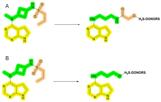
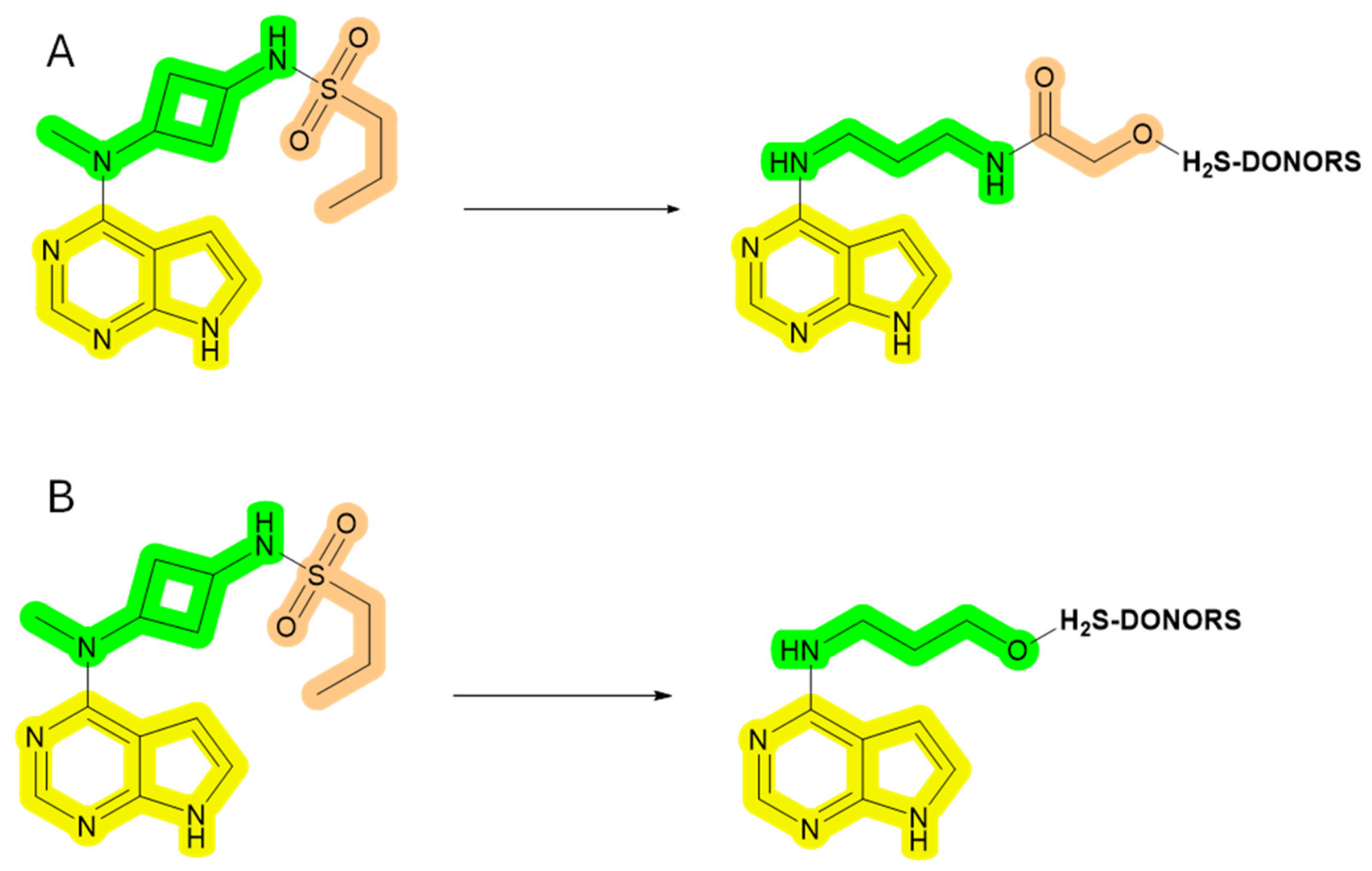
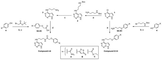
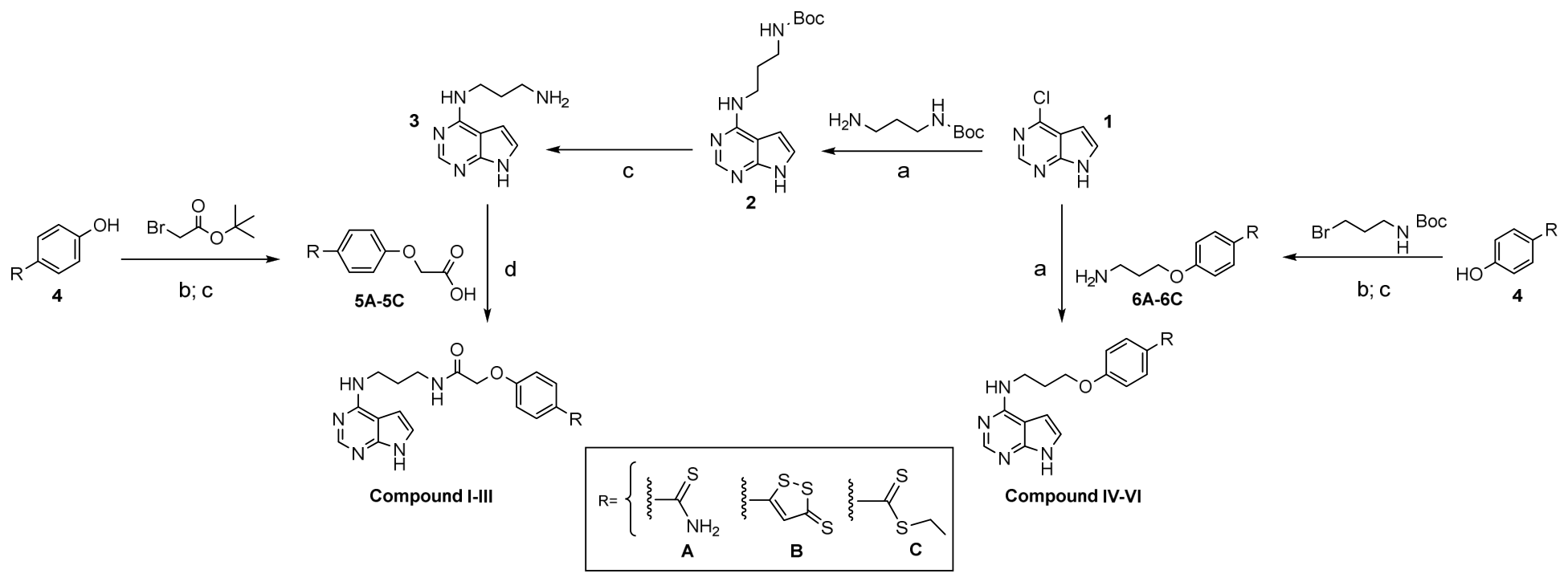
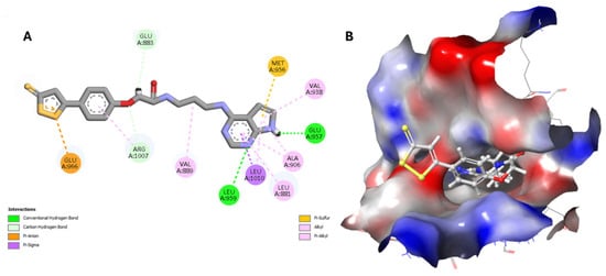
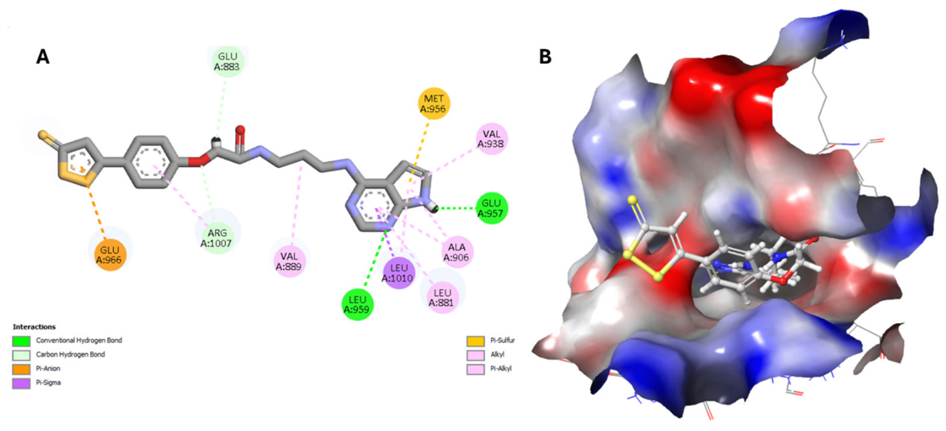
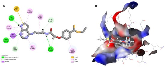
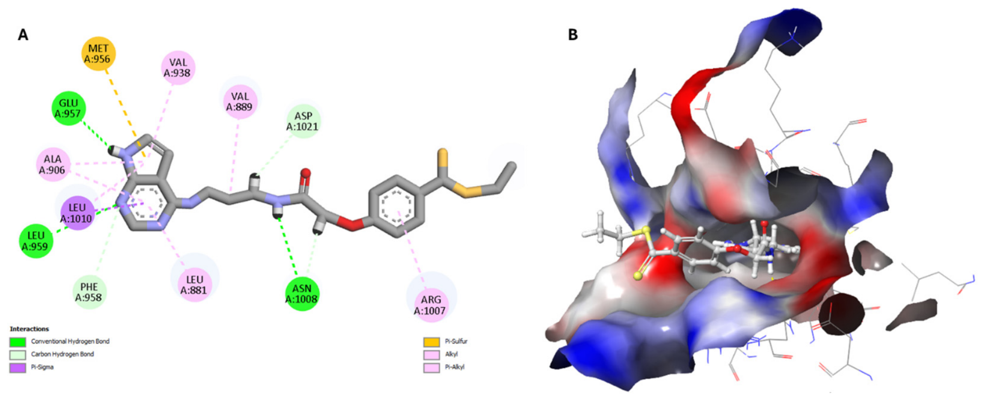
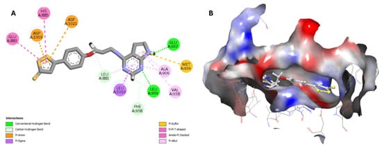
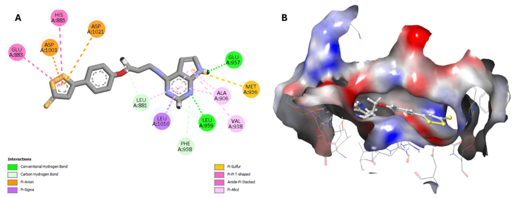
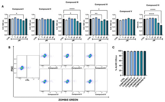
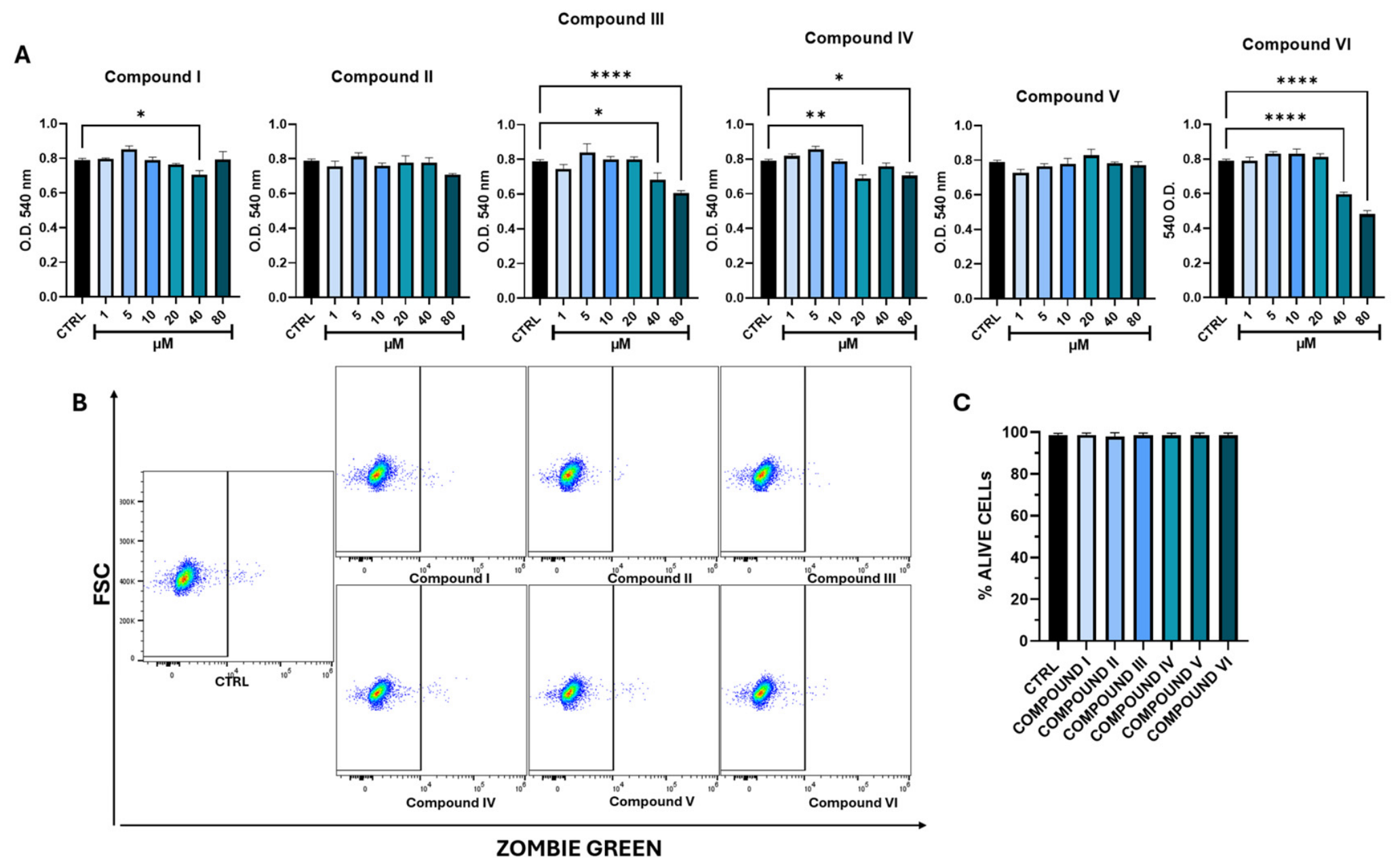
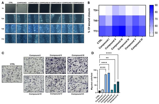
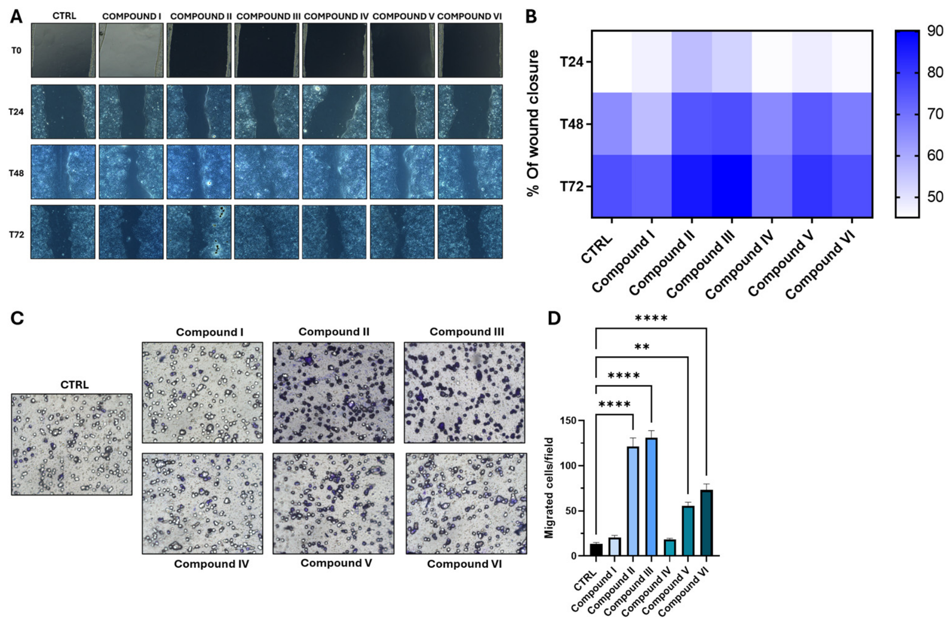
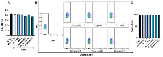
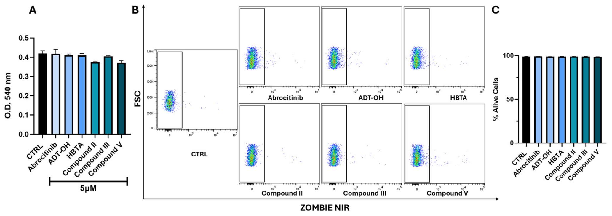
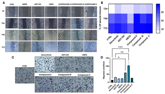
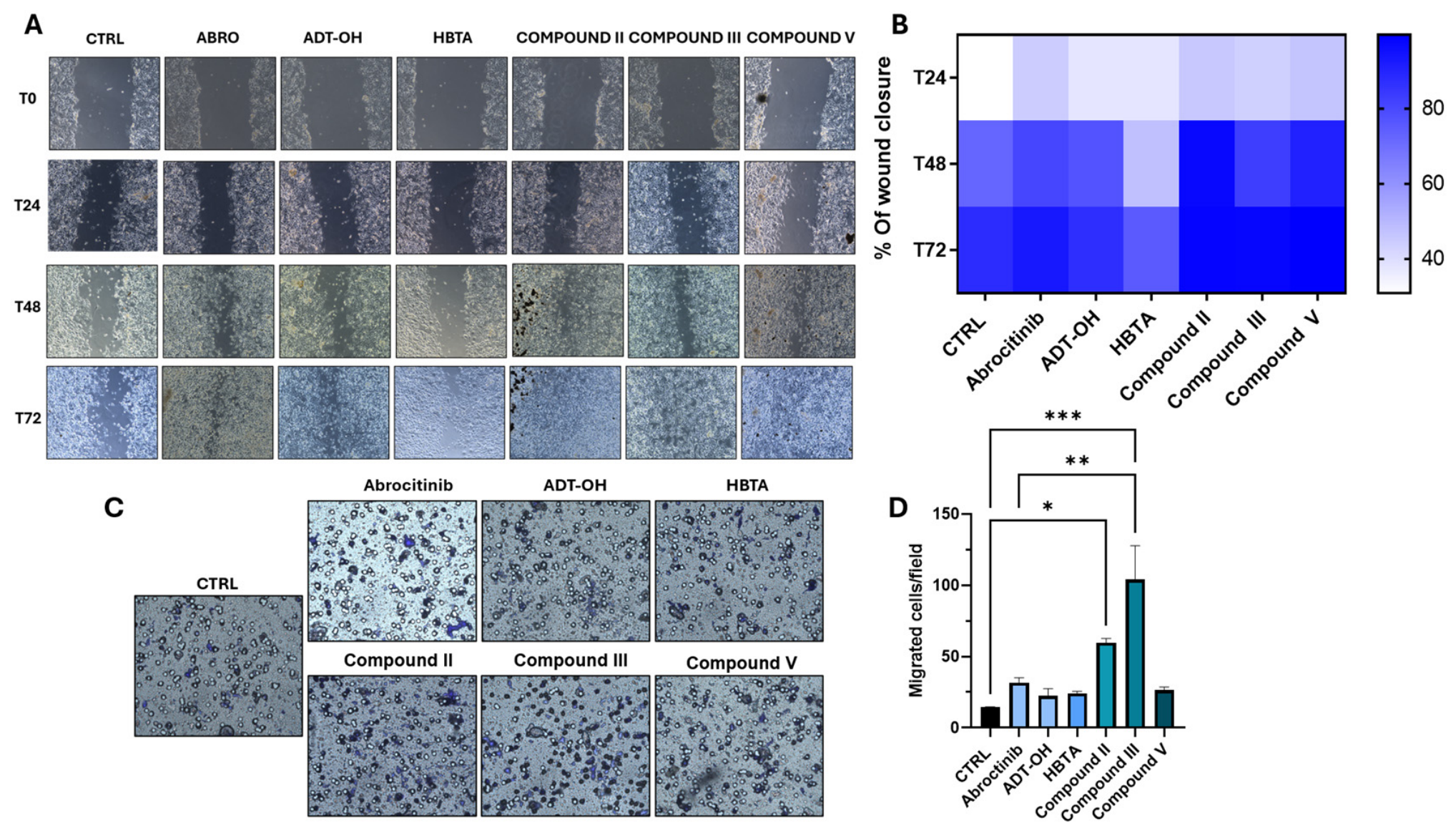
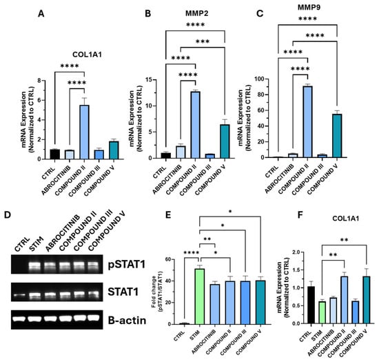
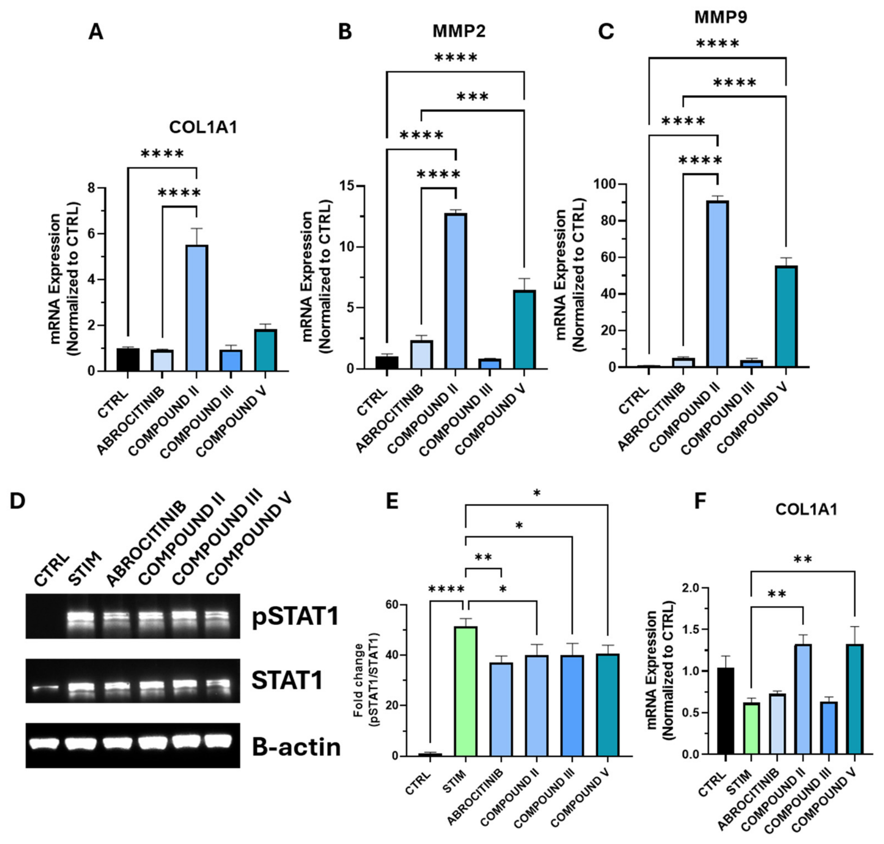
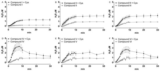
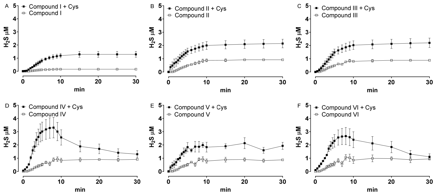

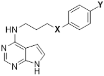
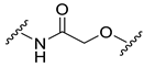


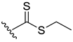


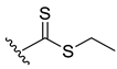
Disclaimer/Publisher’s Note: The statements, opinions and data contained in all publications are solely those of the individual author(s) and contributor(s) and not of MDPI and/or the editor(s). MDPI and/or the editor(s) disclaim responsibility for any injury to people or property resulting from any ideas, methods, instructions or products referred to in the content. |
© 2026 by the authors. Licensee MDPI, Basel, Switzerland. This article is an open access article distributed under the terms and conditions of the Creative Commons Attribution (CC BY) license.