


Abstract
Endotoxemia-associated coagulopathy integrates inflammation, oxidative stress, circulatory dysfunction, fibrin turnover, and organ injury. This study evaluated an amentoflavone-standardized methanolic Juniperus oxycedrus fruit extract in lipopolysaccharide (LPS)-induced endotoxemia in rats and explored candidate targets using network pharmacology and molecular docking. Male Wistar rats were assigned to Control, LPS, LPS + aspirin (200 mg/kg), LPS + J. oxycedrus 100 mg/kg, or LPS + J. oxycedrus 200 mg/kg groups (n = 10/group). HPLC-DAD, thrombo-inflammatory biomarkers, systemic redox indices, antioxidant defenses, hemodynamic and physiological variables, hepatic and renal injury markers, apelin, lung wet/dry ratio, and descriptive histopathology were integrated. The extract contained approximately 0.41% amentoflavone. Network analysis identified 139 core targets, and docking predicted favorable binding poses for amentoflavone–MMP-9 (−9.5 kcal/mol) and luteolin–COX-2 (−9.4 kcal/mol). LPS increased D-dimer, oxidative injury, IL-6, lactate, ALT, AST, and creatinine and reduced MAP, core temperature, apelin, and antioxidant defenses. Both extract doses reduced D-dimer; 200 mg/kg produced the broadest numerical systemic response, lowering MDA by 51.6%, TOS by 50.4%, IL-6 by 65.1%, lactate by 59.2%, ALT by 54.3%, AST by 52.4%, and creatinine by 50.8% versus LPS, while increasing MAP by 46.3%, SOD by 87.4%, CAT by 86.8%, GPx-1 by 102.4%, TAS by 80.4%, and apelin by 132.8%. TxA2, tissue factor, BAL cytokines, and lung wet/dry ratio did not show a consistent extract-related improvement. The findings support a multi-domain association between J. oxycedrus treatment and improved systemic redox, perfusion, inflammatory, and organ-injury profiles, while the docking results remain hypothesis-generating and do not establish target engagement. Responses were endpoint-specific; uniform dose dependence, statistically confirmed between-dose superiority, comprehensive anticoagulation, and definitive pulmonary protection were not established.
1. Introduction
Coagulopathy is a severe systemic disturbance of coagulation and inflammatory pathways, in which thrombin generation and fibrinolysis become imbalanced, fibrin accumulates in small vessels, and platelets are consumed. It typically appears as a secondary complication of endotoxemia, major trauma, or obstetric disorders. In endotoxemia, parallel activation of immune and coagulation pathways drives a state of thrombo-inflammation that promotes microvascular occlusion, tissue hypoxia, and multi-organ dysfunction. The lung is particularly vulnerable, since pulmonary microthrombi impair gas exchange and contribute substantially to poor outcomes in septic shock [1].
Current treatment still focuses on controlling the underlying cause and supporting failing organs. Anticoagulant or antiplatelet drugs may be added in selected cases, but their effects are inconsistent and bleeding risk often limits their use [2]. There is therefore a clear need for strategies that can modulate both inflammation and coagulation without markedly increasing hemorrhagic complications. Natural products with antioxidant, anti-inflammatory, and antithrombotic properties are being investigated as possible phytotherapeutic tools in this context [3].
The genus Juniperus (Cupressaceae) includes several species long used in traditional medicine for pain, inflammation, and infections. Among them, J. oxycedrus has been studied experimentally and shown antioxidant, antimicrobial, and anti-inflammatory effects consistent with its folk uses [4,5]. Phytochemical analyses describe a rich profile of phenolic acids and flavonoids, including catechin, quercetin, luteolin, and the biflavonoid amentoflavone [6,7]. These constituents are known to influence cytokine release, oxidative stress, and platelet aggregation—processes that converge on the thrombo-inflammatory axis driving endotoxemia-associated coagulopathy [8,9].
However, the molecular targets through which J. oxycedrus might counteract endotoxemia-associated coagulopathy remain poorly defined. Plant extracts typically contain dozens of bioactive compounds that may engage multiple proteins at once, making conventional single-target paradigms insufficient to resolve their multi-faceted mechanisms. Network pharmacology offers a systems-level framework to predict candidate compound–target–disease interactions, whereas molecular docking yields computational predictions of plausible binding poses and relative affinities. Importantly, these in silico approaches are strictly exploratory and predictive rather than direct evidence of target engagement or physical protein–ligand binding in vivo. Integrating these computational target-prioritization workflows with systemic in vivo biomarker readouts facilitates the preliminary ranking of candidate phytotherapeutic pathways, narrowing the gap between phytochemical profiling and downstream phenotypic bioactivity [10,11].
Oxidative stress and impaired tissue perfusion provide an additional mechanistic bridge between LPS sensing and organ dysfunction. Excess reactive species promote lipid peroxidation, impair glutathione peroxidase, superoxide dismutase, and catalase defenses, and amplify endothelial and mitochondrial injury. In parallel, hypotension and hyperlactatemia indicate circulatory and metabolic stress, while ALT, AST, and creatinine contextualize hepatic and renal consequences. The apelin-APJ system participates in cardiovascular, endothelial, and inflammatory regulation; therefore, circulating apelin may provide complementary information on vascular-metabolic adaptation. Evaluating these domains alongside coagulation markers allows a more integrated assessment than isolated cytokine or D-dimer measurements [12,13,14,15,16,17,18,19,20,21,22,23,24,25,26].
Accordingly, the present study investigated whether J. oxycedrus fruit extract modifies thrombo-inflammatory, redox, hemodynamic, physiological, and organ-injury responses in LPS-induced endotoxemia. We postulated an endpoint-specific hypothesis rather than assuming a uniform dose-dependent dynamic across all domains: specifically, we evaluated whether the extract differentially lowers D-dimer and selected pro-inflammatory signals, restores systemic antioxidant capacity, improves MAP and lactate, attenuates hepatic and renal injury indices, and modulates circulating apelin, with responses interrogated independently across distinct biological endpoints without the a priori assumption of monotonic dose dependence. In parallel, network pharmacology and molecular docking were implemented as hypothesis-generating, target-prioritization instruments to delineate plausible compound–target interactions rather than to infer direct in vivo target engagement.
2. Materials and Methods
2.1. Plant Material Authentication and Extract Preparation
Mature cone berries of Juniperus oxycedrus L. subsp. oxycedrus were collected in October 2024 from wild shrubs in the vicinity of Beynam, Bala district, Ankara, Türkiye (WGS84: approximately 39°41′ N, 32°55′ E; altitude ~1150 m a.s.l.) (Figure 1a). Taxonomic and botanical identification was conducted by Assist. Prof. Tuğba Ertuğrul through macroscopic and stereomicroscopic morphological comparison against authenticated reference specimens preserved in the Herbarium of Gazi University (GAZI, Ankara, Türkiye; comparative reference identifier: UGB1002). All collected material was processed as a single extraction batch. A collection-specific herbarium voucher specimen was not deposited or retained from this field collection; morphological verification against existing authenticated GAZI reference specimens (comparative reference identifier: UGB1002) was used strictly for comparative taxonomic authentication and does not substitute for a newly accessioned collection voucher, which represents an acknowledged botanical limitation of this study.
Figure 1.
Botanical material of Juniperus oxycedrus L. subsp. oxycedrus: (a) natural habit of the shrub bearing immature cone berries in its native habitat; (b) harvested aerial branches bearing mature cones (galbuli), from which fruits were detached and shade-dried for subsequent extraction.
Following harvest, mature cones (galbuli) were manually separated from the collected aerial branches (Figure 1b), thoroughly cleaned, and destoned (seeds removed). The pericarp and pulp fractions were dried under ambient shaded conditions to a constant weight and mechanically pulverized into a fine powder. The powdered fruit material was extracted by maceration in analytical-grade methanol (1:10, w/v) for 24 h at room temperature, supplemented by ultrasonication. The marc was subsequently re-extracted twice under identical conditions to maximize extraction yield. The combined filtrates were evaporated to dryness under reduced pressure at 50 °C using a rotary evaporator and stored in airtight amber glass containers at −20 °C until phytochemical quantification and in vivo administration. The resulting amentoflavone-standardized fruit extract was utilized across all experimental protocols. HPLC-DAD profiling was employed for chemical fingerprinting and precise quantification of the biflavonoid marker amentoflavone, rather than for exhaustive untargeted metabolomics.
2.2. Phytochemical Characterization
Total phenolic content was determined by the Folin–Ciocalteu method against gallic acid and expressed as mg gallic acid equivalents per gram of dried extract (mg GAE/g); measurements were performed in triplicate. HPLC–DAD analysis was carried out on an Agilent 1260 Infinity system equipped with an ACE 5 C18 column (250 × 4.6 mm, 5 µm) at a constant column temperature of 40 °C. The binary mobile phase consisted of 0.2% (v/v) ortho-phosphoric acid in ultrapure water (Solvent A) and HPLC-grade acetonitrile (Solvent B), delivered at a flow rate of 1.0 mL/min with an injection volume of 10 µL. The gradient profile was specifically adapted for this separation (0 min: 5% B; 41 min: 10% B; 45 min: 10% B; followed by column re-equilibration for 10 min prior to subsequent runs). Dry extract and standard stock solutions were freshly prepared in analytical-grade methanol, filtered through 0.45 µm PTFE syringe filters, and analyzed with diode-array detection scanned across 210–360 nm. Peaks were assigned by comparison of retention times and UV spectra with authentic standards, consistent with 3,4- and 2,4-dihydroxybenzoic, p-coumaric, chlorogenic, caffeic, rosmarinic, sinapic, and dicaffeoylquinic acids; taxifolin, rutin, luteolin, orientin, myricetin, quercetin-3-O-arabinoside, and amentoflavone. Amentoflavone was quantified by external linear calibration at 330 nm; limits of detection (LOD) and quantification (LOQ) were calculated using the signal-to-noise ratio approach (S/N = 3 and 10, respectively; n = 6). The chromatographic method was adapted from previously described procedures [27,28], using the gradient conditions summarized in Table 1. Method performance was characterized in terms of calibration linearity, injection repeatability, and empirical LOD/LOQ values determined by the signal-to-noise ratio method (n = 6).
Table 1.
Gradient elution program used for HPLC analysis (column temperature: 40 °C).
Table 1 presents the gradient elution program used in the HPLC analysis of the plant extract. The mobile phase consisted of acetonitrile and 0.2% o-phosphoric acid in water, applied in a binary gradient mode. The column temperature was maintained at 40 °C throughout the run.
2.3. Animals and Experimental Design
Fifty male Wistar rats (body weight: 200–250 g at the end of the acclimation period; approximately 8–10 weeks of age) were housed under controlled environmental conditions (20 ± 2 °C, 50 ± 10% relative humidity, 12 h light/dark cycle) with standard rodent chow and water ad libitum. Only male rats were included to reduce potential variability associated with estrous-cycle-dependent changes in circulating sex hormones and vascular responsiveness [29]. This design choice limits the extrapolation of the findings to female rats. A sample size of n = 10 per group was determined a priori by power analysis targeting 80% power (α = 0.05, effect size f = 0.57) for one-way ANOVA based on anticipated differences in primary systemic coagulopathy markers (plasma D-dimer). Following 10 days of acclimation, animals were assigned to five experimental groups (n = 10/group): (1) Control, (2) LPS, (3) LPS + aspirin (200 mg/kg, i.p.), (4) LPS + J. oxycedrus 100 mg/kg (i.p.), and (5) LPS + J. oxycedrus 200 mg/kg (i.p.). All procedures were conducted at the Experimental Animals Application and Research Center, Ankara Medipol University. The individual animal was the designated experimental unit; technical assay duplicates did not represent additional independent experimental units. As noted, this statistical power calculation was calibrated for the primary endpoint and does not ensure equivalent statistical power across all exploratory secondary parameters.
Endotoxemia-associated coagulopathy was induced by continuous intravenous infusion of Escherichia coli O55:B5 LPS (Sigma-Aldrich, St. Louis, MO, USA, Cat. No. L2880-100MG). LPS was reconstituted in sterile isotonic saline (pH 7.2–7.4) and delivered via a catheterized femoral vein at a total dose of 5 mg/kg administered continuously over 4 h using an automated syringe infusion pump (infusion rate: ~0.5 mL/h per animal; total infusion volume: ~2.0 mL per rat). Control animals underwent sham femoral catheterization and received vehicle saline under identical infusion conditions. One hour after the initiation of LPS infusion, pharmacological treatments were administered intraperitoneally as a single bolus: acetylsalicylic acid (Sigma-Aldrich, Cat. No. A3160-1VL, 200 mg/kg, suspended in sterile saline) or J. oxycedrus extract (100 or 200 mg/kg, freshly dissolved in sterile saline vehicle). This sustained 5 mg/kg 4 h LPS infusion regimen was adapted from experimental endotoxemia models [30], with protocol-specific modifications including continuous femoral venous delivery, ketamine–xylazine maintenance anesthesia, and a single-dose post-treatment intervention schedule.
All surgical and experimental procedures were performed under maintenance anesthesia with ketamine hydrochloride (Ketalar®, Pfizer, New York, NY, USA, Cat. No. K2753, 50 mg/kg, i.m.) and xylazine hydrochloride (Rompun®, Bayer, Leverkusen, Germany, Cat. No. X1251, 10 mg/kg, i.m.). The timeline was anchored to the initiation of LPS infusion designated as t = 0 h. Treatments were administered at t = 5 h (exactly 1 h following the completion of the 4 h continuous LPS infusion). Animals were maintained under continuous terminal anesthesia and euthanized at t = 8 h from the start of LPS infusion (corresponding to exactly 3 h post-treatment administration) by complete exsanguination under deep surgical anesthesia. Blood was collected from the inferior vena cava into serum-separator and 3.2% sodium-citrate tubes, processed to obtain serum and platelet-poor plasma, and stored at −80 °C until analysis. Lungs were excised immediately for gravimetric edema determination and histopathological evaluation.
Death was not an experimental endpoint in this study. All procedures were performed under non-recovery (terminal) anaesthesia: animals were anaesthetised with ketamine hydrochloride (50 mg/kg, i.m.) and xylazine hydrochloride (10 mg/kg, i.m.) before catheterisation and remained anaesthetised throughout the entire 8 h experimental period, with supplementary doses administered as required to maintain a surgical plane of anaesthesia. Animals were kept under continuous direct observation by the study investigator and an animal technician for the whole period; respiration, spontaneous movement and signs of lethargy were monitored. A humane endpoint of respiratory distress was defined a priori, at which animals would have been immediately euthanised; this endpoint was not reached by any animal. Of the 50 rats used, none died spontaneously, none required early euthanasia and none were excluded. All 50 animals were euthanised at the planned 8 h time point by exsanguination under deep anaesthesia. All personnel involved in animal handling and experimental procedures held a valid Laboratory Animal Use Certificate in accordance with Turkish national regulations.
All procedures, including the terminal anaesthesia design and the anticipated absence of mortality, were reviewed and approved by the Animal Experiments Local Ethics Committee of Ankara Medipol University (Date: 8 March 2024; Decision No: 2024/1-HDY 1/1) and complied with institutional and national guidelines and the ARRIVE guidelines. Under this approval, tissues from the same animals were allocated to separate studies addressing different organs. In accordance with ARRIVE 2.0 recommendations, experimental procedures and reporting boundaries are documented transparently based on verified institutional protocol records; an ARRIVE 2.0 checklist has been prepared reflecting the actual recorded parameters, without retroactively certifying undocumented husbandry or allocation details.
Animals were allocated to the five groups after 10 days of acclimation; a formally documented computational randomisation procedure was not used, and the order of treatment, measurement sequences, and rack/cage locations were not systematically counterbalanced. Samples were submitted for laboratory analysis under animal identification codes rather than explicit group names; however, because these internal alphanumeric codes contained group designation elements, complete masking of laboratory assessors and data processors cannot be guaranteed. All 50 rats (n = 10 per group) completed the 8 h monitoring timeline and were included in the primary datasets across endpoints without attrition. Consequently, because baseline allocation, cage microenvironment distribution, procedural running order, and complete assessor blinding were not formally controlled, potential selection, cage-position, batch, and measurement biases cannot be entirely ruled out; these methodological constraints are transparently recognized as operational limitations.
2.4. Bronchoalveolar Lavage and Thrombo-Inflammatory Measurements
BAL fluid collection was performed 3 h after the final injection. Following tracheotomy, 0.5 mL sterile saline was instilled and gently aspirated three times (Figure 2). Samples were centrifuged (5000 g, 10 min), and supernatants were stored at −80 °C.
Figure 2.
Bronchoalveolar lavage collection: (a) control-group lung and (b) LPS-group lung. Lavage was performed after the final injection as described in Section 2.4.
Serum IL-1β (BT-Lab, E0119Ra), MIP-1α (BT-Lab, E0369Ra), fibrinogen (BT-Lab, E0126Ra), TF (BT-Lab, E0794Ra) and TxA2 (BT-Lab, E0683Ra) were quantified using sandwich ELISA kits from BT-Lab; plasma D-dimer was measured using a competitive ELISA kit (Elabscience, Wuhan, China, E-EL-R0317). BAL supernatants were assayed for IL-1β and MIP-1α with the same BT-Lab kits. All assays followed the manufacturers’ instructions; plates were read at 450 nm on a BioTek ELx800 microplate reader (washer ELx50). Samples were run in duplicate, and concentrations were calculated from four-parameter logistic (4-PL) standard curves. To ensure analytical quality, samples were diluted within the validated calibration range, duplicate CV was required to be ≤15%, and any out-of-range values were re-assayed from stored −80 °C aliquots.
2.5. Assessment of Systemic Redox Balance, Hemodynamics, Organ Function, and Circulating Biomarkers
Serum concentrations of malondialdehyde (MDA), superoxide dismutase (SOD), catalase (CAT), glutathione peroxidase-1 (GPx-1), interleukin-6 (IL-6), and apelin (apelin-13) were quantitatively analyzed using rat-specific enzyme-linked immunosorbent assay (ELISA) kits obtained from Bioassay Technology Laboratory (BT-Lab, Shanghai Korain Biotech Co., Ltd., Shanghai, China), specifically utilizing Cat. No. E0156Ra for MDA, Cat. No. E1375Ra for SOD, Cat. No. E0869Ra for CAT, Cat. No. E1172Ra for GPx-1 (analytical sensitivity: 0.23 ng/mL; linear detection range: 0.5–200 ng/mL), Cat. No. E0135Ra for IL-6 (analytical sensitivity: 0.052 ng/L; linear detection range: 0.1–40 ng/L [0.1–40 pg/mL]), and Cat. No. E1026Ra for apelin (analytical sensitivity: 0.524 ng/mL; linear detection range: 0.1–40 ng/mL), in strict compliance with the analytical protocols provided by the manufacturer. Serum samples exceeding the dynamic linear calibration range—specifically for elevated IL-6 concentrations encountered in the endotoxemic groups—were appropriately pre-diluted (1:5 to 1:20) with standard sample diluent prior to assay execution, and final concentrations were determined by multiplying back with the corresponding dilution factor.
Following the completion of assay-specific incubation and chromogenic development procedures, optical density values were spectrophotometrically acquired at 450 nm on a BioTek ELx800 microplate reader (BioTek Instruments, Winooski, VT, USA), whereby individual sample concentrations were interpolated from standard calibration curves fitted to a four-parameter logistic (4-PL) mathematical model, while ensuring that all analytical runs maintained high assay precision with intra-assay and inter-assay coefficients of variation (CV) strictly remaining below 8% and 10%, respectively.
Serum total antioxidant status (TAS) and total oxidant status (TOS) were determined by automated colorimetric detection methods on the microplate reader platform at 660 nm and 530 nm using commercial assay kits (TAS: Cayman Chemical, Ann Arbor, MI, USA, Cat. No. 709001, calibrated and expressed as mmol Trolox equivalent/L; TOS: commercial spectrophotometric assay kit calibrated against hydrogen peroxide standards and expressed as μmol H2O2 equivalent/L; catalog numbering and lot details verified against analytical logs); the systemic oxidative stress index (OSI, arbitrary unit) was mathematically calculated according to the standard unit-aligned convention: OSI (arbitrary unit) = [TOS (μmol H2O2 Eq/L)/(TAS (mmol Trolox Eq/L) × 1000)] × 100, or reported directly as the ratio TOS/TAS (arbitrary unit) with explicit dimensional scaling preserved; numerical dataset values and statistical group assignments remain unchanged.
Circulating hepatic and renal biochemical indices comprising alanine aminotransferase (ALT), aspartate aminotransferase (AST), and creatinine, alongside the systemic tissue perfusion marker lactate, were quantified by automated kinetic and enzymatic-colorimetric methodologies on a Cobas c501 clinical chemistry autoanalyzer (Roche Diagnostics GmbH, Mannheim, Germany) utilizing original diagnostic reagent cartridges—specifically Cat. No. 20764957322 for ALT (kinetic UV test adhering to IFCC standards), Cat. No. 20764949322 for AST (kinetic UV test adhering to IFCC standards), Cat. No. 04810716190 for creatinine (kinetic alkaline picrate Jaffé method), and Cat. No. 03183742190 for lactate (lactate oxidase enzymatic colorimetric reaction)—with analytical validity and calibration stability confirmed through dedicated calibrator and control matrices yielding intra- and inter-assay CV values below 3.5%.
Continuous hemodynamic recording of mean arterial pressure (MAP) was executed via a pre-calibrated physiological fluid-filled pressure transducer (Harvard Apparatus, Holliston, MA, USA) directly coupled to the catheterized femoral arterial line, whereas rectal core body temperature was continuously captured using an electronic veterinary thermometer integrated with a lubricated digital thermistor probe (Harvard Apparatus, Holliston, MA, USA) positioned 3 cm past the anal sphincter into the rectum; all physiological data points were logged in triplicate at predefined intervals across the entire 8 h post-infusion monitoring timeline and subsequently averaged, with samples identified by animal codes; because these internal identifiers incorporated group designation sequences, complete blinding of experimenters, evaluators, and downstream data analysts could not be rigorously assured across all monitoring and data-entry stages.
2.6. Pulmonary Edema and Histopathology
Pulmonary edema was quantified by the wet/dry lung weight ratio. The right lobe was weighed immediately after dissection (wet mass), then dried at 60 °C for 72 h to constant weight (incubator: ILDAM IN-120). The ratio (wet ÷ dry) was calculated; higher values indicate fluid accumulation [31,32].
Lungs were fixed in 10% neutral buffered formalin (24 h), paraffin-embedded, sectioned (4 µm), and stained with hematoxylin and eosin (H&E). Sections were examined under bright-field microscopy by an experienced histologist. Histological findings are reported descriptively; no semi-quantitative scoring or morphometric index was applied, and histological observations were therefore not compared statistically. Representative fields do not establish group-level histological protection, structural preservation, or quantitative treatment efficacy, as qualitative micrographs cannot substitute for formalized, blinded semi-quantitative scoring.
2.7. Network Pharmacology and Molecular Docking
The fifteen phenolic constituents identified by HPLC were used as the input compound set. Physicochemical descriptors (SMILES, molecular weight, XLogP, hydrogen-bond donors and acceptors, rotatable bonds, topological polar surface area) were retrieved from PubChem [33], and drug-likeness was evaluated by Lipinski’s Rule of Five [34]. Potential human protein targets of each compound were predicted with SwissTargetPrediction using Homo sapiens as the reference organism, and only predictions with a probability score ≥ 0.1 were retained [11]. Disease-related targets were obtained from the Open Targets Platform [35] for nine queries reflecting the clinical spectrum of endotoxemia-associated coagulopathy: sepsis, systemic inflammatory response syndrome, endotoxemia, disseminated intravascular coagulation, acute lung injury, ARDS, coagulopathy, venous thrombosis, and thrombophilia. The intersection between predicted compound targets and disease-related targets defined the core-target set, which was further analysed in the STRING database (v12.0, high-confidence cut-off 0.700) for protein–protein interactions [36]. Pathway enrichment of core targets was performed against the KEGG, Reactome, GO Biological Process, and WikiPathways libraries through Enrichr, and pathways with Benjamini–Hochberg adjusted p < 0.05 were considered significant [37]. For molecular docking, five proteins central to the in vivo biomarker panel were selected—thrombin (PDB 1PPB), COX-2 (5KIR), TLR4–MD2 (3FXI), TF–FVIIa (1DAN), and MMP-9 (1L6J)—and each was docked with its highest-probability ligand from the extract (myricetin, luteolin, chlorogenic acid, p-coumaric acid, and amentoflavone, respectively)using CB-Dock2, which couples cavity detection with AutoDock Vina (version 1.1.2) [38,39].
Receptor structures were obtained from the Protein Data Bank [12]. Vina scores (kcal/mol) and contact residues of the best-ranked pose were recorded. Database queries, target intersection analyses, and network visualisations were performed with custom Python scripts (Python 3.12) using the publicly available APIs of the above resources. The five docking targets were prioritized for biological relevance to coagulation (thrombin and TF–FVIIa), LPS recognition (TLR4–MD2), inflammatory lipid signaling (COX-2), and tissue injury (MMP-9); importantly, these targets were selected as an exploratory computational framework and were not all directly measured in the in vivo animal experiment. Because target prediction databases and available high-resolution crystallographic coordinates primarily represent human macromolecules, these in silico models provide hypothesis-generating insights rather than rat-specific mechanistic validation; cross-species sequence homology, binding-pocket conservation, and rat-specific docking simulations were not formally conducted and are not asserted here.
2.8. Statistical Analysis
Data distribution and normality were evaluated using the Shapiro–Wilk test, and homogeneity of variances across experimental groups was verified using Levene’s test. Continuous variables displaying a normal distribution and equal variances are presented as mean ± standard deviation (SD). Differences among multiple experimental groups were analyzed by one-way analysis of variance (ANOVA) followed by Tukey’s post hoc test for pairwise comparisons. Variables that failed the normality assumption or displayed unequal variance (specifically tissue factor and thromboxane A2) are expressed as median with interquartile range (IQR, 25th–75th percentiles) and were compared using the non-parametric Kruskal–Wallis test followed by Dunn’s post hoc test.
For all omnibus analyses and post hoc pairwise evaluations, a two-tailed p-value < 0.05 was accepted as statistically significant. Statistical calculations, data structuring, and post hoc testing were executed using IBM SPSS Statistics for Windows, version 26.0 (IBM Corp., Armonk, NY, USA) and GraphPad Prism version 9.0 (GraphPad Software, San Diego, CA, USA). Graphical representations and network-associated data workflows were constructed using GraphPad Prism and Python custom scripts (version 3.12). Descriptive percentage changes for the systemic outcomes in these tableswere calculated directly from the displayed group means as 100 × (treatment mean − LPS mean)/LPS mean, unless another reference group is explicitly specified in the column header. Reductions are expressed as positive percentage magnitudes where indicated in the text. Importantly, these calculations represent descriptive group-summary contrasts intended solely for contextual comparison, rather than means of individual within-animal percentage changes or separate inferential hypothesis tests; formal statistical significance is governed exclusively by the omnibus ANOVA and Tukey post hoc tests reported in the tables. Temperature differences are expressed as absolute differences in degrees Celsius (°C) rather than percentages.
3. Results
3.1. Extract Yield
From an initial batch of 150.0 g of finely ground, seed-removed, and shade-dried mature fruits (cone berries) of Juniperus oxycedrus L. subsp. oxycedrus, a final dark-brown crude extract weighing 47.51 g was obtained following the extraction and solvent evaporation procedures. This corresponded to a substantial extraction yield of 31.67% (w/w, dry weight basis). The obtained extract was stored at −20 °C in amber glass containers under dark conditions until further phytochemical quantification and in vivo biological assays to prevent any photo-oxidative degradation.
3.2. Total Phenolic Content
The quantitative estimation of total phenolic constituents in the J. oxycedrus extract was carried out using the Folin–Ciocalteu colorimetric method. Standard calibration was established through linear regression analysis using graded concentrations of gallic acid (0.005–0.04 mg/mL), yielding a linear standard curve (Y = 11.64X + 0.0813, R2 = 0.986; where Y represents absorbance measured spectrophotometrically at 765 nm, and X is the gallic acid concentration in mg/mL) (Table 2). Individual absorbance values obtained across serial dilutions of the crude extract were documented and summarized in Table 3. All determinations were performed in triplicate (n = 3) to ensure analytical reproducibility. Based on the standard regression equation, the total phenolic content of the extract was calculated as 43.30 ± 2.61 mg of gallic acid equivalents per gram of dry extract (mg GAE/g extract), demonstrating that the botanical material is a rich source of phenolic compounds suitable for subsequent bioactivity profiling.
Table 2.
Absorbance measurements for total phenolic content determination using gallic acid standard.
Table 3.
Absorbance values obtained for total phenolic content determination of Juniperus oxycedrus extract.
This table presents the absorbance values obtained from triplicate measurements of gallic acid standards at increasing concentrations for the construction of a calibration curve used in total phenolic content determination. The resulting linear regression equation was Y = 11.64X + 0.0813, where Y represents absorbance and X is the gallic acid concentration (mg/mL). This equation was subsequently used to calculate the total phenolic content of the plant extract.
This table shows the absorbance values obtained at various prepared stock/working concentrations of Juniperus oxycedrus extract for the determination of total phenolic content using the Folin–Ciocalteu method. Each sample dilution was analyzed in technical triplicate (ABS1–3) and the mean absorbance (ABS_mean) was calculated for sample interpolation against the external gallic acid calibration curve. The reported extract concentrations represent working extract dilutions prior to Folin–Ciocalteu reagent and alkaline carbonate addition, rather than gallic acid equivalent concentrations, final spectrophotometric reaction mixture concentrations, or dimensionless dilution factors. Final total phenolic content was calculated using the complete mass-balance equation: TPC = (C_GAE × V × DF)/m_dry, where C_GAE is the interpolated gallic acid equivalent concentration (mg/mL), V is the extract reconstitution volume (mL), DF is the dilution factor, and m_dry is the dry extract mass (g), expressed as mg GAE/g dry extract.
3.3. HPLC-DAD Fingerprinting and Amentoflavone Quantification
The extract chromatogram at 330 nm (Figure 3) showed multiple constituents with prominent peaks at 8.37, 20.49, and 22.02 min. The amentoflavone standard (Figure 4) exhibited a characteristic peak at ≈22.018 min with the expected UV maxima, and its external calibration was linear (Figure 5; R2 = 0.999–1.000), using Y = peak area (330 nm) versus X = concentration (mg/mL). Using this calibration and the 22.018 min chromatographic signal in the sample, amentoflavone was quantified in triplicate as approximately 0.41% (w/w; detailed injection-level mean ± analytical repeatability SD provided in Table 4) in dried plant material. Method sensitivity thresholds, including LOD and LOQ values calculated from peak area signal-to-noise ratios (S/N = 3 and S/N = 10, respectively), are provided in Table 5.
Figure 3.
HPLC-DAD chromatogram of the Juniperus oxycedrus fruit extract recorded at 330 nm. Peak assignments and quantification are described in Section 3.3.
Figure 4.
HPLC chromatogram and ultraviolet spectrum of the amentoflavone reference standard at 330 nm. The major standard peak eluted at approximately 22.018 min.
Figure 5.
External calibration curve for amentoflavone. Chromatographic peak area at 330 nm is plotted against standard concentration (mg/mL). The displayed linear regression is y = 5804x − 1.4919 (R2 = 1.000). Limits of detection and quantification are reported in Table 5.
Table 4.
Amentoflavone content determined by HPLC analysis (λ = 330 nm, retention time ≈ 22.018 min).
Table 5.
Quantified amentoflavone content in the plant extract and corresponding LOD and LOQ values for the analytical method.
This table presents the quantified amount of amentoflavone in the dried. powdered plant material (% w/w), along with the corresponding limit of detection (LOD) and limit of quantification (LOQ) values for the analytical method employed. The results demonstrate the method’s high sensitivity for detecting and quantifying amentoflavone.
3.4. Network Pharmacology
SwissTargetPrediction analysis of the fifteen extract phytochemicals yielded 985 compound–target interactions (probability ≥ 0.1) covering 278 unique human proteins. From the Open Targets Platform, 2532 unique genes were retrieved across nine disease categories related to endotoxemia-associated coagulopathy. The intersection of the two sets defined a core list of 139 putative mechanistic targets. Importantly, these in silico outputs represent candidate human gene identifiers rather than directly quantified rat proteins; candidate genes relevant to the biological domains of our in vivo panel included F3 (tissue factor), F2 (prothrombin/thrombin), PTGS2 (COX-2), ALOX5 (5-lipoxygenase), TLR4, RELA and NFKB1 (NF-κB subunits), TNF, and MMP9 (MMP-9).
Protein–protein interaction analysis of the core targets in STRING (high-confidence cut-off 0.700) generated a network of 125 connected nodes and 572 edges, with STAT3, EGFR, SRC, TNF, AKT1, TLR4, NFKB1, MMP9, RELA, and PTGS2 as top-degree network hubs. Pathway enrichment indicated significant over-representation of Toll-like receptor (adjusted p = 6 × 10⁻¹⁰), NF-κB (6 × 10⁻¹⁰), TNF (9 × 10⁻¹¹), platelet activation (4 × 10⁻⁹), and neutrophil extracellular trap (2 × 10⁻¹¹) pathways in KEGG, and of hemostasis (4 × 10⁻¹³), innate immune system (4 × 10⁻¹¹), cytokine signalling (3 × 10⁻¹⁵), and arachidonic-acid metabolism (1 × 10⁻⁷) in Reactome.
3.5. Molecular Docking
The five protein–ligand pairs identified from the network pharmacology results were subjected to blind docking in CB-Dock2 (Table 6). Vina scores of the best-ranked poses ranged from −5.1 to −9.5 kcal/mol, with a mean of −7.9 kcal/mol across the five complexes.
Table 6.
Molecular docking of the five major J. oxycedrus phytochemicals against key inflammation and coagulation targets.
Docking was performed in CB-Dock2 (cavity detection coupled with AutoDock Vina). The Vina score is a model-dependent computational scoring estimate (kcal/mol); more negative values rank candidate poses more favorably within this in silico workflow, functioning solely as an exploratory target-prioritization metric without establishing experimentally measured binding affinity or physical target engagement.
Amino-acid residues are given in the standard three-letter code. COX-2: cyclooxygenase-2; MD2: myeloid differentiation factor 2; MMP-9: matrix metalloproteinase 9; NSAID: non-steroidal anti-inflammatory drug; PDB: Protein Data Bank; PEX: hemopexin-like (C-terminal) domain; TF–FVIIa: tissue factor–activated factor VII complex; TLR4: Toll-like receptor 4.
The strongest predicted binding was observed for amentoflavone in the hemopexin (PEX) domain of MMP-9 (−9.5 kcal/mol), with contacts at Tyr128, Ser129, Arg134, Asp206/207, Arg279, Tyr311, Trp313, and the Phe328–Asp334 β-propeller interface. Luteolin docked into the NSAID-binding pocket of COX-2 (−9.4 kcal/mol), engaging Arg120, Tyr385, and Ser530—the residue acetylated by aspirin. Myricetin occupied the thrombin active site (−8.0 kcal/mol), with contacts at the catalytic triad (His57, Asp194, Ser195) and the S1 specificity pocket (Asp189). Chlorogenic acid docked at the TLR4–MD2 dimerisation interface (−7.5 kcal/mol), engaging Phe483, Phe532, Phe533, His529, His555, and Lys560. p-Coumaric acid bound the TF–FVIIa catalytic site (−5.1 kcal/mol) at His57, Ser195, Asp189, and the S1 pocket; this modest affinity is consistent with the small molecular size of the ligand.
3.6. Thrombo-Inflammatory Biomarkers
In BAL fluid, IL-1β and MIP-1α concentrations did not differ significantly among the groups (p > 0.05) (Table 7).
Table 7.
Comparison of groups based on bronchoalveolar lavage (BAL) parameters.
Kruskal–Wallis test. p < 0.05 was considered statistically significant. IL-1β: interleukin-1 beta; MIP-1α: macrophage inflammatory protein-1 alpha; BAL: bronchoalveolar lavage.
This table shows the comparison of IL-1β and MIP-1α levels in bronchoalveolar lavage fluid across experimental groups. Data are presented as mean ± standard deviation and median (Q1–Q3). No statistically significant differences were observed among the groups for IL-1β and MIP-1α levels (Kruskal–Wallis test. p > 0.05).
In plasma/serum, IL-1β, MIP-1α, D-dimer, fibrinogen, TF and TxA2 were measured and compared between groups (Table 8). Significant differences were observed for IL-1β (p = 0.007), D-dimer (p < 0.001), TF (p = 0.003), and TxA2 (p = 0.015), whereas MIP-1α and fibrinogen levels did not differ significantly between groups (p > 0.05) (Table 8).
Table 8.
Effects of Juniperus oxycedrus extract on circulating inflammatory cytokines and coagulation markers in LPS-induced endotoxemic rats.
A significant group effect was observed for IL-1β concentrations across the experimental groups (p = 0.007). Groups 1 and 2 had similar values, Group 5 showed lower levels than both of these groups, and Groups 3 and 4 had intermediate concentrations (Table 8).
D-dimer concentrations differed significantly between the study groups (p < 0.001). Group 2 had the highest D-dimer levels, whereas Groups 1, 3, 4 and 5 showed lower and mutually similar concentrations (Table 8).
TF levels also showed a significant group effect (p = 0.003). Group 1 had the lowest TF concentrations; Groups 3, 4 and 5 had higher and comparable levels, and Group 2 showed intermediate values that did not differ significantly from either Group 1 or from Groups 3–5 (Table 8).
TxA2 levels were significantly different among the groups (p = 0.015). Group 1 had the lowest TxA2 values. Groups 3 and 5 exhibited higher and similar concentrations, while Groups 2 and 4 showed intermediate levels without clear statistical separation from either the lowest or highest groups (Table 8).
Statistically significant differences were observed between groups for IL-1β, D-dimer, Tissue Factor and thromboxane A2 levels (p < 0.05). No significant differences were found for MIP-1α and fibrinogen.
3.7. Pulmonary Outcomes and Histopathology
Statistically significant differences were detected in both wet (p = 0.004) and dry (p < 0.001) lung weights among the experimental conditions. The control group had lower wet lung weight than all LPS-challenged groups (LPS, LPS + aspirin, LPS + low-dose J. oxycedrus, LPS + high-dose J. oxycedrus). For dry lung weight, values in the control group were lower than in the LPS and both J. oxycedrus treatment groups, whereas the LPS + aspirin group showed intermediate values. The wet/dry lung weight ratio did not differ significantly among groups (p = 0.104), indicating no detectable between-group difference in pulmonary fluid accumulation (Table 9).
Table 9.
Effects of Juniperus oxycedrus extract on lung gravimetric parameters and pulmonary edema indices in LPS-treated rats.
Statistically significant differences were observed between groups for both wet and dry lung weights (p < 0.05). The control group had significantly lower values compared to the treatment groups. The wet/dry ratio did not differ significantly among groups (p = 0.104).
Histological examination of H&E-stained lung sections showed preserved lung architecture in the control group, without alveolar flooding or interstitial thickening. Sections from the LPS group showed interstitial thickening and inflammatory cell infiltration. These changes appeared less pronounced in the LPS + aspirin group and in both J. oxycedrus-treated groups (Figure 6). As no semi-quantitative scoring was performed, these observations are descriptive and were not compared statistically. The selected images are illustrative and cannot establish a statistically supported group difference, quantitative therapeutic efficacy, or definitive reversal of acute lung injury.
Figure 6.
Representative hematoxylin and eosin (H&E)-stained lung sections from the experimental groups: (a) Control; (b) LPS; (c) LPS+asp; (d) LPS+LD; and (e) LPS+HD. LPS, lipopolysaccharide; asp, aspirin; LD, low-dose Juniperus oxycedrus extract (100 mg/kg); HD, high-dose Juniperus oxycedrus extract (200 mg/kg). Scale bar = 20 µm. Histopathological evaluation was strictly qualitative and descriptive; no semi-quantitative scoring index, morphometric grading, or formal statistical comparisons were performed across groups. Representative photomicrographs illustrate observed morphological fields and do not demonstrate group-level histological protection or quantitative therapeutic reversal.
3.8. Systemic Redox Balance and Antioxidant Defenses
LPS produced a coordinated pro-oxidant phenotype (all omnibus p < 0.001; Table 10). Relative to Control, MDA increased by 165.9%, TOS by 174.8%, and OSI by 449.7%, whereas SOD, CAT, GPx-1, and TAS decreased by 51.7%, 52.5%, 56.5%, and 50.0%, respectively. Both aspirin and J. oxycedrus shifted these outcomes toward Control. Compared with LPS, J. oxycedrus 100 mg/kg reduced MDA by 33.9%, TOS by 30.4%, and OSI by 52.5%, while increasing SOD by 51.7%, CAT by 47.4%, GPx-1 by 57.3%, and TAS by 46.7%. The corresponding changes at 200 mg/kg were −51.6%, −50.4%, and −72.5%, and +87.4%, +86.8%, +102.4% (for GPx-1), and +80.4%. Although the high-dose group shared a Tukey post hoc letter with Control for OSI and with Control and/or aspirin for several antioxidant enzymes, these overlapping statistical groupings represent an endpoint-specific pattern and must not be interpreted as complete physiological normalization or statistically confirmed superiority over the 100 mg/kg dose.
Table 10.
Effects of Juniperus oxycedrus fruit extract on circulating oxidative stress markers and antioxidant enzyme activities in endotoxemic rats.
3.9. Hemodynamic Physiological Organ Function Inflammatory and Apelin Outcomes
LPS caused marked hypotension, hyperlactatemia, hypothermia, hepatic and renal biochemical injury, increased IL-6, and lower apelin (all omnibus p < 0.001; Table 11). Relative to Control, MAP fell by 39.0%, temperature by 1.8 °C, and apelin by 65.2%, whereas lactate increased 3.4-fold, ALT 3.0-fold, AST 2.7-fold, creatinine 2.7-fold, and IL-6 8.0-fold. The 100 mg/kg dose produced intermediate values. Compared with LPS, 200 mg/kg increased MAP by 46.3%, core temperature by 1.5 °C, and apelin by 132.8%, while lowering lactate by 59.2%, ALT by 54.3%, AST by 52.4%, creatinine by 50.8%, and IL-6 by 65.1%. The high-dose group shared the Control category for temperature and overlapped with Control for MAP and apelin, while most injury indices remained numerically above Control, indicating acute modulations in selected circulating biochemical and physiological indices rather than demonstrating definitive organ-level or functional recovery within this acute 8 h experimental window.
Table 11.
Effects of Juniperus oxycedrus fruit extract on systemic physiological, organ-function, inflammatory, and regulatory-peptide biomarkers in endotoxemic rats.
4. Discussion
Endotoxemia is the most common and clinically severe cause of coagulopathy and is usually dominated by intravascular thrombosis and organ dysfunction rather than overt bleeding [13,14]. During endotoxemia, the coagulation response evolves along a continuum from early prothrombotic activation to a decompensated, consumptive phase that is now conceptualized as coagulopathy [15,16]. In this continuum, routine markers (platelet count, PT, fibrinogen, D-dimer, TxA2, TF) provide information beyond basic hemostatic status. Early recognition of endotoxemia-associated coagulopathy using such parameters is therefore critical for the timing of anticoagulant or antiplatelet interventions and for identifying patients who might benefit from novel adjunctive agents [15]. Building on this rationale, the present study evaluated J. oxycedrus fruit extract in an LPS-induced endotoxemia-associated coagulopathy model, focusing on a biomarker panel (IL-1β, MIP-1α, TxA2, TF, fibrinogen, D-dimer) that reflects both inflammatory activation and coagulation pathway engagement.
Phenolic compounds are regarded as key contributors to the anti-inflammatory and vasculoprotective actions of many medicinal plants. In a previous experimental study, a phenolic-rich pomegranate seed oil extract attenuated lung and kidney injury in endotoxemia and ischemia–reperfusion models [17,40]. J. oxycedrus, a traditional remedy with a long history of use, is known to contain a complex mixture of phenolic acids, flavonoids and volatile constituents and has been ascribed antioxidant, antidiabetic, antimicrobial and anti-inflammatory activities [9]. These properties, together with its ethnopharmacological background, make J. oxycedrus a plausible phytotherapeutic candidate for conditions in which systemic inflammation and microvascular thrombosis coexist, such as endotoxemia-associated coagulopathy.
Published phytochemical analyses demonstrate that the phenolic content of J. oxycedrus varies across plant organs, extraction protocols, harvest season and geographical origin, with leaf extracts generally yielding higher total phenolic values than fruit-based preparations [18,19,20,21,22]. In the present work, the total phenolic content of our J. oxycedrus fruit extract was 43.30 ± 2.61 mg GAE/g (Table 2 and Table 3), which lies above previously reported values for fruit extracts while remaining below those of leaf-derived preparations [18,20,22]. This intermediate position suggests that the extract possesses a phenolic density compatible with meaningful biological activity yet still reflects the natural variability typical of plant-based products. From a drug-development perspective, such variability underscores the need for future standardization efforts and for defining phenolic thresholds that correlate with reproducible pharmacodynamic effects.
UV-based chromatographic profiling around 330 nm revealed a structurally diverse phenolic pattern, including flavonoids such as catechin, quercetin, luteolin, myricetin and apigenin, as well as phenolic acids including gallic, caffeic, chlorogenic, p-coumaric and ferulic acids (Figure 3). Among these, amentoflavone was consistently detected and showed retention time and spectral features comparable to the reference standard (Figure 4, Table 4), in line with previous data identifying amentoflavone as one of the major biflavonoids in different Juniperus taxa native to Türkiye [6,23]. The concordance between our chromatographic data and the literature supports the view that amentoflavone may contribute to the observed biological actions of J. oxycedrus and highlights this molecule as a potential lead structure for subsequent fractionation, mechanistic and pharmacokinetic studies. In this context, the present extract can be regarded as an early-stage, chemically characterized matrix from which more refined drug-like candidates could be derived.
Endotoxemia-associated coagulopathy emerges at the interface between innate immunity and coagulation. Monocytes and macrophages respond to pathogen-derived and danger-associated signals through pattern recognition receptors, including TLRs, Fcγ receptors and G-protein-coupled receptors, and thereby trigger cytokine release, neutrophil recruitment and up-regulation of TF and phosphatidylserine on cell surfaces [24,25,26,41]. In our LPS model, plasma D-dimer was significantly elevated in the endotoxemia group, indicating enhanced fibrin turnover. IL-1β, MIP-1α and TxA2 showed numerically higher values in the LPS group, but these differences did not reach statistical significance, suggesting that at this single time point the inflammatory and platelet-activation arms of the response were less pronounced than the coagulation arm. These findings align with previous reports showing that IL-1β promotes microvascular thrombosis in inflamed tissues [42]. Although fibrinogen, an acute-phase reactant and coagulation factor, displayed a tendency to rise in the LPS group, the difference did not reach statistical significance, which may reflect the single time-point design and the relatively short interval between LPS administration and sampling [43].
J. oxycedrus administration modulated this biomarker profile as follows. Both low- and high-dose J. oxycedrus lowered IL-1β and showed a downward trend for MIP-1α, with a statistically significant decrease in IL-1β at the higher dose (Table 8), whereas aspirin had an intermediate IL-1β concentration but did not significantly alter MIP-1α. This pattern suggests that J. oxycedrus exerts a measurable anti-inflammatory effect, broadly compatible with earlier work describing the down-regulation of proinflammatory mediators by Juniperus extracts [44]. TxA2 concentrations were numerically lower in the treatment groups than in the LPS group, but the post hoc pattern did not establish a consistent improvement relative to LPS or a dose-dependent benefit. The present data therefore do not support an effect of J. oxycedrus on TxA2-mediated platelet activation. The most consistent effect was observed for D-dimer: all treatment groups, including both extract doses, showed significantly lower concentrations than LPS alone, indicating lower circulating D-dimer without identifying the mechanism of that difference.
The coagulation markers showed a heterogeneous profile. Tissue factor was not reduced relative to LPS by either extract dose, TxA2 did not show a consistent favorable response, and fibrinogen did not differ significantly among groups. Lower D-dimer cannot determine whether fibrin formation, fibrinolysis, clearance, or their combination changed. Because functional TF activity assays, dynamic thrombin generation, quantitative microvascular fibrin deposition, and fibrinolytic flux were not measured, these findings are strictly restricted to measured circulating biomarker levels and do not establish suppression of the TF pathway, platelet inhibition, or a specific downstream molecular mechanism. The extract therefore cannot be characterized as a confirmed anticoagulant or comprehensive regulator of the thrombo-inflammatory pathway based on the present biomarker panel.
Our network analysis placed TLR4, NF-κB (RELA/NFKB1), TNF, thrombin (F2), tissue factor (F3) and MMP-9 at the core of the inflammation–coagulation interface, in agreement with Salomão et al. (2008) and Iba et al. (2022), who identified TLR4/NF-κB-driven immunothrombosis as central to sepsis [26,41]. TF behaved differently than expected. Zhang et al. (2021) and Gould et al. (2016) showed that TF drives thrombin generation and that blocking TF reduces sepsis-induced coagulopathy [1,25]. In our study, though, J. oxycedrus lowered D-dimer but did not reduce TF, meaning that these data simply reflect altered circulating biomarker levels rather than demonstrating a specific mechanistic effect on fibrin generation, degradation, or clearance. Consequently, attributing these changes to selective modulation of the tissue factor or downstream fibrinolytic cascades is unsupported by the current panel. For IL-1β, our findings concur with Yoshida et al. (2010) and Cambon et al. (2023), who linked IL-1β to thrombosis: the extract significantly reduced IL-1β at the higher dose [42,45].
Docking generated candidate poses relevant to these biological questions, functioning strictly as an exploratory prioritization framework without establishing the proposed mechanistic links or confirming physical in vivo target engagement. Aspirin is known to reduce coagulation in sepsis by blocking COX/TxA2 [31]. Luteolin docked at the same COX-2 residue targeted by aspirin (Ser530); however, TxA2 levels were not significantly altered in vivo, so this predicted interaction remains unvalidated [46]. Similarly, Zheng et al. (2021) reported that suppressing MMP-2/9 alleviates sepsis-induced lung injury [32]; in our analysis the strongest predicted binding was amentoflavone–MMP-9 (−9.5 kcal/mol), nominating this biflavonoid as a candidate effector [7]. These in silico results are hypothesis-generating and require experimental target validation.
Pulmonary involvement is a major determinant of outcome in coagulopathy and contributes to the development of ARDS via endothelial damage, cellular edema and disruption of the alveolar–capillary barrier [47,48]. TF-driven activation of the extrinsic pathway, reduced endogenous anticoagulant activity and neutrophil elastase-mediated tissue injury collectively promote fibrin deposition and microthrombosis in the lung [3,8]. In the present study, we assessed local inflammation by measuring IL-1β and MIP-1α in BAL fluid and evaluated edema by the lung wet-to-dry weight ratio together with histopathology. BAL cytokines did not differ significantly between groups (Table 7), suggesting that at the chosen time point cytokine accumulation in the airway compartment may have been modest or transient. By contrast, histological examination showed interstitial thickening and inflammatory cell infiltration in the LPS group, which appeared less pronounced in the aspirin- and J. oxycedrus-treated groups (Figure 6); these observations are descriptive only. Previous work with Juniperus drupacea in an elastase-induced model [49] concerns a fundamentally distinct botanical taxon, extract fraction, and pathophysiological mechanism; accordingly, those findings cannot be extrapolated to acute endotoxemia and do not provide empirical evidence of pulmonary protection in the present study.
The W/D lung weight ratio did not differ significantly among groups (p = 0.104), indicating that gravimetric pulmonary fluid accumulation was not detectably altered by LPS challenge or by treatment at the single time point examined. Because no semi-quantitative histological scoring was performed, the histological observations are descriptive and cannot be used to support a claim of pulmonary protection. The pulmonary findings of this study are therefore exploratory, and confirmation would require refined models with standardized time points, formal histopathological scoring, and functional endpoints such as gas exchange or respiratory mechanics. For drug-development purposes, lung outcomes would be essential in determining whether J. oxycedrus-derived candidates can meaningfully modify coagulopathy-related organ dysfunction rather than only systemic biomarkers.
4.1. Systemic Redox Restoration
The new redox panel materially extends the thrombo-inflammatory interpretation. LPS simultaneously increased MDA, TOS, and OSI and depleted SOD, CAT, GPx-1, and TAS, demonstrating that lipid peroxidation and loss of enzymatic and non-enzymatic defenses occurred within the same acute window. J. oxycedrus was associated with more favorable numerical redox profiles at the higher dose for several endpoints; however, this descriptive numerical trend must be strictly distinguished from statistically demonstrated dose dependence, as post hoc analyses did not confirm significant pairwise contrasts between the 100 and 200 mg/kg extract doses across all evaluated redox parameters.
This result is consistent with the phenolic density and HPLC fingerprint of the extract and with published in vitro reports of antioxidant activity in J. oxycedrus berries and related Juniperus preparations [6,7,9,18,19,20,21,22,23,44]. The present experiment strengthens those observations by demonstrating an internally coherent in vivo serum phenotype rather than relying on a single antioxidant assay. The redox effect was broadly comparable to aspirin at 200 mg/kg and numerically greater at 200 mg/kg for several endpoints. Nevertheless, this does not establish direct radical scavenging in vivo or identify amentoflavone as the active constituent. Reduced oxidant burden may arise from direct chemical antioxidant activity, lower IL-6-driven cellular activation, improved perfusion, or interactions among multiple phenolics. Differences from studies reporting stronger or weaker Juniperus antioxidant effects can plausibly reflect plant organ, geography, harvest season, extraction solvent, marker-compound abundance, route, dose, and timing [6,7,9,18,19,20,21,22,23,44]. Tissue redox assays, pharmacokinetics, fractionation, and pathway-specific experiments are required before assigning causality to a constituent or signaling pathway.
4.2. Hemodynamics Lactate Organ Injury IL-6 and Apelin
MAP and lactate integrate complementary dimensions of circulatory and metabolic stress. The LPS group showed profound hypotension together with hyperlactatemia, while high-dose J. oxycedrus shifted both variables toward Control. The parallel normalization of core temperature strengthens the whole-animal physiological signal. At the same time, lower ALT, AST, and creatinine indicate that improvement was not restricted to a circulating inflammatory marker but extended to biochemical indices of hepatic and renal injury. These findings are directionally consistent with the established link among endotoxemia, endothelial dysfunction, tissue hypoperfusion, mitochondrial stress, and multiorgan injury [13,14,15,16,24,25,26,41,42,43]. They also align with earlier experimental observations that phenolic-rich botanical preparations can attenuate oxidative and organ injury [17,40].
IL-6 was markedly elevated by LPS and decreased after aspirin and both extract doses, with the greatest reduction at 200 mg/kg. This dose-ordered pattern complements the IL-1beta result but should not be interpreted as global suppression of inflammation because MIP-1alpha and BAL cytokines were unchanged and TF and TxA2 did not show a consistent favorable response. The discordance among compartments and mediators may reflect different production kinetics, cellular sources, clearance rates, and the short interval between treatment and sampling. Thus, the most defensible interpretation is selective modulation within an evolving acute response rather than broad anti-inflammatory efficacy.
Circulating apelin decreased sharply after LPS and recovered with J. oxycedrus, particularly at 200 mg/kg. Because the apelin-APJ axis participates in vascular tone, cardiac performance, endothelial biology, fluid balance, and inflammatory-metabolic signaling, restoration of apelin is biologically compatible with the simultaneous improvement in MAP and lactate. However, circulating concentration alone does not demonstrate APJ activation, and apelin itself may exert context-dependent hemodynamic effects. Consequently, circulating apelin serves strictly as a treatment-responsive phenomenological correlate rather than an experimentally verified mechanistic mediator. Because this investigation did not evaluate APJ receptor expression, downstream intracellular cascades, pharmacological receptor antagonism, or direct vascular endothelial reactivity, attributing the hemodynamic recovery causally to apelinergic pathway activation remains unproven and speculative. Future studies incorporating APJ gene silencing or selective receptor blockade will be necessary to resolve whether apelin plays an obligatory causal role in this model.
4.3. Integrated Interpretation, Strengths, and Limitations
Across the expanded dataset, the clearest treatment signal was a multi-domain pattern observed at 200 mg/kg: lower D-dimer, IL-1β, IL-6, lipid peroxidation, oxidant status, lactate, transaminases, and creatinine, along with higher antioxidant defenses, MAP, temperature, and apelin. Agreement across independent physiological and biochemical domains strengthens internal coherence. Conversely, the absence of statistically significant improvements in TxA2, TF, fibrinogen, BAL cytokines, and lung wet/dry ratio prevents characterization of the extract as a comprehensive anticoagulant, antiplatelet, or pulmonary-protective intervention. Network pharmacology and molecular docking findings offer plausible mechanistic targets, but these in silico predictions remain hypothesis-generating and require subsequent experimental validation. This study has several limitations that constrain the generalizability of the findings. First, all measurements were conducted at a single acute time point (8 h post-infusion), whereas systemic inflammation, microthrombosis, and multi-organ failure in sepsis typically evolve dynamically over extended periods. Classical disseminated intravascular coagulation scoring (e.g., platelet count, prothrombin time, and activated partial thromboplastin time) was not performed; hence, interpretations are necessarily restricted to biomarker-level alterations. This experimental design precluded characterization of temporal kinetics and may have missed late-phase phenomena such as consumptive hypofibrinogenemia or progressive structural organ failure. Second, J. oxycedrus extract was evaluated at two fixed doses without pharmacokinetic profiling or systematic toxicity evaluation, precluding definitive conclusions regarding therapeutic windows or potential off-target effects. Third, histopathological assessment was descriptive, non-semiquantitative, and limited to a single organ and time point. Finally, while the extract was fingerprinted by HPLC-DAD and quantified for its marker biflavone amentoflavone, untargeted high-resolution metabolomic profiling (e.g., LC-MS/MS) was beyond the scope of this investigation; observed bioactivities are therefore attributable to the whole characterized phytochemical matrix rather than isolated constituents. Additional limitations include the absence of an extract-only arm, which precludes the evaluation of baseline physiological effects, inherent pharmacodynamic properties, or potential subacute toxicity in healthy non-endotoxemic animals. Furthermore, the study was conducted exclusively in male rats, limiting the translational generalizability of our findings across sexes, particularly given the known modulating effects of cyclic ovarian hormones on endothelial reactivity and thrombo-inflammatory pathways. Methodologically, the lack of a formally documented randomized allocation procedure, absence of cage counterbalancing, and incomplete evaluator masking during biochemical profiling mean that potential selection, batch, or measurement biases cannot be definitively excluded. Although baseline anesthesia was standardized, surgical catheterization and fluid infusion parameters inherently introduce physiological variability that cannot fully account for cumulative microcirculatory perturbations. Computationally, human-based target predictions and molecular docking cannot be interpreted as rat-specific target engagement, nor do they establish cross-species evolutionary conservation of ligand–protein affinities. Finally, the absence of a newly accessioned voucher specimen from the actual field collection represents an acknowledged constraint on long-term botanical traceability and reproducibility.
Despite these limitations, the study possesses notable strengths. The LPS-induced model provides an established in vivo platform for interrogating thrombo-inflammatory pathophysiology. Combining inflammatory cytokines with key coagulation markers, enzymatic redox defenses, continuous hemodynamics, and circulating apelin allowed a multifaceted characterization of systemic responses. The parallel evaluation of aspirin established an active reference benchmark. Furthermore, analytical fingerprinting and marker quantification provide chemical reproducibility that supports subsequent bioactivity-guided fractionation. To our knowledge, this represents the first comprehensive evaluation of a fruit-derived J. oxycedrus extract in an acute endotoxemic coagulopathy model, offering meaningful insights at the intersection of ethnopharmacology and microvascular pathology. Amentoflavone was quantified strictly as a standardization and chemical marker, rather than established as the principal causal constituent responsible for the observed pharmacodynamic effects. Given the complex phytochemical composition of the crude extract, the systemic benefits likely arise from polypharmacological networks rather than a single chemical entity. Additive, synergistic, or antagonistic interactions among constituents were not empirically tested in this initial investigation. To definitively resolve constituent attribution, future research should systematically employ bioactivity-guided fractionation, assess exposure-matched purified amentoflavone alongside other major identified flavonoids (e.g., luteolin, myricetin), and execute reconstitution (add-back) experiments analyzed through formal mathematical interaction models (such as Loewe additivity or Bliss independence).
5. Conclusions
In this acute LPS-induced rat model, amentoflavone-standardized J. oxycedrus fruit extract was associated with modulations in selected measures of fibrin turnover, systemic redox balance, antioxidant defenses, MAP, lactate, core temperature, IL-6, hepatic and renal injury indices, and circulating apelin. Although the 200 mg/kg group showed the broadest numerical systemic response, a uniform dose-dependent effect or statistically confirmed extract-dose superiority was not established across all endpoints. These findings extend the original D-dimer and IL-1β observations by demonstrating that the treatment-associated phenotype spans oxidative, circulatory, metabolic, inflammatory, and organ-function domains. However, key thrombo-inflammatory and pulmonary endpoints—specifically TxA2, tissue factor, fibrinogen, BAL cytokines, and lung wet/dry ratio—did not demonstrate consistent improvement; histology remained strictly descriptive; and in silico binding predictions provide preliminary hypotheses without establishing physical in vivo target engagement. The extract should therefore be regarded solely as an early preclinical candidate requiring chemical batch replication, verified assay metadata, raw-data reanalysis, pharmacokinetics, comprehensive safety profiling, time-course studies, mechanistic perturbation, and survival-focused validation before any translational claims can be supported. Crucially, these findings are strictly limited to the acute, non-recovery anesthetized male rat model utilized and cannot be extrapolated to infer clinical efficacy, baseline safety in healthy organisms, or general therapeutic protection across polymicrobial or chronic sepsis syndromes.
Author Contributions
Ü.G.B.: Conceptualization, data curation, funding acquisition, investigation, methodology, project administration, resources, supervision, validation, visualization, writing—original draft, and writing—review and editing; B.E.: Conceptualization, data curation, formal analysis, investigation, and methodology; G.B.Ö.: Conceptualization, data curation, formal analysis, investigation, methodology, and writing—original draft; F.U.Ç.: Validation and visualization; D.Ö.: Formal analysis; B.B.: Investigation, resources, validation, writing—review and editing. All authors have read and agreed to the published version of the manuscript.
Funding
This study was supported by the Health Institutes of Türkiye (TÜSEB) under the “2023-A4-03/A Group Emergency R&D Project Call” (Grant/Project No: 34331).
Institutional Review Board Statement
The animal study protocol was approved by the Animal Experiments Local Ethics Committee of Ankara Medipol University (8 March 2024; Decision No. 2024/1-HDY 1/1).
Informed Consent Statement
Not applicable.
Data Availability Statement
Summary data supporting the findings are included in this article. Requests for additional information should be directed to the corresponding author.
Acknowledgments
The authors thank Tuğba Ertuğrul for taxonomic identification, Nazlıcan Bozdemir, and technician Adem Kurtçuoğlu for their technical and laboratory assistance.
Conflicts of Interest
The authors declare no conflicts of interest.
Abbreviations
ALT, alanine aminotransferase; APJ, apelin receptor; AST, aspartate aminotransferase; BAL, bronchoalveolar lavage; CAT, catalase; COX-2, cyclooxygenase-2; GPx-1, glutathione peroxidase-1; HPLC-DAD, high-performance liquid chromatography with diode-array detection; IL, interleukin; JO, Juniperus oxycedrus; LPS, lipopolysaccharide; MAP, mean arterial pressure; MDA, malondialdehyde; MIP-1alpha, macrophage inflammatory protein-1 alpha; MMP-9, matrix metalloproteinase-9; OSI, oxidative stress index; SOD, superoxide dismutase; TAS, total antioxidant status; TF, tissue factor; TLR4, Toll-like receptor 4; TOS, total oxidant status; TxA2, thromboxane A2.
References
- Zhang, R.; Lu, S.; Yang, X.; Li, M.; Jia, H.; Liao, J.; Jing, Q.; Wu, Y.; Wang, H.; Xiao, F.; et al. miR-19a-3p downregulates tissue factor and functions as a potential therapeutic target for sepsis-induced disseminated intravascular coagulation. Biochem. Pharmacol. 2021, 192, 114671. [Google Scholar] [CrossRef] [Scilit] [PubMed]
- Iba, T.; Yamada, A.; Hashiguchi, N.; Nagaoka, I. New therapeutic options for patients with sepsis and disseminated intravascular coagulation. Pol. Arch. Med. Wewn. 2014, 124, 321–328. [Google Scholar] [CrossRef] [Scilit] [PubMed]
- Abdelgawad, S.M.; Hassab, M.A.E.; Abourehab, M.A.; Elkaeed, E.B.; Eldehna, W.M. Olive leaves as a potential phytotherapy in the treatment of COVID-19 disease; A Mini-Review. Front. Pharmacol. 2022, 13, 1036. [Google Scholar] [CrossRef] [Scilit] [PubMed]
- Akkol, E.K.; Güvenç, A.; Yesilada, E. A comparative study on the antinociceptive and anti-inflammatory activities of five Juniperus taxa. J. Ethnopharmacol. 2009, 125, 330–336. [Google Scholar] [CrossRef] [Scilit] [PubMed]
- Djellouli, S.; Larbi, K.S.; Meddah, B.; Rebiai, A.; Touil, A.T.; Sonnet, P. Chemical composition, in vitro antioxidant and anti-inflammatory activities of Juniperus oxycedrus subsp. oxycedrus extracts from Algeria. Eur. J. Biol. Res. 2022, 12, 271–281. [Google Scholar] [CrossRef]
- Taviano, M.F.; Marino, A.; Trovato, A.; Bellinghieri, V.; Melchini, A.; Dugo, P.; Cacciola, F.; Donato, P.; Mondello, L.; Güvenç, A.; et al. Juniperus oxycedrus L. subsp. oxycedrus and Juniperus oxycedrus L. subsp. macrocarpa (Sibth. & Sm.) Ball. “berries” from Turkey: Comparative evaluation of phenolic profile, antioxidant, cytotoxic and antimicrobial activities. Food Chem. Toxicol. 2013, 58, 22–29. [Google Scholar] [CrossRef] [Scilit] [PubMed]
- Ben Mrid, R.; Bouchmaa, N.; Bouargalne, Y.; Ramdan, B.; Karrouchi, K.; Kabach, I.; El Karbane, M.; Idir, A.; Zyad, A.; Nhiri, M. Phytochemical characterization, antioxidant and in vitro cytotoxic activity evaluation of Juniperus oxycedrus Subsp. oxycedrus needles and berries. Molecules 2019, 24, 502. [Google Scholar] [CrossRef] [Scilit] [PubMed]
- Frantzeskaki, F.; Armaganidis, A.; Orfanos, S.E. Immunothrombosis in Acute Respiratory Distress Syndrome: Cross Talks between Inflammation and Coagulation. Respiration 2017, 93, 212–225. [Google Scholar] [CrossRef] [Scilit] [PubMed]
- Moumou, M.; Mokhtari, I.; Tayebi, A.; Milenkovic, D.; Amrani, S.; Harnafi, H. Juniperus oxycedrus L. fruit, leaves, and essential oil: A systematic literature review on bioactive compounds, pharmacological properties and toxicology. Phytochem Rev. 2025, 24, 4759–4789. [Google Scholar] [CrossRef] [Scilit]
- Hopkins, A.L. Network pharmacology: The next paradigm in drug discovery. Nat. Chem. Biol. 2008, 4, 682–690. [Google Scholar] [CrossRef] [Scilit] [PubMed]
- Daina, A.; Michielin, O.; Zoete, V. SwissTargetPrediction: Updated data and new features for efficient prediction of protein targets of small molecules. Nucleic Acids Res. 2019, 47, W357–W364. [Google Scholar] [CrossRef] [Scilit] [PubMed]
- Berman, H.M. The Protein Data Bank. Nucleic Acids Res. 2000, 28, 235–242. [Google Scholar] [CrossRef] [Scilit] [PubMed]
- Adelborg, K.; Larsen, J.B.; Hvas, A. Disseminated intravascular coagulation: Epidemiology, biomarkers, and management. Br. J. Haematol. 2021, 192, 803–818. [Google Scholar] [CrossRef] [Scilit] [PubMed]
- Chang, J.C. Disseminated intravascular coagulation: New identity as endotheliopathy-associated vascular microthrombotic disease based on in vivo hemostasis and endothelial molecular pathogenesis. Thromb. J. 2020, 18, 25. [Google Scholar] [CrossRef] [Scilit] [PubMed]
- Iba, T.; Levy, J.H.; Yamakawa, K.; Thachil, J.; Warkentin, T.E.; Levi, M. Proposal of a two-step process for the diagnosis of sepsis-induced disseminated intravascular coagulation. J. Thromb. Haemost. 2019, 17, 1265–1268. [Google Scholar] [CrossRef] [Scilit] [PubMed]
- Iba, T.; Helms, J.; Connors, J.M.; Levy, J.H. The pathophysiology, diagnosis, and management of sepsis-associated disseminated intravascular coagulation. J. Intensive Care 2023, 11, 24. [Google Scholar] [CrossRef] [Scilit] [PubMed]
- Bozok, Ü.G.; Ergörün, A.İ.; Küçük, A.; Yığman, Z.; Dursun, A.D.; Arslan, M. Effects of Pomegranate Seed Oil on Lower Extremity Ischemia-Reperfusion Damage: Insights into Oxidative Stress, Inflammation, and Cell Death. Medicina 2025, 61, 212. [Google Scholar] [CrossRef] [Scilit] [PubMed]
- Chaouche, T.M.; Haddouchi, F.; Ksouri, R.; Medini, F.; Atik-Bekara, F. In vitro evaluation of antioxidant activity of the hydro-methanolic extracts of Juniperus oxycedrus subsp. oxycedrus. Phytothérapie 2013, 11, 244–249. [Google Scholar] [CrossRef] [Scilit]
- El Jemli, M.; Kamal, R.; Marmouzi, I.; Zerrouki, A.; Cherrah, Y.; Alaoui, K. Radical-Scavenging Activity and Ferric Reducing Ability of Juniperus thurifera (L.), J. oxycedrus (L.), J. phoenicea (L.) and Tetraclinis articulata (L.). Adv. Pharmacol. Sci. 2016, 2016, 6392656. [Google Scholar] [CrossRef] [Scilit] [PubMed]
- Buran, A.; Durğun, M.E. Evaluation of Total Antioxidant Capacities of Juniperus oxycedrus and Juniperus excelsa Seed Extracts. In Proceedings of the Book of 2nd International Conference on Frontiers in Academic Research ICFAR 2023, Konya, Turkey, 4–5 December 2023; Available online: https://alls-academy.com/index.php/as-proceedings/article/view/430 (accessed on 15 October 2024).
- Isik, A.R.; Ozkaya, S.; Erbas, S.; Koknaroglu, H. Katran Ardıcı Meyvesi (Juniperus oxycedrus L.) Aromatik Yağı ve Aromatik Suyunun Antibakteriyel and Antioksidan Aktivitelerinin Belirlenmesi. Erciyes Üniversitesi Vet. Fakültesi Derg. 2020, 17, 88–94. [Google Scholar] [CrossRef] [Scilit]
- Tavares, L.; Pimpão, R.; McDougall, G.; Stewart, D.; Ferreira, R.B.; Santos, C.N. Elucidating Phytochemical Production in Juniperus sp.: Seasonality and Response to Stress Situations. J. Agric. Food Chem. 2013, 61, 4044–4052. [Google Scholar] [CrossRef] [Scilit] [PubMed]
- Miceli, N.; Marino, A.; Köroğlu, A.; Cacciola, F.; Dugo, P.; Mondello, L.; Taviano, M.F. Comparative study of the phenolic profile, antioxidant and antimicrobial activities of leaf extracts of five Juniperus L. (Cupressaceae) taxa growing in Turkey. Nat. Prod. Res. 2020, 34, 1636–1641. [Google Scholar] [CrossRef] [Scilit] [PubMed]
- Futosi, K.; Fodor, S.; Mócsai, A. Reprint of Neutrophil cell surface receptors and their intracellular signal transduction pathways. Int. Immunopharmacol. 2013, 17, 1185–1197. [Google Scholar] [CrossRef] [Scilit] [PubMed]
- Gould, T.J.; Lysov, Z.; Swystun, L.L.; Dwivedi, D.J.; Zarychanski, R.; Fox-Robichaud, A.E.; Liaw, P.C. Extracellular Histones Increase Tissue Factor Activity and Enhance Thrombin Generation by Human Blood Monocytes. Shock 2016, 46, 655–662. [Google Scholar] [CrossRef] [Scilit] [PubMed]
- Iba, T.; Levi, M.; Levy, J.H. Intracellular communication and immunothrombosis in sepsis. J. Thromb. Haemost. 2022, 20, 2475–2484. [Google Scholar] [CrossRef] [Scilit] [PubMed]
- Farjon, A. Juniperus oxycedrus. The IUCN Red List of Threatened Species 2013: E.T42243A2965838. 2011. Available online: https://www.iucnredlist.org/species/42243/2965838 (accessed on 15 October 2024).
- Miceli, N.; Trovato, A.; Marino, A.; Bellinghieri, V.; Melchini, A.; Dugo, P.; Cacciola, F.; Donato, P.; Mondello, L.; Güvenç, A.; et al. Phenolic composition and biological activities of Juniperus drupacea Labill. berries from Turkey. Food Chem. Toxicol. 2011, 49, 2600–2608. [Google Scholar] [CrossRef] [Scilit] [PubMed]
- Shaw, L.; Taggart, M.; Austin, C. Effects of the oestrous cycle and gender on acute vasodilatory responses of isolated pressurized rat mesenteric arteries to 17β-oestradiol. Br. J. Pharmacol. 2001, 132, 1055–1062. [Google Scholar] [CrossRef] [Scilit] [PubMed]
- Suga, Y.; Akita, F.; Yamada, S.; Morishita, E.; Asakura, H. Recombinant human erythropoietin attenuates hepatic dysfunction by suppressing hepatocellular apoptosis in lipopolysaccharide-induced disseminated intravascular coagulation in rats. BioMed Rep. 2021, 16, 5. [Google Scholar] [CrossRef] [Scilit] [PubMed]
- Carestia, A.; Davis, R.P.; Grosjean, H.; Lau, M.W.; Jenne, C.N. Acetylsalicylic acid inhibits intravascular coagulation during Staphylococcus aureus–induced sepsis in mice. Blood 2020, 135, 1281–1286. [Google Scholar] [CrossRef] [Scilit] [PubMed]
- Zheng, B.; Yang, H.; Zhang, J.; Wang, X.; Sun, H.; Hu, F.; Li, Q.; Jiang, L.; Su, Y.; Peng, Q.; et al. Lidocaine Alleviates Sepsis-Induced Acute Lung Injury in Mice by Suppressing Tissue Factor and Matrix Metalloproteinase-2/9. Oxidative Med. Cell. Longev. 2021, 2021, 3827501. [Google Scholar] [CrossRef] [Scilit] [PubMed]
- Kim, S.; Chen, J.; Cheng, T.; Gindulyte, A.; He, J.; He, S.; Li, Q.; Shoemaker, B.A.; Thiessen, P.A.; Yu, B.; et al. PubChem 2025 update. Nucleic Acids Res. 2025, 53, D1516–D1525. [Google Scholar] [CrossRef] [Scilit] [PubMed]
- Lipinski, C.A.; Lombardo, F.; Dominy, B.W.; Feeney, P.J. Experimental and computational approaches to estimate solubility and permeability in drug discovery and development settings. Adv. Drug Deliv. Rev. 2001, 46, 3–26. [Google Scholar] [CrossRef] [Scilit] [PubMed]
- Ochoa, D.; Hercules, A.; Carmona, M.; Suveges, D.; Baker, J.; Malangone, C.; Lopez, I.; Miranda, A.; Cruz-Castillo, C.; Fumis, L.; et al. The next-generation Open Targets Platform: Reimagined, redesigned, rebuilt. Nucleic Acids Res. 2023, 51, D1353–D1359. [Google Scholar] [CrossRef] [Scilit] [PubMed]
- Szklarczyk, D.; Kirsch, R.; Koutrouli, M.; Nastou, K.; Mehryary, F.; Hachilif, R.; Gable, A.L.; Fang, T.; Doncheva, N.T.; Pyysalo, S.; et al. The STRING database in 2023: Protein–protein association networks and functional enrichment analyses for any sequenced genome of interest. Nucleic Acids Res. 2023, 51, D638–D646. [Google Scholar] [CrossRef] [Scilit] [PubMed]
- Chen, E.Y.; Tan, C.M.; Kou, Y.; Duan, Q.; Wang, Z.; Meirelles, G.V.; Clark, N.R.; Ma’ayan, A. Enrichr: Interactive and collaborative HTML5 gene list enrichment analysis tool. BMC Bioinform. 2013, 14, 128. [Google Scholar] [CrossRef] [Scilit] [PubMed]
- Trott, O.; Olson, A.J. AutoDock Vina: Improving the speed and accuracy of docking with a new scoring function, efficient optimization, and multithreading. J. Comput. Chem. 2010, 31, 455–461. [Google Scholar] [CrossRef] [Scilit] [PubMed]
- Liu, Y.; Yang, X.; Gan, J.; Chen, S.; Xiao, Z.X.; Cao, Y. CB-Dock2: Improved protein–ligand blind docking by integrating cavity detection, docking and homologous template fitting. Nucleic Acids Res. 2022, 50, W159–W164. [Google Scholar] [CrossRef] [Scilit] [PubMed]
- Arslan, M.; Küçük, A.; Bozok, Ü.G.; Ergörün, A.İ.; Sezen, Ş.C.; Yavuz, A.; Kavutçu, M. The role of pomegranate seed oil on kidney and lung tissues in the treatment of sepsis: Animal pre-clinical research. J. Infect. Dev. Ctries. 2023, 17, 1791–1797. [Google Scholar] [CrossRef] [Scilit] [PubMed]
- Salomão, R.; Martins, P.S.; Brunialti, M.K.C.; Fernandes, M.D.L.; Martos, L.S.W.; Mendes, M.E.; Gomes, N.E.; Rigato, O. TLR Signaling Pathway in Patients with Sepsis. Shock 2008, 30, 73–77. [Google Scholar] [CrossRef] [Scilit] [PubMed]
- Yoshida, H.; Russell, J.; Senchenkova, E.Y.; Almeida Paula, L.D.; Granger, D.N. Interleukin-1β Mediates the Extra-Intestinal Thrombosis Associated with Experimental Colitis. Am. J. Pathol. 2010, 177, 2774–2781. [Google Scholar] [CrossRef] [Scilit] [PubMed]
- Hulshof, A.M.; Hemker, H.C.; Spronk, H.M.H.; Henskens, Y.M.C.; Ten Cate, H. Thrombin-Fibrin(ogen) Interactions, Host Defense and Risk of Thrombosis. Int. J. Mol. Sci. 2021, 22, 2590. [Google Scholar] [CrossRef] [Scilit] [PubMed] [PubMed Central]
- Zouaoui, Z.; Ennoury, A.; Nhhala, N.; Roussi, Z.; Kabach, I.; Nhiri, M. Juniperus oxycedrus L. phytochemistry and pharmacological properties: A review. Sci. Afr. 2024, 26, e02361. [Google Scholar] [CrossRef] [Scilit]
- Cambon, A.; Rebelle, C.; Bachelier, R.; Arnaud, L.; Robert, S.; Lagarde, M.; Muller, R.; Tellier, E.; Kara, Y.; Leroyer, A.; et al. Macrophage IL-1β-positive microvesicles exhibit thrombo-inflammatory properties and are detectable in patients with active juvenile idiopathic arthritis. Front. Immunol. 2023, 14, 1228122. [Google Scholar] [CrossRef] [Scilit] [PubMed]
- Nakahata, N. Thromboxane A2: Physiology/pathophysiology, cellular signal transduction and pharmacology. Pharmacol. Ther. 2008, 118, 18–35. [Google Scholar] [CrossRef] [Scilit] [PubMed]
- Jin, Y.; Yu, G.; Peng, P.; Zhang, Y.; Xin, X. Down-regulated expression of AQP5 on lung in rat DIC model induced by LPS and its effect on the development of pulmonary edema. Pulm. Pharmacol. Ther. 2013, 26, 661–665. [Google Scholar] [CrossRef] [Scilit] [PubMed]
- Ware, L.B.; Matthay, M.A. The Acute Respiratory Distress Syndrome. N. Engl. J. Med. 2000, 342, 1334–1349. [Google Scholar] [CrossRef] [Scilit] [PubMed]
- Akbulut, H.F.; Vatansev, H.; Colak, B.; Ozdemir, H.; Celik, Z.E.; Akbulut, M. The Therapeutic Effect of Flavan-3-Ols from Organic Extracts of Juniperus drupacea Fruit against Elastase-Induced Chronic Obstructive Pulmonary Disease (COPD) in Rats. Cyprus J. Med. Sci. 2025, 10, 22–32. [Google Scholar] [CrossRef] [Scilit]
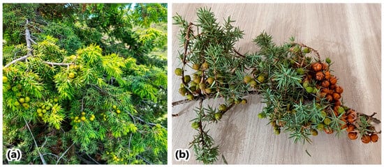
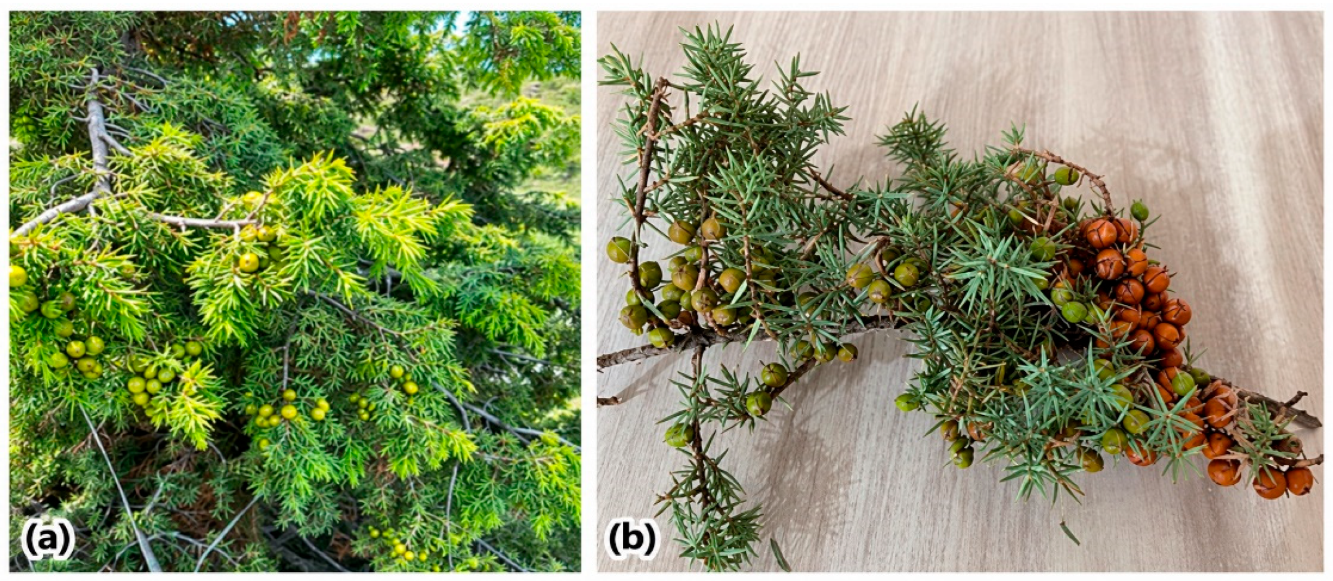
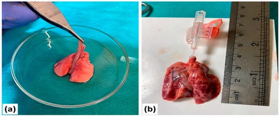
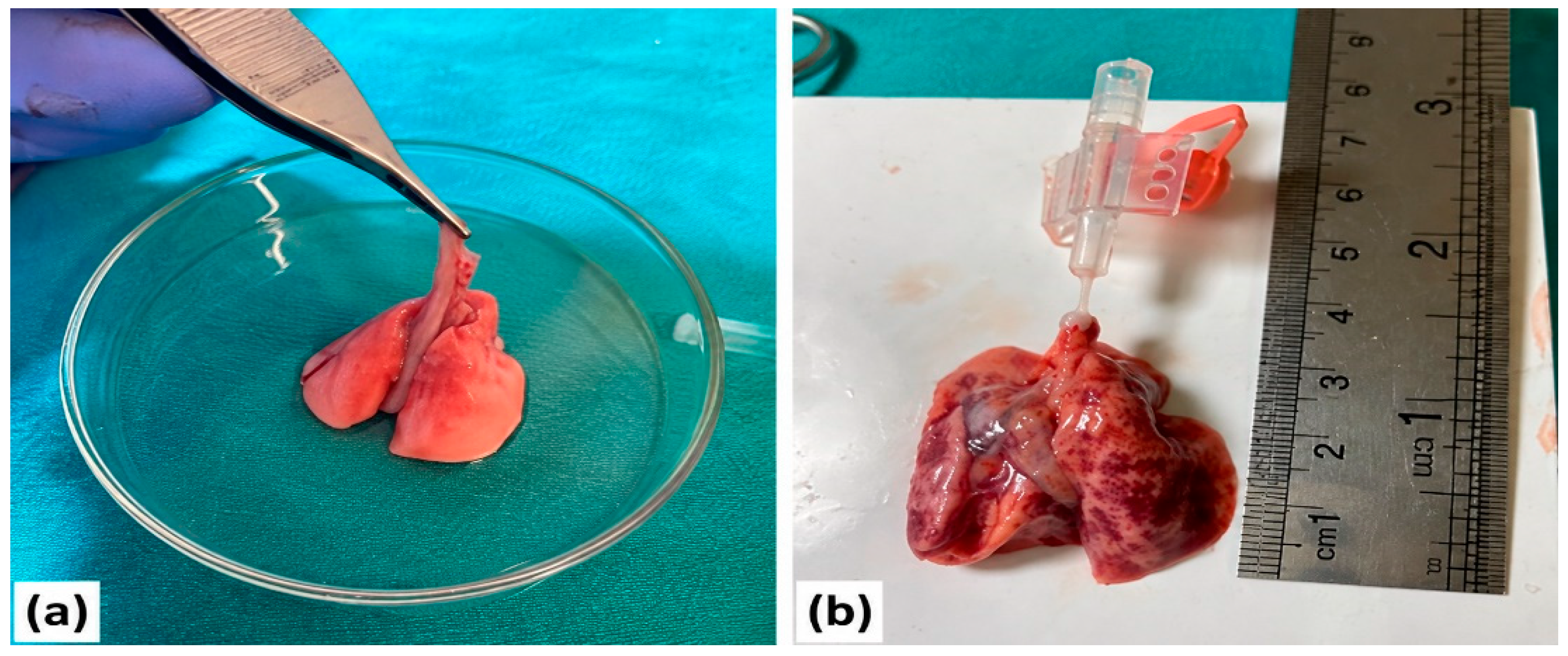
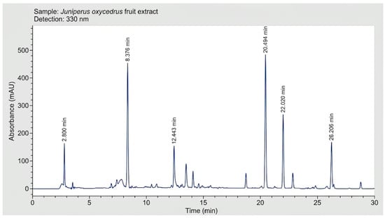
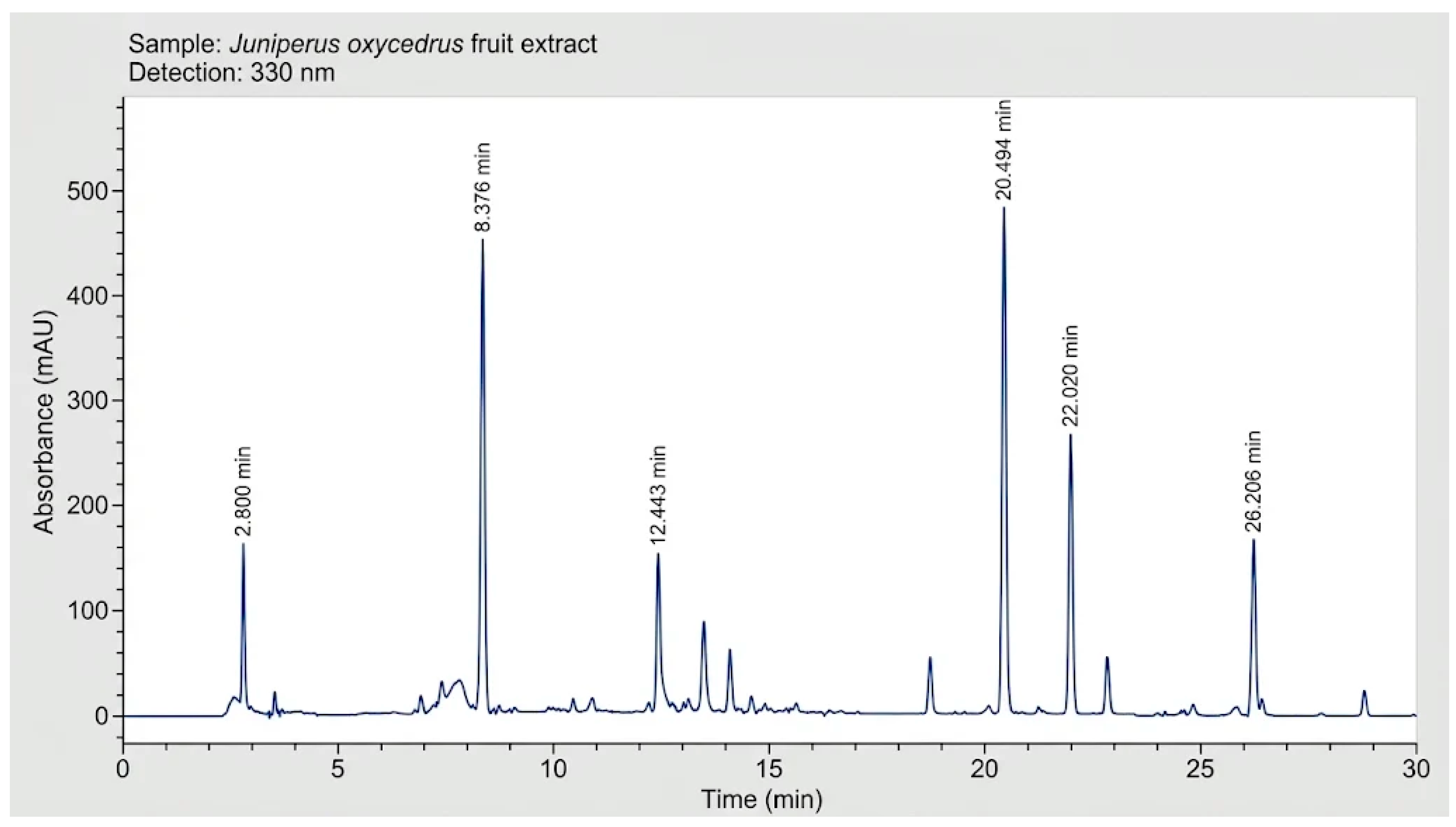
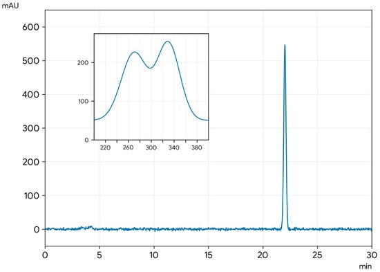
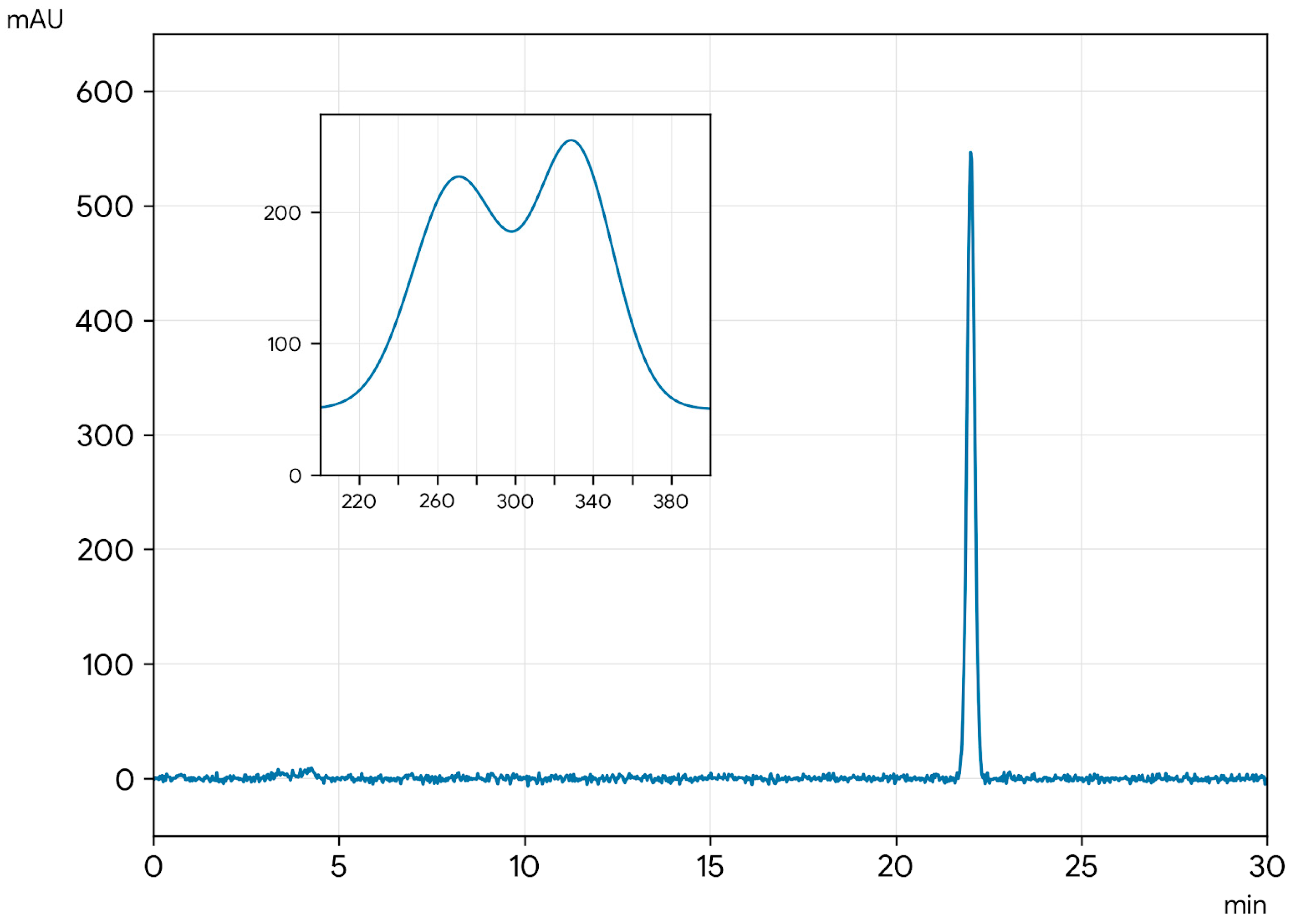
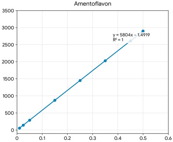
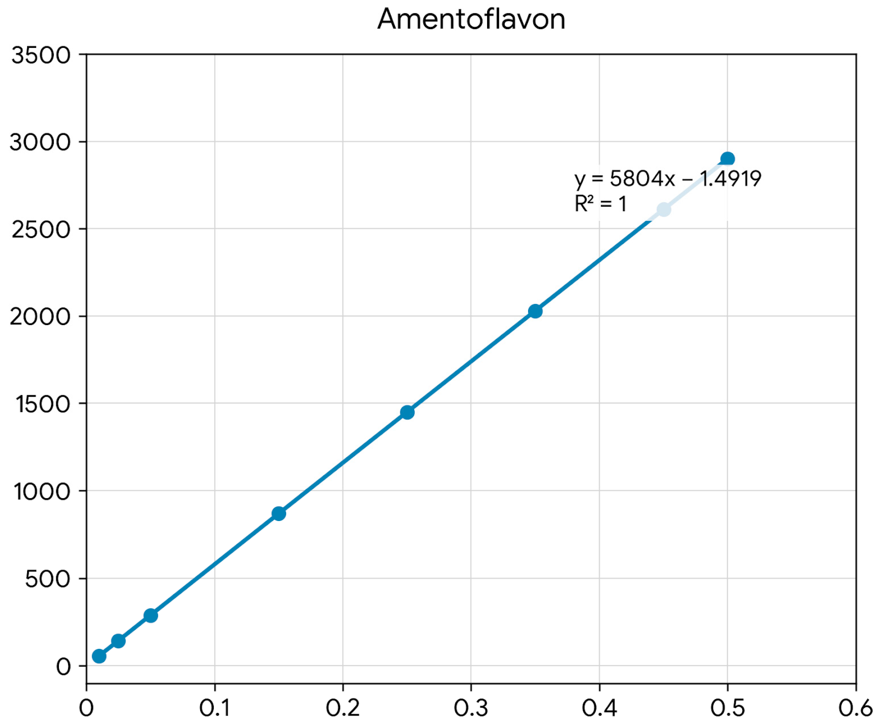
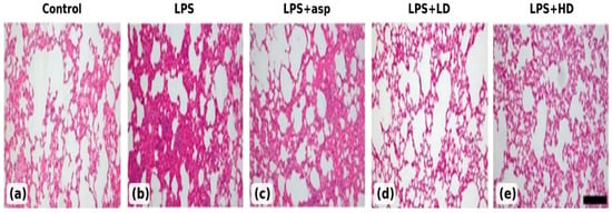
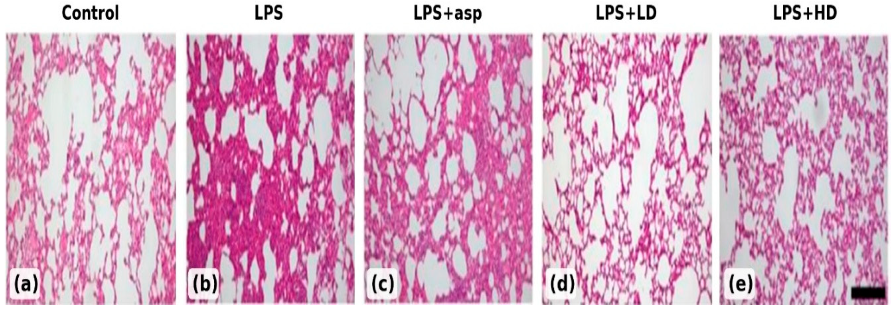

Disclaimer/Publisher’s Note: The statements, opinions and data contained in all publications are solely those of the individual author(s) and contributor(s) and not of MDPI and/or the editor(s). MDPI and/or the editor(s) disclaim responsibility for any injury to people or property resulting from any ideas, methods, instructions or products referred to in the content. |
© 2026 by the authors. Licensee MDPI, Basel, Switzerland. This article is an open access article distributed under the terms and conditions of the Creative Commons Attribution (CC BY) license.