


Abstract
Friedreich’s ataxia (FA) is a progressive neurodegenerative disorder frequently associated with hypertrophic cardiomyopathy and heart failure, a major cause of mortality. FA results from frataxin deficiency, leading to mitochondrial dysfunction, impaired iron–sulfur cluster biogenesis, and oxidative stress. We examined the effect of two clinically relevant Nrf2 activators, dimethyl fumarate (DMF) and omaveloxolone (OMAV), on their ability to restore cardiac function in an inducible and reversible frataxin-knockdown (FXNKD) mouse model of FA. FXNKD mice developed a heart failure with preserved ejection fraction–like phenotype, characterized by left ventricular wall thickening, impaired diastolic relaxation, and reduced stroke volume, with more severe remodeling observed in males. Both treatments activated canonical Nrf2 signaling, confirming target engagement. DMF partially improved cardiac function in males, increasing stroke volume, enhancing metabolic flexibility, and restoring mitochondrial enzyme activity; however, diastolic dysfunction persisted. OMAV treatment was associated with female-specific electrophysiological abnormalities, including increased arrhythmic burden and reduced J-wave amplitude, with persistent fibrotic remodeling. Both treatments attenuated region-specific cardiomyocyte atrophy in FXNKD males and females. These findings suggest that Nrf2 activation alone is insufficient to reverse FA-associated cardiomyopathy and reveal sex-dependent therapeutic responses. Further investigation of complementary treatment strategies may help address persistent cardiac dysfunction and remodeling in FA.
1. Introduction
Friedreich’s ataxia (FA) is a progressive, inherited neuromuscular disorder that affects approximately 1 in 40,000 individuals worldwide and significantly reduces life expectancy [1,2,3]. FA is caused by biallelic pathogenic variants in the FXN gene, with approximately 98% of affected individuals carrying an expanded guanine–adenine–adenine (GAA) trinucleotide repeat in intron 1 of at least one allele, typically in combination with an expansion on the second allele [4,5]. These expansions suppress FXN transcription, resulting in deficient expression of frataxin (FXN), a nuclear-encoded mitochondrial protein essential for iron–sulfur (Fe–S) cluster biogenesis [6]. FXN deficiency disrupts multiple cellular processes, including the function of Fe–S-containing mitochondrial respiratory chain complexes I, II, and III, as well as mitochondrial aconitase. These abnormalities compromise mitochondrial respiration and ATP production, disturb cellular iron homeostasis, and promote oxidative stress [7,8]. Although these molecular abnormalities are well established, the mechanisms linking FXN deficiency to the development and progression of tissue-specific disease manifestations remain incompletely understood.
Cardiac involvement is a major determinant of mortality in FA, accounting for approximately 60–80% of reported deaths, frequently through progressive cardiomyopathy, heart failure (HF), and arrhythmias [1]. Although neurological manifestations often dominate the clinical presentation, cardiac disease can progress independently and may lead to premature death in affected individuals in their 30s or even earlier [1,2,3]. The cardiac phenotype typically involves concentric left ventricular hypertrophy, initially preserved systolic function, and abnormalities in myocardial structure and energetics. In some patients, the disease progresses to myocardial fibrosis, ventricular dilation, and systolic dysfunction. The severity of cardiomyopathy directly correlates with the length of the expanded GAA repeat, which generally correlates inversely with residual FXN expression and age at disease onset [1]. A retrospective study of 133 patients with FA identified clinical heterogeneity associated with the severity of FXN mutations [9]. Approximately 78% of patients with shorter GAA repeat expansions exhibited a comparatively milder clinical course and predominantly hypertrophic cardiac remodeling with preserved ejection fraction, resembling some characteristics of HF with preserved ejection fraction (HFpEF). In contrast, 22% of patients with longer GAA expansions developed severe HF with reduced ejection fraction (HFrEF) [9]. These observations underscore the heterogeneity of FA cardiomyopathy and the need to investigate mechanisms and therapeutic strategies across different stages and phenotypes of cardiac disease. Consistent with a role for redox dysregulation in disease progression, studies of human FA and experimental models have demonstrated suppression of the nuclear factor erythroid 2–related factor 2 (Nrf2) antioxidant response pathway, potentially contributing to impaired antioxidant defenses and cardiac dysfunction [10].
Dimethyl fumarate (DMF) and omaveloxolone (OMAV) are pharmacological activators of the Nrf2 pathway that act through electrophilic modification of reactive cysteine residues in Kelch-like ECH-associated protein 1 (Keap1), thereby disrupting Keap1-mediated repression of Nrf2 and promoting its nuclear accumulation and transcriptional activity. In February 2023, the U.S. Food and Drug Administration approved OMAV, marketed as Skyclarys, as the first treatment for FA in adults and adolescents aged 16 years and older [11]. Clinical trials demonstrated improvements in neurological outcomes measured using the modified Friedreich’s Ataxia Rating Scale (mFARS) and the Scale for the Assessment and Rating of Ataxia (SARA) [12,13]. However, the effects of OMAV on cardiac function and the progression of FA-associated cardiomyopathy have not been established by these clinical trials. Our previous study demonstrated that OMAV was more effective than DMF in improving cardiac contractile function and calcium signaling in a muscle creatine kinase (MCK)-Cre-mediated Fxn conditional knockout (FXN-cKO) mouse model of FA characterized by severe cardiomyopathy and HFrEF [14]. Because this model represents advanced cardiac disease resulting from substantial FXN depletion, it remains unclear whether Nrf2-directed therapies can also modify earlier cardiac abnormalities characterized predominantly by hypertrophic remodeling and preserved systolic function.
In addition to Nrf2 activation, DMF may exert metabolic effects relevant to mitochondrial dysfunction in FA. As a fumaric acid ester, DMF may influence mitochondrial metabolism through fumarate-related pathways and anaplerotic replenishment of tricarboxylic acid (TCA) cycle intermediates, although the contribution of these mechanisms to its cardioprotective effects remains to be established [15]. Our laboratory previously demonstrated that DMF dose-dependently enhanced mitochondrial function and increased FXN expression in FA patient-derived fibroblasts and in skeletal muscle and brain tissue from a frataxin knockdown (FXNKD) mouse model [16,17,18]. Nevertheless, the cardiac effects of DMF in this model remain insufficiently characterized. Given its established clinical use for multiple sclerosis and psoriasis [19,20], DMF represents a potential repurposing candidate for FA-associated cardiomyopathy, provided that its cardiac efficacy, mechanisms of action, and safety are demonstrated in appropriate preclinical studies [21].
The objective of the present study was to investigate the cardiac effects of DMF and OMAV in male and female FXNKD mice, which develop cardiac remodeling characterized predominantly by a restrictive or hypertrophic phenotype with similarities to heart failure with preserved ejection fraction (HFpEF). In this model, doxycycline-inducible RNA interference reduces endogenous Fxn expression in adult mice, allowing the onset and progression of frataxin deficiency to be controlled experimentally [22,23,24]. Systemic FXN knockdown produces multiple abnormalities relevant to human FA, including mitochondrial dysfunction, reduced aconitase activity, cardiac conduction disturbances, increased left ventricular wall thickness, myocardial iron accumulation, and structural abnormalities of cardiac mitochondria [22,25]. The model also exhibits neurological and systemic manifestations, including impaired motor coordination, reduced strength, abnormalities in sensory neurons, and reduced survival, thereby reproducing selected features of the multisystem nature of human FA [22,24]. Importantly, restoration of FXN expression after disease manifestations have developed can improve several functional, pathological, and molecular abnormalities, supporting the utility of this model for investigating disease progression and therapeutic responsiveness [22].
We recently demonstrated that, as in human FA [26,27], sexual dimorphism is evident in experimental models of the disease and that responses to Nrf2-directed therapies may differ between males and females [24,28,29]. These findings highlight the importance of investigating sex as a biological variable when evaluating the efficacy of candidate therapies. Using the inducible FXNKD model, we therefore sought to determine whether pharmacological Nrf2 activation modifies early cardiac dysfunction and whether the responses to DMF and OMAV differ between sexes. This approach may help establish the therapeutic potential of Nrf2-directed interventions for FA-associated cardiomyopathy before the development of advanced systolic dysfunction.
2. Materials and Methods
2.1. In Vivo Frataxin Knockdown Mouse Model of FA: Study Approval and Design
All animal handling and laboratory procedures were in accordance with the approved protocols of the Institutional Animal Care and Use Committee of the University of California, Davis, conforming to the Guide for the Care and Use of Laboratory Animals published by the US National Institutes of Health (8th Edition, 2011). Mice were maintained in a temperature- and humidity-controlled animal facility, with a 12-h light-dark cycle.
Inducible and reversible frataxin (FXN) knockdown (FXNKD) mice were used in this study. This mouse model of FA is based on a transgenic (TG) mouse containing a Tet-On shRNA (against Fxn mRNA) expression cassette [22,24]. To induce the FA phenotype, TG mice were treated with doxycycline chow (Doxy, 200 ppm, Teklad, TD.00502 by Inotiv, West Lafayette, IN, USA) starting at 12–14 weeks of age [24]. Age-matched TG mice littermates that do not contain the shRNA cassette (Wild-type, WT) and TG mice that were not treated with Doxy (TG-) were used as controls. Animals of either sex were randomly distributed into six experimental cohorts: WT without Doxy treatment (WT-), WT treated with Doxy (WT+), and TG without Doxy induction (TG-), TG treated with Doxy and vehicle (FXNKD), FXNKD treated with DMF (82 mg/kg), and FXNKD treated with OMAV (24 mg/kg). Drug treatment started at 4 weeks post-Doxy treatment (at 16–18 weeks of age) and continued for 8 weeks in the presence of Doxy. The drugs were formulated into 200 µL peanut butter (used as a vehicle), which was given daily to individually housed mice every afternoon one hour before the dark cycle. Daily dose consumption was monitored by an animal technician.
2.2. Echocardiography
Cardiac function was examined using 2D echocardiography with a VisualSonics Vevo 2100 echocardiography machine (VisualSonics, Toronto, ON, Canada) and an MS 550D probe (22–55 MHz) under anesthesia with 1% to 1.5% isoflurane in a 100% O2 atmosphere gas chamber [30,31]. Some recordings were performed in conscious animals to exclude the effects of anesthesia on cardiac function. We used the parasternal short- and long-axis views to obtain 2-dimensional and M-mode images [31]. Left ventricular dimensions (EDD, end-diastolic dimension; ESD, end-systolic dimension; PWT, posterior wall thickness) were obtained from 2D guided M-mode images. Indices of contractility such as left ventricular (LV) fractional shortening (FS), ejection fraction (EF), stroke volume, and cardiac output were calculated from the M-mode images. LV mass was calculated using M-mode data as previously described [30,31]. To assess changes in diastolic function, we performed pulsed-wave Doppler imaging in unconscious mice.
2.3. Electrocardiogram
Mice were placed in a supine position on a 37 °C heat pad, and a Lead I surface electrocardiogram (ECG) was recorded in control, FXNKD, and drug-treated FXNKD mice under anesthesia with a ketamine/xylazine mix (0.01 mL/g IP). A blinded observer processed five- and ten-minute recordings using Labchart 8 (ADInstruments, Colorado Springs, CO, USA) for unbiased annotations. Heart rate variability (HRV) was calculated by a single beat through R-to-R labeling using Labchart 8 algorithms (version 8.1.21 software). Averaged waveforms: P, PR, QRS, corrected QT (QTc), and RR intervals were calculated as averaged mean values obtained from a 5-min ECG recording in control, FXNKD, and drug-treated FXNKD mice of either sex. Aggregated frequency distributions: RR intervals were acquired and analyzed by individual beats. A bin size of 1 or 2 was used to reflect the total beats at a specific time interval. Heart rate variability (HRV) is the measured differences in time intervals between adjacent heartbeats. HRV parameters were used to assess ANS activity in FA mice with and without drug treatment. Time parameters examined were the standard deviation of all normal RR intervals (SDRR), the square root of the mean squared difference between successive in RR intervals (RMSSD), and the percentage of RR beats longer than 6 ms (pRR6). The HRV nonlinear domain was utilized to determine the distribution of identities on a scatter plot, Poincaré plot [29].
2.4. Histology
At the end of the study, mice were euthanized, and tissues were collected. Hearts were excised, blotted dry, and weighed, and tibia length was measured for normalization of heart weight. Following dissection, a short-axis cross-section of each heart was obtained and fixed in 10% neutral-buffered formalin before transfer to 70% ethanol. Tissue samples were embedded in paraffin and sectioned at 4 μm by the UC Davis VMTH Histology Laboratory. Sections were stained with Picrosirius Red and hematoxylin and eosin (H&E) to assess cardiac fibrosis and morphology.
Total cardiac fibrosis was quantified as the percentage of fibrotic area from montage images acquired at 10× magnification for each sample, excluding perivascular fibrosis, using the Cytation5 Imager (Agilent, Santa Clara, CA, USA). Cardiomyocyte cross-sectional area was measured from images obtained with the Cytation5 Imager and automatically segmented using Cellpose™ v2.2.2 implemented in Fiji (ImageJ2). Measurements were calculated by averaging two 20× images from each of the following regions: the interventricular septum (IVS), papillary muscle (PM), right ventricular wall (RVW), and left ventricular wall (LVW). Segmentation outputs generated by Cellpose™ were manually inspected to ensure accuracy.
2.5. Quantitative Real-Time PCR
RNA was extracted from flash-frozen heart samples using RNeasy Fibrous Tissue Kit (Qiagen, Hilden, Germany) according to the manufacturer’s instructions. cDNA was created using the iScript cDNA Synthesis kit (BioRad Laboratories, Hercules, CA, USA). Quantitative RT-PCR was carried out using PowerUp Syber Green (ThermoFisher Scientific, Walthan, MA, USA) on a QuantStudio 7 Pro Real-Time PCR System (ThermoFisher, Waltham, MA, USA). The data were analyzed with a delta-delta CT calculation using ribosomal protein 18S (Rps18) as a reference gene.
2.6. Western Blotting and Biochemical Assays
Tissue samples were lysed using CellLytic MT Cell Lysis Reagent (Sigma Aldrich, St. Louis, MO, USA) containing PhosSTOP phosphatase inhibitor and Complete EDTA-free mini protease inhibitor cocktail (Roche Diagnostics, Indianapolis, IN, USA). Protein concentration was determined by Pierce BCA Assay (ThermoFisher Scientific, Waltham, MA, USA) and normalized to 2 mg/mL. Protein separation, detection, and analysis were done using the Jess Chemiluminescent & Fluorescent Capillary Western Blot system by Protein Simple (Biotechne, Minneapolis, MN, USA). Antibodies used were frataxin (ab219414, Abcam, Cambridge, UK) and GAPDH (ab8245, Abcam, Cambridge, UK). Enzyme activities were determined in heart homogenates using Abcam kits for Complex II (ab109908) and aconitase (ab109712) activities.
2.7. Statistical Analysis
Data were analyzed using one-way ANOVA with Tukey’s post-hoc for multiple comparisons or unpaired Welch’s two-tailed t-test for comparison of two means, as appropriate, using Prism version 10 (Graph-Pad Software Inc., San Diego, CA, USA). A p-value < 0.05 was considered statistically significant. Power analyses were performed post hoc and tailored to each experiment, with sample sizes (n) indicated in figure panels. Outliers were determined using Grubbs’ outlier test (alpha = 0.05). All data are shown as mean ± SEM.
3. Results
3.1. Nrf2 Target Engagement and Fxn Expression in FXNKD Hearts
To assess if Nrf2 activation is beneficial for treatment of the restrictive cardiomyopathy in FA, we examined the effect of two Nrf2 activators, DMF and OMAV, on cardiac function in the inducible and reversible FXNKD mouse model of FA [22,24]. Figure 1A shows the experimental design of our study. To generate FXNKD mice, TG mice containing a Tet-On shRNA (against Fxn mRNA) expression cassette were treated with doxycycline (Doxy) chow starting at 12–14 weeks of age (TG+). We have previously reported that FXNKD (TG+) mice started exhibiting signs of ataxia 4 weeks post Doxy treatment [24]. Therefore, both drug treatments were started at this point (i.e., at 16–18 weeks of age) and continued for 8 weeks in the presence of Doxy. Drug doses of DMF (82 mg/kg) and OMAV (24 mg/kg) were converted from their human therapeutic doses (150 mg OMAV and 480 mg DMF daily) to equivalent mouse dosages using body surface area conversions [32]. Age-matched mouse littermates that do not contain the shRNA cassette on control chow (Wild-type, WT-), WT animals on Doxy chow (WT+), and TG mice that were not exposed to Doxy (TG-) were used as controls. The total experiment continued for 12 weeks, at which point (i.e., at 24–26 weeks of age) cardiac echo and ECG were performed, and tissues were collected for analysis.
Figure 1.
Experimental design and Nrf-2 targeting in FXNKD hearts. (A) Experimental study design. Mice were administered doxycycline (Doxy) to induce FXN knockdown and subsequently treated with DMF, OMAV, or vehicle prior to cardiac assessment and tissue collection. Created in BioRender. Dedkova, E. (2026) https://BioRender.com/7eayc60. (B) Schematic illustration of Nrf2 activation and antioxidant response element (ARE) signaling. DMF and OMAV disrupt KEAP1-Nrf2 interaction, allowing Nrf2 nuclear translocation and transcription of antioxidant genes, thereby reducing mitochondrial reactive oxygen species. Created in BioRender. Dedkova, E. (2026) https://BioRender.com/gzynf28. (C) Fxn gene expression in the combined sex FXNKD heart cohort or (D) separated by sex. (E) Nqo1 gene expression in the combined sex FXNKD hearts cohort or (F) separated by sex. (G–I) FXNKD heart protein expression quantified by capillary Western blot and normalized to GAPDH. **** p < 0.0001, *** p < 0.0005, ** p < 0.01, * p < 0.05, ns: not significant.
First, we determined the degree of FXN suppression in FXNKD hearts and the drug’s ability to engage Nrf2-mediated targets in this mouse model of FA. Potentially, both DMF (via MMF formation) and OMAV could be cardioprotective by modifying reactive cysteine residues on Keap1, releasing it from Keap1 in the cytosol with subsequent Nrf2 translocation to the nucleus, where it binds to the antioxidant response element (ARE) in the promoter regions of target antioxidant genes such as Nqo1, Hmox1, Gstp, and Sod2 (Figure 1B). We show that cardiac Fxn mRNA was reduced by 97.6% in the combined-sex FXNKD cohort relative to WT+ (p < 0.0001; n = 24; Figure 1C). When separated by sex, males exhibited a 97.5% reduction (n = 15) and females a 97.8% reduction (n = 9) (Figure 1D). DMF produced a 1.7-fold increase in Fxn gene expression in the combined-sex cohort, but this was not significant due to variability in response. Although non-significant, DMF increased Fxn gene expression in males by 1.5-fold and in females by 2.0-fold. OMAV did not alter Fxn transcript levels.
Among Nrf2 target genes, the Nqo1 gene, which codes for NAD(P)H quinone oxidoreductase 1 (NQO1), was reduced 42% in FXNKD vs. TG- (p < 0.01), then increased 2-fold with DMF (p < 0.05) and 2.25-fold with OMAV (p < 0.001) in combined sex groups (Figure 1E). Sex-stratified analysis revealed a more robust response to Nrf-2 activation in males: Nqo1 decreased 40% (ns) in FXNKD males and increased 2-fold with DMF (p < 0.05) and 2.75-fold with OMAV (p < 0.0001). In females, FXNKD reduced Nqo1 expression by 49% (ns), with substantial but nonsignificant increases in Nqo1 expression following DMF (2.15-fold, p = 0.07) or OMAV (1.58-fold, ns) (Figure 1F). In contrast, the Hmox1 gene, which codes for heme oxidase 1 (HO-1), was strongly upregulated in FXNKD hearts vs. WT-, WT+, and TG- controls (+380–450%, p < 0.005), with additional non-significant increases observed in DMF (+29%, ns) and OMAV (+43%, ns) treated groups (Supplemental Figure S1A).
Both sexes showed significant Hmox1 elevations (males +489%; females +329%), and treatment induced only incremental, nonsignificant increases in Hmox1 expression (Supplemental Figure S1B). No significant changes in Gstp and Sod2 gene expression were detected in FXNKD hearts (see Supplemental Figure S1C–F).
We also verified that FXN protein expression was significantly reduced in FXNKD mice (−60%, p < 0.0001; n = 11) with no improvements noted with DMF or OMAV treatments (Figure 1G,H). FXN protein expression was decreased slightly more in FXNKD males (−65%, p < 0.0001; n = 7) as compared to females (−52%, p < 0.0001; n = 4), and neither DMF nor OMAV had significant effects on FXN protein expression (Figure 1I).
Collectively, these findings confirm successful FXN knockdown in both sexes and demonstrate that only NAD(P)H quinone oxidoreductase 1 (Nqo1) gene expression out of four tested genes in the Nrf2-ARE signaling pathway was significantly decreased in FXNKD hearts and restored almost completely by both DMF and OMAV. Notably, there was a ~4–5-fold increase in Hmox1 gene expression (encodes heme oxidase 1, HO-1) in FXNKD hearts of either sex, which was not affected by DMF or OMAV. FA is a pleiotropic disorder with individualized variability. Here, Nrf2 target engagement was heterogeneous, with a trend toward greater transcriptional response in FXNKD males as compared to females.
3.2. Hypertrophic Cardiomyopathy in FXNKD Males Was Partially Reversed by DMF While OMAV Had Only Limited Benefits
Cardiac function and structure in FXNKD (TG+) mice were evaluated after 12 weeks of Doxy induction using 2-dimensional M-mode echocardiography. Figure 2A shows representative M-mode images recorded at the parasternal short axis dimensions in conscious WT males untreated (WT-) or treated with doxy (WT+), TG males without Doxy induction (TG-), TG animals treated with doxy for 12 weeks (TG+ to induce FXNKD), and FXNKD males treated with either 82 mg/kg DMF (FXNKD+DMF) or 24 mg/kg OMAV (FXNKD+OMAV).
Figure 2.
Cardiac structure and function in FXNKD males. (A) shown are representative echocardiography images recorded in M-mode from WT-, WT+, TG-, and FXNKD mice treated with vehicle (VEH), 82 mg/kg DMF, or 24 mg/kg OMAV. (B–J) shown are summary data for LV mass (B), LVPWd (C), LVAWd (D), LVIDd (E), stroke volume (F), cardiac output (G), heart rate (H), fractional shortening (I), and ejection fraction (J). Data are expressed as mean ± SEM from 8–12 animals. The symbols and numbers in each panel represent the number of individual mice (defined as biological replicates) involved in this study. One-way ANOVA with Tukey’s multiple comparisons test was used to determine significance, with **** p < 0.0001, *** p < 0.0005, ** p < 0.01, * p < 0.05, ns: not significant.
In agreement with findings in FA restricted cardiomyopathy, we did not see any increase in LV mass in FXNKD mice treated with vehicle as compared to controls (Figure 2B); however, a significant increase in LV posterior (LVPW, 163% of WT+, n = 12, p < 0.0001) and anterior (LVAW, 128% of WT+, n = 12, p < 0.01) wall thickness at diastole was noted (Figure 2C,D). Both drugs had no effects on LV mass (Figure 2B); however, both DMF (77% of FXNKD, 23% decrease, n = 8, p < 0.01) and OMAV (78% of FXNKD, 22% decrease, n = 10, p < 0.01) significantly decreased LVPW thickness (Figure 2C) in FXNKD males. Only a non-significant trend to decrease LVAW thickness was noted in DMF (−9%) and OMAV (−15%)-treated males (Figure 2D). This significant increase in wall thickness was associated with a decrease of the LV internal diameter in diastole (LVIDd, −24%, p < 0.0001, n = 12, Figure 2E) leading to the decline in stroke volume (SV, the amount of blood pumped per beat, Figure 2F, −40%, p < 0.0001) and cardiac output (CO, the amount of blood pumped by the heart per minute, −49%, p < 0.0001; Figure 2G). Oral treatment with 82 mg/kg DMF for 8 weeks partially reversed the anatomic and functional abnormalities in FXNKD mice, namely, LV posterior wall thickness (−23%, Figure 2C), a decrease in LVID (+20%, Figure 2E), and stroke volume (+45%, n = 8, Figure 2F) without any improvement in cardiac output (Figure 2G), most probably due to bradycardia observed in FXNKD mice (Figure 2H) [33]. OMAV decreased only posterior LV wall thickness (Figure 2C); however, no improvement of functional parameters was noted with OMAV treatment. No decrease in fractional shortening (FS, which reflects the degree of shortening of the LV diameter between end-diastole and end-systole) or ejection fraction (EF, calculated volumetric fraction of the total blood ejected from the LV with each contraction) was detected in FXNKD mice compared to WT (Figure 2I,J). Overall, these data indicate that DMF was more effective than OMAV in rescuing cardiac parameters in FXNKD males.
3.3. Cardiac Structural Parameters Showed More Deficits in FXNKD Males Compared to Females
While evaluating cardiac function in FXNKD mice, we noticed that FXNKD males (Figure 2A–E) exhibited significantly more pronounced deficits in cardiac structure compared to FXNKD females (Figure 3A–E). For example, LVPW thickness at diastole was increased by 63% (p < 0.0001, n = 12) in FXNKD males (Figure 2C) vs. 34% (p < 0.05, n = 11) in females (Figure 3C), and no changes were noted in LVAW thickness at diastole (Figure 3D). LVIDd was decreased by 24% (p < 0.0001, n = 12) in FXNKD males (Figure 2E), and only by 17% (p < 0.05, n = 11) in females, with no effects of the drugs on this parameter (Figure 3E).
Figure 3.
Cardiac structure and function in FXNKD females. (A), shown are representative echocardiography images recorded in M-mode from WT+, TG-, and FXNKD mice treated with vehicle (VEH), 82 mg/kg DMF, or 24 mg/kg OMAV. (B–J), shown are summary data for LV mass (B), LVPWd (C), LVAWd (D), LVIDd (E), stroke volume (F), cardiac output (G), heart rate (H), fractional shortening (I), and ejection fraction (J). Data are expressed as mean ± SEM from 8–13 female animals. The symbols and numbers in each panel represent the number of individual mice (defined as biological replicates) involved in this study. One-way ANOVA with Tukey’s multiple comparisons test was used to determine significance, with **** p < 0.0001, *** p < 0.0005, ** p < 0.01, * p < 0.05, ns: not significant.
However, when we evaluated cardiac functional parameters, we found that they were decreased to a similar degree in both FXNKD males and females. For example, stroke volume showed 40% decrease in males (p < 0.0001, n = 12) vs. 31% (p < 0.05, n = 11) in females (Figure 2F and Figure 3F), cardiac output was decreased by 49% (p < 0.0001, n = 12) in FXNKD males vs. 43% (p < 0.0001, n = 11) in females (Figure 2G and Figure 3G), and heart rate was decreased by 17% in males vs. 27% (p < 0.01) in FXNKD females (Figure 2H and Figure 3H). Cardiac contractile function was not changed in either FXNKD males or females as compared to WT+ and TG- mice (Figure 2I,J and Figure 3I,J). Both drugs had no significant effects on cardiac structural and functional parameters in FXNKD females, except that they increased FS and EF above those seen in control animals. For example, EF was increased 13% (p < 0.05) by DMF and 15% (p < 0.01) by OMAV, which could contribute to the increased incidence of arrhythmia-like events (prolonged heartbeats) in FXNKD females treated with OMAV. Combined sex data are reported in the Supplemental Figure S2.
3.4. Diastolic Cardiac Function Was Impaired in FXNKD Mice and Not Affected by Nrf2 Inducers
Restrictive cardiomyopathies, such as those seen in FA, are characterized by impaired filling during diastole. We used Pulse-Wave Doppler at the mitral valve (MV) to assess left ventricular filling (Figure 4A), an indication of diastolic function of the heart. Two diastolic waves, E and A, are seen in normal mitral Doppler tracing. The E wave occurs during the rapid filling phase in early diastole, and the A wave occurs in late diastole as a result of atrial contraction. As shown in Figure 4B–G, both MV early filling peak velocity (MV E) and atrial peak velocity (A) were decreased in FXNKD mice, both males and females, indicating reduced blood flow via the mitral valve. Reduction in both MV E and MV A velocities suggests a global reduction in LV filling. Neither DMF nor OMAV had any effects on MV E and A velocities. There was a trend indicating a decrease in MV E/A ratio in FXNKD males and a partial recovery with DMF treatment (Supplementary Figure S3); however, it was not significant. The MV deceleration time (DT) (Figure 4H–J), which reflects how quickly the left atrium (LA)-LV pressure gradient equalizes, was increased significantly only in FXNKD males. FXNKD females only showed a trend in MV deceleration time increase; however, deceleration time was elevated significantly in OMAV-treated FXNKD females. Prolonged deceleration time of early mitral inflow (DT) in FXNKD hearts reflects slow left ventricular relaxation and stiff ventricular walls. This pattern points to impaired early filling, often seen in diastolic dysfunction with normal or low filling pressures. We determined that FXNKD mice displayed an increased isovolumetric relaxation time (IVRT) (Figure 4K–M), indicating a decrease in the rate of active myocardial relaxation and slow LV pressure decline after systole. No significant changes were found in isovolumetric contraction time (IVCT) and MV Ejection Time (ET), which reflects the duration of blood flow during ventricular contraction (Supplementary Figure S3). Taken together, FXNKD mice developed a restrictive cardiomyopathy with diastolic dysfunction consistent with restrictive cardiomyopathy as seen in FA patients. The diastolic dysfunction was not recovered by either DMF or OMAV.
Figure 4.
Diastolic cardiac function in FXNKD mice. (A) Representative images of cardiac Doppler imaging recorded in FXNKD mice were presented, with diastolic dysfunction as evidenced by the increase in MV E/A ratio and by the increase in MV deceleration time. (B–D) Mitral valve E (MV E) wave velocity was significantly decreased in both males and females and was not affected by either DMF or OMAV. (E–G) Mitral valve A (MV A) wave velocity was significantly decreased in both males and females and was not affected by either DMF or OMAV. (H–J) Mitral valve deceleration time was significantly increased in FXNKD males only and was not affected by either DMF or OMAV. In FXNKD females, treatment with OMAV further increased MV deceleration time. (K–M) Mitral valve IVRT time was significantly increased in FXNKD females only and was not affected by either DMF or OMAV. **** p < 0.0001, *** p < 0.0005, ** p < 0.01, * p < 0.05, ns: not significant.
3.5. Nrf2 Activation Induced Sex-Specific Electrophysiological Remodeling in FXN-Deficient Hearts
Next, we investigated changes in electrical activity of the heart using surface ECG in FXNKD males and females and found sex-specific differences (Figure 5). A schematic representation of mouse ECG waveforms is shown in Figure 5A. We performed analysis of the averaged ECG waveform segments and found that there were no changes in P wave duration (representing atrial depolarization) in FXNKD animals (Supplemental Figure S4A). PR segment duration, which reflects the time it takes for the heart’s electrical impulse to travel from the atrioventricular (AV) node through the His-Purkinje system to the ventricles, was unchanged in FXNKD males as compared with WT+ (+0.1%, ns, n = 4) (Figure 5B). However, PR interval was significantly increased in FXNKD females as compared with WT+ (+16%, p < 0.05, n = 5). In contrast, QTc interval, which reflects duration of ventricular repolarization adjusted for heart rate, was significantly increased only in FXNKD males as compared with WT+ (+22.7%, p < 0.01, n = 4), with no changes observed in FXNKD females (Figure 5C). Drugs had no effects on these two parameters (Supplemental Figure S4B,C). QRS segment duration, which reflects ventricular depolarization, also demonstrated opposite changes between sexes (Figure 5D). FXNKD females displayed a shortened QRS interval, reduced by −47% vs. WT+ (p < 0.01, n = 4). FXNKD males exhibited no significant changes in QRS complex duration as compared to WT+ (+29%, ns, n = 4). Neither DMF- nor OMAV altered this parameter in FXNKD animals.
Figure 5.
Conduction abnormalities and altered heart rate variability in FXNKD mice. (A) Schematic illustration of ECG wave components (P, PR, QRS, QT, and J-wave) used for interval measurements. Created in BioRender. Dedkova, E. (2026) https://BioRender.com/cq6vkof. (B–D) Averaged quantification of PR, QTc, and QRS intervals in male and female mice compared to WT+ with or without treatments. (E) Representative ECG tracings showing presence or absence of J-waves across groups; J-waves are absent in FXNKD males but restored by DMF and OMAV, while OMAV abolished J-waves in FXNKD females. (F) Quantification of calcium-handling and ion channel gene expression in male mice: Cacna1c, Ryr2, Atp2a2, Mcu, Kcnq1, and CamkII. (G) Aggregated RR histograms show a bradycardia shift in FXNKD males. (H) Aggregated RR histograms show both bradycardia and tachycardia shifts in FXNKD females. (I,J) Poincaré plots reveal clustered intervals in FXNKD males (I) and females (J), with tighter dispersion in DMF and instability in OMAV groups. ** p < 0.01, * p < 0.05, ns: not significant.
Figure 5E shows representative J-waveforms, which follow the QRS complex on mouse ECG’s [34], exhibiting sex-specific differences in morphology (Figure 5E). The presence of a J-wave was defined as a waveform exceeding 0 mV (above the isoelectric line). In FXNKD males, the J-wave was completely absent and reappeared in 75% of animals treated with DMF (n = 6) and 100% of animals treated with OMAV (n = 8). In contrast, the J-wave in females was preserved in 83% of WT+ (n = 8), 71% of FXNKD (n = 6), and 75% of DMF-treated (n = 4) animals but was completely absent in all OMAV-treated females (n = 5), indicating a female-specific loss of early repolarization under OMAV treatment.
To investigate the potential molecular basis of the observed cardiac electrophysiological changes, we analyzed the expression of genes encoding proteins involved in Ca2+ handling and K+ channel function (Figure 5F). In FXNKD males, transcripts encoding several proteins involved in cardiac excitation–contraction (EC) coupling were elevated relative to WT+ controls, whereas treatment with DMF or OMAV produced no significant changes in most of these genes as compared to vehicle-treated FXNKD hearts. Cacna1c, encoding the α1C subunit of the L-type Ca2+ channel, increased by 107% (p < 0.01, n = 5), while Ryr2, encoding ryanodine receptor 2, increased by 120% (not significant, n = 5). Atp2a2, encoding sarco/endoplasmic reticulum Ca2+-ATPase 2 (SERCA2), increased by 205% (p < 0.05, n = 5), and Mcu, encoding the mitochondrial calcium uniporter, increased by 186% (p < 0.05, n = 5). Camk2d, encoding Ca2+/calmodulin-dependent protein kinase IIδ (CaMKIIδ), increased by 161% (p < 0.01, n = 5); neither DMF nor OMAV significantly altered its expression. Kcnq1, encoding the pore-forming α-subunit of the slowly activating delayed rectifier K+ channel (IKs), increased by 129% in untreated FXNKD males as compared to WT+ hearts, although this difference was not statistically significant (n = 4). DMF-treated FXNKD males showed a further 108% increase relative to the vehicle-treated FXNKD hearts (p < 0.05, n = 3). FXNKD females exhibited only modest, nonsignificant changes in these transcripts (Supplementary Figure S4F).
Collectively, these findings indicate sex-dependent transcriptional remodeling of cardiac Ca2+-handling and repolarization-associated pathways in FXNKD mice. The coordinated elevation of several EC-coupling-related transcripts in males may represent a compensatory response that helps maintain systolic function, consistent with preserved fractional shortening and ejection fraction. However, transcript abundance alone cannot establish enhanced Ca2+ cycling or EC-coupling efficiency. Similarly, increased Kcnq1 expression could reflect an adaptive response to altered electrical activity, but its functional consequences for ventricular repolarization remain uncertain. KCNQ1 alpha subunits assemble into a tetramer and combine with KCNE beta subunits (such as KCNE1) to function properly and generate the IKs current, which contributes to action-potential repolarization and repolarization reserve. Whether the observed transcriptional changes alter channel abundance, current density, or arrhythmia susceptibility requires direct functional investigation. These sex-dependent molecular changes provide a basis for further studies of electrophysiological remodeling and potential sex-specific therapeutic responses in FA-associated cardiomyopathy.
3.6. Heart Rate Variability Analysis Revealed Arrhythmic Susceptibility with OMAV in FXNKD Females
Next, we performed heart rate variability (HRV) analysis of beat-to-beat (RR) intervals. Figure 5G shows the histogram of RR interval distributions in FXNKD males. The median RR interval increased from 315 ms in WT+ mice to 355 ms in FXNKD males, consistent with relative bradycardia. DMF further shifted the distribution toward longer RR intervals, with a median of 416 ms. In contrast, OMAV-treated males exhibited a broad, bimodal distribution containing both tachycardic and bradycardic intervals despite an apparently near-normal median RR interval of 264 ms, indicating greater beat-to-beat variability.
In FXNKD females (Figure 5H), the median RR interval increased from 252 ms in WT+ mice to 280 ms in FXNKD animals and further increased to 316 ms following DMF treatment. Although OMAV-treated females exhibited a near-normal median RR interval (281 ms), the distribution contained a distinct tachycardic peak centered at approximately 150 ms, suggesting intermittent episodes of rapid rhythm and unstable cardiac pacing.
Poincaré analysis (Figure 5I) further corroborated these findings. A Poincaré plot is a nonlinear geometric method for assessing HRV by plotting each RR interval against the subsequent interval, thereby providing a visual representation of beat-to-beat dynamics and autonomic regulation. In males, FXNKD hearts displayed dispersed tachycardic and bradycardic clusters accompanied by reduced long-term variability (SD2 = 9 ms in FXNKD versus 25 ms in WT+ males). Both DMF and OMAV partially restored the clustering pattern toward that of WT+ controls and increased SD2 to approximately 20 ms. In females, FXNKD increased long-term variability (SD2 = 31 ms), whereas both DMF and OMAV reduced SD2 to 7 and 8 ms, respectively (Figure 5J). Despite this apparent normalization, OMAV-treated females exhibited discrete tachycardic and bradycardic clusters rather than the compact elliptical distribution observed in healthy hearts, indicating abnormal oscillatory behavior consistent with rhythm instability.
To further characterize nonlinear HRV dynamics, we performed Second-Order Difference Plot (SODP) and Central Tendency Measure (CTM) analyses (Supplemental Figure S4K–M). SODP visualizes the differences between consecutive RR intervals, providing a sensitive assessment of short-term rhythm fluctuations, whereas CTM quantifies the proportion of data points contained within a predefined radius around the origin, with lower CTM values reflecting greater beat-to-beat variability and rhythm instability. Consistent with the Poincaré analysis, DMF increased clustering around the origin and improved central tendency, indicative of a more stable cardiac rhythm. In contrast, OMAV-treated FXNKD females exhibited greater dispersion of SODP points and reduced central tendency, confirming persistent irregular beat-to-beat oscillations and increased arrhythmic susceptibility.
In summary, DMF consistently improved RR interval dynamics across complementary HRV analyses by narrowing RR interval distributions, restoring more physiological Poincaré oscillations, and increasing rhythm stability, with the greatest benefit observed in males. In contrast, OMAV failed to normalize nonlinear HRV patterns in FXNKD females and was associated with tachycardic interval clustering, abnormal Poincaré and SODP distributions, reduced rhythm stability, and a female-specific loss of the J-wave, collectively indicating an increased susceptibility to cardiac arrhythmias.
3.7. Nrf2 Activation Does Not Recover Pathological Remodeling in FXNKD Mouse Hearts
Picrosirius red staining (Figure 6A–C) demonstrated markedly increased fibrosis (dark red staining) in male FXNKD hearts (+179%, p < 0.05, Figure 6A,B) relative to WT+. Fibrosis remained high in both DMF-treated (+228%) and OMAV-treated (+138%) males, indicating that Nrf2 activation did not attenuate collagen accumulation. In FXNKD females, collagen levels increased by 74% from WT+, but it was not significant, and neither DMF nor OMAV had significant effects on collagen formation in FXNKD females (Figure 6C). Lgals3 gene expression, which encodes the fibrosis marker galectin-3 (Figure 6D), was strongly upregulated in FXNKD male hearts (+374% vs. WT+, p < 0.05) and remained elevated in DMF (+494%, p < 0.01) and OMAV-treated (+677%, p < 0.0001) hearts as compared to WT+. In females, Lgals3 increased by ~200% across FXNKD groups relative to WT+, but none of these changes reached statistical significance. Supplemental Figure S5A–C shows combined sex data for Picrosirius Red and Lgals3 gene expression for all experimental groups, with a similar severe increase in fibrosis in the vehicle-treated FXNKD mice, which was not affected by the drugs.
Figure 6.
Fibrosis and cardiac remodeling in FXNKD hearts. (A–C) Quantification and representative Picrosirius red staining of male (B) and female (C) hearts. Fibrosis remained elevated in FXNKD compared to WT+, with no reduction by DMF or OMAV. (D) Lgals3 gene expression was increased in FXNKD males and remained elevated with treatment. (E) Nppb gene expression was not significantly changed in FXNKD animals. (F) Myh7 gene expression showed consistent remodeling under Nrf2 activation. (G) Gdf15 gene expression was significantly upregulated in OMAV-treated FXNKD male and female hearts. (H) Aldh1a3 gene expression was increased in FXNKD males and females treated with OMAV. (I) Mcp1/Ccl2 gene expression was increased in FXNKD males treated with OMAV. **** p < 0.0001, ** p < 0.01, * p < 0.05, ns: not significant.
Next, we examined gene expression of B-type natriuretic peptide (BNP), a classical heart failure (HF) marker, and several markers of cardiac stress and inflammation which could contribute to HF development. When the heart undergoes stress or severe stretching, it releases BNP and NT-proBNP into the blood, making these proteins primary diagnostic biomarkers for HF. We found that Nppb gene expression, which encodes BNP, was elevated (but not significant) in FXNKD hearts vs. WT+ (Figure 6E). Neither DMF nor OMAV had significant effects in FXNKD animals separated by sex when compared to the vehicle-treated FXNKD groups; however, when these cohorts were combined (Supplemental Figure S5D), we observed that both DMF and OMAV significantly increased Nppb expression as compared to the WT+ Vehicle cohort.
Myh7 encodes the β-myosin heavy chain (β-MHC), one of the major motor proteins responsible for cardiac contraction. Myh7 is a canonical marker of cardiac remodeling, which typically correlates with dilated hypertrophic cardiomyopathy. As shown in Figure 6F, there were no changes in Myh7 expression in FXNKD males (−3.5%, ns). Both DMF and OMAV increased Myh7 expression in FXNKD males (+130% and +100%, respectively) as compared to vehicle-treated FXNKD hearts, but this effect was not significant. In FXNKD females, Myh7 decreased in FXNKD vs. WT+ (−40%, ns), and it was non-significantly increased by DMF vs. FXNKD (+63%, ns) but elevated significantly by OMAV (+118% vs. FXNKD, p < 0.05) (Figure 6F). Similar to Nppb, the effect of both Nrf2 drugs became significant in the combined sex cohorts for Myh7 gene expression (Supplemental Figure S5E). Because the β-MHC isoform generates more mechanical force but uses less ATP than its counterpart (the faster α-MHC), upregulating Myh7 could represent the heart’s attempt to adapt and pump more efficiently when subjected to increased workloads.
Gdf15 expression, growth differentiation factor 15, a stress-response cytokine, was robustly induced in FXNKD male hearts (+93%, ns) relative to WT+, which was markedly amplified by OMAV (+680% vs. WT+, p < 0.01 and +305% vs. FXNKD+Veh, p < 0.01) treatment only (Figure 6G). Female hearts showed a more attenuated response in FXNKD animals (+135% vs. WT+), with OMAV producing a significant +319% increase (vs. WT+, p < 0.05) induction in Gdf15 expression.
Further, we found that there was a significant increase in Aldh1a3 gene expression in DMF- and OMAV-treated FXNKD male hearts (Figure 6H). Aldehyde Dehydrogenase 1 Family Member A3 (encoded by Aldh1a3) is heavily upregulated in cytokine-stimulated vascular smooth muscle cells (VSMCs). Its chronic activation increases cell proliferation and vascular wall thickening (narrowing arteries), which overloads the heart and contributes to mortality from heart failure.
We also found that monocyte chemoattractant protein-1 (MCP-1/CCL2), which is a key inflammatory cytokine that drives cardiovascular disease, was significantly elevated in OMAV-treated FXNKD males (Figure 6I). Elevated MCP-1 levels promote monocyte recruitment to blood vessels, initiating fatty streak and plaque formation. In the heart, chronic MCP-1 expression can lead to severe ischemic heart disease and heart failure. Combined sex data for Aldh1a3, Gdf15, and Mcp1/Ccl2 expression are reported in Supplementary Figure S5F–H.
To conclude, neither DMF nor OMAV prevented cardiac remodeling and fibrosis. Remarkably, both drug-treated groups exhibited a significant increase in Nppb in the combined sex cohort as compared to WT+ groups. This indicates that the FA heart is compensating for the decreased LV and stroke volume with increased contractility, possibly inducing the overstretching of cardiac muscle leading to cell damage and NPB release. Combined sex data for Gdf15, Aldh1a3, and Mcp1/Ccl2 expression are reported in Supplementary Figure S5F–H and indicate that OMAV was also increasing these inflammatory markers in FXNKD mice.
3.8. DMF and OMAV Restored Region- and Sex-Specific Cardiomyocyte Atrophy in FXNKD Hearts
In a different set of experiments, hearts were stained with hematoxylin and eosin (H&E) to assess cardiac morphology (Figure 7). As shown in Figure 7A, the size of the hearts did not change significantly between WT+ and FXNKD animals; however, in agreement with echocardiography data, the thickness of the chamber walls was significantly increased. Before H&E staining, heart weights were taken and normalized to tibial length (HW/TL). No significant changes in HW/TL were detected in FXNKD males and females as compared to WT+ controls (Figure 7B).
Figure 7.
DMF and OMAV restored region- and sex-specific cardiomyocyte atrophy in FXNKD hearts. (A) Representative H&E-stained mid-sections from WT, FXNKD, FXNKD+DMF, and FXNKD+OMAV hearts illustrating overall cardiac morphology and regional landmarks. Scale bar = 2000 µm. (B) Quantification of heart weight normalized to tibia length (HW/TL, mg/mm) in male (left) and female (right) mice across experimental groups. (C) Left—Schematic illustration of mouse heart parasternal short-axis dissection indicating regions used for morphometric analysis: left ventricular wall (LVW), papillary muscle (PM), interventricular septum (IVS), and right ventricular wall (RVW). Created in BioRender. Dedkova, E. (2026) https://BioRender.com/wnon9z0. Right—Representative CellposeTM images of IVS from WT+, FXNKD, FXNKD+DMF, and FXNKD+OMAV males demonstrating myocardial structural differences across experimental groups (scale bar = 100 μM). (D–G) Quantification of cardiomyocyte cross-sectional area in LVS, PM, IVS, and RVW regions in male and female mice, demonstrating ventricular remodeling in FXNKD hearts with differential effects of DMF and OMAV treatment. Each dot represents an individual animal, and bars represent the mean SEM. **** p < 0.0001. One-way ANOVA with Tukey’s multiple comparisons test was used to determine significance; ns, not significant.
However, we found that FXNKD hearts exhibited sex-specific reduction in cardiomyocyte size (atrophy), which was restored by Nrf2-activation with DMF and OMAV. Figure 7C highlights specific areas of the heart that were included in spatial analysis of the parasternal short-axis midsection of the heart: left ventricular wall (LVW), papillary muscle (PM), interventricular septum (IVS), and right ventricular wall (RVW). Representative mosaic images of cardiomyocyte cross-sections from the IVS region show decreased area size in FXNKD mice vs. WT+, and increased area size in DMF- and OMAV-treated groups (Figure 7C). Figure 7D–G show the total sum of every cell’s area from 2 independent and region-specific images for each mouse (8 or 10 images/region total). Male FXNKD hearts showed significant atrophy in IVS and RVW (Figure 7D,E), whereas LVW and PM were not significantly affected (Figure 7F,G). Both DMF and OMAV increased myocyte size across all regions (p < 0.0001). Female FXNKD mice exhibited global cardiomyocyte atrophy, with DMF significantly increasing cardiomyocyte size in all regions (p < 0.0001). OMAV increased cardiomyocyte area in PM, IVS, and RV (p < 0.0001); however, LV cross-sectional size remained unchanged. Collectively, these data reinforce that the FXNKD mouse develops a restrictive hypertrophic cardiomyopathy where the wall thickness of heart chambers is increased without a corresponding increase in cardiomyocyte size and heart size.
3.9. Sex-Specific Glycolytic and Mitochondrial Responses to Nrf2 Activation
Cardiomyocyte atrophy, the shrinkage of heart muscle cells, is primarily driven by an imbalance where protein breakdown becomes much higher than protein production. This happens due to factors like lower physical activity, decreased blood flow, and shifts in cell metabolism. We found that both glycolytic pathways and mitochondrial function were significantly compromised in FXNKD hearts (Figure 8). First, we examined the glycolytic transcriptional responses across the sexes (Figure 8A). In the combined cohort, Hk2 gene expression of Hexokinase 2, an enzyme that catalyzes the first step of glycolysis by phosphorylating glucose to glucose-6-phosphate, was significantly decreased by ~34–43% (p < 0.05) as compared to WT+ (Figure 8B). DMF increased Hk2 expression by 86% in the combined sex cohort (p < 0.01), and this effect was mostly mediated by the increase in Hk2 expression (147%, p < 0.05) in FXNKD females relative to vehicle-treated females. OMAV had no effect on Hk2 expression in both sexes (+17% in males, +65% in females, ns). Next, we evaluated expression of the Pfkm gene, which codes the muscle-type subunit of the phosphofructokinase (PFK) enzyme, a critical component of glycolysis that converts fructose-6-phosphate to fructose-1,6-bisphosphate (Figure 8C). We found that Pfkm gene expression was not changed in FXNKD male hearts as compared to WT+; however, both DMF (+205%, p < 0.001) and OMAV (+130%, p < 0.05) significantly increased Pfkm gene expression. In FXNKD females, Pfkm expression declined ~18% vs. WT+ and was not significantly increased by DMF (92%) or OMAV (123%). Next, we examined Pkm gene expression, which codes for the muscle type of pyruvate kinase, and followed a different pattern, with reduced expression in FXNKD males (−42%) vs. WT+, but both DMF (+131%, p < 0.05) and OMAV (+110%, p < 0.05) increased Pkm gene expression. FXNKD females showed a −39% reduction vs. WT+ hearts, with smaller increases from DMF (+52%, ns) and OMAV (+43%, ns) treatments (Figure 8D). Pyruvate kinase catalyzes the conversion of phosphoenolpyruvate (PEP) to pyruvate, generating ATP in the process. Together, these results show that DMF transcriptionally enhanced glycolytic signaling through Pfkm and Pkm in males and through Hk2 in females, whereas OMAV produced weaker and less consistent glycolytic transcriptional responses. Next, mitochondrial function was evaluated in FXNKD hearts. Illustration of important Fe-S cluster involvement in the mitochondrial TCA cycle, notably Complex II (CII) and aconitase, is highlighted in Figure 8E. Consistent with Fe-S cluster vulnerability in FA, mitochondrial enzyme activities were impaired in FXNKD hearts and selectively improved by DMF. CII activity was reduced by 30% in FXNKD (p < 0.05) and increased significantly by DMF (+87%, p < 0.005), but not by OMAV (+34%, ns). Aconitase activity was decreased by 35% in FXNKD vs. WT+ hearts (p < 0.05) and restored by DMF (+52%, p < 0.05), with OMAV again showing only a modest non-significant effect (+41%, ns). Both males and females exhibited the same trends for complex II activity and aconitase activity. In summary, our transcriptional and activity assay findings demonstrate that DMF provides the most efficient metabolic rescue in FXNKD hearts compared to OMAV. To fully understand the impact of DMF on glycolysis and oxidative phosphorylation in FXNKD hearts, mitochondrial function needs to be assessed in more detail, but our data agree with previously published data that DMF could support cellular function via non-Nrf2-dependent mechanisms [35].
Figure 8.
Glycolytic remodeling and Fe-S enzyme activity in FXNKD hearts. (A) Schematic illustration of the glycolytic pathway highlighting steps commonly impaired in FA (red) and genes analyzed in this study (HK2, PFKM, PKM; yellow). Created in BioRender. Dedkova, E. (2026) https://BioRender.com/okgxrsq. (B–D) Gene expression analysis of glycolytic enzymes in male and female control, FXNKD, and FXNKD mice with DMF or OMAV. Results demonstrate sex-dependent transcriptional remodeling of glycolysis in FXNKD hearts and differential responses to DMF and OMAV treatment. (E) Schematic illustration of mitochondrial oxidative phosphorylation and Fe-S-dependent enzymes analyzed in this study (Complex II, aconitase; yellow). Created in BioRender. Dedkova, E. (2026) https://BioRender.com/768yp2g. (F) Complex II and (G) aconitase enzymatic activity. *** p < 0.0005, ** p < 0.01, * p < 0.05, ns: not significant.
4. Discussion
4.1. FXNKD as a Model of Restrictive Hypertrophic Cardiomyopathy in FA
The inducible FXNKD mouse provides a useful model for investigating cardiac dysfunction associated with Friedreich’s ataxia (FA), particularly the hypertrophic and diastolic abnormalities that can precede the development of advanced systolic dysfunction [9,36]. In the present study, FXNKD mice exhibited an approximately 98% reduction in Fxn transcript levels (Figure 1), accompanied by impaired mitochondrial function (Figure 8). These findings are consistent with the central role of frataxin deficiency in disrupting mitochondrial function and support the utility of this model for investigating the consequences of FXN depletion in the heart. Accordingly, the model is particularly valuable for studying the cardiac consequences of FXN deficiency and evaluating therapeutic interventions, but its findings should be interpreted in the context of these differences.
Previous studies have reported progressive cardiac remodeling and HFpEF-like features in FXNKD mice, including increases in ejection fraction (EF) at earlier time points, around 20 weeks of age [37], and at later stages, between 30 and 34 weeks [23]. In contrast, we did not observe significant increases in fractional shortening (FS) or EF in vehicle-treated FXNKD animals (Figure 2 and Figure 3). Interestingly, female FXNKD mice treated with either dimethyl fumarate (DMF) or omaveloxolone (OMAV) exhibited significant increases in FS and EF (Figure 3). These differences may reflect variation in disease progression, sex-dependent responses, experimental conditions, or treatment effects. Because increased EF does not necessarily indicate improved myocardial performance in a hypertrophied, poorly compliant ventricle, these findings should be interpreted alongside the other functional and structural measurements.
The cardiac phenotype observed in our study included preserved EF, relatively preserved ventricular dimensions, increased ventricular wall thickness, impaired diastolic function, reduced stroke volume (Figure 2 and Figure 4), conduction abnormalities and reduced heart rate variability (HRV) (Figure 5), as well as fibrosis and cardiomyocyte atrophy (Figure 6). Collectively, these findings support a hypertrophic and diastolic dysfunction phenotype with features consistent with restrictive cardiac remodeling [36]. This phenotype is relevant to human FA, in which cardiac involvement frequently includes concentric hypertrophy and preserved systolic function, although a subset of patients progresses to ventricular dilation and heart failure with reduced EF [9,33,38].
Several qualifications are important when interpreting this translational relationship. Restrictive cardiomyopathy is defined by impaired ventricular filling and restrictive physiology, whereas heart failure with preserved ejection fraction (HFpEF) is a clinical syndrome requiring evidence of HF in addition to preserved EF. These entities overlap but are not interchangeable. Thus, the findings support the use of FXNKD mice to investigate selected features of FA-associated hypertrophic and diastolic cardiac disease rather than establishing that the model fully recapitulates clinical restrictive cardiomyopathy or HFpEF. Furthermore, the human disease develops in the setting of lifelong, genetically determined FXN deficiency, whereas inducible RNA interference produces experimentally controlled knockdown. Despite these differences, the combination of profound FXN depletion, mitochondrial dysfunction, cardiac remodeling, and electrophysiological abnormalities makes the FXNKD model valuable for evaluating whether pharmacological interventions can modify cardiac dysfunction associated with FA.
4.2. Differential Cardiac Responses to DMF and OMAV: Contributions Beyond Canonical Nrf2 Signaling
Our findings indicate that DMF and OMAV do not produce identical cardiac responses in FXNKD mice, despite both drugs being recognized activators of the nuclear factor erythroid 2–related factor 2 (Nrf2) pathway. DMF improved selected cardiac functional parameters, calcium-handling-related transcriptional markers, and mitochondrial Fe–S activity, whereas OMAV provided more limited benefits across several of the outcomes examined. However, neither treatment consistently corrected the full spectrum of cardiac abnormalities associated with FXN deficiency. These findings suggest that pharmacological activation of Nrf2 may improve selected aspects of cardiac function without being sufficient to reverse established structural, diastolic, and electrophysiological abnormalities.
An important consideration is that demonstration of canonical Nrf2 target engagement does not establish that all functional effects of either drug are mediated by Nrf2. Both DMF and OMAV can potentially influence biological processes beyond canonical Nrf2-dependent transcription, and their downstream pharmacological effects may differ. DMF is a fumaric acid ester that can influence cellular metabolism and redox-related processes in addition to activating Nrf2. Its potential effects on mitochondrial substrate availability and TCA cycle metabolism may be relevant to FA, in which frataxin deficiency compromises mitochondrial Fe–S-dependent processes and energy metabolism. The improvements in mitochondrial Fe–S activity and selected cardiac functional parameters observed in the present study are compatible with a contribution from metabolic mechanisms, although the experiments do not establish whether these effects are directly mediated by DMF, indirectly related to Nrf2 activation, or attributable to interactions between these pathways.
Similarly, OMAV-mediated Nrf2 activation may improve antioxidant responses without necessarily correcting all downstream consequences of FXN deficiency. Persistent abnormalities in diastolic function, cardiac remodeling, and electrophysiological parameters indicate that the restoration of selected antioxidant or metabolic responses may not be sufficient to normalize myocardial function. It is also possible that the magnitude, timing, or downstream consequences of Nrf2 activation differ between the two treatments. However, direct comparisons of pathway activation, together with experiments that inhibit or genetically disrupt Nrf2, would be required to determine the relative contributions of Nrf2-dependent and Nrf2-independent mechanisms.
These considerations are particularly relevant to the interpretation of DMF’s effects. Although the results support a potential therapeutic advantage of DMF for selected outcomes in this model, they do not establish that DMF is uniformly superior to OMAV across all cardiac endpoints or that its effects are independent of Nrf2. Instead, the findings suggest that the overall cardiac response depends on the drug, the outcome measured, the sex of the animal, and the underlying disease phenotype. Future studies should therefore distinguish Nrf2 target engagement from functional Nrf2 dependence and investigate whether differences in mitochondrial metabolism, calcium handling, inflammatory signaling, or electrophysiological regulation contribute to the distinct responses to these treatments.
4.3. Sex-Dependent Responses to Nrf2-Directed Therapies in FXNKD Mouse Model
An important finding of the present study is that the cardiac responses to DMF and OMAV differed between male and female FXNKD mice. These observations extend previous work demonstrating sex-dependent disease manifestations and responses to Nrf2-directed therapies in experimental models of FA [14,24,28] and are consistent with reports of sex-related differences in the clinical presentation of FA [26,27]. The results underscore the importance of considering biological sex when evaluating candidate treatments for FA-associated cardiomyopathy. However, the mechanisms underlying the observed differences remain to be established.
Several pathways may contribute to sex-dependent treatment responses. First, differences in mitochondrial metabolism and redox homeostasis may influence the capacity of cardiomyocytes to respond to pharmacological interventions. Because FXN deficiency disrupts mitochondrial function and Fe–S-dependent processes, differences in the baseline severity of these abnormalities or in the capacity to restore mitochondrial function could influence the response to treatment. DMF’s potential effects beyond canonical Nrf2 activation may be particularly relevant in this context, although the present study does not establish whether metabolic mechanisms account for the differences between sexes.
Second, differences in calcium handling and electrophysiological regulation may contribute to the observed functional responses. Calcium cycling influences cardiac contraction and relaxation, while ion-channel function and repolarization reserve contribute to electrical stability. The effects of DMF on selected calcium-handling-related transcriptional markers and the electrophysiological abnormalities observed in OMAV-treated females suggest that contractile and electrical responses should be considered separately when evaluating treatment efficacy. In particular, the reported loss of the J-wave signal and arrhythmic instability in OMAV-treated females warrant further investigation. These findings identify a potential safety signal in this experimental model but do not, by themselves, establish the underlying electrophysiological mechanism or predict a corresponding clinical risk in women with FA.
Third, sex hormones may influence mitochondrial function, antioxidant signaling, calcium cycling, and cardiac ion-channel regulation. Such effects could contribute to differences in disease progression or drug responsiveness. Nevertheless, because circulating hormone concentrations, hormonal status, and hormone-dependent signaling pathways were not directly assessed, hormonal regulation remains a plausible hypothesis rather than an established explanation for the present findings. Similarly, although the observed differences may involve sex-dependent Nrf2 signaling, the current data cannot determine whether they arise from differences in Nrf2 activation, downstream pathway responsiveness, or Nrf2-independent drug actions.
4.4. Nrf2 Therapy Is Insufficient and Should Be Paired with Accompanying Therapies
Cardiac structural and functional findings align with prior FA reports on Nrf2 therapies [10,14,23,39,40]. The differential responses to DMF and OMAV also highlight the need to consider cardiac function, electrophysiology, and structural remodeling as distinct but interrelated treatment outcomes. Improvements in selected functional parameters do not necessarily indicate resolution of the underlying cardiomyopathy. In the present study, fibrosis and remodeling-associated markers, including Lgals3, Myh7, and Gdf15, remained elevated in treated FXNKD mice, particularly in males; Gdf15 expression was also significantly increased in OMAV-treated females (Figure 6). These markers are associated with adverse remodeling and worse survival in FA, suggesting a potential for anti-fibrotic therapy paired with Nrf2 activation for functional benefit [41,42]. Similarly, Nrf2 activation led to decreased HRV variability, with small improvements under Nrf2 activation. Ion-channel regulation is strongly linked to autonomic nervous system (ANS) activity, both of which are significant contributors of ventricular dysfunction [43,44]. Given the role of sympathetic overdrive in both restrictive cardiomyopathy and FA [40,45,46], our results suggest that Nrf2 inducers alone are insufficient to counter this effect. The overall effect of Nrf2 activation in FXNKD hearts was cardiomyocyte enlargement without parallel improvements in diastolic filling. Altogether, the data mentioned here aligns with evidence that sustained Nrf2 drive can generate reductive stress and impair relaxation [40].
The persistence of fibrosis, diastolic dysfunction, and maladaptive remodeling suggests that Nrf2-directed treatment alone may be insufficient to address all components of FA-associated cardiomyopathy. This does not negate the potential benefits of Nrf2 activation; rather, it indicates that antioxidant and metabolic interventions may need to be complemented by approaches targeting additional disease mechanisms. A more effective strategy will require sex- and dose-stratified Nrf2 activation combined with paired therapies that target side-effects. For example, strategies directed at galectin-3 (Gal-3) and transforming growth factor-β (TGF-β) signaling could be investigated for their potential to limit profibrotic remodeling [41,46,47]. Likewise, autonomic and rhythm stabilizers, such as β-blockers or compounds that preserve repolarization reserve and balance Ca2+ could counteract arrhythmic susceptibility, particularly in OMAV-treated females [43,44,48]. Modulation of autophagy and related pathways may also offer a potential approach to maladaptive myocardial remodeling [49,50]. These strategies should be regarded as candidates for future preclinical testing rather than established combination treatments for FA cardiomyopathy.
The persistence of diastolic dysfunction, fibrotic remodeling, and electrophysiological abnormalities despite Nrf2-directed treatment suggests that targeting oxidative stress alone may be insufficient to address the multiple mechanisms contributing to FA-associated cardiomyopathy. These findings support several testable therapeutic hypotheses. First, combining Nrf2 activation with interventions targeting profibrotic pathways, including galectin-3 or transforming growth factor-β signaling, could be investigated to determine whether myocardial fibrosis and adverse remodeling can be attenuated more effectively. Second, the observed electrophysiological abnormalities support evaluating complementary approaches that stabilize cardiac calcium handling and repolarization, with particular attention to treatment-associated changes in arrhythmic susceptibility. Third, the sex-dependent treatment responses suggest that therapeutic selection may need to account for sex-specific differences in cardiac remodeling, metabolism, and electrophysiology. These strategies should be evaluated experimentally, including assessment of treatment interactions, cardiac function, fibrosis, and arrhythmia burden. Importantly, the present findings do not establish the efficacy of any proposed combination, and the potential benefits and risks of these approaches in FA-associated cardiomyopathy remain to be determined.
5. Conclusions
The FXNKD mouse models the restrictive cardiomyopathy seen in FA patients. Within this model, DMF produced clear improvements in FXNKD males across metabolism, structure, repolarization, and heart rhythm, while OMAV offered minimal benefits and introduced unique vulnerabilities in female mice, including J-wave loss in mice and arrhythmic instability. Neither drug reversed fibrosis, diastolic dysfunction, maladaptive remodeling, or averted arrhythmias, highlighting the limitations of Nrf2 monotherapy. For instance, several markers were exacerbated in the presence of OMAV or DMF, such as Myh7, a biomarker of cardiac hypertrophy, compared to vehicle-treated FXNKD hearts. These results strongly argue for a therapeutic strategy that is sex- and dose-stratified, where DMF is favored in progressive restrictive phenotypes, and caution should be taken with OMAV in females. Moreover, drug combinations targeting fibrosis, autonomic imbalance, and maladaptive growth will be necessary to optimize Nrf2 activation for progressive FA cardiomyopathy. Translating these findings to the clinical setting will require sex-specific trials that integrate metabolic, functional, electrophysiological, and structural endpoints to ensure that Nrf2-based therapies are effective and safe for FA patients.
Supplementary Materials
The following supporting information can be downloaded at: https://www.mdpi.com/article/10.3390/antiox15111490/s1, Figure S1: Gene expression analysis of ARE transcripts in cardiac tissue from control and FXNKD mice; Figure S2: Cardiac structure and function in FXNKD mice; Figure S3: Diastolic cardiac function in FXNKD mice; Figure S4: Conduction abnormalities and HRV analysis in FXNKD mice; Figure S5: Fibrosis and heart failure marker expression in FXNKD mice.
Author Contributions
All authors contributed significantly to this work. E.N.D. was the Principal Investigator for this study: she designed, sponsored, and supervised this project. F.F., L.S. (Lili Salinas), R.S., S.L.G., P.N.T. and E.N.D. performed experiments. C.B.M.-B. and G.P.C. maintained the colony and performed genotyping. F.F., L.S. (Lili Salinas), L.S. (Landon Sims), C.B.M.-B., R.S., S.L.G. and E.N.D. analyzed the data. F.F. and E.N.D. wrote the manuscript. N.C. and G.C. provided expertise and edited the manuscript. Part of this work has been presented at the Gordon Conference in Lucca, Italy. Conceptualization, E.N.D.; methodology, E.N.D. and F.F.; validation, E.N.D., F.F., L.S. (Lili Salinas), L.S. (Landon Sims), R.S., S.L.G. and P.N.T.; formal analysis, E.N.D., F.F., L.S. (Lili Salinas), L.S. (Landon Sims), R.S.; investigation, E.N.D., F.F., L.S. (Lili Salinas), L.S. (Landon Sims), R.S., S.L.G.; resources, E.N.D.; data curation, E.N.D.; writing—original draft preparation, F.F.; writing—review and editing, E.N.D.; visualization, E.N.D.; supervision, E.N.D.; project administration, E.N.D.; funding acquisition, E.N.D. All authors have read and agreed to the published version of the manuscript.
Funding
This research was funded by the National Institutes of Health 1R01HL155907-1 (to E.N.D.), FARA Research Award (to E.N.D.), American Heart Association Transformational Research Project 25TPA1478903, University of California Davis CRCF Pilot & Feasibility Award 181031, University of California Innovative Development Award (to E.N.D.), pre-doctoral fellowship from NIH T32 HL086350 Training Grant in Basic & Translational Cardiovascular Science (to F.F.), pre-doctoral fellowship from NIH R01HL155907-02S1 Diversity Supplement and postdoctoral fellowship from T32 HL007013 (to L.S.).
Institutional Review Board Statement
All animal handling and laboratory procedures were in accordance with the approved protocols #23613 and 25119 on 2 October 2025 of the Institutional Animal Care and Use Committee of the University of California, Davis, conforming to the Guide for the Care and Use of Laboratory Animals published by the US National Institutes of Health (8th Edition, 2011).
Informed Consent Statement
Not applicable.
Data Availability Statement
The original contributions presented in this study are included in the article/Supplementary Material. Further inquiries can be directed to the corresponding author.
Acknowledgments
We would like to thank Tiffany Chang and Paulina Pena for their help with animal husbandry. During the preparation of this manuscript, the authors used ChatGPT Edu (Open AI; version GPT-6) for the purposes of generating the Graphical Abstract. The authors have reviewed and edited the output and take full responsibility for the content of this publication.
Conflicts of Interest
The authors declare no conflicts of interest.
Abbreviations
The following abbreviations are used in this manuscript:
| ANS | Autonomic nervous system |
| ARE | Antioxidant responsive element |
| Cacna1c | Calcium voltage-gated channel subunit alpha1C gene |
| CamkII | Calcium/calmodulin-dependent protein kinase II gene |
| CTM | Central Tendency Measure |
| DMF | Dimethyl fumarate |
| ECG | Electrocardiogram |
| EF | Ejection Fraction |
| ECHO | Echocardiography |
| FA | Friedreich’s ataxia |
| FXN | Frataxin |
| FS | Fractional shortening |
| GAA | Guanine-adenine-adenine |
| Kcnq1 | Potassium voltage-gated channel subfamily Q member 1 gene |
| ISC | Iron-sulfur clusters |
| Lgals3 | Galectin-3 gene |
| LV | Left ventricle |
| LVEF | Left ventricular ejection fraction |
| LVFS | Left ventricular fractional shortening |
| Mcu | Mitochondrial calcium uniporter gene |
| Myh7 | Myosin heavy chain 7 gene |
| NCX | Sodium-calcium exchanger |
| NOS | Nitric oxide synthase |
| NQO1 | NAPDH quinone dehydrogenase 1 |
| Nrf2 | Nuclear factor erythroid 2-related factor 2 |
| OMAV | Omaveloxolone |
| PNS | Parasympathetic nervous system |
| ROS | Reactive oxygen species |
| RV | Right ventricle |
| Ryr2 | Ryanodine receptor 2 gene |
| Scn5a | Sodium voltage-gated channel alpha subunit 5 gene |
| SERCA | Sarco-Endoplasmic Reticulum Calcium ATPase |
| SNS | Sympathetic nervous system |
| SOD2 | Superoxide dismutase 2 |
| SODP | Second Order Difference Plot |
References
- Tsou, A.Y.; Paulsen, E.K.; Lagedrost, S.J.; Perlman, S.L.; Mathews, K.D.; Wilmot, G.R.; Ravina, B.; Koeppen, A.H.; Lynch, D.R. Mortality in Friedreich ataxia. J. Neurol. Sci. 2011, 307, 46–49. [Google Scholar] [CrossRef] [Scilit] [PubMed]
- Indelicato, E.; Reetz, K.; Maier, S.; Nachbauer, W.; Amprosi, M.; Giunti, P.; Mariotti, C.; Durr, A.; de Rivera Garrido, F.J.R.; Klopstock, T.; et al. Predictors of Survival in Friedreich’s Ataxia: A Prospective Cohort Study. Mov. Disord. 2024, 39, 510–518. [Google Scholar] [CrossRef] [Scilit] [PubMed]
- Payne, R.M. Cardiovascular Research in Friedreich Ataxia: Unmet Needs and Opportunities. JACC Basic. Transl. Sci. 2022, 7, 1267–1283. [Google Scholar] [CrossRef] [Scilit] [PubMed]
- Campuzano, V.; Montermini, L.; Molto, M.D.; Pianese, L.; Cossee, M.; Cavalcanti, F.; Monros, E.; Rodius, F.; Duclos, F.; Monticelli, A.; et al. Friedreich’s ataxia: Autosomal recessive disease caused by an intronic GAA triplet repeat expansion. Science 1996, 271, 1423–1427. [Google Scholar] [CrossRef] [Scilit] [PubMed]
- Durr, A.; Cossee, M.; Agid, Y.; Campuzano, V.; Mignard, C.; Penet, C.; Mandel, J.L.; Brice, A.; Koenig, M. Clinical and genetic abnormalities in patients with Friedreich’s ataxia. N. Engl. J. Med. 1996, 335, 1169–1175. [Google Scholar] [CrossRef] [Scilit] [PubMed]
- Pandolfo, M. The molecular basis of Friedreich ataxia. Adv. Exp. Med. Biol. 2002, 516, 99–118. [Google Scholar] [CrossRef] [Scilit] [PubMed]
- Schulz, J.B.; Boesch, S.; Burk, K.; Durr, A.; Giunti, P.; Mariotti, C.; Pousset, F.; Schols, L.; Vankan, P.; Pandolfo, M. Diagnosis and treatment of Friedreich ataxia: A European perspective. Nat. Rev. Neurol. 2009, 5, 222–234. [Google Scholar] [CrossRef] [Scilit] [PubMed]
- Mottram, P.M.; Delatycki, M.B.; Donelan, L.; Gelman, J.S.; Corben, L.; Peverill, R.E. Early changes in left ventricular long-axis function in Friedreich ataxia: Relation with the FXN gene mutation and cardiac structural change. J. Am. Soc. Echocardiogr. 2011, 24, 782–789. [Google Scholar] [CrossRef] [Scilit] [PubMed]
- Pousset, F.; Legrand, L.; Monin, M.L.; Ewenczyk, C.; Charles, P.; Komajda, M.; Brice, A.; Pandolfo, M.; Isnard, R.; Tezenas du Montcel, S.; et al. A 22-Year Follow-up Study of Long-term Cardiac Outcome and Predictors of Survival in Friedreich Ataxia. JAMA Neurol. 2015, 72, 1334–1341. [Google Scholar] [CrossRef] [Scilit] [PubMed]
- Tiberi, J.; Segatto, M.; Fiorenza, M.T.; La Rosa, P. Apparent Opportunities and Hidden Pitfalls: The Conflicting Results of Restoring NRF2-Regulated Redox Metabolism in Friedreich’s Ataxia Pre-Clinical Models and Clinical Trials. Biomedicines 2023, 11, 1293. [Google Scholar] [CrossRef] [Scilit] [PubMed]
- Lynch, D.R.; Perlman, S.; Schadt, K. Omaveloxolone for the treatment of Friedreich ataxia: Clinical trial results and practical considerations. Expert. Rev. Neurother. 2024, 24, 251–258. [Google Scholar] [CrossRef] [Scilit] [PubMed]
- Lynch, D.R.; Farmer, J.; Hauser, L.; Blair, I.A.; Wang, Q.Q.; Mesaros, C.; Snyder, N.; Boesch, S.; Chin, M.; Delatycki, M.B.; et al. Safety, pharmacodynamics, and potential benefit of omaveloxolone in Friedreich ataxia. Ann. Clin. Transl. Neurol. 2019, 6, 15–26. [Google Scholar] [CrossRef] [Scilit] [PubMed]
- Lynch, D.R.; Chin, M.P.; Delatycki, M.B.; Subramony, S.H.; Corti, M.; Hoyle, J.C.; Boesch, S.; Nachbauer, W.; Mariotti, C.; Mathews, K.D.; et al. Safety and Efficacy of Omaveloxolone in Friedreich Ataxia (MOXIe Study). Ann. Neurol. 2021, 89, 212–225. [Google Scholar] [CrossRef] [Scilit] [PubMed]
- Salinas, L.; Figueroa, F.; Montgomery, C.B.; Thai, P.N.; Chiamvimonvat, N.; Cortopassi, G.; Dedkova, E.N. Omaveloxolone, But Not Dimethyl Fumarate, Improves Cardiac Function in Friedreich’s Ataxia Mice With Severe Cardiomyopathy. J. Am. Heart Assoc. 2025, 14, e038505. [Google Scholar] [CrossRef] [Scilit] [PubMed]
- Kourakis, S.; Timpani, C.A.; de Haan, J.B.; Gueven, N.; Fischer, D.; Rybalka, E. Dimethyl Fumarate and Its Esters: A Drug with Broad Clinical Utility? Pharmaceuticals 2020, 13, 306. [Google Scholar] [CrossRef] [Scilit] [PubMed]
- Hayashi, G.; Jasoliya, M.; Sahdeo, S.; Sacca, F.; Pane, C.; Filla, A.; Marsili, A.; Puorro, G.; Lanzillo, R.; Brescia Morra, V.; et al. Dimethyl fumarate mediates Nrf2-dependent mitochondrial biogenesis in mice and humans. Hum. Mol. Genet. 2017, 26, 2864–2873. [Google Scholar] [CrossRef] [Scilit]
- Jasoliya, M.; Sacca, F.; Sahdeo, S.; Chedin, F.; Pane, C.; Brescia Morra, V.; Filla, A.; Pook, M.; Cortopassi, G. Dimethyl fumarate dosing in humans increases frataxin expression: A potential therapy for Friedreich’s Ataxia. PLoS ONE 2019, 14, e0217776. [Google Scholar] [CrossRef] [Scilit] [PubMed]
- Hui, C.K.; Dedkova, E.N.; Montgomery, C.; Cortopassi, G. Dimethyl fumarate dose-dependently increases mitochondrial gene expression and function in muscle and brain of Friedreich’s ataxia model mice. Hum. Mol. Genet. 2021, 29, 3954–3965. [Google Scholar] [CrossRef] [Scilit] [PubMed]
- Havrdova, E.; Hutchinson, M.; Kurukulasuriya, N.C.; Raghupathi, K.; Sweetser, M.T.; Dawson, K.T.; Gold, R. Oral BG-12 (dimethyl fumarate) for relapsing-remitting multiple sclerosis: A review of DEFINE and CONFIRM. Expert. Opin. Pharmacother. 2013, 14, 2145–2156. [Google Scholar] [CrossRef] [Scilit] [PubMed]
- Morrison, P.J.; Suhrkamp, I.; Gerdes, S.; Mrowietz, U. Oral dimethyl fumarate induces changes within the peripheral neutrophil compartment of patients with psoriasis that are linked with skin improvement. Br. J. Dermatol. 2021, 185, 605–615. [Google Scholar] [CrossRef] [Scilit]
- Pane, C.; Marra, A.M.; Aliberti, L.; Campanile, M.; Coscetta, F.; Crisci, G.; D’Assante, R.; Marsili, A.; Puorro, G.; Salzano, A.; et al. Rationale and protocol of a double-blind, randomized, placebo-controlled trial to test the efficacy, safety, and tolerability of dimethyl fumarate in Friedreich Ataxia (DMF-FA-201). Front. Neurosci. 2023, 17, 1260977. [Google Scholar] [CrossRef] [Scilit] [PubMed]
- Chandran, V.; Gao, K.; Swarup, V.; Versano, R.; Dong, H.; Jordan, M.C.; Geschwind, D.H. Inducible and reversible phenotypes in a novel mouse model of Friedreich’s Ataxia. Elife 2017, 6, e30054. [Google Scholar] [CrossRef] [Scilit] [PubMed]
- Perry, C.E.; Halawani, S.M.; Mukherjee, S.; Ngaba, L.V.; Lieu, M.; Lee, W.D.; Davis, J.G.; Adzika, G.K.; Bebenek, A.N.; Bazianos, D.D.; et al. NAD+ precursors prolong survival and improve cardiac phenotypes in a mouse model of Friedreich’s Ataxia. JCI Insight 2024, 9, e177152. [Google Scholar] [CrossRef] [Scilit] [PubMed]
- Montgomery, C.B.; Salinas, L.; Cox, G.P.; Adcock, L.E.; Chang, T.; Figueroa, F.; Cortopassi, G.; Dedkova, E.N. Robust behavioral assessment of the inducible Friedreich’s ataxia mouse does not show improvement with NRF2 induction. Dis. Model. Mech. 2025, 18, dmm052128. [Google Scholar] [CrossRef] [Scilit] [PubMed]
- Salinas, L.; Figueroa, F.; Montgomery-Bowie, C.B.; Cox, G.P.; Cortopassi, G.; Dedkova, E.N. Mitochondrial dynamics and repair differences in two mouse models of Friedreich’s ataxia. J. Physiol. 2026, 604, 8360–8378. [Google Scholar] [CrossRef] [Scilit] [PubMed]
- Ghorbani, M.; Pousset, F.; Tucker, A.; Swift, S.; Giunti, P.; Parkinson, M.; Gilbert, D.; Liu, X.-H.; Payne, A. Analysis of Friedreich’s ataxia patient clinical data reveals importance of accurate GAA repeat determination in disease prognosis and gender differences in cardiac measures. Inform. Med. Unlocked 2019, 17, 100266. [Google Scholar] [CrossRef] [Scilit] [PubMed]
- Schadt, K.A.; Friedman, L.S.; Regner, S.R.; Mark, G.E.; Lynch, D.R.; Lin, K.Y. Cross-sectional analysis of electrocardiograms in a large heterogeneous cohort of Friedreich ataxia subjects. J. Child. Neurol. 2012, 27, 1187–1192. [Google Scholar] [CrossRef] [Scilit] [PubMed]
- Salinas, L.; Montgomery, C.B.; Figueroa, F.; Thai, P.N.; Chiamvimonvat, N.; Cortopassi, G.; Dedkova, E.N. Sexual dimorphism in a mouse model of Friedreich’s ataxia with severe cardiomyopathy. Commun. Biol. 2024, 7, 1250. [Google Scholar] [CrossRef] [Scilit]
- Figueroa, F.; Salinas, L.; Thai, P.N.; Montgomery, C.B.; Chiamvimonvat, N.; Cortopassi, G.; Dedkova, E.N. Poincare plot analysis of electrocardiogram uncovers beneficial effects of omaveloxolone in a mouse model of Friedreich’s ataxia. Heart Rhythm. 2025, 22, 1829–1842. [Google Scholar] [CrossRef] [Scilit] [PubMed]
- Thai, P.N.; Daugherty, D.J.; Frederich, B.J.; Lu, X.; Deng, W.; Bers, D.M.; Dedkova, E.N.; Schaefer, S. Cardiac-specific Conditional Knockout of the 18-kDa Mitochondrial Translocator Protein Protects from Pressure Overload Induced Heart Failure. Sci. Rep. 2018, 8, 16213. [Google Scholar] [CrossRef] [Scilit] [PubMed]
- Thai, P.N.; Miller, C.V.; King, M.T.; Schaefer, S.; Veech, R.L.; Chiamvimonvat, N.; Bers, D.M.; Dedkova, E.N. Ketone Ester D-beta-Hydroxybutyrate-(R)-1,3 Butanediol Prevents Decline in Cardiac Function in Type 2 Diabetic Mice. J. Am. Heart Assoc. 2021, 10, e020729. [Google Scholar] [CrossRef] [Scilit] [PubMed]
- Reagan-Shaw, S.; Nihal, M.; Ahmad, N. Dose translation from animal to human studies revisited. FASEB J. 2008, 22, 659–661. [Google Scholar] [CrossRef] [Scilit] [PubMed]
- Pousset, F.; Kalotka, H.; Durr, A.; Isnard, R.; Lechat, P.; Le Heuzey, J.Y.; Thomas, D.; Komajda, M. Parasympathetic activity in Friedrich’s ataxia. Am. J. Cardiol. 1996, 78, 847–850. [Google Scholar] [CrossRef] [Scilit] [PubMed]
- Boukens, B.J.; Rivaud, M.R.; Rentschler, S.; Coronel, R. Misinterpretation of the mouse ECG: ‘musing the waves of Mus musculus’. J. Physiol. 2014, 592, 4613–4626. [Google Scholar] [CrossRef] [Scilit] [PubMed]
- Brennan, M.S.; Patel, H.; Allaire, N.; Thai, A.; Cullen, P.; Ryan, S.; Lukashev, M.; Bista, P.; Huang, R.; Rhodes, K.J.; et al. Pharmacodynamics of Dimethyl Fumarate Are Tissue Specific and Involve NRF2-Dependent and -Independent Mechanisms. Antioxid. Redox Signal 2016, 24, 1058–1071. [Google Scholar] [CrossRef] [Scilit] [PubMed]
- Rapezzi, C.; Aimo, A.; Barison, A.; Emdin, M.; Porcari, A.; Linhart, A.; Keren, A.; Merlo, M.; Sinagra, G. Restrictive cardiomyopathy: Definition and diagnosis. Eur. Heart J. 2022, 43, 4679–4693. [Google Scholar] [CrossRef] [Scilit] [PubMed]
- Vasquez-Trincado, C.; Patel, M.; Sivaramakrishnan, A.; Bekeova, C.; Anderson-Pullinger, L.; Wang, N.; Tang, H.Y.; Seifert, E.L. Adaptation of the heart to Frataxin depletion: Evidence that integrated stress response can predominate over mTORC1 activation. Hum. Mol. Genet. 2021, 33, 637–654. [Google Scholar] [CrossRef] [Scilit] [PubMed]
- Weidemann, F.; Rummey, C.; Bijnens, B.; Stork, S.; Jasaityte, R.; Dhooge, J.; Baltabaeva, A.; Sutherland, G.; Schulz, J.B.; Meier, T.; et al. The heart in Friedreich ataxia: Definition of cardiomyopathy, disease severity, and correlation with neurological symptoms. Circulation 2012, 125, 1626–1634. [Google Scholar] [CrossRef] [Scilit] [PubMed]
- Baumann, P.Q.; Sobel, B.E.; Tarikuz Zaman, A.K.; Schneider, D.J. Gender-dependent differences in echocardiographic characteristics of murine hearts. Echocardiography 2008, 25, 739–748. [Google Scholar] [CrossRef] [Scilit] [PubMed]
- Wafi, A.M. Nrf2 and autonomic dysregulation in chronic heart failure and hypertension. Front. Physiol. 2023, 14, 1206527. [Google Scholar] [CrossRef] [Scilit] [PubMed]
- Blair, I.A.; Farmer, J.; Hersch, S.; Larkindale, J.; Lynch, D.R.; Napierala, J.; Napierala, M.; Payne, R.M.; Subramony, S.H. The current state of biomarker research for Friedreich’s ataxia: A report from the 2018 FARA biomarker meeting. Future Sci. OA 2019, 5, FSO398. [Google Scholar] [CrossRef] [Scilit] [PubMed]
- Tian, T.; Liu, M.; Little, P.J.; Strijdom, H.; Weng, J.; Xu, S. Emerging Roles of GDF15 in Metabolic and Cardiovascular Diseases. Research 2025, 8, 0832. [Google Scholar] [CrossRef] [Scilit] [PubMed]
- Reed, M.J.; Robertson, C.E.; Addison, P.S. Heart rate variability measurements and the prediction of ventricular arrhythmias. QJM 2005, 98, 87–95. [Google Scholar] [CrossRef] [Scilit] [PubMed]
- Mitra, M.; Chakrabarty, S.; Mia, M.S.; Rahman, A. Identification of Arrhythmia in Electrocardiogram(ECG) Using Statistical Tools and Non-LinearAnalysis. Int. J. Bioinform. Biomed. Eng. 2018, 4, 31–44. [Google Scholar]
- Sayegh, A.L.C.; Dos Santos, M.R.; Sarmento, A.O.; de Souza, F.R.; Salemi, V.M.C.; Hotta, V.T.; Marques, A.; Kramer, H.H.; Trombetta, I.C.; Mady, C.; et al. Cardiac and peripheral autonomic control in restrictive cardiomyopathy. ESC Heart Fail 2017, 4, 341–350. [Google Scholar] [CrossRef] [Scilit] [PubMed]
- Dhyani, N.; Tian, C.; Gao, L.; Rudebush, T.L.; Zucker, I.H. Nrf2-Keap1 in Cardiovascular Disease: Which Is the Cart and Which the Horse? Physiology 2024, 39, 288–301. [Google Scholar] [CrossRef] [Scilit] [PubMed]
- Masarone, D.; Falco, L. Restrictive cardiomyopathies: The need for better characterization of a deadly disease. Kardiol. Pol. 2023, 81, 1189–1190. [Google Scholar] [CrossRef] [Scilit] [PubMed]
- dos Santos, L.; Barroso, J.J.; Macau, E.E.; de Godoy, M.F. Assessment of heart rate variability by application of central tendency measure. Med. Biol. Eng. Comput. 2015, 53, 1231–1237. [Google Scholar] [CrossRef] [Scilit] [PubMed]
- Oka, S.; Alcendor, R.; Zhai, P.; Park, J.Y.; Shao, D.; Cho, J.; Yamamoto, T.; Tian, B.; Sadoshima, J. PPARalpha-Sirt1 complex mediates cardiac hypertrophy and failure through suppression of the ERR transcriptional pathway. Cell Metab. 2011, 14, 598–611. [Google Scholar] [CrossRef] [Scilit] [PubMed]
- Lian, C.Y.; Chu, B.X.; Xia, W.H.; Wang, Z.Y.; Fan, R.F.; Wang, L. Persistent activation of Nrf2 in a p62-dependent non-canonical manner aggravates lead-induced kidney injury by promoting apoptosis and inhibiting autophagy. J. Adv. Res. 2023, 46, 87–100. [Google Scholar] [CrossRef] [Scilit] [PubMed]
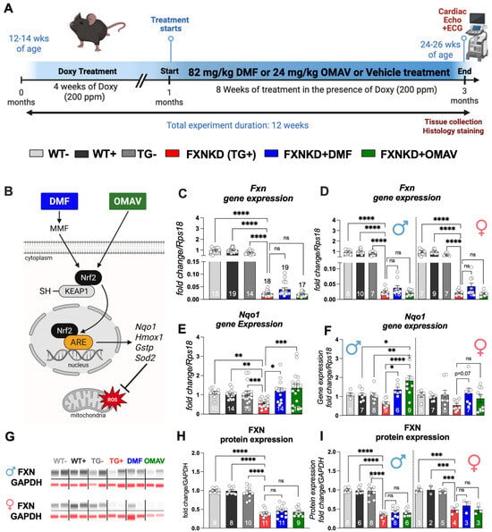
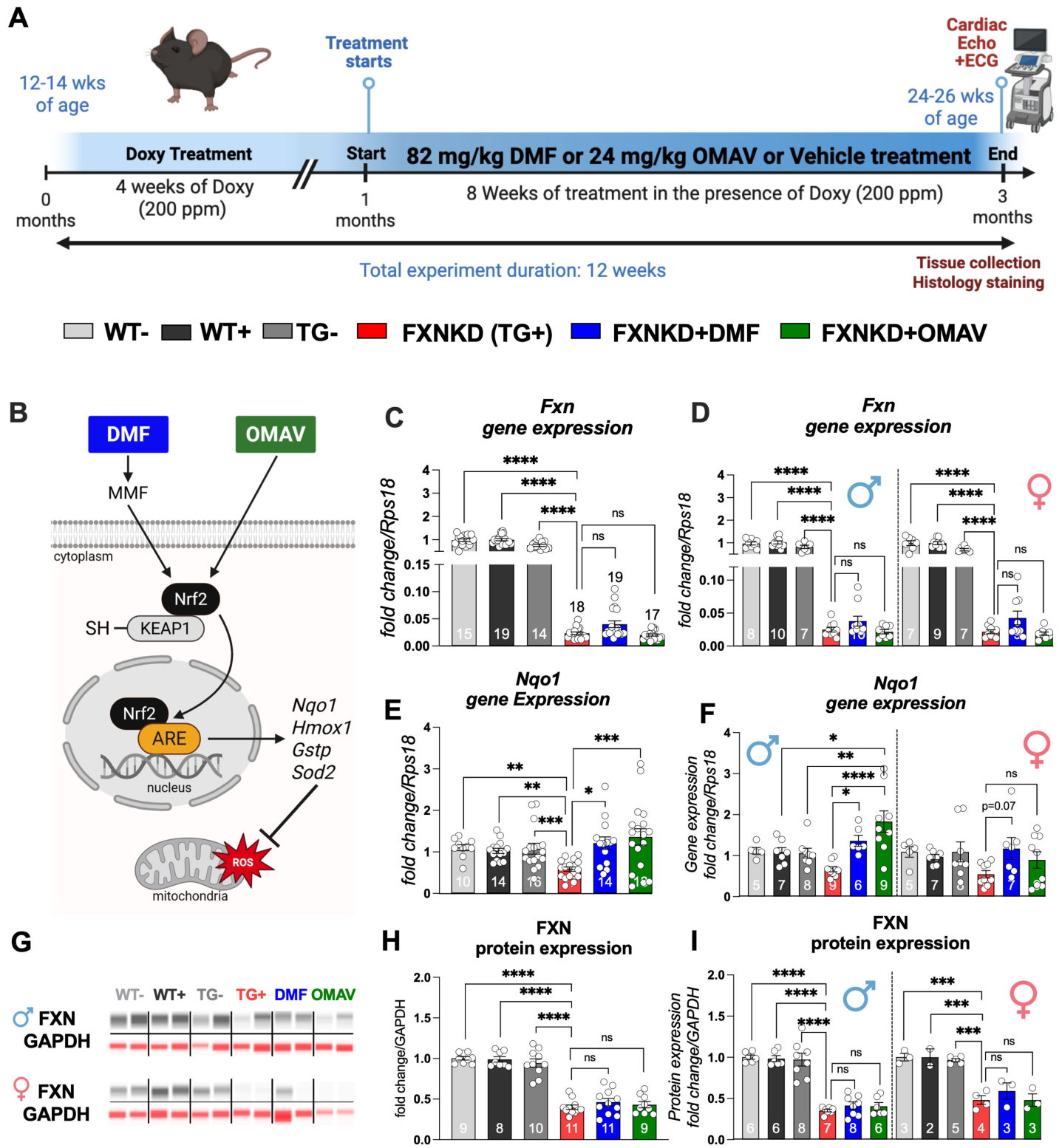
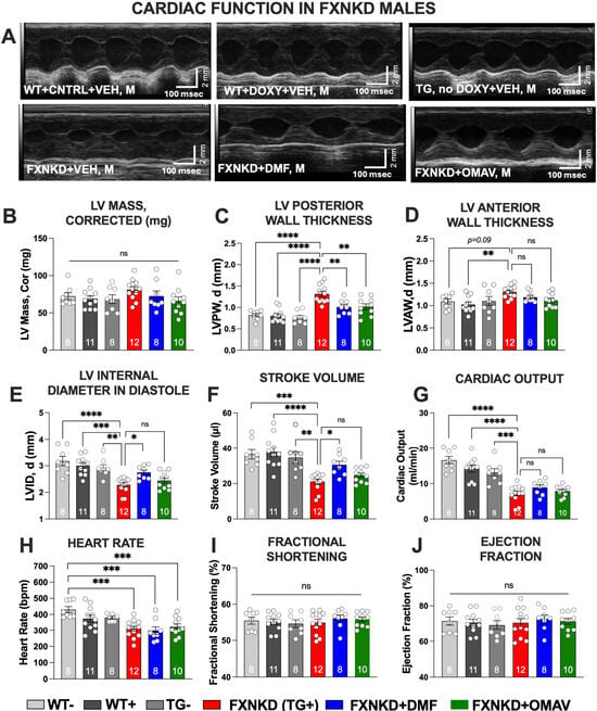
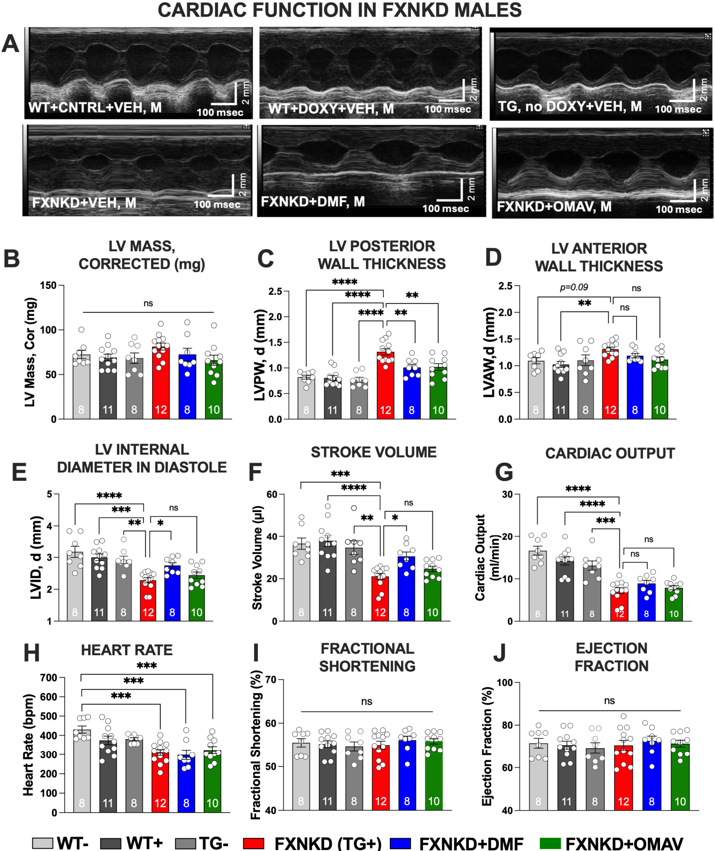
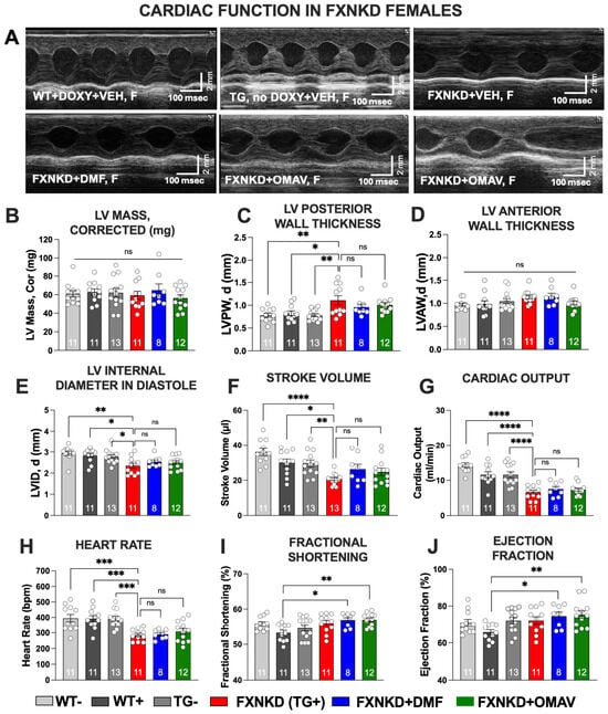
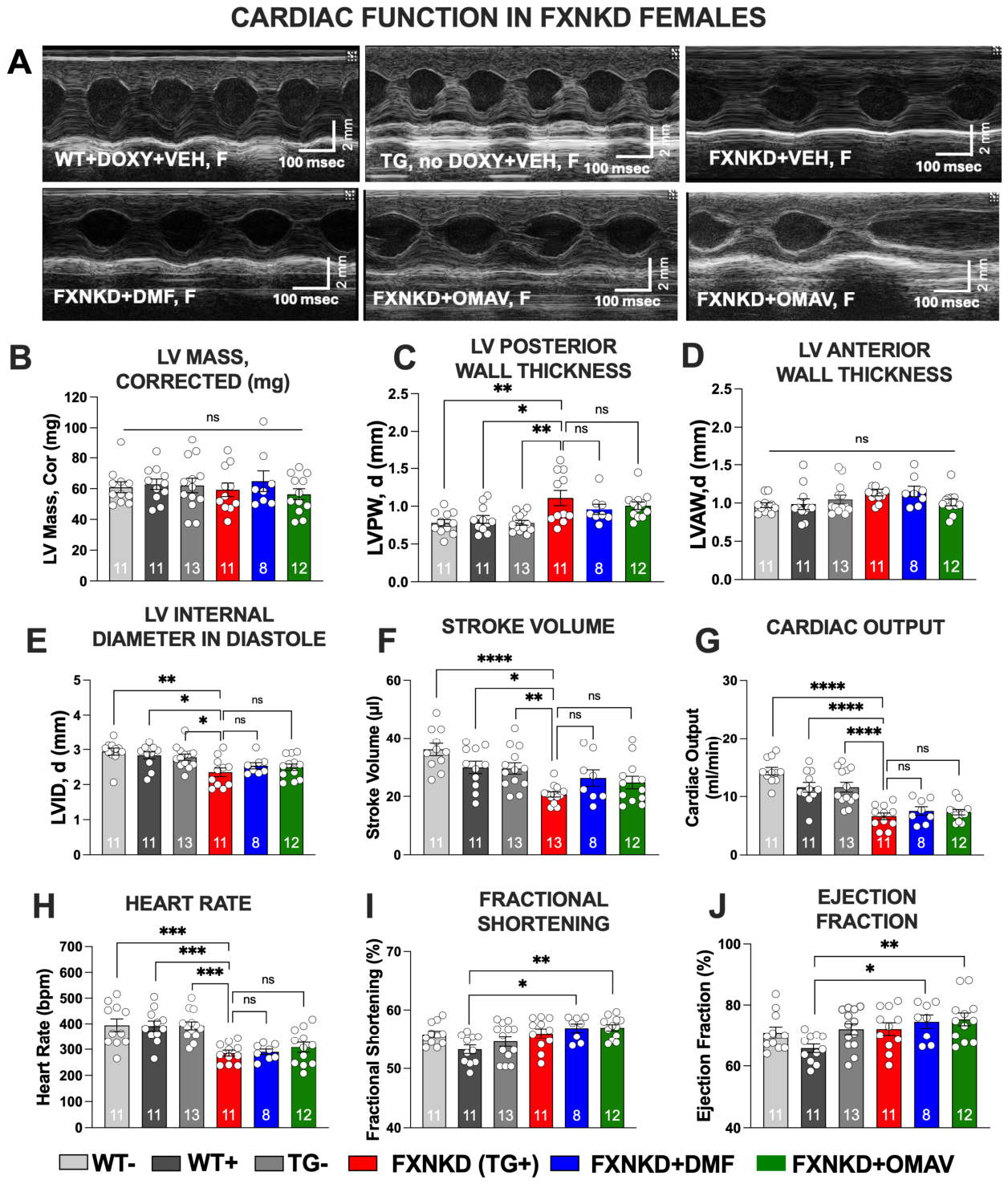
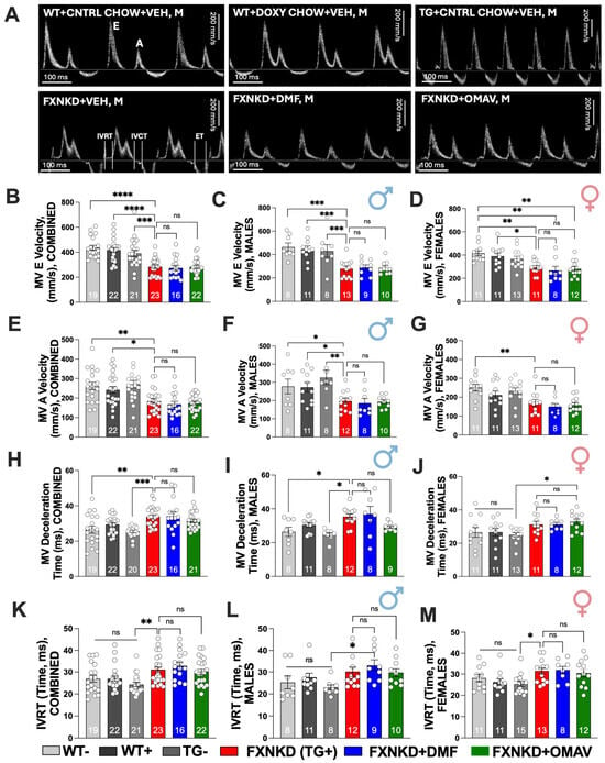
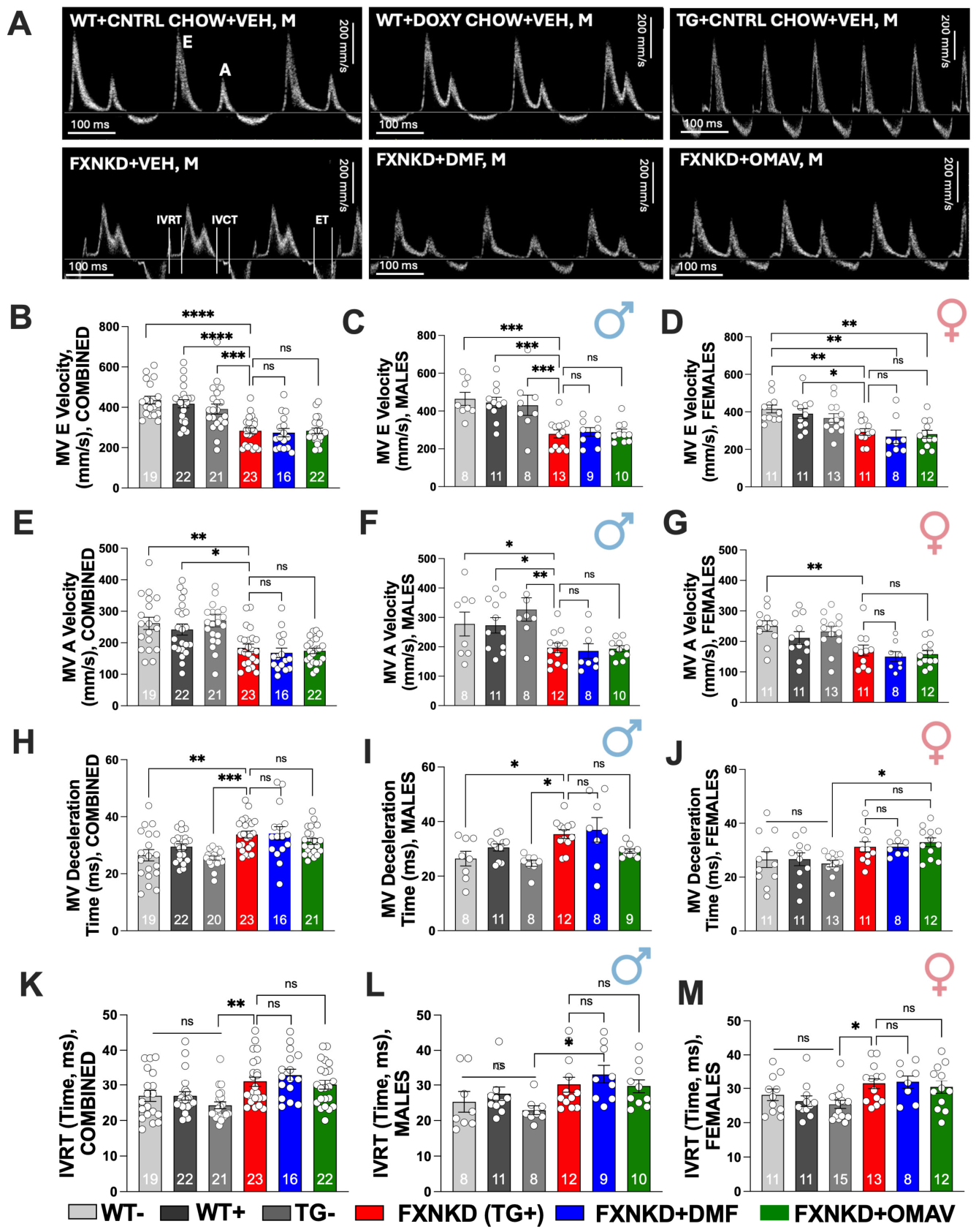
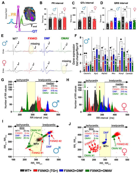
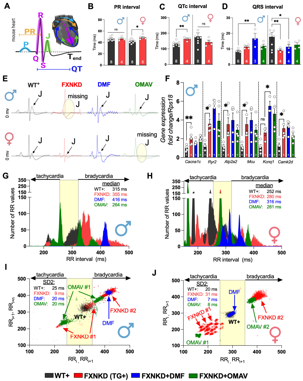
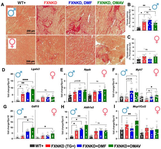
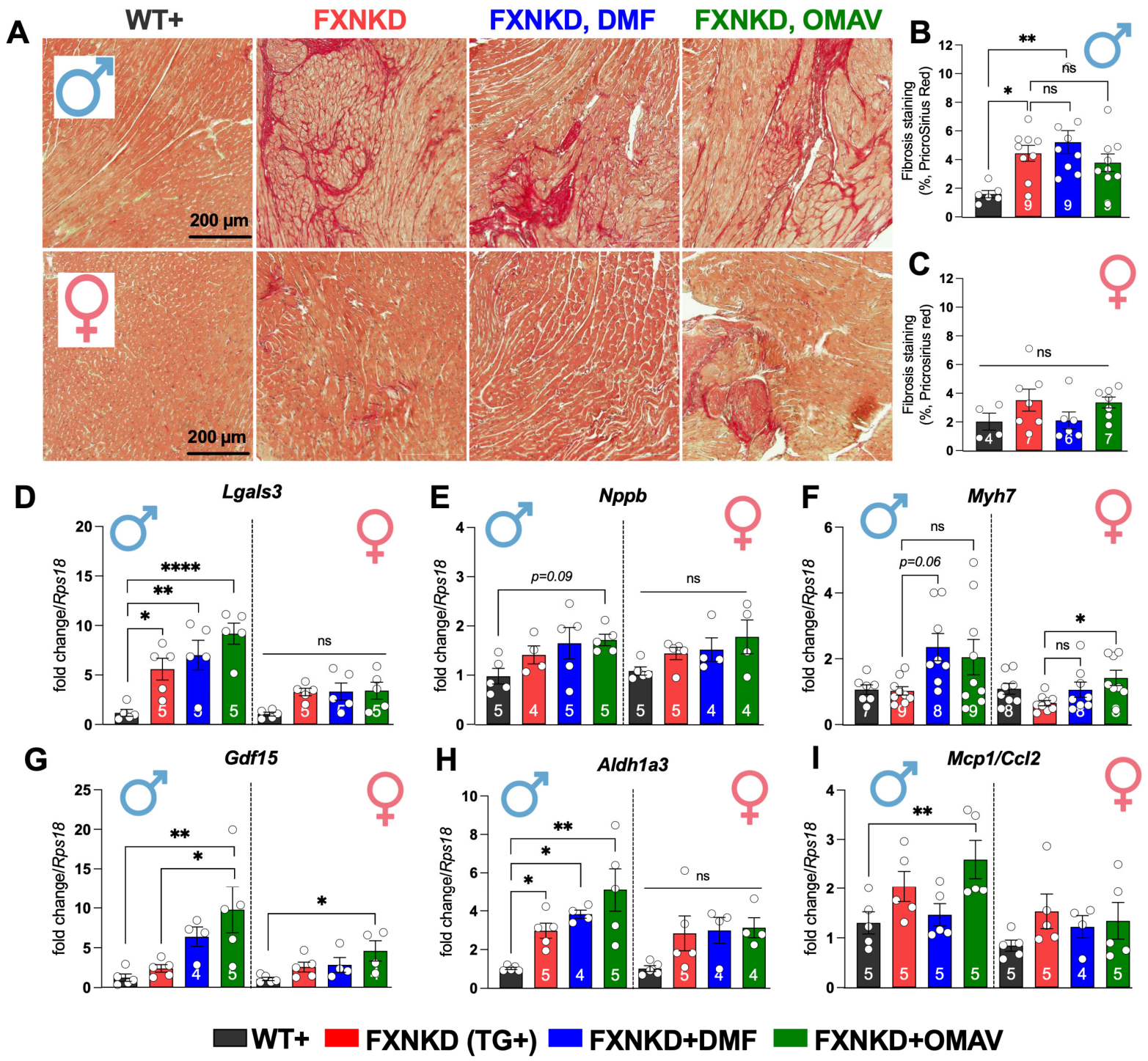
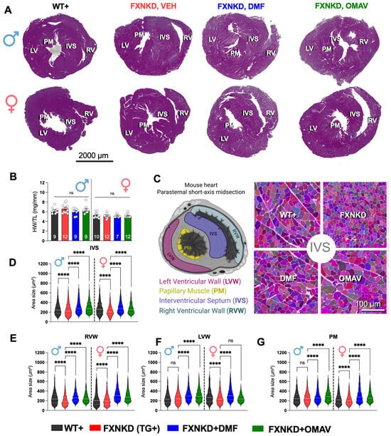
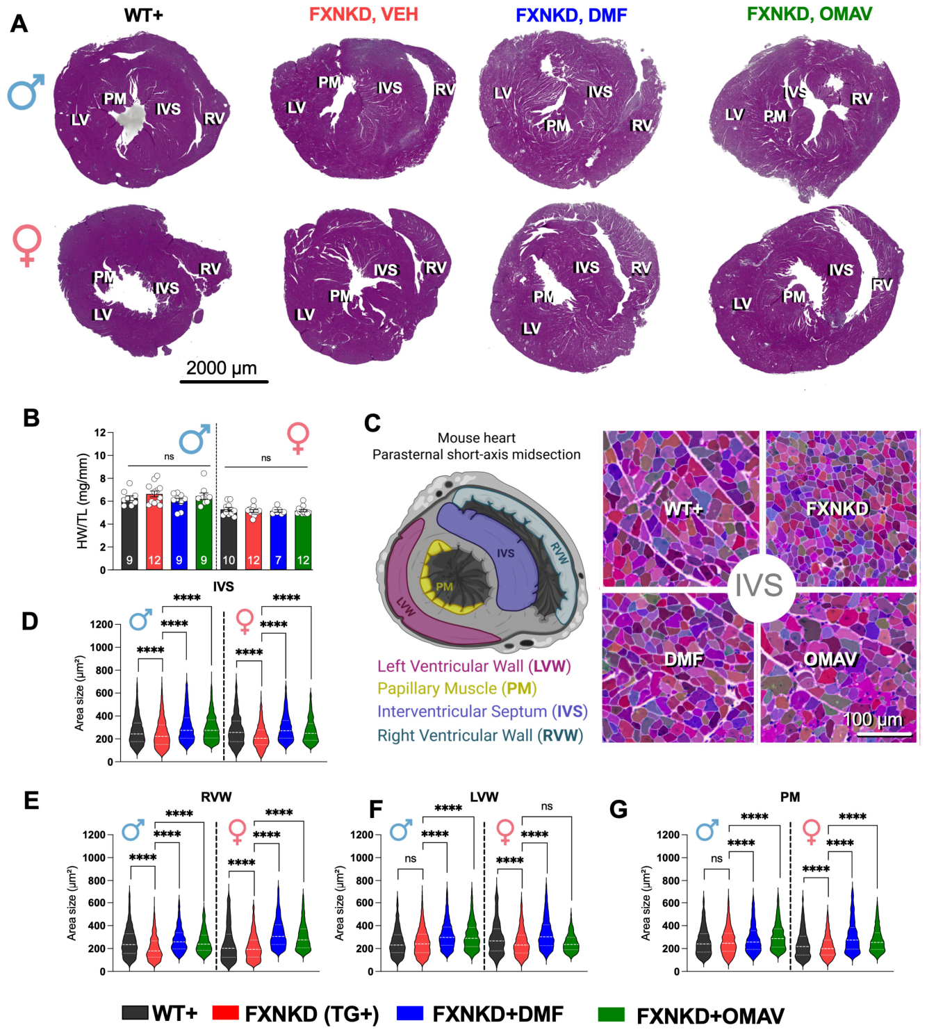
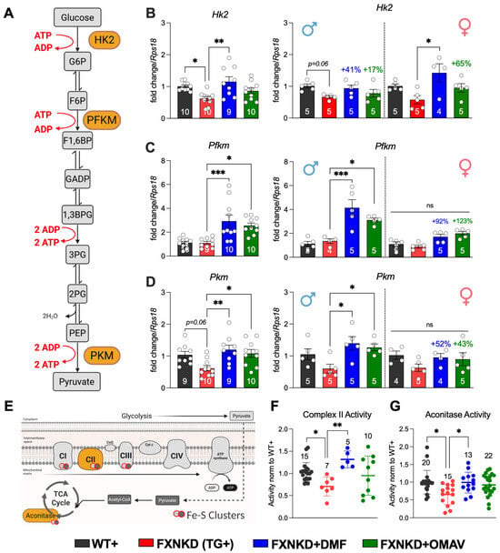
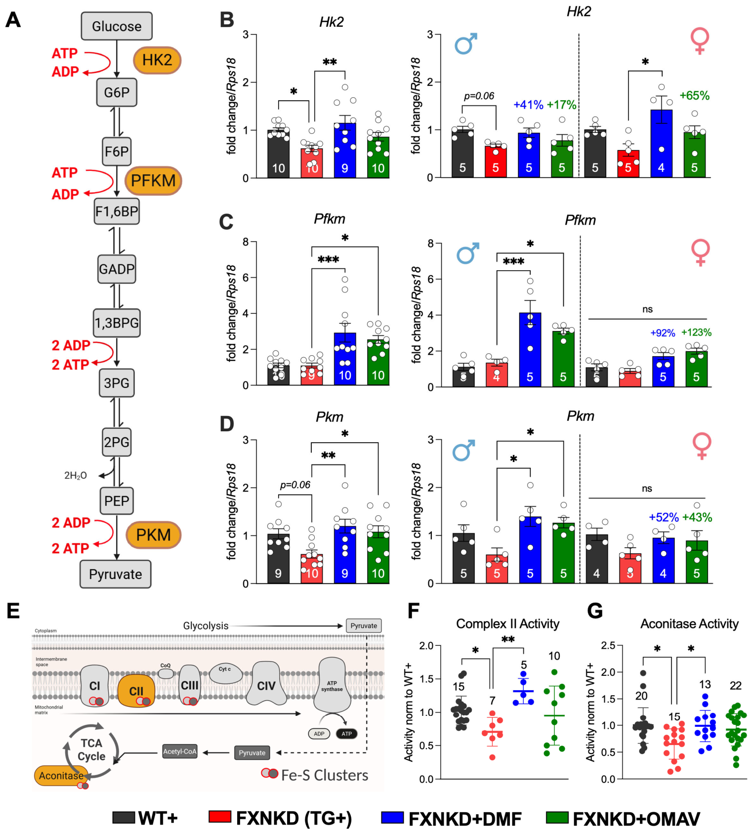
Disclaimer/Publisher’s Note: The statements, opinions and data contained in all publications are solely those of the individual author(s) and contributor(s) and not of MDPI and/or the editor(s). MDPI and/or the editor(s) disclaim responsibility for any injury to people or property resulting from any ideas, methods, instructions or products referred to in the content. |
© 2026 by the authors. Licensee MDPI, Basel, Switzerland. This article is an open access article distributed under the terms and conditions of the Creative Commons Attribution (CC BY) license.