


Abstract
Oxidative stress-mediated cellular damage is involved in various pathological processes. It is of great significance to discover active components with antioxidant protection potential from natural sources. Astragalus [Astragalus membranaceus (Fisch.) Bge. or Astragalus membranaceus (Fisch.) Bge. var. mongholicus (Bge.) Hsiao] is a perennial herbaceous plant in the Fabaceae family, and its anti-tumor, anti-inflammatory, and antioxidant pharmacological activities have been well documented. Notably, Astragalus seeds are rich in protein, but research on the screening and mechanism of antioxidant peptides derived from its seeds remains limited. In this study, Astragalus seed protein hydrolysates (UAMPH) were prepared by ultrasonic-assisted enzymatic hydrolysis. Candidate active peptides were screened using LC-MS/MS identification, multi-database virtual screening, and molecular docking techniques. Two novel pentapeptides, FAVGG and YLGPG, were obtained via solid-phase synthesis. Furthermore, their antioxidant effects were systematically evaluated using in vitro chemical antioxidant assays and an H2O2-induced oxidative damage model in HepG2 cells. The results showed that UAMPH exhibited good structural flexibility, thermal stability, and in vitro gastrointestinal digestibility. Both peptides scavenged various free radicals in a concentration-dependent manner and alleviated intracellular ROS accumulation, restored mitochondrial membrane potential, and inhibited cell apoptosis by suppressing Keap1 expression and increasing the protein expression of Nrf2 and its downstream target HO-1. Concurrently, they increased SOD and CAT activities and GSH levels while decreasing MDA levels and LDH activity. Following intervention with ML385, a selective Nrf2 inhibitor, the peptide-mediated cytoprotective effects were significantly reversed. In conclusion, two previously unreported antioxidant peptides were identified from Astragalus seed protein hydrolysate, and the results suggest that their cytoprotective effects may be associated with activation of the Nrf2-mediated antioxidant response, providing new evidence for the development of traditional Chinese medicinal resources and natural antioxidants.
1. Introduction
Oxidative stress is an essential pathological trigger in the occurrence and development of various chronic diseases [1,2]. Developing natural bioactive components with antioxidant protection potential and exploring their biological effects can provide a new material basis for research on oxidative damage-related issues. Antioxidant peptides have become a research hotspot in the field of natural active substances due to their advantages such as low toxicity [3], easy absorption by the body [4], and structural modifiability [5]. In recent years, the discovery of related active peptides from food-borne and medicinal plant proteins has received widespread attention [6]. However, existing studies have mostly focused on traditional protein sources such as soybean, cereals, and marine organisms [7,8]. Although there have been reports on antioxidant peptides derived from some medicinal plants [9,10], active peptides derived from Astragalus seeds and their antioxidant mechanisms have not been systematically investigated.
Astragalus belongs to the genus Astragalus of the Fabaceae family and has two botanical origins: Astragalus membranaceus (Fisch.) Bge. var. mongholicus (Bge.) Hsiao and Astragalus membranaceus (Fisch.) Bge. Both are used as Astragalus herbs and have a long history of use in China. It was described as the “chief tonic” in the Compendium of Materia Medica during the Ming dynasty. Traditionally, it has been used for its roots. Modern pharmacology has confirmed that polysaccharides, saponins, and flavonoids in Astragalus roots possess significant antioxidant activity [11,12,13,14]. In contrast, although Astragalus seeds are rich in protein, the exploration of protein-derived active peptides from this source has rarely been reported. Previous work by our group demonstrated that seed proteins from these two Astragalus species (AMP) are promising raw materials for developing bioactive peptides. Bioactive peptides from Astragalus membranaceus (Fisch.) Bge. seeds exhibited marked immunomodulatory activity [15], while seed proteins of Astragalus membranaceus (Fisch.) Bge. var. mongholicus (Bge.) Hsiao possessed favorable antioxidant activity. However, natural AMP has a large molecular weight and low bioavailability, which limits its direct application. Degrading macromolecular proteins into small-molecule peptides through enzymatic hydrolysis is an effective strategy to improve their bioavailability [16,17]. Numerous studies have confirmed that protein-derived active peptides can exert excellent antioxidant and cytoprotective effects [18,19]. Nevertheless, most studies focus on conventional protein sources, and the identification and activity evaluation of novel active peptides derived from medicinal plant seeds remain scarce. Accordingly, we investigated the antioxidant potential of peptides from Astragalus membranaceus (Fisch.) Bge. var. mongholicus (Bge.) Hsiao seed proteins to provide experimental reference data for research on bioactive peptides derived from medicinal plant seeds.
Based on this, to discover novel antioxidant peptides from Astragalus seed protein hydrolysates, UAMPH was prepared by ultrasonic-assisted enzymatic hydrolysis in this study. Candidate active peptides were subsequently obtained through sequential screening using LC-MS/MS peptide sequence identification, bioactivity prediction, and molecular docking techniques. Their antioxidant activities were systematically evaluated in both in vitro chemical models and an H2O2-induced HepG2 cell oxidative damage model. Finally, a controlled experiment was conducted using the Nrf2 selective inhibitor ML385 to further substantiate the relevant molecular mechanisms by which the two novel peptides participate in protecting hepatocytes against oxidative damage. This provides an experimental basis for the high-value utilization of Astragalus seed protein resources and the development of natural antioxidant peptides.
2. Materials and Methods
2.1. Materials and Reagents
Three batches of Astragalus [Astragalus membranaceus (Fisch.) Bge. var. mongholicus (Bge.) Hsiao] seeds were obtained from Guyang County, Inner Mongolia Autonomous Region, China, from Hebei Changze Seed Industry Co., Ltd. (Chengde, China) and were authenticated as seeds of Astragalus membranaceus (Fisch.) Bge. var. mongholicus (Bge.) Hsiao, a Fabaceae plant, at Changchun University of Chinese Medicine. Alkaline protease was purchased from Shanghai Yuanye Bio-Technology Co., Ltd. (Shanghai, China); 1,1-diphenyl-2-picrylhydrazyl (DPPH), 2,2’-azino-bis(3-ethylbenzothiazoline-6-sulfonic acid) diammonium salt (ABTS), 1,10-phenanthroline, and vitamin C (VC) (all analytical grade) were purchased from Beijing Solarbio Science & Technology Co., Ltd. (Beijing, China). Human hepatocellular carcinoma HepG2 cells and high-glucose DMEM medium were purchased from Wuhan Procell Life Science & Technology Co., Ltd. (Wuhan, China); Normal human hepatocyte MIHA was purchased from Shanghai Zeye Biotechnology Co., Ltd. (Shanghai, China); human AC16 cardiomyocytes were purchased from Zhejiang Baidi Biotechnology Co., Ltd. (Hangzhou, China); human normal lung epithelial BEAS-2B cells were obtained from the Cell Bank of the Chinese Academy of Sciences (Shanghai, China); human embryonic kidney HEK293 cells were purchased from Wuhan Procell Biotechnology Co., Ltd. (Wuhan, China); mouse BV2 microglial cell line was obtained from Shanghai Jinyuan Biotechnology Co., Ltd. (Shanghai, China).; fetal bovine serum (FBS) and trypsin were purchased from Gibco (New York, NY, USA); the Cell Counting Kit-8 (CCK-8) and phosphate-buffered saline (PBS) were purchased from Beijing Bioss Biotechnology Co., Ltd. (Beijing, China); the Calcein-AM/PI Double Stain Apoptosis Detection Kit was purchased from Shanghai BestBio Biotechnology Co., Ltd. (Shanghai, China); the Reactive Oxygen Species (ROS) Assay Kit was purchased from Beyotime Biotechnology (Shanghai, China). Enzyme-linked immunosorbent assay (ELISA) kits for superoxide dismutase (SOD), malondialdehyde (MDA), and glutathione (GSH) were all purchased from Shanghai Youxuan Biotechnology Co., Ltd. (Shanghai, China).
2.2. Preparation of UAMPH
Astragalus seed protein (AMP) was previously prepared and stored in our laboratory. We also evaluated the effects of different preparation methods on Astragalus seed protein hydrolysates. Compared with hydrolysates prepared by ultrasonication alone or enzymatic hydrolysis alone, ultrasound-assisted enzymatic hydrolysis produced a hydrolysate with higher peptide content, a higher degree of hydrolysis, improved in vitro digestibility, and stronger antioxidant activity. Therefore, ultrasound-assisted enzymatic hydrolysis was selected for subsequent experiments. The relevant preliminary methods and results are provided in the Supplementary Materials. To optimize the preparation process of UAMPH, single-factor experiments combined with the Box–Behnken response surface methodology were employed to optimize the ultrasound-assisted enzymatic hydrolysis process for preparing UAMPH. The complete experimental data, regression models, variance analysis, and relevant charts are provided in Supplementary Material.
Under the optimal conditions, an appropriate amount of AMP was dissolved in distilled water to prepare a 2% (w/v) protein solution, and the pH was adjusted to 9.5 using 1 mol/L NaOH. Alkaline protease was added at an enzyme-to-substrate ratio of 2% (w/w), and ultrasonic-assisted enzymatic hydrolysis was performed in a 55 °C water bath. The ultrasonic treatment conditions were as follows: power 150 W, time 20 min, pulse mode (2s on, 2s off). The enzymatic hydrolysis reaction lasted for 3 h, during which the pH was maintained constantly at 9.5. After the enzymatic hydrolysis, the reaction mixture was heated in a 95 °C water bath for 10 min to inactivate the enzyme, cooled to room temperature, and the pH was adjusted to 7.0 using 1 mol/L HCl. The mixture was centrifuged at 8000 rpm for 15 min at 4 °C. The supernatant was collected and ultrafiltered using a 3 kDa PES membrane. The filtrate was lyophilized to obtain UAMPH, which was stored at −20 °C for future use.
2.3. Characterization of Physicochemical Properties
2.3.1. Sodium Dodecyl Sulfate-Polyacrylamide Gel Electrophoresis (SDS-PAGE)
The resolving and stacking gels were prepared according to the instructions of the SDS-PAGE protein kit. AMP and UAMPH samples were dissolved in protein loading buffer to a final concentration of 5.0 mg/mL. An aliquot of 10 µL of each sample was loaded and electrophoresed in a PowerPac Basic electrophoresis system. After electrophoresis, the gel was stained with Coomassie Brilliant Blue G250, destained with a methanol-acetic acid-water (3:1:6, v/v/v) solution, and the results were observed and recorded using an I Bright 1500 chemiluminescence imaging system.
2.3.2. Molecular Weight Distribution
The molecular weight distribution of UAMPH was determined using size exclusion chromatography-high performance liquid chromatography (SEC-HPLC). The chromatographic column was a TSKgel G2000 SWXL (7.8 mm × 300 mm, Tosoh, Japan). The mobile phase was an aqueous solution of 30% acetonitrile containing 0.1% trifluoroacetic acid (TFA), the flow rate was 0.5 mL/min, the column temperature was 35 °C, and the detection wavelength was 220 nm. A molecular weight standard curve was plotted using cytochrome C (12,384 Da), aprotinin (6512 Da), bacitracin (1422 Da), oxidized glutathione (612 Da), and Gly-Tyr (238 Da) as standards.
2.3.3. Degree of Hydrolysis
The degree of hydrolysis of UAMPH was determined by the formaldehyde titration method [20]. Referring to the national standard GB/T 5009.5-2016 [21], the nitrogen content in UAMPH was determined using the Kjeldahl method [22]. The degree of hydrolysis (DH) was calculated according to the following formula:
where m1 is the sample mass (g); m2 is the total nitrogen mass of the sample (g); C is the molar concentration of NaOH (mol/L); V1 is the volume of NaOH consumed to adjust the pH to 9.2 after adding formaldehyde to the diluted sample (mL); V2 is the volume of NaOH consumed to adjust the pH to 9.2 after adding formaldehyde in the blank experiment (mL); V3 is the sampling volume of the diluted sample (mL); and V4 is the fixed volume of the diluted sample (mL).
2.3.4. Thermal Stability
Following the method in the literature [23], the thermal stability of UAMPH was determined using Differential Scanning Calorimetry (DSC). An appropriate amount of the sample was weighed and sealed in an aluminum crucible and scanned in the range of 25–140 °C at a heating rate of 5 °C/min, recording the heat flow curve.
2.3.5. Determination of Amino Acid Composition
A UAMPH sample (200 mg) was weighed and hydrolyzed with 6 mol/L HCl at 110 °C for 24 h. After the sample was completely dried, it was redissolved and filtered through a 0.22 µm microporous membrane. An aliquot of 20 µL of the filtrate was taken, and the amino acid composition was determined using an automatic amino acid analyzer (L-8900, Hitachi, Tokyo, Japan) [24]. Amino acid contents are expressed as g/100 g sample and normalized according to the total protein content of the sample.
2.3.6. In Vitro Digestibility Determination
The in vitro digestibility of UAMPH was determined using a simulated two-step gastrointestinal digestion model [25]. An appropriate amount of the sample was weighed and dissolved in deionized water to prepare a 1 mg/mL solution, and the pH was adjusted to 2.0 with 1 mol/L HCl. Pepsin was added at an enzyme-to-substrate ratio of 4% (w/w), and the mixture was incubated at 37 °C with constant shaking for 2 h. After the reaction, it was immediately heated in a boiling water bath for 10 min to inactivate the enzyme. After cooling, the pH was adjusted to 7.0 with 1 mol/L NaOH, and the mixture was centrifuged at 8000 rpm for 15 min. The collected supernatant was the gastric digest. The gastric digest was taken, the pH was adjusted to 8.0 with 1 mol/L NaOH, and trypsin was added at an enzyme-to-substrate ratio of 4% (w/w). The mixture was incubated at 37 °C with constant shaking for 2 h. After the reaction, it was inactivated in a boiling water bath for 10 min, cooled, adjusted to pH 7.0 with 1 mol/L HCl, and centrifuged at 8000 rpm for 15 min. The collected supernatant was the intestinal digest. The protein content in the gastric digest, intestinal digest, and undigested samples was determined according to the GB 5009.5-2016 method. In vitro digestibility was calculated according to the following formula:
2.4. Structural Characterization
2.4.1. FT-IR Spectroscopic Analysis
Samples were prepared using the KBr pellet method. UAMPH was mixed with dried KBr powder, fully ground in an agate mortar, and then pressed into a transparent and uniform thin pellet using a tablet press. Absorption spectra were acquired using a Fourier Transform Infrared Spectrometer (Thermo Fisher Scientific, Madison, WI, USA) in the range of 4000–400 cm−1, with 32 scans and a resolution of 4 cm−1.
2.4.2. CD Spectroscopic Analysis
Referring to the method determined by previous researchers with slight modifications [26], UAMPH was dissolved in Tris-HCl buffer (10 mmol/L, pH 7.4) to prepare a homogeneous protein solution. A circular dichroism (CD) spectrometer (Applied Photophysics Limited, Leatherhead, UK) was used to scan in the wavelength range of 190–260 nm, with a scanning speed of 100 nm/min, a bandwidth of 1.0 nm, a response time of 0.5 s, and a path length of 0.1 cm. CDNN software(version 2.0c) was used to calculate the secondary structure contents.
2.4.3. SEM Analysis
A scanning electron microscope was used to observe the microstructure of UAMPH [27]. The lyophilized UAMPH powder was evenly adhered to a conductive tape, sputtered with gold using an ion sputter coater, and the micro-morphology was observed using a scanning electron microscope (Hitachi High-Tech Corporation, Tokyo, Japan) at an accelerating voltage of 5–10 kV.
2.5. Identification, Virtual Screening, and Synthesis of Antioxidant Peptides
2.5.1. LC-MS/MS Peptide Sequence Identification
Liquid chromatography-tandem mass spectrometry (LC-MS/MS) was used to identify the peptide sequences in UAMPH. After desalting the sample with a C18 desalting column (Millipore Corporation, Billerica, USA), analysis was performed using an EASY-nLC 1200 liquid chromatography system (Thermo Fisher Scientific, Waltham, MA, USA) coupled with a Q Exactive mass spectrometer (Thermo Fisher Scientific, Waltham, MA, USA). The chromatographic column was a Reprosil-Pur 120 C18-AQ column (150 μm × 170 mm, 1.9 μm, Dr. Maisch, Germany). Mobile phase A was a 0.1% aqueous formic acid solution, and mobile phase B was an 80% acetonitrile solution containing 0.1% formic acid. The gradient elution program was: 0–2 min, 4–8% B; 2–35 min, 8–28% B; 35–55 min, 28–40% B; 55–56 min, 40–98% B; 56–66 min, 98% B. The flow rate was 600 nL/min. The mass spectrometer operated in data-dependent acquisition (DDA) mode, with a full scan range of 100–1500 m/z and a resolution of 70,000 (at m/z 200); the secondary mass spectrometry resolution was 17,500, with an HCD collision energy of 28%. The raw data was searched against the UniProtKB database (restricted to Viridiplantae) using PEAKS Studio (Bioinformatics Solutions Inc., Waterloo, ON, Canada), and the false discovery rate (FDR) for peptide identification was controlled within 1%.
2.5.2. Bioactivity Prediction
The Peptide Ranker database (http://distilldeep.ucd.ie/PeptideRanker/ (accessed on 25 January 2025)) was used to predict the bioactivity of the peptide sequences identified by LC-MS/MS, and peptides with a score > 0.5 were considered to have potential bioactivity [28]. Subsequently, the AnOxPePred-1.0 database (https://services.healthtech.dtu.dk/services/AnOxPePred-1.0/ (accessed on 25 January 2025)) was used to evaluate the free radical scavenging and metal ion chelating abilities of the candidate peptides [29]; the PreAIP database (http://kurata14.bio.kyutech.ac.jp/PreAIP/index.php (accessed on 26 January 2025)) was used to predict their anti-inflammatory activity [30]; and the ToxinPred database (https://webs.iiitd.edu.in/raghava/toxinpred/ (accessed on 26 January 2025)) was used to evaluate their toxicity and physicochemical properties [31].
2.5.3. Molecular Docking
AutoDockTools-1.5.6 software was used to analyze the binding affinity of the candidate peptide sequences for Keap1 and the Keap1–Nrf2 complex. Crystal structures of the Keap1 Kelch domain (PDB: 1ZGK) and the Keap1–Nrf2 complex (PDB: 2FLU) were obtained from the PDB database. For Keap1, the grid was centered on the Nrf2-binding pocket within the Kelch domain, with docking-center coordinates of x = 18.111, y = 16.083, and z = 7.417. The grid dimensions along the x, y, and z axes were 60.75 × 60.75 × 60.75 Å, with a grid spacing of 0.486 Å, covering residues critical for Keap1–peptide interactions. For the Keap1–Nrf2 complex, the grid was centered on the Nrf2 peptide-binding site within the Keap1 Kelch domain, with docking-center coordinates of x = 11.871, y = 16.086, and z = 6.782. The grid dimensions along the x, y, and z axes were 64.25 × 64.25 × 64.25 Å, with a grid spacing of 0.514 Å, covering residues critical for interactions between the Keap1–Nrf2 complex and peptides.
Polar hydrogens and Gasteiger charges were subsequently added in AutoDockTools-1.5.6, and three-dimensional structures of candidate peptides were constructed for molecular docking. The docking grid box was centered on the native active pocket of each target protein, such that docking was restricted to the native functional binding region rather than performed as whole-protein blind docking. Docking results were visualized using PyMOL (version 2.5) to identify peptide–target interaction sites and hydrogen-bond networks within the active pocket. All docking results were used only for candidate-peptide screening and the generation of mechanistic hypotheses and were not regarded as evidence of direct binding or functional target regulation.
2.5.4. Solid-Phase Synthesis
Based on LC-MS/MS identification, Peptide Ranker scores (>0.5), AnOxPePred/PreAIP/ToxinPred predictions of function and physicochemical properties, and rankings of molecular docking binding energies, candidate antioxidant peptides were screened stepwise. FAVGG and YLGPG, which exhibited the lowest binding energies and suitable physicochemical properties, were selected and synthesized by the standard Fmoc solid-phase synthesis method. The synthesized peptides were purified by reversed-phase high-performance liquid chromatography (RP-HPLC). The chromatographic conditions were: column SHIMADZU Inertsil ODS-SP (4.6 × 250 mm, 5 μm); mobile phase A was an aqueous solution containing 0.1% TFA; and mobile phase B was an acetonitrile solution containing 0.1% TFA. The linear gradient elution program was: 0–25 min, 1–50% B; followed by an instantaneous switch to 100% B at 25.01 min and maintained until 30 min; flow rate 1.0 mL/min, detection wavelength 220 nm, and injection volume 30 µL. The purity of the synthesized peptides was confirmed by HPLC to be ≥95%, and they were lyophilized and stored at −20 °C for future use.
2.6. Toxicity Evaluation of FAVGG and YLGPG on Five Normal Cell Lines
Cell viability was determined using the Cell Counting Kit-8 (CCK-8) according to the method of Zhang et al. [32] with slight modifications. MIHA hepatocytes, AC16 cardiomyocytes, BEAS-2B bronchial epithelial cells, HEK293 kidney cells, and BV2 microglia were seeded in 96-well plates at a density of 5 × 10³ cells per well and cultured overnight. Then, the cells were treated with different concentrations of FAVGG and YLGPG (10, 15, 20, 25, 50, 100, 150, 200, 400, 600, 800, and 1000 μg/mL) for 24 h. Five technical replicate wells were set for each concentration, and the experiment was independently repeated three times. CCK-8 reagent was added to each well, incubated at 37 °C for 30 min, and the absorbance at 450 nm was measured using a microplate reader (Tecan Group Ltd., Männedorf, Switzerland).
2.7. Determination of Chemical Antioxidant Activity
Referring to literature methods, the scavenging abilities of the synthesized peptides FAVGG and YLGPG against DPPH free radicals [33], ABTS+ free radicals [34], hydroxyl radicals (·OH) [35], and superoxide anions (O2-) [36] were determined. Each peptide was prepared at 0.2, 0.6, 1.0, 1.2, 1.6, and 2.0 mg/mL. Three independent preparations of each peptide solution at each concentration were measured. Using VC as the positive control, the free radical scavenging rates at each concentration were determined, and the half-maximal inhibitory concentration (IC50) was calculated.
2.8. Evaluation of Cellular Antioxidant Activity
2.8.1. Cell Culture and Grouping
HepG2 cells were cultured in high-glucose DMEM medium containing 10% fetal bovine serum (FBS), 100 U/mL penicillin, and 100 µg/mL streptomycin in a cell culture incubator at 37 °C with 5% CO2 [37]. The experimental groups were as follows: blank control group (DMEM medium); model group (oxidative stress damage model established by stimulation with DMEM medium containing 1000 µmol/L H2O2 for 2 h, then replaced with fresh DMEM medium); Treatment groups (DMEM containing 1000 µmol/mL H2O2 for 2 h to establish the oxidative stress injury model, followed by treatment for 24 h with DMEM containing FAVGG or YLGPG); positive control group (DMEM containing 1000 µmol/mL H2O2 for 2 h to establish the oxidative stress injury model, followed by treatment for 24 h with DMEM containing 50 µg/mL VC). All cell experiments were independently repeated three times using independently cultured cells for each experiment.
2.8.2. Determination of Intracellular ROS Levels
After the cells were treated according to the grouping in Section 2.8.1, the DCFH-DA fluorescent probe was added to each well at a final concentration of 10 µmol/L, and incubated at 37 °C in the dark for 30 min. The probe was removed, and the cells were washed twice with pre-cooled PBS. After centrifugation to discard the supernatant, the cells were resuspended in 400 µL PBS and passed through a 300-mesh cell strainer. The fluorescence intensity was then measured using a flow cytometer (BD Biosciences, San Jose, CA, USA) to quantitatively analyze intracellular ROS levels.
2.8.3. Calcein-AM/PI Double Stain Apoptosis Detection
After the cells were treated according to the grouping in Section 2.8.1, they were washed twice with PBS. Calcein-AM staining working solution (2 μmol/L) was added to cover the cells, and incubated at 37 °C in the dark for 20 min. After washing with PBS, a staining working solution containing 4 μmol/L PI was added and incubated at 37 °C in the dark for 5 min. After washing with PBS, the cells were observed and photographed under a fluorescence microscope (Thermo Fisher Scientific, Waltham, MA, USA). Images of green fluorescence (Calcein-AM, Ex/Em = 494/517 nm) and red fluorescence (PI, Ex/Em = 535/617 nm) were captured separately.
2.8.4. JC-1 Staining for Mitochondrial Membrane Potential
After the cells were treated according to the grouping in Section 2.8.1, they were washed once with PBS, and 1 mL of fresh medium was added. Following the instructions of the mitochondrial membrane potential assay kit (C2006, Beyotime, Shanghai, China), JC-1 staining working solution was added and incubated at 37 °C in the dark for 20 min. After washing, the changes in red/green fluorescence intensity were observed under a fluorescence microscope, and the red/green fluorescence ratio was used to reflect the mitochondrial membrane potential level.
2.8.5. Determination of Antioxidant Factors and Damage Markers
After the cells were treated according to the grouping in Section 2.8.1, the cell culture supernatant or cell lysate was collected. Referring to the instructions of the corresponding ELISA kits, MDA, GSH, SOD, LDH, and CAT were quantitatively determined.
2.9. Molecular Mechanism Study
2.9.1. Western Blotting
After the cells were treated according to the grouping in Section 2.8.1, total cellular protein was extracted using a total protein extraction kit, protein concentration was determined by the BCA method, and the loading amounts were standardized. A 20 μg aliquot of protein sample was separated by SDS-PAGE (12% resolving gel and 5% stacking gel) and transferred to a PVDF membrane. After blocking with 5% non-fat milk at room temperature for 1 h, the membranes were incubated with Nrf2 rabbit monoclonal antibody (1:1000, Cell Signaling Technology, USA), HO-1 rabbit monoclonal antibody (1:1000, Abcam, UK), Keap1 rabbit monoclonal antibody (1:1000, Proteintech, Wuhan, China), and β-actin mouse monoclonal antibody (1:5000, Sigma-Aldrich, St. Louis, MO, USA) at 4 °C overnight. After washing with PBST 3 times, the membranes were incubated with horseradish peroxidase (HRP)-conjugated goat anti-rabbit/anti-mouse secondary antibodies for 1 h. After washing with PBST another 3 times, the bands were developed using an ECL chemiluminescence kit (Beyotime, Shanghai, China). Images were acquired using a chemiluminescence imaging system (iBright 1500, Thermo Fisher Scientific, Singapore), and quantitative grayscale analysis was performed using ImageJ software (version 1.53t). The relative expression of each protein was calculated using β-actin as an internal reference. Western blot experiments were independently performed three times. Representative bands are shown, and quantitative protein data are presented as the mean ± standard deviation of three independent experiments.
2.9.2. Nrf2 Inhibition Experiment
The selective Nrf2 inhibitor ML385 (purity 99.96%, MedChemExpress, USA) was used to verify the key role of Nrf2 in peptide-mediated antioxidant protection. ML385 was dissolved in DMSO to prepare a 10 mmol/L stock solution. HepG2 cells were first stimulated with 1000 μmol/mL H2O2 for 2 h to establish an oxidative damage model. The H2O2-containing medium was discarded and replaced with fresh medium. ML385 was added for 1 h of incubation, followed by treatment with peptides, with a total incubation time of 24 h. Four experimental groups were set up: blank group, model group, H2O2 + peptide group, and H2O2 + ML385 + peptide group. The peptide concentration was 100 μg/mL in all peptide-treated groups. Cells were harvested after incubation, and Western Blot was used to detect the protein expression of Nrf2 and HO-1 (method same as 2.9.1); simultaneously, the levels of MDA, GSH, and SOD were determined (method same as 2.8.5).
2.10. Data Processing
Except for molecular docking and in silico predictions, which were computational simulations and did not involve experimental replication, all experiments were independently performed in triplicate (n = 3), and results are expressed as mean ± standard deviation (SD). Technical replicates, such as replicate wells, repeated injections, or multiple fields of view, were averaged before statistical analysis and were not considered as n. For UAMPH characterization, biological replicates represent three independently prepared batches of UAMPH. For cell experiments, biological replicates represent three independent cell-culture and treatment experiments. For chemical antioxidant assays, biological replicates represent measurements of three independently prepared peptide solutions. For Western blotting, biological replicates represent three independent experiments, each using lysates from independently cultured cells. All statistical analyses were performed using SPSS 26.0 software (SPSS Inc., Chicago, IL, USA). One-way analysis of variance (One-way ANOVA) was used for comparison among multiple groups, and Duncan’s multiple range test was used for post hoc testing. p < 0.05 was considered statistically significant. GraphPad Prism 9.0 (GraphPad Software, San Diego, CA, USA) and Origin 2021 (OriginLab Corp., Northampton, MA, USA) were used for plotting.
3. Results
3.1. Molecular Weight Distribution, Structural Characteristics, and In Vitro Digestibility of UAMPH
The molecular weight distributions of UAMPH and AMP were compared using SDS-PAGE and SEC-HPLC. As shown in the SDS-PAGE pattern (Figure 1a), the protein subunits of AMP were mainly distributed between 17 and 63 kDa (Lane 1). The subunit molecular weight of UAMPH, prepared via ultrasonic-assisted enzymatic hydrolysis, decreased significantly and was distributed below 11 kDa (Lane 2). The SEC-HPLC results (Figure 1b) further quantified the molecular weight distribution of UAMPH, where fractions < 0.1 kDa, 0.1–1 kDa, 1–1.5 kDa, and >1.5 kDa accounted for 5.024%, 15.35%, 78.783%, and 0.842%, respectively. Fractions < 1.5 kDa accounted for 99.157% in total, indicating that the product consisted predominantly of low-molecular-weight peptides below 1.5 kDa. This proportion exceeded the reported 83.24% of <3 kDa fractions in ultrasound-assisted enzymatic hydrolysates of soy protein isolate, suggesting that the ultrasound-assisted enzymatic hydrolysis process used in this study achieved more extensive peptide-chain degradation [38]. The degree of hydrolysis result (Figure 1c) showed that the degree of hydrolysis of UAMPH reached 35.62 ± 1.13%, indicating thorough enzymatic hydrolysis where proteins were degraded into a large number of small peptides. This high degree of protein hydrolysis is consistent with the lower molecular weight of UAMPH protein bands in SDS-PAGE and the high proportion (99.16%) of small-molecule peptides < 1.5 kDa in SEC-HPLC results. These results directly demonstrate that the ultrasonic-assisted enzymatic hydrolysis process can effectively enhance the hydrolysis of Astragalus seed protein, establishing the clear practical significance of ultrasonic pretreatment.
Figure 1.
This figure shows the structural characteristics and physicochemical properties of UAMPH. (a) SDS–PAGE electrophoretic patterns (Lane 1: AMP; Lane 2: UAMPH); (b) SEM image (200–fold magnification); (c) bar charts of degree of hydrolysis and gastrointestinal digestibility; (d) SEC–HPLC molecular–weight–distribution chromatogram of UAMPH with quantitative analysis results; (e) DSC thermogram; (f) FT-IR spectrum (4000–400 cm−1); (g) CD spectrum (190–260 nm) together with quantitative analysis results.
FT-IR spectroscopy was employed to characterize the functional group composition and the main-chain chemical bond environment of UAMPH [39]. As shown in Figure 1d, the sample exhibited characteristic absorption in the amide band (3600–3100 cm−1), which was attributed to the N–H stretching vibration of the peptide bond, indicating that the product predominantly consisted of intact peptide bonds and had not completely degraded into free amino acids. The absorption peak at 2946 cm−1 corresponds to the aliphatic C–H stretching vibration of methyl and methylene groups in the amino acid side chains. In the characteristic amide band regions, UAMPH displayed absorption peaks at 1658 cm−1 (Amide I band, mainly C=O stretching), 1536 cm−1 (Amide II band, coupling of N–H bending and C–N stretching), and 1238 cm−1 (Amide III band), with the peak shapes of the Amide I and Amide II bands being relatively sharpened. CD spectroscopy quantitatively resolved changes in secondary structure. As shown in Figure 1f, UAMPH exhibited a strong negative absorption band at 198 nm and a positive absorption band at 212 nm. Quantitative analysis revealed that the secondary structure of UAMPH consisted of 14.8% α-helix, 13.6% β-sheet, 35.1% β-turn, and 36.5% random coil. Compared with the secondary-structure composition of AMPH measured in our previous experiments (17.3% α-helix, 14.2% β-sheet, 32.8% β-turn, and 35.7% random coil), the α-helix and β-sheet contents of UAMPH decreased by 14.45% and 4.23%, respectively, whereas β-turn and random-coil contents increased by 7.01% and 2.24%, respectively. These changes indicate that ultrasound-assisted enzymatic hydrolysis induced secondary-structure rearrangement. The UAMPH backbone mainly adopted a loose, extended conformation rich in random coils and β-turns, with a small proportion of α-helices, while the sharpening of infrared peaks suggested relatively uniform secondary-structure classes. The increased random-coil proportion caused by ultrasonication is consistent with the increased conformational flexibility reported for ultrasonicated collagen peptides [40], corroborating that ultrasonic cavitation can promote conversion of protein backbones from ordered structures to flexible conformations. DSC analysis (Figure 1e) showed that the denaturation temperature of UAMPH was 79.49 °C. SEM visually presented the microscopic morphological features of the product. As shown in Figure 1g, UAMPH exhibited a high degree of fragmentation, with the macromolecular protein skeleton fully disrupted and degraded into smaller peptide fragments with looser structures, displaying a porous microstructure. The results of the simulated in vitro two-step gastrointestinal digestion (Figure 1c) showed that the digestibility of UAMPH was 58.64% during the gastric digestion phase and 79.25% during the intestinal digestion phase, indicating higher digestibility in the intestinal phase than in the gastric phase. This result is consistent with findings in grape-seed protein hydrolysates that enzymatic modification can significantly improve in vitro protein digestibility [41].
3.2. Amino Acid Composition Results
Amino acid composition is an important indicator for evaluating the nutritional properties and functional activities of peptides [42]. The amino acid composition of UAMPH was determined using an automatic amino acid analyzer. Amino acid contents in UAMPH are reported on both a g/100 g sample and a g/100 g protein basis (Supplementary Table S3); comparisons with the FAO/WHO (2013) [43] reference pattern were made on a g/100 g protein basis.
A total of 17 amino acids, excluding tryptophan, were detected in UAMPH using an automatic amino acid analyzer. These included seven essential amino acids (EAAs) and ten nonessential amino acids (NEAAs), indicating that the product retained diverse amino acid composition after ultrasound-assisted enzymatic hydrolysis (Figure 2). The total amino acid (TAA) content of UAMPH was 99.73 g/100 g, with total EAA and NEAA contents of 44.90 and 54.83 g/100 g, respectively. According to the FAO/WHO (2013) [43] recommended amino acid pattern, UAMPH showed an overall favorable EAA profile: threonine, valine, isoleucine, leucine, and phenylalanine + tyrosine met or exceeded the recommended values, with valine, isoleucine, and leucine substantially exceeding the recommendations. Only lysine and methionine (including cysteine) were relatively low. In terms of composition, UAMPH was enriched in hydrophobic and aromatic amino acids, such as tyrosine, leucine, and glutamic acid, and had a relatively high proportion of flavor amino acids (DAAs), with EAA/TAA and EAA/NEAA ratios of 45.02% and 81.89%, respectively. This composition, dominated by hydrophobic and aromatic amino acids, provides an important structural basis for radical scavenging by antioxidant peptides and supports the antioxidant activity of UAMPH. These results indicate that ultrasound-assisted enzymatic hydrolysis can effectively increase the release of total amino acids, EAAs, and functionally relevant amino acids, thereby improving the amino acid nutritional profile of the hydrolysate.
Figure 2.
Amino-acid analysis of UAMPH. Notes: Essential amino acids: Leu, Val, Ile, Thr, Phe, Lys, Met; Hydrophilic amino acids: Ser, Thr, Cys, Tyr; Hydrophobic amino acids: Gly, Ala, Val, Leu, Pro, Met, Phe, Ile; Acidic amino acid: Glu; Alkaline amino acids: Arg, Lys, His; Aromatic amino acids: Tyr, Phe.
3.3. Screening and Identification of Antioxidant Peptides
LC-MS/MS was applied to identify the peptide sequences of UAMPH. In total, 331 peptides were identified, with m/z values between 252.13 and 1282.63 and lengths of 5–16 amino acids. The total ion chromatogram is presented in Figure 3a. The bioactivity of the 331 peptides was predicted using the Peptide Ranker database, resulting in 34 peptides with a score > 0.5 (score range 0~0.98, Supplementary Table S4), among which FSLMFLF, RWLGR, and YKFFN ranked in the top three. Toxicity prediction by ToxinPred showed that all 331 peptides had no potential toxicity, indicating good safety. A multi-dimensional virtual screening was conducted on the 34 candidate peptides. In the AnOxPePred-1.0 free radical scavenging activity prediction, YLGPG, QGFHNWTLNP, GHPWGNAPGAVA, GHPWGNAPGA, and TYMLFY ranked in the top five. In the metal chelating activity prediction, FNTPN, KLFPQ, FSPTRN, SSPRG, and FSLMFLF ranked in the top five. In the PreAIP anti-inflammatory activity prediction, GHPWGNAPGAVA, DWVSLPG, GHPWGNAPGAV, GHPWGNAPGAVAN, and GHPWGNAPGA scored > 0.5. ToxinPred physicochemical property analysis showed that the molecular weights of the 34 peptides ranged from 449.57 to 1247.52 Da (Supplementary Table S5), the top three peptides ranked by molecular weight (from lowest to highest) are FAVGG, TGGVF, and SSPRG. Hydrophobicity analysis revealed that 53% of the peptides had positive hydrophobicity values, with FAVGG, FVDLL, and GFVDLL ranking in the top three. Combining molecular weight and hydrophobicity data, FAVGG, possessing both the lowest molecular weight and the highest hydrophobicity, emerged as a high-quality candidate antioxidant peptide.
Figure 3.
This figure illustrates the LC-MS/MS identification of antioxidant peptides in UAMPH. (a) Total ion chromatogram (TIC) of UAMPH; (b) MS/MS spectrum and chemical structure of FAVGG; (c) MS/MS spectrum and chemical structure of YLGPG.
The 34 candidate peptides were further docked separately with Keap1 (PDB: 1ZGK) and the Keap1–Nrf2 complex (PDB: 2FLU) using AutoDockTools, with binding energy as the core screening criterion. Binding energies for the 34 peptide sequences are listed in Supplementary Table S5. In docking with Keap1, binding energies ranged from −9.09 to −2.86 kcal/mol, with YLGPG (−9.09 kcal/mol) and FAVGG (−8.5 kcal/mol) showing the lowest values. In docking with the Keap1–Nrf2 complex, YLGPG (−8.30 kcal/mol) and FAVGG (−6.48 kcal/mol) also showed the lowest binding energies. According to reported docking results for food-derived antioxidant peptides, binding energies of different peptides with Keap1 are mostly in the range of −7.8 to −10.1 kcal/mol, and peptides generally interfere with Keap1–Nrf2 interactions by binding in the Kelch-domain pocket [44]. The binding energies obtained in this study fell within this reported range. We also observed that YLGPG and FAVGG could form hydrogen-bond networks with hydrophobic residues of Keap1, including Val418 and Val465; similar interaction patterns have been reported in studies of other antioxidant peptides [45]. Considering all virtual-screening results, FAVGG had low molecular weight, high hydrophobicity, no predicted toxicity, and favorable docking binding energies with multiple targets. YLGPG had the highest AnOxPePred antioxidant prediction score and similarly showed favorable binding in multi-target molecular docking. Both peptides met multiple screening criteria and were therefore selected as target peptides for subsequent solid-phase synthesis and functional validation. Analysis of intermolecular interactions in the best binding conformations (Figure 4) showed that YLGPG formed five hydrogen bonds with Keap1 involving VAL418, VAL420, VAL465, VAL467, and ASN469, as well as electrostatic attractive interactions. YLGPG formed five hydrogen bonds with the Nrf2 protein in the Keap1–Nrf2 complex involving VAL418, VAL465, VAL512, VAL561, and VAL604. FAVGG formed six hydrogen bonds with Keap1 involving GLY423, ASP422, VAL420, VAL465, VAL467, and ILE559. FAVGG formed six hydrogen bonds with the Nrf2 protein in the Keap1–Nrf2 complex involving VAL465, VAL512, VAL561, GLY367, THR560, and ARG326. The hydrogen-bonding and hydrophobic interactions observed in this study share features with those reported for some Keap1-targeting antioxidant peptides, suggesting that the two pentapeptides may have the potential to interfere with Keap1–Nrf2 interactions.
Figure 4.
Three-dimensional conformations obtained from molecular docking of FAVGG and YLGPG with Keap1 and the Keap1–Nrf2 complex. (a) Docking conformation of FAVGG with Keap1; (b) Docking conformation of YLGPG with Keap1; (c) Docking conformation of FAVGG with the Keap1–Nrf2 complex; (d) Docking conformation of YLGPG with the Keap1–Nrf2 complex. Yellow dashed lines represent hydrogen-bond interactions, and residues are shown in stick representation.
LC-MS/MS identification was performed on the solid-phase synthesized FAVGG and YLGPG. As shown in Figure 3b, consecutive b/y ion series were detected in the MS/MS spectrum of FAVGG, and all fragment ion peaks perfectly matched the theoretically predicted peptide sequence, confirming its amino acid sequence as Phe-Ala-Val-Gly-Gly, with its molecular weight consistent with the theoretical value. Similarly, a complete b/y ion series was observed in the MS/MS spectrum of YLGPG (Figure 3c), confirming its sequence as Tyr-Leu-Gly-Pro-Gly. The above results indicate that the target peptides were successfully obtained through solid-phase synthesis, and the purity met the requirements for subsequent cell experiments.
3.4. In Vitro Toxicity Evaluation of FAVGG and YLGPG
The toxicity of FAVGG and YLGPG on five representative normal cell lines (MIHA hepatocytes, AC16 cardiomyocytes, BEAS-2B bronchial epithelial cells, HEK293 kidney cells, and BV2 microglia) was evaluated using the CCK-8 assay. The results showed that in the concentration range of 10–1000 μg/mL, neither FAVGG nor YLGPG exhibited obvious toxicity to any of the tested cells, with cell viability maintained above 90% (Figure 5), showing no significant difference compared with the blank control group. The selection of the above five cell lines, corresponding to the five major organs of the liver, heart, lungs, kidneys, and nervous system, respectively, preliminarily indicated the safety of FAVGG and YLGPG.
Figure 5.
In vitro cytotoxicity evaluation in five cell types. (a) In vitro cytotoxicity evaluation of FAVGG; (b) In vitro cytotoxicity evaluation of YLGPG. Data are expressed as mean ± standard deviation. ns, no significant difference; * p < 0.05, ** p < 0.01, *** p < 0.001 versus control group (n = 3, independent biological replicates).
3.5. Chemical Antioxidant Activity of FAVGG and YLGPG
The scavenging abilities of FAVGG and YLGPG against DPPH free radicals, ABTS+ free radicals, hydroxyl radicals (·OH), and superoxide anions (O2−) were determined, and the results are shown in Figure 6a–d. The scavenging rates of both peptides for the four free radicals increased in a concentration-dependent manner as the concentration increased (0.2–1.6 mg/mL). At a concentration of 1.6 mg/mL, the scavenging rates of FAVGG for DPPH, ABTS+, hydroxyl radicals, and superoxide anions were 90.15%, 73.05%, 71.83%, and 52.26%, respectively. At the same concentration, the scavenging rates of YLGPG for the four free radicals were 92.09%, 82.45%, 83.82%, and 65.07%, respectively. The positive control VC (1.6 mg/mL) exhibited scavenging rates of 94.23%, 89.67%, 90.14%, and 72.58% against DPPH, ABTS+, hydroxyl radicals, and superoxide anions, respectively. The IC50 values of FAVGG for DPPH, ABTS+, hydroxyl radicals, and superoxide anions were 0.1989, 0.7872, 0.5918, and 1.665 mg/mL, respectively. The IC50 values of YLGPG were 0.1809, 0.5609, 0.4693, and 1.163 mg/mL, respectively. Under the four in vitro chemical radical-scavenging systems used in this study, YLGPG consistently exhibited lower IC50 values. However, peptide antioxidant activity is strongly assay-dependent; this activity advantage applies only under the present experimental conditions and cannot be directly extrapolated to all radical types or oxidative environments. The two peptides differed in their scavenging activities toward different radicals. Pearson correlation analysis of the four free radical scavenging rates for both peptides is shown in Figure 6e,f. The DPPH scavenging rate of FAVGG showed a highly significant positive correlation with the ABTS+ scavenging rate (r = 0.997, p < 0.01) and a significant positive correlation with the hydroxyl radical scavenging rate (r = 0.923, p < 0.05). The DPPH scavenging rate of YLGPG showed a highly significant positive correlation with the ABTS+ scavenging rate (r = 0.996, p < 0.01) and also with the superoxide anion scavenging rate (r = 0.992, p < 0.01).
Figure 6.
This figure illustrates the chemical antioxidant activities and correlation analysis of FAVGG and YLGPG. (a) DPPH radical–scavenging rate curves; (b) ABTS+ radical–scavenging rate curves; (c) Hydroxyl radical (·OH)–scavenging rate curves; (d) Superoxide anion radical (O2−)–scavenging rate curves; (e) Pearson correlation analysis of four radical–scavenging rates for FAVGG; (f) Pearson correlation analysis of four radical–scavenging rates for YLGPG. Data are expressed as mean ± standard deviation (n = 3, independent biological replicates).
3.6. Repairing Effect of FAVGG and YLGPG on H2O2-Induced HepG2 Cell Damage
3.6.1. Cell Viability and Intracellular ROS Levels
The potential cytotoxicity of FAVGG and YLGPG on HepG2 cells was evaluated using the CCK-8 assay. As shown in Figure 7a, in the concentration range of 12.5–100 µg/mL, the cell viability of both peptides was above 90%, showing no significant difference compared with the blank control group (p > 0.05). Therefore, 100 µg/mL was selected for subsequent intervention experiments. The effect of different concentrations of H2O2 (0~2.0 mmol/L) treatment for 2 h on cell viability is shown in Figure 7b. The cell survival rate gradually decreased with increasing H2O2 concentration. At 1.0 mmol/L H2O2 for 2 h, the cell survival rate was 56.81%, and this condition was selected to establish the oxidative stress model. As shown in Figure 7c, compared with the blank group, the cell viability of the model group significantly dropped to 50.05% (p < 0.01). Compared with the model group, cell viability increased to 77.64% (p < 0.01) following intervention with FAVGG (100 µg/mL), and to 86.94% (p < 0.01) following intervention with YLGPG (100 µg/mL). The cell viability of the positive control group (50 µg/mL VC) was 91.23% (p < 0.01).
Figure 7.
This figure illustrates the reparative effects of FAVGG and YLGPG on H2O2-induced oxidative damage in HepG2 cells. (a) Effects of peptides at different concentrations on HepG2 cell viability; Values with the same letter are not significantly different, while values with different letters are significantly different (p < 0.05). (b) Effects of different H2O2 concentrations on HepG2 cell viability; Values with the same letter are not significantly different, while values with different letters are significantly different (p < 0.05). (c) Effects of peptides on cell viability of H2O2-induced oxidatively damaged HepG2 cells; (d) Representative histograms of intracellular ROS levels detected by flow cytometry; (e) Quantitative analysis of ROS levels; (f) Representative fluorescence images of mitochondrial membrane potential (ΔΨm) detected using the JC-1 fluorescent probe and quantification of red-to-green fluorescence ratio; (g) Representative fluorescence images for cell apoptosis detected by Calcein-AM/PI double-staining and quantification of live-cell proportion. Data are expressed as mean ± standard deviation (n = 3, independent biological replicates). Compared with the blank group, ### p < 0.001; compared with the model group, ** p < 0.01, *** p < 0.001. The DCFH-DA fluorescent probe was used to detect intracellular ROS levels, and the results are shown in Figure 7d,e. Compared with the blank group, the ROS fluorescence intensity in HepG2 cells of the model group significantly increased (p < 0.01). Compared with the model group, the ROS fluorescence intensity decreased by 10.2% (p < 0.05) after FAVGG intervention, by 23.5% (p < 0.01) after YLGPG intervention, and by 31.8% (p < 0.01) in the positive control group. Notably, DCFH-DA is a non-specific fluorescent probe for detecting reactive oxygen species (ROS). Its signal is susceptible to interference from photooxidation, pH conditions, and the redox properties of test substances. This assay only demonstrates that FAVGG and YLGPG can ameliorate the overall cellular oxidative stress level and cannot achieve absolute quantification of specific ROS subtypes. Further studies using multiple specific probes or flow cytometry coupled with mass spectrometry are required to distinguish and quantify the dynamic changes in different ROS.
3.6.2. Mitochondrial Membrane Potential and Cell Apoptosis
The JC-1 fluorescent probe was used to detect changes in mitochondrial membrane potential (ΔΨm), and the results are shown in Figure 7f. The red/green fluorescence ratio of the blank group was 9.62; the red/green ratio of the model group dropped to 1.04 (p < 0.01). Following FAVGG intervention, the red/green ratio recovered to 3.51 (p < 0.01); after YLGPG intervention, it recovered to 5.53 (p < 0.01); the positive control group reached 6.87 (p < 0.01). Calcein-AM/PI double staining was used to detect cell apoptosis, and the results are shown in Figure 7g. The proportion of living cells in the blank group was 98.2%; this proportion dropped to 46.3% in the model group (p < 0.01). After FAVGG intervention, the proportion of living cells increased to 77.1% (p < 0.01); after YLGPG intervention, it increased to 91.8% (p < 0.01); the positive control group was 95.7% (p < 0.01).
3.6.3. Antioxidant Enzyme Activity and Oxidative Damage Markers
The effects of FAVGG and YLGPG on antioxidant enzyme activities (SOD, CAT), non-enzymatic antioxidants (GSH), lipid peroxidation products (MDA), and cell membrane damage markers (LDH) in H2O2-induced HepG2 cells were measured, with results shown in Figure 8. Compared with the blank group, SOD and CAT activities and GSH content in the model group were significantly reduced (p < 0.01), while MDA content and LDH activity were significantly increased (p < 0.01). Compared with the model group, after 24 h of intervention with FAVGG and YLGPG (100 µg/mL), the aforementioned indicators were significantly reversed: SOD activity increased by 4.37 and 5.79 U/mL, CAT activity increased by 1.43 and 3.25 U/mg prot, and GSH content increased by 8.88 and 11.36 µmol/mL, respectively; MDA content decreased by 5.93 and 7.27 nmol/mL, and LDH activity decreased by 81.69 and 147.96 U/L, respectively (p < 0.01). Among the indicators evaluated in this experiment, YLGPG showed stronger restorative effects than FAVGG and approached the effect of the positive control VC.
Figure 8.
Effects of FAVGG and YLGPG on antioxidant-enzyme activities and oxidative-damage biomarkers in H2O2-stimulated HepG2 cells. (a) SOD activity; (b) CAT activity; (c) GSH content; (d) MDA content; (e) LDH activity. Data are expressed as mean ± standard deviation (n = 3, independent biological replicates). Compared with the blank group, ### p < 0.001; compared with the model group, * p < 0.05, ** p < 0.01, *** p < 0.001.
3.7. Mechanism Verification of FAVGG and YLGPG Activating Keap1/Nrf2/HO-1 Signaling Pathway
3.7.1. Keap1/Nrf2/HO-1 Pathway Protein Expression
WB was used to detect the protein expression levels of Keap1, Nrf2, and HO-1, with results shown in Figure 9a. Compared with the blank group, the Keap1 protein expression level in the model group increased from 0.42 to 0.87 (p < 0.01), while Nrf2 protein expression decreased from 1.24 to 0.51 (p < 0.01), and HO-1 protein expression decreased from 1.18 to 0.46 (p < 0.01). Compared with the model group, after FAVGG (100 µg/mL) intervention, Keap1 expression decreased from 0.87 to 0.62 (p < 0.05), Nrf2 expression increased from 0.51 to 0.89 (p < 0.01), and HO-1 expression increased from 0.46 to 0.78 (p < 0.01). After YLGPG (100 µg/mL) intervention, Keap1 expression decreased from 0.87 to 0.48 (p < 0.01), Nrf2 expression increased from 0.51 to 1.16 (p < 0.01), and HO-1 expression increased from 0.46 to 0.96 (p < 0.01).
Figure 9.
Effects of FAVGG and YLGPG on the regulation of Keap1/Nrf2/HO-1 signaling pathway and the reversal effect of ML385. (a) Representative Western Blot bands and quantitative analysis for Keap1, Nrf2 and HO-1 protein expression; (b) Reversal effect of ML385 on FAVGG-mediated Nrf2 and HO-1 protein expression; (c) Reversal effect of ML385 on YLGPG-mediated Nrf2 and HO-1 protein expression; (d) Reversal effect of ML385 on FAVGG- and YLGPG-mediated changes in MDA, SOD and GSH levels. Data are expressed as mean ± standard deviation (n = 3, independent biological replicates). Individual square symbols represent the values of independent biological replicates. Compared with the model group, * p < 0.05, ** p < 0.01, *** p < 0.001.
3.7.2. Reversal of Peptide-Mediated Antioxidant Effects by Nrf2 Inhibitor ML385
The Nrf2 selective inhibitor ML385 was used for reverse target verification, detecting Nrf2 and HO-1 protein expression levels, with results shown in Figure 9b–c. In the FAVGG intervention group, after pretreatment with ML385 (5 µmol/L), Nrf2 expression decreased from 0.89 to 0.54 (p < 0.01), and HO-1 expression decreased from 0.78 to 0.49 (p < 0.01). In the YLGPG intervention group, after ML385 pretreatment, Nrf2 expression decreased from 1.16 to 0.62 (p < 0.01), and HO-1 expression decreased from 0.96 to 0.56 (p < 0.01). The effects of ML385 on oxidative damage markers MDA, SOD, and GSH were further measured, with results shown in Figure 9d. In the FAVGG intervention group, after ML385 pretreatment, MDA content increased from 7.20 nmol/mL to 10.55 nmol/mL (p < 0.05), SOD activity decreased from 18.50 U/mL to 14.20 U/mL (p < 0.05), and GSH content decreased from 41.80 μmol/mL to 33.50 μmol/mL (p < 0.05). In the YLGPG intervention group, after ML385 pretreatment, MDA content increased from 4.50 nmol/mL to 8.20 nmol/mL (p < 0.05), SOD activity decreased from 22.30 U/mL to 16.80 U/mL (p < 0.05), and GSH content decreased from 48.20 μmol/mL to 37.60 μmol/mL (p < 0.05). These findings demonstrate that ML385 pretreatment significantly weakened the FAVGG- and YLGPG-mediated upregulation of Nrf2/HO-1 expression and restorative effects on SOD and GSH, suggesting that the cytoprotective effects of both peptides may be associated with activation of the Nrf2 signaling pathway.
4. Discussion
In this study, Astragalus seed protein hydrolysates were prepared using ultrasonic-assisted enzymatic hydrolysis technology, and two novel antioxidant peptides, FAVGG and YLGPG, were identified from them. Their mechanism of alleviating H2O2-induced HepG2 cell oxidative damage by regulating the Keap1/Nrf2/HO-1 pathway was systematically verified, providing an experimental basis for the development of traditional Chinese medicine resources and natural antioxidants.
4.1. Remodeling Effects of Ultrasonic-Assisted Enzymatic Hydrolysis on the Structure and Function of UAMPH
The optimization of the ultrasonic-assisted enzymatic hydrolysis process is a crucial step in peptide preparation [46]. In this study, the interactive effects of ultrasonic power, temperature, and time significantly influenced the comprehensive score (Supplementary Table S5), attributed to the synergistic action of ultrasonic cavitation and enzymatic thermal effects. Appropriate intensity of ultrasound can disrupt the advanced structure of proteins, increasing the contact probability between enzymes and substrates, thereby enhancing hydrolysis efficiency [47,48]; however, excessive ultrasonic treatment may lead to peptide chain degradation or free radical-mediated oxidative damage, resulting in a decrease in activity [40,49]. The indicators plateaued after the enzyme-to-substrate ratio exceeded 2%, indicating that substrate saturation might be the main limiting factor for further hydrolysis.
Various structural characterization methods systematically elucidated the structural remodeling patterns of UAMPH by ultrasonic-assisted enzymatic hydrolysis at the molecular level. SDS-PAGE and SEC-HPLC results showed that ultrasonic treatment successfully degraded macromolecular proteins into products dominated by small-molecule peptides (fractions < 1.5 kDa accounting for 99.157%). A high proportion of small-molecule peptides helps improve diffusion efficiency and cell membrane permeability, thereby rapidly exerting antioxidant effects within hepatocytes [50,51]. FT-IR and CD spectroscopy revealed that ultrasonic treatment reduced α-helices and increased random coils (from 30.9% to 36.5%), enhancing peptide chain flexibility. Flexible peptide chains can more easily undergo adaptive conformational adjustments according to the shape of free radical substrates, thereby more effectively capturing and neutralizing free radicals; they are also more easily recognized and cleaved by proteases [52], which is consistent with the result of significantly improved in vitro digestibility of UAMPH. DSC results showed a significant improvement in the thermal stability of UAMPH, which may be attributed to the ultrasonic cavitation effect reshaping the aggregation structure of peptide molecules or enhancing intermolecular hydrophobic interactions by exposing hydrophobic groups [53]. The loose and porous micro-morphology visually presented by SEM is conducive to increasing the specific surface area and solubility, allowing active sites to be fully exposed [54]. Amino acid composition analysis further confirmed that the hydrophobic amino acid content increased by 46.25 g/100 g, corroborating the red shift in the C-H peak (exposure of hydrophobic groups) in FT-IR. These structural features collectively form the molecular basis for the antioxidant activity of UAMPH.
4.2. Screening, Structure-Activity Relationship, and Nrf2 Pathway Regulatory Mechanism of FAVGG and YLGPG
The active peptide screening and identification process integrated mass spectrometry sequencing, multi-database activity prediction, and molecular docking analysis, successfully isolating two target peptides from 331 sequences. Molecular docking results showed that FAVGG and YLGPG exhibited the lowest binding energies for Keap1 and the Keap1-Nrf2 complex. Notably, the binding energy of YLGPG with Keap1 reached −9.09 kcal/mol, and that of FAVGG was −8.50 kcal/mol. The extensive hydrogen bond network and electrostatic interactions ensured the structural stability of the peptide-target protein complex. This binding pattern is similar to the Keap1 interaction mode observed by Niu et al. [55] in peanut seed meal antioxidant peptides—interfering with Keap1-Nrf2 binding by occupying the active pocket of Keap1, thereby releasing Nrf2 and initiating the downstream antioxidant defense system. Notably, that study also validated antioxidant protection by peptides in an H2O2-induced HepG2 cell model, which is consistent with the cell model and intervention strategy used in the present study and further suggests the potential of legume-derived antioxidant peptides for hepatocyte protection.
In vitro chemical activity assays showed that both peptides scavenged DPPH, ABTS+, hydroxyl, and superoxide anion radicals in a concentration-dependent manner. YLGPG contains a tyrosine (Tyr) residue, which can efficiently neutralize free radicals through electron transfer and resonance stabilization of its aromatic ring [56]. Its IC50 values were lower than those of FAVGG in the four radical systems examined in this study, although the magnitude of the activity difference between the two peptides varied substantially according to the radical tested. Valine (Val) and alanine (Ala) residues in FAVGG confer high hydrophobicity (0.34) [57,58], but the peptide lacks aromatic amino acids; therefore, its radical-scavenging mechanism differs from that of YLGPG [59]. Studies of antioxidant peptides from peas have likewise shown that enrichment in hydrophobic amino acids is an important structural basis for antioxidant activity, corroborating the structure–activity analysis in this study.
The cellular antioxidant protective effects of the two peptides were further evaluated in an H2O2-induced oxidative injury model in HepG2 cells. Oxidative stress causes excessive accumulation of intracellular reactive oxygen species, disrupts mitochondrial membrane integrity, and triggers an apoptotic cascade, while also altering the homeostasis of antioxidant enzymes and oxidative products. Results of this study showed that both FAVGG and YLGPG effectively alleviated H2O2-induced oxidative injury in HepG2 cells by decreasing intracellular ROS accumulation, maintaining mitochondrial membrane potential, and inhibiting oxidative stress-mediated apoptosis. Both peptides also increased intracellular SOD and CAT activities and endogenous GSH content, while decreasing the levels of the oxidative product MDA and the cell injury marker LDH. These findings suggest that both peptides can mitigate oxidative stress-induced hepatocyte injury by regulating endogenous cellular antioxidant systems.
Western blotting results showed that both peptides exerted protective effects by downregulating Keap1 expression, increasing total Nrf2 protein levels, synchronously activating its downstream effector HO-1, and coordinately enhancing intracellular antioxidant enzyme systems. As nuclear and cytoplasmic fractions were not separated in this study, Nrf2 nuclear translocation was not directly verified. However, HO-1 is a classical downstream target gene of Nrf2, and its significant increase in protein expression suggests enhanced Nrf2 transcriptional activity and effective activation of the Nrf2 signaling axis. Notably, FAVGG showed lower intracellular ROS scavenging efficiency (approximately 10.2%) than YLGPG (approximately 23.5%) but still produced a significant improvement in cell viability (27.59%). Furthermore, its binding affinities to Keap1 (−8.50 kcal/mol) and the Keap1-Nrf2 complex (−6.48 kcal/mol) were prominent, suggesting that the protective action of FAVGG relies more heavily on upregulating endogenous antioxidant enzyme systems via Keap1-Nrf2 pathway activation rather than directly scavenging free radicals. Further reversal experiments using the Nrf2-specific inhibitor ML385 showed that inhibition of Nrf2 significantly attenuated the upregulation of Nrf2 and HO-1 protein expression by both peptides and reversed their restorative effects on MDA, SOD, and GSH, suggesting that Nrf2 may be an important target through which these peptides exert antioxidant protection.
In addition, legumes are protein-rich raw materials and have become a focus of bioactive-peptide research in recent years. Miao et al. [60] identified antioxidant peptides from mung bean protein hydrolysates and demonstrated that they alleviated H2O2-induced oxidative injury in hepatocytes through the Keap1/Nrf2 pathway, a mechanism highly similar to that in the present study. Zhang et al. [61] systematically analyzed the physicochemical properties and storage stability of mung bean protein hydrolysates, as well as their inhibitory effects on lipid peroxidation. In addition, isolation and identification studies of bioactive peptides from chickpeas, common beans, and other legumes have been reported. However, these studies have mainly focused on common edible legumes. Astragalus seeds, derived from a traditional medicinal legume, remain insufficiently studied as a source of protein-derived antioxidant peptides. The FAVGG and YLGPG sequences identified in this study have not been previously reported, enriching the sequence repertoire of legume-derived bioactive peptides and expanding perspectives for the development and utilization of medicinal legume seed resources.
Overall, in the H2O2-induced oxidative injury model in HepG2 cells, YLGPG outperformed FAVGG in most endpoints, including recovery of cell viability, reduction in ROS, restoration of mitochondrial membrane potential, inhibition of apoptosis, increases in SOD/CAT/GSH, and decreases in MDA/LDH. This may be attributable to its higher aromatic amino acid content, which enhances free-radical scavenging ability [62], and its stronger binding affinity for Keap1, allowing superior performance in both direct radical scavenging and activation of endogenous antioxidant defenses. These findings suggest that there might be two synergistic mechanisms for the cytoprotective effects of antioxidant peptides: “direct scavenging” and “pathway activation.” The former relies on the abundance of aromatic amino acids, while the latter depends on the specific binding capability to the Keap1 active pocket.
4.3. Research Significance, Limitations, and Perspectives
This study identified two novel antioxidant peptides, FAVGG and YLGPG, from Astragalus seed proteins. A series of investigations covering structural characterization, chemical activity, cellular protective function, pathway regulation, and target validation was conducted, preliminarily elucidating the process and molecular mechanisms by which they may exert antioxidant protection through regulation of Keap1–Nrf2 interactions. This discovery not only opens up a new avenue for the high-value utilization of Astragalus seeds but also provides novel lead compounds with clear structures and defined mechanisms for the research and development of natural active substances regulating the Nrf2 pathway. Compared to reported Nrf2 pathway regulatory peptides, FAVGG and YLGPG possess the advantages of a smaller molecular weight (<600 Da) and a shorter sequence, theoretically endowing them with a higher potential for oral absorption.
This study still has certain limitations. First, the screening of active peptides mainly relied on computer-based virtual predictions and in vitro experiments; the stability and transmembrane absorption efficiency of the peptides during the digestion process have not been systematically evaluated. Second, this study only validated the activity in the HepG2 cell model and lacks in vivo pharmacodynamic and pharmacokinetic data at the animal level. Furthermore, it remains to be explored whether the two peptides exert synergistic protective effects through other signaling pathways. Finally, this study determined only total Nrf2 expression in whole-cell protein lysates. Nuclear–cytoplasmic protein fractionation and immunofluorescence experiments were not performed, and direct experimental evidence for Nrf2 nuclear translocation is lacking. The mechanism of Nrf2 nuclear translocation and activation requires further verification.
Future research should verify the in vivo antioxidant activity and bioavailability of FAVGG and YLGPG in animal models, and fully resolve their molecular regulatory networks using omics techniques, in order to provide more solid experimental support for their application in functional foods or natural antioxidants.
5. Conclusions
Two novel antioxidant peptides, FAVGG and YLGPG, were identified from Astragalus seeds. By suppressing Keap1 expression and increasing Nrf2/HO-1 protein levels, both peptides effectively alleviated H2O2-induced oxidative injury in HepG2 cells, as evidenced by reduced ROS accumulation, restoration of mitochondrial membrane potential, increased antioxidant enzyme activity, and reduced lipid peroxidation. The Nrf2-specific inhibitor ML385 significantly reversed these protective effects, suggesting that the protective effects may be associated with the Nrf2 signaling axis. This study provides a new route for high-value utilization of Astragalus seeds and novel lead compounds with defined structures for the development of natural bioactive substances that regulate the Keap1–Nrf2 pathway. Further studies are needed to validate their in vivo antioxidant activity and oral bioavailability in animal models and to directly verify Nrf2 nuclear translocation through nuclear–cytoplasmic fractionation or immunofluorescence.
Supplementary Materials
The following supporting information can be downloaded at: https://www.mdpi.com/article/10.3390/antiox15111481/s1.
Author Contributions
Conceptualization, M.Y.; S.S.; methodology, H.T.; software, X.Y.; Y.D.; validation, X.Y.; Y.D. and Z.X.; formal analysis, X.Y.; Y.D. investigation, B.W.; resources, B.F.; data curation, B.W.; writing—original draft preparation, X.Y.; writing—review and editing, M.Y.; S.S.; G.L. visualization, X.Y.; H.T.; supervision, M.Y.; S.S.; G.L. project administration, M.Y.; S.S.; funding acquisition, M.Y.; S.S. X.Y. and Y.D. contributed equally to the experimental design, data acquisition and manuscript writing. All authors have read and agreed to the published version of the manuscript.
Funding
1. This research was funded by the Jilin Provincial Department of Science and Technology, grant number 20250204043YY. 2. This research was funded by the Jilin Provincial Development and Reform Commission, grant number 2024C012-3.
Institutional Review Board Statement
Not applicable.
Informed Consent Statement
Not applicable.
Data Availability Statement
The original contributions presented in this study are included in the article/Supplementary Materials. Further inquiries can be directed to the corresponding authors.
Conflicts of Interest
The authors declare no conflicts of interest.
Abbreviations
The following abbreviations are used in this manuscript:
| AMP | Astragalus membranaceus seed protein |
| UAMPH | Ultrasound-assisted Astragalus membranaceus protein hydrolysate |
References
- Huang, P.; Luo, F.-J.; Ma, Y.-C.; Wang, S.-X.; Huang, J.; Qin, D.-D.; Xue, F.-F.; Liu, B.-Y.; Wu, Q.; Wang, X.-L.; et al. Dual antioxidant activity and the related mechanisms of a novel pentapeptide GLP4 from the fermented mycelia of Ganoderma lingzhi. Food Funct. 2022, 13, 9032–9048. [Google Scholar] [CrossRef] [Scilit] [PubMed]
- Li, J.; Pan, L.; Pan, W.; Li, N.; Tang, B. Recent progress of oxidative stress associated biomarker detection. Chem. Commun. 2023, 59, 7361–7374. [Google Scholar] [CrossRef] [Scilit] [PubMed]
- Xu, B.; Dong, Q.; Yu, C.; Chen, H.; Zhao, Y.; Zhang, B.; Yu, P.; Chen, M. Advances in Research on the Activity Evaluation, Mechanism and Structure-Activity Relationships of Natural Antioxidant Peptides. Antioxidants 2024, 13, 479. [Google Scholar] [CrossRef] [Scilit] [PubMed]
- Zhu, Y.; Lao, F.; Pan, X.; Wu, J. Food Protein-Derived Antioxidant Peptides: Molecular Mechanism, Stability and Bioavailability. Biomolecules 2022, 12, 1622. [Google Scholar] [CrossRef] [Scilit] [PubMed]
- Kim, G.H.; Kim, B.M. In Vitro and In Silico Evaluation of a Novel Multifunctional Cyclic Peptide with Antioxidant, Tyrosinase-Inhibitory, and Extracellular Matrix-Modulating Activities. Int. J. Mol. Sci. 2025, 26, 10878. [Google Scholar] [CrossRef] [Scilit] [PubMed]
- Kim, E.; Jang, J.; Seo, H.H.; Lee, J.H.; Moh, S.H. Cannabis sativa (Hemp) seed-derived peptides WVYY and PSLPA modulate the Nrf2 signaling pathway in human keratinocytes. PLoS ONE 2024, 19, e0298487. [Google Scholar] [CrossRef] [Scilit] [PubMed]
- Zakariya, M.; Montuori, E.; Kopp, G.; Coppola, A.; Giordano, D.; Bruno, S.; Lauritano, C. Antioxidant, Anti-Inflammatory and Anticancer Peptides from Extreme Marine Environments. Antioxidants 2026, 15, 615. [Google Scholar] [CrossRef] [Scilit] [PubMed]
- Huerta-Rodriguez, E.; Sanchez-Jimenez, O.; Chuck-Hernández, C.; Martinez-Fierro, M.L.; Garza-Veloz, I.; Cárdenas-Chávez, D.L.; Ponce-Ponce de Leon, C.A.; Rocha-Pizaña, M.D.R. Ultrasound-Assisted Enzymatic Hydrolysates from Common Bean and Pumpkin Seed Proteins: Antioxidant and Anti-Inflammatory Properties. Antioxidants 2026, 15, 578. [Google Scholar] [CrossRef] [Scilit] [PubMed]
- Hwang, Y.-H.; Samad, A.; Muazzam, A.; Alam, A.N.; Kim, S.; Kim, C.; Joo, S.-T. Modern Trends in Alternative Proteins and Processing Technologies for Sustainable Food Systems with Antioxidant Implications. Antioxidants 2026, 15, 535. [Google Scholar] [CrossRef] [Scilit] [PubMed]
- Manzanilla-Valdez, M.L.; Montaño, S.; Martinez-Villaluenga, C.; Zúiga, F.; Boesch, C.; Hernandez-Alvarez, A.J. Antioxidant, Hypotensive, and Antidiabetic Breakthroughs: Bromelain Hydrolysis Unlocks Quinoa’s Peptide Potential—In Silico and In Vitro Approach. J. Agric. Food Chem. 2025, 73, 22877–22894. [Google Scholar] [CrossRef] [Scilit] [PubMed]
- Shahrivari-Baviloliaei, S.; Erdogan Orhan, I.; Abaci Kaplan, N.; Konopacka, A.; Waleron, K.; Plenis, A.; Viapiana, A. Characterization of Phenolic Profile and Biological Properties of Astragalus membranaceus Fisch. ex Bunge Commercial Samples. Antioxidants 2024, 13, 993. [Google Scholar] [CrossRef] [Scilit] [PubMed]
- Yao, J.; Peng, T.; Shao, C.; Liu, Y.; Lin, H.; Liu, Y. The Antioxidant Action of Astragali radix: Its Active Components and Molecular Basis. Molecules 2024, 29, 1691. [Google Scholar] [CrossRef] [Scilit] [PubMed]
- Borowicz, K.K.; Jach, M.E. Astragalus Membranaceus—Can It Delay Cellular Aging? Nutrients 2025, 17, 1299. [Google Scholar] [CrossRef] [Scilit] [PubMed]
- Lee, J.Y.; Park, H.M.; Kang, C.-H. Antioxidant Effect via Bioconversion of Isoflavonoid in Astragalus membranaceus Fermented by Lactiplantibacillus plantarum MG5276 In Vitro and In Vivo. Fermentation 2022, 8, 34. [Google Scholar] [CrossRef] [Scilit]
- Zhao, Y.; Teng, H.; Yu, Z. Identification, characterization, and molecular docking of immunomodulatory peptides in Astragalus (Astragalus membranaceus (Fisch.) Bge) seed protein hydrolysates. Food Chem. 2025, 480, 143631. [Google Scholar] [CrossRef] [Scilit] [PubMed]
- d’Adduzio, L.; Fanzaga, M.; Capriotti, A.L.; Taglioni, E.; Boschin, G.; Laganà, A.; Rueller, L.; Robert, J.; van Gemmern, A.; Bollati, C.; et al. Ultrasonication coupled to enzymatic hydrolysis of soybean okara proteins for producing bioactive and bioavailable peptides. Curr. Res. Food Sci. 2024, 9, 100919. [Google Scholar] [CrossRef] [Scilit] [PubMed]
- Pacheco, A.F.C.; Pacheco, F.C.; Cunha, J.S.; Nalon, G.A.; Gusmão, J.V.F.; Santos, F.R.D.; Andressa, I.; Paiva, P.H.C.; Tribst, A.A.L.; Leite Junior, B.R.C. Physicochemical Properties and In Vitro Antioxidant Activity Characterization of Protein Hydrolysates Obtained from Pumpkin Seeds Using Conventional and Ultrasound-Assisted Enzymatic Hydrolysis. Foods 2025, 14, 782. [Google Scholar] [CrossRef] [Scilit] [PubMed]
- Lv, X.R.; Yuan, S.; Sun, Y.C.; Zhang, Y.F.; Yang, W.Q.; Xu, H.L.; Yan, W.J.; Bai, Q.H.; Bai, F.L.; Cui, F.C.; et al. Isolation and Identification of a Novel Antioxidant Peptide from Fermented Sea Cucumber (Stichopus japonicus) Intestine and Its Protective Effects on HepG2 Cells from Oxidative Damage. J. Agric. Food Chem. 2025, 73, 9062–9075. [Google Scholar] [CrossRef] [Scilit] [PubMed]
- Yang, C.; Yang, C.; Zhou, Y.; Ma, J.; Wei, Y.; Huang, J. Quinoa protein hydrolysates: Antioxidant properties and cytoprotection against D-galactose-induced oxidative stress. Front Nutr. 2026, 12, 1707365. [Google Scholar] [CrossRef] [Scilit] [PubMed]
- Zhang, H.; Liu, Y.; Gao, L.; Wang, J. Analysis of flavor changes in Huangshan floral mushroom hydrolysates obtained by different enzyme treatments. Food Chem. 2024, 443, 138554. [Google Scholar] [CrossRef] [Scilit] [PubMed]
- GB 5009.5-2016; National Food Safety Standard—Determination of Protein in Foods. Standards Press of China: Beijing, China, 2017.
- Fan, Z.; Dong, Z.; Ji, X.; Zhang, B.; Li, H. Study on the detection of protein nitrogen content in soybean and wheat protein precipitated with trichloroacetic acid by kjeldahl nitrogen determination method and Dumas combustion method. J. Food Saf. Qual. 2024, 15, 271–277. [Google Scholar] [CrossRef]
- Wang, H.; Zhang, H.; Zhou, H.; Meng, L.; Zhang, H.; Yi, C.; Li, G.; Yan, M. Physicochemical and functional properties of two kinds of Schisandra proteins and their antioxidant activity in H2O2-treated HepG2 cells. Food Res. Int. 2025, 209, 116319. [Google Scholar] [CrossRef] [Scilit] [PubMed]
- Fei, X.; Hu, H.; Luo, Y.; Shi, Q.; Wei, A. Widely targeted metabolomic profiling combined with transcriptome analysis provides new insights into amino acid biosynthesis in green and red pepper fruits. Food Res. Int. 2022, 160, 111718. [Google Scholar] [CrossRef] [Scilit] [PubMed]
- Zhao, G.; Yang, X.; Wang, Y.; Zhao, Y.; Chi, C.; Wang, B. Antioxidant peptides from the protein hydrolysate of Spanish mackerel (Scomberomorous niphonius) muscle by in vitro gastrointestinal digestion and their in vitro activities. Mar. Drugs 2019, 17, 531. [Google Scholar] [CrossRef] [Scilit] [PubMed]
- Deng, F.; Luo, S.; Hu, X.; Liu, C. Preparation, structural characterization and properties of feruloyl oligosaccharide–rice protein hydrolysate conjugates. Food Res. Int. 2024, 176, 113844. [Google Scholar] [CrossRef] [Scilit] [PubMed]
- Ge, M.; Chen, R.; Zhang, L.; Wang, Y.; Chi, C.; Wang, B. Novel Ca-chelating peptides from protein hydrolysate of Antarctic krill (Euphausia superba): Preparation, characterization, and calcium absorption efficiency in Caco-2 cell monolayer model. Mar. Drugs 2023, 21, 579. [Google Scholar] [CrossRef] [Scilit] [PubMed]
- Purcell, D.; Packer, M.A.; Hayes, M. Identification of bioactive peptides from a Laminaria digitata protein hydrolysate using in silico and in vitro methods to identify angiotensin-1-converting enzyme (ACE-1) inhibitory peptides. Mar. Drugs 2023, 21, 90. [Google Scholar] [CrossRef] [Scilit] [PubMed]
- Muangrod, P.; Charoenchokpanich, W.; Roytrakul, S.; Rungsardthong, V.; Charoenlappanit, S.; Wonganu, B.; Tabtimmai, L.; Chamsodsai, P.; Casanova, F.; Thumthanaruk, B. Bioactivity assessment of peptides derived from salted jellyfish (Rhopilema hispidum) byproducts. PLoS ONE 2025, 20, e0318781. [Google Scholar] [CrossRef] [Scilit] [PubMed]
- Hayes, M.; Naik, A.; Mora, L.; Iñarra, B.; Ibarruri, J.; Bald, C.; Cariou, T.; Reid, D.; Gallagher, M.; Dragøy, R.; et al. Generation, characterisation and identification of bioactive peptides from mesopelagic fish protein hydrolysates using in silico and in vitro approaches. Mar. Drugs 2024, 22, 297. [Google Scholar] [CrossRef] [Scilit] [PubMed]
- Mane, S.; Jamdar, S.N. Purification and identification of Ace-inhibitory peptides from poultry viscera protein hydrolysate. J. Food Biochem. 2017, 41, e12275. [Google Scholar] [CrossRef] [Scilit]
- Zhang, J.; Li, M.; Zhang, G.; Tian, Y.; Kong, F.; Xiong, S.; Zhao, S.; Jia, D.; Manyande, A.; Du, H. Identification of novel antioxidant peptides from snakehead (Channa argus) soup generated during gastrointestinal digestion and insights into the anti-oxidation mechanisms. Food Chem. 2021, 337, 127921. [Google Scholar] [CrossRef] [Scilit] [PubMed]
- Yu, X.; Chen, Y.; Qi, Z.; Chen, Q.; Cao, Y.; Kong, Q. Preparation and identification of a novel peptide with high antioxidant activity from corn gluten meal. Food Chem. 2023, 424, 136389. [Google Scholar] [CrossRef] [Scilit] [PubMed]
- Cai, B.; Wan, P.; Chen, H.; Huang, J.; Ye, Z.; Chen, D.; Pan, J. Purification and Identification of Novel Myeloperoxidase Inhibitory Antioxidant Peptides from Tuna (Thunnas albacares) Protein Hydrolysates. Molecules 2022, 27, 2681. [Google Scholar] [CrossRef] [Scilit] [PubMed]
- Wang, C.; Cui, C.; Li, N.; Sun, X.; Wen, L.; Gao, E.; Wang, F. Antioxidant activity and protective effect of wheat germ peptides in an in vitro celiac disease model via Keap1/Nrf2 signaling pathway. Food Res. Int. 2022, 161, 111864. [Google Scholar] [CrossRef] [Scilit] [PubMed]
- Teng, L.; Wang, X.; Yu, H.; Li, R.; Geng, H.; Xing, R.; Liu, S.; Li, P. Jellyfish Peptide as an Alternative Source of Antioxidant. Antioxidants 2023, 12, 742. [Google Scholar] [CrossRef] [Scilit] [PubMed]
- Zhou, Y.; Zhang, R.; Wang, J.; Tong, Y.; Zhang, J.; Li, Z.; Zhang, H.; Abbas, Z.; Si, D.; Wei, X. Isolation, Characterization, and Functional Properties of Antioxidant Peptides from Mulberry Leaf Enzymatic Hydrolysates. Antioxidants 2024, 13, 854. [Google Scholar] [CrossRef] [Scilit] [PubMed]
- Yolandani, M.H.; Liu, D.; Cheng, Y.; Raynaldo, F.A.; Dabbour, M.; Chao, J.; Ali, A.; Yang, S. Ultrasound viscous reduction effects on the proteolysis of soy protein isolate at a limited degree of hydrolysis: Changes in the functional characteristics and protein structure. Ultrason. Sonochemistry 2024, 104, 106847. [Google Scholar] [CrossRef] [Scilit] [PubMed]
- Zhang, Y.; Wei, S.; Xiong, Q.; Meng, L.; Li, Y.; Ge, Y.; Guo, M.; Luo, H.; Lin, D. Ultrasonic-Assisted Extraction of Dictyophora rubrovolvata Volva Proteins: Process Optimization, Structural Characterization, Intermolecular Forces, and Functional Properties. Foods 2024, 13, 1265. [Google Scholar] [CrossRef] [Scilit] [PubMed]
- Qi, L.; Mao, L.; Qin, X.; Guo, Y.; Zhang, C. Ultrasonic pretreatment assisted enzymolysis for preparation of low molecular weight osteogenic collagen peptides: Kinetics, thermodynamics, and osteogenic activity. Ultrason Sonochem. 2025, 120, 107525. [Google Scholar] [CrossRef] [Scilit] [PubMed]
- Ubaid, M.; Saini, S.C. Enzymatic hydrolysis of grape seed protein: In vitro digestibility, functional, and structural insights as effected by enzyme concentration and enzymolysis time. Int. J. Biol. Macromol. 2025, 309, 143077. [Google Scholar] [CrossRef] [Scilit] [PubMed]
- Li, X.; Deng, J.; Shen, S.; Li, T.; Yuan, M.; Yang, R.; Ding, C. Antioxidant activities and functional properties of enzymatic protein hydrolysates from defatted Camellia oleifera seed cake. J. Food Sci. Technol. 2015, 52, 5681–5690. [Google Scholar] [CrossRef] [Scilit] [PubMed]
- Food and Agriculture Organization of the United Nations (FAO). Dietary Protein Quality Evaluation in Human Nutrition; FAO Food and Nutrition Paper No. 92; FAO: Rome, Italy, 2013; ISBN 978-92-5-107417-6. [Google Scholar]
- Gao, D.; Wang, J.; Wang, J.; Wang, C.; Yang, X.; Li, H.; Chen, Y.; Wu, D.; Song, L. Yak Casein Peptides Exhibit Antioxidant Activity via Regulating Keap1-Nrf2/ARE Pathway in H2O2-Induced HEK-293 Cells. J. Agric. Food Chem. 2026, 74, 2378–2391. [Google Scholar] [CrossRef] [Scilit] [PubMed]
- Lv, S.; Hu, B.; Ran, S.-Z.; Zhang, M.; Chi, C.-F.; Wang, B. Antioxidant Peptides from Hizikia fusiformis: A Study of the Preparation, Identification, Molecular Docking, and Cytoprotective Function of H2O2-Damaged A549 Cells by Regulating the Keap1/Nrf2 Pathway. Foods 2025, 14, 400. [Google Scholar] [CrossRef] [Scilit] [PubMed]
- Silla, A.; Barbalace, M.C.; Caliceti, C.; Punzo, A.; Hrelia, S.; Angeloni, C.; Malaguti, M. From Waste to Beauty: Agri-Food By-Products as Sources of Antioxidant Compounds for Cosmeceutical Applications. Antioxidants 2026, 15, 949. [Google Scholar] [CrossRef] [Scilit] [PubMed]
- Qian, J.; Chen, D.; Zhang, Y.; Gao, X.; Xu, L.; Guan, G.; Wang, F. Ultrasound-Assisted Enzymatic Protein Hydrolysis in Food Processing: Mechanism and Parameters. Foods 2023, 12, 4027. [Google Scholar] [CrossRef] [Scilit] [PubMed]
- Sharifi, E.; Undeland, I.; Abdollahi, M. Unveiling the efficiency of pulsed electric field and ultrasonication in enhancing collagen susceptibility to enzymatic hydrolysis. Ultrason Sonochem. 2025, 122, 107633. [Google Scholar] [CrossRef] [Scilit] [PubMed]
- Cropotova, J.; Kvangarsnes, K.; Rustad, T.; Stangeland, J.; Roda, G.; Fanzaga, M.; Bartolomei, M.; Lammi, C. Effect of ultrasound treatment on quality parameters and health promoting activity of fish protein hydrolysates extracted from side streams of Atlantic mackerel (Scomber scombrus). Front. Nutr. 2024, 11, 1446485. [Google Scholar] [CrossRef] [Scilit] [PubMed]
- Quan, Y.; Chen, L.; Fan, M.; Zhao, X.; Hao, J. Antioxidant Peptides from Tiger Nut (Cyperus esculentus L.): Chemical Analysis and Cytoprotective Functions on HepG2 and Caco-2 Cells. Foods 2025, 14, 349. [Google Scholar] [CrossRef] [Scilit] [PubMed]
- Dou, L.; Zhang, Z.; Yang, W.; Chen, Y.; Luo, K.; Kan, J. Separation and purification of antioxidant peptides from Idesia polycarpa Maxim. cake meal and study of conformational relationship between them. Food Sci. Nutr. 2024, 12, 6206–6225. [Google Scholar] [CrossRef] [Scilit] [PubMed]
- Shen, S.; Wu, H.; Li, T.; Sun, H.; Wang, Y.; Ning, J. Formation of aroma characteristics driven by volatile components during long-term storage of An tea. Food Chem. 2023, 411, 135487. [Google Scholar] [CrossRef] [Scilit] [PubMed]
- Ubaid, M.; Saini, C.S. Ultrasound-Induced Modification of Grape Seed Protein Hydrolysate: Effect of Treatment Time on Functional, Structural, and Morphological Properties. J. Food Process Eng. 2026, 49, e70624. [Google Scholar] [CrossRef] [Scilit]
- Ma, R.; Pan, X.; Zhuang, Y.; Han, Y.; Li, S.; Fang, Z.; Xia, M.; Hu, L.; Chen, H. Temperature-Mediated Structure–Functionality Changes in Soybean Meal Protein via Extrusion. Foods 2026, 15, 2866. [Google Scholar] [CrossRef] [Scilit] [PubMed]
- Niu, J.; Tian, Q.; Hui, M.; Xu, Y.; Yi, Y. Purification, identification, and synthesis of novel antioxidant peptides from peanut meal and their protective effects against H2O2-induced oxidative damage in HepG2 cells. Food Chem. 2025, 486, 144568. [Google Scholar] [CrossRef] [Scilit] [PubMed]
- Tao, W.; Zeng, Z.; Qiu, C.; Qu, W.; Zhuang, Y.; Gu, Y.; Hao, H.; Zhao, Z. Measurement of Zinc Ions in Seawater Samples Using a Microfluidic System Based on the GR/CeO2/Nafion Material. Molecules 2024, 29, 2867. [Google Scholar] [CrossRef] [Scilit] [PubMed]
- Fan, L.; Mao, X.; Wu, Q. Purification, Identification and Molecular Docking of Novel Antioxidant Peptides from Walnut (Juglans regia L.) Protein Hydrolysates. Molecules 2022, 27, 8423. [Google Scholar] [CrossRef] [Scilit] [PubMed]
- Ma, Y.; Zhang, D.; Liu, M.; Li, Y.; Lv, R.; Li, X.; Wang, Q.; Ren, D.; Wu, L.; Zhou, H. Identification of Antioxidant Peptides Derived from Tilapia (Oreochromis niloticus) Skin and Their Mechanism of Action by Molecular Docking. Foods 2022, 11, 2576. [Google Scholar] [CrossRef] [Scilit] [PubMed]
- Zhao, D.; Liu, X. Purification, Identification and Evaluation of Antioxidant Peptides from Pea Protein Hydrolysates. Molecules 2023, 28, 2952. [Google Scholar] [CrossRef] [Scilit] [PubMed]
- Miao, X.; Liu, X.; Chen, H.; Wang, C.; Diao, J. Restoration and preservation effects of mung bean antioxidant peptides on H2O2-induced WRL-68 cells via Keap1-Nrf2 pathway. Food Sci. Nutr. 2023, 11, 7130–7144. [Google Scholar] [CrossRef] [Scilit] [PubMed]
- Zhang, Y.; Li, W.; Hou, P.; Yang, T.; Xie, J. Physicochemical and stability analysis of mung bean protein hydrolysates with lipid peroxidation inhibition. Food Chem. 2025, 463, 141135. [Google Scholar] [CrossRef] [Scilit] [PubMed]
- Huang, J.; Wong, K.; Tay, S.V.; Serra, A.; Sze, S.K.; Tam, J.P. Astratides: Insulin-Modulating, Insecticidal, and Antifungal Cysteine-Rich Peptides from Astragalus membranaceus. J. Nat. Prod. 2019, 82, 194–204. [Google Scholar] [CrossRef] [Scilit] [PubMed]
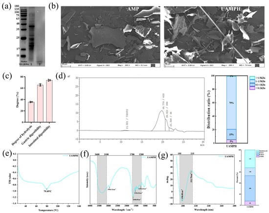
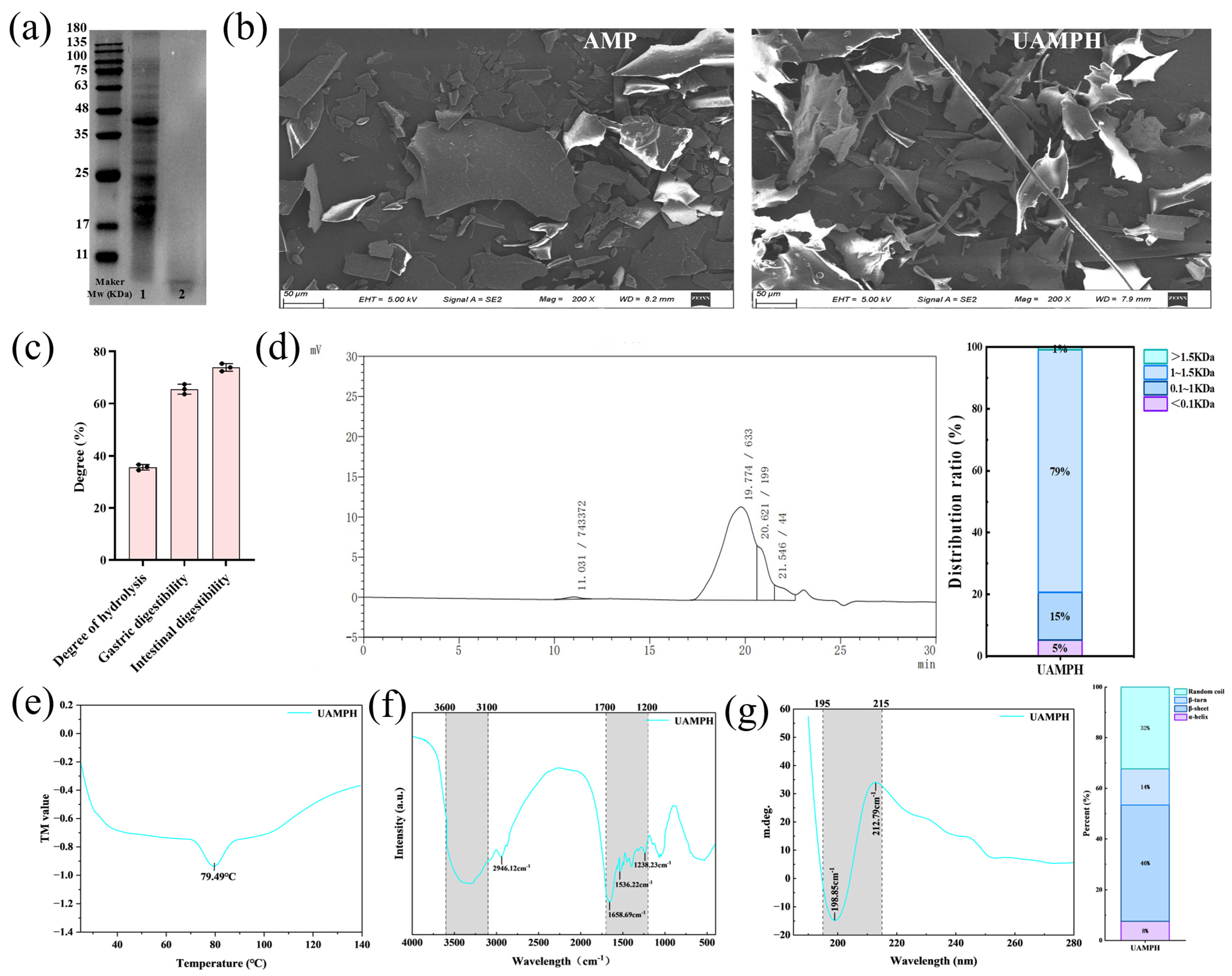
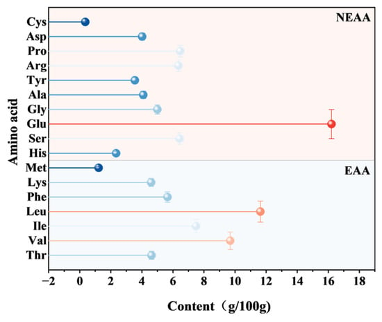
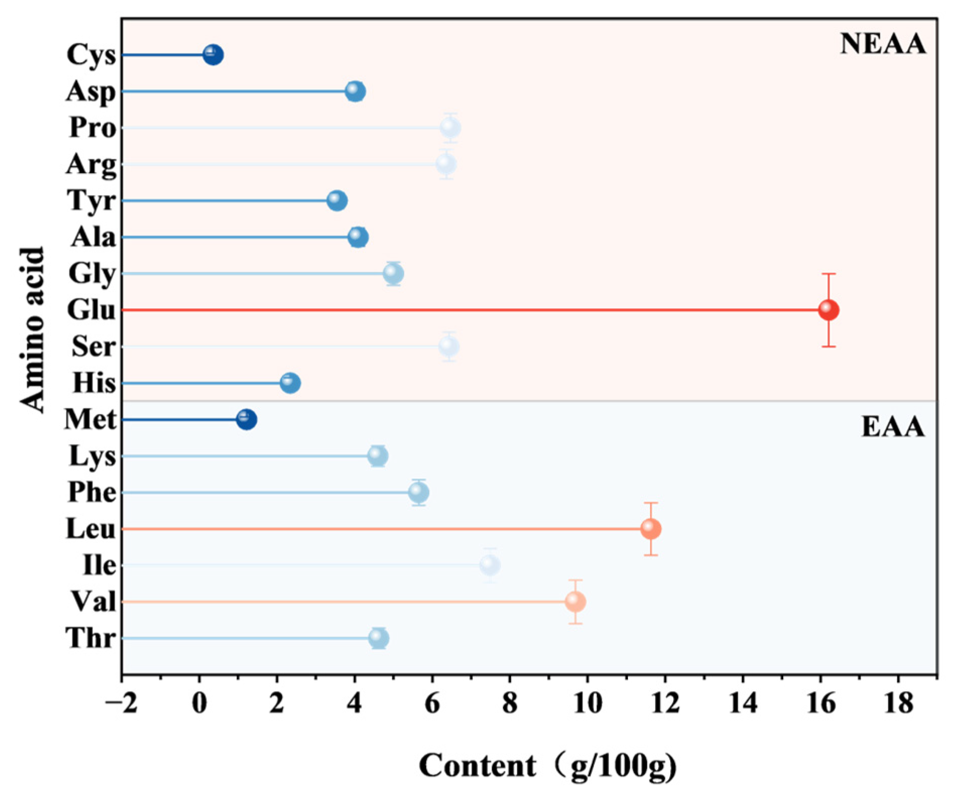
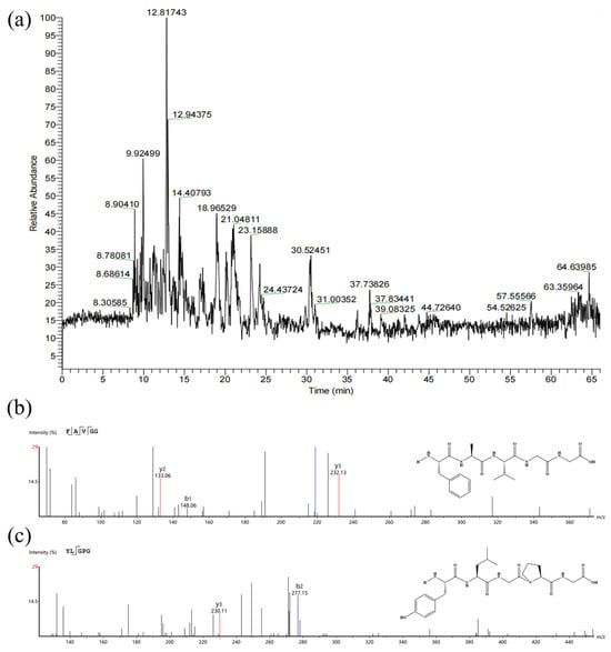
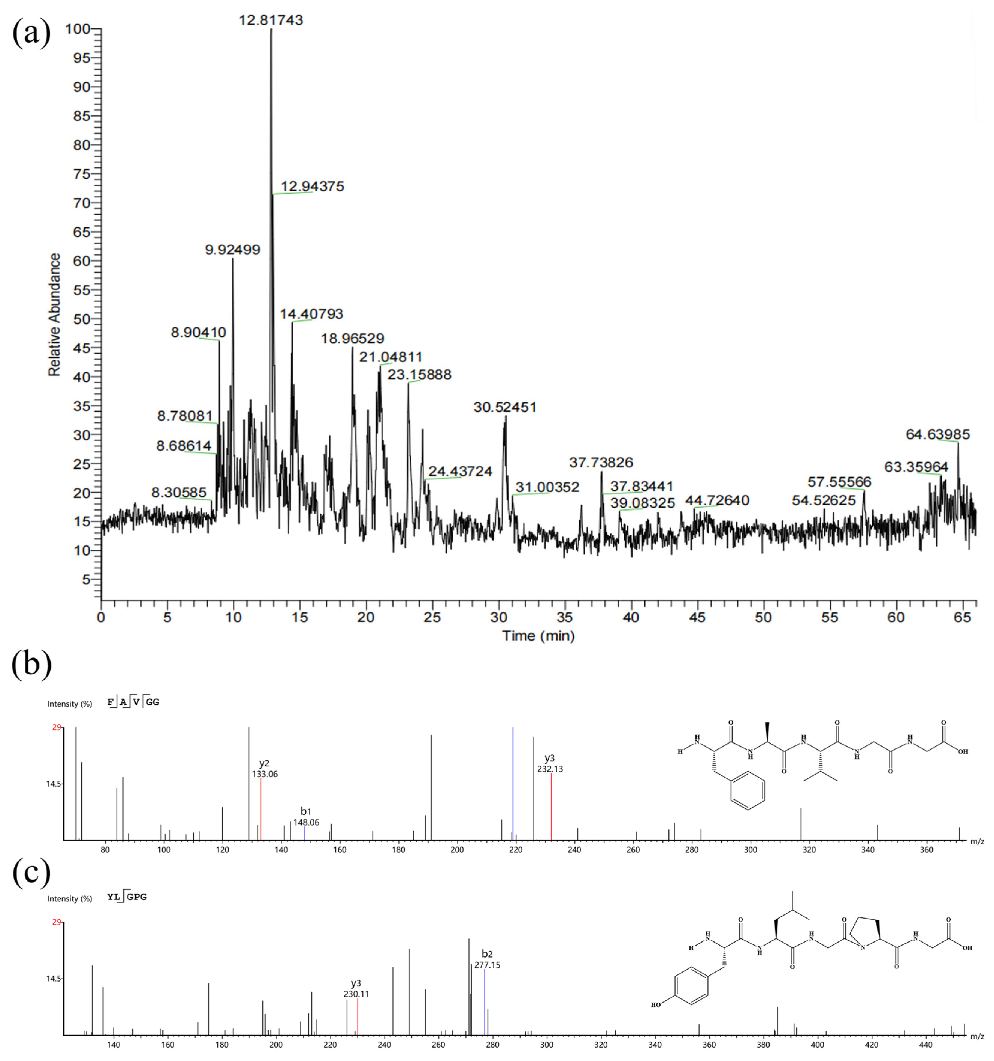
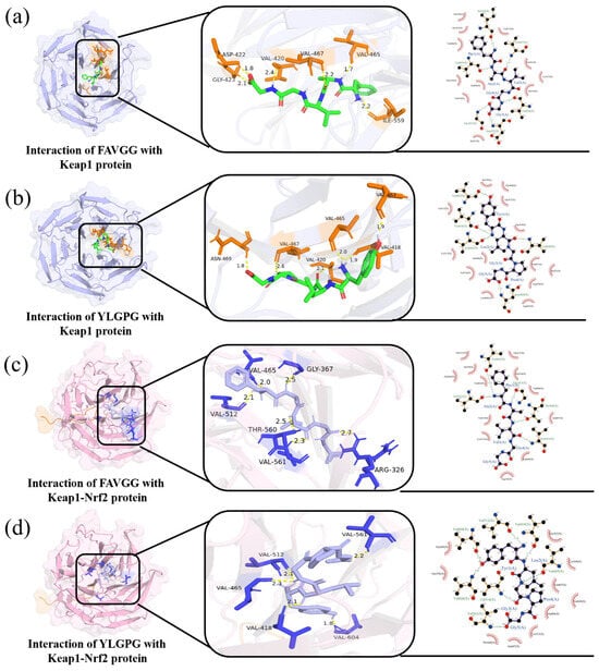
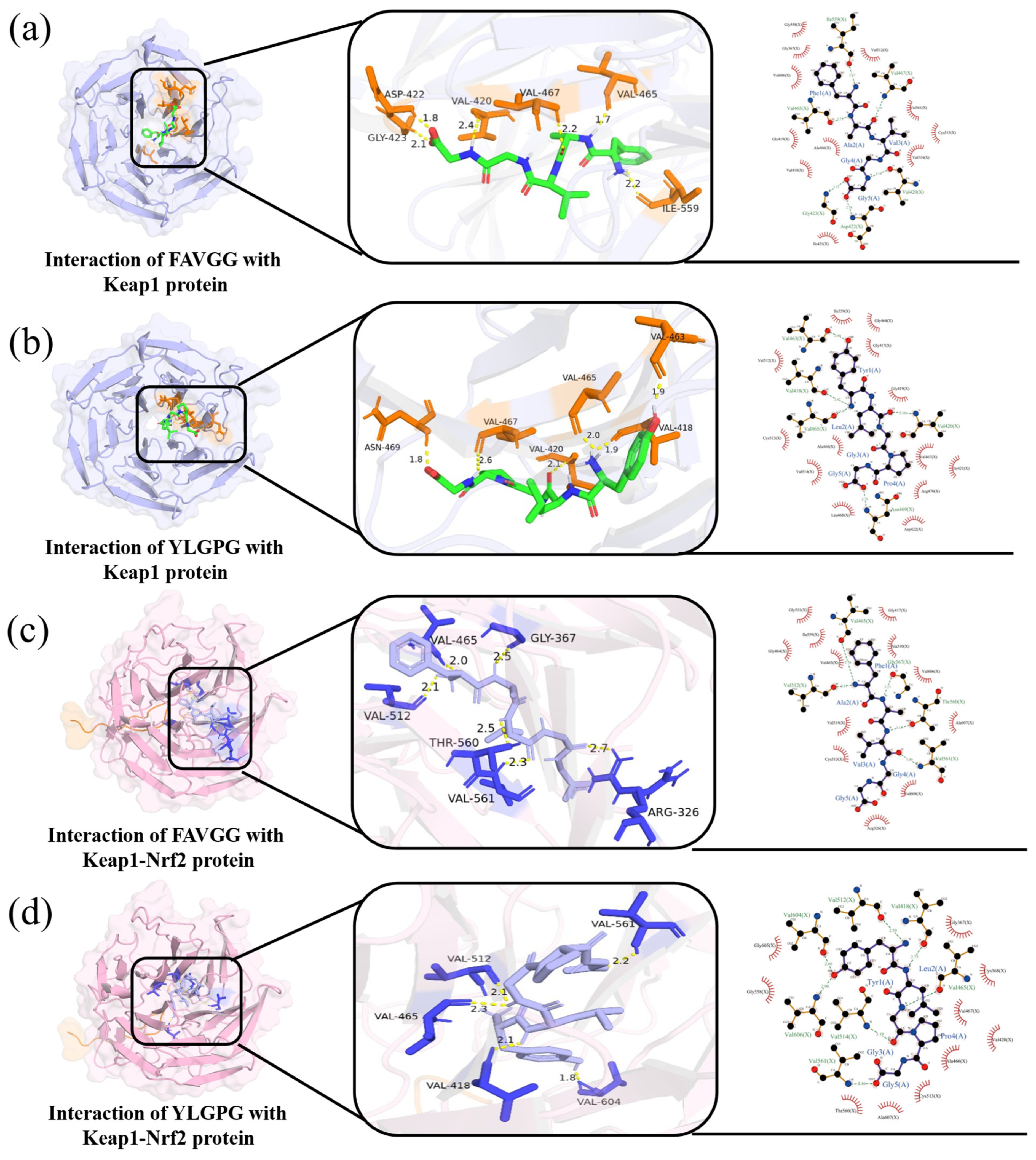
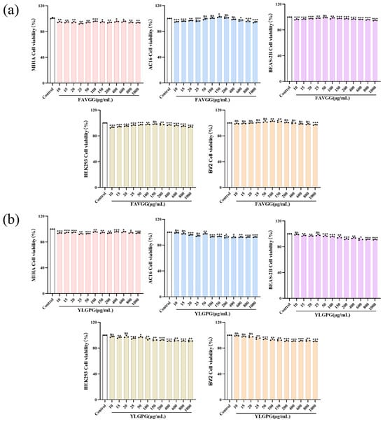
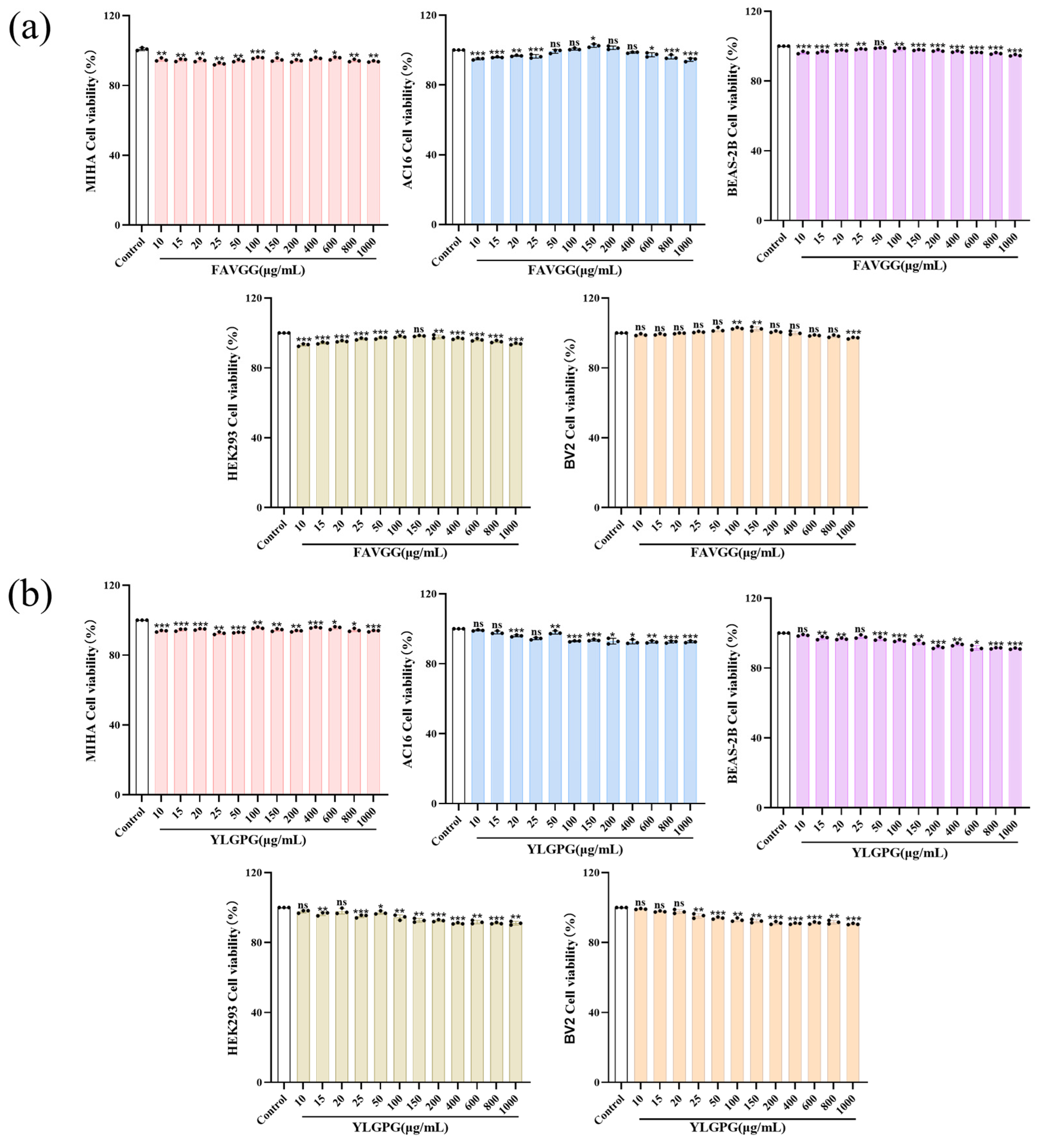
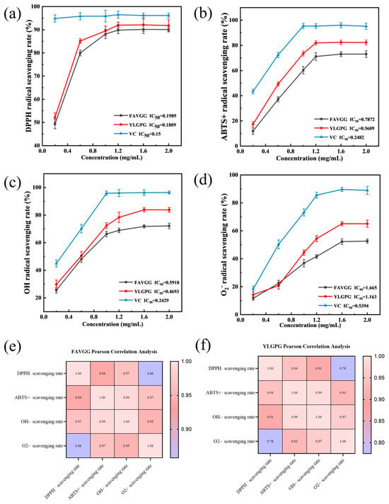
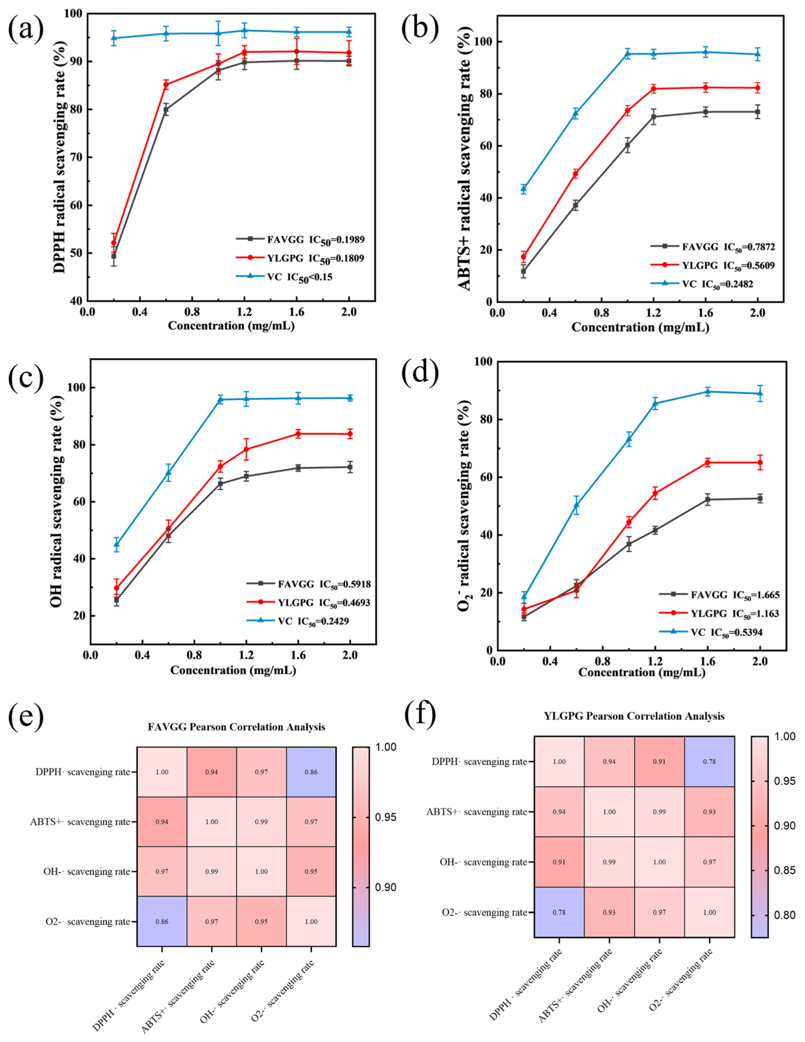
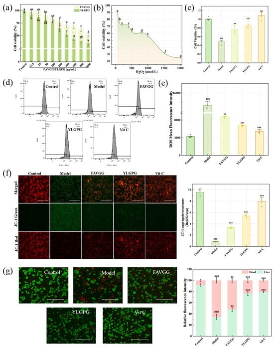
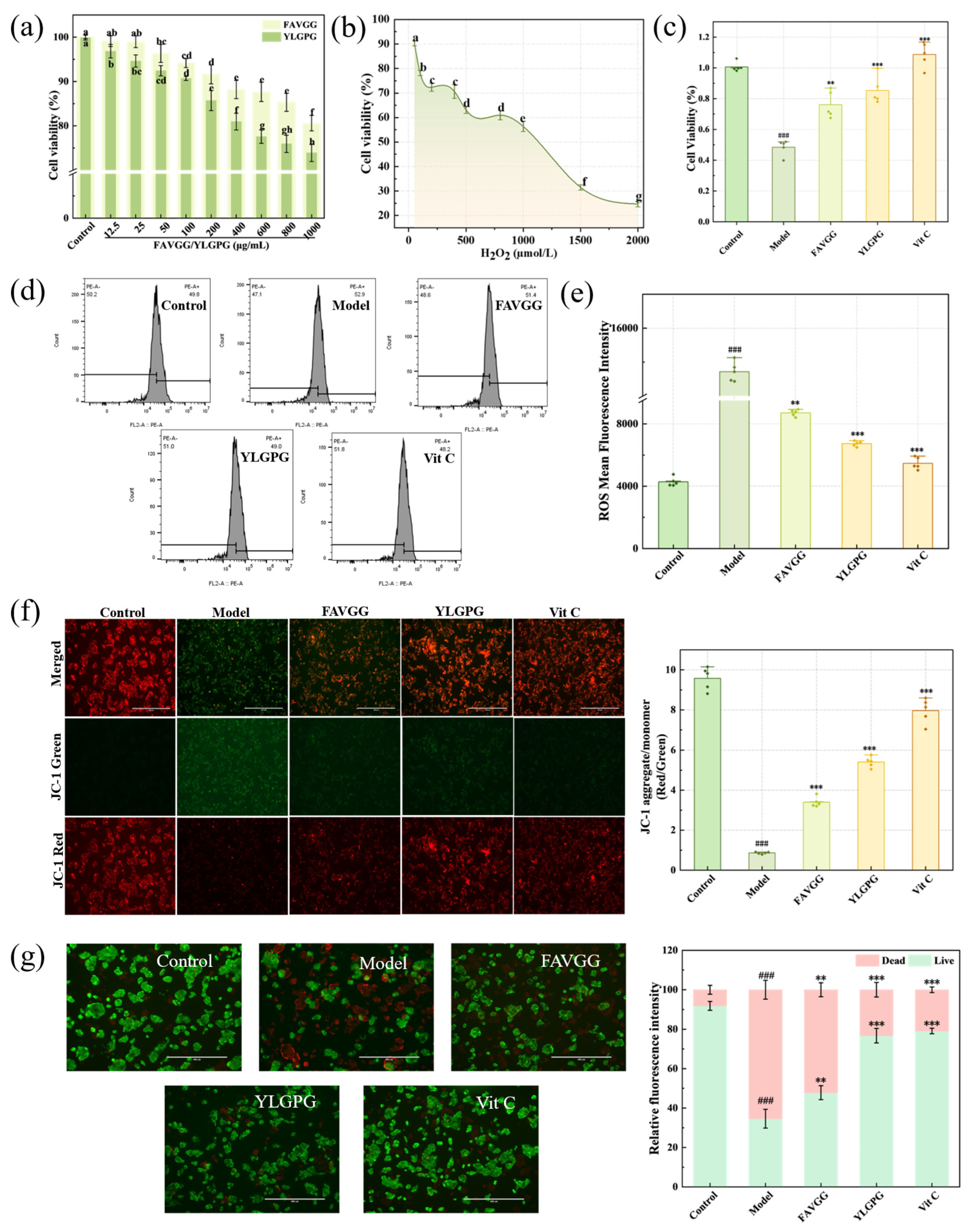
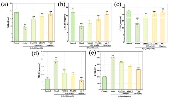
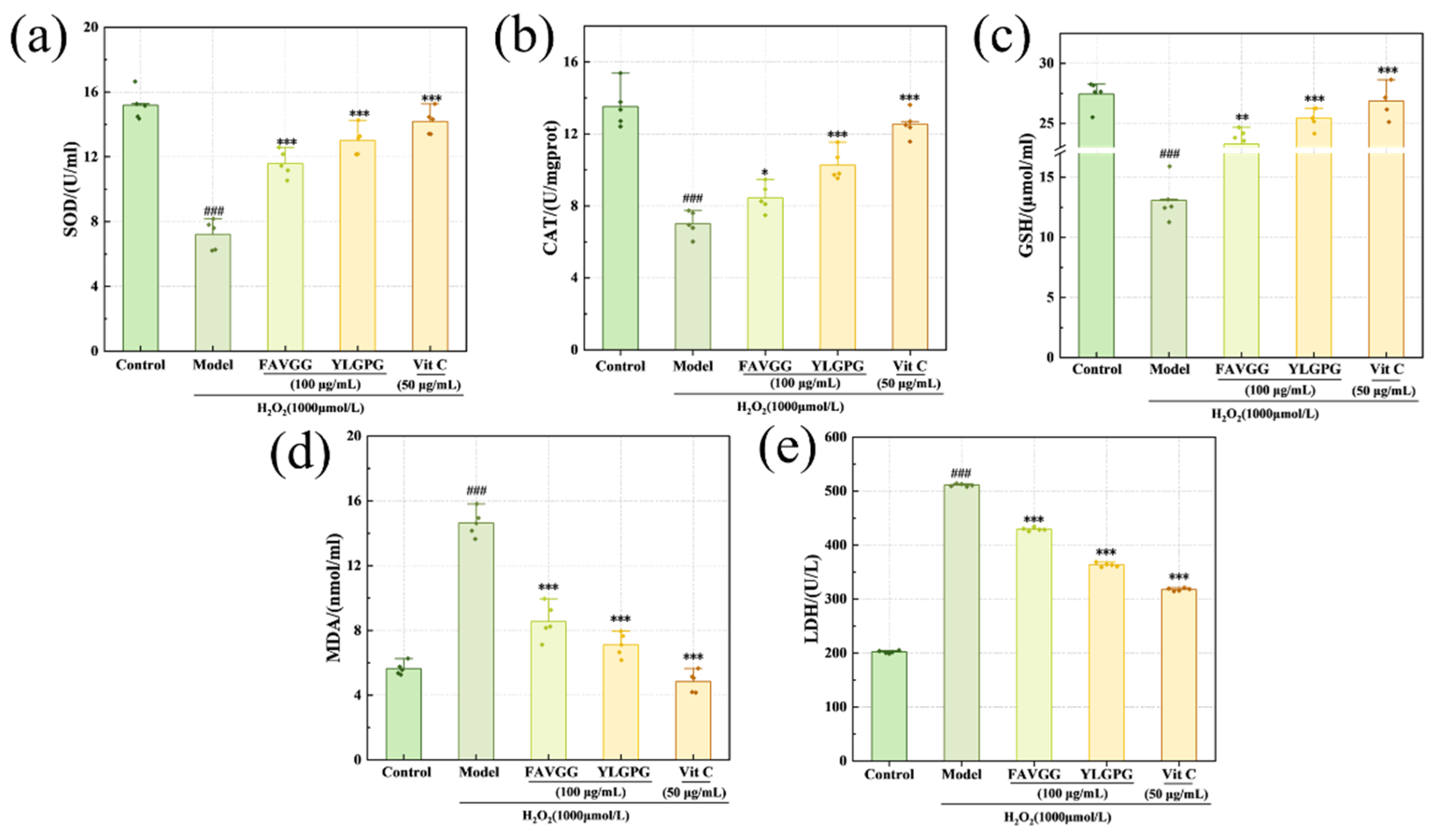
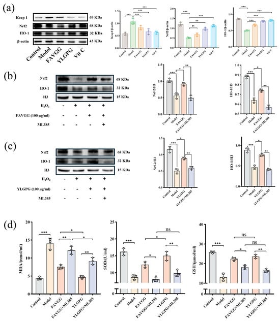
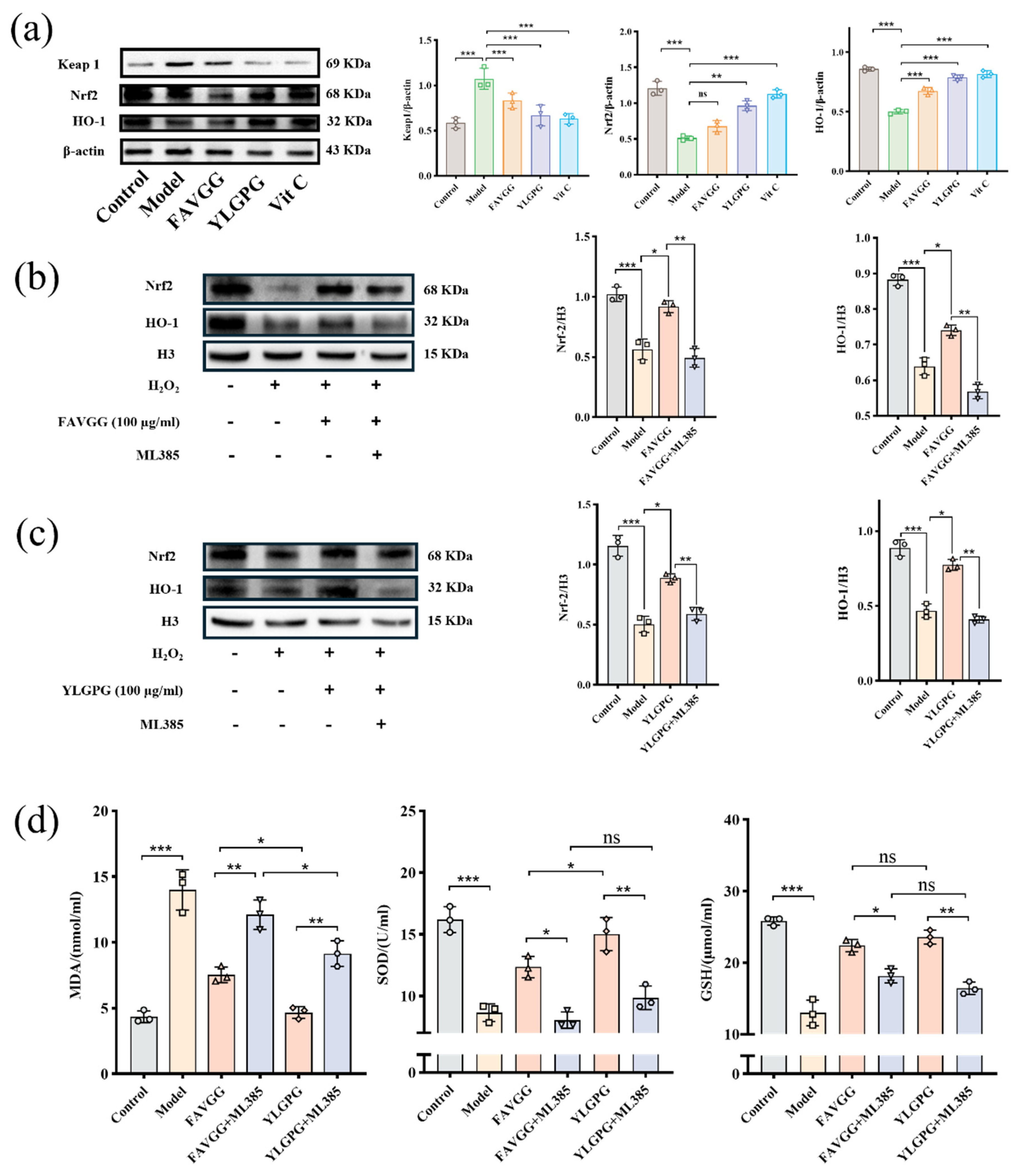
Disclaimer/Publisher’s Note: The statements, opinions and data contained in all publications are solely those of the individual author(s) and contributor(s) and not of MDPI and/or the editor(s). MDPI and/or the editor(s) disclaim responsibility for any injury to people or property resulting from any ideas, methods, instructions or products referred to in the content. |
© 2026 by the authors. Licensee MDPI, Basel, Switzerland. This article is an open access article distributed under the terms and conditions of the Creative Commons Attribution (CC BY) license.