


Abstract
Chemotherapy-induced cognitive impairment (CICI) is an increasingly recognized complication of cancer treatment, particularly among breast cancer survivors, characterized by persistent difficulties in memory, attention, executive function, and processing speed. This review summarizes current evidence on the clinical phenotype and biological mechanisms of CICI and examines the rationale for investigating medical ozone as a redox-modulating intervention. A translational narrative review was conducted using literature published between 2000 and September 2026, retrieved from PubMed/MEDLINE, Scopus, Web of Science, and Google Scholar, complemented by manual reference-list screening. Clinical, preclinical, mechanistic, and evidence-synthesis studies were examined to critically assess the pathophysiology of CICI and the biological rationale for investigating ozone-induced redox modulation. Current evidence points to a multifactorial pathophysiology of CICI and provides a rationale for testing whether controlled redox modulation can influence some of the pathways involved, including activation of endogenous antioxidant and adaptive pathways. Preliminary evidence from chemotherapy-induced peripheral neuropathy and selected neurological or cognitive conditions provides indirect translational support, but does not establish efficacy in CICI. Medical ozone should therefore be considered investigational in this context. The hypothesis should be tested in randomized, sham-controlled trials using validated cognitive outcomes, mechanistic biomarkers, and appropriate oncological safety monitoring to determine whether ozone-induced redox adaptation can produce clinically meaningful cognitive benefit.
1. Introduction
Improved cancer survival has increased attention to the long-term functional consequences of cancer and its treatment. Among these consequences, chemotherapy-induced cognitive impairment (CICI) has emerged as a clinically relevant problem, particularly among patients treated for breast cancer because of its high prevalence, high use of chemotherapy, and long survival. Patients may report difficulties with memory, concentration, attention, executive functioning, and mental processing speed, commonly described as “brain fog” or “chemobrain.” Although the magnitude and persistence of these alterations vary considerably, longitudinal studies and evidence syntheses indicate that cognitive changes can occur during and after systemic treatment and may persist beyond completion of therapy [1,2,3,4,5,6].
Breast cancer provides a particularly informative setting in which to investigate CICI. Modern systemic management frequently involves combinations of chemotherapy, endocrine therapy, targeted agents, and, in selected patients, radiotherapy. Cognitive outcomes, however, cannot be attributed exclusively to a particular anticancer drug. Prospective studies indicate that some patients already exhibit cognitive difficulties before systemic treatment, while post-treatment trajectories are heterogeneous [3,4,6]. Fatigue, sleep disturbances, psychological distress, menopausal symptoms, pain, and other cancer-related factors may further influence subjective complaints and objective neuropsychological performance [1,2,6]. Current evidence therefore supports a multifactorial model in which cancer, treatment, and patient characteristics may all contribute to cognitive dysfunction.
The biological mechanisms underlying CICI remain incompletely characterized. Converging clinical and experimental evidence implicates interconnected processes including systemic and central inflammation, oxidative stress, mitochondrial dysfunction, altered neuroplasticity, glial activation, and possible neurovascular or blood–brain barrier disturbances [7,8,9,10,11]. In breast cancer populations, inflammatory biomarkers such as interleukin-6 (IL-6) and tumor necrosis factor-α (TNF-α) have been associated with poorer cognitive outcomes, although findings vary according to treatment stage, biomarker, and cognitive assessment [9,10]. Experimental models further suggest chemotherapy-associated microglial activation and inflammatory signaling involving nuclear factor-κB (NF-κB) and the NOD-like receptor family pyrin domain-containing 3 (NLRP3) inflammasome [12]. Oxidative stress may interact closely with these inflammatory processes, as redox imbalance can activate inflammatory signaling while inflammation can further increase reactive oxygen species (ROS). The resulting redox–inflammatory interface therefore represents a biologically plausible component of treatment-related neurological dysfunction.
These observations provide a rationale for investigating interventions that modify endogenous redox responses. Medical ozone therapy, which explicitly excludes ozone inhalation, encompasses several administration routes, including systemic approaches such as major autohemotherapy (MAH) and rectal insufflation, as well as local applications, including intra-articular and other tissue-directed procedures. These modalities differ in their exposure characteristics and should not be considered interchangeable when interpreting the available evidence.
Ozone is a potent oxidizing molecule and should not be conceptualized as a conventional antioxidant; rather, controlled medical oxygen–ozone (O2/O3) exposure has been proposed to generate transient oxidative signals capable of activating endogenous adaptive pathways, including nuclear factor erythroid 2-related factor 2 (Nrf2)-dependent antioxidant and cytoprotective responses [13,14,15,16]. Experimental and selected clinical studies have reported changes in glutathione metabolism, antioxidant enzymes, oxidative damage markers, and inflammatory mediators following medical ozone administration. However, these findings derive from heterogeneous conditions and experimental models, cannot be directly extrapolated to CICI, and therefore provide only a hypothetical rationale that requires direct experimental validation. Ozone has also been investigated in chemotherapy-associated toxicity, including experimental models involving doxorubicin, cisplatin, methotrexate, and bleomycin [17]. Preliminary clinical studies have also reported possible improvements in chemotherapy-induced peripheral neuropathy (CIPN), providing an early human translational signal in another form of treatment-related neurotoxicity [18]. Two registered phase II/III randomized controlled trials are currently recruiting patients with CIPN: NCT04299893 (O3NPIQ), which evaluates ozone therapy for neuropathic pain, and NCT06706544 (OzoParQT), which focuses on numbness and tingling. According to ClinicalTrials.gov records updated in September 2026, their estimated study completion dates are December 2027 and March 2030, respectively. Neither trial has reported results to date, and neither directly investigates CICI [19,20]. These observations do not establish efficacy for cognitive impairment but provide a rationale for examining whether related redox and inflammatory mechanisms may be relevant to other manifestations of chemotherapy-induced neurological dysfunction.
Additional translational clues arise from non-oncological neurological conditions. Brain fog and cognitive dysfunction have been described following systemic diseases such as Long COVID, while experimental studies have reported neuroprotective or cognitive effects of ozone in models of cerebral ischemia, hypoxic–ischemic injury, and Alzheimer’s disease [21,22,23,24,25]. Preliminary retrospective evidence has also suggested cognitive changes following O2/O3 MAH in patients with mild cognitive impairment [26]. These observations remain indirect: Long COVID studies do not establish a cognitive-specific effect of ozone, animal models cannot establish clinical efficacy, and mild cognitive impairment should not be equated with Alzheimer’s disease. They therefore provide translational clues rather than evidence of efficacy in CICI.
In this review, we examine the biological basis of CICI, with particular emphasis on breast cancer and the interconnected roles of oxidative stress, inflammation, mitochondrial dysfunction, and neuroinflammation. The available experimental and clinical evidence for medical ozone as a potential redox-modulating intervention is critically evaluated, including evidence from chemotherapy-associated toxicity and CIPN and selected neurological conditions that may provide mechanistic insights. The evidence gaps are then considered within a translational framework designed to determine whether controlled medical O2/O3 exposure can modify pathways implicated in CICI and whether this hypothesis warrants prospective clinical investigation in appropriately selected patients with cancers outside the central nervous system (CNS).
2. Materials and Methods
2.1. Review Design and Objectives
This manuscript was designed as a narrative translational review integrating clinical, mechanistic, preclinical, and emerging translational evidence relevant to CICI and the biological rationale for investigating medical ozone.
The review addressed four objectives: (i) to characterize CICI, with particular emphasis on breast cancer and systemic chemotherapy; (ii) to examine the biological mechanisms potentially contributing to CICI, including oxidative stress, inflammation, mitochondrial dysfunction, neuroimmune activation, and neurovascular alterations; (iii) to evaluate the biological rationale and available clinical evidence concerning medical ozone as a redox-modulating intervention; and (iv) to integrate evidence from CIPN and selected non-oncological neurological or cognitive conditions—including post-acute sequelae of severe acute respiratory syndrome coronavirus 2 (SARS-CoV-2) infection, cerebrovascular disease, mild cognitive impairment, and neurodegenerative disorders—as indirect translational evidence.
The review was not intended to provide a quantitative estimate of treatment efficacy or to establish the clinical effectiveness of ozone therapy for CICI.
2.2. Search Strategy and Information Sources
This study was designed as a translational narrative review aimed at integrating clinical, preclinical, and mechanistic evidence concerning CICI and the biological rationale for investigating medical ozone as a redox-modulating intervention.
A narrative literature search was conducted in PubMed/MEDLINE, Scopus, Web of Science, and Google Scholar between 1 and 15 September 2026, covering publications from January 2000 through September 2026. Additional relevant publications were identified through manual screening of the reference lists of key original studies, systematic reviews, and narrative reviews.
Searches combined controlled vocabulary, including Medical Subject Headings (MeSH) where applicable, with free-text terms. The principal search domains included: (i) “cancer-related cognitive impairment”, “cancer therapy-related cognitive impairment”, “chemotherapy-related cognitive impairment”, “chemobrain”, and “chemofog”; (ii) “breast cancer” combined with “cognition” or “cognitive dysfunction”; (iii) “chemotherapy” combined with “oxidative stress”, “neuroinflammation”, “mitochondrial dysfunction”, “microglia”, or “neurovascular”; (iv) “medical ozone”, “ozone therapy”, “oxygen–ozone therapy”, and “major autohemotherapy”; (v) “ozone” combined with “Nrf2”, “glutathione”, “oxidative stress”, “inflammation”, or “mitochondria”; and (vi) “ozone therapy” in relation to chemotherapy-induced peripheral neuropathy and selected neurological or cognitive conditions.
The broader term “cancer-related cognitive impairment” (CRCI) was included to capture studies addressing cancer-associated cognitive vulnerability and potentially overlapping mechanisms. However, CRCI and CICI were not considered interchangeable: the primary focus of this review remained cognitive impairment associated with chemotherapy, while studies addressing cancer-related cognitive alterations independently of chemotherapy were considered only when relevant to the broader pathophysiological context.
Search terms were adapted to the indexing characteristics of each database and refined iteratively during literature retrieval to capture relevant clinical, experimental, and translational evidence. The purpose of this search was not to perform an exhaustive systematic assessment of therapeutic efficacy, but to develop a critical synthesis connecting established CICI pathophysiological mechanisms with experimental evidence concerning ozone-induced adaptive redox responses.
Accordingly, the review was not conducted under a systematic review protocol, and no formal Preferred Reporting Items for Systematic Reviews and Meta-Analyses (PRISMA)-based screening or quantitative meta-analysis was performed. The findings are therefore interpreted within the methodological scope of a translational narrative review, with particular attention to distinguishing direct clinical evidence from indirect mechanistic and translational observations.
2.3. Scope and Eligibility of Evidence
Because this was a narrative translational review, evidence was selected according to relevance to the predefined conceptual framework rather than through formal systematic-review eligibility criteria. The evidence considered included: 1. prospective and retrospective clinical studies evaluating cognitive function in patients with cancer; 2. observational studies and clinical trials addressing CICI, particularly in breast cancer; 3. systematic reviews, meta-analyses, umbrella reviews, and relevant narrative reviews concerning CICI; 4. preclinical and translational studies investigating oxidative stress, neuroinflammation, mitochondrial dysfunction, neuroimmune mechanisms, neurovascular alterations, and neuronal plasticity in the context of cancer or anticancer therapy; 5. experimental and clinical studies addressing the biological effects of medical ozone, particularly redox signaling, Nrf2-related pathways, glutathione homeostasis, inflammation, mitochondrial function, and adaptive responses; 6. clinical and preclinical studies of ozone therapy in CIPN and other chemotherapy-associated toxicities; 7. studies of ozone therapy in neurological or cognitive conditions outside oncology, including post-acute sequelae of SARS-CoV-2 infection, cerebrovascular disease, mild cognitive impairment, and neurodegenerative disorders, when considered relevant to the proposed translational hypothesis.
Studies involving patients with primary brain tumors or brain metastases were not considered part of the target clinical population for CICI, because structural CNS disease represents a distinct biological context and would introduce substantial confounding into the proposed framework. Such studies were considered only when they provided relevant mechanistic information that could be clearly separated from the CICI clinical phenotype.
Priority was given to peer-reviewed studies providing direct clinical evidence, well-characterized experimental models, systematic evidence syntheses, and recent publications relevant to the current state of the field. Conference abstracts, non-peer-reviewed material, and publications for which the available information was insufficient to evaluate their relevance were generally excluded from the evidentiary synthesis.
No language restrictions were applied during the literature search. Publications in English and other languages were considered for inclusion, provided that their scientific content could be adequately evaluated by the authors.
2.4. Evidence Interpretation and Critical Appraisal
Evidence was critically appraised according to its clinical relevance, methodological strength, and directness in relation to CICI and the proposed therapeutic hypothesis. For interpretative purposes, the available evidence was considered within four broad, non-mutually exclusive categories:
Direct clinical evidence: Studies evaluating cognitive impairment or cognitive outcomes in cancer populations, particularly patients with breast cancer.
Mechanistic and experimental evidence: Preclinical and translational investigations addressing biological pathways potentially relevant to CICI or ozone-induced adaptive responses.
Indirect clinical evidence: Clinical studies investigating ozone therapy in related conditions, particularly CIPN and neurological or cognitive disorders outside oncology.
Hypothesis-generating evidence: Preliminary clinical observations or mechanistic findings providing biological plausibility without establishing efficacy or causality in CICI.
These categories were used as a conceptual framework to distinguish clinical observations from experimental findings, indirect clinical evidence, and unvalidated therapeutic hypotheses throughout the narrative synthesis. They were not intended as a formal study-level grading system, and individual publications could contribute to more than one category.
The critical appraisal considered study design, sample size, control groups, blinding, outcome definition, follow-up duration, intervention reproducibility, and potential sources of bias. Observational findings and uncontrolled clinical reports were not considered equivalent to evidence from randomized controlled trials, and mechanistic plausibility was not interpreted as proof of clinical efficacy.
2.5. Narrative Synthesis and Translational Integration
Given the substantial heterogeneity in clinical populations, interventions, experimental models, cognitive outcomes, and biological endpoints, a qualitative narrative synthesis was performed rather than a quantitative meta-analysis. Evidence was organized into interconnected thematic domains: 1. clinical phenotype of CICI, with emphasis on breast cancer; 2. biological mechanisms of cancer therapy-related cognitive dysfunction, including oxidative stress, inflammation, mitochondrial dysfunction, neuroimmune activation, and neurovascular alterations; 3. redox biology of medical ozone, including adaptive oxidative signaling, Nrf2-related responses, and glutathione homeostasis; 4. clinical evidence in chemotherapy-associated neurological toxicity, particularly CIPN; 5. evidence from non-oncological neurological and cognitive conditions; 6. integrated translational model linking systemic redox modulation with potential effects on CNS function; 7. clinical translation, safety considerations, methodological requirements, and priorities for future research.
We distinguished associative findings from mechanistic evidence and from evidence of clinical efficacy. Particular care was taken to avoid extrapolating findings from CIPN or non-oncological neurological conditions directly to CICI.
2.6. Development of the Translational Hypothesis
The evidence was subsequently used to formulate a mechanistic hypothesis linking systemic cancer therapy-related biological perturbations with cognitive dysfunction and to examine whether controlled redox modulation by medical ozone could theoretically influence this pathway.
The proposed model was developed from convergent evidence concerning oxidative and inflammatory signaling, mitochondrial and neurovascular function, neuroimmune mechanisms, and endogenous antioxidant responses. The model was explicitly formulated as a testable hypothesis rather than as an established therapeutic mechanism.
The translational framework was subsequently used to identify key requirements for future clinical investigation, including patient selection, timing of treatment, standardized ozone exposure, sham-controlled trial design, cognitive outcome assessment, mechanistic biomarkers, and oncological safety monitoring.
2.7. Generative Artificial Intelligence Disclosure
Generative artificial intelligence tools were used to assist with language editing, text organization, literature exploration, and the development of the conceptual framework. These tools did not replace independent scientific assessment by the authors. The selection of relevant publications, verification of original source information, evaluation of evidence, and approval of the final scientific interpretations remained the responsibility of the authors. AI-generated suggestions were critically reviewed against the original sources before incorporation into the manuscript.
3. Chemotherapy-Induced Cognitive Impairment
3.1. Clinical Definition and Phenotype
CICI refers to cognitive alterations occurring in temporal association with chemotherapy, although their development and severity may also be influenced by cancer-related, treatment-related, and individual factors. The clinical phenotype is heterogeneous and may involve both objectively measurable neuropsychological deficits and subjective cognitive complaints reported by patients. The latter are frequently described using terms such as “brain fog,” “chemobrain,” or “chemotherapy-related cognitive dysfunction.” Importantly, subjective cognitive complaints and objective neuropsychological impairment do not necessarily coincide, reflecting the contribution of psychological, behavioral, fatigue-related, and other cancer-associated factors to the patient’s perception of cognitive functioning [1,6,9].
In breast cancer, the cognitive domains most consistently reported as affected include episodic and working memory, attention and concentration, executive functioning, verbal fluency, and processing speed [1,2,5,6,9]. The magnitude and pattern of impairment, however, vary substantially across studies according to the neuropsychological instruments employed, timing of assessment, definition of impairment, treatment regimen, and characteristics of the study population. This methodological heterogeneity has contributed to considerable variability in estimates of the frequency and persistence of CICI [1].
Subjective cognitive complaints and objectively measured impairment should be considered separately. Subjective complaints may be clinically meaningful even in the absence of deficits on conventional neuropsychological testing, particularly when they interfere with occupational, social, or everyday functioning. Conversely, subtle objective deficits may occur without prominent subjective complaints. Contemporary approaches to CICI consequently favor multidimensional assessment combining patient-reported outcomes with standardized neuropsychological measures rather than relying on either approach alone [1,27].
3.2. Evidence from Breast Cancer Populations
Breast cancer represents the most extensively investigated solid tumor population in the CICI literature. An umbrella review published in 2024 synthesized evidence from systematic reviews and meta-analyses evaluating the cognitive effects of breast cancer treatments and concluded that both chemotherapy and endocrine therapy are consistently associated with cognitive concerns or impairment, particularly involving memory, attention/concentration, executive functions, and processing speed [1]. These findings support the recognition of CICI as a clinically relevant phenomenon rather than an isolated anecdotal complaint.
A 2023 meta-analysis and meta-regression including 20 studies and more than 2500 women with breast cancer similarly found an overall association between chemotherapy and poorer cognitive performance, with working memory showing one of the clearest effects [2]. The authors also identified patient characteristics that could influence the magnitude of cognitive impairment, emphasizing that treatment exposure alone does not fully explain interindividual variability.
Longitudinal studies are particularly informative because they provide a pretreatment reference for subsequent cognitive changes. In a prospective study of 71 women receiving doxorubicin and cyclophosphamide with or without a taxane, 23% showed cognitive impairment before chemotherapy. Following treatment, significant decreases were observed in visuospatial ability, attention, delayed memory, and motor function, followed by partial improvement six months after completion of chemotherapy; immediate memory, language, and executive function did not show significant longitudinal changes [3]. These findings illustrate both the potential acute impact of chemotherapy and the heterogeneity of recovery across cognitive domains.
Other prospective studies have demonstrated more persistent or delayed changes. Wefel and colleagues evaluated women with breast cancer before chemotherapy, during and shortly after treatment, and approximately one year after treatment. Cognitive dysfunction was already present in 21% before chemotherapy; 65% showed cognitive decline during the acute treatment interval, and 61% of those evaluated at long-term follow-up demonstrated decline relative to baseline. Learning and memory, executive function, and processing speed were particularly affected. Notably, a subgroup exhibited newly emerging delayed cognitive decline after treatment rather than simple persistence of an early deficit [5]. Although the sample was relatively small, these observations support the possibility that cognitive trajectories following chemotherapy are not uniform and may include both transient and delayed patterns.
The importance of pretreatment assessment is further illustrated by studies comparing patients receiving different forms of breast cancer treatment. In a prospective study of 81 women with non-metastatic breast cancer (including 41 patients receiving chemotherapy and 40 receiving radiotherapy without chemotherapy), cognitive alterations were observed following breast cancer treatment irrespective of whether chemotherapy was administered, while chemotherapy was additionally associated with impaired verbal fluency [28]. Similarly, another prospective study found measurable pretreatment cognitive impairment in a substantial proportion of women and demonstrated heterogeneous cognitive trajectories following chemotherapy [3]. Collectively, these observations argue against a simplistic model in which chemotherapy alone initiates cognitive dysfunction in every affected patient.
More recent longitudinal evidence reinforces this interpretation. The nationwide prospective cohort study by Janelsins and colleagues followed 580 patients with breast cancer receiving chemotherapy and 363 control participants, assessing trajectories in memory, attention, and executive function from before chemotherapy through treatment and follow-up [29]. Such large prospective cohorts are particularly valuable because they permit adjustment for factors such as age, education, cognitive reserve, anxiety, and depressive symptoms, all of which may influence cognitive performance independently of treatment.
The recently reported CICARO study (NCT02753036), a prospective longitudinal observational cohort rather than an interventional clinical trial, examined cognitive function in patients with breast cancer receiving first-line chemotherapy compared with breast cancer controls not receiving chemotherapy. Cognitive assessments were performed before treatment, approximately six months after chemotherapy, and again two to three years later. Overall cognitive function declined significantly after chemotherapy compared with controls, with figural memory contributing substantially to the observed difference [30]. The long-term follow-up of such cohorts is important because the persistence of cognitive alterations remains one of the clinically most relevant unresolved questions.
3.3. Temporal Trajectory: Acute, Persistent, or Delayed Impairment?
The temporal course of CICI is complex; evidence does not support a single stereotyped trajectory. Some patients experience cognitive deterioration during chemotherapy followed by partial or substantial recovery after treatment, whereas others demonstrate persistent deficits or delayed deterioration. In a meta-analysis specifically examining subjective cognitive impairment, the largest association with chemotherapy was observed within the first month after treatment, suggesting that at least part of the subjective cognitive burden may be particularly pronounced during the acute treatment period [31]. However, this finding should not be interpreted as evidence that all cognitive effects are transient.
The longitudinal study by Wefel et al. is particularly informative in this respect because it identified both continuing decline and newly emerging delayed impairment one year after chemotherapy [5]. Conversely, the study by Jansen et al. observed improvement in several cognitive domains six months after chemotherapy [3]. These differences may reflect genuine interindividual heterogeneity, variations in baseline vulnerability, treatment regimens, assessment instruments, sample sizes, and the distinction between subjective and objective cognitive outcomes.
The available longitudinal data are therefore difficult to reconcile with a single, uniform “chemobrain” phenotype. CICI is more appropriately considered a spectrum of cognitive trajectories, ranging from pre-existing cognitive vulnerability through acute treatment-associated deterioration to partial recovery, persistent dysfunction, or delayed cognitive decline.
3.4. Chemotherapy Is Important, but It Is Not the Whole Explanation
One of the most important conclusions emerging from the longitudinal literature is that chemotherapy should not be regarded as the sole determinant of cognitive dysfunction. Cognitive impairment may be present before the first chemotherapy cycle, indicating that cancer itself, associated systemic inflammation, metabolic alterations, psychological distress, sleep disturbance, fatigue, anemia, pain, hormonal changes, and other factors may contribute to cognitive vulnerability [1,3,4,9].
This limitation is relevant when interpreting studies that lack pretreatment cognitive assessment. Without a pretreatment assessment, cognitive deficits identified after chemotherapy cannot reliably be attributed to the anticancer treatment. Prospective studies incorporating baseline neuropsychological evaluation have repeatedly demonstrated that a subset of patients already performs below expected levels before systemic treatment [3,4,5]. Consequently, the most appropriate conceptual model is not simply “chemotherapy causes cognitive impairment,” but rather that systemic anticancer therapy may interact with pre-existing and cancer-related biological and psychosocial vulnerabilities to produce clinically heterogeneous cognitive trajectories.
This multifactorial interpretation is also consistent with the observation that different treatments may affect cognition through partially overlapping pathways. Hormone therapy, targeted therapies, and immunotherapy have resulted in improved survival rates for some patients with, however, potential impact on cognition [6]. Hormone therapy has been investigated extensively in breast cancer. A systematic review of 12 studies involving 2756 patients found evidence suggesting alterations in verbal memory, verbal fluency, motor speed, attention, and working memory in association with endocrine therapy, although methodological heterogeneity limited definitive conclusions [7]. A more recent comprehensive review similarly concluded that cognitive adverse effects of endocrine therapy are clinically relevant but remain incompletely characterized [8]. These observations are particularly relevant in breast cancer because endocrine therapy is often administered for several years, potentially extending the period during which cognitive symptoms may be experienced.
3.5. Subjective Cognitive Impairment and “Brain Fog”
The terminology of “brain fog” deserves particular attention because it is widely used by patients but does not represent a standardized neuropsychological diagnosis. Patients may use this term to describe difficulties with concentration, word finding, multitasking, mental clarity, memory, or the subjective slowing of cognitive processing. Such complaints can have substantial consequences for professional and social functioning even when conventional neuropsychological testing identifies only subtle abnormalities.
Meta-analytic evidence indicates that chemotherapy is associated with subjective cognitive impairment, with the strongest effect occurring shortly after treatment [31]. However, subjective cognitive symptoms are influenced by fatigue, anxiety, depression, sleep quality, menopausal symptoms, pain, and perceived overall health. Consequently, subjective cognitive impairment should neither be dismissed as purely psychological nor assumed to represent direct neuronal toxicity. Instead, it should be considered one component of a multidimensional clinical phenotype.
The distinction also affects the design and interpretation of intervention studies. A treatment that improves subjective “brain fog” without measurable changes in objective cognitive performance may still have clinical value, but it should not be described as reversing neurocognitive impairment unless appropriate objective endpoints demonstrate such an effect. Conversely, improvements in neuropsychological test performance without corresponding improvement in everyday cognitive functioning may have uncertain clinical significance. Future interventional studies should therefore incorporate both patient-reported cognitive outcomes and validated objective neuropsychological measures.
3.6. Scope of the Present Review
For the purposes of this review, CICI is considered primarily in patients with “non-central nervous system cancers”, with particular emphasis on breast cancer, and in the context of systemic chemotherapy. Patients whose cognitive dysfunction is primarily attributable to primary brain tumors or documented brain metastases are outside the intended clinical framework, because these conditions introduce direct structural CNS pathology and substantially different mechanisms of neurological dysfunction.
Although breast cancer provides the principal clinical model discussed in this review, CICI has also been investigated in patients with other non-CNS malignancies, including colorectal, lung, and hematological cancers [32,33,34]. Cognitive alterations in these populations may involve overlapping domains, such as memory, attention, executive function, and processing speed, although their frequency and trajectories vary according to treatment regimens, age, comorbidities, and assessment methods. Sex-related differences also warrant consideration, given the potential influence of hormonal status, treatment-related biological susceptibility, and psychosocial factors on cognitive outcomes. However, the predominance of female breast cancer cohorts, together with the limited availability of adequately powered sex-stratified analyses in other cancer populations, restricts direct comparisons between men and women [35]. These considerations support a broader, sex-sensitive approach to future CICI research without altering the breast cancer focus of the present review.
This distinction is essential when considering the potential role of redox-modulating interventions. The biological hypothesis developed in the following sections concerns cognitive dysfunction arising in the setting of systemic cancer and its treatment, rather than cognitive impairment caused by intracranial tumor involvement. The relevant mechanistic question is therefore whether systemic treatment-associated oxidative stress, inflammation, mitochondrial dysfunction, and neuroimmune alterations could contribute to cognitive dysfunction in the absence of direct CNS tumor infiltration.
Taken together, current evidence supports CICI as a heterogeneous and clinically meaningful complication of breast cancer and its treatment. Chemotherapy appears capable of contributing to cognitive deterioration in a subset of patients, but pretreatment impairment and substantial interindividual variability demonstrate that chemotherapy alone cannot explain the phenotype. The resulting model is consequently one of multifactorial cognitive vulnerability, in which systemic treatment may interact with inflammatory, metabolic, endocrine, redox, mitochondrial, and neuroimmune processes. This framework provides the rationale for examining the biological mechanisms underlying CICI in greater detail and, subsequently, for considering whether interventions capable of modulating these pathways could have neuroprotective potential.
4. From Clinical CICI to Its Neurobiological Substrate
The clinical heterogeneity of CICI is mirrored by the complexity of its proposed biological mechanisms. Rather than representing a single form of direct neuronal toxicity, CICI is increasingly understood as the potential consequence of interacting systemic and central processes involving inflammation, oxidative stress, mitochondrial dysfunction, altered neuroplasticity, glial activation, and, potentially, neurovascular and blood–brain barrier abnormalities [9,10,11,36]. These mechanisms may be influenced by the anticancer treatment itself but also by the underlying malignancy and by host-related factors, providing a biological framework for the heterogeneous cognitive trajectories observed in breast cancer survivors.
It is important to emphasize that much of the mechanistic evidence linking oxidative stress, mitochondrial dysfunction, and neurovascular alterations to CICI derives from preclinical studies, particularly rodent models involving doxorubicin, and from secondary literature. Although these findings provide biological plausibility, human studies directly correlating these molecular alterations with validated cognitive outcomes remain limited. Therefore, these mechanisms should be interpreted as potential contributors rather than established causal pathways in patients with CICI.
4.1. Systemic Inflammation and Neuroinflammatory Signaling
Inflammation is one of the most consistently investigated biological mechanisms in CICI. Cancer and its treatment can alter circulating cytokine profiles, and several studies have investigated whether systemic inflammatory mediators are associated with subsequent cognitive performance. In breast cancer populations, systematic reviews have identified associations between circulating cytokines (including interleukin (IL) IL-6, IL-1β, and tumor necrosis factor-α (TNF-α) and cognitive outcomes, although the magnitude and consistency of these associations vary across studies [10,11].
Several mechanisms could link peripheral inflammation with CNS function, including the capacity of peripheral inflammatory signals to influence the central nervous system. Circulating cytokines may affect brain function through several partially overlapping mechanisms, including signaling across the blood–brain barrier, activation of endothelial and perivascular cells, neural and humoral pathways, and modulation of resident immune cells. Persistent inflammatory signaling may consequently alter neuronal function without requiring direct penetration of the causative anticancer agent into neural tissue.
Microglia are of particular interest in this context. As the resident innate immune cells of the CNS, microglia can respond to systemic inflammatory signals and undergo functional activation in response to tissue stress. Sustained or dysregulated microglial activation may result in the production of pro-inflammatory mediators and ROS, potentially affecting synaptic function and neuronal homeostasis. Experimental studies of CICI provide support for this concept. In a mouse model, doxorubicin administration produced neuroinflammatory changes accompanied by cognitive impairment, while attenuation of neuroinflammation was associated with reversal of the cognitive phenotype [36]. Although such findings cannot be directly extrapolated to humans, they provide experimental evidence for a potentially causal relationship between chemotherapy exposure, neuroinflammation, and cognitive dysfunction.
More recently, experimental work using a breast cancer model has implicated the NF-κB/NLRP3 inflammatory axis in doxorubicin-associated neurobehavioral abnormalities. Pharmacological modulation of C-C chemokine receptor type 5 (CCR5) signaling attenuated neuroinflammation and behavioral deficits, supporting the possibility that chemotherapeutic exposure can activate interconnected inflammatory pathways within the CNS [12]. These findings remain preclinical, but they strengthen the rationale for considering neuroinflammation as a mechanistic component rather than merely a biomarker of CICI.
Recent mechanistic reviews have further emphasized that glial involvement in CICI should not be restricted to microglial activation. Astrocytes and oligodendrocytes may also contribute to chemotherapy-related cognitive dysfunction through altered neuroinflammatory signaling, glutamate homeostasis, synaptic support, myelin integrity, and white-matter vulnerability [37].
4.2. Oxidative Stress
Oxidative stress is another potential component of CICI. ROS are physiological mediators of cellular signaling, but excessive or inadequately controlled production can disrupt proteins, lipids, nucleic acids, mitochondrial function, and cellular membranes. Neurons may be particularly vulnerable to persistent redox imbalance because of their high energetic requirements, substantial oxygen consumption, abundance of polyunsaturated fatty acids, and limited regenerative capacity [9].
Several anticancer drugs can alter cellular redox homeostasis. Doxorubicin, for example, can participate in redox cycling and mitochondrial injury, generating oxidative stress in susceptible tissues. Other agents, including platinum compounds and antimetabolites, can also interfere with mitochondrial and cellular homeostasis [9]. Importantly, the presence of systemic oxidative stress does not by itself demonstrate that oxidative damage is responsible for cognitive dysfunction. The relevant question is whether treatment-associated redox disturbances reach biological compartments capable of influencing CNS function and whether these alterations are linked temporally and mechanistically to cognitive outcomes.
The relationship between oxidative stress and inflammation is particularly relevant. ROS can activate redox-sensitive inflammatory pathways, whereas inflammatory cytokines can increase cellular oxidative burden. This bidirectional interaction provides a potential mechanism through which an initially transient systemic perturbation could contribute to more sustained alterations in neuronal and glial function. Thus, oxidative stress and inflammation should not necessarily be regarded as independent mechanisms of CICI but rather as interconnected components of a broader redox–inflammatory network [9,10,11].
4.3. Mitochondrial Dysfunction and Impaired Cellular Energetics
Mitochondria are another potential mechanistic interface between systemic cancer therapy and CNS dysfunction. Neurons depend heavily on oxidative phosphorylation to maintain membrane potentials, synaptic transmission, axonal transport, and neurotransmitter homeostasis. Consequently, mitochondrial dysfunction can affect neuronal performance even in the absence of overt neuronal death.
Anticancer drugs have been reported to influence mitochondrial bioenergetics, mitochondrial membrane integrity, ROS generation, and apoptotic signaling [9]. Doxorubicin is particularly relevant because of its well-established mitochondrial effects in peripheral tissues and experimental evidence of mitochondrial dysfunction in neural tissue. However, the extent to which these mechanisms contribute to human CICI remains uncertain.
Mitochondrial dysfunction may nevertheless provide a mechanistic bridge between oxidative stress and neuroinflammation. Impaired electron transport can increase ROS production, while mitochondrial damage can release molecular signals capable of activating innate immune pathways. Conversely, inflammatory mediators can further compromise mitochondrial function. Such reciprocal interactions could establish a feed-forward loop in which oxidative stress, mitochondrial dysfunction, and inflammation reinforce one another.
This mechanism could also explain why cognitive symptoms may persist after systemic drug concentrations have declined. A transient systemic exposure could potentially initiate downstream biological alterations that persist after the drug itself has been eliminated. This remains a mechanistic hypothesis rather than a clinically demonstrated sequence, but it is consistent with the persistent or delayed cognitive trajectories observed in some longitudinal breast cancer cohorts [5].
4.4. Neuroplasticity, Neurogenesis, and Synaptic Function
Cognitive performance depends not only on neuronal survival but also on dynamic processes of synaptic plasticity, dendritic remodeling, neurogenesis, and network connectivity. Experimental studies summarized in mechanistic reviews of “chemobrain” suggest that chemotherapy can influence several of these processes, including hippocampal neurogenesis, dendritic architecture, synaptic proteins, and neurotrophic signaling [9].
Brain-derived neurotrophic factor (BDNF) has attracted particular attention because of its role in neuronal survival, synaptic plasticity, learning, and memory. A recent systematic review of cytokines and BDNF in breast CICI identified associations between alterations in inflammatory mediators and BDNF and cognitive outcomes, although considerable heterogeneity was present between studies [11]. These findings are compatible with a model in which inflammatory and oxidative disturbances may alter neuroplasticity without producing gross structural CNS injury.
The relevance of this mechanism to CICI is potentially substantial. A modest reduction in synaptic efficiency or hippocampal plasticity may manifest clinically as impaired learning, memory retrieval, attention, or processing speed while remaining below the threshold of conventional structural neuroimaging detection. Such a mechanism could contribute to the frequent discrepancy between substantial patient-reported cognitive difficulties and relatively subtle findings on routine neurological investigations.
4.5. Blood–Brain Barrier and Neurovascular Mechanisms
The blood–brain barrier (BBB) represents another potential interface between systemic cancer therapy and CNS function. Under physiological conditions, the BBB tightly regulates the passage of circulating molecules and immune mediators into the brain. Systemic inflammation, oxidative stress, endothelial dysfunction, and altered mitochondrial signaling can affect BBB integrity and endothelial function.
Evidence concerning BBB dysfunction in CICI remains considerably less mature than evidence concerning inflammation or oxidative stress. Nevertheless, experimental and translational studies have suggested that chemotherapy may alter neurovascular homeostasis and endothelial signaling [9]. If BBB function is impaired, circulating inflammatory mediators and other systemic signals could exert greater effects on the CNS, potentially amplifying neuroinflammatory and oxidative pathways.
This mechanism is particularly relevant to a translational model because it provides a possible explanation for how systemic treatment could affect brain function without requiring substantial direct exposure of neural tissue to the parent anticancer compound. At present, however, BBB dysfunction should be considered a plausible contributing mechanism rather than an established defining feature of human CICI.
A preliminary human observation involving cerebral perfusion and metabolic changes following systemic ozone therapy is discussed in Section 7.3 [38]. Although potentially relevant to neurovascular mechanisms, this single-patient report does not establish BBB modulation or a causal therapeutic effect in CICI.
4.6. Interactions Among Biological Pathways
These mechanisms are likely to interact rather than operate independently. A possible sequence is:
Cancer and systemic treatment → systemic inflammatory and metabolic perturbation → oxidative stress and mitochondrial dysfunction → endothelial/BBB and glial responses → microglial activation and inflammatory signaling → altered neuroplasticity, synaptic function and neuronal energetics → cognitive dysfunction.
This model is necessarily conceptual. No single pathway has been demonstrated to account for CICI across all patients or treatment regimens. Rather, the available evidence suggests that different biological mechanisms may predominate in different individuals according to treatment exposure, age, hormonal status, pre-existing vulnerabilities, cognitive reserve, systemic inflammation, and other host factors [1,9,10,11].
Beyond the CICI and the redox–inflammatory mechanisms emphasized in this review, it is worth mentioning other potentially relevant contributors to cancer therapy-related cognitive dysfunction include endocrine therapy and estrogen deprivation, cellular senescence and deoxyribonucleic acid (DNA) damage responses, gut microbiota alterations and metabolite-mediated gut–brain signaling, as well as individual genetic susceptibility, including apolipoprotein E (APOE)-related variability. Neuroimaging studies have also reported changes in white-matter integrity and functional brain connectivity, providing complementary evidence of CNS alterations. These factors may interact with oxidative and inflammatory pathways, although their relative contributions and causal relationships with cognitive outcomes remain incompletely established.
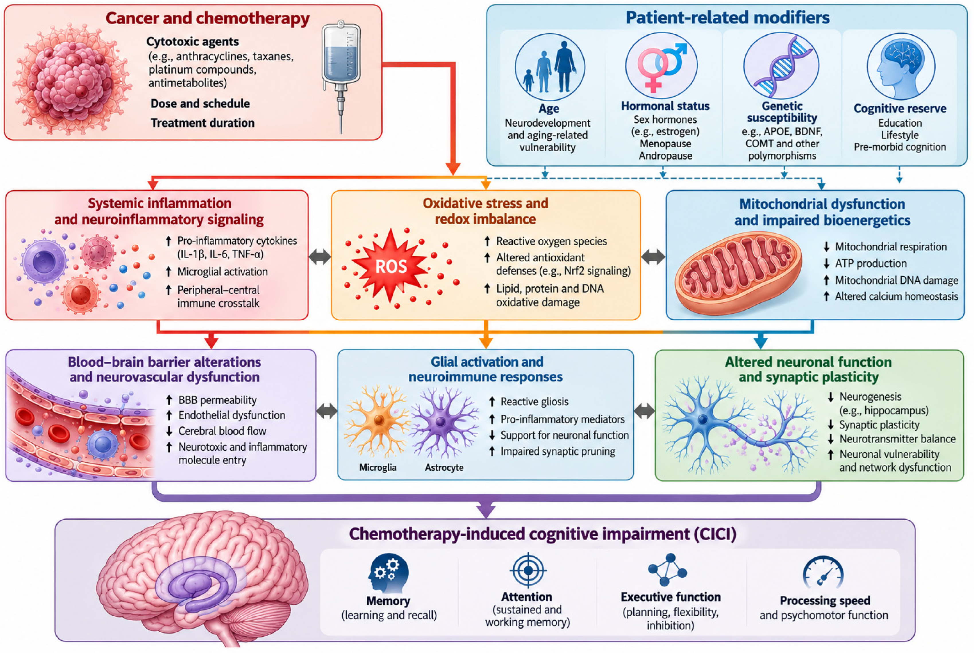
Taken together, these observations support a multifactorial conceptual framework for CICI, in which redox–inflammatory disturbances represent one potentially relevant component among several interacting biological, treatment-related, and host-dependent factors. The relative contribution of each mechanism remains uncertain and may vary across patients and treatment regimens. This proposed pathophysiological framework is summarized in Figure 1.
4.7. Therapeutic Implications of the Redox–Inflammatory Interface
The convergence of oxidative stress, mitochondrial dysfunction, and inflammation raises the possibility that therapeutic modulation of endogenous cellular defense mechanisms could influence pathways implicated in CICI. In this way, it has been described in animal models that the administration of compounds with strong antioxidant effects (such as N-acetylcysteine and others) can counteract both oxidative stress and cognitive alterations induced by several chemotherapeutic drugs [39]. However, rather than directly scavenging ROS, which may interfere with their physiological signaling functions, activation of endogenous cytoprotective pathways such as the Nrf2 system may represent an alternative strategy for restoring redox homeostasis. This concept provides the rationale for considering medical ozone as a potential redox-modulating intervention: although ozone is itself an oxidizing stimulus and should not be regarded as a conventional antioxidant, controlled medical O2/O3 exposure has been proposed to induce transient redox signals capable of activating endogenous adaptive responses, including Nrf2-related pathways [14,15,16,20]. Whether such modulation can influence the redox–inflammatory processes implicated in CICI remains unknown and requires validation through controlled studies incorporating both cognitive and mechanistic endpoints.
Figure 1.
Proposed pathophysiological framework underlying chemotherapy-induced cognitive impairment (CICI). Cancer- and treatment-associated systemic perturbations may promote interconnected inflammatory, oxidative, and mitochondrial disturbances. These processes may influence neurovascular integrity, blood–brain barrier function, glial activation, neuronal energetics, and synaptic plasticity, potentially contributing to cognitive dysfunction. Age, hormonal status, genetic susceptibility, comorbidities, and cognitive reserve may modify these responses. The proposed relationships are supported by varying degrees of clinical and preclinical evidence and should not be interpreted as a fully established causal sequence in patients with CICI. Dashed arrows indicate hypothesized or incompletely validated mechanistic connections. Abbreviations: APOE, apolipoprotein E; ATP, adenosine triphosphate; BBB, blood–brain barrier; BDNF, brain-derived neurotrophic factor; CICI, chemotherapy-induced cognitive impairment; COMT, catechol-O-methyltransferase; DNA, deoxyribonucleic acid; IL-1β, interleukin-1 beta; IL-6, interleukin-6; Nrf2, nuclear factor erythroid 2-related factor 2; ROS, reactive oxygen species; TNF-α, tumor necrosis factor-alpha.
5. Medical Ozone as a Candidate Redox-Modulating Intervention
5.1. From Oxidative Stress to Adaptive Redox Signaling
The potential relevance of medical ozone to CICI derives primarily from its capacity to induce a controlled oxidative stimulus rather than from a conventional antioxidant effect. Ozone (O3) is a highly reactive oxidizing molecule and should not be regarded as an antioxidant in itself. Its proposed therapeutic effects are instead attributed to the biological responses elicited by its interaction with readily oxidizable components of biological fluids and tissues [14,15,16].
When an oxygen–ozone gas mixture is administered by an established systemic route, ozone reacts rapidly with molecules present in biological fluids, particularly antioxidants, proteins, and polyunsaturated fatty acids (PUFAs). Consequently, intact ozone is unlikely to circulate for prolonged periods or reach the brain as an unchanged molecule. Rather, its biological effects are proposed to arise predominantly from transient secondary reaction products, including hydrogen peroxide (H2O2) and lipid ozonation products (LOPs), which can function as short-lived or relatively longer-lived signaling mediators [16].
This distinction has major implications for a potential neurological application. A hypothesis based on direct delivery of ozone to the brain would be biologically inappropriate for systemic ozone administration. The relevant hypothesis is instead that controlled peripheral redox signaling may modify endogenous antioxidant, inflammatory, metabolic, or neurovascular pathways that ultimately influence CNS homeostasis.
5.2. Ozone-Derived Second Messengers: H2O2 and Lipid Ozonation Products
Two broad classes of secondary mediators have attracted particular attention: hydrogen peroxide and lipid-derived oxidation products. Hydrogen peroxide is generated during the rapid reactions initiated by ozone in aqueous biological environments. Although H2O2 is conventionally considered a ROS, at controlled concentrations it can also function as a signaling molecule involved in cellular redox regulation. Its relatively greater stability compared with highly reactive free radicals permits transient signaling between extracellular and intracellular compartments [14].
The second group comprises lipid ozonation products generated when ozone reacts with unsaturated lipids. These include hydroperoxides and aldehydic products, among which 4-hydroxynonenal (4-HNE) has been extensively studied as a biologically active electrophilic molecule [20,40]. At sufficiently low concentrations, such products may participate in adaptive signaling; at excessive concentrations, however, lipid peroxidation products can themselves contribute to cellular injury.
The biological response to ozone depends on exposure concentration, route, duration, and the prevailing redox state. The distinction between an adaptive oxidative signal and oxidative injury is not merely theoretical: the same chemical reactivity that may initiate cytoprotective signaling at controlled exposure levels can become deleterious when the oxidative burden exceeds cellular adaptive capacity [17].
In this context, the hormetic window should be understood as a dose- and context-dependent biological response rather than a universally established therapeutic range. Although consensus documents issued by scientific organizations, including the International Scientific Committee of Ozone Therapy (ISCO3) [41], provide recommended concentrations, volumes, and administration routes, these recommendations do not establish a validated hormetic window for patients undergoing cancer treatment. Such a window would require prospective evaluation of exposure parameters, treatment tolerability, and longitudinal redox and inflammatory biomarkers, together with clinically relevant outcomes. In particular, the balance between adaptive signaling and potentially harmful lipid oxidation remains to be established in this population.
Potential pharmacodynamic endpoints could include changes in Nrf2-regulated gene expression, glutathione-related indices, antioxidant enzyme activity, and lipid peroxidation markers such as 4-HNE, assessed alongside treatment exposure, safety parameters, and clinical outcomes. However, no individual biomarker or combination of biomarkers has yet been validated to define an optimal ozone-induced hormetic response in patients with CICI.
5.3. Activation of the Nrf2–Keap1 Pathway
Among the signaling pathways proposed to mediate the adaptive response to medical ozone, the Nrf2 pathway is particularly relevant. Under basal conditions, Nrf2 is maintained at relatively low intracellular concentrations through interaction with Kelch-like ECH-associated protein 1 (Keap1), which facilitates its ubiquitination and proteasomal degradation. Oxidative or electrophilic modification of critical Keap1 residues can alter this regulatory process, allowing Nrf2 to accumulate and translocate to the nucleus. There, Nrf2 interacts with antioxidant response elements (AREs) and regulates the expression of a broad range of cytoprotective genes [20].
Experimental studies of medical ozone have reported activation of Nrf2-associated antioxidant responses. Galiè and colleagues demonstrated that mild ozonisation activates an antioxidant cellular response through a Keap1/Nrf2-dependent pathway, providing experimental support for the concept that controlled ozone exposure can act as a redox signal rather than simply as a source of oxidative damage [14]. A subsequent review specifically addressing Nrf2 in medical ozone exposure concluded that Nrf2 signaling represents one of the principal mechanistic pathways potentially underlying the adaptive response to ozone [16].
Activation of Nrf2 may increase cellular capacity for maintaining redox homeostasis through induction of enzymes and proteins involved in glutathione metabolism, peroxide detoxification, reduced nicotinamide adenine dinucleotide phosphate (NADPH)-dependent redox control, heme metabolism, and xenobiotic defense. These include, among others, glutathione-related enzymes, NADPH quinone dehydrogenase 1 (NQO1), heme oxygenase-1 (HO-1), and glutathione S-transferases [20].
This pathway is relevant to the CICI hypothesis because oxidative stress and impaired endogenous antioxidant defenses have been implicated in the biological response to systemic anticancer therapy [9]. However, evidence of ozone-associated Nrf2 activation derives predominantly from cellular, ex vivo, and preclinical investigations, with limited clinical observations. Direct demonstration of Nrf2 pathway engagement in the human CNS following systemic ozone therapy is lacking. Furthermore, Nrf2 activation in peripheral tissues cannot be assumed to translate into neuroprotection or improved cognitive function in patients with CICI.
5.4. Interaction Between Nrf2 and Inflammatory Signaling
Nrf2 signaling in cancer may play a double-edged role, because Nrf2 activation in tumor cells may support cellular survival, metabolic adaptation, and resistance to anticancer therapy [42]. Medical ozone should not be regarded as a direct or selective pharmacological Nrf2 activator; its proposed effects derive from transient peripheral redox signaling and adaptive cytoprotective responses. Nevertheless, whether systemic ozone exposure can meaningfully affect Nrf2 signaling in residual tumor cells or influence tumor biology remains unknown and should be specifically assessed in clinical studies. Therefore, future randomized controlled trials evaluating medical ozone in CICI should preferably include patients after completion of acute cytotoxic chemotherapy, with stable disease or no evidence of active disease, and should incorporate oncological safety monitoring.
On the other hand, redox regulation and inflammation are closely interconnected. Nrf2 does not operate independently from inflammatory signaling pathways, and the interaction between Nrf2 and nuclear factor kappa B (NF-κB) has been proposed as one mechanism through which redox status can influence inflammatory responses [14,18].
Experimental ozone studies have reported modulation of inflammatory mediators and signaling pathways, including changes involving NF-κB and cytokine production. The direction and magnitude of these effects, however, depend on the biological context and exposure conditions. It is therefore preferable to describe ozone as a modulator of redox-sensitive inflammatory signaling rather than as intrinsically “anti-inflammatory.”
This issue deserves particular attention in oncology. Inflammation is not uniformly pathological in cancer; it is involved in host defense, tissue repair, immune surveillance, and tumor biology. Similarly, oxidative signaling has physiological functions and may participate in the cytotoxic activity of several anticancer treatments. Consequently, an intervention intended to modify oxidative stress during cancer therapy must be evaluated carefully to determine whether it alters tumor control or treatment efficacy.
At present, available evidence does not establish that appropriately administered medical ozone compromises anticancer efficacy, but neither does the existing evidence justify assuming that it is universally neutral with respect to tumor biology. This issue warrants specific investigation in future clinical studies, particularly when ozone is administered concomitantly with systemic anticancer therapy.
5.5. Glutathione and Endogenous Antioxidant Defenses
The glutathione system represents a particularly relevant downstream component of redox adaptation. Reduced glutathione (GSH) participates directly in the detoxification of reactive species and maintains the redox state of cellular proteins. The GSH/oxidized glutathione (GSSG) ratio is therefore commonly used as an indicator of cellular redox balance.
The proposed interaction between ozone exposure and glutathione homeostasis is not based on ozone acting as a direct antioxidant. Rather, the transient oxidative stimulus generated by ozone-derived mediators may activate cellular pathways that increase endogenous antioxidant capacity [43]. Experimental literature has reported modulation of glutathione and antioxidant enzyme systems following controlled ozone exposure, although the magnitude and reproducibility of these effects vary according to experimental model and treatment protocol.
This mechanism is potentially relevant to chemotherapy-associated toxicity. Clavo and colleagues reviewed experimental and clinical studies in which ozone therapy was investigated as a means of modulating oxidative stress associated with chemotherapy. The review included studies involving cisplatin, methotrexate, doxorubicin, and bleomycin and reported that ozone exposure was associated in several experimental models with changes in oxidative-stress and antioxidant parameters and with reduced toxicity [17,18]. Importantly, however, the authors emphasized the limited number of available studies and the need for randomized controlled trials.
The relevance to CICI is therefore indirect but conceptually important. If chemotherapy-induced systemic oxidative stress contributes to downstream neuroinflammatory or mitochondrial disturbances, an intervention capable of modifying endogenous redox defenses could theoretically influence this process. Whether such modulation occurs in the CNS, or translates into measurable cognitive benefit, remains unknown.
5.6. Mitochondrial Redox Regulation
Moderate changes in cellular redox signaling can activate adaptive mechanisms, whereas sustained or excessive oxidative stress can impair mitochondrial membranes, respiratory-chain function, adenosine triphosphate (ATP) production, and mitochondrial quality control.
A recent systematic review of mitochondrial dysfunction and low-dose medical ozone proposed that ozone-associated redox signaling may influence mitochondrial homeostasis through interactions involving glutathione metabolism, Nrf2 signaling, and related adaptive pathways [16]. However, much of the mechanistic evidence remains experimental, and the distinction between direct mitochondrial effects and secondary consequences of systemic redox adaptation is not always established.
The potential relevance to CICI lies in the convergence between these mechanisms and the mitochondrial hypothesis of chemotherapy-related cognitive dysfunction described above. If chemotherapy contributes to a persistent imbalance between ROS generation and endogenous antioxidant capacity, restoration of redox homeostasis could theoretically attenuate downstream mitochondrial dysfunction.
This remains a testable hypothesis rather than an established therapeutic mechanism. Demonstrating it would require studies measuring both cognitive outcomes and mechanistically relevant biomarkers, ideally before and after treatment.
5.7. Why the Concept of Hormesis Requires Caution
The biological model proposed for medical ozone is frequently described using the concept of hormesis: a low-intensity stressor induces an adaptive response that increases resistance to subsequent or ongoing stress [44].
This framework provides a plausible explanation for why an oxidizing molecule could theoretically produce antioxidant effects. It also defines the principal limitation of the approach. Hormetic responses are typically dose-dependent, and the beneficial and detrimental ranges may be separated by a relatively narrow window [45].
Consequently, the expression “more ozone” cannot be equated with “greater antioxidant effect.” Excessive oxidative exposure may overwhelm cellular defenses and cause lipid, protein, DNA, and mitochondrial damage. The therapeutic concept therefore depends on controlled exposure within a biologically tolerable range, not on maximal oxidative stimulation.
This dose–response relationship is particularly relevant in patients undergoing chemotherapy, in whom endogenous redox homeostasis may already be disturbed. An intervention that is adaptive under one physiological condition could theoretically become excessive under another. Treatment protocols therefore need to specify ozone concentration, gas volume, oxygen–ozone ratio, route, frequency, cumulative exposure, and timing relative to anticancer treatment.
5.8. Could Peripheral Redox Modulation Influence the Brain?
The proposed relevance of ozone to CICI does not require ozone itself to cross the blood–brain barrier. Instead, several indirect pathways could theoretically connect peripheral redox modulation with CNS function. One possibility is modulation of circulating inflammatory mediators. A second is alteration of systemic oxidative status and endogenous antioxidant capacity. A third involves endothelial and microvascular signaling, potentially influencing the neurovascular interface. A fourth involves peripheral immune-cell signaling capable of modifying neuroimmune activity. These mechanisms remain hypothetical in the specific context of ozone and CICI. A preliminary human imaging observation potentially relevant to this peripheral-to-central hypothesis is discussed in Section 7.3 [38], although it provides no direct evidence of ozone-induced CNS redox modulation or cognitive benefit in CICI.
However, direct evidence that systemic ozone therapy activates Nrf2 within human brain tissue is currently lacking. Similarly, there is no convincing clinical evidence demonstrating that ozone reverses chemotherapy-induced microglial activation or restores hippocampal neuroplasticity in cancer patients.
Accordingly, the proposed pathway should presently be formulated as: controlled O2/O3 exposure induces transient peripheral oxidative signaling → Nrf2/Keap1 and related redox responses → modulation of endogenous antioxidant and inflammatory pathways → potential effects on systemic mitochondrial, endothelial, and immune homeostasis → possible downstream influence on CNS inflammatory/redox environment → potential modification of cognitive dysfunction.
The proposed sequence comprises mechanistic links supported by different levels of evidence. Ozone-induced secondary redox signaling and associated adaptive responses have been characterized mainly in experimental systems, whereas their reproducibility, dose dependence, and clinical relevance in cancer patients remain insufficiently established. The subsequent links involving CNS Nrf2 engagement, neuroprotection, and cognitive improvement in CICI are currently hypothetical and require direct clinical validation.
5.9. The Translational Gap
The main limitation is the absence of direct clinical evidence in CICI. Experimental studies indicate that controlled ozone exposure can modify redox signaling and antioxidant responses, while chemotherapy models implicate oxidative stress and neuroinflammation in cognitive dysfunction. Separate clinical and translational studies have investigated ozone in chemotherapy-associated toxicity and CIPN, but no clinical study has yet demonstrated an effect of ozone therapy on CICI. Thus, evidence of altered oxidative biomarkers, improvement in CIPN, or effects in Alzheimer’s disease models cannot be considered evidence of cognitive benefit in cancer survivors. Medical ozone should therefore be regarded as a candidate redox-modulating intervention whose potential effects on CICI remain to be tested, ideally through studies combining validated cognitive outcomes with mechanistically relevant biomarkers (Table 1) [46].
Table 1.
Proposed mechanisms of medical ozone and their potential relevance to chemotherapy-induced cognitive impairment (CICI).
6. Evidence of Ozone Therapy in Cancer Treatment-Related Toxicity and Chemotherapy-Induced Peripheral Neuropathy
6.1. Rationale for Investigating Ozone in Treatment-Related Toxicity
The potential application of medical ozone to cancer treatment-related neurological dysfunction is preceded by a broader body of experimental and clinical work investigating ozone in chemotherapy-associated toxicity. This literature is relevant because several chemotherapeutic agents affect systemic redox and inflammatory pathways that may contribute to treatment-related toxicity [18]. Nevertheless, the existence of a shared redox component does not imply that all chemotherapy toxicities respond similarly to redox modulation.
A systematic review by Clavo et al. examined experimental and clinical studies evaluating ozone therapy in the prevention or treatment of chemotherapy-induced toxicity [17]. The reviewed literature included models involving anthracyclines, platinum compounds, antimetabolites, and other cytotoxic agents. Across experimental studies, ozone treatment was associated with changes in markers of oxidative stress and endogenous antioxidant defenses and, in some models, with attenuation of organ toxicity. These findings provide a rationale for considering ozone as a potential modulator of treatment-associated oxidative injury.
However, this evidence should be interpreted cautiously. Most studies were preclinical, treatment protocols varied substantially, and the relationship between biochemical changes and clinically meaningful outcomes was not always established. Furthermore, attenuation of systemic oxidative stress does not necessarily imply protection of the CNS, and any intervention administered during active anticancer treatment must be evaluated for potential effects on tumor response.
Thus, the chemotherapy-toxicity literature provides biological and translational plausibility, rather than evidence that ozone prevents or treats CICI.
6.2. Chemotherapy-Induced Peripheral Neuropathy as the Closest Clinical Neurological Model
Among cancer treatment-related neurological toxicities, CIPN currently provides the most relevant human clinical experience with ozone therapy. CIPN is a frequent complication of neurotoxic anticancer agents, particularly platinum compounds, taxanes, vinca alkaloids, and proteasome inhibitors. Clinical manifestations include numbness, tingling, burning pain, sensory loss, and, in some patients, motor or autonomic dysfunction. Its pathophysiology is multifactorial and includes mitochondrial dysfunction, oxidative stress, neuroinflammation, axonal injury, altered ion-channel function, and impaired neuronal homeostasis. CIPN is predominantly a peripheral neurotoxicity, although its clinical consequences may extend beyond sensory symptoms. Observational prospective data in breast cancer survivors indicate that CIPN onset is associated with an early increased risk of depression and anxiety, although this association does not establish causality [47]. This supports the inclusion of mood, fatigue, quality of life, and functional endpoints in multidimensional phenotyping of chemotherapy-related neurotoxicity, as we have included in our randomized controlled trial (RCT).
Although the anatomical and functional consequences of CIPN differ fundamentally from CICI, the involvement of oxidative and inflammatory pathways provides a mechanistic point of convergence. Nevertheless, an emerging line of research links peripheral chemotherapy-induced neurotoxicity with central dysfunction and cognitive symptoms, particularly through shared neuroinflammatory mechanisms connecting neuropathic pain, peripheral nerve injury, and CNS pathology [48]. This does not make CIPN a surrogate for CICI, but it strengthens its value as a clinically relevant translational bridge between peripheral and central chemotherapy-related neurotoxicity. This makes CIPN a useful, but limited, translational precedent for investigating whether redox-modulating interventions may influence cancer treatment-related neurological dysfunction.
6.3. Initial Clinical Evidence: Chronic Pain Associated with CIPN
One of the first clinical reports evaluating ozone in CIPN was published by Clavo et al. in 2022 [18]. In this preliminary study, patients with chronic pain secondary to chemotherapy-induced peripheral neuropathy received ozone treatment and were followed longitudinally. The authors reported sustained improvement in pain-related outcomes following treatment.
The study is important primarily as a proof-of-concept clinical signal. It demonstrated that ozone administration could be investigated in patients with established chemotherapy-associated neurological toxicity and suggested the possibility of clinically meaningful symptom improvement.
Nevertheless, the study has important limitations. It was preliminary, the sample size was limited, and the study design does not permit definitive attribution of the observed improvement to ozone. Pain is also a subjective endpoint influenced by multiple factors, and improvement in neuropathic pain cannot be assumed to indicate restoration of peripheral nerve structure or function.
Consequently, the findings should be interpreted as hypothesis-generating rather than confirmatory.
6.4. Persistent Sensory Symptoms After Chemotherapy
Subsequent retrospective work examined patients with persistent numbness and tingling following chemotherapy, symptoms without effective evidence-based treatment [49,50]. Clavo et al. reported long-term improvement in sensory symptoms after ozone treatment in a retrospective cohort [51]. The reported persistence of improvement is of interest given the often-prolonged course of CIPN.
The retrospective nature of the study, however, substantially limits causal inference. Without randomized allocation and an appropriate control group, spontaneous improvement, regression toward the mean, placebo effects, changes in concomitant treatment, and other confounding factors cannot be excluded. Furthermore, numbness and tingling are symptoms rather than direct measures of neuronal repair. The study therefore provides evidence of a potential symptomatic effect but not definitive evidence that ozone reverses the underlying neurotoxic process.
6.5. Randomized Clinical Investigation: The O3NPIQ and OzoParQT Trials
The most important development in this field is the ongoing randomized, triple-blind clinical trials specifically designed to evaluate ozone treatment in persistent CIPN [17,19]. As noted above, both registered randomized trials (with an oxygen-based control group) investigating ozone therapy for CIPN (NCT04299893 and NCT06706544) remain in the recruitment phase, with no results reported as of October 2026. Their findings may provide future evidence regarding chemotherapy-associated neurotoxicity, but they will not directly establish efficacy in CICI. The trials incorporate clinical outcomes together with biological measures intended to characterize oxidative and inflammatory changes.
This design is methodologically important because it addresses several limitations of previous observational reports. Randomization reduces confounding, triple blinding reduces expectancy and assessment bias, and the inclusion of mechanistic biomarkers create an opportunity to test whether clinical changes are accompanied by measurable biological alterations.
However, the existence of a randomized trial should not be confused with evidence of efficacy. The protocol establishes the methodological framework for determining whether ozone is effective; until results are available, it does not itself demonstrate that treatment improves CIPN.
The distinction is particularly important for the present review. Even positive results in these trials would establish efficacy for a specific peripheral neurological toxicity, not for CICI. Their greatest relevance to CICI would be to provide evidence that ozone treatment may influence a chemotherapy-associated neurological phenotype in humans and, potentially, that clinical effects are accompanied by redox or inflammatory changes.
6.6. Evidence Hierarchy and Current Limitations
The current evidence can be considered across four levels: experimental studies showing that ozone can modulate oxidative stress and endogenous antioxidant responses in models of chemotherapy-associated toxicity [17]; preliminary and retrospective clinical studies reporting improvements in pain and persistent sensory symptoms in CIPN [18,51]. Ongoing investigation through randomized clinical CIPN trials will provide a stronger framework for evaluating clinical efficacy and associated biological changes [19,20]; and, finally, CICI-specific evidence, which remains absent. The first three levels provide progressively stronger support for biological plausibility and clinical feasibility, but their findings cannot be directly extrapolated to cognitive outcomes in cancer survivors. In particular, observational CIPN studies remain susceptible to bias and confounding, while experimental models cannot establish clinical efficacy. Thus, the principal translational gap remains the lack of an adequately designed clinical trial demonstrating whether ozone therapy can improve patient-reported or objective cognitive outcomes associated with cancer treatment.
7. Ozone Therapy in Non-Oncological Conditions Associated with Cognitive Dysfunction: Translational Evidence
7.1. Why Consider Evidence Outside Oncology?
Because direct clinical evidence in CICI is unavailable, studies from other neurological and systemic conditions were considered as indirect translational evidence, provided that their limitations are explicitly recognized. Several disorders associated with cognitive dysfunction, including post-acute sequelae of SARS-CoV-2 infection, ischemic brain injury, mild cognitive impairment (MCI), and neurodegenerative disease, share biological features with the mechanistic framework proposed for CICI, including oxidative stress, inflammation, mitochondrial dysfunction, endothelial abnormalities, and altered neuroimmune signaling.
These similarities do not establish therapeutic equivalence. Rather, they provide an opportunity to determine whether the biological effects attributed to medical ozone can be observed in human conditions involving cognitive dysfunction and whether experimental evidence supports effects on pathways relevant to CNS homeostasis.
The translational evidence is therefore best considered at three levels: clinical evidence in cognitive or brain-related conditions, experimental evidence of neuroprotection, and mechanistic evidence linking ozone exposure to redox and inflammatory pathways.
Aging is accompanied by progressive changes in redox homeostasis, mitochondrial function, inflammatory regulation, and cognitive reserve, which may increase susceptibility to cognitive dysfunction and treatment-related neurotoxicity [52]. These processes overlap with several pathways implicated in CICI and age-associated neurological disorders, including Alzheimer’s disease and cerebrovascular disease. Although experimental studies suggest that controlled medical ozone exposure may modulate selected oxidative and inflammatory responses, evidence that ozone therapy can delay biological aging, prevent age-related cognitive decline, or modify the clinical course of neurodegenerative disorders remains insufficient [53]. Thus, aging should be considered primarily as a potential modifier of vulnerability and therapeutic response rather than an established indication for ozone therapy. Future investigations should assess whether age-related biological characteristics influence the safety and pharmacodynamic responses to medical ozone in patients at risk of CICI.
7.2. Post-COVID Cognitive Dysfunction and “Brain Fog”
Post-acute sequelae of SARS-CoV-2 infection (PASC), commonly referred to as Long COVID or Post-COVID Condition (PCC), provide an interesting parallel because cognitive dysfunction or “brain fog” is a recognized component of the syndrome. Neurocognitive impairment in Long COVID has been associated with multiple potentially interacting mechanisms, including systemic inflammation, endothelial dysfunction, immune dysregulation, mitochondrial abnormalities, and persistent alterations in CNS function.
The relevance to CICI is therefore conceptual rather than disease-specific. Both conditions may involve persistent systemic abnormalities capable of influencing CNS function without direct structural destruction of the brain. However, the etiological context is fundamentally different: PASC follows viral infection, whereas CICI occurs in the context of malignancy and anticancer treatment.
Early clinical studies have investigated O2/O3 MAH in PASC. Tirelli et al. reported preliminary results in 100 patients with post-COVID fatigue treated with O2/O3 MAH and described substantial improvement in fatigue scores [54]. This study is relevant because fatigue and cognitive dysfunction frequently coexist in PASC, and because the authors investigated a systemic condition characterized by persistent symptoms after the acute disease.
However, the study did not establish an effect on objective cognitive performance. It was also not a randomized controlled trial, and fatigue improvement cannot be interpreted as evidence of improvement in “brain fog.” Consequently, it provides evidence of a possible systemic symptomatic effect, but not direct evidence of cognitive neuroprotection.
More recently, a pilot randomized controlled trial evaluated O2/O3 MAH in patients with PASC [55]. Seventy-three participants were randomized to O2/O3 MAH plus conventional treatment or conventional treatment alone. The ozone group demonstrated greater improvement in global symptom scores, functional capacity, and selected inflammatory and coagulation parameters. The study therefore provides stronger clinical evidence than earlier observational reports that O2/O3 MAH can influence some manifestations of PASC [55].
Nevertheless, cognitive function was not established as the principal outcome, and the study should not be interpreted as demonstrating efficacy against Long-COVID brain fog. This distinction is particularly important because cognitive symptoms constitute only one component of the broader PASC phenotype.
A further retrospective study published in 2026 evaluated 40 patients with post-COVID syndrome receiving 10 sessions of major ozone autohemotherapy. Improvements were reported in fatigue, anxiety, depression, and health-related quality of life [56]. Again, these findings suggest possible systemic effects but do not establish an effect on objective cognitive impairment.
Taken together, the PASC literature provides limited clinical evidence that systemic ozone therapy may influence some persistent post-infectious symptoms and inflammatory or functional parameters, but there is currently insufficient evidence to conclude that ozone specifically improves cognitive dysfunction or brain fog.
7.3. Cognitive Dysfunction in Cerebral Ischemia: Experimental and Emerging Clinical Evidence
Cerebral ischemia offers another, although biologically distinct, model for examining ozone-related effects on brain injury. Oxidative stress, mitochondrial dysfunction, excitotoxicity, inflammation, endothelial injury, and disruption of the neurovascular unit all contribute to ischemic brain damage. Several of these pathways overlap with mechanisms proposed for CICI, although their magnitude and temporal dynamics are very different.
Clavo et al. reported a single patient with vascular risk factors and radiation-induced brain injury following stereotactic radiosurgery, in whom serial single-photon emission computed tomography (SPECT) and fluorodeoxyglucose positron emission tomography (FDG-PET) examinations showed increased cerebral perfusion and glucose metabolism during oxygen–ozone autohemotherapy, accompanied by clinical improvement [38]. However, the uncontrolled design, concomitant vascular disease, and radiation-related injury preclude causal attribution. This observation remains hypothesis-generating and cannot be extrapolated to CICI.
Experimental studies have reported neuroprotective effects of ozone treatment in models of cerebral ischemia. In ischemic rats, ozone treatment has been associated with reduced brain injury and modulation of NF-κB signaling and autophagy [24]. Other experimental work has suggested that controlled ozone exposure may influence antioxidant defenses and inflammatory responses following cerebral injury [57].
The translational evidence has recently expanded with a clinical randomized study of ozone autohemotherapy in acute ischemic stroke (NCT06525792) [58]. In this study, 62 patients were allocated to control, oxygen placebo, or ozone treatment groups, with treatment initiated within 24 h of symptom onset. The investigators reported improvements in neurological and functional outcomes and changes in oxidative-stress markers and Nrf2-related signaling in the ozone group [58].
Although potentially interesting, these findings require cautious interpretation. The study was relatively small and focused on acute ischemic stroke rather than chronic cognitive impairment and the authors reported a “45%” ozone mixture without a concentration stated in the conventional µg/mL units, making the exposure difficult to interpret or compare with other medical-ozone protocols; this reporting ambiguity precludes its use to define a hormetic range. The reported cognitive outcome was part of a broader neurological assessment rather than evidence specifically addressing persistent cognitive dysfunction. Furthermore, replication by independent groups and larger multicenter trials would be required before clinical conclusions could be drawn.
Taken together, experimental studies and the limited human imaging evidence suggest that ozone-associated redox modulation may influence biological processes relevant to cerebral perfusion, metabolism, and brain injury. However, these observations do not demonstrate that the same mechanisms operate in CICI, nor do they establish that changes in cerebral perfusion or metabolism translate into cognitive improvement.
7.4. Mild Cognitive Impairment and the Alzheimer Continuum
The most direct human evidence currently relevant to cognition comes from a recent retrospective cohort study of O2/O3 MAH in individuals with mild cognitive impairment (MCI) [26]. Micarelli et al. evaluated 81 individuals with MCI who completed ten O2/O3 MAH sessions over five weeks and compared them with 93 matched healthy controls. Olfactory function was assessed using the Sniffin’ Sticks threshold-discrimination-identification (TDI) test, while global cognition was evaluated using the Mini-Mental State Examination (MMSE) and Montreal Cognitive Assessment (MoCA).
Following treatment, the proportion of participants with normosmia increased, TDI scores improved, and MoCA scores increased, including improvements in selected cognitive domains. In contrast, MMSE scores did not show a significant change [26]. This study is particularly relevant because it provides a human clinical signal involving an objective cognitive measure rather than only fatigue, pain, or quality of life. It also provides an interesting biological hypothesis linking olfactory and cognitive function.
However, its evidentiary limitations are substantial. The study was retrospective and lacked a sham-treated MCI control group undergoing the same intervention. Therefore, regression to the mean, practice effects associated with repeated cognitive testing, expectancy effects, and other confounding factors cannot be excluded. Most importantly, the authors themselves concluded that causal attribution to O2/O3 MAH requires randomized, double-blind, sham-controlled trials [26].
MCI should also not be equated with Alzheimer’s disease (AD). MCI is a heterogeneous clinical state and may arise from several etiologies. Consequently, improvement in MoCA scores in an MCI cohort does not constitute evidence that ozone modifies Alzheimer’s pathology.
7.5. Experimental Evidence in Alzheimer’s Disease
Experimental evidence provides a more mechanistically focused perspective. In an amyloid precursor protein/presenilin-1 (APP/PS1) transgenic mouse model, Lin et al. reported that ozone treatment inhibited amyloid precursor protein (APP)/amyloid-β (Aβ) production and was associated with improved cognitive performance [25]. The study is relevant because it connects ozone exposure with both a pathological hallmark of Alzheimer’s disease and behavioral outcomes.
However, animal models of AD reproduce only selected aspects of the human disease. Improvement in cognitive behavior in a transgenic mouse model cannot establish clinical efficacy in patients with Alzheimer’s disease or MCI. The principal value of this study for the present review is therefore mechanistic. It suggests that ozone-associated redox modulation may potentially interact with pathways extending beyond generic antioxidant defense and may influence disease-associated molecular processes. Nevertheless, this remains a hypothesis requiring independent replication and clinical validation.
7.6. Neuroprotection Versus Cognitive Improvement
The evidence reviewed above highlights an important conceptual distinction between neuroprotection and cognitive improvement. Neuroprotection refers broadly to preservation of neuronal structure or function in the context of injury or pathological stress. Experimental studies may infer neuroprotection from reductions in neuronal injury markers, tissue damage, apoptosis, oxidative stress, or inflammatory signaling. Cognitive improvement, by contrast, requires demonstration of improved cognitive function using validated clinical or neuropsychological endpoints.
The two outcomes should therefore be interpreted separately. An intervention may reduce oxidative stress without improving cognition; it may improve cognitive performance through mechanisms unrelated to neuronal protection; or it may influence systemic symptoms without producing measurable CNS effects.
For this reason, evidence that ozone reduces oxidative stress in an experimental stroke model, improves neurological recovery after ischemia, or alters APP/Aβ biology in an AD mouse model cannot be directly translated into a claim of cognitive benefit in patients with CICI.
7.7. Converging Evidence, but Heterogeneous Clinical Contexts
Across PASC, stroke, MCI, and experimental AD, several findings recur, particularly changes in redox and inflammatory markers. Ozone exposure has been associated in different experimental or clinical settings with some combination of: modulation of oxidative-stress markers; changes in endogenous antioxidant defenses; Nrf2-related signaling; modulation of inflammatory pathways; improvement in selected functional or symptomatic outcomes; and, in selected experimental or clinical studies, changes in cognitive measures.
The heterogeneity of the evidence is substantial, however. The interventions differ in route, dose, treatment schedule, disease stage, patient characteristics, and outcome measures. Some studies are randomized, whereas others are retrospective or uncontrolled. Some evaluate biological biomarkers, others symptoms, and only a small subset directly assess cognition.
The appropriate conclusion is therefore that these studies provide convergent but indirect translational support for investigating redox-modulating approaches in cognitive dysfunction. Their principal findings and methodological limitations are summarized in Table 2. Collectively, they do not constitute evidence that ozone is effective for CICI (Figure 2).
Table 2.
Representative studies of medical ozone in chemotherapy-associated toxicity and neurological conditions: findings, limitations, and relevance to CICI.
Figure 2.
From Chemotherapy-Induced Cognitive Impairment to a Translational Hypothesis for Ozone Therapy. Abbreviations: CICI, chemotherapy-induced cognitive impairment; ROS, reactive oxygen species; BBB, blood–brain barrier; O2/O3, oxygen–ozone mixture; H2O2, hydrogen peroxide; LOPs, lipid ozonation products; Nrf2, nuclear factor erythroid 2-related factor 2; GSH, reduced glutathione; HO-1, heme oxygenase-1; NQO1, NAD(P)H quinone dehydrogenase 1; CNS, central nervous system. Note: The diagram highlights selected redox–inflammatory mechanisms relevant to the translational hypothesis and does not represent an exhaustive or clinically validated causal model of CICI pathophysiology. The proposed involvement of blood–brain barrier alterations and impaired synaptic plasticity is supported predominantly by experimental and translational evidence, while their direct contribution to cognitive impairment in patients remains incompletely established. Other contributing factors, including endocrine, genetic, metabolic, and neuroimaging-related alterations, are discussed in the text. How to read this model: Panel (A) summarizes the principal pathophysiological mechanisms proposed to link cancer and systemic anticancer therapy with cognitive impairment, including systemic inflammation, oxidative stress, mitochondrial dysfunction, endothelial and blood–brain barrier alterations, neuroimmune activation, and impaired synaptic function and plasticity. The small blue arrows adjacent to the synapse in Panel (A) schematically represent synaptic signal transmission and do not indicate additional mechanistic links. Panel (B) illustrates the proposed biological response to controlled oxygen–ozone exposure, in which transient redox signaling may activate endogenous adaptive pathways, including the Keap1/Nrf2 system and glutathione-dependent cytoprotective mechanisms. Panel (C) integrates these processes into a translational hypothesis in which peripheral redox and inflammatory modulation may influence systemic homeostasis and, secondarily, the CNS environment, potentially affecting neuroinflammation, neurovascular function, neuronal and glial homeostasis, and hypothesized cognitive effects (not clinically established). Evidence interpretation: Solid arrows represent established or relatively well-supported biological links; dashed arrows indicate hypothesized links that remain unproven in the specific context of CICI; dotted or sequential arrows indicate the proposed temporal progression of biological events. The color coding distinguishes different levels of evidence, ranging from clinical and experimental/translational evidence to mechanistic evidence and currently unproven hypotheses. Important: The terminal pathway linking ozone-induced peripheral redox modulation to cognitive enhancement remains undemonstrated and warrants validation through adequately designed clinical trials. Accordingly, the figure should be interpreted as a hypothesis-generating translational model rather than an established therapeutic pathway.
8. Current Approaches, Clinical Translation and Research Priorities
8.1. Current Approaches
In CICI, non-pharmacological strategies appear more promising and are generally associated with a favorable safety profile.
In a comprehensive network meta-analysis including 6843 participants from 84 randomized controlled trials and evaluating 15 non-pharmacological interventions, Tai Chi/Qigong showed the highest effectiveness for improving subjective cognitive function, cognitive rehabilitation was the most effective intervention for improving overall objective cognitive function, executive function, and language, and acupoint stimulation showed the greatest effect on visuospatial and motor domains [59].
Similarly, an overview of 64 systematic reviews concluded that cognitive training and cognitive rehabilitation had the strongest evidence of efficacy. Physical activity and exercise also showed promising effects on cognitive outcomes, although findings were heterogeneous and no consistent intervention characteristics emerged as predictors of benefit. This variability may partly reflect the broad classification of exercise interventions used across the literature (e.g., aerobic, resistance, or combined exercise), with limited characterization of key training variables such as intensity, volume, frequency, progression, and overall exercise dose. In this context, lower levels of exercise supervision were associated with smaller cognitive improvements in some analyses, which could support the hypothesis that supervised exercise by facilitating greater adherence to the prescribed stimulus and potentially ensuring a higher effective exercise intensity, may produce more consistent cognitive benefits. More detailed characterization of exercise dose and supervision may therefore help clarify dose–response relationships and identify the most effective exercise stimuli for cancer-related cognitive impairment. Mind–body and psychological or behavioral interventions were supported by more limited evidence, although short-term benefits were reported [60].
Moreover, multimodal or complex interventions have been proposed as potentially advantageous, given the multifactorial nature of CICI, although their components, mechanisms, and implementation strategies remain poorly standardized and are supported by a limited number of trials [60]. Within this context, medical ozone may be considered an investigational complementary approach for a syndrome in which inflammation, oxidative stress, mitochondrial dysfunction, and neuroimmune alterations may be involved. However, it must be emphasized once again that adequately designed clinical trials are required to determine whether this mechanistic rationale translates into clinically meaningful cognitive benefit.
8.2. Target Population and Timing
The available experimental and indirect clinical evidence provides a preliminary biological rationale for investigating medical ozone in a controlled proof-of-concept study of CICI, but does not support its clinical use for this indication. An initial proof-of-concept trial should therefore focus on a relatively homogeneous population, preferably breast cancer survivors with persistent cognitive complaints after completion of the acute systemic treatment phase. Eligibility could include histologically confirmed breast cancer, previous systemic anticancer therapy, persistent cognitive complaints documented using predefined patient-reported and/or objective criteria, and adequate oncological stability, while excluding patients with primary brain tumors or brain metastases because structural CNS disease represents a fundamentally different clinical phenotype. The initial therapeutic strategy should preferably address established persistent CICI rather than prevention during active chemotherapy, thereby reducing immediate concerns regarding potential interactions with anticancer treatment and allowing assessment of cognitive outcomes in a more stable clinical setting.
8.3. Trial Design and Outcome Measures
The first clinical study should use a prospective randomized, sham-controlled design with blinded outcome assessment. Cognitive efficacy should be evaluated using both validated patient-reported measures and objective neuropsychological testing, covering domains such as processing speed, attention, memory, and executive function. Relevant determinants of cognitive performance—including fatigue, sleep, mood, pain, menopausal status, endocrine therapy, physical activity, medication use, and baseline cognitive status—should be systematically assessed. This approach would help distinguish a genuine cognitive effect from nonspecific improvements in symptoms or general well-being.
A translational design should additionally incorporate predefined biological endpoints capable of testing the proposed mechanism. Biomarkers of oxidative stress, glutathione redox status, inflammatory signaling, and, where feasible, Nrf2-related responses could be assessed alongside cognitive outcomes to determine whether biological changes accompany or precede clinical improvement. Neuroimaging may provide complementary mechanistic information but should not be considered essential for an initial proof-of-concept study. The key objective would be to establish concordant evidence of biological activity and clinically meaningful cognitive benefit rather than relying on either biomarker or subjective outcomes alone.
8.4. Oncological and Procedural Safety
Oncological safety should be a primary consideration, particularly if ozone is administered during active anticancer treatment. Several anticancer therapies exploit oxidative stress, DNA damage, mitochondrial dysfunction, or related mechanisms to induce tumor-cell death; therefore, claims that ozone protects normal tissues without affecting tumor cells require direct verification. An initial trial should consequently preferentially evaluate patients after completion of acute cytotoxic treatment and should document previous and ongoing anticancer therapies in detail. Appropriate oncological outcomes, adverse events, and procedural safety parameters should be systematically monitored. This precaution is especially relevant in breast cancer, where treatment combinations may include chemotherapy, endocrine therapy, anti-human epidermal growth factor receptor 2 (HER2) therapy, immunotherapy, radiotherapy, and other targeted agents.
An additional concern is the limited evidence regarding the long-term safety of repeated systemic ozone exposure in cancer survivors. Although controlled ozone administration is intended to induce transient adaptive redox responses, the consequences of these responses for residual malignant cells, tumor recurrence, and interactions with ongoing anticancer treatments remain insufficiently characterized. Moreover, the available clinical studies generally lack the sample sizes, standardized adverse-event reporting, and prolonged follow-up required to assess uncommon or delayed complications. Therefore, future investigations should include systematic monitoring of treatment-related adverse events, oncological outcomes, and potential long-term effects, without assuming that short-term tolerability establishes long-term safety.
8.5. Translational Development Pathway
The broader ozone literature remains heterogeneous, with inconsistent findings and generally limited certainty across clinical indications; methodological heterogeneity, variable safety reporting, and lack of standardized treatment protocols further constrain interpretation [61]. For CICI, clinical development should therefore begin with mechanistic validation and a randomized sham-controlled proof-of-concept study, only if biological, cognitive, and safety signals are concordant, to larger multicenter studies with longer follow-up. This staged approach would allow optimization of patient selection, treatment parameters, cognitive endpoints, and mechanistic biomarkers while avoiding premature clinical adoption. At present, medical ozone should therefore be considered a testable translational hypothesis rather than an established treatment for CICI.
8.6. Ozone Should Be Investigated as an Adjunct, Not a Substitute for Cancer Treatment
The proposed role of ozone should remain strictly supportive. The intervention should not be positioned as an anticancer therapy, a substitute for systemic treatment, or an intervention intended to modify tumor progression. Its hypothetical indication is the management of treatment-related cognitive dysfunction. This distinction is consistent with the available literature, which has described ozone as a potential adjunctive intervention but has not established it as a primary cancer treatment [62]. The clinical trial protocol should therefore explicitly state that standard oncological treatment remains unchanged and that ozone administration is investigational and adjunctive.
Moreover, ozone treatment should not replace established evidence-based supportive or symptomatic interventions, including physical exercise, that may contribute to the management of CICI-related symptoms and to improvements in overall quality of life. Any potential role of ozone should therefore be considered complementary to, rather than substitutive of, standard oncological and supportive care.
8.7. Standardization of Ozone Exposure
One of the major problems in the ozone literature is heterogeneity in treatment protocols. Studies differ in route of administration, ozone concentration, gas volume, frequency, duration, number of sessions, and treatment schedule. Such heterogeneity makes cross-study comparisons difficult and complicates dose–response interpretation. The recent umbrella review identified protocol standardization as an important priority for future trials [61].
A CICI trial should therefore prespecify the complete intervention: route of administration; oxygen–ozone concentration; treatment volume; frequency; number of sessions; treatment duration; interval between sessions; and timing relative to previous anticancer therapy. Intervention reporting should be sufficiently detailed to allow replication and meaningful comparison between studies. This is particularly important because the biological concept underpinning the intervention is a controlled adaptive redox response. The therapeutic hypothesis therefore depends on exposure remaining within a defined biological range.
8.8. The Control Intervention Is Critical
An adequate control group is essential because cognitive symptoms are highly susceptible to expectancy effects, repeated-testing effects, changes in fatigue, sleep, mood, and other nonspecific influences. A randomized sham-controlled design would therefore be preferable.
The control intervention should reproduce, as far as ethically and technically possible, the treatment context without generating the active ozone exposure. Blinding should be maintained for participants, outcome assessors, and investigators whenever feasible. This is particularly important because uncontrolled ozone studies cannot reliably distinguish pharmacological or biological effects from nonspecific improvement.
The recent evidence synthesis of ozone therapy strongly reinforces this methodological requirement: despite substantial mechanistic literature, randomized evidence remains inconsistent and frequently limited by methodological weaknesses [61].
A rigorous clinical evaluation of ozone therapy for CICI should combine subjective and objective cognitive outcomes, while controlling for major determinants of cognitive performance such as fatigue, sleep, mood, pain, menopausal status, endocrine therapy, physical activity, medications, and baseline cognitive status. Patient-reported cognition should be assessed with validated instruments, complemented by predefined neuropsychological endpoints covering key domains such as processing speed, attention, memory, executive function, and verbal fluency. The translational value of the intervention would be strengthened by incorporating mechanistic biomarkers of redox homeostasis, antioxidant response, inflammation, and, where feasible, mitochondrial function. This would allow testing whether ozone exposure produces measurable biological changes that temporally precede and potentially mediate cognitive improvement. Neuroimaging may provide complementary mechanistic information but is not essential for an initial proof-of-concept study.
Any future trial should incorporate comprehensive procedural, systemic, and oncological safety monitoring and include sufficiently long follow-up to assess durability of cognitive effects and cancer-related outcomes. A convincing proof-of-concept study should demonstrate a predefined and clinically meaningful cognitive benefit, ideally supported by concordant patient-reported and objective measures, a biological response compatible with the proposed redox mechanism, an appropriate temporal relationship between biological and cognitive changes, and an acceptable safety profile. The current evidence supports ozone therapy only as an investigational translational hypothesis: there is sufficient mechanistic rationale to justify controlled research, but insufficient direct clinical evidence to recommend its use for CICI. Accordingly, the proposed development pathway should progress from mechanistic validation to randomized sham-controlled proof-of-concept trials, followed by biomarker–clinical integration, optimization of patient selection and treatment protocols, and ultimately larger multicenter studies with long-term cognitive and oncological follow-up.
9. Discussion
The present review identifies a convergence between biological pathways implicated in CICI and mechanisms potentially modulated by medical ozone. However, this convergence should be interpreted cautiously. CICI is a heterogeneous clinical condition involving multiple interacting biological and patient-related factors, and the contribution of redox disturbances to cognitive dysfunction remains incompletely established in humans. Moreover, evidence that systemic ozone administration induces adaptive redox responses does not demonstrate that these responses occur within the central nervous system or translate into clinically meaningful cognitive benefits. These uncertainties represent fundamental limitations of the proposed therapeutic hypothesis.
The available evidence also presents substantial challenges for clinical translation. Findings from chemotherapy-induced peripheral neuropathy and non-oncological neurological conditions provide indirect support for biological plausibility but cannot establish efficacy in CICI. Experimental observations, uncontrolled clinical studies, and retrospective analyses differ considerably in their ability to support causal inference. Furthermore, heterogeneity in ozone administration protocols, patient populations, and outcome assessments limits comparisons across studies. The broader clinical literature on ozone therapy also indicates that randomized evidence remains heterogeneous and frequently of low certainty [61]. Consequently, the proposed relationship between ozone-induced redox adaptation and cognitive improvement must be regarded as an unvalidated hypothesis rather than an established therapeutic mechanism.
A central implication of this review is that future investigations should move beyond demonstrating changes in peripheral oxidative or inflammatory biomarkers and determine whether such changes are associated with meaningful cognitive outcomes. This requires distinguishing biological activity from clinical efficacy and establishing whether the proposed intervention can be administered without compromising oncological safety. The translational value of medical ozone in CICI will ultimately depend on evidence linking a reproducible biological response to clinically relevant cognitive improvement under appropriately controlled conditions. Until such evidence becomes available, ozone therapy should remain an investigational approach rather than a recommended treatment for CICI. Particular caution is warranted regarding potential interactions with tumor-related redox signaling and the absence of sufficiently robust long-term safety data in cancer survivors.
10. Conclusions and Translational Perspective
CICI is a clinically relevant and multifactorial complication of cancer treatment, particularly in breast cancer, for which no established treatment is currently available. Current evidence implicates interacting mechanisms involving oxidative stress, inflammation, mitochondrial dysfunction, neuroimmune signaling, and neurovascular alterations. Medical ozone may influence some of these pathways through transient redox signaling and endogenous adaptive responses; however, these effects have not been demonstrated in CICI, and no direct clinical evidence currently establishes its efficacy in preventing or treating this condition.
Accordingly, ozone therapy should currently be regarded as a testable translational hypothesis rather than an established neuroprotective treatment for CICI. The next step should be a rigorously designed randomized, sham-controlled clinical trial in well-characterized patients, integrating validated subjective and objective cognitive outcomes with mechanistic biomarkers and comprehensive oncological safety monitoring. Progression to larger studies should depend on evidence of biological activity, clinically meaningful cognitive benefit, and an acceptable safety profile.
Author Contributions
Conceptualization, B.C., G.M.-S. and F.R.-E.; methodology, B.C., G.M.-S. and F.R.-E.; investigation, B.C., G.M.-S., S.E.C.-R., Á.C.-M., J.C.R.-L., L.C.-H., S.R.-V., C.G.P.-S., J.A.D.-G., M.F., M.G., R.M.-P., D.M.-A. and F.R.-E.; resources, B.C. and F.R.-E.; writing—original draft preparation, B.C. and G.M.-S.; writing—review and editing, B.C., G.M.-S., S.E.C.-R., Á.C.-M., J.C.R.-L., L.C.-H., S.R.-V., C.G.P.-S., J.A.D.-G., M.F., M.G., R.M.-P., D.M.-A. and F.R.-E.; visualization, G.M.-S. and B.C.; supervision, B.C., G.M.-S. and F.R.-E.; project administration, B.C. and F.R.-E. All authors have read and agreed to the published version of the manuscript.
Funding
Some authors affiliated with the Research Unit and Instituto de Investigación Sanitaria de Canarias (IISC, Las Palmas, Spain) were supported by grant CGC/2025/12551 from the Cabildo de Gran Canaria (Las Palmas, Spain).
Institutional Review Board Statement
Not applicable.
Informed Consent Statement
Not applicable.
Data Availability Statement
No new data were created or analyzed in this study. Data sharing is not applicable to this article.
Acknowledgments
AI figures are based on original sketches produced by the authors. During the preparation of this manuscript, the authors used ChatGPT (GPT-6, OpenAI, October 2026) to assist with linguistic editing, including improvements in grammar, clarity, and scientific writing style. Additionally, ChatGPT (GPT-6, OpenAI, October 2026), including its integrated AI image-generation tool, was used to assist in the design, visual organization, and refinement of the graphical abstract, Figure 1 and Figure 2. This included adjustments to layout, formatting, image composition, text legibility, and color schemes to meet the journal’s graphical requirements. The prompts used included: (1) “Adjust the graphical abstract and Figure 1 to meet the journal’s requirements regarding image quality, dimensions, text legibility, spacing, and concise scientific content”; (2) “Prepare the graphical abstract in the horizontal format required by the journal (minimum 1100 × 560 pixels)”; and (3) “Correct the overlapping and illegible subtitle ‘Chemotherapy-induced cognitive impairment (CICI)’ in Panel 1.” The authors have reviewed and edited the AI-generated outputs, verified their scientific accuracy and consistency with the manuscript, and take full responsibility for the content of this publication.
Conflicts of Interest
In 2023, BC received financial support for a European grant application from Hänsler Medical GmbH (Iffezheim, Germany). The authors declare no other conflicts of interest. The funders had no role in the design of the study; in the collection, analyses, or interpretation of data; in the writing of the manuscript, or in the decision to publish the results. All authors confirm that they had full access to all the data in the study and accept responsibility to submit for publication.
Abbreviations
The following abbreviations are used in this manuscript:
| 4-HNE | 4-Hydroxynonenal |
| AD | Alzheimer’s disease |
| APOE | Apolipoprotein E |
| APP | Amyloid precursor protein |
| AREs | Antioxidant response elements |
| ATP | Adenosine triphosphate |
| Aβ | Amyloid-beta |
| BBB | Blood–brain barrier |
| BDNF | Brain-derived neurotrophic factor |
| CCR5 | C-C chemokine receptor type 5 |
| CICI | Chemotherapy-induced cognitive impairment |
| CIPN | Chemotherapy-induced peripheral neuropathy |
| CNS | Central nervous system |
| COMT | Catechol-O-methyltransferase |
| CRCI | Cancer-related cognitive impairment |
| DNA | Deoxyribonucleic acid |
| FDG-PET | Fluorodeoxyglucose positron emission tomography |
| GSH | Reduced glutathione |
| GSSG | Oxidized glutathione |
| HER2 | Human epidermal growth factor receptor 2 |
| HO-1 | Heme oxygenase-1 |
| IL-6 | Interleukin-6 |
| IL-1β | Interleukin-1 beta |
| ISCO3 | International Scientific Committee of Ozone Therapy |
| Keap1 | Kelch-like ECH-associated protein 1 |
| LOPs | Lipid ozonation products |
| MAH | Major autohemotherapy |
| MCI | Mild cognitive impairment |
| MeSH | Medical Subject Headings |
| MMSE | Mini-Mental State Examination |
| MoCA | Montreal Cognitive Assessment |
| NADPH | Reduced nicotinamide adenine dinucleotide phosphate |
| NF-κB | Nuclear factor kappa B |
| NLRP3 | NOD-like receptor family pyrin domain-containing 3 |
| NQO1 | NAD(P)H quinone dehydrogenase 1 |
| Nrf2 | Nuclear factor erythroid 2-related factor 2 |
| O2/O3 | Oxygen–ozone mixture |
| PASC | Post-acute sequelae of SARS-CoV-2 infection |
| PCC | Post-COVID condition |
| PRISMA | Preferred Reporting Items for Systematic Reviews and Meta-Analyses |
| PS1 | Presenilin-1 |
| PUFAs | Polyunsaturated fatty acids |
| RCT | Randomized controlled trial |
| ROS | Reactive oxygen species |
| SPECT | Single-photon emission computed tomography |
| TDI | Threshold-discrimination-identification |
| TNF-α | Tumor necrosis factor-alpha |
References
- Oliva, G.; Giustiniani, A.; Danesin, L.; Burgio, F.; Arcara, G.; Conte, P. Cognitive impairment following breast cancer treatments: An umbrella review. Oncologist 2024, 29, e848–e863. [Google Scholar] [CrossRef] [Scilit]
- Oliveira, M.E.C.; Torres, G.S.V.; Franklin, R.G.; Gomes, K.A.L.; Nóbrega, W.F.S.; Fernandes, T.P.; Santos, N.A. Cognitive impairments associated with chemotherapy in women with breast cancer: A meta-analysis and meta-regression. Braz. J. Med. Biol. Res. 2023, 56, e12947. [Google Scholar] [CrossRef] [Scilit]
- Jansen, C.E.; Cooper, B.A.; Dodd, M.J.; Miaskowski, C.A. A prospective longitudinal study of chemotherapy-induced cognitive changes in breast cancer patients. Support. Care Cancer 2011, 19, 1647–1656. [Google Scholar] [CrossRef] [Scilit]
- Wefel, J.S.; Lenzi, R.; Theriault, R.L.; Davis, R.N.; Meyers, C.A. The cognitive sequelae of standard-dose adjuvant chemotherapy in women with breast carcinoma: Results of a prospective, randomized, longitudinal trial. Cancer 2004, 100, 2292–2299. [Google Scholar] [CrossRef] [Scilit]
- Wefel, J.S.; Saleeba, A.K.; Buzdar, A.U.; Meyers, C.A. Acute and late onset cognitive dysfunction associated with chemotherapy in women with breast cancer. Cancer 2010, 116, 3348–3356. [Google Scholar] [CrossRef] [Scilit]
- Lange, M.; Joly, F.; Vardy, J.; Ahles, T.; Dubois, M.; Tron, L.; Winocur, G.; De Ruiter, M.B.; Castel, H. Cancer-related cognitive impairment: An update on state of the art, detection, and management strategies in cancer survivors. Ann. Oncol. 2019, 30, 1925–1940. [Google Scholar] [CrossRef] [Scilit]
- Bakoyiannis, I.; Tsigka, E.A.; Perrea, D.; Pergialiotis, V. The Impact of Endocrine Therapy on Cognitive Functions of Breast Cancer Patients: A Systematic Review. Clin. Drug Investig. 2016, 36, 109–118. [Google Scholar] [CrossRef] [Scilit]
- Haggstrom, L.R.; Vardy, J.L.; Carson, E.K.; Segara, D.; Lim, E.; Kiely, B.E. Effects of Endocrine Therapy on Cognitive Function in Patients with Breast Cancer: A Comprehensive Review. Cancers 2022, 14, 920. [Google Scholar] [CrossRef] [Scilit]
- Onzi, G.R.; D’Agustini, N.; Garcia, S.C.; Guterres, S.S.; Pohlmann, P.R.; Rosa, D.D.; Pohlmann, A.R. Chemobrain in Breast Cancer: Mechanisms, Clinical Manifestations, and Potential Interventions. Drug Saf. 2022, 45, 601–621. [Google Scholar] [CrossRef] [Scilit]
- Tyagi, K.; Masoom, M.; Majid, H.; Garg, A.; Bhurani, D.; Agarwal, N.B.; Khan, M.A. Role of Cytokines in Chemotherapy-related Cognitive Impairment of Breast Cancer Patients: A Systematic Review. Curr. Rev. Clin. Exp. Pharmacol. 2023, 18, 110–119. [Google Scholar] [CrossRef] [Scilit]
- Fierro-Salgado, Y.T.; Reiriz, M.; Beltrán-Velasco, A.I.; Calleja-Conde, J.; Hernández-Oñativia, X.; Uceda, S.; Echeverry-Alzate, V. Cytokines and Brain-Derived Neurotrophic Factor as Biomarkers of Cognitive Impairment Related to Breast Cancer and Its Treatments: A Systematic Review. Int. J. Mol. Sci. 2025, 26, 10074. [Google Scholar] [CrossRef] [Scilit]
- Wu, Y.; Che, J.; Dong, J.; Zhang, X.; Deng, Y.; Chen, W.; Zhang, J. CCR5 antagonist maraviroc alleviates doxorubicin-induced neuroinflammation and neurobehavioral deficiency by regulating NF-κB/NLRP3 signaling in a breast cancer mouse model. Neuropharmacology 2024, 254, 109981. [Google Scholar] [CrossRef] [Scilit]
- Galie, M.; Covi, V.; Tabaracci, G.; Malatesta, M. The Role of Nrf2 in the Antioxidant Cellular Response to Medical Ozone Exposure. Int. J. Mol. Sci. 2019, 20, 4009. [Google Scholar] [CrossRef] [Scilit]
- Galie, M.; Costanzo, M.; Nodari, A.; Boschi, F.; Calderan, L.; Mannucci, S.; Covi, V.; Tabaracci, G.; Malatesta, M. Mild ozonisation activates antioxidant cell response by the Keap1/Nrf2 dependent pathway. Free Radic. Biol. Med. 2018, 124, 114–121. [Google Scholar] [CrossRef] [Scilit]
- Delgado-Roche, L.; Riera-Romo, M.; Mesta, F.; Hernandez-Matos, Y.; Barrios, J.M.; Martinez-Sanchez, G.; Al-Dalaien, S.M. Medical ozone promotes Nrf2 phosphorylation reducing oxidative stress and pro-inflammatory cytokines in multiple sclerosis patients. Eur. J. Pharmacol. 2017, 811, 148–154. [Google Scholar] [CrossRef] [Scilit]
- Viebahn-Haensler, R.; Leon Fernandez, O.S. Mitochondrial Dysfunction, Its Oxidative Stress-Induced Pathologies and Redox Bioregulation through Low-Dose Medical Ozone: A Systematic Review. Molecules 2024, 29, 2738. [Google Scholar] [CrossRef] [Scilit]
- Clavo, B.; Rodriguez-Esparragon, F.; Rodriguez-Abreu, D.; Martinez-Sanchez, G.; Llontop, P.; Aguiar-Bujanda, D.; Fernandez-Perez, L.; Santana-Rodriguez, N. Modulation of Oxidative Stress by Ozone Therapy in the Prevention and Treatment of Chemotherapy-Induced Toxicity: Review and Prospects. Antioxidants 2019, 8, 588. [Google Scholar] [CrossRef] [Scilit]
- Clavo, B.; Rodriguez-Abreu, D.; Galvan, S.; Federico, M.; Martinez-Sanchez, G.; Ramallo-Farina, Y.; Antonelli, C.; Benitez, G.; Rey-Baltar, D.; Jorge, I.J.; et al. Long-term improvement by ozone treatment in chronic pain secondary to chemotherapy-induced peripheral neuropathy: A preliminary report. Front. Physiol. 2022, 13, 935269. [Google Scholar] [CrossRef] [Scilit]
- Clavo, B.; Cánovas-Molina, A.; Cazorla-Rivero, S.; Martínez-Sánchez, G.; Galván, S.; Benítez, G.; Federico, M.; Fabelo, H.; González-Martín, J.M.; García-Bello, M.A.; et al. Effectiveness and cost-effectiveness of ozone treatment in patients with paraesthesia (numbness, tingling) secondary to chemotherapy-induced peripheral neuropathy: Randomized, triple-blind clinical trial (OzoParQT). BMC Cancer 2026, 26, 260. [Google Scholar] [CrossRef] [Scilit]
- Clavo, B.; Martinez-Sanchez, G.; Rodriguez-Esparragon, F.; Rodriguez-Abreu, D.; Galvan, S.; Aguiar-Bujanda, D.; Diaz-Garrido, J.A.; Canas, S.; Torres-Mata, L.B.; Fabelo, H.; et al. Modulation by Ozone Therapy of Oxidative Stress in Chemotherapy-Induced Peripheral Neuropathy: The Background for a Randomized Clinical Trial. Int. J. Mol. Sci. 2021, 22, 2802. [Google Scholar] [CrossRef] [Scilit]
- Panagea, E.; Messinis, L.; Petri, M.C.; Liampas, I.; Anyfantis, E.; Nasios, G.; Patrikelis, P.; Kosmidis, M. Neurocognitive Impairment in Long COVID: A Systematic Review. Arch. Clin. Neuropsychol. 2025, 40, 125–149. [Google Scholar] [CrossRef] [Scilit]
- Faseeh, A.; Rida, M.; Karim, N.E.; Zafar, A.; Shah, A.; Perveen, A. Prevalence and long-term outcomes of brain fog and cognitive impairment in individuals with long COVID: A systematic review. Medicine 2026, 105, e49022. [Google Scholar] [CrossRef] [Scilit]
- Resitoglu, B.; Celik, Y.; Komur, M.; Polat, A.; Erdogan, S.; Arslankoylu, A.E.; Beydagi, H. The efficacy of ozone therapy in neonatal rats with hypoxic ischemic brain injury. Bratisl. Lek. Listy 2018, 119, 81–85. [Google Scholar] [CrossRef] [Scilit]
- Zhu, L.; Ding, S.; Xu, L.; Wu, Z. Ozone treatment alleviates brain injury in cerebral ischemic rats by inhibiting the NF-κB signaling pathway and autophagy. Cell Cycle 2022, 21, 406–415. [Google Scholar] [CrossRef] [Scilit]
- Lin, S.Y.; Ma, J.; An, J.X.; Qian, X.Y.; Wang, Y.; Cope, D.K.; Williams, J.P. Ozone Inhibits APP/Aβ Production and Improves Cognition in an APP/PS1 Transgenic Mouse Model. Neuroscience 2019, 418, 110–121. [Google Scholar] [CrossRef] [Scilit]
- Micarelli, A.; Mrakic-Sposta, S.; Malacrida, S.; Vezzoli, A.; Micarelli, R.X.; Micarelli, B.; Granito, I.; Alessandrini, M. Olfactory and Cognitive Performance Improvement After Oxygen-Ozone Major Autohemotherapy in Mild Cognitive Impairment: A Retrospective Cohort Study. Neurol. Int. 2026, 18, 41. [Google Scholar] [CrossRef] [Scilit]
- Janelsins, M.C.; Kesler, S.R.; Ahles, T.A.; Morrow, G.R. Prevalence, mechanisms, and management of cancer-related cognitive impairment. Int. Rev. Psychiatry 2014, 26, 102–113. [Google Scholar] [CrossRef] [Scilit]
- Quesnel, C.; Savard, J.; Ivers, H. Cognitive impairments associated with breast cancer treatments: Results from a longitudinal study. Breast Cancer Res. Treat. 2009, 116, 113–123. [Google Scholar] [CrossRef] [Scilit]
- Janelsins, M.C.; Heckler, C.E.; Peppone, L.J.; Ahles, T.A.; Mohile, S.G.; Mustian, K.M.; Palesh, O.; O’Mara, A.M.; Minasian, L.M.; Williams, A.M.; et al. Longitudinal Trajectory and Characterization of Cancer-Related Cognitive Impairment in a Nationwide Cohort Study. J. Clin. Oncol. 2018, 36, 3231–3239. [Google Scholar] [CrossRef] [Scilit]
- Kerkmann, A.; Schinke, C.; Dordevic, A.; Kern, J.; Bangemann, N.; Finck, J.; Blohmer, J.U.; Ruprecht, K.; Göpfert, J.C.; Otto, C.; et al. Chemotherapy-induced cognitive impairment and its long-term development in patients with breast cancer: Results from the observational CICARO study. Oncologist 2025, 30, oyae268. [Google Scholar] [CrossRef] [Scilit]
- Kim, H.J.; Jung, S.O.; Kim, E.; Abraham, I. Association of chemotherapy and subjective cognitive impairment in breast cancer patients: Meta-analysis of longitudinal prospective cohort studies. Eur. J. Oncol. Nurs. 2022, 57, 102099. [Google Scholar] [CrossRef] [Scilit]
- Simó, M.; Vaquero, L.; Alemany, M.; Padrones, S.; Olivé, G.; Caravaca, A.; Padró-Miquel, A.; Nadal, E.; Camara, E.; Graus, F.; et al. Cancer-Related Cognitive Impairment in Patients with Lung Cancer. Neurology 2025, 104, e213651. [Google Scholar] [CrossRef] [Scilit]
- Sun, H.; Gada, U.; Alieldin, R.; Zhao, T.; Teets, H.; Bearden, J.D.; Esparaz, B.T.; Misleh, J.; Loh, K.P.; Magnuson, A.; et al. Longitudinal assessment of cognitive function in patients with breast cancer and lymphoma receiving chemotherapy. JNCI Cancer Spectr. 2025, 9, pkaf105. [Google Scholar] [CrossRef] [Scilit]
- Dwek, M.R.; Newman, S.P.; Brini, S.; Holder, P.; Machesney, M.; Propper, D.; Rixon, L.R.; Hirani, S.P.; Hurt, C.S. The impact of chemotherapy on cognitive performance post-surgery in patients with colorectal cancer: A prospective cohort study. Psychooncology 2023, 32, 1057–1066. [Google Scholar] [CrossRef] [Scilit]
- Santaballa-Bertrán, A.; Marcos, R.J.A.; Cardeña-Gutiérrez, A.; Martinez-Callejo, V.; Higuera, O.; Bernardez, B.; Moreno-Martínez, M.E.; Majem, M. Sex-related differences in the efficacy and toxicity of cancer treatments. Clin. Transl. Oncol. 2025, 27, 3636–3646. [Google Scholar] [CrossRef] [Scilit]
- Allen, B.D.; Apodaca, L.A.; Syage, A.R.; Markarian, M.; Baddour, A.A.D.; Minasyan, H.; Alikhani, L.; Lu, C.; West, B.L.; Giedzinski, E.; et al. Attenuation of neuroinflammation reverses Adriamycin-induced cognitive impairments. Acta Neuropathol. Commun. 2019, 7, 186. [Google Scholar] [CrossRef] [Scilit]
- Barbosa-Azevedo, M.; Dias-Carvalho, A.; Carvalho, F.; Costa, V.M. Chemotherapy-induced cognitive impairment and glia: A new take on chemobrain? Toxicol. Appl. Pharmacol. 2024, 492, 117085. [Google Scholar] [CrossRef] [Scilit]
- Clavo, B.; Suarez, G.; Aguilar, Y.; Gutierrez, D.; Ponce, P.; Cubero, A.; Robaina, F.; Carreras, J.L. Brain ischemia and hypometabolism treated by ozone therapy. Forsch. Komplementärmedizin 2011, 18, 283–287. [Google Scholar] [CrossRef] [Scilit]
- Cauli, O. Oxidative Stress and Cognitive Alterations Induced by Cancer Chemotherapy Drugs: A Scoping Review. Antioxidants 2021, 10, 1116. [Google Scholar] [CrossRef] [Scilit]
- Pecorelli, A.; Bocci, V.; Acquaviva, A.; Belmonte, G.; Gardi, C.; Virgili, F.; Ciccoli, L.; Valacchi, G. NRF2 activation is involved in ozonated human serum upregulation of HO-1 in endothelial cells. Toxicol. Appl. Pharmacol. 2013, 267, 30–40. [Google Scholar] [CrossRef] [Scilit]
- International Scientific Committee of Ozone Therapy (ISCO3). Madrid Declaration on Ozone Therapy, 4th ed.; International Scientific Committee of Ozone Therapy (ISCO3): Madrid, Spain, 2025; p. 103. [Google Scholar]
- Wu, S.; Lu, H.; Bai, Y. Nrf2 in cancers: A double-edged sword. Cancer Med. 2019, 8, 2252–2267. [Google Scholar] [CrossRef] [Scilit]
- Casale, R.; Bernatoniene, J.; Petrikonis, K.; Fornasari, D.; Sarzi-Puttini, P.; Varrassi, G. Oxygen-ozone autohaemotherapy in fibromyalgia: Oxidative stress, Nrf2 activation, small fibre neuropathy and a critical narrative review of the evidence. Clin. Exp. Rheumatol. 2026, 44, 1208–1216. [Google Scholar] [CrossRef] [Scilit]
- Vinciguerra, C.; Cernera, M.; Comegna, M.; Amoroso, M.R.; Franco, G.R.; Bellia, L.; De Stefano, F.; Castaldo, A.; Corbi, G.; Rengo, G.; et al. Ozone in neurodegeneration: An environmental toxicant or a therapeutic hormetin? Neurotoxicology 2026, 114, 103471. [Google Scholar] [CrossRef] [Scilit]
- Chirumbolo, S.; Tirelli, U.; Franzini, M.; Pandolfi, S.; Ricevuti, G.; Vaiano, F.; Valdenassi, L. Ozone in the adjunct medical treatment. The round personality of a molecule with hormetic properties. Hum. Exp. Toxicol. 2023, 42, 9603271231218926. [Google Scholar] [CrossRef] [Scilit]
- Lietz-Kijak, D.; Skibicka, K.; Skomro, P.; Brodkiewicz, A.; Budkowska, M.; Kijak, K.; Gronwald, H.; Kijak, K.; Szczegielniak, J.; Bogacz, K.; et al. Medical ozone: Mechanisms, biological effects and clinical evidence—A narrative review. Front. Med. 2026, 13, 1859221. [Google Scholar] [CrossRef] [Scilit]
- McNeish, B.L.; Richardson, J.K.; Whitney, D.G. Chemotherapy-induced peripheral neuropathy onset is associated with early risk of depression and anxiety in breast cancer survivors. Eur. J. Cancer Care 2022, 31, e13648. [Google Scholar] [CrossRef] [Scilit]
- Cherchi, L.; Zippo, A.G.; Fantoni, L.; Carozzi, V.A.; Marmiroli, P. From peripheral neurotoxicity to central dysfunction: Linking neuropathic pain and cognition in chemotherapy-induced peripheral neuropathy. Rev. Neurosci. 2026, 37, 553–576. [Google Scholar] [CrossRef] [Scilit]
- Jordan, B.; Margulies, A.; Cardoso, F.; Cavaletti, G.; Haugnes, H.S.; Jahn, P.; Le-Rhun, E.; Preusser, M.; Scotté, F.; Taphoorn, M.J.B.; et al. Systemic anticancer therapy-induced peripheral and central neurotoxicity: ESMO-EONS-EANO Clinical Practice Guidelines for diagnosis, prevention, treatment and follow-up. Ann. Oncol. 2020, 31, 1306–1319. [Google Scholar] [CrossRef] [Scilit]
- Loprinzi, C.L.; Lacchetti, C.; Bleeker, J.; Cavaletti, G.; Chauhan, C.; Hertz, D.L.; Kelley, M.R.; Lavino, A.; Lustberg, M.B.; Paice, J.A.; et al. Prevention and Management of Chemotherapy-Induced Peripheral Neuropathy in Survivors of Adult Cancers: ASCO Guideline Update. J. Clin. Oncol. 2020, 38, 3325–3348. [Google Scholar] [CrossRef] [Scilit]
- Clavo, B.; Rodriguez-Abreu, D.; Galvan-Ruiz, S.; Federico, M.; Canovas-Molina, A.; Ramallo-Farina, Y.; Antonilli, C.; Benitez, G.; Fabelo, H.; Garcia-Lourve, C.; et al. Long-Term Effects of Ozone Treatment in Patients with Persistent Numbness and Tingling Secondary to Chemotherapy-Induced Peripheral Neuropathy. A Retrospective Study. Integr. Cancer Ther. 2025, 24, 15347354241307038. [Google Scholar] [CrossRef] [Scilit]
- López-Otín, C.; Blasco, M.A.; Partridge, L.; Serrano, M.; Kroemer, G. Hallmarks of aging: An expanding universe. Cell 2023, 186, 243–278. [Google Scholar] [CrossRef] [Scilit]
- Chirumbolo, S.; Valdenassi, L.; Bertossi, D.; Loprete, F.; Tirelli, U.; Franzini, M. A conceptual model of oxygen-ozone therapy as a modulator of aging via the HMGB1 pathway. Biogerontology 2025, 27, 28. [Google Scholar] [CrossRef] [Scilit]
- Tirelli, U.; Franzini, M.; Valdenassi, L.; Pisconti, S.; Taibi, R.; Torrisi, C.; Pandolfi, S.; Chirumbolo, S. Fatigue in post-acute sequelae of SARS-CoV2 (PASC) treated with oxygen-ozone autohemotherapy—Preliminary results on 100 patients. Eur. Rev. Med. Pharmacol. Sci. 2021, 25, 5871–5875. [Google Scholar] [CrossRef] [Scilit]
- He, Y.; Liu, X.; Zha, S.; Wang, Y.; Zhang, J.; Zhang, Q.; Hu, K. A pilot randomized controlled trial of major ozone autohemotherapy for patients with post-acute sequelae of COVID-19. Int. Immunopharmacol. 2024, 139, 112673. [Google Scholar] [CrossRef] [Scilit]
- Kuculmez, O. Efficacy of major ozone autohemotherapy in patients with post-COVID syndrome. Front. Med. 2026, 13, 1720578. [Google Scholar] [CrossRef] [Scilit]
- Wu, X.; Liu, X.; Huang, H.; Li, Z.; Xiong, T.; Xiang, W.; Liu, L.; Tao, Z. Effects of major ozonated autoheamotherapy on functional recovery, ischemic brain tissue apoptosis and oxygen free radical damage in the rat model of cerebral ischemia. J. Cell. Biochem. 2019, 120, 6772–6780. [Google Scholar] [CrossRef] [Scilit]
- Cheng, H.; Lu, R.; Du, J.; Zhao, X.; Zhuang, Z. A clinical study on ozone autohemotherapy for the treatment of acute ischemic stroke. Front. Med. 2025, 12, 1595568. [Google Scholar] [CrossRef] [Scilit]
- Ho, M.H.; Wang, L.; Wong, J.W.H.; Xin, G.; Zhang, Q.; Lin, Y.K.; Cheung, D.S.T.; Vardy, J.L.; Lin, C.C. Comparative effectiveness of various nonpharmacological interventions, including traditional Chinese medicine-based interventions for cancer-related cognitive impairment: A comprehensive network meta-analysis. J. Natl. Cancer Inst. 2025, 117, 1784–1796. [Google Scholar] [CrossRef] [Scilit]
- Haywood, D.; Henneghan, A.M.; Chan, A.; Chan, R.J.; Dhillon, H.M.; Lustberg, M.B.; Vardy, J.L.; O’Connor, M.; Elvidge, N.; Dauer, E.; et al. The effect of non-pharmacological interventions on cognitive function in cancer: An overview of systematic reviews. Support. Care Cancer 2025, 33, 151. [Google Scholar] [CrossRef] [Scilit]
- Cacciatore, S.; Abbatecola, G.; Calvani, R.; Veronese, N. Effectiveness and Safety of Ozone Therapy in Humans: An Umbrella Review of Systematic Reviews with Meta-Analyses of Randomized Clinical Trials. Med. Sci. 2026, 14, 289. [Google Scholar] [CrossRef] [Scilit]
- Clavo, B.; Santana-Rodríguez, N.; Llontop, P.; Gutiérrez, D.; Suárez, G.; López, L.; Rovira, G.; Martínez-Sánchez, G.; González, E.; Jorge, I.J.; et al. Ozone Therapy as Adjuvant for Cancer Treatment: Is Further Research Warranted? Evid.-Based Complement. Altern. Med. 2018, 2018, 7931849. [Google Scholar] [CrossRef] [Scilit]
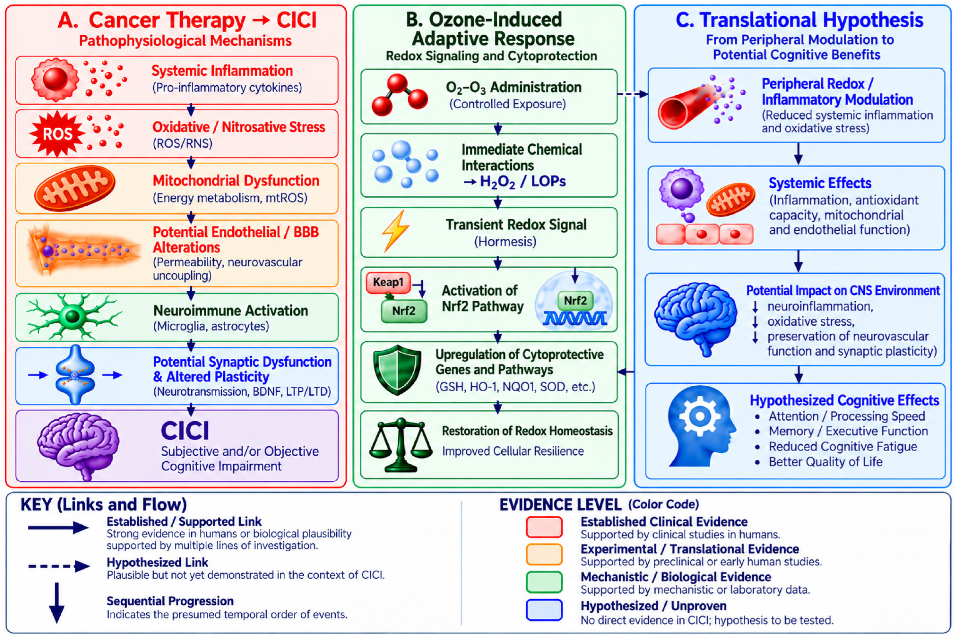

Disclaimer/Publisher’s Note: The statements, opinions and data contained in all publications are solely those of the individual author(s) and contributor(s) and not of MDPI and/or the editor(s). MDPI and/or the editor(s) disclaim responsibility for any injury to people or property resulting from any ideas, methods, instructions or products referred to in the content. |
© 2026 by the authors. Licensee MDPI, Basel, Switzerland. This article is an open access article distributed under the terms and conditions of the Creative Commons Attribution (CC BY) license.