


Abstract
Childhood, adolescent, and young adult cancer survivors (CAYACS) are at increased risk of late morbidity, potentially reflecting premature biological aging. Cellular senescence, oxidative stress, and chronic inflammation may contribute via interconnected pathways, including signaling mediated by advanced glycation end products (AGEs) and their receptor, RAGE. This study investigated biomarkers related to these pathways in CAYACS-treated Hodgkin lymphoma (HL) patients, comparing their biological profile with that of age-matched (Controls 1) and older (aged 50–65) healthy controls (Controls 2). HL survivors exhibited accumulation of AGEs, higher expression of their receptor RAGE and lower circulating levels of soluble RAGE compared to age-matched Controls 1. A similar profile of AGE-RAGE signaling was observed in Controls 2. HL survivors and Controls 2 showed an altered oxidative status. Moreover, HL survivors reported higher plasma levels of IL-6 than age-matched controls. Furthermore, HL survivors and Controls 2 showed alterations in autophagy and mitophagy. Finally, CAYACS had higher plasma MMP9 levels and higher p16 mRNA levels than age-matched Controls 1. Data show that CAYACS exhibit a biological profile consistent with premature cellular senescence, with several features resembling those observed in older healthy individuals. Alterations in AGE-RAGE signaling, oxidative stress-related pathways, and chronic inflammation may lead to the onset of this phenotype. Further longitudinal studies are needed to clarify the potential role of these biological alterations in the development of long-term treatment-related complications.
1. Introduction
In recent decades, advances in oncology have markedly improved survival, with cure rates for childhood, adolescent and young adult cancer now exceeding 80%. Consequently, the population of childhood, adolescent and young adult cancer survivors (CAYACS) is steadily increasing worldwide [1].
However, remission from malignancy does not imply complete recovery, as many survivors develop treatment-related late effects emerging or persisting more than five years after the end of treatment [2]. Compared with the general population, CAYACS face an increased risk of morbidity and premature mortality: while early deaths are mainly due to relapse, long-term mortality is primarily driven by cardiovascular disease (CVD) and second malignant neoplasms [3,4]. Moreover, endocrine and metabolic complications, such as gonadal failure, hypothyroidism, obesity, altered body composition, dyslipidemia, diabetes, and hypertension, have all been frequently described in CAYACS [2,5]. It has been reported that, by age 50, childhood cancer survivors (CCS) may develop an average of 17 chronic conditions (vs. five in the general population), four of which are severe [6]. Strikingly, the burden of chronic disease in 24-year-old CCS often resembles that of their 50-year-old siblings [7]. Given the established associations between metabolic dysfunction, CVD, and cancer development and progression, increasing attention has been focused on the biological mechanisms that may connect cancer treatment with increased risk of morbidity and premature mortality. In this scenario, premature aging could be associated with increased vulnerability in HL survivors [8]. One mechanism that may contribute to this process is cellular senescence. Although aging involves a progressive decline in physiological function over time, cellular senescence can occur throughout life and participate in both physiological and pathological processes [9,10]. Thus, the persistence and accumulation of senescent cells may contribute to premature biological aging in cancer survivors. In this setting, cancer treatments may promote stress-induced premature senescence through chronic DNA damage, oxidative stress, and mitochondrial dysfunction [11]. This process should be distinguished from replicative senescence, which typically develops following repeated cell divisions and progressive telomere shortening [10]. Senescent cells can develop a senescence-associated secretory phenotype (SASP)—characterized by the production of cytokines, chemokines, growth factors and enzymes that modify the extracellular matrix [9,10], and further promoting inflammaging, a state of chronic, low-grade inflammation leading to premature aging. This persistent condition may contribute to the onset of long-term treatment-related complications, as well as atherosclerosis, diabetes, neurodegeneration, and cancer [8,12]. Furthermore, autophagy and mitophagy are additional mechanisms involved in cellular senescence [13,14]. Both represent cellular defense mechanisms that help maintain cellular homeostasis and limit inflammatory responses. They preserve redox balance and regulate cellular senescence.
These processes are closely connected to the cellular stress pathways, which may also be activated by advanced glycation end products (AGEs) [15]. Oxidative stress, inflammation, aging, and exogenous factors accelerate AGE formation. AGEs contribute to cellular dysfunction by crosslinking long-lived proteins (e.g., collagen) and, more importantly, through receptor-mediated effects, mainly via the receptor for AGE (RAGE) [16]. RAGE activation can stimulate NF-κB, a key transcription factor involved in inflammatory responses, consequently promoting a self-perpetuating pro-inflammatory and pro-oxidant loop [17]. Moreover, reactive oxygen species (ROS) produced through nicotinamide adenine dinucleotide phosphate oxidase (NADPH oxidase) promote further AGE accumulation [18]. Oncologic treatments may activate this AGE-RAGE axis, promoting chronic inflammation and oxidative stress. Furthermore, defensive mechanisms such as soluble RAGE (sRAGE) or glyoxalase-mediated detoxification are often impaired in aging and disease, amplifying inflammation in this context and promoting premature senescence [19].
Elevated circulating levels of pro-inflammatory cytokines, including CRP, IL-6, IL-1β, IL-17, and TNF-α, have been reported in CAYACS, particularly after hematopoietic stem cell transplantation or intensive chemotherapy [20,21]. Our group previously demonstrated increased IL-1β, IL-17, and CRP in acute lymphoblastic leukemia survivors, together with AGE levels nearly sevenfold higher than controls [22]. HL survivors likewise showed impaired AGE clearance and activation of NF-κB, NLRP3 inflammasome, and NADPH oxidase [23]. These findings suggest that alkylating agents, anthracyclines, and radiotherapy may fuel a vicious cycle of ROS, AGE-RAGE signaling, and chronic inflammation that persists long after therapy [23]. AGE accumulation and RAGE activation have been proposed to contribute to cancer recurrence, second malignancies, and neurocognitive impairment following cancer treatment [24]. Additional processes implicated in therapy-related premature aging include immunosenescence, characterized by reduced naïve lymphocyte pools and altered immune cell function [25]; telomere shortening [26]; and persistent inflammatory signaling associated with the SASP [24].
This study was designed to characterize biomarkers related to cellular senescence, AGE-RAGE signaling, redox regulation, chronic low-grade inflammation, and autophagy/mitophagy in a cohort of CAYACS treated for HL during childhood or adolescence. By comparing survivors with both age-matched and older healthy controls, we sought to identify features consistent with accelerated cellular senescence that may reflect premature biological aging rather than chronological age alone. Although these processes are interconnected, their individual contributions and interplay in long-term HL survivors remain incompletely understood.
2. Materials and Methods
2.1. Subjects
During routine follow-up visits at Città della Salute e della Scienza Hospital in Turin, 35 consecutive patients meeting the following eligibility criteria were enrolled: (a) previous diagnosis of HL before the age of 30 years; (b) completion of oncologic treatment between 2010 and 2017; (c) age at the time of the study between 18 and 40 years. The following exclusion criteria were applied: (a) current smokers or those who quit less than 5 years prior; (b) obesity (BMI > 29.9 kg/m2); (c) previous hematopoietic stem cell transplantation (autologous or allogeneic); (d) diabetes mellitus; (e) arterial hypertension; (f) chronic pre-existing conditions prior to cancer diagnosis or severe intercurrent illnesses; (g) treatment with medications potentially interfering with inflammatory mechanisms (e.g., HMG-CoA inhibitors, acetylsalicylic acid, or other NSAIDs). We also excluded patients unable to understand the study objectives or provide informed consent. Two healthy control populations were also enrolled among hospital employees volunteering during routine occupational health surveillance visits: Controls 1 (51 individuals aged 18–40 years) and Controls 2 (49 individuals aged 50–65 years). Although sex matching was initially planned, it could not be fully achieved due to the limited participation of eligible volunteers. The same exclusion criteria applied to the CAYACS cohort were applied to the control populations. Given the exploratory nature of the study, we did not perform a formal a priori sample size calculation to determine the number of participants enrolled in the clinical cohort. The local Ethics Committee approved the study (31 May 2023, protocol no. 96/2023), and all participants provided written informed consent.
2.2. Plasma Separation
Blood samples were collected after a fasting period of at least 8 h in EDTA tubes. Plasma, obtained following a centrifugation of 15 min at 1500 RPM, was then aliquoted and stored at −80 °C.
2.3. Peripheral Blood Mononuclear Cell (PBMC) Extraction
PBMCs were isolated from whole blood samples collected from participants enrolled in the study, using Ficoll (Ficoll-Paque Plus, GE Healthcare Europe GmbH, Freiburg, Germany) and gradient-based centrifugation (1400 RPM, RT for 35 min). This procedure separated the blood into a plasma layer, an intermediate PBMC layer, and a lower fraction containing polymorphonuclear cells and erythrocytes. After isolation, PBMCs were washed twice with sterile 0.9% saline solution, pelleted, and stored at −80 °C for no longer than two weeks before RNA extraction. Frozen cell pellets were kept frozen until the start of the RNA extraction procedure.
2.4. Plasma AGE Quantification
Total concentration of plasma AGEs was evaluated with the OxiSelect™ Advanced Glycation End Product (AGE) Competitive ELISA Kit (STA–817, Cell Biolabs, Inc., San Diego, CA, USA) as per the manufacturer’s instructions. For all ELISA assays described in this section and in the following sections (Section 2.6, Section 2.7, Section 2.8 and Section 2.9), biological samples were analyzed in single replicate, without technical replicates, whereas assay standards and kit controls were analyzed in replicate. Carboxy-methyl-lysine (CML), carboxy-ethyl-lysine (CEL) and pentosidine (PENTO) were measured by ultra-high-performance liquid chromatography–tandem mass spectrometry (UHPLC-MS/MS). In brief, 50 µL of sample was hydrolyzed with 500 µL of 0.6 M trichloroacetic acid and 50 mL of 6 M hydrochloric acid for 2 h at 60 °C. The analyses were performed on a UHPLC coupled to a triple quadrupole mass spectrometer (AB-Sciex Triple Quad 5500, Milan, Italy), equipped with a Turbo ion ESI source. Analytes were separated on a reversed-phase C18 column (Kinetex 100 × 2.1 mm, 1.7 µm particle size, Phenomenex, Torrance, CA, USA) at a flow rate of 0.35 µL/min. A gradient mobile phase composition of 95/5–40/60 over 25 min in 5 Nm heptafluorobutanoic acid/acetonitrile was adopted. The liquid chromatograph column eluent was delivered to the Turbo ion source using nitrogen as a sheath (GS1) and curtain (CUR) gas, and air as reagent gas (GS2). The source voltage was set at 4.5 kV in positive mode, CUR 26 arbitrary units (arb), GS1 45 arb, and GS2 50 arb. The heated capillary was maintained at 275 °C. The MRM transitions and parameters were as follows: Nɛ-(carboxymethyl)-lysine (m/z) 205@84, declustering potential (DP) 100 V, entrance potential (EP) 7 V, and collision energy (CE) 32 V. Quantification of analytes was performed using CML standard calibration curve at concentrations of 10, 50, 100, 250, 300, and 500 µg L−1. Measured concentration of analytes in samples was always within the linear range of calibration. The coefficient of variation for the above analyses ranged from 8% to 14%.
2.5. Quantitative Real-Time PCR (Polymerase Chain Reaction)
Total RNA was extracted from the PBMC using the Total-RNA Purification Kit (#17250 Norgen Biotek Corporation, Thorold, ON, Canada) according to the manufacturer’s instructions. RNA concentration (ng/µL) and purity were determined using the NanoDrop™ One/OneC Microvolume UV-Vis Spectrophotometer (Thermo Fisher, Waltham, MA, USA). In total, 800 ng of RNA was reverse-transcribed into complementary DNA (cDNA) according to the manufacturer’s instructions with the SensiFAST cDNA Synthesis Kit 250 reactions (#BIO-65054 Meridian Bioscience, Cincinnati, OH, USA). Quantitative real-time PCR was performed in the CFX Connect Real-Time PCR Detection System (Bio-Rad, Hercules, CA, USA) using 50 ng of cDNA and the SensiFAST™ SYBR® No-ROX Kit (#BIO-98020, Meridian Bioscience, Cincinnati, OH, USA). Relative gene expression was obtained after normalization to the housekeeping gene (β-2-microglobulin) using the formula 2−ΔΔCT as previously described [27], and fold change was determined by comparison to the Controls 1 group. Primer sequences were designed using NCBI Primer-BLAST and purchased from Metabion (Nrf2, p16, β-2-microglobulin-Metabion, Planegg, Germany), or Merck KGaA (p62, PINK1, parkin, LC3B-Merck KGaA, Darmstadt, Germany). Commercially available pre-designed primer assays were purchased from Qiagen (advanced glycation end product receptor, RAGE/AGER, QT00000119, RefSeq: NM_001136; NADPH oxidase 1, NOX-1, QT00025585, RefSeq: NM_007052–Qiagen, Hilden, Germany). Table 1 provides the custom-designed primer sequences and the corresponding NCBI RefSeq accession numbers for representative target transcripts.
Table 1.
Primer sequences used for quantitative real-time PCR (qRT-PCR) and corresponding NCBI RefSeq accession numbers.
2.6. Quantification of Plasma Concentration of the Soluble Receptor of AGEs in Plasma
The soluble plasma form of the receptor for AGEs was evaluated, following the manufacturer’s instructions, with the Quantikine ELISA Human RAGE immunoassay (DRG00, R&D Systems, Inc., Minneapolis, MN, USA).
2.7. Evaluation of Urinary Levels of 8-Hydroxy-2-Deoxyguanosine (8-OHdG)
8-OHdG has been proposed as a urinary biomarker for in vivo oxidative DNA lesions [28]. As per the manufacturer’s instructions, urine collected from the three groups was centrifuged for 20 min at 1000× g at 4 °C; supernatants were collected and tested with the 8-OHdG Competitive ELISA Kit (EEL004, Thermo Fisher Scientific, Waltham, Massachusetts, MA, USA).
2.8. Evaluation of Interleukin 6 (IL-6) in Plasma
Plasma IL-6 was evaluated in the three groups, as per the manufacturer’s instructions, with the Human IL-6 ELISA Kit (RK00004, Woburn, MA, USA).
2.9. Matrix Metalloproteinase 9 (MMP9) Quantification in Plasma
Plasma levels of MMP9, as an indirect marker of senescence [29,30,31], were evaluated with the MMP9 Human ELISA Kit (BMS2016-2, Thermo Fisher Scientific, Waltham, Massachusetts, MA, USA), following the manufacturer’s instructions.
2.10. Statistical Analysis
Prior to the experimental biomarker analyses, an a priori sample size calculation was performed using G*Power 3.1, based on previously published data from our research group [23]. We used the estimated minimum sample sizes as reference values to guide planning for the different experimental analyses.
Prior to statistical analysis, potential outliers were identified using the robust regression and outlier removal (ROUT) method (Q = 1%) in GraphPad Prism. Following evaluation of potential outliers and any justified exclusions, normality was assessed separately within each group using the Shapiro–Wilk test, while homogeneity of variances was evaluated using Bartlett’s test. Variables showing non-normal distributions were logarithmically transformed to reduce skewness and improve their suitability for parametric analysis. After transformation, we reassessed normality and homogeneity of variances using the corresponding tests.
Data satisfying the assumptions for parametric analysis were analyzed using one-way ANOVA followed by Bonferroni post hoc test. For plasma AGEs measured by ELISA, the assumptions for parametric analysis were not adequately satisfied even after logarithmic transformation; therefore, the Kruskal–Wallis test followed by Dunn’s multiple-comparison test was applied. Statistical comparisons were not adjusted for sex and BMI. Given the exploratory nature of the study and the relatively limited sample size, we adopted an unadjusted analytical approach aimed at identifying preliminary differences in biomarker profiles across the three study groups.
Data are presented as violin plots with individual data points overlaid, showing the median and interquartile range (IQR) to illustrate data distribution, central tendency, and variability. The application of logarithmic transformation and the statistical tests used are specified in the corresponding figure legends. A p-value < 0.05 was considered statistically significant. All statistical analyses were performed using GraphPad Prism version 8 (GraphPad Software, San Diego, CA, USA).
3. Results
3.1. Demographic and Clinical Characteristics of the Study Population
As shown in Table 2, the HL survivor group comprised 35 individuals (10 males and 25 females), with a mean age at study participation of 30.65 ± 5.42 years and a mean BMI of 20.94 ± 3.93 kg/m2. The mean follow-up time since cancer diagnosis was 9.95 ± 3.01 years, and the mean age at diagnosis was 20.02 ± 5.38 years. Regarding disease severity, 21 patients had stage II HL, 6 had stage III, and 8 had stage IV. Most survivors were treated with ABVD chemotherapy (n = 27), while smaller subsets received COPP/ABV (n = 2) or other regimens (n = 6). Radiotherapy was administered to the majority of the cohort: 23 received mediastinal irradiation, 6 cervical irradiation, and 1 abdominal irradiation, whereas 5 patients did not undergo radiotherapy. Control group 1 included 51 participants (26 males and 25 females), with a mean age of 31.76 ± 4.07 years and a mean BMI of 22.7 ± 2.80 kg/m2. Control group 2 consisted of 49 individuals (14 males and 35 females), who were older on average (56.03 ± 4.58 years) and had a mean BMI of 23.37 ± 3.99 kg/m2.
Table 2.
Demographic and clinical characteristics of HL survivors and controls.
3.2. HL Survivors Exhibit AGE-RAGE Axis Dysregulation Comparable to Older Volunteers
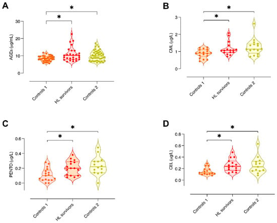
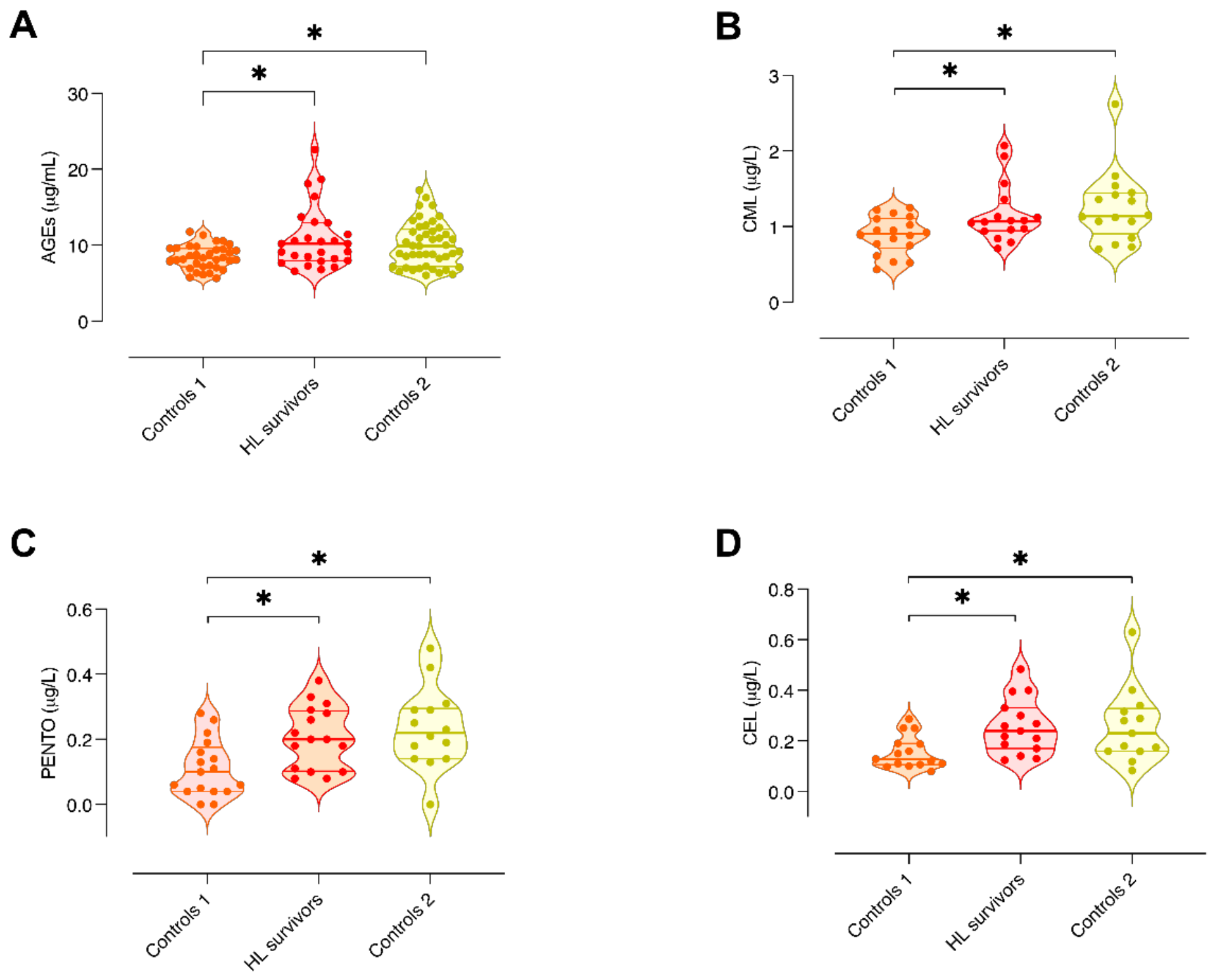
A significant accumulation of both total AGEs (Figure 1A) and specific AGEs, namely, CEL, PENTO and CML (Figure 1B–D), was observed in the plasma of HL survivors and Controls 2 compared to Controls 1. Interestingly, the level of AGEs detected in HL survivors was similar to that recorded in Controls 2.
Figure 1.
Evaluation of circulating AGE levels in HL survivors and controls. (A) Plasma levels of total AGEs measured by ELISA; (B) plasma levels of CML measured by UHPLC-MS/MS; (C) plasma levels of pentosidine (PENTO) measured by UHPLC-MS/MS; (D) plasma levels of CEL measured by UHPLC-MS/MS. The groups included Controls 1 (younger healthy volunteers), HL survivors, and Controls 2 (older healthy volunteers). Data are presented as violin plots with individual data points overlaid, showing the median and interquartile range (IQR). Sample sizes (Controls 1/HL survivors/Controls 2) were n = 35/27/45 (A), 18/16/16 (B), 17/16/14 (C), and 15/15/13 (D). Logarithmic transformation was applied to CML (B) and CEL (D) before statistical analysis. Group comparisons were performed using the Kruskal–Wallis test followed by Dunn’s multiple-comparison test for AGEs (A), and one-way ANOVA followed by Bonferroni-adjusted post hoc comparisons for CML, PENTO, and CEL (B–D). * p-value < 0.05.
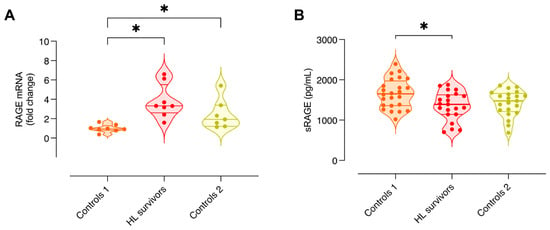
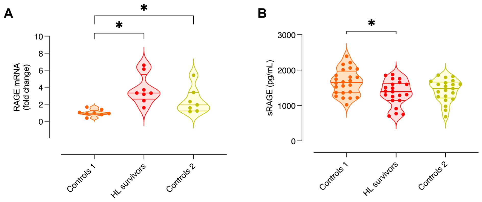
The gene expression of RAGE, measured in PBMC, was significantly higher in HL survivors and Controls 2 compared with Controls 1, supporting the activation of the AGE-RAGE signaling induced by high levels of AGEs in these two groups (Figure 2A). The plasma concentrations of the detoxifying soluble receptor sRAGE were significantly lower in HL survivors than in Controls 1. Although sRAGE levels were also lower in Controls 2 than in Controls 1, this difference did not reach statistical significance. These findings suggest a reduction in the protective defenses against AGE accumulation (Figure 2B).
Figure 2.
Measurement of membrane-bound and soluble RAGE in HL survivors and controls. (A) RAGE mRNA expression measured by qRT-PCR in PBMCs; (B) soluble RAGE (sRAGE) levels measured by ELISA in plasma. The groups included Controls 1 (younger healthy volunteers), HL survivors, and Controls 2 (older healthy volunteers). Data are presented as violin plots with individual data points overlaid, showing the median and interquartile range (IQR). Sample sizes (Controls 1/HL survivors/Controls 2) were n = 8/8/7 (A) and 24/21/21 (B). Logarithmic transformation was applied to RAGE mRNA expression (A) before statistical analysis, whereas sRAGE data (B) were analyzed without transformation. Group comparisons were performed using one-way ANOVA followed by Bonferroni-adjusted post hoc comparisons. * p-value < 0.05.
3.3. Altered Oxidative Homeostasis and Inflammatory Profile in HL Survivors
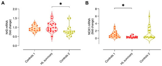
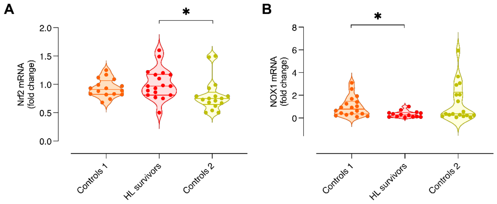
One of the main contributors to AGE formation, and a hallmark of aging, is oxidative stress. The gene expression of NRF2, a transcriptional factor regulating antioxidant genes, was significantly higher in HL survivors than in Controls 2 (Figure 3A). There were no differences compared to Controls 1. A lower gene expression of NOX1, a pro-oxidant enzyme, was significantly lower in HL survivors than Controls 1 (Figure 3B). Taken together, these results suggest that Nrf2 activation could inhibit the NADPH oxidase transcription in HL survivors, consistent with other studies [32]. We also assessed urinary 8-OHdG as it has been proposed as a urinary biomarker for in vivo oxidative DNA lesions. However, we did not identify significant differences among groups (Supplementary Figure S1).
Figure 3.
Assessment of oxidative stress-related markers in HL survivors and controls. (A) NRF2 mRNA expression evaluated by qRT-PCR in PBMCs; (B) NOX1 mRNA expression evaluated by qRT-PCR in PBMCs. The groups included Controls 1 (younger healthy volunteers), HL survivors, and Controls 2 (older healthy volunteers). Data are presented as violin plots with individual data points overlaid, showing the median and interquartile range (IQR). Sample sizes (Controls 1/HL survivors/Controls 2) were n = 16/18/17 (A) and 18/14/15 (B). Logarithmic transformation was applied to both NRF2 and NOX1 data before statistical analysis. Group comparisons were performed using one-way ANOVA followed by Bonferroni-adjusted post hoc comparisons. * p-value < 0.05.
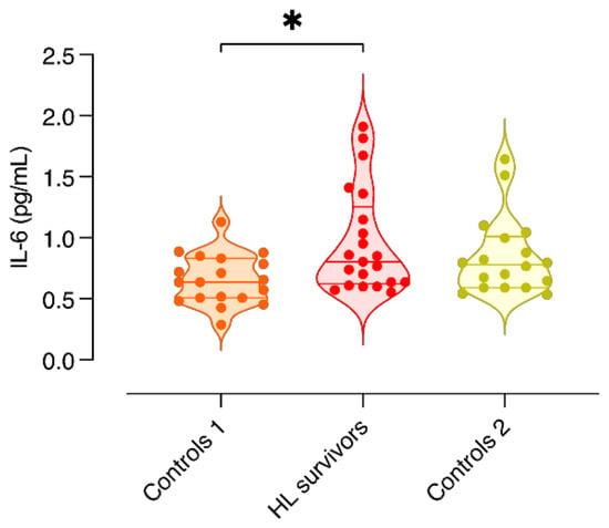
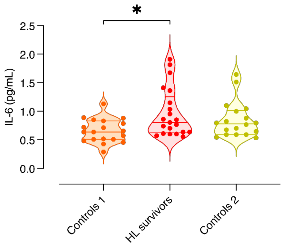
Together with oxidative stress, inflammation is another key driver of AGE formation. Moreover, chronic, low-grade inflammation is associated with the aging process, a condition known as “inflammaging”. In Figure 4, the plasma level of IL-6, one of the canonical inflammaging cytokines, was significantly higher in HL survivors compared to Controls 1. Controls 2 showed a slight increase, without reaching statistical significance.
Figure 4.
Measurement of circulating IL-6 levels in HL survivors and controls. Plasma IL-6 levels, measured by ELISA and expressed in pg/mL, in Controls 1 (younger healthy volunteers), HL survivors, and Controls 2 (older healthy volunteers). Data are presented as violin plots with individual data points overlaid, showing the median and interquartile range (IQR). Sample sizes (Controls 1/HL survivors/Controls 2) were n = 19/18/21. Logarithmic transformation was applied before statistical analysis. Group comparisons were performed using one-way ANOVA followed by Bonferroni-adjusted post hoc comparisons. * p-value < 0.05.
3.4. HL Survivors Exhibit Aged-like Features of Autophagy Dysfunction and Cellular Senescence
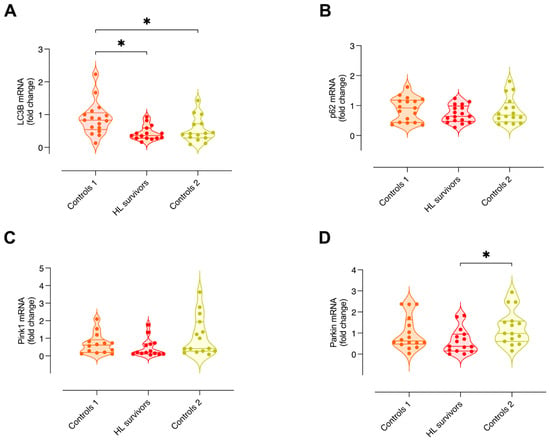
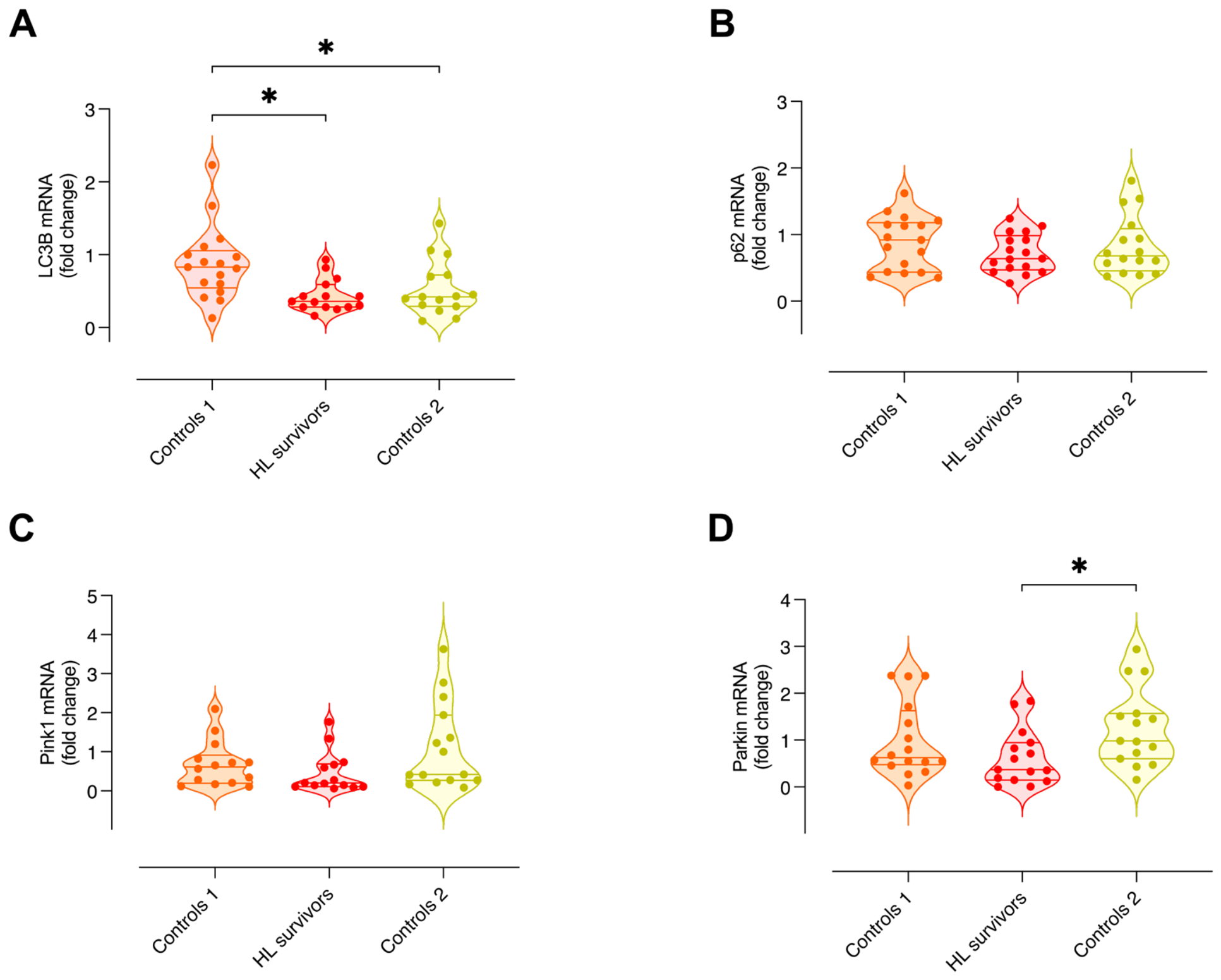
Aging is associated with the dysregulation of fundamental cellular processes, such as autophagy and mitophagy. HL survivors and Controls 2 showed a statistically lower LC3B expression than Controls 1, indicating reduced autophagy (Figure 5A). p62, another autophagy marker, did not show any difference among groups (Figure 5B). Also, the expression of PINK1, a mitophagy protein, did not show any difference among groups (Figure 5C). However, Parkin expression was statistically higher in Controls 2 compared to HL survivors (Figure 5D). This result suggests that mitophagy is upregulated in Controls 2.
Figure 5.
Evaluation of autophagy and mitophagy-related markers in HL survivors and control groups. (A) LC3B mRNA expression evaluated by qRT-PCR in PBMCs; (B) p62 mRNA expression evaluated by qRT-PCR in PBMCs; (C) PINK1 mRNA expression evaluated by qRT-PCR in PBMCs. The groups included Controls 1 (younger healthy volunteers), HL survivors, and Controls 2 (older healthy volunteers). Data are presented as violin plots with individual data points overlaid, showing the median and interquartile range (IQR). Sample sizes (Controls 1/HL survivors/Controls 2) were n = 17/15/15 (A), 17/17/16 (B), 14/14/15 (C), and 16/15/16 (D). Logarithmic transformation was applied to LC3B (A), PINK1 (C), and PARKIN (D) before statistical analysis, whereas p62 (B) was analyzed without transformation. Group comparisons were performed using one-way ANOVA followed by Bonferroni-adjusted post hoc comparisons. * p-value < 0.05.
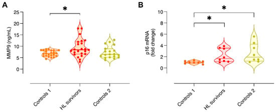
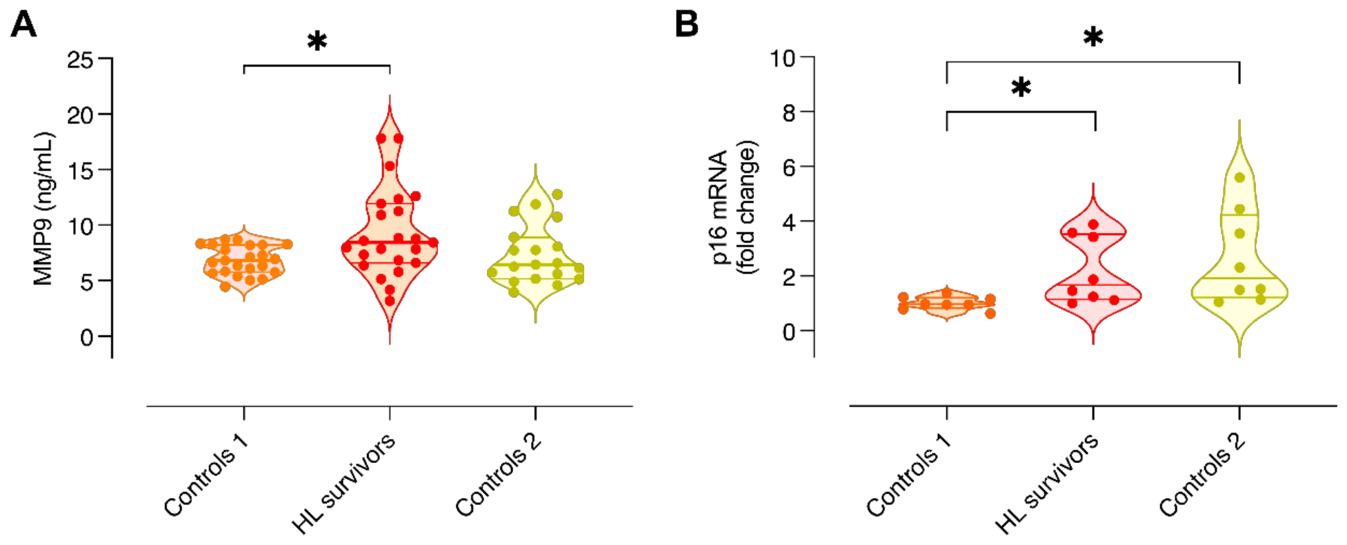
The plasma level of MMP9, an indirect marker of aging [33], was significantly higher in HL survivors than in Controls 1 (Figure 6A). In Controls 2, no difference was observed. The expression of p16, a cellular senescence marker [34], was statistically increased in HL survivors and Controls 2 compared to Controls 1 (Figure 6B). The increase in these markers confirms the presence of senescence-associated features in both groups.
Figure 6.
Measurement of cellular senescence-related markers in plasma and PBMCs. (A) MMP9 levels measured by ELISA in plasma; (B) p16 mRNA expression evaluated by qRT-PCR in PBMCs. The groups included Controls 1 (younger healthy volunteers), HL survivors, and Controls 2 (older healthy volunteers). Data are presented as violin plots with individual data points overlaid, showing the median and interquartile range (IQR). Sample sizes (Controls 1/HL survivors/Controls 2) were n = 23/23/19 (A) and 8/8/8 (B). Logarithmic transformation was applied to both MMP9 (A) and p16 (B) before statistical analysis. Group comparisons were performed using one-way ANOVA followed by Bonferroni-adjusted post hoc comparisons. * p-value < 0.05.
4. Discussion
Advances in the treatment of CAYACS have substantially increased the number of survivors within the general population. However, these individuals experience health problems far more frequently than their siblings or the general population. This increased burden is attributed to treatment-related cellular alterations that lead to premature aging in CAYACS.
In the present study, we confirmed previous observations of elevated inflammaging markers in young HL survivors and extended these data by demonstrating senescence-associated features consistent with accelerated cellular senescence, some of which more closely resembled those observed in older healthy controls than in age-matched controls. Our results indicate that, five years or more after the end of cancer therapies, young survivors of HL show higher plasma levels of total AGEs, and specifically CEL, CML, and pentosidine, compared to age-matched controls. Notably, the AGE profile of cancer survivors was comparable to that of controls aged 50–65 years, supporting a possible association between AGE accumulation and premature aging.
Consistent with AGE accumulation, we found that the expression of RAGE in PBMCs of HL survivors and older controls was significantly higher than in young controls. In plasma, we found significantly lower sRAGE concentrations in HL survivors than in age-matched controls, potentially indicating reduced availability of circulating decoy receptors that counteract AGE–RAGE signaling.
Interestingly, AGE-induced oxidative stress may activate NRF2-dependent antioxidant defenses. In our study, NRF2 expression was higher in HL survivors than in older controls, whereas the difference from age-matched controls did not reach statistical significance. This pattern may reflect a compensatory response to AGE-associated oxidative stress.
Alongside the NRF2 expression pattern, the expression of Nox1, instead, was significantly lower in HL survivors than in the age-matched healthy groups, and a similar trend was observed for older controls, even if not statistically significant. It has been demonstrated that Nrf2 may inhibit Nox1 transcription, thus antagonizing inflammatory reactions and oxidative stress [32]. Our findings may therefore reflect a coordinated adaptive response involving antioxidant defenses and reduced expression of a ROS-generating enzyme. However, other regulatory mechanisms may also contribute to the observed NOX1 expression pattern.
Our study demonstrated that HL survivors exhibited significantly higher plasma IL-6 concentrations than age-matched controls. This finding is consistent with the inflammatory profile described by Latoch et al. in childhood ALL survivors, characterized by alterations in multiple SASP-related biomarkers [21]. Elevated IL-6 levels and age-associated immune alterations have also been reported in other cohorts of young adult leukemia survivors [35,36]. However, IL-6 results have not been consistent across other survivor cohorts. Ariffin and collaborators reported numerically higher plasma IL-6 concentrations in young adult ALL survivors, but the difference from matched controls was not statistically significant [26]. Differences in cancer diagnosis, treatment exposure, age at assessment, time since treatment, and analytical methods may contribute to this heterogeneity. These observations suggest that persistent inflammation in cancer survivors may involve different cytokine profiles rather than a uniform increase in individual markers.
In the present study, IL-6 and MMP9 were selected as representative inflammatory markers due to their involvement in both AGE-RAGE-mediated inflammatory signaling [9,24] and SASP, providing a potential link between chronic inflammation and senescence-associated processes. Nevertheless, SASP comprises a heterogeneous combination of cytokines, chemokines, growth factors, and extracellular matrix-remodeling enzymes, whose composition varies depending on the cell type and biological context. Although circulating IL-6 levels and MMP 9 alone cannot establish the presence of SASP or cellular senescence, these observations could suggest that persistent inflammation may be part of the senescence-associated profile observed in HL survivors.
Aging is characterized by the accumulation of molecular damage, due to the dysfunction of cellular repair/detoxification systems. Among the cellular processes affected, autophagy is believed to progressively decline with age [37]. Defective autophagy in T cells from older subjects is associated with redox imbalance and activation of a Th17 inflammaging profile [38]. We found significantly lower gene expression of LC3B in PBMC of HL survivors and older controls compared to young controls. However, p62 mRNA expression did not differ among the groups studied. The expression of important autophagy genes, like LC3B, can be reduced in aging individuals through promoter hypermethylation [39]. While LC3B is a marker of autophagosome formation, p62 reflects more the autophagic flux: when autophagy is impaired, p62 mRNA levels can remain unchanged, while accumulation of p62 protein is usually observed. In addition to bulk autophagy, aging also affects selective autophagy and cellular quality control, particularly mitophagy [40]. We report that HL survivors have significantly lower expression of Parkin, a key mitophagy regulator, compared to Controls 2. In certain conditions, mitophagy defects derive from the downregulation of components of the mitophagy machinery, with consequent deficient removal and increased accumulation of damaged mitochondria. Conversely, other studies report increased levels of mitophagy components with aging and diseases, as a compensatory response to a status of excessive mitochondrial damage. With respect to Pink1 expression, we observed a similar trend to Parkin, but not statistically significant.
Among the late effects of oncologic therapies, CAYACS are at increased risk of cardiovascular disease. Increased arterial stiffness contributes to the onset of cardiovascular diseases in aged individuals. In the plasma of survivors, we found a higher concentration of the MMP9 soluble isoform, which seems to be particularly important in age-related extracellular matrix alterations and in the onset of the SASPs [9,41]. It has been reported that cancer patients who have undergone anthracycline chemotherapy have higher arterial stiffness compared to age-matched controls [42], and similar or even greater than what is observed in older adults, supporting the idea of anthracycline chemotherapy as a cause of accelerated vascular aging. Commonly used chemotherapeutic drugs can also induce the persistent presence of senescent cells in normal, noncancerous tissues, including the heart, contributing to pathogenesis [11,43]. We found significantly higher expression of p16, a validated marker of senescence [34,44], in PBMC from survivors and older controls compared to their younger counterparts, suggesting the presence of senescence-associated alterations in both groups. The selection of p16INK4a was supported by previous evidence demonstrating its strong association with chronological age in peripheral blood T lymphocytes [45], its correlation with tissue p16 expression [34], and its increased expression in blood cells from cancer survivors previously exposed to chemotherapy [11,46,47]. Nevertheless, cellular senescence involves multiple regulatory pathways, including the p53-p21 axis, which plays an important role in stress-induced cell-cycle arrest [9]. Although our findings are consistent with a senescence-associated phenotype, increased p16INK4a expression alone cannot establish cellular senescence. The absence of complementary assessments, including p21/p53 signaling, senescence-associated β-galactosidase activity, and persistent DNA damage markers, represents a limitation of the present study and warrants further investigation.
This study has several strengths. The use of two control groups, including an older population, allowed a direct comparison with physiological aging, strengthening the interpretation of the results. Furthermore, the simultaneous assessment of biomarkers related to glycation, redox regulation, inflammation, cellular senescence, and autophagy/mitophagy offered an integrated characterization of potentially interconnected biological processes.
However, several limitations should be acknowledged. The cross-sectional design precludes any inference regarding causality or the temporal relationship between biological alterations and clinical outcomes. The relatively small sample size, the single-center design, and the absence of an a priori sample size calculation for clinical cohort recruitment represent additional limitations. In particular, the limited sample size may have reduced the statistical power to detect smaller differences between groups, increasing the risk of type II error. The incomplete sex matching, particularly between HL survivors and younger controls, together with the absence of statistical adjustment for potential confounders such as sex and BMI, may have introduced residual confounding and should be considered when interpreting the observed differences between groups. Furthermore, transcriptional measurements in bulk PBMCs may reflect differences in immune cell subset composition, as well as changes in gene expression within individual cells. Therefore, these data alone cannot distinguish variation in immune cell populations from changes associated with senescence within individual cells.
Moreover, the use of highly selective inclusion criteria, excluding subjects with major cardiometabolic risk factors, reduced potential confounding but may have resulted in a cohort that is not fully representative of the broader population of cancer survivors. The limited sample size and heterogeneous treatment exposures did not allow reliable subgroup analyses according to chemotherapy regimens or radiotherapy exposure, preventing the assessment of treatment-specific associations with the investigated biomarkers. In addition, our findings are specific to HL survivors and therefore cannot be directly extrapolated to survivors of adult-onset cancers or non-hematopoietic malignancies, although similar alterations have been reported in other cancer survivor populations.
Our biomarker findings provide a basis for further investigation of senescence-associated changes in HL survivors. Complementary cellular assessments could strengthen the characterization of the phenotype suggested by p16, IL-6, and MMP9 and help identify the cellular sources of circulating mediators. Likewise, protein-level and functional analyses could extend the transcriptional findings on redox regulation and autophagy/mitophagy, which do not directly measure pathway activity or autophagic flux. Characterizing PBMC subsets would also help clarify the contribution of immune cell composition to the observed expression patterns. Larger multicenter longitudinal studies are needed to validate these findings, characterize biomarker trajectories over time, and investigate their association with incident late effects.
5. Conclusions
In conclusion, HL survivors exhibited AGE accumulation, increased inflammatory biomarkers, and accelerated cellular senescence, some of which resembled those observed in older healthy controls. The results support the hypothesis of premature biological aging in HL survivors and provide a basis for investigating whether premature activation of aging pathways adds to long-term morbidity. However, the contribution of cellular senescence to late morbidity remains unclear, and further studies are needed to distinguish the relative roles and time dynamics of replicative and treatment-induced senescence.
Longitudinal studies including complementary biological assessments, detailed treatment exposure data, and clinical outcomes are needed to clarify the mechanisms underlying these alterations and their relationship with late complications.
Supplementary Materials
The following supporting information can be downloaded at: https://www.mdpi.com/article/10.3390/antiox15111457/s1, Figure S1: Assessment of DNA oxidative damage in HL survivors and control groups.
Author Contributions
Conceptualization, F.C.F., M.A., M.C. and S.P.; methodology, A.G., F.D.B., F.C.F., G.F.A. and M.A.; validation, B.B. (Barbara Botto), B.B. (Benedetto Bruno), E.A. (Emanuela Arvat), F.C., M.C., M.L., M.P., R.F. and U.R.; formal analysis, A.G., C.R., E.A. (Eleonora Aimaretti), E.P. and F.D.B.; investigation, A.G., C.R., E.A. (Eleonora Aimaretti), E.P., F.D.B. and M.P.; resources, B.B. (Barbara Botto), B.B. (Benedetto Bruno), E.A. (Emanuela Arvat), F.C., F.C.F., M.L., M.P., R.F. and U.R.; writing—original draft, C.R., E.A. (Eleonora Aimaretti) and F.C.F.; writing—review and editing, F.C.F., G.F.A., M.A., M.C., R.F. and S.P.; visualization, C.R., E.A. (Eleonora Aimaretti), E.P. and G.F.A.; supervision, F.C.F., M.A., M.C. and S.P.; funding acquisition, F.C.F., M.A., M.C. and S.P. All authors have read and agreed to the published version of the manuscript.
Funding
This research was funded by the CRT Foundation [“Hodgkin Lymphoma Disease survivors STUDY-AGE, Inflammation and aging”. Grant number RF= 2022.1814] (F.C.F. and M.A.); this funding source had no involvement in study design, data collection, analysis or interpretation, in the writing of the report, and in the decision to submit the article for publication. This research was also funded by University of Turin, Department of Clinical and Biological Sciences (local funds to M.A., S.P.), and Department of Neurosciences “Rita Levi Montalcini” (local funds to M.C.).
Institutional Review Board Statement
The study was conducted in accordance with the Declaration of Helsinki and approved by the Ethics Committee of Città della Salute e della Scienza Hospital (31 May 2023, protocol no. 96/2023).
Informed Consent Statement
Informed consent was obtained from all subjects involved in the study.
Data Availability Statement
The data presented in this study are available on request from the corresponding authors due to ethical restrictions and the terms of the informed consent, which state that data will only be shared in an anonymized form.
Acknowledgments
The authors would like to thank the nurses of the “CAS” group at the Città della Salute e della Scienza Hospital for their support in patient recruitment. The Graphical Abstract was created in BioRender. COLLINO, M. (2026) https://app.biorender.com/illustrations/6977a244ec9985694dd2bb41?slideId=22f998b2-8d4d-4644-95ee-aedf2ecf1926.
Conflicts of Interest
The authors declare no conflicts of interest.
Abbreviations
The following abbreviations are used in this manuscript:
| 8-OHdG | 8-hydroxy-2′-deoxyguanosine |
| ABVD | doxorubicin, bleomycin, vinblastine, and dacarbazine |
| AGE(s) | advanced glycation end product(s) |
| ALL | acute lymphoblastic leukemia |
| ANOVA | analysis of variance |
| BMI | body mass index |
| CAYACS | childhood, adolescent, and young adult cancer survivors |
| CCS | childhood cancer survivors |
| cDNA | complementary DNA |
| CEL | Nε-(carboxyethyl)-lysine |
| CML | Nε-(carboxymethyl)-lysine |
| COPP/ABV | cyclophosphamide, vincristine, procarbazine, and prednisone/doxorubicin, bleomycin, vinblastine |
| CRP | C-reactive protein |
| CVD | cardiovascular disease |
| ELISA | enzyme-linked immunosorbent assay |
| HL | Hodgkin lymphoma |
| HMG-CoA | 3-hydroxy-3-methylglutaryl coenzyme A |
| LC3B | microtubule-associated protein 1 light chain 3 beta |
| MMP9 | matrix metalloproteinase 9 |
| mTOR | mechanistic target of rapamycin |
| NADPH | nicotinamide adenine dinucleotide phosphate |
| NF-κB | nuclear factor kappa-light-chain-enhancer of activated B cells |
| NLRP3 | NOD-like receptor protein 3 |
| NOX1 | NADPH oxidase 1 |
| NRF2 | nuclear factor erythroid 2-related factor 2 |
| NSAIDs | non-steroidal anti-inflammatory drugs |
| p16 | p16INK4a, cyclin-dependent kinase inhibitor 2A |
| p62 | sequestosome 1 |
| PBMC(s) | peripheral blood mononuclear cell(s) |
| PENTO | pentosidine |
| PINK1 | PTEN-induced kinase 1 |
| qRT-PCR | quantitative real-time polymerase chain reaction |
| RAGE | receptor for advanced glycation end products |
| ROS | reactive oxygen species |
| SASP | senescence-associated secretory phenotype |
| sRAGE | soluble receptor for advanced glycation end products |
| Th17 | T helper 17 |
| TNF-α | tumor necrosis factor alpha |
| UHPLC-MS/MS | ultra-high-performance liquid chromatography–tandem mass spectrometry |
| UV–Vis | ultraviolet–visible |
References
- Howlader, N.; Noone, A.M.; Krapcho, M.; Miller, D.; Brest, A.; Yu, M.; Ruhl, J.; Tatalovich, Z.; Mariotto, A.; Lewis, D.R.; et al. (Eds.) SEER Cancer Statistics Review, 1975–2018; National Cancer Institute: Bethesda, MD, USA, 2021; Based on November 2020 SEER Data Submission, Posted to the SEER Web Site, April 2021. Available online: https://seer.cancer.gov/csr/1975_2018/ (accessed on 10 April 2026).
- Brignardello, E.; Felicetti, F.; Castiglione, A.; Chiabotto, P.; Corrias, A.; Fagioli, F.; Ciccone, G.; Boccuzzi, G. Endocrine health conditions in adult survivors of childhood cancer: The need for specialized adult-focused follow-up clinics. Eur. J. Endocrinol. 2013, 168, 465–472. [Google Scholar] [CrossRef] [Scilit] [PubMed]
- Oeffinger, K.C.; Mertens, A.C.; Sklar, C.A.; Kawashima, T.; Hudson, M.M.; Meadows, A.T.; Friedman, D.L.; Marina, N.; Hobbie, W.; Kadan-Lottick, N.S.; et al. Chronic health conditions in adult survivors of childhood cancer. N. Engl. J. Med. 2006, 355, 1572–1582. [Google Scholar] [CrossRef] [Scilit] [PubMed]
- Armstrong, G.T.; Liu, Q.; Yasui, Y.; Neglia, J.P.; Leisenring, W.; Robison, L.L.; Mertens, A.C. Late mortality among 5-year survivors of childhood cancer: A summary from the childhood cancer survivor study. J. Clin. Oncol. 2009, 27, 2328–2338. [Google Scholar] [CrossRef] [Scilit] [PubMed]
- de Haas, E.C.; Oosting, S.F.; Lefrandt, J.D.; Wolffenbuttel, B.H.; Sleijfer, D.T.; Gietema, J.A. The metabolic syndrome in cancer survivors. Lancet Oncol. 2010, 11, 193–203. [Google Scholar] [CrossRef] [Scilit] [PubMed]
- Bhakta, N.; Liu, Q.; Ness, K.K.; Baassiri, M.; Eissa, H.; Yeo, F.; Chemaitilly, W.; Ehrhardt, M.J.; Bass, J.; Bishop, M.W.; et al. The cumulative burden of surviving childhood cancer: An initial report from the St Jude Lifetime Cohort Study (SJLIFE). Lancet 2017, 390, 2569–2582. [Google Scholar] [CrossRef] [Scilit] [PubMed]
- Armstrong, G.T.; Kawashima, T.; Leisenring, W.; Stratton, K.; Stovall, M.; Hudson, M.M.; Sklar, C.A.; Robinson, L.L.; Oeffinger, K.C. Aging and risk of severe, disabling, life-threatening, and fatal events in the childhood cancer survivor study. J. Clin. Oncol. 2014, 32, 1218–1227. [Google Scholar] [CrossRef] [Scilit] [PubMed]
- Liberale, L.; Badimon, L.; Montecucco, F.; Lüscher, T.F.; Libby, P.; Camici, G.G. Inflammation, Aging, and Cardiovascular Disease: JACC Review Topic of the Week. J. Am. Coll. Cardiol. 2022, 79, 837–847. [Google Scholar] [CrossRef] [Scilit] [PubMed]
- Suryadevara, V.; Hudgins, A.D.; Rajesh, A.; Pappalardo, A.; Karpova, A.; Dey, A.K.; Hertzel, A.; Agudelo, A.; Rocha, A.; Soygur, B.; et al. SenNet recommendations for detecting senescent cells in different tissues. Nat. Rev. Mol. Cell Biol. 2024, 25, 1001–10023. [Google Scholar] [CrossRef] [Scilit] [PubMed]
- Schmeer, C.; Kretz, A.; Wengerodt, D.; Stojiljkovic, M.; Witte, O.W. Dissecting Aging and Senescence-Current Concepts and Open Lessons. Cells 2019, 8, 1446. [Google Scholar] [CrossRef] [Scilit] [PubMed]
- Demaria, M.; O’Leary, M.N.; Chang, J.; Shao, L.; Liu, S.; Alimirah, F.; Koenig, K.; Le, C.; Mitin, N.; Deal, A.M.; et al. Cellular Senescence Promotes Adverse Effects of Chemotherapy and Cancer Relapse. Cancer Discov. 2017, 7, 165–176. [Google Scholar] [CrossRef] [Scilit] [PubMed]
- Dugan, B.; Conway, J.; Duggal, N.D. Inflammaging as a target for healthy ageing. Age Ageing 2023, 52, afac328. [Google Scholar] [CrossRef] [Scilit] [PubMed]
- Tabibzadeh, S. Role of autophagy in aging: The good, the bad, and the ugly. Aging Cell 2022, 22, e13753. [Google Scholar] [CrossRef] [Scilit] [PubMed]
- Picca, A.; Fatig, J.; Auwerx, J.; Ferrucci, L.; D’amico, D. Mitophagy in human health, ageing and disease. Nat. Metab. 2023, 5, 2047–2061. [Google Scholar] [CrossRef] [Scilit] [PubMed]
- Vistoli, G.; De Maddis, D.; Cipak, A.; Zarkovic, N.; Carini, M.; Aldini, G. Advanced glycoxidation and lipoxidation end products (AGEs and ALEs): An overview of their mechanisms of formation. Free Radic. Res. 2013, 47, 3–27. [Google Scholar] [CrossRef] [Scilit] [PubMed]
- Twarda-Clapa, A.; Olczak, A.; Białkowska, A.M.; Koziołkiewicz, M. Advanced Glycation End-Products (AGEs): Formation, Chemistry, Classification, Receptors, and Diseases Related to AGEs. Cells 2022, 11, 1312. [Google Scholar] [CrossRef] [Scilit] [PubMed]
- Dong, H.; Zhang, Y.; Huang, Y.; Deng, H. Pathophysiology of RAGE in inflammatory diseases. Front. Immunol. 2022, 13, 931473. [Google Scholar] [CrossRef] [Scilit] [PubMed]
- Tobon-Velasco, J.; Cuevas, E.; Torres-Ramos, M. Receptor for AGEs (RAGE) as Mediator of NF-kB Pathway Activation in Neuroinflammation and Oxidative Stress. CNS Neurol. Disord. Drug Targets 2014, 13, 1615–1626. [Google Scholar] [CrossRef] [Scilit] [PubMed]
- Hollenbach, M. The role of glyoxalase-I (Glo-I), advanced glycation endproducts (AGEs), and their receptor (RAGE) in chronic liver disease and hepatocellular carcinoma (HCC). Int. J. Mol. Sci. 2017, 18, 2466. [Google Scholar] [CrossRef] [Scilit] [PubMed]
- Ferrucci, L.; Fabbri, E. Inflammageing: Chronic inflammation in ageing, cardiovascular disease, and frailty. Nat. Rev. Cardiol. 2018, 15, 505–522. [Google Scholar] [CrossRef] [Scilit] [PubMed]
- Latoch, E.; Konończuk, K.; Konstantynowicz-Nowicka, K.; Muszyńska-Rosłan, K.; Sztolsztener, K.; Chabowski, A.; Krawczuk-Rybak, M. Asymptomatic Survivors of Childhood Acute Lymphoblastic Leukemia Demonstrate a Biological Profile of Inflamm-Aging Early in Life. Cancers 2022, 14, 2522. [Google Scholar] [CrossRef] [Scilit] [PubMed]
- Felicetti, F.; Cento, A.S.; Fornengo, P.; Cassader, M.; Mastrocola, R.; D’Ascenzo, F.; Settanni, F.; Benso, A.; Arvat, E.; Collino, M.; et al. Advanced glycation end products and chronic inflammation in adult survivors of childhood leukemia treated with hematopoietic stem cell transplantation. Pediatr. Blood Cancer 2020, 67, e28106. [Google Scholar] [CrossRef] [Scilit] [PubMed]
- Felicetti, F.; Aimaretti, E.; Dal Bello, F.; Gatti, F.; Godono, A.; Saba, F.; Einaudi, G.; Collino, M.; Fagioli, F.; Aragno, M.; et al. Advanced glycation end products and their related signaling cascades in adult survivors of childhood Hodgkin lymphoma: A possible role in the onset of late complications. Free Radic. Biol. Med. 2022, 178, 76–82. [Google Scholar] [CrossRef] [Scilit] [PubMed]
- Rossi, F.; Di Paola, A.; Pota, E.; Argenziano, M.; Di Pinto, D.; Marrapodi, M.M.; Di Leva, C.; Di Martino, M.; Tortora, C. Biological aspects of inflamm-aging in childhood cancer survivors. Cancers 2021, 13, 4933. [Google Scholar] [CrossRef] [Scilit] [PubMed]
- Sulicka-Grodzicka, J.; Surdacki, A.; Seweryn, M.; Mikołajczyk, T.; Rewiuk, K.; Guzik, T.; Grodzicki, T. Low-grade chronic inflammation and immune alterations in childhood and adolescent cancer survivors: A contribution to accelerated aging? Cancer Med. 2021, 10, 1772–1782. [Google Scholar] [CrossRef] [Scilit] [PubMed]
- Ariffin, H.; Azanan, M.S.; Abd Ghafar, S.S.; Oh, L.; Lau, K.H.; Thirunavakarasu, T.; Sedan, A.; Ibrahim, K.; Chan, A.; Chin, T.F. Young adult survivors of childhood acute lymphoblastic leukemia show evidence of chronic inflammation and cellular aging. Cancer 2017, 123, 4207–4214. [Google Scholar] [CrossRef] [Scilit] [PubMed]
- Livak, K.J.; Schmittgen, T.D. Analysis of relative gene expression data using real-time quantitative PCR and the 2-ΔΔCT method. Methods 2001, 25, 402–408. [Google Scholar] [CrossRef] [Scilit] [PubMed]
- Graille, M.; Wild, P.; Sauvain, J.J.; Hemmendinger, M.; Canu, I.G.; Hopf, N.B. Urinary 8-OHdG as a biomarker for oxidative stress: A systematic literature review and meta-analysis. Int. J. Mol. Sci. 2020, 21, 3743. [Google Scholar] [CrossRef] [Scilit] [PubMed]
- Consoli, A.; Devangelio, E. Thiazolidinediones and inflammation. Lupus 2005, 14, 794–797. [Google Scholar] [CrossRef] [Scilit] [PubMed]
- Pittayapruek, P.; Meephansan, J.; Prapapan, O.; Komine, M.; Ohtsuki, M. Role of matrix metalloproteinases in photoaging and photocarcinogenesis. Int. J. Mol. Sci. 2016, 17, 868. [Google Scholar] [CrossRef] [Scilit] [PubMed]
- Iannetta, M.; Zingaropoli, M.A.; Latronico, T.; Pati, I.; Pontecorvo, S.; Prezioso, C.; Pietropaolo, V.; Cortese, A.; Frontoni, M.; D’Agostino, C.; et al. Dynamic changes of MMP-9 plasma levels correlate with JCV reactivation and immune activation in natalizumab-treated multiple sclerosis patients. Sci. Rep. 2019, 9, 311. [Google Scholar] [CrossRef] [Scilit] [PubMed]
- Wu, W.; Zhang, J.; Su, X.; Lin, X.; Zhu, L.; Zhuang, Z.; Liu, C.; Zhu, Z.; Zeng, Y. Nrf2 regulates the expression of NOX1 in TNF-α-induced A549 cells. Allergol. Immunopathol. 2023, 51, 54–62. [Google Scholar] [CrossRef] [Scilit] [PubMed]
- He, K.; Zhang, H.; Tan, B.; Song, C.; Liang, Z.; Zhang, L.; Tian, D.; Xiao, L.; Xue, H.; Guo, Q.; et al. Hydrogen Sulfide Ameliorates Heart Aging by Downregulating Matrix Metalloproteinase-9. Cardiovasc. Drugs Ther. 2025, 39, 967–981. [Google Scholar] [CrossRef] [Scilit] [PubMed]
- Sorokina, A.G.; Orlova, Y.A.; Grigorieva, O.A.; Novoseletskaya, E.S.; Basalova, N.A.; Alexandrushkina, N.A.; Vigovskiy, M.A.; Kirillova, K.I.; Balatsky, A.V.; Samokhodskaya, L.M.; et al. Correlations between biomarkers of senescent cell accumulation at the systemic, tissue and cellular levels in elderly patients. Exp. Gerontol. 2023, 177, 112176. [Google Scholar] [CrossRef] [Scilit] [PubMed]
- Azanan, M.S.; Abdullah, N.K.; Chua, L.L.; Lum, S.H.; Abdul Ghafar, S.S.; Kamarulzaman, A.; Kamaruzzaman, S.; Lewin, S.R.; Woo, Y.L.; Ariffin, H.; et al. Immunity in young adult survivors of childhood leukemia is similar to the elderly rather than age-matched controls: Role of cytomegalovirus. Eur. J. Immunol. 2016, 46, 1715–1726. [Google Scholar] [CrossRef] [Scilit] [PubMed]
- Kientega, T.; Marcoux, S.; Bourbonnais, J.; Montpetit, J.; Caru, M.; Cardin, G.B.; Arbour, N.; Marcil, V.; Curnier, D.; Laverdière, C.; et al. Premature thymic functional senescence is a hallmark of childhood acute lymphoblastic leukemia survivorship. Blood Cancer J. 2024, 14, 96. [Google Scholar] [CrossRef] [Scilit] [PubMed]
- Kaushik, S.; Tasset, I.; Arias, E.; Pampliega, O.; Wong, E.; Martinez-Vicente, M.; Cuervo, A.M. Autophagy and the hallmarks of aging. Ageing Res. Rev. 2021, 72, 101468. [Google Scholar] [CrossRef] [Scilit] [PubMed]
- Bharath, L.P.; Agrawal, M.; McCambridge, G.; Nicholas, D.A.; Hasturk, H.; Liu, J.; Jiang, K.; Liu, R.; Guo, Z.; Deeney, J.; et al. Metformin Enhances Autophagy and Normalizes Mitochondrial Function to Alleviate Aging-Associated Inflammation. Cell Metab. 2020, 32, 44–55.e6. [Google Scholar] [CrossRef] [Scilit] [PubMed]
- Khalil, H.; Tazi, M.; Caution, K.; Ahmed, A.; Kanneganti, A.; Assani, K.; Kopp, B.; Marsh, C.; Dakhlallah, D.; Amer, A.O. Aging is associated with hypermethylation of autophagy genes in macrophages. Epigenetics 2016, 11, 381–388. [Google Scholar] [CrossRef] [Scilit] [PubMed]
- Palmer, J.E.; Wilson, N.; Son, S.M.; Obrocki, P.; Wrobel, L.; Rob, M.; Palmer, J.E.; Korolchuk, V.I.; Rubinsztein, D.C. Autophagy, aging, and age-related neurodegeneration. Neuron 2025, 113, 29–48. [Google Scholar] [CrossRef] [Scilit] [PubMed]
- Yabluchanskiy, A.; Ma, Y.; Chiao, Y.A.; Lopez, E.F.; Voorhees, A.P.; Toba, H.; Hall, M.E.; Han, H.; Lindsey, M.L.; Jin, Y. Cardiac aging is initiated by matrix metalloproteinase-9-mediated endothelial dysfunction. Am. J. Physiol. Heart Circ. Physiol. 2014, 306, H1398–H1407. [Google Scholar] [CrossRef] [Scilit] [PubMed]
- Clayton, Z.S.; Hutton, D.A.; Mahoney, S.A.; Seals, D.R. Anthracycline chemotherapy-mediated vascular dysfunction as a model of accelerated vascular aging. Aging Cancer 2021, 2, 45–69. [Google Scholar] [CrossRef] [Scilit] [PubMed]
- Jain, A.; Casanova, D.; Padilla, A.V.; Paniagua Bojorges, A.; Kotla, S.; Ko, K.A.; Samanthapudi, V.S.K.; Chau, K.; Nguyen, M.T.H.; Wen, J.; et al. Premature senescence and cardiovascular disease following cancer treatments: Mechanistic insights. Front. Cardiovasc. Med. 2023, 10, 1212174. [Google Scholar] [CrossRef] [Scilit] [PubMed]
- Guan, L.; Crasta, K.C.; Maier, A.B. Assessment of cell cycle regulators in human peripheral blood cells as markers of cellular senescence. Ageing Res. Rev. 2022, 78, 101634. [Google Scholar] [CrossRef] [Scilit] [PubMed]
- Liu, Y.; Sanoff, H.K.; Cho, H.; Burd, C.E.; Torrice, C.; Ibrahim, J.G.; Thomas, N.E.; Sharpless, N.E. Expression of p16(INK4a) in peripheral blood T-cells is a biomarker of human aging. Aging Cell 2009, 8, 439–448. [Google Scholar] [CrossRef] [Scilit] [PubMed]
- Bourlon, M.T.; Velazquez, H.E.; Hinojosa, J.; Orozco, L.; Rios-Corzo, R.; Lima, G.; Llorente, L.; Hernandez-Ramirez, D.F.; Valentin-Cortez, F.J.; Medina-Rangel, I.; et al. Immunosenescence profile and expression of the aging biomarker (p16INK4a) in testicular cancer survivors treated with chemotherapy. BMC Cancer 2020, 20, 882. [Google Scholar] [CrossRef] [Scilit] [PubMed]
- Sanoff, H.K.; Deal, A.M.; Krishnamurthy, J.; Torrice, C.; Dillon, P.; Sorrentino, J.; Ibrahim, J.G.; Jolly, T.A.; Williams, G.; Carey, L.A.; et al. Effect of cytotoxic chemotherapy on markers of molecular age in patients with breast cancer. J. Natl. Cancer Inst. 2014, 106, dju057. [Google Scholar] [CrossRef] [Scilit] [PubMed]

Disclaimer/Publisher’s Note: The statements, opinions and data contained in all publications are solely those of the individual author(s) and contributor(s) and not of MDPI and/or the editor(s). MDPI and/or the editor(s) disclaim responsibility for any injury to people or property resulting from any ideas, methods, instructions or products referred to in the content. |
© 2026 by the authors. Licensee MDPI, Basel, Switzerland. This article is an open access article distributed under the terms and conditions of the Creative Commons Attribution (CC BY) license.