


Abstract
Environmental and biochemical factors can compromise the DNA integrity of equine spermatozoa. Several sperm DNA damage assays exist, but their relative prognostic value has not been assessed in the horse. This study compared four assays for detecting DNA damage in stallion spermatozoa: the sperm chromatin structure assay (SCSA), the sperm chromatin dispersion assay (SCD), the single-cell gel electrophoresis (comet) assay, and the 8-hydroxy-2′-deoxyguanosine (8-OHdG) ELISA. Experiment 1 assessed the effect of hydrogen peroxide (H2O2), a driver of oxidative stress, on sperm DNA damage across increasing concentrations and incubation times. Experiment 2 assessed arachidonic acid (AA), an upstream driver of endogenous oxidative stress. Experiment 3 evaluated the prognostic value of each assay using semen from Thoroughbred stallions during periods of high and low fertility. Across experiments, the comet assay detected DNA damage at lower stressor doses and shorter incubation times than the other assays and was the only assay to distinguish semen from low-fertility stallions from that of high-fertility stallions. The SCSA, SCD, and 8-OHdG ELISA were comparatively less sensitive in detecting significant differences under the same conditions. In conclusion, the comet assay demonstrated the most consistent responsiveness in detecting sperm DNA damage in stallions under the tested conditions.
1. Introduction
Stallion fertility is routinely assessed by conventional semen analyses, yet many stallions with normal sperm motility, morphology and concentration return poor per-cycle pregnancy rates, and as these measures reveal nothing about the integrity of the paternal genome delivered at fertilisation, they are unreliable predictors of fertility [1,2]. Thanks in large part to the invention of the sperm chromatin structure assay (SCSA) by Donald Evenson in 1980 [3], and its validation in the horse in 1995 [4], sperm DNA damage assays have increasingly gained popularity. Sperm DNA damage encompasses several distinct molecular lesions, including oxidative base modifications, abasic sites, single- and double-strand breaks, and DNA cross-links, each of which may affect genome integrity through different mechanisms [5]. Oxidative base modifications, such as 8-hydroxy-2′-deoxyguanosine (8-OHdG), can compromise the fidelity of genetic information, whereas abasic sites and strand breaks can disrupt DNA continuity [6]. Cross-links can impede strand separation and replication, potentially interfering with paternal genome processing after fertilisation [7]. The biological consequences of these lesions depend on their type and extent, as well as the capacity of the oocyte to repair paternal DNA after fertilisation [8,9]. Consequently, elevated sperm DNA damage has been associated with reduced fertilisation success, impaired pre-implantation embryo development, an increased risk of early pregnancy loss, and lower fertility outcomes following conception [10,11,12,13,14,15].
Unlike other livestock species, stallions are selected as breeding animals based on athletic performance, pedigree and conformation rather than reproductive traits. Over generations, this has led to a high degree of fertility variability between stallions, despite reproductive efficiency being of substantial economic importance to horse breeders, with significant costs associated with veterinary management and the financial losses incurred when mares experience repeated early embryonic death (EED) or fail to establish pregnancy. Diagnostic tools that predict fertility before, or early in the breeding season are therefore of considerable practical value. The SCSA has historically been the DNA damage assay of choice in the horse, with early studies revealing significant differences between fertile and subfertile stallions [4], and later studies reporting an inverse relationship between DNA fragmentation and pregnancy rates [16].
Oxidative stress is arguably the most prevalent driver of DNA damage in spermatozoa [17,18]. Spermatozoa are particularly susceptible to oxidative stress due to their limited cell repair mechanisms and low antioxidant capacity [19,20,21], as well as their polyunsaturated fatty acid-rich plasma membranes [22,23], which are vulnerable to oxidation. Exacerbating this in the stallion is a dependence on oxidative phosphorylation to produce ATP for motility [24], a process which produces large quantities of ROS. Despite playing essential roles in signalling pathways, unchecked ROS lead to oxidative stress, which activates the intrinsic apoptotic pathway [25], involving mitochondrial permeability transition pore formation and loss of mitochondrial membrane potential [26], activation of caspases, phosphatidylserine exposure, and finally, oxidative DNA damage [17]. In contrast to somatic cells, mature spermatozoa cannot fully repair oxidative DNA damage [21], and this truncated repair pathway leaves behind ‘abasic’ sites on the DNA backbone that weaken its integrity and promote the formation of DNA strand breaks that can be measured using various sperm DNA damage assays.
H2O2 is a principal ROS implicated in sperm oxidative damage [27], generating highly reactive radicals that readily target guanine bases and the sugar–phosphate backbone of DNA, resulting in oxidative lesions including 8-hydroxy-2′-deoxyguanosine (8-OHdG), as well as single- and double-strand DNA breaks and chromatin cross-linking [28]. Arachidonic acid, a highly unsaturated fatty acid, is an endogenous driver of oxidative stress, and is routinely used to initiate oxidative stress and lipid peroxidation in vitro [26], resulting in the generation of reactive aldehydes such as malondialdehyde (MDA) and 4-hydroxynonenal (4-HNE), and subsequently sperm DNA damage [29].
Numerous sperm DNA damage assays exist. The assays used to evaluate sperm DNA integrity differ in their underlying principles and the molecular features they measure. The SCSA and the sperm chromatin dispersion (SCD, also referred to as the ‘halo’) assay measure the vulnerability of chromatin to acid-induced DNA denaturation [30,31], thereby assessing the susceptibility of spermatozoa to DNA denaturation, as opposed to existing DNA damage; in contrast, the single-cell gel electrophoresis (comet) assay is most appropriate [32]. The comet assay may be run under various pH conditions, with alkaline conditions being preferred for the detection of both single- and double-strand breaks and alkali-labile sites at the individual cell level [33]. For the detection of oxidative DNA damage, a flow-cytometric assay using a FITC conjugated biotinylated probe is well described [28]; however, this product is no longer available, so today the gold standard for measuring sperm oxidative DNA damage (8-hydroxy-2′-deoxyguanosine; 8-OHdG) requires a combination of DNA extraction and a commercial 8-OHdG ELISA kit. Full protocols describing these assays have recently been published [34], with each having distinct advantages and limitations; while the SCSA is rapid, non-biased and enables the analysis of large numbers of cells, it requires flow cytometry; the SCD assay is simple and cost-effective, but it is subjective; the comet assay is time-consuming, labour-intensive and requires specialised equipment and software; and while the 8-OHdG ELISA provides a quantitative measure of oxidative DNA damage, it requires a suite of specialised and expensive laboratory equipment, consumables, and reagents, so it is not feasible outside of a research setting. A further confounding issue is a lack of agreement between assay outputs in previous studies [21] which can lead to a lack of confidence in their outputs and interpretation.
Therefore, the aim of this study was to compare the SCSA, SCD, comet and 8-OHdG ELISA assays in the stallion to determine the level of agreement between their outputs and to ascertain which is most sensitive for the detection of sperm DNA damage induced either in vitro or in vivo. We hypothesise that the alkaline comet assay will exhibit the greatest sensitivity by virtue of its capacity to reveal both single- and double-strand breaks.
2. Materials and Methods
2.1. Materials
All chemicals were purchased from Sigma-Aldrich (St. Louis, MO, USA) unless otherwise stated. Following semen collection and processing (Section 2.2), spermatozoa were resuspended in modified Biggers, Whitten, and Whittingham (BWW) [35] medium containing 95 mM sodium chloride, 1.7 mM calcium chloride dihydrate, 4.7 mM potassium chloride, 1.2 mM magnesium sulfate heptahydrate, 1.2 mM monopotassium phosphate, 5.6 mM D-glucose, 25 mM sodium bicarbonate, 275 µM sodium pyruvate, 50 U/mL penicillin, 50 µg/mL streptomycin, 3.7 µL/mL of 60% sodium lactate syrup, 20 mM HEPES, and 0.1% (w/v) polyvinyl alcohol, with an osmolarity of approximately 310 mOsm/kg and a pH of 7.4, for all experimental work.
2.2. Collection and Preparation of Spermatozoa
Institutional ethical approval for the collection of equine semen was obtained from the University of Newcastle Animal Care and Ethics Committee (ARA number A-2021-139). All pony stallions were maintained at institutionally approved facilities with uniform husbandry and management practices throughout the experimental period. Pony stallions had ad libitum access to water, native pasture and mineral blocks, and were supplemented with grass and lucerne hay as required.
All stallions utilised for this study were deemed reproductively sound based on their breeding records (commercial Thoroughbred stallions) and routine semen quality assessments (pony stallions). For the in vitro studies, all experiments were conducted using multiple ejaculates from four healthy, normozoospermic Shetland and miniature crossbred pony stallions aged 5–18 years using a pony-sized Missouri artificial vagina (AV; Minitube, Ballarat, VIC, Australia) equipped with an in-line semen filter. For the comparison of high vs. low fertility sperm samples, postcoital dismount semen samples were collected from commercial Thoroughbred stallions in the Upper Hunter Valley, NSW during the 2023/2024 breeding season. Immediately after dismounting from the mare, residual semen was gently milked from the urethra into a warmed vial, passed through a semen filter to remove debris, and diluted (2:1 ratio of extender to semen) using EquiPlus semen extender (Minitube, Ballarat, VIC, Australia) prior to processing. All equipment and extenders were maintained at a temperature between 35 and 37 °C throughout the collection and dilution process. The extended semen for the in vitro experiment was then transported to the laboratory at ambient temperature and processed within 1 h of collection. Semen samples collected from Thoroughbred stallions were processed using an EquiPure colloidal gradient at the farm immediately after collection.
For both pony and Thoroughbred semen samples, up to 6 mL of extended semen was gently layered over 3 mL of EquiPure colloidal gradient (Tek-Event Pty Ltd., Rouse Hill, NSW, Australia) and centrifuged at 400 RCF for 20 min to isolate high-quality spermatozoa. After centrifugation, the supernatant was carefully removed, and the resulting sperm pellet was gently resuspended in BWW to a concentration of 20 × 106 sperm/mL for in vitro treatments. Following in vitro treatments (Section 3.1 and Section 3.2), for the SCSA, SCD and comet assays, sperm concentration was adjusted to 10 × 106/mL via addition of BWW, snap-frozen in liquid nitrogen (LN), and stored at −80 °C until further use. For the 8-OHdG assay, sperm were prepared as outlined in Section 2.6. Thoroughbred semen samples used for the fertility comparison underwent the same gradient processing and sperm preparation procedures but were not subjected to in vitro treatments.
2.3. Sperm Chromatin Structure Assay (SCSA)
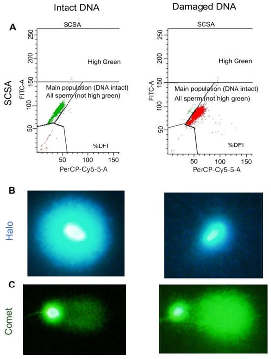
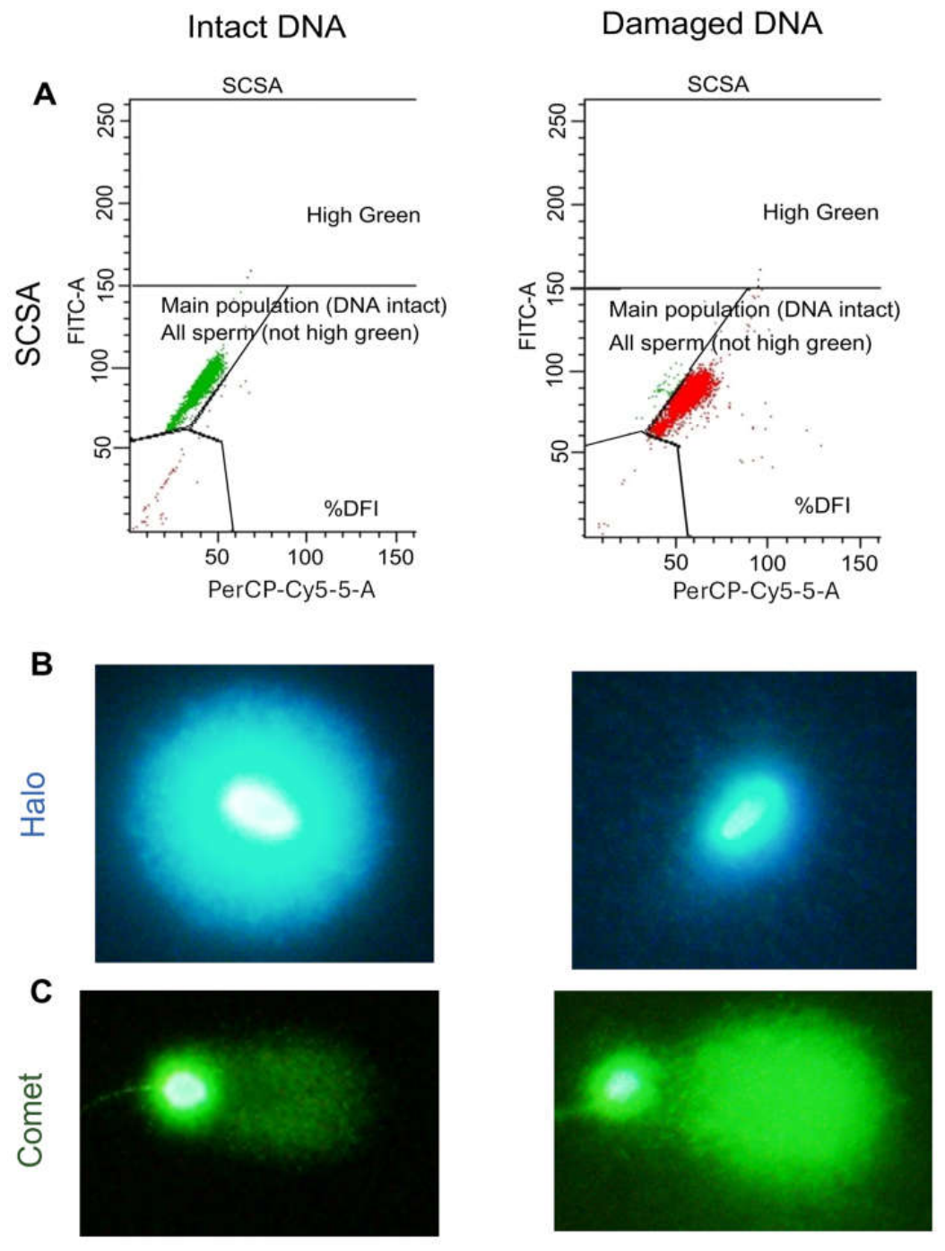
Samples were thawed at RT, and 100 µL of sperm suspension was mixed with 200 µL of acid detergent solution (0.08 N HCl, 0.15 M NaCl, 0.1% Triton X-100; pH 1.2) and incubated for exactly 30 s at room temperature, after which 600 µL of acridine orange solution (0.1 M citric acid, 0.2 M Na2PO4, 1 mM EDTA, 0.15 M NaCl, 1 M acridine orange) was added, and samples were allowed to equilibrate for 2.5 min at RT prior to flow cytometric analysis. Measurements were conducted using a BD FACS Canto II flow cytometer (FACSDiva software, version 9.0.1) equipped with a 488 nm Argon-ion laser. Fluorescence was detected using 530/30 nm (green) and 670/30 nm (red) band-pass filters to quantify double-stranded and denatured single-stranded DNA respectively using a linear scale. DNA fragmentation was quantified as the percentage of spermatozoa outside the main population, expressed as the DNA fragmentation index (%DFI; Figure 1A) [36].
Figure 1.
Representative images of sperm DNA damage assays. (A) Flow cytometric dot plot from the sperm chromatin structure assay (SCSA). Spermatozoa with intact DNA exhibit predominantly green fluorescence, whereas spermatozoa with fragmented DNA show increased red fluorescence. (B) Sperm chromatin dispersion (SCD) assay. Spermatozoa with intact DNA display large, dispersed halos of fluorescence, while spermatozoa with high levels of DNA damage exhibit small halos. (C) Single-cell gel electrophoresis (Comet assay). Spermatozoa with intact DNA show minimal tail length and low tail intensity, whereas DNA-damaged spermatozoa exhibit elongated comet tails with increased fluorescence intensity.
2.4. Sperm Chromatin Dispersion Assay (SCD)
Prior to assessment, samples were thawed at room temperature and combined with pre-warmed (37 °C) 1% low-melting point agarose to achieve a final agarose concentration of 0.7% (30 µL sperm suspension mixed with 70 µL agarose). Approximately 70 µL of the sperm–agarose mixture was spread evenly onto microscope slides precoated with 0.65% standard agarose. Coverslips were gently placed, and slides were incubated at 4 °C for at least 5 min to allow agarose solidification.
After this, coverslips were carefully removed, and slides were exposed to 0.08 N HCl for 7 min to initiate DNA denaturation. Slides were then subsequently treated with denaturation solution 1 (50 mM EDTA, 0.4 M Tris, 800 mM DTT, 1% SDS; pH 7.5) for 10 min, then denaturation solution 2 (2 M NaCl, 0.4 M Tris, 1% SDS; pH 7.5) for 5 min. Slides were then rinsed in 1× TBE buffer at room temperature for 2 min, dehydrated through a graded ethanol series (70%, 90%, and 100%; 2 min each), and air-dried. For visualisation, slides were stained with DAPI (1:2000 in PBS) for 3 min in the dark, rinsed with PBS, and mounted with Mowiol under a coverslip. DNA fragmentation was evaluated using a fluorescence microscope (Carl Zeiss Microscopy GmbH, Jena, Germany) equipped with an Olympus DP80 camera (Evident Corporation, Tokyo, Japan) and Olympus cellSens standard 4.2 software, with at least 100 spermatozoa assessed per group and categorised as either having intact DNA (large halos) or fragmented DNA (small or no halo; Figure 1B) [34].
2.5. Alkaline Comet Assay
After thawing, sperm concentration was adjusted to 4000 cells/µL in PBS, followed by mixing the sperm suspension with low-gelling-temperature agarose maintained at 37 °C (10 µL of sperm suspension was combined with 70 µL of 1% agarose). A 70 µL aliquot of this mixture was placed onto slides precoated with 1% low-gelling-temperature agarose. Coverslips were applied gently, and slides were allowed to solidify for 1 h at 4 °C. Coverslips were gently removed, and slides were treated by flooding each slide with 100 µL of lysis solution 1 (0.8 M Tris base, 0.8 M dithiothreitol [DTT], 1% SDS, pH 7.5) and covering them with coverslips for 30 min at room temperature. Coverslips were then removed, and the slides were incubated with lysis solution 2 (0.4 M Tris base, 50 mM ethylenediaminetetraacetic acid [EDTA], 2 M NaCl, 0.4 M DTT, pH 7.5) under coverslips for 30 min at room temperature. Then slides were washed in TBE buffer (0.445 M Tris base, 0.445 M boric acid, 10 mM EDTA, pH 7.5) for 10 min, followed by incubation in the alkaline solution (0.03 M NaOH, 1 M NaCl) for 15 min at 4 °C. Electrophoresis was performed in a Bio-Rad horizontal gel electrophoresis tank in alkaline buffer (0.03 M NaOH) at 1 V/cm (25 V constant) for 4 min at room temperature. Slides were washed in neutralisation buffer (0.4 M Tris base, pH 7.5) for 5 min, stained with SYBRTM green (1:10,000) nucleic acid stain (Thermo Fisher Scientific, Rockland, ME, USA) and a minimum of 50 sperm cells per replicate were imaged immediately under a fluorescent microscope (Carl Zeiss, Germany) equipped with Olympus DP80 camera and Olympus cellSens standard 4.2 software. DNA fragmentation was quantified using COMET IVTM software (version 4.2) by measuring the fluorescent intensity of comet tail relative to comet head, expressed as DNA tail intensity (TI%; Figure 1C).
2.6. 8-Hydroxy-2′-Deoxyguanosine Assay (8-OHdG)
8-OHdG is a product of oxidative DNA damage and a marker of cellular oxidative stress. 8-OHdG adducts in sperm DNA were quantified using a commercial DNA/RNA Oxidative Damage (High Sensitivity) ELISA kit (Cayman Chemical, Ann Arbor, MI, USA; Cat. No. 589320), as previously described [34]. After in vitro treatment, sperm cells were centrifuged to separate the sperm pellet and supernatant, rapidly frozen in liquid nitrogen, and stored at −80 °C until use. Genomic DNA was isolated using the Sbeadex™ Livestock DNA purification Kit (LGC Biosearch Technologies, Berlin, Germany) according to the manufacturer instructions, with adaptions for automated processing. For each sample, 10–50 µL of semen was used depending on sample origin and quality. Samples were diluted to a final volume of 100 µL with Lysis Buffer PVP supplemented with 50 mM dithiothreitol (DTT) to facilitate disruption of sperm nuclear proteins. Proteolysis was performed by adding 10 µL of protease solution and incubating the samples at 55 °C for at least 1 h, with overnight incubation allowed for samples requiring extensive lysis. After lysis, 110 µL of the lysate was transferred to a new tube and supplemented with 100 µL Lysis Buffer SB, optionally incubated at 60 °C for 5–10 min to ensure complete lysis. A total of 210 µL of lysate was then prepared for automated DNA purification. Samples with high viscosity were split and extracted as two separate extractions each utilising 105 µL of lysate.
All DNA extractions were performed on a KingFisher Flex Prime automated purification platform (Thermo Fisher Scientific) using the manufacturer-installed sbeadex protocol sbx_livestock_standard2_flex. This magnetic bead-based programme automates DNA binding, washing, and elution steps to ensure consistent sample processing. DNA binding was achieved by adding 180 µL of the kit-provided Sbeadex binding mix to the lysate and incubating at room temperature for 5 min with constant shaking. Washing of bound DNA was performed sequentially using the kit-supplied proprietary wash buffers (BN1, TN1, and TN2), which are included as part of the sbeadex kit. Each wash involved repeated resuspension and magnetic separation steps to remove proteins, salts, and other contaminants. DNA was eluted from the magnetic particles using 180 µL of the kit-supplied Elution Buffer (AMP) by incubation at 55 °C for 10 min with agitation. During elution, magnetic separation was used to immobilise the beads, and approximately 90% of the eluate was carefully recovered to minimise magnetic particle carryover into the final DNA preparation.
Extracted DNA from the sperm pellets and supernatants was quantified using NanoDropTM 2000/2000c spectrophotometers (Thermo Fisher Scientific). For ELISA sample preparation, 1 µg of DNA per assay was used. Samples were subjected to three freeze–thaw cycles (100 °C for 3 min and −80 °C for 5 min), followed by enzymatic digestion to nucleosides through sequential incubation with nuclease P1 (1 µL/2.5 µg DNA; 56 °C for 2 h) in ammonium acetate/zinc chloride buffer (20 mM NH4CH3CO2, 5 mM ZnCl2, 50 mM NaCl, 50% glycerol (v/v), pH 5.3) and alkaline shrimp phosphatase (2 µL/µg DNA; 37 °C for 30 min) in 15 mM Tris. Enzymes were then heat-inactivated at 100 °C for 10 min, and samples were placed on ice until ELISA plate preparation. The DNA/RNA Oxidative Damage (High Sensitivity) ELISA was prepared according to the manufacturer’s instructions using the supplied standards and controls, and 8-OHdG concentrations were determined colorimetrically using SPECTROstar Nano microplate reader (BMG LABTECH GmbH, Ortenberg, Germany).
3. Experimental Design
3.1. Experiment 1: Effect of H2O2 Exposure on Sperm DNA Damage Detection
H2O2 is used as an exogenous oxidative stressor to induce controlled oxidative DNA damage in spermatozoa. Following semen processing (Section 2.2), spermatozoa were incubated with increasing doses of H2O2 (0 mM, 0.125 mM, 1.25 mM, 2.5 mM and 5 mM) at 37 °C for 1 h. In the second part of the experiment, spermatozoa were incubated with a 1 mM dose of H2O2 at 37 °C for 0 h, 0.5 h, 1 h, 2 h and 4 h. The H2O2 concentrations were selected based on previous research. Following incubation with H2O2, samples were centrifuged at 500 RCF for 3 min, and the sperm cells were resuspended in BWW; sperm concentration was adjusted to 10 × 106/mL, and aliquots were snap-frozen in liquid nitrogen (LN) and stored at −80 °C until assessment via SCSA, comet and SCD, while for the 8-OHdG assay, following centrifugation, the sperm pellet was snap frozen for DNA extraction and analyses.
3.2. Experiment 2: Effect of Arachidonic Acid Exposure on Sperm DNA Damage Detection
In the same manner as Experiment 1, spermatozoa were initially exposed to increasing concentrations of arachidonic acid (AA; 0 µM, 5 µM, 10 µM, 20 µM and 40 µM) for 1 h at 37 °C. In a follow-up experiment, sperm cells were incubated with a single dose of arachidonic acid (20 µM) for 0 h, 0.5 h, 1 h, 2 h and 4 h. An untreated control was also incubated for these durations. These concentrations were previously employed to induce endogenous oxidative stress in other studies [37,38]. Following treatment, all samples were washed via centrifugation at 500 RCF for 3 min, resuspended in BWW; sperm concentration was adjusted to 10 × 106/mL, and aliquots were snap-frozen in LN and stored at −80 °C until assessment via SCSA, comet and SCD, while for the 8-OHdG assay, following centrifugation, the sperm pellet was snap frozen for DNA extraction and analyses.
3.3. Experiment 3: Comparing the Ability of DNA Damage Assays to Detect Differences in Sperm DNA Damage Levels at Periods of High or Low Fertility
Spermatozoa were collected from Thoroughbred stallions, processed and stored according to Section 2.2. Samples were assigned to high-fertility and low-fertility groups based on the following pregnancy data: per-cycle pregnancy rates were collected for three days before and after the semen collection day, with an average of 25 mares covered by each stallion during this period. Mare selection for breeding was made entirely by individual horse owners and farm managers. Mares were generally bred once per cycle; however, rebreeding was only performed when mares were confirmed to still be in oestrus. Pregnancy scan results for each breeding performed by stallion were provided by the stud farm. Stallions with per-cycle pregnancy rates ≥ 80% during this period were categorised into the high-fertility group, while six stallions with per-cycle pregnancy rates ≤ 30% during this period were categorised into the low-fertility group. After collection, spermatozoa were washed by centrifugation and extended in BWW at room temperature for DNA integrity analysis, described in Section 3.2.
3.4. Statistics
Data was analysed using JMP Student Edition 18 (SAS Institute Inc., Cary, NC, USA). Statistical significance was defined at p ≤ 0.05, and all data are presented as mean ± SEM. The Shapiro–Wilk test was applied to assess the normality of the data, with a p-value ≤ 0.05 indicating a significant deviation from normality. Data were normalised using the Box–Cox or logit transformation where appropriate. Data that did not meet normality assumptions were analysed using the non-parametric Kruskal–Wallis test as an alternative to one-way ANOVA. When a significant effect of treatment was observed, post hoc pairwise comparisons among all groups were conducted using Dunn’s multiple comparison test.
For normally distributed data, a one-way ANOVA was performed to evaluate the effect of H2O2 and arachidonic acid dose responses. Where the ANOVA revealed a significant treatment effect, treatment group means were compared with the corresponding control using Dunnett’s post hoc test to determine which treatment groups showed significant differences from the control. A full factorial mixed-effects model was used to evaluate treatment × time interactions for H2O2 and arachidonic acid treatments across incubation intervals. When a significant treatment × time interaction was detected, simple effects were examined using post hoc comparisons. In the absence of significant interaction, data was pooled to assess the main effect of treatment and time separately. Comparisons with control were performed using Dunnett’s post hoc test. For the comparison between high- and low-fertility commercial Thoroughbred stallions, an unpaired, two-tailed t-test was used.
Agreement between assay outputs was assessed using the in vitro data from Experiments 1 and 2, in which every sample was assessed by the SCSA, SCD and comet assays; the 8-OHdG ELISA was performed on a subset of treatments only and was therefore not included. Two comparisons were made. To determine whether the assays ranked individual samples in the same way, the values obtained for untreated control spermatozoa were correlated with one another (Pearson correlation; n = 138 to 144 paired observations, depending on the assay pair). To determine whether the assays detected the same treatment effects, the mean value of each treatment group was correlated with the corresponding mean for a second assay (n = 30 treatment groups), allowing for the differing baseline levels of damage between the four in vitro datasets. Consistent differences between stallions were assessed by one-way analysis of variance on the control values for each assay.
4. Results
4.1. Effect of H2O2 Exposure on Sperm DNA Damage Detection
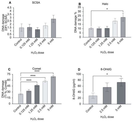
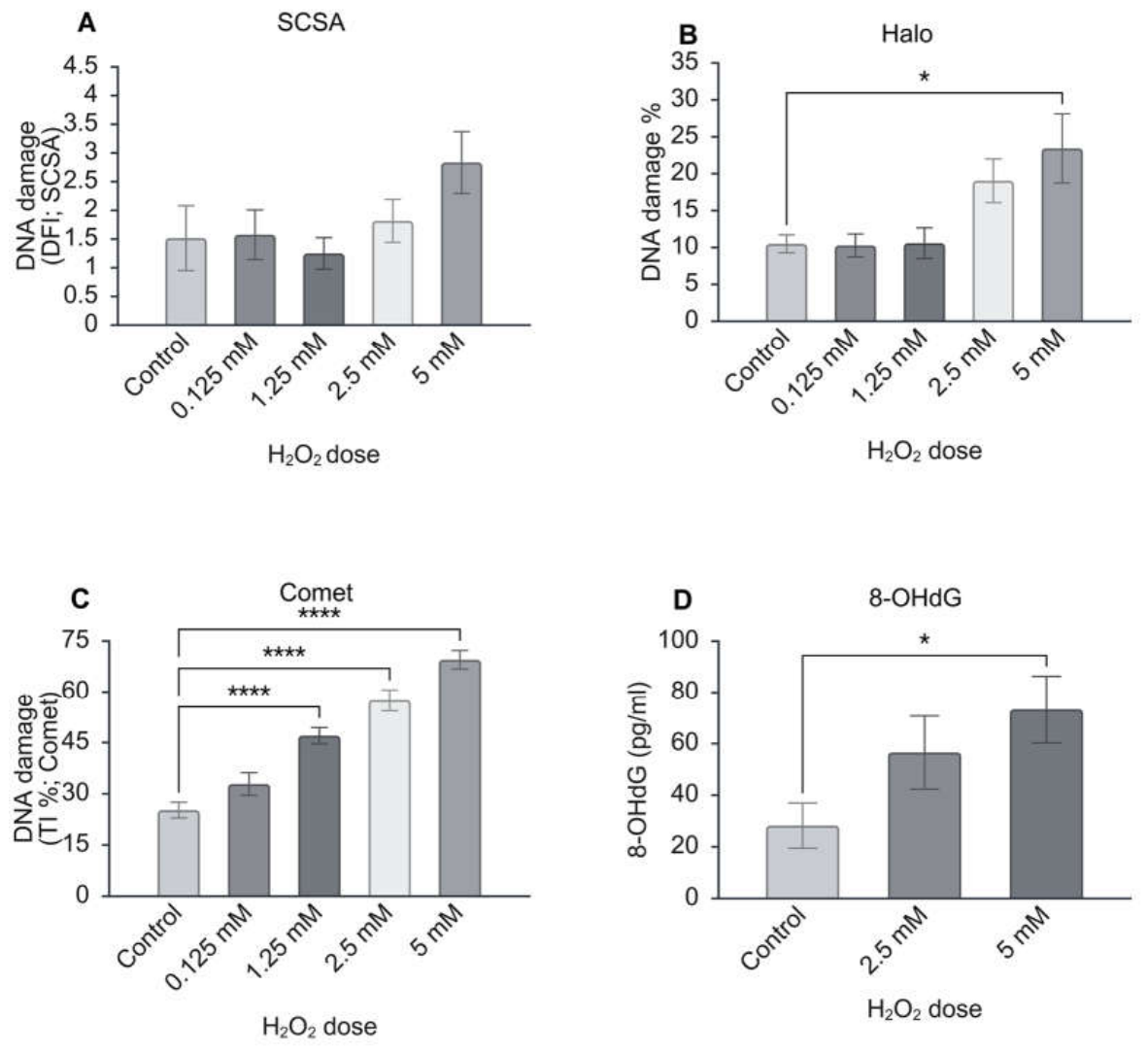
DNA damage was assessed using the four assays (SCSA, SCD, comet, and the 8-OHdG ELISA) to evaluate the effects of in vitro H2O2 exposure. Representative images of spermatozoa with intact and fragmented DNA for the SCSA, SCD, and comet assays are shown in Figure 1. The SCSA revealed no significant differences in DNA fragmentation index (DFI%) across H2O2 treatments (Figure 2A). The SCD assay did reveal a significant treatment effect: the percentage of DNA-damaged spermatozoa (without a halo) increased at 5 mM H2O2 (23.43 ± 4.68%) compared with the control (10.49 ± 1.21; p ≤ 0.05; Figure 2B). The comet assay was able to detect a highly significant increase in DNA damage (p ≤ 0.0001), expressed as tail intensity (TI%), at H2O2 doses of 1.25 mM and above compared to the control (control: 25.20 ± 2.30; 1.25 mM: 47.01 ± 2.39, p ≤ 0.0001; 2.5 mM: 57.47 ± 3.11, p ≤ 0.0001; 5 mM: 69.51 ± 2.71, p ≤ 0.0001, Figure 2C). Interestingly, while the 8-OHdG ELISA was able to detect a treatment effect in the sperm pellet, it could only detect a difference between the control (28.21 ± 8.73 pg/mL) and the highest dose (5 mM: 73.29 ± 13.04 pg/mL, p ≤ 0.05, Figure 2D).
Figure 2.
Effect of increasing H2O2 concentrations on DNA damage in stallion spermatozoa as measured using (A) sperm chromatin structure assay (SCSA; DNA fragmentation index, DFI), (B) sperm chromatin dispersion (SCD) assay, (C) The comet assay, expressed as tail intensity (TI%), representing the percentage of total comet fluorescence detected in the tail relative to the combined fluorescence of the head and tail, (D) The 8-hydroxy-2′-deoxyguanosine (8-OHdG) ELISA assay (pg/mL). Significant differences relative to the control are indicated by * p ≤ 0.05, **** p ≤ 0.0001 (n = 12).
A full factorial mixed-effects model was used to evaluate the effects of time, treatment and the time × treatment interaction. For all assays, there was no significant time × treatment interaction on DNA damage (Table 1), and so data were pooled across time points to isolate the main effect of physiological H2O2 exposure. Neither the SCSA nor SCD assay was able to detect a significant increase in DNA damage (Table 2), but the comet assay was able to detect differences in DNA damage when the data were pooled to isolate the main effects of incubation time and H2O2 treatment. According to the comet assay, TI% increased by 1 h of incubation (from 37.37 ± 0.98% to 44.59 ± 1.60% at 1 h, p ≤ 0.05), and this difference continued to increase up to the 4 h time point (52.46 ± 1.91%, p ≤ 0.001, Table 2). When isolating the treatment effects, the comet assay revealed that H2O2-treated spermatozoa had higher levels of DNA damage compared to the control (42.30 ± 1.20% vs. 47.75 ± 1.18%, p ≤ 0.001, Table 2).
Table 1.
Effect of incubation time on equine sperm DNA damage assessed by SCSA, SCD assay, and comet assay following physiological H2O2 exposure (1 mM).
Table 2.
Main effect of physiological (1 mM) H2O2 treatment on pooled equine sperm DNA damage across all incubation time points, assessed by SCSA, SCD, and comet assay.
4.2. Effect of Arachidonic Acid Exposure on Sperm DNA Damage Detection
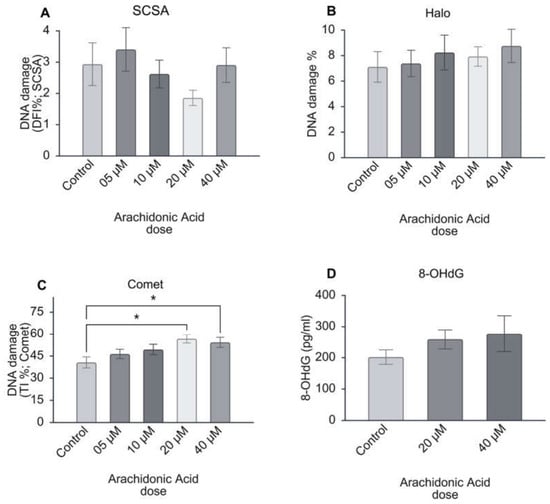
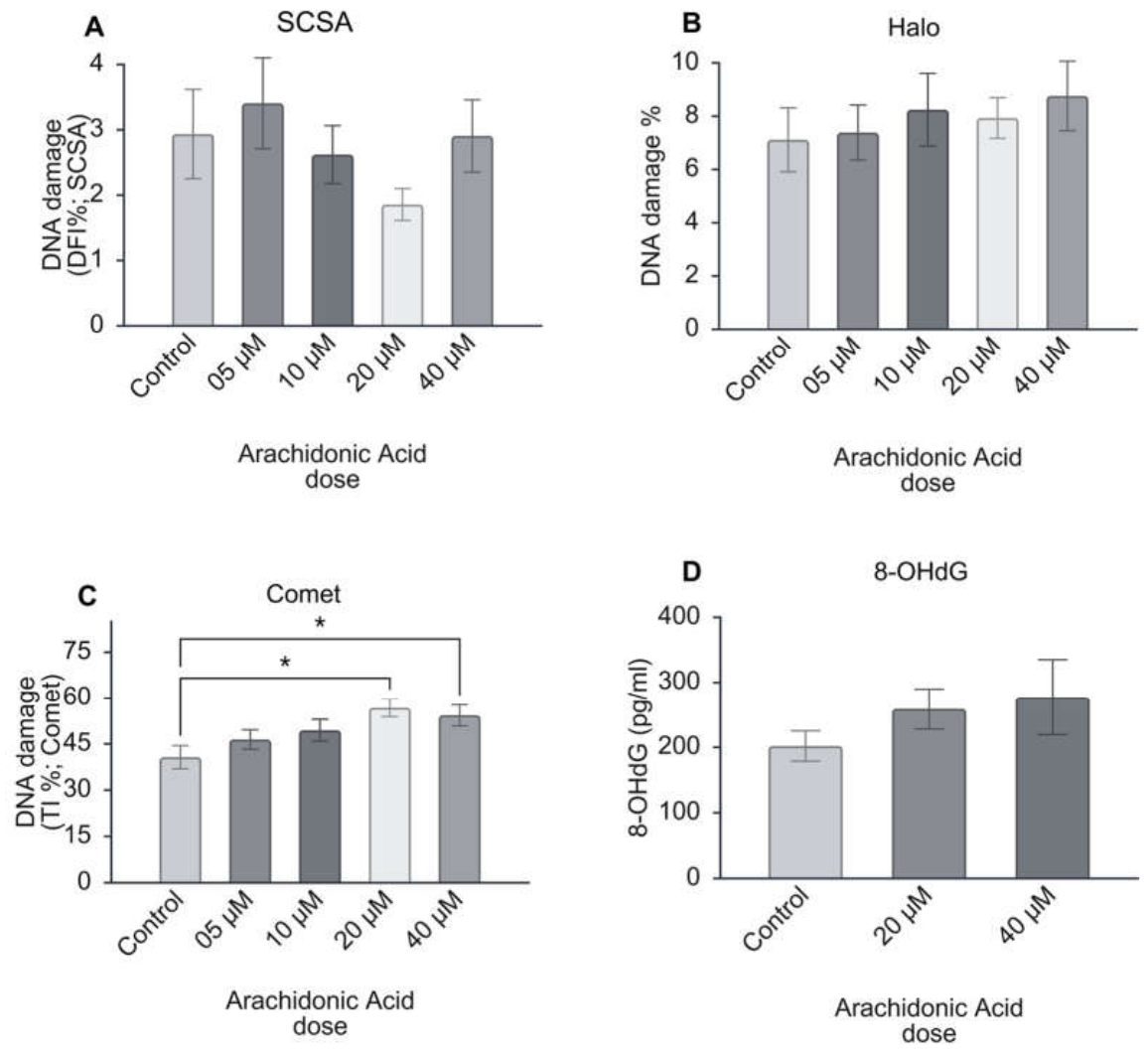
DNA damage was assessed using the four assays (SCSA, SCD, comet and the 8-OHdG ELISA) to evaluate the effects of in vitro AA exposure (Figure 3). The sperm chromatin structure assay (SCSA) and SCD assay revealed no significant differences in the DNA fragmentation index (DFI%; Figure 3A) or percentage of DNA damage spermatozoa (Figure 3B) across AA treatments. The comet assay, however, did reveal a significant treatment effect (ANOVA, p < 0.05); the percentage of DNA damage, expressed as tail intensity (TI%), increased significantly at 20 µM and 40 µM AA compared with the control (control: 40.67 ± 3.76; 20 µM: 56.72 ± 2.90, p ≤ 0.05; 40 µM: 54.26 ± 3.44, p ≤ 0.05; Figure 3C). The 8-OHdG ELISA did not detect a significant treatment effect on the levels of 8-OHdG adducts in sperm cells (Figure 3D).
Figure 3.
Effect of increasing arachidonic acid (AA) concentrations on DNA damage in stallion spermatozoa as measured by (A) sperm chromatin structure assay (SCSA; DNA fragmentation index, DFI), (B) sperm chromatin dispersion (SCD) assay, (C) The comet assay, expressed as tail intensity (TI%), representing the percentage of total comet fluorescence detected in the tail relative to the combined fluorescence of the head and tail, (D) The 8-hydroxy-2′-deoxyguanosine (8-OHdG) ELISA assay (pg/mL). Significant differences relative to the control are indicated by * p ≤ 0.05.
A full factorial mixed-effects model was used to evaluate the effects of time, treatment and the time × treatment interaction. No significant time × treatment interaction on DNA damage was detected using any assay (Table 3), so data were pooled across time points to isolate the main effect of exposure to a physiological concentration of AA. Neither SCSA nor comet assays were able to detect a significant increase in DNA damage, but the SCD assay was able to detect difference in DNA damage when the data were pooled to isolate the main effects of incubation time (Table 4). DNA damage increased significantly after 4 h compared to 0 h (0 h: 14.16 ± 0.78 vs. 4 h: 18.78 ± 0.94, p ≤ 0.001; Table 4). When isolating the treatment effects, AA-treated spermatozoa had significantly higher levels of DNA damage compared to the control according to the SCD assay (control: 15.54 ± 0.50%; AA-treatment: 17.24 ± 0.57%, p ≤ 0.05, Table 4). The comet assay also detected an increase in DNA damage in AA-treated spermatozoa compared to the control (control: 37.14 ± 1.14%; AA treatment: 40.09 ± 0.98%, p ≤ 0.05, Table 4).
Table 3.
Effect of incubation time on stallion sperm DNA damage assessed by SCSA, SCD assay, and comet assay following physiological AA exposure.
Table 4.
Main effect of physiological AA treatment on pooled equine sperm DNA damage across all incubation time points, assessed by SCSA, SCD, and comet assay.
4.3. Comparing the Ability of DNA Damage Assays to Detect Differences in Sperm DNA Damage Levels at Periods of High or Low Fertility
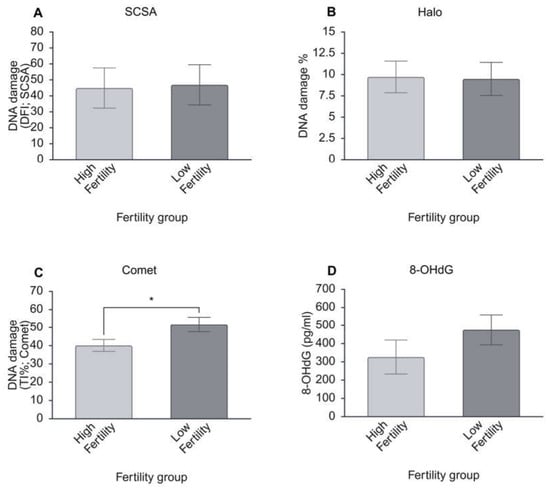
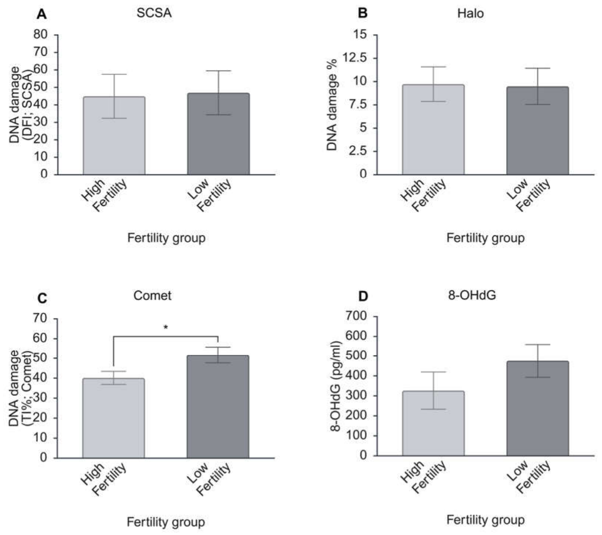
Differences between the levels of in vivo-induced DNA damage in the spermatozoa of commercial Thoroughbred stallions of high and low fertility were assessed, and neither the SCSA, SCD, nor the 8-OHdG ELISA were able to detect a significant difference between the two cohorts (Figure 4A,B,D). In contrast, the comet assay revealed higher DNA damage in spermatozoa of the low-fertility group compared with the high-fertility group (low fertility: 51.69 ± 4.03% vs. high fertility: 40.12 ± 3.27%, p ≤ 0.05, Figure 4C).
Figure 4.
Comparison between DNA damage levels in spermatozoa of stallions of high (n = 6) and low (n = 6) fertility as measured by (A) sperm chromatin structure assay (SCSA; DNA fragmentation index, DFI), (B) sperm chromatin dispersion (SCD) assay, (C) The comet assay, expressed as tail intensity (TI%), representing the percentage of total comet fluorescence detected in the tail relative to the combined fluorescence of the head and tail, and (D) The 8-hydroxy-2′-deoxyguanosine (8-OHdG) ELISA assay (pg/mL). Significant differences are indicated by * p ≤ 0.05.
4.4. Agreement Between Assay Outputs
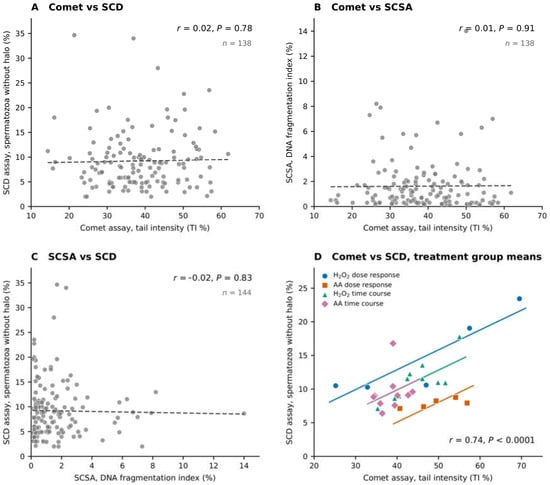
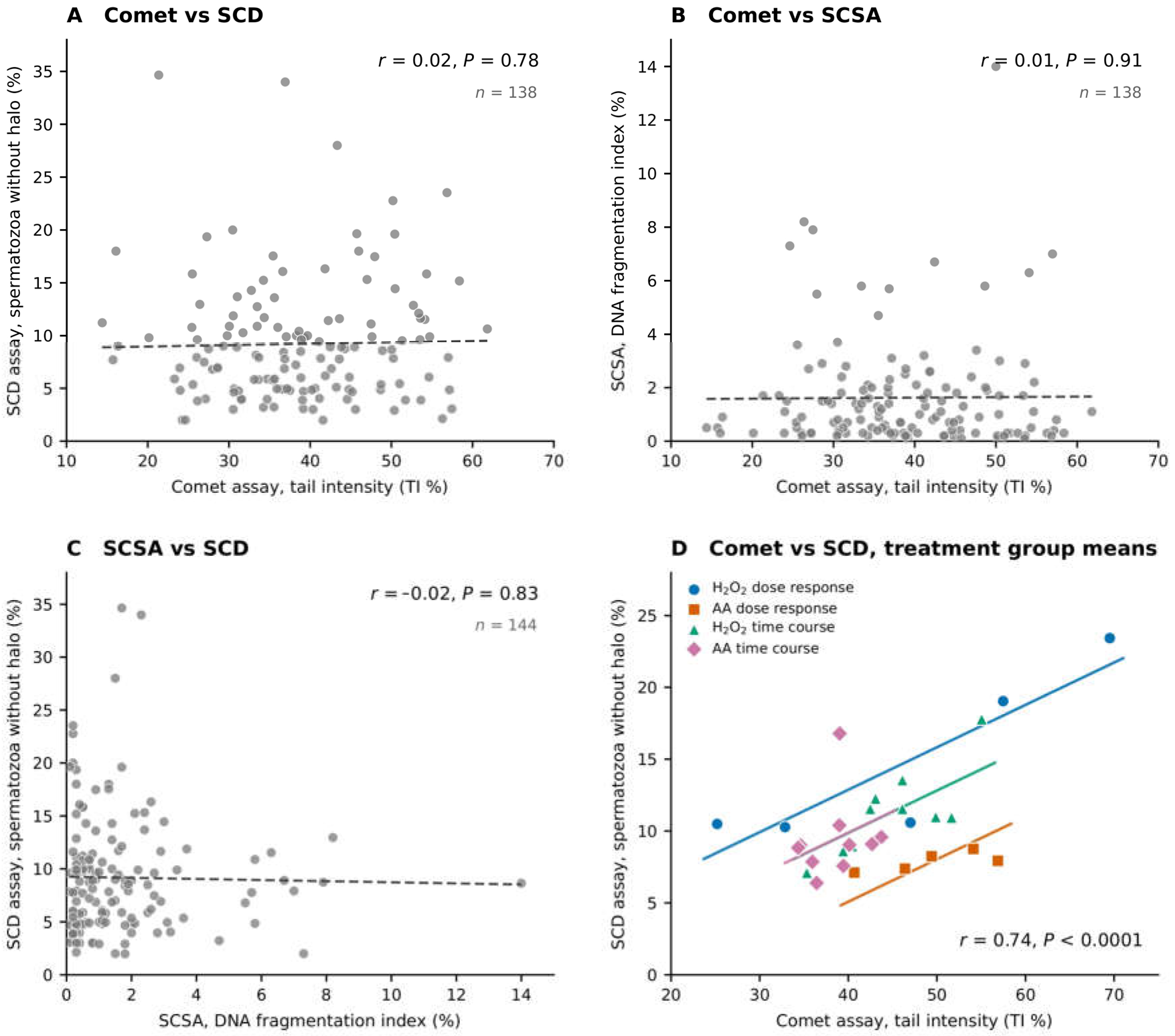
Agreement between the SCSA, SCD and comet assays was assessed at two levels using the in vitro data from Experiments 1 and 2 (Figure 5). In untreated control spermatozoa, no pair of assays showed any significant relationship (comet vs. SCD: r = 0.02, p = 0.78, n = 138; comet vs. SCSA: r = 0.01, p = 0.91, n = 138; SCSA vs. SCD: r = −0.02, p = 0.83, n = 144; Figure 5A–C); a sample identified as heavily damaged by one assay was therefore no more likely than any other to be identified as heavily damaged by a second assay.
Figure 5.
Agreement between sperm DNA damage assays in Experiments 1 and 2. (A–C) Untreated control spermatozoa, each point representing one sample assessed by two assays. (D) Mean values for each treatment group in the four in vitro datasets (n = 30 groups). A separate line is fitted for each dataset because baseline levels of damage differed between them; the lines are parallel because the relationship between the two assays was the same in each.
However, when treatment groups were compared, the comet and SCD assays agreed well (r = 0.74, p ≤ 0.0001; Figure 5D). This agreement was consistent: the relationship was positive within each of the four in vitro datasets (H2O2 dose–response r = 0.90; AA dose–response r = 0.75; H2O2 time course r = 0.80; AA time course r = 0.19), and exclusion of the most damaged treatment group (5 mM H2O2) left it essentially unchanged (r = 0.58, p ≤ 0.01). The weaker value obtained for the AA time course reflects both the narrower range of damage spanned by that dataset and the fact that the SCD assay, but not the comet assay, detected the increase in damage at 4 h (Table 4). The SCSA showed no relationship with either of the other assays at either level (comet vs. SCSA: r = 0.23, p = 0.25; SCSA vs. SCD: r = 0.01, p = 0.94).
The assays also differed in their ability to detect differences between individual stallions, that is, differences arising in vivo rather than those induced by in vitro treatment. The SCD assay revealed consistent differences between the four stallions, with the proportion of spermatozoa lacking a halo approximately twice as high in two stallions compared to the other two (Stallion 1: 6.1%, Stallion 3: 5.9%, Stallion 2: 11.5%, Stallion 4: 13.2%; p ≤ 0.0001), accounting for approximately a third of the variation between control samples. Neither the comet assay (36.9 to 40.3% TI, p = 0.58) nor the SCSA (1.3 to 2.2% DFI, p = 0.26) distinguished between stallions. The SCD assay therefore detected a stallion effect that was not detected by the comet assay, despite the comet assay being the more sensitive of the two to treatment-induced damage.
5. Discussion
This is the first study to compare the sensitivity of the SCSA, SCD, comet and 8-OHdG ELISA assays for detecting DNA damage in stallion spermatozoa. We hypothesised that the alkaline comet assay would prove most sensitive, by virtue of its capacity to detect single- and double-strand breaks as well as alkali-labile sites. This was supported: the comet assay alone detected damage across all experiments, including in vitro-driven DNA damage caused by exogenous (Figure 2C, Table 2) and endogenous (Figure 3, Table 4) oxidative stressors, as well as differences in inherent DNA damage between stallions of high or low fertility (Figure 4). Comparison of the assays with one another further revealed that their agreement depended on whether treatment effects or individual sample results were being compared (Figure 5).
DNA damage assays may measure existing damage directly, as for the comet assay; assess susceptibility to damage based on DNA packaging, as for the SCSA and SCD; or, as with the 8-OHdG ELISA, assess an upstream instigator of damage: oxidised guanine nucleotides, which once excised by OGG1 leave abasic sites particularly susceptible to single strand breaks [21]. This distinction between assay mechanisms is central to interpreting the results of the present study.
The difference between the alkaline comet assay and other techniques for measuring sperm DNA damage may be attributed to the electrophoresis step, which likely enhances the sensitivity for detecting DNA strand breaks compared with other methods [39]. In low-fertility stallions, DNA fragmentation measured by the alkaline comet assay was significantly higher than in high-fertility stallions, a clinically relevant difference which was not detected by any other assay (Figure 4). However, the sample size for this experiment was small, and it should be noted that, following much larger studies, the SCSA has been reported to discriminate between stallions of differing fertility to the extent that reference ranges have been established [40,41]. Indeed, the SCSA is one of the longest-standing and clinically recognised DNA damage assays, and it is not the intent of the authors to discredit its validity; while the SCSA is useful for identifying extensive DNA damage, it lacks the sensitivity to resolve the more moderate differences observed in the present study.
Studies comparing sperm DNA damage assays in other mammalian species have been conducted, and with the exception of the comet assay, which performs strongly across all species, the SCSA and SCD assays often perform exceptionally well in other species. In other livestock species, the comet assay is highly correlated with in vivo fertility and in vitro embryo development [42,43], and in humans it is associated with an increased incidence of miscarriage [44]. In contrast to the finding of the present study, SCSA has good prognostic value in humans [45], bulls [46,47] and rams [48] with clear reference range thresholds established for the diagnosis of infertility. However, in the present study, SCSA could not detect a difference between high and low fertility stallions (Figure 4A), nor in vitro-induced damage, even when spermatozoa were exposed to supraphysiological levels of oxidative stressors (Figure 2A and Figure 3A). Other studies have also reported the limited predictive value of the SCSA-derived DNA fragmentation index (DFI%) for pregnancy outcomes and in vitro fertilisation success in stallions [16], boars [49] and humans [50]. The reduced sensitivity of SCSA observed in the present study may be explained by oxidative over-modification of thiol groups, leading to excessive sperm chromatin condensation [51]. This hyper-condensation may stabilise chromatin structure following exposure to high concentrations of H2O2, thereby limiting further increases in DFI% values despite ongoing DNA damage [52]. Consequently, these structural changes reduce susceptibility to acid-induced denaturation, diminishing the capacity of SCSA to detect additional DNA lesions. More generally, the SCSA reflects a composite of DNA strand breaks and chromatin packaging defects, both of which increase susceptibility to denaturation, so it lacks the specificity to identify the nature of the damage. This limits its utility in mechanistic studies such as those investigating in vitro oxidative stress-induced DNA fragmentation.
The SCD assay detected a significant increase in DNA damage only at the highest concentration of H2O2 (Figure 2B). This contrasts with previous work in which no differences were detected by the SCD assay in stallion spermatozoa across a range of H2O2 concentrations [53] or in thawed bull spermatozoa [54], although smaller halos have been reported in infertile compared with fertile men [33,55]. Together these observations suggest limited sensitivity of the SCD assay for low levels of sperm DNA damage. The SCD assay evaluates DNA integrity following chromatin decondensation and quantifies damage based on the extent of DNA dispersion from the sperm nucleus [56]. Consequently, this method is more effective at identifying disrupted chromatin architecture established during spermatogenesis, rather than subtle oxidative DNA lesions arising from downstream stressors.
Similarly, the 8-OHdG assay showed significantly increased oxidative DNA damage only at supraphysiological concentrations of H2O2 (Figure 2D), while no significant differences were observed following arachidonic acid treatment (Figure 3D) or between fertility groups (Figure 4D). These results support the findings of previous studies, in which equine spermatozoa exhibit a comparatively high tolerance to H2O2-induced oxidative stress [53,57], which could account for the absence of significant changes in 8-OHdG levels under less severe oxidative conditions.
The limited responses detected by the SCSA and 8-OHdG assays may also reflect the particular oxidative biology of stallion spermatozoa. Species whose spermatozoa rely primarily on glycolysis for ATP production exhibit DNA damage at very low doses of H2O2 [58,59], whereas stallion spermatozoa, which depend on OXPHOS [60,61,62,63], do not, possibly by virtue of greater antioxidant defences [53,57]. Both in vitro treatments used in the present study compromised sperm DNA integrity, but this was reliably detected only by the comet assay, indicating that the resilience of stallion spermatozoa places a particular premium on assay sensitivity.
Previous studies have reported poor agreement between sperm DNA damage assays [21]. The present results suggest that whether the assays agree depends on what is being compared, and that the pattern of agreement is itself informative about what each assay measures. The comet and SCD assays agreed closely when treatment groups were compared, both detecting more damage in the more oxidatively stressed treatments in every in vitro dataset. Yet when untreated ejaculates were compared with one another, the two assays showed no relationship at all: the individual sample identified as most damaged by one assay was not the sample identified as most damaged by the other (Figure 5). The results of the assays across stallions support this interpretation. In untreated control spermatozoa, the SCD assay showed a significant stallion effect, whereas the comet assay did not distinguish between them at all. Differences between male animals in sperm protamination, disulphide cross-linking and the resulting degree of chromatin compaction [64] determine how readily DNA loops disperse and create the halos in the SCD assay, and these are inherent properties of the spermatozoon following spermatogenesis, as opposed to the result of a post-spermatogenesis oxidative assault, which may occur as a result of in vitro manipulations, or as a result of in vivo oxidative stress during epididymal maturation and storage. The comet assay, which measures the migration of fragmented DNA under an electric field, is largely blind to such packaging differences and instead reports the strand breaks that are present. This is consistent with the mechanistic distinction between assays that measure existing damage and those that measure susceptibility to damage, and it explains why two assays that both respond to oxidative stress can nonetheless rank ejaculates independently of one another. Any conclusion regarding the SCSA must be more guarded, since DFI% varied little in this study under any condition, and an assay that does not vary cannot be shown to agree or disagree with another. What the present data do not support is the substitution of one assay for the other when assessing an individual animal or ejaculate: a comet result cannot be used to infer what an SCD assay would have shown for the same sample, and vice versa. It should be noted that oxidative DNA damage represents only one contributor to sperm dysfunction, and stallion fertility is influenced by genetic, endocrine, infectious and environmental factors that no DNA damage assay captures. These assays are therefore best interpreted alongside conventional semen analysis and reproductive history.
In conclusion, the alkaline comet assay was the most sensitive method for detecting DNA strand breaks in stallion spermatozoa under the tested conditions. However, sensitivity alone should not determine which assay is appropriate in clinical or research settings. Where the concern is damage acquired after spermatogenesis, such as during epididymal transit, following testicular or systemic heat stress, or because of semen handling, cooling, extension or cryopreservation, the comet assay is the appropriate choice. Where the concern relates to the quality of chromatin packaging during spermatogenesis, which may be indicated by a persistent drop in fertility or increase in abortion rates despite otherwise normal semen parameters, the SCD assay is more informative. The SCSA measures a similar property to the SCD and offers rapid, objective, high-throughput assessment, but was insufficiently sensitive to discriminate between stallions in the present study. A limitation of the comet assay is that, in its current form, it is labour-intensive and technically demanding. In addition, chromatin decondensation was performed at a single dithiothreitol (DTT) concentration, which may have influenced the efficiency of sperm deprotamination and, consequently, DNA migration. Future research should therefore establish an optimal DTT concentration for equine sperm lysis and develop a simplified, standardised comet assay kit compatible with conventional light microscopy. Such advances would improve the assay’s accessibility and clinical applicability, supporting its wider use for assessing sperm DNA damage in clinical decision-making.
Author Contributions
M.Y. conducted experimental work, performed statistical analyses and co-wrote the manuscript. A.J.M., R.A.G., C.J., A.S.-J. and A.S. assisted with experimental work. Z.G. conceived the idea for the manuscript, conducted experimental work and statistical analyses, and co-wrote the manuscript. All authors have read and agreed to the published version of the manuscript.
Funding
Project costs and Roisin Griffin’s salary were funded through the by AgriFutures Australia Thoroughbred Horses Scheme (FRP-015570). Additional project costs, Zamira Gibb’s salary and Muhammad Yaseen’s PhD stipend were funded by an Australian Research Council Future Fellowship (FT220100557). Aleona Swegen’s salary was funded through an Australian Research Council DECRA Fellowship (DE220100121), and Ashlee Medica’s salary was funded by an Australian Research Council Linkage Grant (LP230100156).
Institutional Review Board Statement
Institutional ethical approval (University of Newcastle Animal Care and Ethics Committee, Approval No. A-2021-139) was secured for the collection of horse spermatozoa.
Informed Consent Statement
Not applicable.
Data Availability Statement
The raw data supporting the conclusions of this manuscript will be made available by the authors on request.
Conflicts of Interest
The authors declare no conflicts of interest.
References
- Griffin, R.A.; Baker, M.; Aitken, R.J.; Swegen, A.; Gibb, Z. What makes a fertile sperm? Unique molecular attributes of stallion fertility. Reprod. 2019, 158, R125–R137. [Google Scholar] [CrossRef] [Scilit] [PubMed]
- Egyptien, S.; Deleuze, S.; Ledeck, J.; Ponthier, J. Sperm quality assessment in stallions: How to choose relevant assays to answer clinical questions. Animals 2023, 13, 3123. [Google Scholar] [CrossRef] [Scilit] [PubMed]
- Evenson, D.; Darzynkiewicz, Z.; Melamed, M. Relation of mammalian sperm chromatin heterogeneity to fertility. Science 1980, 210, 1131–1133. [Google Scholar] [CrossRef] [Scilit] [PubMed]
- Kenney, R.M.; Evenson, D.P.; Garcia, M.C.; Love, C.C. Relationships between sperm chromatin structure, motility, and morphology of ejaculated sperm, and seasonal pregnancy rate. Biol. Reprod. 1995, 52, 647–653. [Google Scholar] [CrossRef] [Scilit]
- Rashki Ghaleno, L.; Alizadeh, A.; Drevet, J.R.; Shahverdi, A.; Valojerdi, M.R. Oxidation of sperm DNA and male infertility. Antioxidants 2021, 10, 97. [Google Scholar] [CrossRef] [Scilit] [PubMed]
- Aitken, R.J. DNA damage in human spermatozoa; important contributor to mutagenesis in the offspring. Transl. Andro. Urol. 2017, 6, S761. [Google Scholar] [CrossRef] [Scilit] [PubMed]
- Marchetti, F.; Bishop, J.; Gingerich, J.; Wyrobek, A.J. Meiotic interstrand DNA damage escapes paternal repair and causes chromosomal aberrations in the zygote by maternal misrepair. Sci. Rep. 2015, 5, 7689. [Google Scholar] [CrossRef] [Scilit] [PubMed]
- Tamburrino, L.; Marchiani, S.; Montoya, M.; Marino, F.E.; Natali, I.; Cambi, M.; Forti, G.; Baldi, E.; Muratori, M. Mechanisms and clinical correlates of sperm DNA damage. Asian J. Androl. 2011, 14, 24. [Google Scholar] [CrossRef] [Scilit] [PubMed]
- Newman, H.; Catt, S.; Vining, B.; Vollenhoven, B.; Horta, F. DNA repair and response to sperm DNA damage in oocytes and embryos, and the potential consequences in ART: A systematic review. Mol. Hum. Reprod. 2022, 28, gaab071. [Google Scholar] [CrossRef] [Scilit] [PubMed]
- Noblanc, A.; Damon-Soubeyrand, C.; Karrich, B.; Henry-Berger, J.; Cadet, R.; Saez, F.; Guiton, R.; Janny, L.; Pons-Rejraji, H.; Alvarez, J.G. DNA oxidative damage in mammalian spermatozoa: Where and why is the male nucleus affected? Free Radic. Biol. Med. 2013, 65, 719–723. [Google Scholar] [CrossRef] [Scilit] [PubMed]
- Aitken, R.J.; De Iuliis, G.N. Origins and consequences of DNA damage in male germ cells. Reprod. Biomed. Online 2007, 14, 727–733. [Google Scholar] [CrossRef] [Scilit] [PubMed]
- Zini, A.; Sigman, M. Are tests of sperm DNA damage clinically useful? Pros and cons. J. Androl. 2009, 30, 219–229. [Google Scholar] [CrossRef] [Scilit] [PubMed]
- Love, C.; Kenney, R. The relationship of increased susceptibility of sperm DNA to denaturation and fertility in the stallion. Theriogenology 1998, 50, 955–972. [Google Scholar] [CrossRef] [Scilit] [PubMed]
- Lewis, S.E.; Simon, L. Clinical implications of sperm DNA damage. Hum. Fertil. 2010, 13, 201–207. [Google Scholar] [CrossRef] [Scilit] [PubMed]
- Abah, K.O.; Ligocka-Kowalczyk, Z.; Itodo, J.I.; Ameh, G.; Partyka, A.; Nizanski, W. Association between sperm DNA fragmentation and fertility parameters in farm animals: A systematic review and meta-analysis. BMC Vet. Res. 2025, 21, 204. [Google Scholar] [CrossRef] [Scilit] [PubMed]
- Morrell, J.M.; Johannisson, A.; Dalin, A.-M.; Hammar, L.; Sandebert, T.; Rodriguez-Martinez, H. Sperm morphology and chromatin integrity in Swedish warmblood stallions and their relationship to pregnancy rates. Acta Vet. Scand. 2008, 50, 2. [Google Scholar] [CrossRef] [Scilit] [PubMed]
- Aitken, R.; De Iuliis, G. On the possible origins of DNA damage in human spermatozoa. MHR Basic. Sci. Reprod. Med. 2009, 16, 3–13. [Google Scholar] [CrossRef] [Scilit] [PubMed]
- Wright, C.; Milne, S.; Leeson, H. Sperm DNA damage caused by oxidative stress: Modifiable clinical, lifestyle and nutritional factors in male infertility. Reprod. Biomed. Online 2014, 28, 684–703. [Google Scholar] [CrossRef] [Scilit] [PubMed]
- Aitken, R.J. Impact of oxidative stress on male and female germ cells: Implications for fertility. Reprod. 2020, 159, R189–R201. [Google Scholar] [CrossRef] [Scilit] [PubMed]
- Dutta, S.; Majzoub, A.; Agarwal, A. Oxidative stress and sperm function: A systematic review on evaluation and management. Arab. J. Urol. 2019, 17, 87–97. [Google Scholar] [CrossRef] [Scilit] [PubMed]
- Smith, T.B.; Dun, M.D.; Smith, N.D.; Curry, B.J.; Connaughton, H.S.; Aitken, R.J. The presence of a truncated base excision repair pathway in human spermatozoa that is mediated by OGG1. J. Cell Sci. 2013, 126, 1488–1497. [Google Scholar] [CrossRef] [Scilit] [PubMed]
- Koppers, A.J.; Garg, M.L.; Aitken, R.J. Stimulation of mitochondrial reactive oxygen species production by unesterified, unsaturated fatty acids in defective human spermatozoa. Free Radic. Biol. Med. 2010, 48, 112–119. [Google Scholar] [CrossRef] [Scilit] [PubMed]
- Jones, R.; Mann, T.; Sherins, R. Peroxidative breakdown of phospholipids in human spermatozoa, spermicidal properties of fatty acid peroxides, and protective action of seminal plasma. Fertil. Steril. 1979, 31, 531–537. [Google Scholar] [CrossRef] [Scilit] [PubMed]
- Gibb, Z.; Lambourne, S.R.; Aitken, R.J. The paradoxical relationship between stallion fertility and oxidative stress. Biol. Reprod. 2014, 91, 77. [Google Scholar] [CrossRef] [Scilit] [PubMed]
- Xu, Z.; Yan, Q.; Zhang, K.; Lei, Y.; Zhou, C.; Ren, T.; Gao, N.; Wen, F.; Li, X. Mitochondrial regulation of spermatozoa function: Metabolism, oxidative stress and therapeutic insights. Animals 2025, 15, 2246. [Google Scholar] [CrossRef] [Scilit] [PubMed]
- Gibb, Z.; Aitken, R.J.; Sheridan, A.R.; Holt, B.; Waugh, S.; Swegen, A. The effects of oxidative stress and intracellular calcium on mitochondrial permeability transition pore formation in equine spermatozoa. FASEB BioAdv 2024, 6, 143. [Google Scholar] [CrossRef] [Scilit] [PubMed]
- Alvarez, J.G.; Touchstone, J.C.; Blasco, L.; Storey, B.T. Spontaneous lipid peroxidation and production of hydrogen peroxide and superoxide in human spermatozoa Superoxide dismutase as major enzyme protectant against oxygen toxicity. J. Androl. 1987, 8, 338–348. [Google Scholar] [CrossRef] [Scilit] [PubMed]
- De Iuliis, G.N.; Thomson, L.K.; Mitchell, L.A.; Finnie, J.M.; Koppers, A.J.; Hedges, A.; Nixon, B.; Aitken, R.J. DNA damage in human spermatozoa is highly correlated with the efficiency of chromatin remodeling and the formation of 8-hydroxy-2′-deoxyguanosine, a marker of oxidative stress. Biol. Reprod. 2009, 81, 517–524. [Google Scholar] [CrossRef] [Scilit] [PubMed]
- Wathes, D.C.; Abayasekara, D.R.E.; Aitken, R.J. Polyunsaturated fatty acids in male and female reproduction. Biol. Reprod. 2007, 77, 190–201. [Google Scholar] [CrossRef] [Scilit] [PubMed]
- Henkel, R.; Hoogendijk, C.; Bouic, P.; Kruger, T. TUNEL assay and SCSA determine different aspects of sperm DNA damage. Andrologia 2010, 42, 305–313. [Google Scholar] [CrossRef] [Scilit] [PubMed]
- Evenson, D.P.; Kasperson, K.; Wixon, R.L. Analysis of sperm DNA fragmentation using flow cytometry and other techniques. Soc. Reprod. Fertil. Suppl. 2007, 65, 93–113. [Google Scholar] [PubMed]
- Collins, A.R.; Oscoz, A.A.; Brunborg, G.; Gaivao, I.; Giovannelli, L.; Kruszewski, M.; Smith, C.C.; Štětina, R. The comet assay: Topical issues. Mutagenesis 2008, 23, 143–151. [Google Scholar] [CrossRef] [Scilit] [PubMed]
- Ribas-Maynou, J.; García-Peiró, A.; Fernández-Encinas, A.; Abad, C.; Amengual, M.; Prada, E.; Navarro, J.; Benet, J. Comprehensive analysis of sperm DNA fragmentation by five different assays: TUNEL assay, SCSA, SCD test and alkaline and neutral Comet assay. Andrology 2013, 1, 715–722. [Google Scholar] [CrossRef] [Scilit] [PubMed]
- Gibb, Z.; Griffin, R.A.; Bromfield, E.G.; Lambourne, S.R.; Wilkins, A.; Jenkins, C.; Swegen, A.; Aitken, R.J.; De Iuliis, G.N. Evaluation of Oxidative Stress in Mammalian Spermatozoa. In Spermatology: Methods and Protocols; Springer: Berlin/Heidelberg, Germany, 2025; pp. 363–415. [Google Scholar]
- Biggers, J.; Whitten, W.; Whittingham, D.; Daniel, J. Methods in Mammalian Embryology; W. H. Freeman: San Francisco, CA, USA, 1971; pp. 86–116. [Google Scholar]
- Evenson, D.P. Sperm chromatin structure assay (SCSA®) for fertility assessment. Curr. Protoc. 2022, 2, e508. [Google Scholar] [CrossRef] [Scilit] [PubMed]
- Aitken, R.J.; Wingate, J.K.; De Iuliis, G.N.; Koppers, A.J.; McLaughlin, E.A. Cis-unsaturated fatty acids stimulate reactive oxygen species generation and lipid peroxidation in human spermatozoa. J. Clin. Endocrinol. Metab. 2006, 91, 4154–4163. [Google Scholar] [CrossRef] [Scilit] [PubMed]
- Moazamian, R.; Polhemus, A.; Connaughton, H.; Fraser, B.; Whiting, S.; Gharagozloo, P.; Aitken, R.J. Oxidative stress and human spermatozoa: Diagnostic and functional significance of aldehydes generated as a result of lipid peroxidation. MHR Basic. Sci. Reprod. Med. 2015, 21, 502–515. [Google Scholar] [CrossRef] [Scilit] [PubMed]
- Javed, A.; Talkad, M.S.; Ramaiah, M.K. Evaluation of sperm DNA fragmentation using multiple methods: A comparison of their predictive power for male infertility. Clin. Exp. Reprod. Med. 2019, 46, 14. [Google Scholar] [CrossRef] [Scilit] [PubMed]
- Love, C.C. The sperm chromatin structure assay: A review of clinical applications. Anim. Reprod. Sci. 2005, 89, 39–45. [Google Scholar] [CrossRef] [Scilit] [PubMed]
- Evenson, D.P. The Sperm Chromatin Structure Assay (SCSA®) and other sperm DNA fragmentation tests for evaluation of sperm nuclear DNA integrity as related to fertility. Anim. Reprod. Sci. 2016, 169, 56–75. [Google Scholar] [CrossRef] [Scilit] [PubMed]
- Eid, L.N.; Shamiah, S.M.; El-Regalaty, H.; El-Keraby, F. Sperm DNA damage and embryonic development as related to fertility potential of buffalo bulls. J. Anim. Poult. Prod. 2011, 2, 65–74. [Google Scholar] [CrossRef] [Scilit]
- Ahmed, H.; Andrabi, S.M.H.; Jahan, S. Semen quality parameters as fertility predictors of water buffalo bull spermatozoa during low-breeding season. Theriogenology 2016, 86, 1516–1522. [Google Scholar] [CrossRef] [Scilit] [PubMed]
- Drakeley, A.; Lunt, R.L.; Newton, E.; Gregoire, R.C.; Watkins, L.; Bambang, K.; Ellard, A.-M.; Ledgerwood, C.J.; Rae, L.; Sloan, A. Double-stranded sperm DNA fragmentation assessed using comet assay is associated with recurrent pregnancy loss. Reprod. BioMed. Online 2026, 52, 105184. [Google Scholar] [CrossRef] [Scilit] [PubMed]
- Zhang, Z.; Zhu, L.L.; Jiang, H.S.; Chen, H.; Chen, Y.; Dai, Y.T. Predictors of pregnancy outcome for infertile couples attending IVF and ICSI programmes. Andrologia 2016, 48, 962–969. [Google Scholar] [CrossRef] [Scilit] [PubMed]
- Kumaresan, A.; Johannisson, A.; Al-Essawe, E.M.; Morrell, J.M. Sperm viability, reactive oxygen species, and DNA fragmentation index combined can discriminate between above-and below-average fertility bulls. J. Dairy. Sci. 2017, 100, 5824–5836. [Google Scholar] [CrossRef] [Scilit] [PubMed]
- Narud, B.; Klinkenberg, G.; Khezri, A.; Zeremichael, T.T.; Stenseth, E.-B.; Nordborg, A.; Haukaas, T.H.; Morrell, J.M.; Heringstad, B.; Myromslien, F.D. Differences in sperm functionality and intracellular metabolites in Norwegian Red bulls of contrasting fertility. Theriogenology 2020, 157, 24–32. [Google Scholar] [CrossRef] [Scilit] [PubMed]
- Nordstoga, A.; Krogenæs, A.; Nødtvedt, A.; Farstad, W.; Waterhouse, K. The Relationship Between Post-Thaw Sperm DNA Integrity and Non-Return Rate Among Norwegian Cross-Bred Rams. Reprod. Domest. Anim. 2013, 48, 207–212. [Google Scholar] [CrossRef] [Scilit] [PubMed]
- Waberski, D.; Schapmann, E.; Henning, H.; Riesenbeck, A.; Brandt, H. Sperm chromatin structural integrity in normospermic boars is not related to semen storage and fertility after routine AI. Theriogenology 2011, 75, 337–345. [Google Scholar] [CrossRef] [Scilit] [PubMed]
- Speyer, B.; Pizzey, A.; Ranieri, M.; Joshi, R.; Delhanty, J.; Serhal, P. Fall in implantation rates following ICSI with sperm with high DNA fragmentation. Hum. Reprod. 2010, 25, 1609–1618. [Google Scholar] [CrossRef] [Scilit] [PubMed]
- Rufas, O.; Fisch, B.; Seligman, J.; Tadir, Y.; Ovadia, J.; Shalgi, R. Thiol status in human sperm. Mol. Reprod. Dev. 1991, 29, 282–288. [Google Scholar] [CrossRef] [Scilit] [PubMed]
- Bittner-Schwerda, L.; Malama, E.; Siuda, M.; van Loon, B.; Bollwein, H. The sperm chromatin structure assay does not detect alterations in sperm chromatin structure induced by hydrogen peroxide. Anim. Reprod. Sci. 2022, 247, 107094. [Google Scholar] [CrossRef] [Scilit] [PubMed]
- Medica, A.J.; Swegen, A.; Seifi-Jamadi, A.; McIntosh, K.; Gibb, Z. Catalase in Unexpected Places: Revisiting H2O2 Detoxification Pathways in Stallion Spermatozoa. Antioxidants 2025, 14, 718. [Google Scholar] [CrossRef] [Scilit] [PubMed]
- Martínez-Pastor, F.; Del Rocío Fernández-Santos, M.; Domínguez-Rebolledo, Á.E.; Esteso, M.C.; Garde, J.J.; Group, B.o.R. DNA status on thawed semen from fighting bull: A comparison between the SCD and the SCSA tests. Reprod. Domest. Anim. 2009, 44, 424–431. [Google Scholar] [CrossRef] [Scilit] [PubMed]
- Chohan, K.R.; Griffin, J.T.; Lafromboise, M.; De Jonge, C.J.; Carrell, D.T. Comparison of chromatin assays for DNA fragmentation evaluation in human sperm. J. Androl. 2006, 27, 53–59. [Google Scholar] [CrossRef] [Scilit] [PubMed]
- Fernández, J.L.; Muriel, L.; Rivero, M.T.; Goyanes, V.; Vazquez, R.; Alvarez, J.G. The sperm chromatin dispersion test: A simple method for the determination of sperm DNA fragmentation. J. Androl. 2003, 24, 59–66. [Google Scholar] [CrossRef] [Scilit]
- Gibb, Z.; Lambourne, S.R.; Curry, B.J.; Hall, S.E.; Aitken, R.J. Aldehyde dehydrogenase plays a pivotal role in the maintenance of stallion sperm motility. Biol. Reprod. 2016, 94, 133. [Google Scholar] [CrossRef] [Scilit] [PubMed]
- Duru, N.K.; Morshedi, M.; Oehninger, S. Effects of hydrogen peroxide on DNA and plasma membrane integrity of human spermatozoa. Fertil. Steril. 2000, 74, 1200–1207. [Google Scholar] [CrossRef] [Scilit] [PubMed]
- Ben Abdallah, F.; Zribi, N.; Ammar-Keskes, L. Antioxidative potential of Quercetin against hydrogen peroxide induced oxidative stress in spermatozoa in vitro. Andrologia 2011, 43, 261–265. [Google Scholar] [CrossRef] [Scilit] [PubMed]
- Ferrusola, C.O.; Fernandez, L.G.; Sandoval, C.S.; Garcia, B.M.; Martinez, H.R.; Tapia, J.; Pena, F. Inhibition of the mitochondrial permeability transition pore reduces “apoptosis like” changes during cryopreservation of stallion spermatozoa. Theriogenology 2010, 74, 458–465. [Google Scholar] [CrossRef] [Scilit] [PubMed]
- Aitken, R.J.; Gibb, Z.; Mitchell, L.A.; Lambourne, S.R.; Connaughton, H.S.; De Iuliis, G.N. Sperm motility is lost in vitro as a consequence of mitochondrial free radical production and the generation of electrophilic aldehydes but can be significantly rescued by the presence of nucleophilic thiols. Biol. Reprod. 2012, 87, 110. [Google Scholar] [CrossRef] [Scilit] [PubMed]
- Darr, C.R.; Cortopassi, G.A.; Datta, S.; Varner, D.D.; Meyers, S.A. Mitochondrial oxygen consumption is a unique indicator of stallion spermatozoal health and varies with cryopreservation media. Theriogenology 2016, 86, 1382–1392. [Google Scholar] [CrossRef] [Scilit] [PubMed]
- Darr, C.R.; Varner, D.D.; Teague, S.; Cortopassi, G.A.; Datta, S.; Meyers, S.A. Lactate and pyruvate are major sources of energy for stallion sperm with dose effects on mitochondrial function, motility, and ROS production. Biol. Reprod. 2016, 95, 34. [Google Scholar] [CrossRef] [Scilit] [PubMed]
- Brewer, L.R.; Corzett, M.; Balhorn, R. Protamine-induced condensation and decondensation of the same DNA molecule. Science 1999, 286, 120–123. [Google Scholar] [CrossRef] [Scilit] [PubMed]

Disclaimer/Publisher’s Note: The statements, opinions and data contained in all publications are solely those of the individual author(s) and contributor(s) and not of MDPI and/or the editor(s). MDPI and/or the editor(s) disclaim responsibility for any injury to people or property resulting from any ideas, methods, instructions or products referred to in the content. |
© 2026 by the authors. Licensee MDPI, Basel, Switzerland. This article is an open access article distributed under the terms and conditions of the Creative Commons Attribution (CC BY) license.