


Abstract
In intracerebral hemorrhage (ICH), an elevated oxidant level may lead to perihematoma edema (PHE) and adversely affect its outcome. We report circulating oxidative stress markers in ICH patients at different time points and their association with PHE on computerized tomographic (CT) scans. A total of 87 patients with CT confirmed ICH within 24 h of ictus were prospectively included, and a follow-up CT scan was performed on day 7. The location and volume of hematoma, hematoma edema complex (HEC), PHE, and intraventricular extension of hemorrhage were noted on both CT scans. Blood samples were collected on days 1, 7, and 15, and reactive oxygen species (ROS), malondialdehyde (MDA), catalase (CAT), and glutathione peroxidase (GPx) levels were measured using a microplate reader. These biomarkers were also measured in 57 age-matched healthy controls. The patients had elevated ROS (p < 0.001) and MDA (p < 0.001) levels, whereas CAT (p < 0.001) and GPx (p < 0.001) levels were reduced compared to the controls. The oxidants remained elevated on day 7 and decreased on day 15 but did not reach the control levels. Furthermore, the CAT and GPx showed recovery on day 7 and day 15 but did not achieve the control levels. ROS (r = 0.28, p = 0.02) and GPx (r = −0.29, p = 0.02) correlated with PHE, suggesting their role in the pathogenesis. The hematoma volume, HEC, and PHE at day 1 correlated with the day 15 CAT level and MDA with PHE. At one month, 15 (17.24%) patients died, 56 (64.36%) had poor recovery, and 16 (18.39%) had good recovery. The patients with higher antioxidant levels had better survival and good outcomes. Future studies may investigate the efficacy of antioxidant therapy in improving clinical outcomes and modulating oxidative stress biomarkers in ICH patients.
1. Introduction
Stroke is the second leading cause of death and the third leading cause of mortality and disability combined globally [1]. In 2021, about 11.9 million incident cases and 93.8 million prevalent stroke cases were estimated, resulting in approximately 7.3 million deaths [2]. Ischemic stroke is the most common type, accounting for nearly 80–90% of cases, while intracerebral hemorrhage (ICH) constitutes about 10–20% of stroke cases [3,4]. Hemorrhagic stroke is notably more prevalent in Asia, accounting for 20–30% of reported cases [5,6].
Post-stroke cerebral edema is an important determinant of clinical worsening, mortality and functional outcome [7]. On the basis of pathophysiology, cerebral edema can be categorized as cytotoxic, vasogenic, interstitial, osmotic, or combined. Cytotoxic edema occurs in arterial infarction and small vessel disease; vasogenic edema occurs in ICH, brain tumors, and cerebral venous thrombosis; interstitial edema occurs in hydrocephalus; osmotic edema occurs with rapid lowering of serum osmolality; and a combination of cytotoxic and vasogenic edema may occur in stroke, tumor, and infections [8]. The outcomes of ICH depend not only on the primary brain damage caused by the hematoma but may also depend on the secondary brain injury induced by hemoglobin breakdown products such as hemin and iron. Hemin (ferric iron-protoporphyrin IX) and its constituent iron play a dual role in generating reactive oxygen species (ROS), acting as a direct catalyst for free radical production as well as a substrate for intracellular, pro-oxidant reactions. Iron promotes Fenton chemistry and may sustain Haber–Weiss-type redox cycling, generating highly reactive hydroxyl radicals (•OH). These radicals may trigger lipid peroxidation-mediated cell death, dysregulate autophagy and oxidize nucleic acid; therefore, they may promote secondary brain injury and neurodegeneration following ICH [9,10,11,12,13,14]. The brain is more vulnerable to oxidative injury due to its rich lipid and iron content, as well as the relative deficiency of glutathione peroxidase (GPx), superoxide dismutase (SOD), and nuclear factor erythroid-2-related factor 2 (Nrf-2) [10,15]. Both animal and human studies have shown increased oxidants and reduced antioxidants during the acute stage of stroke [16]. A collagenase-induced rodent ICH study has reported a 2.1-fold higher level of oxidation protein in the perihematomal region than in the contralateral striatum. Moreover, there are elevated levels of nitrosylated and carbonylated proteins and matrix metallopeptidase-9 (MMP-9) [10,17,18]. These findings indicate that a redox-imbalanced environment surrounding the hematoma may cause endothelial dysfunction and blood–brain barrier disruption, exacerbating perihematomal edema (PHE) and worsening patient outcomes.
In principle, disease biomarkers are measured in the target tissue/organs; however, direct assessment of oxidative stress biomarkers in the central nervous system (CNS) is often not feasible in clinical studies [16]. Because most reactive oxygen species (ROS) are highly unstable and short-lived, those generated within brain tissue are rarely detected directly in peripheral blood. However, an acute CNS insult may trigger a surge in catecholamines, inducing systemic oxidative stress. Consequently, circulating biomarkers of oxidative stress can serve as valuable indirect indicators of CNS pathology [16,19,20].
Clinical studies investigating oxidative stress biomarkers in ICH patients are limited; a PubMed search on 12 September 2026, using the keywords ‘intracerebral hemorrhage’ and ‘oxidative stress’, identified only 12 relevant human studies [21,22,23,24,25,26]. The majority of these studies reported elevated markers of circulating oxidative stress and reduced levels of antioxidants [27,28,29,30]; five studies correlated the findings with radiological findings (CT/MRI) [27,28,29,30,31] and three with death [28,29,30]. However, one study evaluated the temporal kinetics of oxidants and antioxidants in ICH patients [32] and none evaluated the association of oxidative stress with day 1 and day 7 PHE. Based on this knowledge gap, we hypothesized that elevated oxidants may result in PHE, and these patients may experience higher mortality and poorer outcomes at one month. In this communication, we report the temporal kinetics of circulating oxidative stress markers [ROS and malondialdehyde (MDA)] and antioxidants [catalase (CAT) and GPx] on days 1, 7, and 15 of ICH and the influence of these biomarkers on the hematoma edema complex (HEC) and PHE on a repeat CT scan on day 7.
2. Subjects and Methods
This is a prospective observational single-center hospital-based study of a cohort of patients with ICH admitted to our care between January 2023 and December 2024. The study protocol was approved by the Ethics Committee of Sanjay Gandhi Postgraduate Institute of Medical Sciences, Lucknow, India (Ethics No. 2022-20-IMP-125, 8 July 2022). Eligible patients or their guardians gave informed consent.
2.1. Inclusion Criteria
Patients admitted within 24 h of the acute onset of a neurological deficit with or without headache, vomiting, altered sensorium, or seizure, and whose cranial CT scan revealed an ICH were included.
2.2. Exclusion Criteria
Patients with subarachnoid hemorrhage, subdural hematoma, vascular malformation, tumor bleed, bleeding diathesis, head injury, malignancy, anticoagulant or thrombolysis-induced ICH, or venous thrombosis; children (≤18 years), elderly (>80 years), and those with pregnancy, lactation, organ transplantation, immunosuppression, acquired immune deficiency syndrome, or did not provide consent were excluded.
2.3. Clinical Evaluation
A detailed history, including demographic information (age and gender) and stroke risk factors (hypertension, diabetes, smoking, obesity, and sedentary habit) was recorded. The presenting symptoms, including hemiplegia, quadriplegia, visual abnormalities, seizures, headaches, vomiting, and alterations in consciousness were noted. Consciousness was assessed using the Glasgow Coma Scale (GCS), and stroke severity was assessed using the National Institute of Health Stroke Scale (NIHSS).
2.4. Investigations
Hemoglobin, blood counts, erythrocyte sedimentation rate at the first hour, blood glucose, serum creatinine, sodium, potassium, albumin, calcium, and alkaline phosphatase were measured. Electrocardiography and chest radiography were performed.
2.5. Computerized CT Scan
A cranial CT scan was done using a 64-slice CT scanner (Philips Brilliance 64-slice, Philips Medical System, Eindhoven, The Netherlands), and the location and volume of the hematoma, the midline shift, and intraventricular extension were recorded. A repeat CT scan was done on the 7th day. The hematoma volume, HEC, and absolute PHE were measured using Philips IntelliSpace Portal Version 12.1, in which volumetric analysis was done by clip and 3D segmentation. A hand-drawn ROI (region of interest)-based method was used to outline the hematoma and the PHE in each slice, and an automated software calculated the volume.
2.6. Biomarker Assay
Five milliliters of venous blood from the antecubital vein were drawn in an ethylenediamine tetra-acetic acid vial on the 1st, 7th, and 15th days of stroke. Serum was separated and kept at −80 °C until analyzed. The following oxidants and antioxidants were measured.
Reactive Oxygen Species: A 25 µL aliquot of serum was added in triplicate to each well of the 96-well plate along with a blank. The volume was made up by adding 174 µL of 1x phosphate-buffer (pH = 7.4) along with 1 µL of 2,7-dichlorofluorescein diacetate (DCFH) dye (Cat. No. 85048, from Sisco Research Laboratories Private Limited, Mumbai, Maharashtra, India) to achieve a final volume of 200 µL in each well. The plate was then incubated at 20 °C in the dark on an orbital shaker for 30 min. The fluorescence intensity was measured at 485 nm for excitation and 535 nm for emission using a microplate reader (Synergy H1, BioTek Instruments Inc., Winooski, VT, USA) [33].
Malondialdehyde: A 25 µL aliquot of serum was mixed with 825 µL of distilled water in a test tube, followed by brief vortexing for 5 s. A 50 µL aliquot of 10% sodium dodecyl sulfate was added, and the mixture was allowed to incubate for 5 min at room temperature. Next, 300 µL of 20% glacial acetic acid (GAA) (Cat. No. 1.93402.0521, from Merck Life Science Private Limited, Mumbai, India) was added and incubated for 5 min. 300 µL of 0.8% thiobarbituric acid (TBA) (Cat. No. AST2873, Avra Synthesis Private Limited, Hyderabad, India) solution was added, and the mixture was incubated in a water bath at 95 °C for an hour, during which the color of the sample changed from colorless to pink. The samples were then cooled at 4 °C for 15 min and centrifuged at 5000 rpm for 5 min. The pellet was discarded, and the supernatant was transferred to each well of a 96-well plate. The absorbance was measured at 532 nm using a microplate reader [34,35].
Catalase: Four tubes were labeled as experimental test, control test, standard, and blank, respectively. A 1 mL aliquot of 0.01 M sodium phosphate buffer was added in each tube, and 100 µL of serum (1 mg/mL protein) was added to the experimental and control test tubes only. After that, distilled water was added as follows: 1000 µL to the control tube, 100 µL to the standard, and 1100 µL to the blank tube. A 1000 µL aliquot of 0.4 M H2O2 (Cat. No. Q18755, from Thermo Fisher Scientific India Private Limited, Mumbai, India) was added to the experimental and standard tubes, respectively. The samples were briefly vortexed and incubated at 37 °C for 3 min, followed by the addition of 2 mL of 5% dichromate acetic acid (DAA) to each of the four tubes. All the tubes were incubated at 100 °C for 10 min. The tubes were allowed to cool under running tap water and centrifuged at 2500× g for 5 min to separate the precipitated protein. The absorbance was measured at 470 nm using a microplate reader [36,37].
Glutathione peroxidase: Aliquots of 60 µL of 0.1 M phosphate buffer (pH = 7.4), 40 µL of 2 mM reduced glutathione (GSH) (Cat No. GLR09.022149, GLR Innovations, New Delhi, India), 20 µL of 10 mM sodium azide (NaN3) (Cat. No. 84070, Sisco Research Laboratories Private Limited, Mumbai, Maharashtra, India), and 20 µL of 1 mM H2O2 were added to 60 µL of plasma sample in a microcentrifuge tube, followed by brief mixing, and allowed to incubate for 15 min at 37 °C. A 100 µL aliquot of 5% trichloroacetic acid (TCA) (Cat No. GLRIN19088672, GLR Innovations, New Delhi, India) was added, then the mixture was centrifuged at 2000 rpm for 5 min. After that, 25 µL of supernatant was transferred to a new microcentrifuge tube with 35 µL of 0.1 M phosphate buffer, and 140 µL of 0.4 mg/mL 5,5-dithionitrobenzoic acid (DTNB) (Cat No. GLRIN19013648, GLR Innovations, New Delhi, India) was further added after vortexing for 30 s. The color of the sample changed from colorless to yellow. The sample was transferred to each well of the 96-well plate. Absorbance was measured at 420 nm using a microplate reader [38].
2.7. Control Group
Fifty-seven healthy age- [56 (Q1, 51–Q3, 59) vs. 58 (Q1, 50–Q3, 68) years, p = 0.10] and gender-matched [females 18 (30.5%) vs. 33 (37.9%), p = 0.38] controls were recruited from among hospital employee or patients’ relatives to establish reference values for oxidative stress biomarkers. They did not have hypertension, diabetes mellitus, hyperlipidemia, or a history of stroke and were nonsmokers and nonalcoholics.
2.8. Treatment
All the patients were managed in the high-dependency unit (HDU) with continuous monitoring of the electrocardiogram, blood pressure, respiratory rate, and oxygen saturation. Patients who developed respiratory distress with evidence of arterial blood gas abnormalities, including hypoxia, hypercarbia, or acidosis, were intubated and mechanically ventilated. These patients were managed in the intensive care unit (ICU). Antihypertensive drugs (amlodipine, metoprolol, and hydrochlorothiazide in isolation or in combination) were prescribed if blood pressure was more than 160/100 mm Hg. Patients with clinical signs of raised intracranial pressure, such as pupillary asymmetry and hyperventilation, received 100 mL of 10% mannitol intravenously. None of the patients underwent surgical evaluation for hematoma or extra-ventricular drainage.
2.9. Outcome
Outcome at one month was defined on the basis of the modified Rankin Scale (mRS) and was categorized as death (mRS = 6), poor outcome (mRS > 2), and good outcome (mRS ≤ 2) [39].
2.10. Sample Size
Sample size was calculated using G*Power 3.1.9.7. In the literature, GPx values in patients with ICH and healthy controls were 51.6 ± 14.8 U/g Hb and 64.0 ± 21.0 U/g Hb, respectively (Cohen’s d effect size = 0.682) [30]. With an α error of 0.05, a study power of 95%, and an effect size of 0.682, the estimated sample size for each group was 57. We included 87 patients with ICH and 57 healthy controls.
2.11. Statistical Analysis
The normality of continuous data was verified by the Shapiro–Wilk test. The baseline biomarkers of oxidative stress and antioxidants in ICH patients and healthy controls were compared using the Mann–Whitney U test. These oxidative stress biomarkers at different time points (day 1, day 7, and day 15) were compared using the related samples Friedman’s two-way analysis of variance test with Bonferroni correction. The sizes of ICH, PHE, and HEC on day 1 and day 7 were compared using the Wilcoxon signed-rank test. The relationships of oxidants and antioxidants with clinical data, ICH, PHE, and HEC were evaluated using Spearman’s rank correlation test. The relationships of biomarkers with death, poor recovery, and good recovery were evaluated using one-way analysis of variance. The predictive value of clinical data, CT scan findings, and biomarkers in defining death and good outcome at 1 month was evaluated using univariate analysis. The variables having a two-tailed p-value of ≤0.05 in the univariate analysis were included in the multivariate backward-Wald logistic regression analysis with the Hosmer–Lemeshow goodness-of-fit test to derive the best set of predictors of death and good outcome. SPSS 20v was used for statistical analysis, and graphs were prepared using GraphPad Prism 7. A variable with a two-sided p-value of <0.05 was considered significant.
3. Results
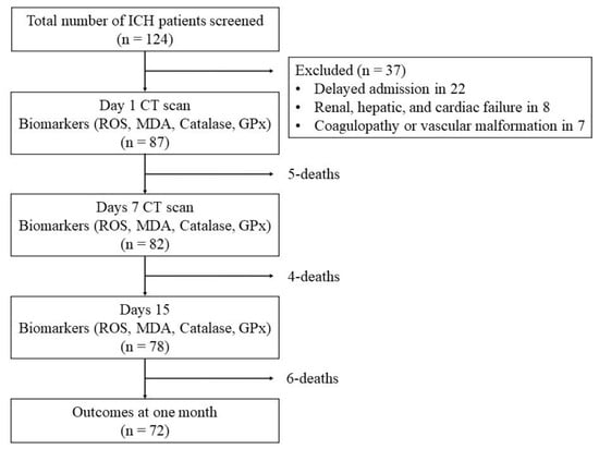
During the study period, 124 ICH patients were admitted; 37 were excluded due to delayed admission in 22, renal, hepatic, or cardiac failure in eight, and coagulopathy or vascular malformation in seven patients. Eight of these 37 (21.6%) patients died within 2 weeks. Therefore, this study was based on 87 patients; of them, five patients died within 1 week, and another four died between 1 and 2 weeks. Hence, biomarkers could be studied in 87, 82, and 78 patients on day 1, day 7, and day 15, respectively (Figure 1). Their median age was 58 (Q1, 50–Q3, 68) years, and 33 (37.9%) were females. Seventy-nine (90.8%) patients were hypertensive, 19 (21.8%) diabetic, 11 (12.6%) smokers, 29 (33.3%) tobacco chewers, and 19 (21.8%) consumed alcohol. The median NIHSS score at admission was 18 (range 11–36), and the GCS score was 12 (ranged 3–15). The details are presented in Supplementary Table S1.
Figure 1.
Flow chart showing the work plan.
3.1. CT Scan Findings
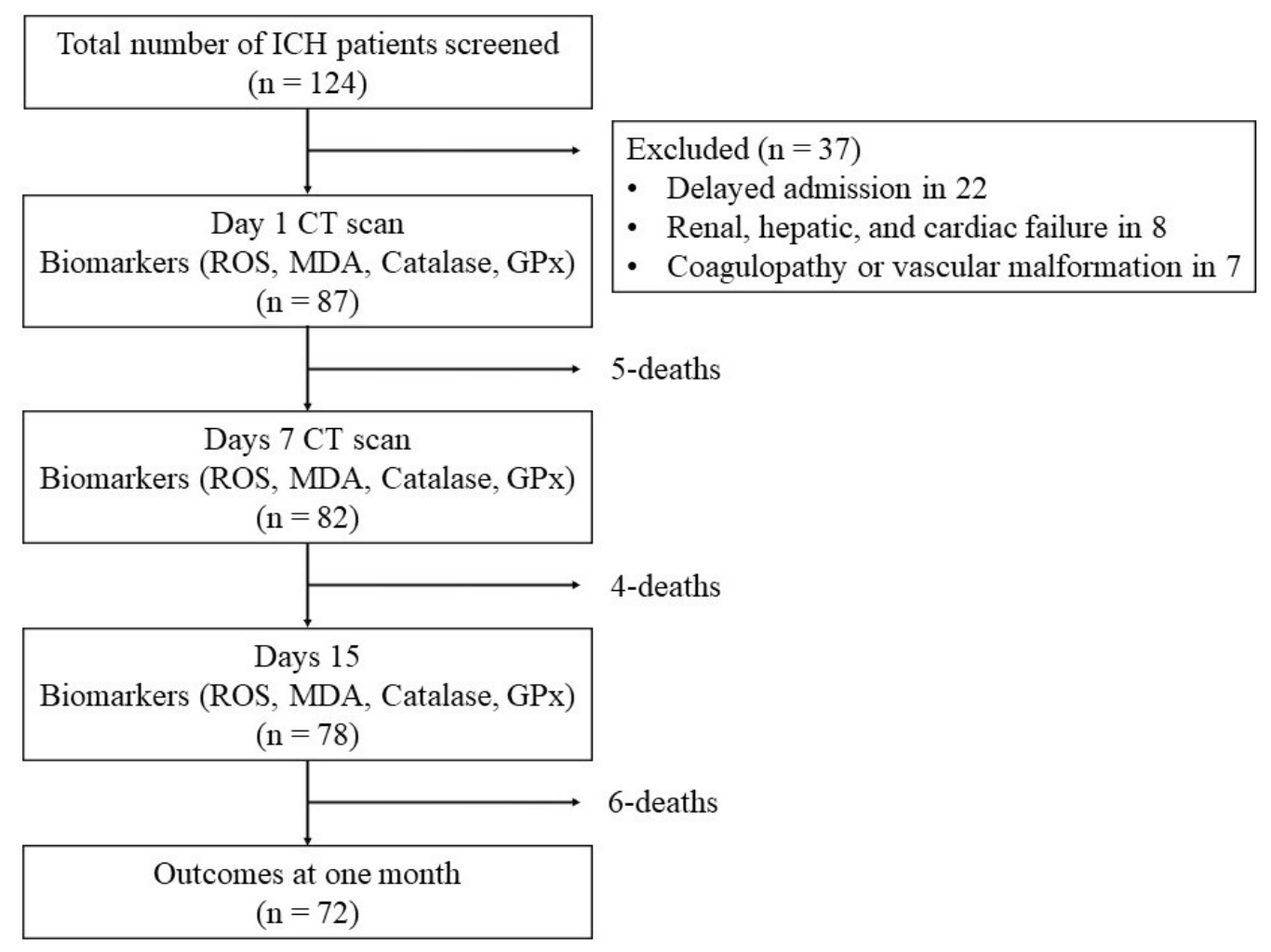
Seventy-one (81.6%) patients had basal ganglia or thalamic ICH, and 16 (18.4%) had lobar ICH. On day 1 CT scan, the median hematoma volume was 25.0 (Q1, 19.0–Q3, 50.0) mL, HEC was 30.0 (Q1, 22.0–Q3, 53.5) mL, and PHE was 4.0 (Q1, 2.0–Q3, 7.0) mL; 39 (44.8%) patients had intraventricular extension. A repeat CT scan on day 7 revealed an increase in PHE (p < 0.001) (Figure 2). Hematoma volume and HEC, however, did not increase (Table 1).
Figure 2.
Volumetric analysis of intracerebral hematoma (ICH) on day 1 and day 7. (A1–A3) images show computerized CT scan findings: (A1) ICH volume, (A2) hematoma edema complex (HEC), and (A3) perihematoma edema (PHE) on day 1. (B1–B3) are the images on day 7, and show reduction in (B1) ICH volume, but (B2) HEC and (B3) PHE volume increased from the day 1.
Table 1.
Cranial computerized tomographic scan findings.
3.2. Comparison of Biomarkers in the Patients and the Healthy Controls
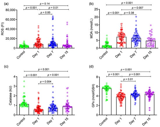
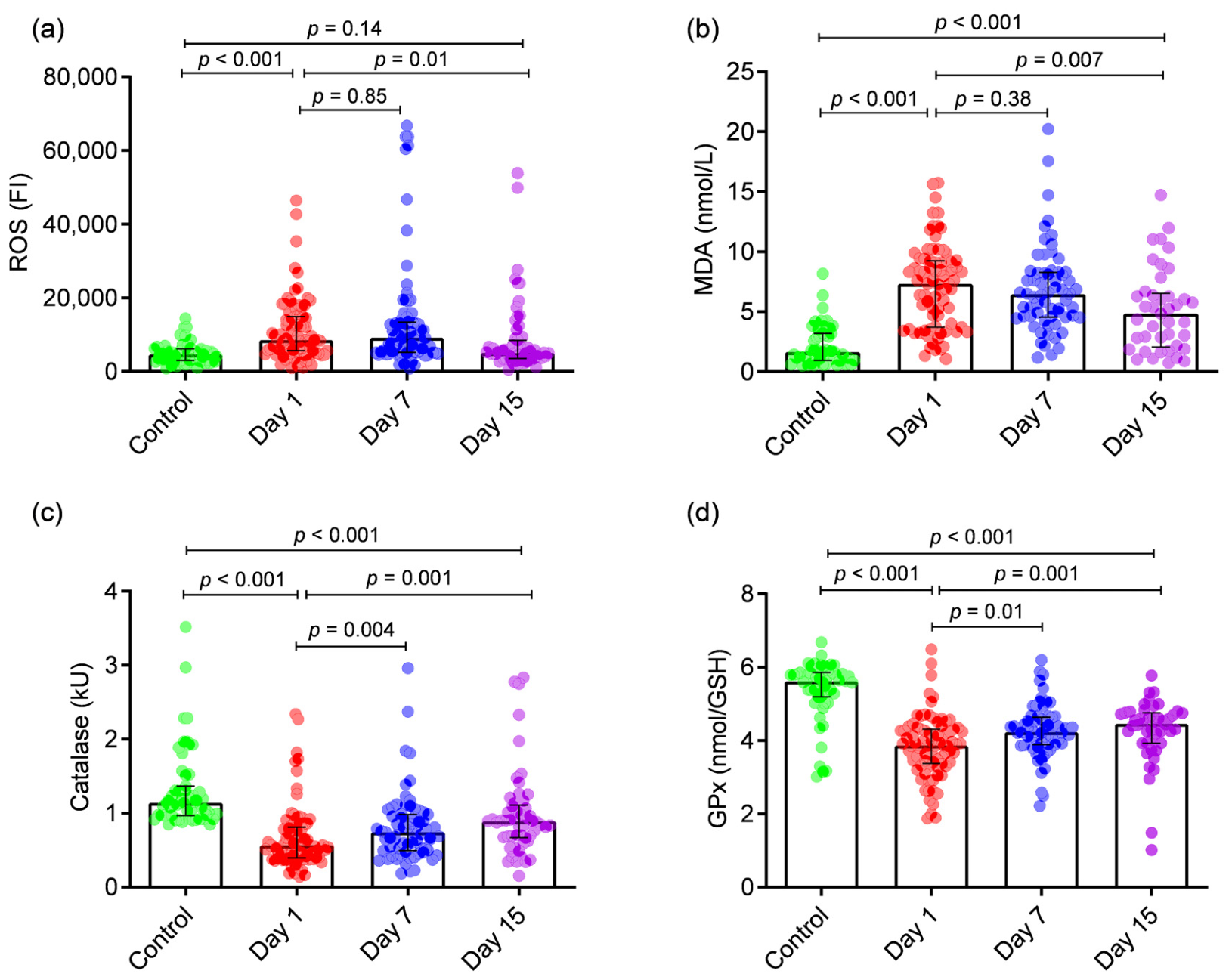
On day 1, the patients had elevated ROS (p < 0.001) and MDA (p < 0.001) levels, whereas CAT (p < 0.001) and GPx (p < 0.001) were reduced compared to the controls (Table 2). There was recovery of CAT (p = 0.004) and GPx (p = 0.01) on day 7 compared to day 1. On day 15, CAT recovered further compared to day 7 (p = 0.012), but the recovery of GPx was not significant (p = 0.167). The oxidative stress markers (ROS and MDA) remained elevated on day 7 but were reduced significantly on day 15, compared to day 1. However, ROS (p = 0.144) and MDA (p < 0.001) levels remained elevated even on day 15 compared to the controls. Similarly, the patients had lower levels of GPx (p < 0.001) and CAT (p < 0.001) on day 15 compared to the controls (Table 2 and Figure 3).
Table 2.
Biomarkers in the patients with intracerebral hemorrhage (ICH) and normal healthy control at different time points.
Figure 3.
The error bar diagram shows a comparison of biomarkers in patients at different time points with normal healthy controls. (a) ROS (reactive oxygen species) and (b) MDS (malondialdehyde) levels were higher the patients compared to the controls, and remained high till day 7. On day 15, the ROS level in the patients reached that of the controls, but the MDA level remained elevated. (c) Catalase and (d) GPx (glutathione peroxidase) levels in the patients were reduced compared to the healthy controls on day 1 and recovered on day 7 and 15 but remained low compared to the controls.
3.3. Correlation of Biomarkers with CT Scan Findings
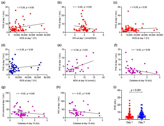
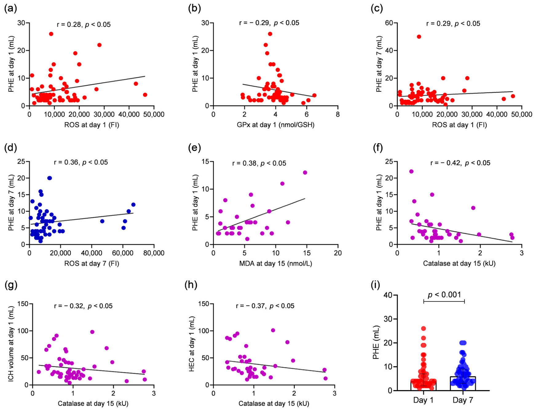
The day 1 biomarkers revealed a weak to moderate correlation of ROS (r = 0.28, p = 0.02) and GPx (r = −0.29, p = 0.02) with PHE volume. There was no correlation of ICH volume and HEC with any of the oxidant or antioxidant biomarkers. On the 7th day, PHE had a moderate correlation with ROS (r = 0.36, p = 0.005). On day 15, the biomarkers had a moderate correlation with admission CT scan parameters: CAT with hematoma volume (r = −0.32; p = 0.02), PHE (r = −0.43; p = 0.006), and HEC (r = −0.37; p = 0.02), and MDA with PHE (r = 0.38; p = 0.04). On day 15, the biomarkers also correlated with day 7 CT scan parameters. The details are presented in Supplementary Table S2, and significant parameters are shown in Figure 4.
Figure 4.
Linear regression graphs showing correlations of biomarkers with CT scan findings. Day 1 ROS (reactive oxygen species) correlated with (a) day 1 and (c) day 7 perihematoma edema (PHE). Day 7 ROS also correlated with (d) day 7 PHE. (b) Day 1 GPx (glutathione peroxidase) inversely correlated with day 1 PHE. (e) Day 15 MDA (malondialdehyde) correlated with day 1 PHE, and catalase correlated with (f) day 1 PHE, (g) ICH volume and (h) HEC (hematoma edema complex). (i) The error bar diagram shows an increase in PHE volume on day 7 compared to day 1. The solid black line represents the linear trendline.
3.4. Association of Biomarkers with Outcome
At 1 month, 15 (17.24%) patients died, 56 (64.36%) had a poor outcome, and 16 (18.39%) had a good outcome. Among the biomarkers, GPx was associated with survival (Table 3). GPx levels were higher in the patients who survived compared to those who died [3.94 (Q1, 3.55–Q3, 4.36) vs. 3.30 (Q1, 2.62–Q3, 3.85) nmol/GSH, p = 0.02] (Table 4). Day 1 ROS and MDA levels did not show an association with the outcome. On univariate analysis, death was also associated with hematoma volume (p < 0.001), HEC volume (p < 0.001), NIHSS score (p < 0.001), GCS score (p < 0.001), GPx (p = 0.01), and the requirement for an antiedema drug (p = 0.003) and mechanical ventilation (p < 0.001). The requirement for antihypertensive, antidiabetic, antibiotic, and antiseizure drugs was not associated with death (Table 4). On multivariate analysis, after including all the parameters that were significant in the univariate analysis, the independent predictors of death were the NIHSS score [adjusted odds ratio (AOR) 1.18; 95% confidence interval (CI) 1.00–1.39; p = 0.054) and mechanical ventilation (AOR 0.027; 95% CI 0.002–0.351; p = 0.006). Biomarkers did not achieve significance in the multivariate analysis.
Table 3.
Outcome of patients at one month.
Table 4.
Predictors of death at one month in patients with intracerebral hemorrhage on univariate analysis.
4. Discussion
This study has revealed elevated circulating oxidants (MDA and ROS) and reduced antioxidants (CAT and GPx) in ICH patients compared to the healthy controls. Although the oxidant levels started declining after the 7th day, they remained elevated till day 15 compared to the healthy controls. Similarly, antioxidants (CAT and GPx) were reduced at admission and continued to improve but remained lower than those in the healthy controls even on day 15. Admission PHE correlated with day 1 ROS and GPx. Furthermore, day 1 hematoma volume, PHE, and HEC were significant predictors of CAT and MDA levels on day 15. High antioxidant levels on day 1 were associated with survival and good outcomes at one month. Overall, this study has comprehensively evaluated the temporal kinetics of circulating oxidative stress and antioxidant biomarkers in a large cohort of primary ICH patients and their association with clinical stroke severity, CT scan findings, and one-month outcomes.
There is a paucity of studies on circulating oxidative stress markers in ICH patients. The stress generated by the ICH and raised intracranial pressure is likely to increase catecholamine drive, leading to systemic oxidative stress through activation of circulating neutrophils and monocytes or macrophages, as well as the generation of more stable secondary oxidation products. Therefore, the observed changes in peripheral blood biomarkers are indirect indicators of the systemic response to ICH rather than a direct measure of oxidative stress within the brain [10,13,16,40]. Xia et al. studied MDA, SOD, IL-6, and TNF-α in serum and drain fluid obtained through minimally invasive drainage in the patients with hypertensive ICH and compared them with those treated conservatively. They reported reduced TNF-α, IL-6, and MDA levels and elevated SOD levels in the minimally invasive drainage group compared to the conservatively treated group. Additionally, there was improvement in NIHSS, hematoma volume, and HEC on the 7th, 14th, and 28th days in the minimally invasive drainage group [31]. Rendevski et al. studied CAT and advanced oxidation protein products, along with IL-6 and IL-10, in 73 conservatively treated ICH patients. However, there was no association between these biomarkers and PHE; rather, C-reactive protein (CRP), erythrocyte sedimentation rate (ESR), and neutrophil-to-lymphocyte ratio (NLR) predicted PHE [27]. Another study evaluated oxidative stress markers in the brain tissue overlying the hematoma and compared these with the brain tissue from an aneurysmal bleed or peritumor area. No significant difference was observed in protein oxidation, GSH, GPx, glutathione reductase, SOD, CAT, and total antioxidant status between the two groups [41]. This may be due to the inclusion of adjacent brain tissue from the aneurysm and tumor, as these areas may also have oxidative stress. In our study, we have observed elevated levels of oxidants and reduced levels of antioxidants in the ICH patients compared to the controls, which did not reach normal levels even on the 15th day. This may be attributed to the kinetics of hematoma resolution. The resolution of a hematoma depends upon its volume; smaller hematomas resolve within weeks, whereas larger hematomas may take months [42].
The majority of reported studies did not evaluate the role of day 1 oxidants and antioxidants on subsequent PHE. Perihematoma edema is a result of complex molecular and inflammatory cascades which develops at the beginning and increases after 72 h till 1 week [43]. Oxidative stress may induce both cytotoxic and vasogenic edema [44]. Cytotoxic edema induced by oxidative stress has been attributed to the destruction of ion transporters in the cell membrane, increased calcium influx, reduced ATP formation, activation of phospholipase, destruction of organelles and the cell membrane, and acidosis [45]. Free radicals stimulate nitric oxide (NO), matrix metalloproteinases (MMPs), and cytokines, thereby altering the blood–brain barrier and contributing to vasogenic edema [44]. Xia et al., demonstrated that improved PHE after minimally invasive craniopuncture is driven by the reduction of oxidative stress and inflammatory cytokines [31]. We noted a correlation between ROS and GPx and admission PHE. Day 1 volumes of ICH, PHE, and HEC also determined the antioxidant and oxidant status on day 15. Catalase and GPx resist oxidative stress by neutralizing free radicals [46]. The decrease in antioxidant status may be due to utilization and exhaustion. In a mouse model, treatment with exogenous GSH has shown neuronal protection and improvement [47]. Elevated CSF MDA and lower plasma total antioxidant status have also been reported an association with unfavorable outcomes in patients with ICH [28,29,32].
To reduce oxidative stress and enhance antioxidants, various strategies have been used in experimental studies, including deferoxamine (an iron chelator), statins (reduce inducible nitric oxide synthase), PPAR-γ (downregulates the tumor necrosis factor gene and nitric oxide synthase), minocycline (reduces malondialdehyde by inhibiting metalloproteinases and chelating iron), NXY-059 (scavenges ROS), edaravone (scavenges ROS), and targeted gene therapy against heme oxygenase-1 and heme oxygenase-2 (downregulates the rate-limiting enzyme for heme catabolism and iron production), and Nrf2 (upregulates the expression of a wide range of antioxidants) [10,48,49,50,51,52,53,54,55,56]. Glutathione peroxidase (GPx) is upregulated by baicalein, baicalin, curcumin, luteolin, quercetin, astragaloside IV, gastrodin, oleuropein, bakuchiol, isorhynchophylline, crocin, and chrysophanol in a rat model of ICH [26,57,58,59,60,61,62,63,64,65,66,67,68]. In a systematic review and meta-analysis including 38 randomized controlled trials involving 3454 patients, the role of edaravone administered within 7 days of ICH was evaluated. Edaravone could not reduce all-cause mortality or improve long-term functional outcomes [69]. However, edaravone was safe and reduced hematoma edema, NIHSS scores, and Barthel index scores [69,70]. The addition of both GPx enhancers and oxidant depletors for a period of 15 days or more may be helpful in the patients with ICH and warrants future studies.
In our study, the overall fatality rate at 1 month was 18.5%, and that in the study cohort was 17.2%. In a systematic analysis, the case fatality rate of ICH at one month ranged between 14% and 25% [71]. The strongest predictors of death in ICH are GCS score, NIHSS score, mechanical ventilation, hematoma volume, and HEC [7,72,73,74]. In the present study, the independent predictors of death were NIHSS score and mechanical ventilation.
5. Limitations
The ages of the patients and the volume of the hematoma were variable, which is inevitable in human studies. We did not measure oxidative and antioxidative stress biomarkers after day 15 of stroke. The observational design of this study limits causal interpretation, and the oxidative stress biomarkers were detected at predefined time points, which may not fully capture the temporal kinetics. Furthermore, other key factors contributing to PHE expansion, including inflammatory cytokines, ER stress, and metabolic factors, were not evaluated. These factors might also interact with oxidative stress signaling pathways. We used the microplate reader for oxidative biomarker quantification. Liquid chromatography–mass spectrometry (LC-MS) would have been a more sensitive method for quantification. Moreover, we measured circulating biomarkers, which provide indirect evidence of the systemic stress response following ICH. We did not assess inter-rater and intra-rater variability in volumetric CT scan measurements. Although magnetic resonance imaging (MRI) is the best method for measuring brain edema, we performed CT scans to reduce costs and image acquisition time. Cranial MRI requires at least 30 min, while a CT scan can be done in 5 min. Patients with ICH often have altered sensorium, hyperventilation, and extensor posturing. Therefore, subjecting them to MRI was not considered safe by the Ethics Committee.
6. Conclusions
Patients with ICH had higher oxidative stress and lower antioxidant levels, which remained abnormal until day 15. Oxidative stress biomarkers correlated with PHE, suggesting their role in the pathogenesis. Patients with higher levels of antioxidants had better survival and good outcome. Our findings provide an understanding of secondary brain damage and identify GPx as a potential therapeutic target. Future studies are required to evaluate the role of antioxidants in PHE and clinical outcomes, which will add further validity to the current findings and interpretations.
Supplementary Materials
The following supporting information can be downloaded at: https://www.mdpi.com/article/10.3390/antiox15101260/s1, Table S1. Baseline characteristics of intracerebral hemorrhage patients. Table S2. Correlation biomarkers with volumetric parameters on computerized tomographic-scan.
Author Contributions
All authors contributed to the study conception and design. S.K.G.: biomarker analysis, data curation, investigation, formal analysis, writing-original draft, review, and editing. J.K.: funding acquisition, conceptualization, formal analysis, writing—original draft, and supervision. P.C.P.: patient management. D.K.: patient management and data curation. V.S.: reporting of CT scans. R.M.: patient management. All authors commented on previous versions of the manuscript. All authors have read and agreed to the published version of the manuscript.
Funding
Sandeep Kumar Gupta received a fellowship from the National Centre for Disease Informatics and Research, Indian Council of Medical Research, Government of India. The other authors declare that no funds, grants, or other support was received during the preparation of this manuscript.
Institutional Review Board Statement
This study was performed in line with the principles of the Declaration of Helsinki. Approval was granted by the Institutional Ethics Committee, SGPGIMS, Lucknow (Ethics No: 2022-20-IMP-125 8 July 2022).
Informed Consent Statement
Informed consent was obtained from the patients or their legal guardians. The authors affirm that no images or videos of human research participants are disclosed.
Data Availability Statement
The datasets generated during and/or analyzed during the current study are included in the main text, figures, tables, and Supplementary Materials. Further inquiries or additional datasets will be available from the first author and corresponding author upon request for scientific research purposes.
Acknowledgments
Sandeep Kumar Gupta, student, received a fellowship from the National Centre for Disease Informatics and Research, Indian Council of Medical Research, Government of India. We thank Prabhakar Mishra, of the Department of Biostatistics and Health Informatics, Sanjay Gandhi Postgraduate Institute of Medical Sciences, India, for statistical analysis.
Conflicts of Interest
The authors have no relevant financial or non-financial interests to disclose. The authors declare that there are no conflicts of interest.
References
- Katan, M.; Luft, A. Global Burden of Stroke. Semin. Neurol. 2018, 38, 208–211. [Google Scholar] [CrossRef] [Scilit] [PubMed]
- GBD 2021 Stroke Risk Factor Collaborators. Global, regional, and national burden of stroke and its risk factors, 1990–2021: A systematic analysis for the Global Burden of Disease Study 2021. Lancet Neurol. 2024, 23, 973–1003. [CrossRef] [Scilit] [PubMed]
- Faghih-Jouybari, M.; Raof, M.T.; Abdollahzade, S.; Jamshidi, S.; Padegane, T.; Ehteshami, S.; Fateh, S. Mortality and morbidity in patients with spontaneous intracerebral hemorrhage: A single-center experience. Curr. J. Neurol. 2021, 20, 32–36. [Google Scholar] [CrossRef] [Scilit] [PubMed]
- González-Pérez, A.; Gaist, D.; Wallander, M.A.; McFeat, G.; García-Rodríguez, L.A. Mortality after hemorrhagic stroke: Data from general practice (The Health Improvement Network). Neurology 2013, 81, 559–565. [Google Scholar] [PubMed]
- Rangamani, S.; Huliyappa, D.; Kulothungan, V.; Saravanan, S.; Murugan, P.K.; Mahadevan, R.; Rachel Packiaseeli, C.; Bobby, E.; Sunitha, K.; Mallick, A.K. Stroke incidence, mortality, subtypes in rural and urban populations in five geographic areas of India (2018–2019): Results from the National Stroke Registry Programme. Lancet Reg. Health Southeast Asia 2023, 23, 100308. [Google Scholar] [CrossRef] [Scilit] [PubMed]
- O’Carroll, C.B.; Brown, B.L.; Freeman, W.D. Intracerebral Hemorrhage: A Common yet Disproportionately Deadly Stroke Subtype. Mayo Clin. Proc. 2021, 96, 1639–1654. [Google Scholar] [PubMed]
- Kalita, J.; Kumar, D.; Gupta, S.K.; Pandey, P.C.; Mahajan, R.; Singh, V. Association Between Hematoma Edema Complex and Outcomes of Primary Intracerebral Hemorrhage. Neurocrit. Care 2025, 43, 763–774. [Google Scholar] [CrossRef] [Scilit] [PubMed]
- Ho, M.L.; Rojas, R.; Eisenberg, R.L. Cerebral edema. Am. J. Roentgenol. 2012, 199, 258–273. [Google Scholar] [CrossRef] [Scilit] [PubMed]
- Wei, C. The role of glutathione peroxidase 4 in neuronal ferroptosis and its therapeutic potential in ischemic and hemorrhagic stroke. Brain Res. Bull. 2024, 217, 111065. [Google Scholar] [CrossRef] [Scilit] [PubMed]
- Zhang, Y.; Khan, S.; Liu, Y.; Wu, G.; Yong, V.W.; Xue, M. Oxidative Stress Following Intracerebral Hemorrhage: From Molecular Mechanisms to Therapeutic Targets. Front. Immunol. 2022, 13, 847246. [Google Scholar] [CrossRef] [Scilit] [PubMed]
- Zhang, R.; Bai, Q.; Liu, Y.; Zhang, Y.; Sheng, Z.; Xue, M.; Yong, V.W. Intracerebral hemorrhage in translational research. Brain Hemorrhages 2020, 1, 13–18. [Google Scholar] [CrossRef] [Scilit]
- Aronowski, J.; Zhao, X. Molecular pathophysiology of cerebral hemorrhage: Secondary brain injury. Stroke 2011, 42, 1781–1786. [Google Scholar] [PubMed]
- Zhang, Y.; Khan, S.; Liu, Y.; Zhang, R.; Li, H.; Wu, G.; Tang, Z.; Xue, M.; Yong, V.W. Modes of Brain Cell Death Following Intracerebral Hemorrhage. Front. Cell. Neurosci. 2022, 16, 799753. [Google Scholar] [CrossRef] [Scilit] [PubMed]
- Sbodio, J.I.; Snyder, S.H.; Paul, B.D. Redox Mechanisms in Neurodegeneration: From Disease Outcomes to Therapeutic Opportunities. Antioxid. Redox Signal. 2019, 30, 1450–1499. [Google Scholar] [CrossRef] [Scilit] [PubMed]
- Xu, W.; Gao, L.; Zheng, J.; Li, T.; Shao, A.; Reis, C.; Chen, S.; Zhang, J. The Roles of MicroRNAs in Stroke: Possible Therapeutic Targets. Cell Transplant. 2018, 27, 1778–1788. [Google Scholar] [CrossRef] [Scilit] [PubMed]
- Duan, X.; Wen, Z.; Shen, H.; Shen, M.; Chen, G. Intracerebral Hemorrhage, Oxidative Stress, and Antioxidant Therapy. Oxid. Med. Cell. Longev. 2016, 2016, 1203285. [Google Scholar] [CrossRef] [Scilit] [PubMed]
- Chen, L.; Zhang, X.; Chen-Roetling, J.; Regan, R.F. Increased striatal injury and behavioral deficits after intracerebral hemorrhage in hemopexin knockout mice. J. Neurosurg. 2011, 114, 1159–1167. [Google Scholar] [CrossRef] [Scilit] [PubMed]
- Katsu, M.; Niizuma, K.; Yoshioka, H.; Okami, N.; Sakata, H.; Chan, P.H. Hemoglobin-induced oxidative stress contributes to matrix metalloproteinase activation and blood-brain barrier dysfunction in vivo. J. Cereb. Blood Flow. Metab. 2010, 30, 1939–1950. [Google Scholar] [CrossRef] [Scilit] [PubMed]
- Mrakic-Sposta, S.; Gussoni, M.; Montorsi, M.; Porcelli, S.; Vezzoli, A. Assessment of a standardized ROS production profile in humans by electron paramagnetic resonance. Oxid. Med. Cell. Longev. 2012, 2012, 973927. [Google Scholar] [CrossRef] [Scilit] [PubMed]
- Pierzchala, K.; Simicic, D.; Sienkiewicz, A.; Sessa, D.; Mitrea, S.; Braissant, O.; McLin, V.A.; Gruetter, R.; Cudalbu, C. Central nervous system and systemic oxidative stress interplay with inflammation in a bile duct ligation rat model of type C hepatic encephalopathy. Free Radic. Biol. Med. 2022, 178, 295–307, Corrigendum in Free Radic. Biol. Med. 2022, 182, 289. [Google Scholar] [CrossRef] [Scilit] [PubMed]
- Alexandrova, M.L.; Danovska, M.P. Serum C-reactive protein and lipid hydroperoxides in predicting short-term clinical outcome after spontaneous intracerebral hemorrhage. J. Clin. Neurosci. 2011, 18, 247–252. [Google Scholar] [CrossRef] [Scilit] [PubMed]
- Atik, İ.; Kozacı, N.; Beydilli, İ.; Avcı, M.; Ellidağ, H.; Keşaplı, M. Investigation of oxidant and antioxidant levels in patients with acute stroke in the emergency service. Am. J. Emerg. Med. 2016, 34, 2379–2383. [Google Scholar] [CrossRef] [Scilit] [PubMed]
- Kim, M.; Byun, J.; Chung, Y.; Lee, S.U.; Park, J.E.; Park, W.; Park, J.C.; Ahn, J.S.; Lee, S. Reactive Oxygen Species Scavenger in Acute Intracerebral Hemorrhage Patients: A Multicenter, Randomized Controlled Trial. Stroke 2021, 52, 1172–1181. [Google Scholar] [PubMed]
- Liu, C.; Tian, W.; Lei, D. GSTO2 ameliorates human neuroblastoma cell apoptosis, inflammation, ferroptosis, and oxidative stress by upregulating GPX4 expression in intracerebral hemorrhage. Drug Dev. Res. 2024, 85, e22245. [Google Scholar] [CrossRef] [Scilit] [PubMed]
- Pera, J.; Slowik, A.; Dziedzic, T.; Pulyk, R.; Wloch, D.; Szczudlik, A. Glutathione peroxidase 1 C593T polymorphism is associated with lobar intracerebral hemorrhage. Cerebrovasc. Dis. 2008, 25, 445–449. [Google Scholar] [CrossRef] [Scilit] [PubMed]
- Wang, F.; Li, W.L.; Shen, L.J.; Jiang, T.T.; Xia, J.J.; You, D.L.; Hu, S.Y.; Wang, L.; Wu, X. Crocin Alleviates Intracerebral Hemorrhage-Induced Neuronal Ferroptosis by Facilitating Nrf2 Nuclear Translocation. Neurotox. Res. 2022, 40, 596–604. [Google Scholar] [CrossRef] [Scilit] [PubMed]
- Rendevski, V.; Aleksovski, B.; Mihajlovska Rendevska, A.; Hadzi-Petrushev, N.; Manusheva, N.; Shuntov, B.; Gjorgoski, I. Inflammatory and oxidative stress markers in intracerebral hemorrhage: Relevance as prognostic markers for quantification of the edema volume. Brain Pathol. 2023, 33, e13106. [Google Scholar] [CrossRef] [Scilit] [PubMed]
- Lorente, L.; Martín, M.M.; González-Rivero, A.F.; Pérez-Cejas, A.; Abreu-González, P.; Sabatel, R.; Ramos, L.; Argueso, M.; Cáceres, J.J.; Solé-Violán, J.; et al. High Serum DNA and RNA Oxidative Damage in Non-surviving Patients with Spontaneous Intracerebral Hemorrhage. Neurocrit. Care 2020, 33, 90–96. [Google Scholar] [CrossRef] [Scilit] [PubMed]
- Lorente, L.; Martín, M.M.; Abreu-González, P.; Sabatel, R.; Ramos, L.; Argueso, M.; Solé-Violán, J.; Riaño-Ruiz, M.; Jiménez, A.; García-Marín, V. Serum Malondialdehyde Levels and Mortality in Patients with Spontaneous Intracerebral Hemorrhage. World Neurosurg. 2018, 113, e542–e547. [Google Scholar] [CrossRef] [Scilit] [PubMed]
- Chen, Y.C.; Chen, C.M.; Liu, J.L.; Chen, S.T.; Cheng, M.L.; Chiu, D.T. Oxidative markers in spontaneous intracerebral hemorrhage: Leukocyte 8-hydroxy-2′-deoxyguanosine as an independent predictor of the 30-day outcome. J. Neurosurg. 2011, 115, 1184–1190. [Google Scholar] [CrossRef] [Scilit] [PubMed]
- Xia, L.; Han, Q.; Ni, X.Y.; Chen, B.; Yang, X.; Chen, Q.; Cheng, G.L.; Liu, C.F. Different Techniques of Minimally Invasive Craniopuncture for the Treatment of Hypertensive Intracerebral Hemorrhage. World Neurosurg. 2019, 126, e888–e894. [Google Scholar] [CrossRef] [Scilit] [PubMed]
- Masomi-Bornwasser, J.; Kurz, E.; Frenz, C.; Schmitt, J.; Wesp, D.M.A.; König, J.; Lotz, J.; Ringel, F.; Kerz, T.; Krenzlin, H.; et al. The Influence of Oxidative Stress on Neurological Outcomes in Spontaneous Intracerebral Hemorrhage. Biomolecules 2011, 11, 1615. [Google Scholar]
- Niknahad, H.; Heidari, R.; Mohammadzadeh, R.; Ommati, M.M.; Khodaei, F.; Azarpira, N.; Abdoli, N.; Zarei, M.; Asadi, B.; Rasti, M. Sulfasalazine induces mitochondrial dysfunction and renal injury. Ren. Fail. 2017, 39, 745–753. [Google Scholar] [CrossRef] [Scilit] [PubMed]
- Placer, Z.A.; Cushman, L.L.; Johnson, B.C. Estimation of product of lipid peroxidation (malonyl dialdehyde) in biochemical systems. Anal. Biochem. 1966, 16, 359–364. [Google Scholar] [CrossRef] [Scilit] [PubMed]
- Ohkawa, H.; Ohishi, N.; Yagi, K. Assay for lipid peroxides in animal tissues by thiobarbituric acid reaction. Anal. Biochem. 1979, 95, 351–358. [Google Scholar] [CrossRef] [Scilit] [PubMed]
- Sinha, A.K. Colorimetric assay of catalase. Anal. Biochem. 1972, 47, 389–394. [Google Scholar] [CrossRef] [Scilit] [PubMed]
- Hawan, M.H.; Khabt, H. Simple Spectrophotometric Method for Analysis of Serum Catalase Activity. J. Clin. Diagn. Res. 2018, 12, 13. [Google Scholar] [CrossRef] [Scilit]
- Rotruck, J.T.; Pope, A.L.; Ganther, H.E.; Swanson, A.B.; Hafeman, D.G.; Hoekstra, W.G. Selenium: Biochemical role as a component of glutathione peroxidase. Science 1973, 179, 588–590. [Google Scholar] [CrossRef] [Scilit] [PubMed]
- Kalita, J.; Bastia, J.; Bhoi, S.K.; Misra, U.K. Systemic Inflammatory Response Syndrome Predicts Severity of Stroke and Outcome. J. Stroke Cerebrovasc. Dis. 2015, 24, 1640–1648. [Google Scholar] [CrossRef] [Scilit] [PubMed]
- Zhu, H.; Wang, Z.; Yu, J.; Yang, X.; He, F.; Liu, Z.; Che, F.; Chen, X.; Ren, H.; Hong, M.; et al. Role and mechanisms of cytokines in the secondary brain injury after intracerebral hemorrhage. Progress. Neurobiol. 2019, 178, 101610. [Google Scholar] [CrossRef] [Scilit] [PubMed]
- Mantle, D.; Siddique, S.; Eddeb, F.; Mendelow, A.D. Comparison of protein carbonyl and antioxidant levels in brain tissue from intracerebral haemorrhage and control cases. Clin. Chim. Acta Int. J. Clin. Chem. 2001, 312, 185–190. [Google Scholar] [CrossRef] [Scilit] [PubMed]
- Wan, Y.; Holste, K.G.; Hua, Y.; Keep, R.F.; Xi, G. Brain edema formation and therapy after intracerebral hemorrhage. Neurobiol. Dis. 2023, 176, 105948. [Google Scholar] [CrossRef] [Scilit] [PubMed]
- Venkatasubramanian, C.; Mlynash, M.; Finley-Caulfield, A.; Eyngorn, I.; Kalimuthu, R.; Snider, R.W.; Wijman, C.A. Natural history of perihematomal edema after intracerebral hemorrhage measured by serial magnetic resonance imaging. Stroke 2011, 42, 73–80. [Google Scholar] [CrossRef] [Scilit] [PubMed]
- Yu, Y.; Zhao, W.; Zhu, C.; Kong, Z.; Xu, Y.; Liu, G.; Gao, X. The clinical effect of deferoxamine mesylate on edema after intracerebral hemorrhage. PLoS ONE 2015, 10, e0122371. [Google Scholar] [CrossRef] [Scilit] [PubMed]
- Vlodavsky, E.; Palzur, E.; Shehadeh, M.; Soustiel, J.F. Post-traumatic cytotoxic edema is directly related to mitochondrial function. J. Cereb. Blood Flow Metab. 2017, 37, 166–177. [Google Scholar] [CrossRef] [Scilit] [PubMed]
- Ighodaro, O.M.; Akinloye, O.A. First line defence antioxidants-superoxide dismutase (SOD), catalase (CAT) and glutathione peroxidase (GPX): Their fundamental role in the entire antioxidant defence grid. Alex. J. Med. 2018, 54, 287–293. [Google Scholar] [CrossRef] [Scilit]
- Diao, X.; Zhou, Z.; Xiang, W.; Jiang, Y.; Tian, N.; Tang, X.; Chen, S.; Wen, J.; Chen, M.; Liu, K.; et al. Glutathione alleviates acute intracerebral hemorrhage injury via reversing mitochondrial dysfunction. Brain Res. 2020, 1727, 146514. [Google Scholar] [CrossRef] [Scilit] [PubMed]
- Nakamura, T.; Keep, R.F.; Hua, Y.; Schallert, T.; Hoff, J.T.; Xi, G. Deferoxamine-induced attenuation of brain edema and neurological deficits in a rat model of intracerebral hemorrhage. J. Neurosurg. 2004, 100, 672–678. [Google Scholar] [CrossRef] [Scilit] [PubMed]
- Catalão, C.H.R.; Santos-Júnior, N.N.; da Costa, L.H.A.; Souza, A.O.; Alberici, L.C.; Rocha, M.J.A. Brain Oxidative Stress During Experimental Sepsis Is Attenuated by Simvastatin Administration. Mol. Neurobiol. 2017, 54, 7008–7018. [Google Scholar] [CrossRef] [Scilit] [PubMed]
- Chen, C.J.; Ding, D.; Ironside, N.; Buell, T.J.; Elder, L.J.; Warren, A.; Adams, A.P.; Ratcliffe, S.J.; James, R.F.; Naval, N.S.; et al. Statins for neuroprotection in spontaneous intracerebral hemorrhage. Neurology 2019, 93, 1056–1066. [Google Scholar] [CrossRef] [Scilit] [PubMed]
- Zhao, X.; Sun, G.; Zhang, J.; Strong, R.; Song, W.; Gonzales, N.; Grotta, J.C.; Aronowski, J. Hematoma resolution as a target for intracerebral hemorrhage treatment: Role for peroxisome proliferator-activated receptor gamma in microglia/macrophages. Ann. Neurol. 2007, 61, 352–362. [Google Scholar] [CrossRef] [Scilit] [PubMed]
- Chen-Roetling, J.; Chen, L.; Regan, R.F. Minocycline attenuates iron neurotoxicity in cortical cell cultures. Biochem. Biophys. Res. Commun. 2009, 386, 322–326. [Google Scholar] [CrossRef] [Scilit] [PubMed]
- Peeling, J.; Del Bigio, M.R.; Corbett, D.; Green, A.R.; Jackson, D.M. Efficacy of disodium 4-[(tert-butylimino) methyl] benzene-1, 3-disulfonate N-oxide (NXY-059), a free radical trapping agent, in a rat model of hemorrhagic stroke. Neuropharmacology 2001, 40, 433–439. [Google Scholar] [CrossRef] [Scilit] [PubMed]
- Miao, H.; Jiang, Y.; Geng, J.; Zhang, B.; Zhu, G.; Tang, J. Edaravone Administration Confers Neuroprotection after Experimental Intracerebral Hemorrhage in Rats via NLRP3 Suppression. J. Stroke Cerebrovasc. Dis. 2020, 29, 104468. [Google Scholar] [CrossRef] [Scilit] [PubMed]
- Yang, Y.; Xi, Z.; Xue, Y.; Ren, J.; Sun, Y.; Wang, B.; Zhong, Z.; Yang, G.Y.; Sun, Q.; Bian, L. Hemoglobin pretreatment endows rat cortical astrocytes resistance to hemin-induced toxicity via Nrf2/HO-1 pathway. Exp. Cell Res. 2017, 361, 217–224. [Google Scholar] [CrossRef] [Scilit] [PubMed]
- Wang, J.; Fields, J.; Zhao, C.; Langer, J.; Thimmulappa, R.K.; Kensler, T.W.; Yamamoto, M.; Biswal, S.; Doré, S. Role of Nrf2 in protection against intracerebral hemorrhage injury in mice. Free Radic. Biol. Med. 2007, 43, 408–414. [Google Scholar] [CrossRef] [Scilit] [PubMed]
- Zheng, Y.; Li, R.; Fan, X. Targeting Oxidative Stress in Intracerebral Hemorrhage: Prospects of the Natural Products Approach. Antioxidants 2022, 11, 1811. [Google Scholar] [CrossRef] [Scilit] [PubMed]
- Wei, N.; Wei, Y.; Li, B.; Pang, L. Baicalein Promotes Neuronal and Behavioral Recovery After Intracerebral Hemorrhage Via Suppressing Apoptosis, Oxidative Stress and Neuroinflammation. Neurochem. Res. 2017, 42, 1345–1353. [Google Scholar] [CrossRef] [Scilit] [PubMed]
- Li, N.; Feng, L.; Tan, Y.; Xiang, Y.; Zhang, R.; Yang, M. Preparation, Characterization, Pharmacokinetics and Biodistribution of Baicalin-Loaded Liposome on Cerebral Ischemia-Reperfusion after i.v. Administration in Rats. Molecules 2018, 23, 1747. [Google Scholar] [CrossRef] [Scilit] [PubMed]
- Li, X.; Zhao, L.; Yue, L.; Liu, H.; Yang, X.; Wang, X.; Lin, Y.; Qu, Y. Evidence for the protective effects of curcumin against oxyhemoglobin-induced injury in rat cortical neurons. Brain Res. Bull. 2016, 120, 34–40. [Google Scholar] [CrossRef] [Scilit] [PubMed]
- Tan, X.; Yang, Y.; Xu, J.; Zhang, P.; Deng, R.; Mao, Y.; He, J.; Chen, Y.; Zhang, Y.; Ding, J. Luteolin Exerts Neuroprotection via Modulation of the p62/Keap1/Nrf2 Pathway in Intracerebral Hemorrhage. Front. Pharmacol. 2020, 10, 1551. [Google Scholar] [CrossRef] [Scilit] [PubMed]
- Galho, A.R.; Cordeiro, M.F.; Ribeiro, S.A.; Marques, M.S.; Antunes, M.F.; Luz, D.C.; Hädrich, G.; Muccillo-Baisch, A.L.; Barros, D.M.; Lima, J.V. Protective role of free and quercetin-loaded nanoemulsion against damage induced by intracerebral haemorrhage in rats. Nanotechnology 2016, 27, 175101. [Google Scholar] [CrossRef] [Scilit] [PubMed]
- Shao, A.; Guo, S.; Tu, S.; Ammar, A.B.; Tang, J.; Hong, Y.; Wu, H.; Zhang, J. Astragaloside IV alleviates early brain injury following experimental subarachnoid hemorrhage in rats. Int. J. Med. Sci. 2014, 11, 1073–1081. [Google Scholar] [CrossRef] [Scilit] [PubMed]
- Liu Xi, C.; Wu, C.Z.; Hu, X.F.; Wang, T.L.; Jin, X.P.; Ke, S.F.; Wang, E.; Wu, G. Gastrodin Attenuates Neuronal Apoptosis and Neurological Deficits after Experimental Intracerebral Hemorrhage. J. Stroke Cerebrovasc. Dis. 2020, 29, 104483. [Google Scholar] [CrossRef] [Scilit] [PubMed]
- Shi, J.; Wu, G.; Zou, X.; Jiang, K. Oleuropein protects intracerebral hemorrhage-induced disruption of blood-brain barrier through alleviation of oxidative stress. Pharmacol. Rep. 2017, 69, 1206–1212. [Google Scholar] [CrossRef] [Scilit] [PubMed]
- Liu, H.; Guo, W.; Guo, H.; Zhao, L.; Yue, L.; Li, X.; Feng, D.; Luo, J.; Wu, X.; Cui, W. Bakuchiol Attenuates Oxidative Stress and Neuron Damage by Regulating Trx1/TXNIP and the Phosphorylation of AMPK After Subarachnoid Hemorrhage in Mice. Front. Pharmacol. 2020, 11, 712. [Google Scholar] [CrossRef] [Scilit] [PubMed]
- Zhao, H.; Li, X.; Yang, L.; Zhang, L.; Jiang, X.; Gao, W.; Chen, P.; Cheng, Y.; Wang, F.; Liu, J. Isorhynchophylline Relieves Ferroptosis-Induced Nerve Damage after Intracerebral Hemorrhage Via miR-122-5p/TP53/SLC7A11 Pathway. Neurochem. Res. 2021, 46, 1981–1994. [Google Scholar] [CrossRef] [Scilit] [PubMed]
- Jadaun, K.S.; Mehan, S.; Sharma, A.; Siddiqui, E.M.; Kumar, S.; Alsuhaymi, N. Neuroprotective Effect of Chrysophanol as a PI3K/AKT/mTOR Signaling Inhibitor in an Experimental Model of Autologous Blood-induced Intracerebral Hemorrhage. Curr. Med. Sci. 2022, 42, 249–266. [Google Scholar] [CrossRef] [Scilit] [PubMed]
- Qin, M.; Feng, L.; Yang, C.; Wei, D.; Li, T.; Jiang, P.; Guan, J.; Zhang, X.; Shi, X.; Liang, N. Edaravone use in acute intracerebral hemorrhage: A systematic review and meta-analysis of randomized controlled trials. Front. Pharmacol. 2022, 13, 935198. [Google Scholar] [CrossRef] [Scilit] [PubMed]
- Yang, J.; Liu, M.; Zhou, J.; Zhang, S.; Lin, S.; Zhao, H. Edaravone for acute intracerebral haemorrhage. Cochrane Database Syst. Rev. 2011, 2, CD007755. [Google Scholar] [CrossRef] [Scilit] [PubMed]
- Feigin, V.L.; Lawes, C.M.; Bennett, D.A.; Barker-Collo, S.L.; Parag, V. Worldwide stroke incidence and early case fatality reported in 56 population-based studies: A systematic review. Lancet. Neurol. 2009, 8, 355–369. [Google Scholar] [CrossRef] [Scilit] [PubMed]
- Weir, C.J.; Bradford, A.P.; Lees, K.R. The prognostic value of the components of the Glasgow Coma Scale following acute stroke. QJM Mon. J. Assoc. Physicians 2003, 96, 67–74. [Google Scholar] [CrossRef] [Scilit] [PubMed]
- Mahdy, M.E.; Ghonimi, N.A.; Elserafy, T.S.E.; Mahmoud, W. The NIHSS score can predict the outcome of patients with primary intracerebral hemorrhage. Egypt. J. Neurol. Psychiatry Neurosurg. 2019, 55, 21. [Google Scholar] [CrossRef] [Scilit]
- Misra, U.K.; Kalita, J.; Srivastava, M.; Mandal, S.K. A study of prognostic predictors of supratentorial haematomas. J. Neurol. 1996, 243, 96–100. [Google Scholar] [CrossRef] [Scilit] [PubMed]

Disclaimer/Publisher’s Note: The statements, opinions and data contained in all publications are solely those of the individual author(s) and contributor(s) and not of MDPI and/or the editor(s). MDPI and/or the editor(s) disclaim responsibility for any injury to people or property resulting from any ideas, methods, instructions or products referred to in the content. |
© 2026 by the authors. Licensee MDPI, Basel, Switzerland. This article is an open access article distributed under the terms and conditions of the Creative Commons Attribution (CC BY) license.