


Abstract
Background/Objectives: Cognitive impairment and neurological dysfunction in Multiple Sclerosis are increasingly understood as consequences of altered brain network connectivity. Magnetoencephalography offers high temporal resolution for investigating functional connectivity patterns, which may underlie various clinical symptoms. This exploratory pilot study aims to characterize preliminary FC patterns at rest and during cognitive processing in MS patients with a high fatigue burden compared with healthy controls using MEG, complemented by a systematic review of existing MEG literature in MS. Methods: In this pilot study, MEG was utilized to investigate connectivity in MS patients and HCs. Data were acquired during resting-state and a digitized version of the Symbol Digit Modalities Test. A systematic review was also conducted following PRISMA guidelines to synthesize the current evidence on frequency-specific MEG connectivity in MS. Results: MS patients exhibited a connectivity pattern paralleling the directionality of neuronal slowing, described in the spectral power MS literature, during resting-state, characterized by reduced interhemispheric coherence in alpha and beta bands alongside region-specific increases in frontal–parietal and parietal–occipital alpha coherence, reflecting a bidirectional spatial reorganization. Conversely, during the SDMT task, the MS group demonstrated significant compensatory recruitment with increased coherence in theta and gamma bands (specifically in frontal and striatal circuits). The systematic review of 44 MEG studies corroborated these findings, highlighting a consistent trend of frequency-dependent dysconnectivity that aligns with clinical load and disease pathology. Conclusions: We found that in MS, network alterations are state-dependent, shifting from reduced connectivity at rest to increased coherence during cognitive effort. These preliminary findings suggest that MEG-based connectivity may serve as a functional neuroimaging biomarker of underlying network reorganization, for monitoring early cognitive disease progression in MS.
1. Introduction
Understanding the neurophysiological basis of Multiple Sclerosis (MS) is crucial, as the disease is increasingly recognized as a “disconnection syndrome” [1,2,3]. In this context, neurological deficits arise not only from focal lesions but from the widespread disruption of functional brain networks. Several functional imaging techniques for assessing the impact of MS on cognition are available, including functional magnetic resonance imaging (fMRI), positron emission tomography (PET), and magnetoencephalography (MEG). Numerous studies using these techniques have provided valuable insights into the neural mechanisms underlying cognitive dysfunction in patients with MS [1,2,4,5,6]. fMRI and PET measure secondary neural response from hemodynamics and glucose consumption, while MEG measures the magnetic field associated with actual neuronal activity. Functional brain activity associated with cognition during a modified Symbol Digit Modalities Test (SDMT), a validated measure of cognitive processing speed in MS and other neurodegenerative disorders, which examinees have 90 s to match abstract symbols to paired digits using a reference key, has been assessed with fMRI, revealing no differences in accuracy between patients with MS and healthy controls. Nonetheless, individuals with MS had significantly slower processing speeds than controls [7]. Moreover, fMRI activation during the SDMT was seen in the precentral gyrus and occipital cortex in both groups, but those with MS showed significantly less cerebral activity in the bilateral frontal and parietal regions [7]. Complementing these imaging modalities, pure neurophysiological techniques, such as TMS-EEG, have provided evidence of cortical excitability alterations, functional connectivity disruption, and network-level reorganization in MS, revealing deficits in cortical inhibition and plasticity [8]. Overall, these studies have provided a detailed picture of changes in neuronal activity in patients with MS but fall short of providing the temporal resolution of brain activity processing. To understand the temporal dynamics of underlying brain processes, a brain imaging technique such as MEG becomes necessary.
MEG is a noninvasive functional brain imaging technique that measures the magnetic brain waves arising from neuronal activity during rest or a specific task, and this technique is capable of real-time functional imaging with a spatial resolution down to several millimeters and a temporal resolution of less than 1 ms. A study combining MEG and fMRI observed reduced neuronal and hemodynamic responses during visual stimulation in patients with MS compared to controls. This materialized as a significant reduction in peak gamma power in MS patients compared to control subjects. This reduction could indicate gray matter dysfunction, possibly mediated by GABAergic changes [1]. However, findings regarding gamma oscillations remain inconsistent, particularly when comparing passive stimulation to active cognitive tasks. Also, MEG showed that mental fatigue suppresses activities in the anterior cingulate cortex elicited by mental imagery of physical effort [2]. Another study investigating MEG aimed at identifying the neurophysiological markers of cognitive impairment in patients with MS analyzed global spectral and connectivity, revealing that cognitively impaired patients had higher theta and alpha1 power (8–10 Hz) and lower alpha2 and beta power (10–13 Hz) than patients with no cognitive impairment in several regions of interest [3]. Yet other studies implementing MEG to investigate MS have focused on disrupted networks in the resting state (i.e., theta, alpha, and beta frequency bands) [9,10,11,12,13,14,15,16], leaving a gap in understanding how these connectivity patterns shift during active cognitive challenge.
Despite these insights, the neurophysiological transition between resting-state and active cognitive effort and how it relates to the overall network reorganization in MS remains incompletely characterized. Therefore, this exploratory pilot study aims to investigate preliminary frequency-specific connectivity signatures within four neuronal activity bands during both rest and a cognitive task (SDMT) in MS patients compared with controls. By integrating our experimental results with a comprehensive systematic review, we seek to provide a robust profile of MEG functional connectivity changes in MS that could serve as an objective imaging biomarker for monitoring disease status and progression.
2. Materials and Methods
In this pilot study conducted at a single center, we investigated the impact of MS-related fatigue on cognition compared to age- and gender-matched healthy individuals. We used the SDMT, a validated standardized cognitive scale widely used in MS research as a sensitive measure to assess cognitive processing speed and attention; it is scored as the total number of correct substitutions made in 90 s under timed conditions [17,18]. For this study, a digitized version adapted for MEG administration was employed. Additionally, we used MEG to map the underlying brain areas involved in cognitive processes. Patients were evaluated during a resting state and while performing a cognitive task.
2.1. Subject Population and Eligibility
Participants were recruited from the Henry Ford Health MS Clinic. All participants underwent a full Informed Consent process and provided written consent to participate in the study. This study was approved by the Henry Ford Health Institutional Review Board [IRB No. 15400].
2.1.1. Inclusion Criteria
Eligible patients were adults (18–60 years old), diagnosed with RRMS or clinically isolated syndrome meeting McDonald’s 2017 diagnostic criteria for MS, with a history of MS-related fatigue; currently taking disease-modifying therapies (DMT); and Kurtzke’s Expanded Disability Status Scale (EDSS) score ≤ 4.0. Patients taking anti-fatigue medications, such as amantadine, modafinil, and methylphenidate, were given a 4-week washout period (based on the medications’ elimination half-lives) before study enrollment [19].
2.1.2. Exclusion Criteria
Patients were excluded if they had used steroids for MS exacerbation within the past 30 days, had a history of severe anxiety, depression, psychiatric disorders, stroke, excessive alcohol use, or traumatic brain injury. Additionally, patients with uncontrolled medical risk factors such as hypertension, hyperlipidemia, and diabetes, which could increase the risk for chronic ischemic microvascular disease and potential cognitive impairment, were also excluded.
2.2. Experimental Setups
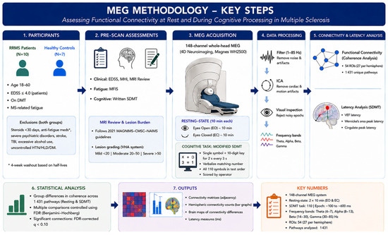
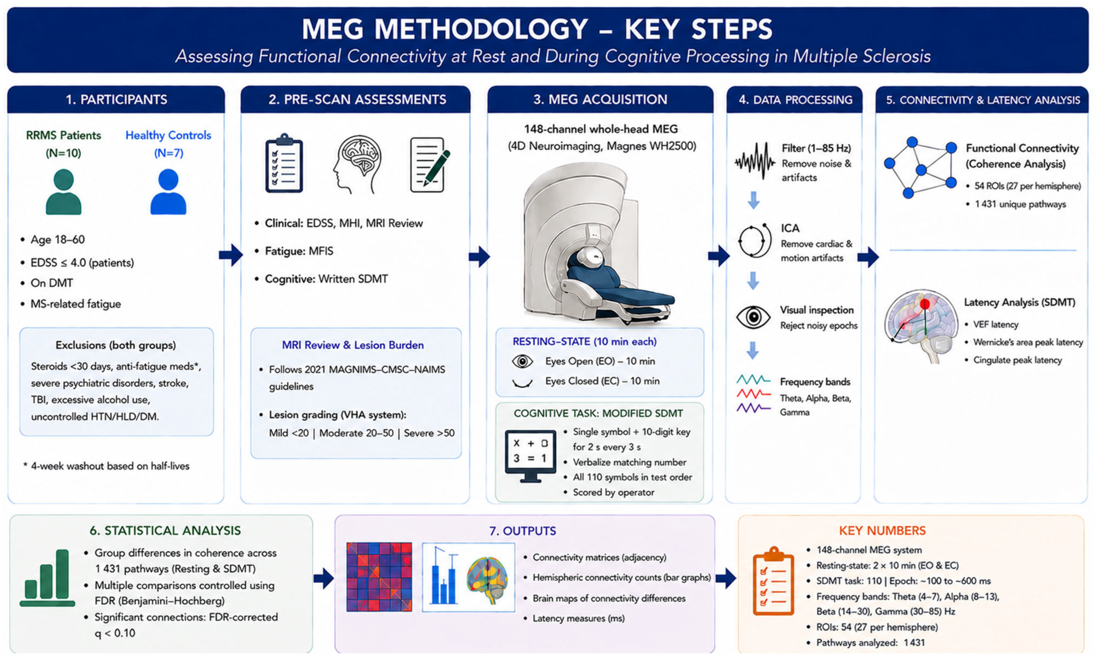
Before MEG sessions, MS patients underwent clinical evaluations, including the EDSS, Mental Health Inventory (MHI), and written SDMT. The Modified Fatigue Impact Scale (MFIS) assessed baseline physical fatigue. Healthy controls also completed the MFIS and SDMT before scanning. During MEG, patients performed a modified SDMT task, adapted from prior fMRI studies, after obtaining required approval for test use [7,20]. Demographics, medical history, disease/fatigue duration, radiographic findings, and disease-modifying therapies (DMTs) were retrieved from EPIC (electronic medical records). Brain and spinal cord MRI images were reviewed to assess disease burden. MRI interpretation followed the 2021 MAGNIMS–CMSC–NAIMS consensus guidelines [21]. Lesion burden was graded using the Veterans Health Administration (VHA) system: mild (<20 lesions), moderate (20–50), or severe (>50) [22]. While this grading approach aligned with MAGNIMS–CMSC–NAIMS criteria, severity stratification was not explicitly recommended by the consensus. A summary of the experimental workflow, including MEG data acquisition, preprocessing steps, and connectivity analysis, is illustrated in Figure 1.
Figure 1.
MEG setup steps illustration.
2.3. Brain Imaging Measurements and Analyses
2.3.1. Recording
Cortical activity was recorded using a 148-channel whole-head MEG system (Magnes WH2500; 4D Neuroimaging, San Diego, CA, USA) with magnetometer sensors. Participants wore a standard medical gown and removed all metal objects. Five fiducial coils were placed on each subject: three across the forehead and one in front of each ear. These coils were digitized to map scalp locations, and head shapes were recorded for MRI co-registration. Before and after each run, the coils were activated with a low current to detect head movement. Runs were repeated if motion exceeded 5 mm; no participants moved beyond this threshold during scanning. Subjects lay supine with their head positioned inside a helmet-shaped sensor array within a magnetically shielded room. A bowl-shaped head holder minimized motion, and the MEG helmet was secured without obstructing the face. MEG measures functional brain activity, allowing for the analysis of network dynamics and connectivity patterns during cognitive processing.
2.3.2. MEG Data Acquisition and Analysis
During acquisition, data were band-pass filtered 0.1 to 100 Hz and digitally sampled at 508.63 Hz, the standard native sampling rate of the 4D Neuroimaging Magnes WH2500 system, and continuously recorded for later analysis. The total time of MEG testing was approximately 30 min. The resting-state scans consisted of two continuous 10 min acquisitions of spontaneous brain activity (one scan with eyes open and one with eyes closed). The cognitive processing task was the SDMT, which was modified and adapted for MEG, similar to approaches used with fMRI [7,20]. A mirror was placed above the subjects, angled so they could view a projected image on the screen. A single SDMT symbol was visually displayed along with the 10-digit key above it for 2 s every 3 s. Subjects verbalized responses as soon as they identified the correct number to match the displayed symbol. A person outside of the room scored the correct answers. All 110 SDMT symbols from the test were displayed in the same order as in the original test.
2.3.3. MEG Data Processing
Resting-state and SDMT data were processed with an additional 1–85 Hz band-pass filter to optimize artifact rejection and remove noise outside the frequency bands of interest. Independent Component Analysis (ICA) was utilized to isolate and remove cardiac and motion-related artifacts. Additionally, data epochs were reviewed to exclude trials contaminated by excessive noise prior to connectivity analysis. Data were then filtered into four frequency bands: theta (4–7 Hz), alpha (8–13 Hz), beta (14–30 Hz), and gamma (30–85 Hz). The upper gamma boundary of 85 Hz was chosen to capture high-gamma activity (>60 Hz), which has been shown in MEG studies to be functionally relevant to cognitive and working-memory processing, and was accommodated by the 1–85 Hz preprocessing band-pass filter. We note that gamma definitions in the systematic review literature range from 30–70 Hz to 35–80 Hz, and cross-study gamma comparisons should account for this variability. The delta frequency band (0.5–4 Hz) was excluded from the connectivity analysis, aligning with the vast majority of prior MS MEG literature. It was implemented to minimize the contamination of low-frequency non-neural artifacts, such as respiratory variations and subtle head or jaw movements, which are highly pronounced during active verbal tasks like the SDMT. Coherence analysis of the strength of the functional network connectivity across 1431 pathways derived from all unique pairwise combinations of 54 ROIs, determined using MEG TOOLS, a MATLAB-based software program (RRID:SCR_000661; https://www.nitrc.org/projects/meg-tools; Accessed on 3 June 2026) [23,24,25,26]. The SDMT MEG data were averaged based on the trigger recorded every time the image was displayed (N = 110 trials). All epochs had a baseline of 100 ms before stimuli onset and 600 ms of data after stimuli onset. These data were then visually inspected to determine the latencies of the visual evoked response and the cognitive peak in Wernicke’s area. Source reconstruction was performed using MR-FOCUSS, a current distribution algorithm that estimates cortical sources from MEG data. Activity was mapped to standard MRI space through co-registration, allowing localization to specific areas, such as Wernicke’s region.
Co-registration enabled precise correspondence between anatomical structures and MEG-identified cortical activation areas, with errors of less than 5 mm. A standard MRI was transformed into the digitized head shape for each patient to compare group brain activations. Each patient’s brain activity was mapped onto the standard MRI with MEG TOOLS.
2.3.4. MEG Coherence Analysis
Synchronization of neuronal activity was quantified by calculating coherence between 27 cortical sites in each hemisphere [24,25]. The functional connectivity between brain areas can be determined by detecting highly coherent regions in MEG data. Coherence was quantified as magnitude-squared coherence, as implemented in MEG TOOLS, following prior validated applications of this pipeline [23,24]. To calculate coherence [24], MEG data were first divided into 80 segments, each containing a 7.5 s data segment. The high amplitude of cortical activity in each segment was imaged onto the standard MRI using MRFOCUSS, a current distribution imaging technique [26]. Using the time sequence of imaged activity, coherence between active cortical model sites was calculated for each data segment and then averaged across the resting state scan. Additionally, connectivity for each cortical model site was quantified using a histogram of the number of sites with which it shared the same level of coherence. Statistical analysis of cortical coherence levels (ranging from 0 to 1) was used to quantify differences in network connectivity between groups. For additional details of MEG coherence imaging, see Lajiness-O’Neill et al. [23]. Because magnitude-squared coherence can be affected by field spread and volume conduction, we computed it in source space after MR-FOCUSS cortical reconstruction rather than at the sensor level, thereby reducing this contamination. However, source reconstruction does not fully eliminate zero-lag spatial leakage, and this remains a limitation. Phase-invariant metrics such as imaginary coherence or the weighted phase-lag index, are recommended for future work. A region-of-interest (ROI) tool implemented in MEG Tools was used to identify 54 brain regions (27 per hemisphere). MEG Tools utilizes a non-linear volumetric transformation of the patient’s brain to convert MEG coordinates into a standard MRI coordinate system [27,28,29]. This enables the ROI tool to access an atlas of Brodmann’s area identifiers and an atlas of cortical structures [28].
2.3.5. Group Differences in MEG Data
For each MEG frequency band (theta, alpha, beta, and gamma) within each condition (resting state and cognitive SDMT), group differences in functional connectivity were assessed by comparing coherence values across all 1431 possible regional pairs. To address the multiple comparisons problem, a False Discovery Rate (FDR) correction was applied using the Benjamini–Hochberg procedure at a threshold of 0.10 to maximize detection of potentially relevant signals [30]. This threshold was selected over the conventional q < 0.05, given the exploratory, hypothesis-generating nature of this pilot study. No prior power calculation was performed, as published effect-size benchmarks for MEG coherence differences in this specific MS-fatigue phenotype were unavailable at study design. Significant connections surviving the FDR-corrected threshold were then aggregated using a z-score computed according to the Efron method to summarize the difference in coherence values between groups. Positive z-scores indicate higher coherence in the MS group and negative z-scores indicate higher coherence in the HC group. The connections were counted and visualized in connectivity matrices and brain maps to represent the frequency-specific signatures for each group.
2.4. Additional Statistical Analyses
SAS software, SAS Institute Inc., Cary, NC, USA (version 9.4) was used for the remaining statistical analyses. Continuous data were described as mean ± standard deviation (SD), and categorical data were described as number and percentage. Independent sample t-tests and chi-square tests were performed to compare groups. For demographic and clinical score comparisons, a two-tailed p-value < 0.05 was considered statistically significant.
2.5. Systematic Review
We conducted our review in accordance with the Preferred Reporting Items for Systematic Reviews and Meta-Analyses (PRISMA) guidelines [31]. This systematic review was not prospectively registered; we appended it to complement the imaging study. We systematically searched MEDLINE (via PubMed), Scopus, Web of Science Core Collection, and the Cochrane Library from database inception to November 2025, using a combination of controlled vocabulary (e.g., MeSH/Emtree) and free-text terms related to multiple sclerosis and magnetoencephalography, with the core strategy (“Multiple Sclerosis”) AND (“Magnetoencephalography” OR MEG OR “magneto-encephalography”). The complete search strategies for all databases are provided in Supplementary Table S1. The reference lists of included studies and relevant reviews were manually searched, and forward citation tracking was performed to identify all relevant articles. Eligibility criteria were defined a priori using the PICO framework: population: individuals with multiple sclerosis; exposure: magnetoencephalographic data acquisition; comparator: healthy controls; and outcomes: any reported MEG-derived parameter, including connectivity metrics, amplitude, power, latency, and topographic measures. We included original English-language studies enrolling at least five patients with multiple sclerosis that quantified at least one MEG parameter, encompassing resting-state, evoked, and event-related paradigms, with no date restrictions. Two reviewers independently screened titles and abstracts, assessed full texts for eligibility, and extracted data on study characteristics (year, country, sample size), population descriptors (MS subtype, baseline EDSS), and MEG findings (frequency bands, analytical metrics, resting-state and task-related results or modulation from baseline), with discrepancies resolved by discussion or third-reviewer adjudication. The quality of the studies was assessed using the National Institutes of Health (NIH) quality assessment tools for observational studies, with domains rated as yes/no/cannot determine/not applicable, and an overall risk-of-bias judgment of “good,” “fair,” or “poor” assigned. The completed PRISMA 2020 checklist is provided in Supplementary Table S2.
3. Results
3.1. Study Population
A total of 17 participants were included: ten MS patients and seven HCs. The mean ± SD age was 46.7 ± 9.6 years for the controls and 40.3 ± 9.2 years for the MS group. Groups did not significantly differ by sex or age. For MS patients, the median EDSS was 2.75 (range, 1.5–3.5), and the median duration of MS was 5.5 years (range, 1–26 years). The MS group had a significantly lower mean paper SDMT score than the control group (43.3 ± 11.2 vs. 57.7 ± 12.9; p = 0.03), suggesting potential cognitive deficits; however, MEG SDMT scores did not differ between groups (Table 1). The paper SDMT is timed and self-paced, so it is sensitive to the processing-speed deficit typical of MS. The MEG version instead displayed each symbol for 2 s and scored accuracy rather than speed. Because each item allowed adequate time, accuracy approached ceiling in both groups. MS Patients reported a significantly greater fatigue impact than control patients, and no differences were observed between groups in MEG latency measures of cognitive processing speed, as identified in the evoked MEG data (Table 1).
Table 1.
Baseline characteristics of the included participants.
3.2. MEG Resting and SDMT Connectivity Patterns in MS Patients vs. Controls:
The neurophysiological assessment revealed distinct frequency-specific connectivity signatures in patients with MS compared to HCs. Overall, MS patients demonstrated fewer interhemispheric connections during resting-state, particularly in the alpha, and beta bands. However, a significant shift was observed during the SDMT task, with the MS group showing increased connectivity (higher coherence) in the theta and gamma bands, suggesting state-dependent neural reorganization.
During rest, MS patients exhibited altered connectivity across all bands: Theta Band (4–7 Hz): MS patients showed strong regional coherence, with a distribution involving the right hemisphere (131 significant pathways in MS vs. 79 significant pathways in HC) and left hemisphere (117 pathways in MS vs. 60 pathways in HC), while interhemispheric connections were comparable between groups (209 in MS vs. 218 in HC) (Supplementary Figure S1). Alpha Band (8–13 Hz): MS patients showed higher coherence in frontal–frontal, frontal–parietal, fusiform–striatal, and parietal–striatal pairs, whereas controls showed markedly higher coherence in posterior pairs (e.g., temporal–occipital, occipital–occipital, occipital–parietal), consistent with their higher overall interhemispheric connectivity (298 vs. 134) (Supplementary Figure S2). Beta (14–30 Hz) and Gamma Bands (30–85 Hz): HCs generally showed more extensive connectivity counts compared to MS patients (beta: 814 vs. 25 total significant pathways in HC vs. MS; gamma: 530 vs. 365). In the MS group, connections were more localized, particularly in beta, where left-hemisphere connections were markedly fewer (20 in MS vs. 161 in HC) (Supplementary Figures S3 and S4).
The performance of the SDMT triggered a different connectivity profile: Theta Band: During the task, MS patients showed a marked increase in connectivity counts compared to HCs (612 vs. 262 total significant pathways), particularly in the left hemisphere (181 in MS vs. 43 in HC) and temporal–occipital regions (Supplementary Figure S5). Alpha Band: Unlike the resting state, task-related alpha connectivity was more comparable between groups (414 vs. 476 total significant pathways in MS vs. HC), with interhemispheric connections predominating in both (202 in MS, 272 in HC) (Supplementary Figure S6). Beta Band: HCs continued to show higher overall connectivity (553 vs. 214 total significant pathways), but MS patients demonstrated a shift toward increased left-hemisphere coherence during the task compared to rest (67 during SDMT vs. 20 at rest) (Supplementary Figure S7). Gamma Band: During cognitive effort, MS patients exhibited enhanced connectivity (632 vs. 265 total significant pathways), showing a higher count of left-hemispheric connections compared to HCs (183 in MS vs. 46 in HC) (Supplementary Figure S8).
A summary of these MEG-resting and SDMT differences is presented in Table 2. Connectivity differences are shown in Supplementary Figures S1–S8, where red areas indicate the MS group had more connectivity than the control group, whereas green areas indicate the control group had more connectivity. In these images, only differences between the groups are shown, while brain areas that were active in both groups are shown in the histograms and heatmaps.
Table 2.
Summary of Functional Connectivity Differences in MEG-Resting and MEG-SDMT states across different frequency bands in Multiple Sclerosis patients and healthy controls.
3.3. Systematic Review Results
3.3.1. Included Studies Characteristics and Their Participant Demographics
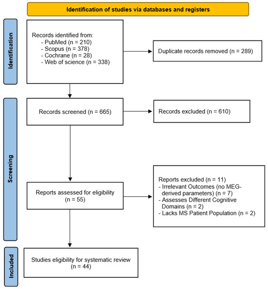
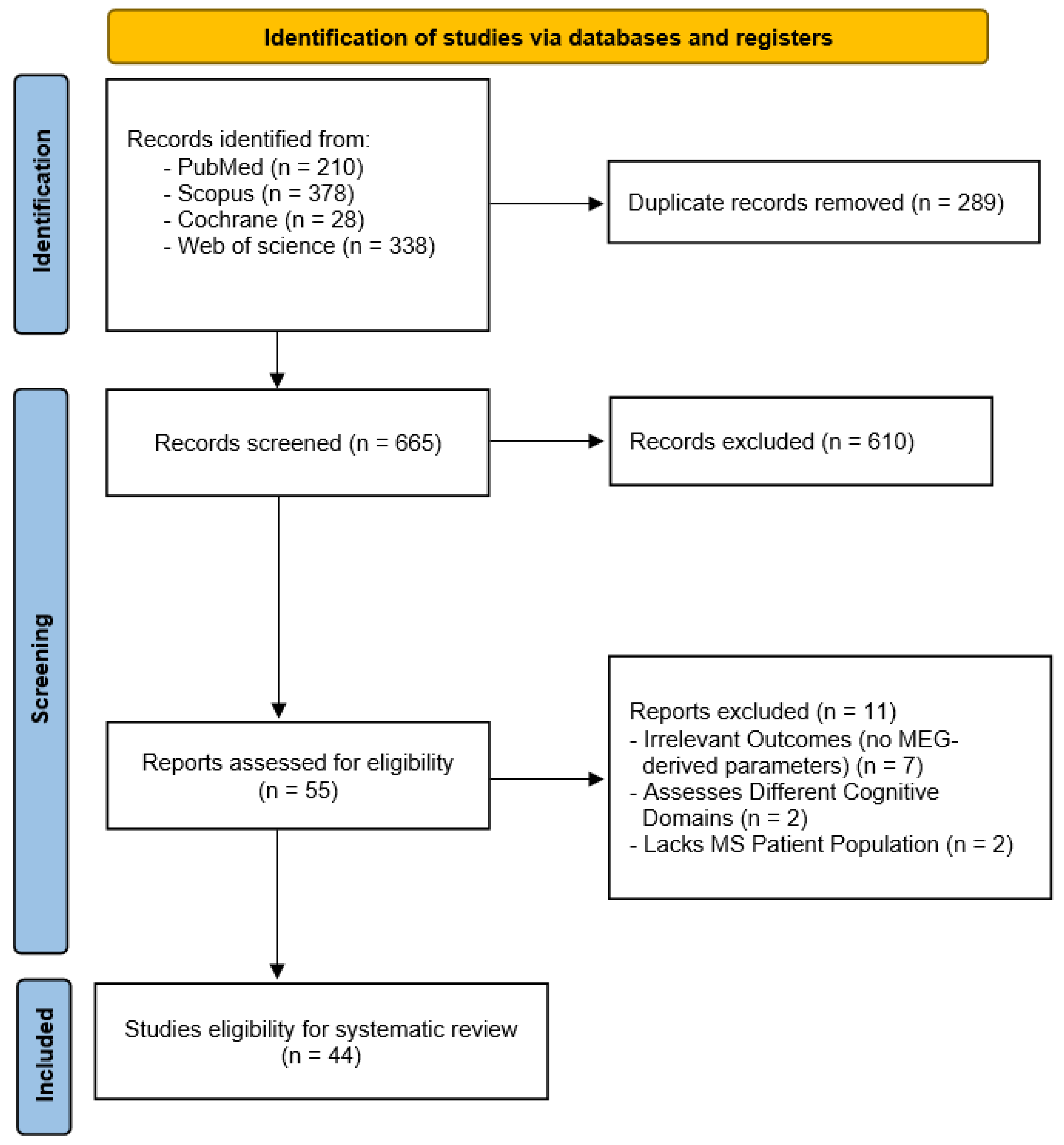
The initial database search yielded 954 records. Following the removal of 289 duplicates and the screening of 665 titles and abstracts, 55 full-text reports were assessed for eligibility. Eleven reports were excluded at the full-text stage (The 11 reports, along with their specific reasons for exclusion, are listed in Supplementary Table S3). Ultimately, 44 studies [1,3,9,10,11,12,13,14,15,16,32,33,34,35,36,37,38,39,40,41,42,43,44,45,46,47,48,49,50,51,52,53,54,55,56,57,58,59,60,61,62,63,64,65] met the inclusion criteria for this systematic review (Figure 2). The included studies analyzed diverse MS populations, predominantly RRMS. Sample sizes varied significantly, ranging from small pilots (N = 10, Kotini 2007) [50] to large-scale cohort analyses (N = 209, Bijvank 2021) [54]. The majority of studies compared MS patients to HC, with MS groups frequently exhibiting a female predominance (e.g., 72–80% female in several cohorts). Age demographics generally ranged from 30 to 55 years, except for one pediatric-onset study (N = 29, mean age 17.5 ± 3.1). Disability levels, quantified by the EDSS, ranged from minimal disability (EDSS 1.0–2.0) to moderate-severe disability (median EDSS up to 5.5 in Arpin 2017) [35]. Detailed characteristics and results are shown in Table 3. Regarding Quality Assessment, most included studies were rated as “fair” quality, while three studies (Has Silemek 2023 [15]; Nauta 2021, 2024 [13,53]) achieved a “good” rating. The quality assessment for all 44 included studies is provided in Supplementary Table S4.
Figure 2.
PRISMA Flow diagram.
Table 3.
Findings from the previous studies about MEG Functional Connectivity Studies in Multiple Sclerosis.
3.3.2. Resting-State Neural Dynamics: Spectral Power and Slowing
A consistent finding across resting-state studies was the phenomenon of “neuronal slowing,” characterized by a shift in spectral power from higher to lower frequency bands. Multiple studies reported alterations in the alpha band (8–13 Hz). Specifically, MS patients demonstrated a decrease in alpha band power and a shift in the alpha peak toward lower frequencies compared to controls. In cognitively impaired patients (N = 117), Schoonhoven et al. (2019) reported increased relative power in the theta (4–8 Hz) and lower alpha (8–10 Hz) bands, alongside a significantly reduced peak frequency (9.2 Hz vs. 9.7 Hz in controls) [10]. Van Der Meer et al. (2013) corroborated this, noting increased alpha1 power and decreased alpha2 power, with the former associated with slower information-processing speed [63]. Regarding broadband dynamics, Akbarian et al. (2023) used aperiodic 1/f slope analysis [33] and found that cognitively impaired MS patients (N = 139) exhibited a steeper 1/f slope than preserved patients and controls, suggesting an imbalance in excitation/inhibition. However, a subsequent study by the same group found no significant difference in the pre-stimulus baseline 1/f slope between groups, indicating potential variability based on medication status (e.g., Benzodiazepines) or cohort differences.
3.3.3. Functional Connectivity and Network Topology
Analyses of functional connectivity (FC) and graph theory metrics revealed frequency-dependent network reorganization.
- -
- Alpha Band: Decreased connectivity was a recurrent theme. Sjøgård et al. (2021) reported reduced alpha-band FC within the Default Mode Network (DMN) [12], while Tewarie et al. (2013) observed lower FC in the alpha2 band in visual and DMNs [11].
- -
- Beta Band: Findings in the beta band (13–30 Hz) were mixed. Sjøgård et al. (2021) found reduced FC in the sensorimotor network, which correlated with physical disability [12]. Conversely, Tewarie et al. (2013) and Schoonheim et al. (2013) reported higher beta connectivity or synchronization likelihood in MS patients compared to controls [11,58].
- -
- Network Topology: Graph theory approaches using Minimum Spanning Trees (MST) indicated topological shifts. Tewarie et al. (2014) described a shift toward a “star-like” topology in the theta band (higher leaf fraction) but a more “line-like” topology in the alpha2 band (lower leaf fraction) in early RRMS [14]. Has Silemek et al. (2023) identified a progressive loss of hub connectivity in the DMN and somatomotor networks over a 2-year longitudinal period (N = 76) [15].
3.3.4. Event-Related Dynamics and Task Modulation
Studies utilizing active tasks demonstrated significant deviations in neural processing efficiency and inhibition in MS patients.
- -
- Somatosensory Processing: MS patients exhibited impaired somatosensory gating. In paired-pulse stimulation paradigms, healthy controls showed significant attenuation of the secondary response (Peak 2 < Peak 1), whereas MS patients failed to suppress the second stimulus, resulting in a higher gating ratio. This lack of inhibition was correlated with mobility impairments. Additionally, Karhu et al. (1992) reported abnormally large-amplitude deflections at 60–80 ms (P60m) [46].
- -
- Motor Systems (Beta Rebound): A robust finding across multiple studies was the attenuation of the Post-Movement Beta Rebound (PMBR). Arpin et al. (2017), Barratt et al. (2017), and Sanders et al. (2025) consistently reported that MS patients exhibited weaker or delayed PMBR following motor tasks (e.g., knee extension, button press) compared to controls [37,38,57].
- -
- Visual Processing (Gamma Oscillations): Visual stimulation tasks revealed diminished gamma band (30–80 Hz) responses. Reduced gamma amplitude or power has been reported in adult mixed-subtype cohorts (Barratt, 2017; Stickland, 2019) and pediatric-onset MS (N = 29, Waldman, 2020) [1,38,65].
- -
- Cognitive Modulation: During auditory oddball and n-back tasks, MS patients showed altered modulation of the 1/f slope, specifically attenuated slope steepening after distractor stimuli, indicating a deficit in dynamic inhibition.
3.3.5. Clinical and Structural Correlations
Physiological abnormalities were frequently linked to clinical outcomes.
- -
- Cognition: Cognitive impairment was strongly associated with spectral slowing (increased theta/alpha1 power), steeper 1/f slopes, and specific topological changes such as increased clustering in the lower alpha band. Lower tree hierarchy in the alpha2 band was also associated with poorer overall cognitive performance.
- -
- Physical Disability: Physical impairment (EDSS) was correlated with reduced beta-band functional connectivity in the sensorimotor network and higher beta DMN connectivity.
- -
- Structure-Function Coupling: Several studies examined the relationship between structural damage and functional metrics. Kulik et al. (2022) found that cognitively impaired patients showed stronger structure-function coupling in the theta band [51]. Furthermore, Mazzara et al. (2025) employed computational modeling to demonstrate that conduction delays caused by lesions were inversely correlated with alpha peak amplitude, indicating that lesion topography, rather than total volume, drives spectral changes [52].
4. Discussion
Our study utilized MEG to characterize the dynamic reorganization of functional brain networks in MS patients with high fatigue burden compared with healthy controls. While MS patients exhibited a pattern of neural disconnection during rest (reduced interhemispheric coherence in alpha and beta bands), they demonstrated compensatory hyperconnectivity during the SDMT task. This shift was prominent in the gamma and theta bands, hinting that MS patients recruit additional neural resources to maintain cognitive performance levels comparable to healthy controls. This compensation is further evidenced by the absence of significant differences between MS patients and controls in task accuracy and MEG processing latencies (e.g., VEF, Wernicke, and Cingulate latencies), despite significant neurological disruption and high fatigue levels. Such network hyperconnectivity may alternatively reflect a maladaptive phenomenon driven by diffuse demyelination and a pathological loss of network inhibition, rather than a purely adaptive compensatory response.
The cognitive impact of MS and the elevated fatigue levels often seen in these patients are well documented [18,66], and the association between clinical-cognitive manifestations and lesion load has been detailed previously [67]. Recent resting-state MEG studies have demonstrated its utility for mapping neural activity and altered connectivity in MS; we applied this approach to examine network reorganization in patients with MS with high fatigue burden at rest, identifying compensatory connections related to cognitive processing speed [11,12]. Our resting-state findings, which show reduced connectivity in alpha and beta bands, align with those of Sjøgård et al. [12], who concluded that MS-induced dysconnectivity is a hallmark of the disease. However, our study extends this work by demonstrating that during cognitive demand (SDMT), these networks undergo compensatory reorganization.
The failure of long-range neural communication is likely a direct consequence of white matter degradation, which renders standard physiological pathways less efficient. Conversely, the hyper-synchrony in theta and gamma bands during cognitive effort (SDMT) represents a compensatory functional shift, rather than a simple manifestation of disease. By employing higher-frequency oscillations, the MS brain appears to integrate information through alternative cortical hubs. While this process preserves processing speed and accuracy, as evidenced by our MEG latency and SDMT performance data, it is likely metabolically demanding, which aligns with the significantly elevated cognitive and physical fatigue scores (MFIS) reported in the MS group. This provides a potential explanation for why, despite significant neurological burden and subjective fatigue, MS patients may maintain performance levels indistinguishable from healthy controls in specific cognitive domains. This functional shift is consistent with evidence that frontal gamma oscillations are crucial for modulating working memory [68] and that theta rhythms facilitate long-range communication during memory-intensive tasks [69]. Furthermore, the increased connectivity we observed in the gamma band aligns with findings that link gamma power and distribution to the preservation of cognitive performance [69,70].
Further supporting the compensatory role of gamma oscillations, a long-term cohort study assessed cognitive function in healthy individuals at the ages of 63 and 68 through multiple measures, including a comprehensive neuropsychological battery and electroencephalography with scalp electrodes (measured at steady-state with visual evoked potentials at alpha and gamma frequencies) [70] have shown that while cognitive performance remains stable over five years, the power of gamma oscillations shifts from posterior to anterior brain regions with age. This reorganization is thought to be a neural response to maintaining cognitive stability. Our findings align with this concept, suggesting that MS patients might undergo a process paralleling accelerated cognitive aging. In this context, the disrupted neural pathways in both MS and aging may drive a similar compensatory shift. Thus, the intense interhemispheric gamma-band coherence observed in our patients may be an attempt to recruit alternative networks to bypass structural deficits and maintain performance. This analogy remains speculative, as the aforementioned study examined healthy elderly rather than MS patients, and requires validation in a longitudinal MS cohort.
Our experimental findings provide a critical context for the variability observed in our systematic review of 44 MEG studies. The review confirms both spectral-power neuronal slowing and reduced alpha/beta connectivity at rest [10]; our resting-state coherence findings are parallel to the latter. The task-related dynamics in which our MS cohort demonstrated a significant hyper-synchrony in the gamma and theta bands during the SDMT. Some previous studies, however, reported diminished gamma responses during simple visual stimulation [1,38]. These findings may reflect different neural processes. Passive visual stimulation is thought to drive local, feed forward gamma activity in the visual cortex, which appears attenuated in MS. The SDMT, by contrast, is a high-load cognitive task that may require more distributed information integration, potentially recruiting inter-regional gamma coherence across frontal and parietal networks. If so, reduced local sensory gamma and increased network-level gamma coherence could coexist in the same patient. This interpretation would be consistent with the preserved processing latencies observed in our cohort, although it remains speculative and warrants confirmation in larger studies.
Our study combines MEG analysis with a systematic review of 44 studies, offering a robust framework for interpreting MS-related dysconnectivity. By examining multiple frequency bands during rest and during cognitive challenge (SDMT), we captured compensatory mechanisms in gamma and theta bands that are often missed in resting-state research. Limitations include a small, single-center sample size, limiting statistical power, and a lack of individualized MRI co-registration. Delta band exclusion was necessary to avoid speech artifacts, but future non-verbal paradigms could explore delta dynamics. Longitudinal designs and multimodal imaging are needed to assess compensatory stability and structure–function relationships.
5. Conclusions
In summary, our study highlights dynamic neural reorganization in MS patients. While the disease causes a disconnection syndrome at rest (reduced alpha/beta), the brain compensates during cognitive tasks through hyper-synchrony in theta and gamma bands. This neural overdrive offers a potential framework to explain how patients maintain normal processing speed and accuracy despite their underlying neurological burden and high fatigue levels. Supported by a systematic review, these findings position MEG as a potential biomarker for neuroplasticity and provide a foundation for future interventions targeting specific frequency bands to manage cognitive fatigue.
Supplementary Materials
The following supporting information can be downloaded at: https://www.mdpi.com/article/10.3390/brainsci16080862/s1. Supplementary Table S1: Full search strategies for each database, including all controlled vocabulary and free-text terms. Supplementary Table S2. Completed PRISMA 2020 checklist indicating the location of each reporting item within the manuscript. Supplementary Table S3: Full-text reports excluded from the systematic review (n = 11), with the specific reason for exclusion for each study. Supplementary Table S4: Risk-of-bias and quality assessment for all 44 included studies, rated using the NIH quality assessment tool. Supplementary Figure S1: Resting-state theta band connectivity differences between MS patients and HCs. (A) Spatial distribution of theta band connections on MRI; green indicates higher coherence in HC, red in MS; only differences between groups are shown. (B) Hemispheric connectivity counts comparison between groups. (C) Adjacency matrix of region-to-region connectivity in MS patients. (D) Adjacency matrix of region-to-region connectivity in healthy controls; Supplementary Figure S2: Resting-state alpha band connectivity differences between MS patients and HC. (A) Spatial distribution of alpha band connections on MRI; green indicates higher coherence in HC, red in MS; only differences between groups are shown. (B) Hemispheric connectivity counts comparison between groups. (C) Adjacency matrix of region-to-region connectivity in MS patients. (D) Adjacency matrix of region-to-region connectivity in healthy controls; Supplementary Figure S3: Resting-state beta band connectivity differences between MS patients and HC. (A) Spatial distribution of beta band connections on MRI; green indicates higher coherence in HC, red in MS; only differences between groups are shown. (B) Hemispheric connectivity counts comparison between groups. (C) Adjacency matrix of region-to-region connectivity in MS patients. (D) Adjacency matrix of region-to-region connectivity in healthy controls; Supplementary Figure S4: Resting-state gamma band connectivity differences between MS patients and healthy controls. (A) Spatial distribution of gamma band connections on MRI; green indicates higher coherence in HC, red in MS; only differences between groups are shown. (B) Hemispheric connectivity counts comparison between groups. (C) Adjacency matrix of region-to-region connectivity in MS patients. (D) Adjacency matrix of region-to-region connectivity in healthy controls; Supplementary Figure S5: Theta band connectivity differences during SDMT performance between MS patients and healthy controls. (A) Task-related theta band connectivity displayed on MRI; green indicates higher coherence in HC, red in MS; only differences between groups are shown. (B) Hemispheric connectivity counts during SDMT. (C) Adjacency matrix of region-to-region connectivity in MS patients during task performance. (D) Adjacency matrix of region-to-region connectivity in healthy controls during task performance; Supplementary Figure S6: Alpha band connectivity differences during SDMT performance between MS patients and healthy controls. (A) Task-related alpha band connectivity displayed on MRI; green indicates higher coherence in HC, red in MS; only differences between groups are shown. (B) Hemispheric connectivity counts during SDMT. (C) Adjacency matrix of region-to-region connectivity in MS patients during task performance. (D) Adjacency matrix of region-to-region connectivity in healthy controls during task performance; Supplementary Figure S7: Beta band connectivity differences during SDMT performance between MS patients and healthy controls. (A) Task-related beta band connectivity displayed on MRI; green indicates higher coherence in HC, red in MS; only differences between groups are shown. (B) Hemispheric connectivity counts during SDMT. (C) Adjacency matrix of region-to-region connectivity in MS patients during task performance. (D) Adjacency matrix of region-to-region connectivity in healthy controls during task performance; Supplementary Figure S8: Gamma band connectivity differences during SDMT performance between MS patients and healthy controls. (A) Task-related gamma band connectivity displayed on MRI; green indicates higher coherence in HC, red in MS; only differences between groups are shown. (B) Hemispheric connectivity counts during SDMT. (C) Adjacency matrix of region-to-region connectivity in MS patients during task performance. (D) Adjacency matrix of region-to-region connectivity in healthy controls during task performance.
Author Contributions
A.B.M.: Conceptualization, Methodology, Data Curation, Formal Analysis, Writing—Original Draft Preparation, Writing—Review and Editing, Project Administration, Supervision, Funding Acquisition; A.Z.N.: Methodology, Data Curation, Visualization, Writing—Review and Editing; M.H.: Methodology, Writing—Review and Editing, Resources; B.M.: Investigation, Visualization, Writing—Review and Editing; A.H.F.: Systematic review, Writing—Review and Editing; F.A.B.: Data Curation, Methodology, Writing—Review and Editing; M.C.: Conceptualization, Resources, Review & Editing; L.R.S.: Formal Analysis, Data Curation, Methodology, Writing—Review and Editing; S.M.B.: Conceptualization, Methodology, Data Curation, Formal Analysis, Writing—Original Draft Preparation, Writing—Review and Editing, Project Administration, Supervision, Funding Acquisition. All authors have read and agreed to the published version of the manuscript.
Funding
This research was funded by Henry Ford Health System, Grant number A30994, Detroit, MI, USA.
Institutional Review Board Statement
This study was approved by the Henry Ford Health Institutional Review Board (IRB No. 15400 and date of approval 20 January 2022).
Informed Consent Statement
All participants underwent a full Informed Consent process and provided written consent to participate in the study.
Data Availability Statement
The experimental MEG datasets analyzed in the current study are not publicly available due to patient privacy and ethical restrictions but are available from the corresponding author upon reasonable request. The data supporting the systematic review findings are available within the article and its Supplementary Materials.
Acknowledgments
The authors thank all patients and controls at Henry Ford Health in Detroit for their participation in this study. The authors thank Karla D Passalacqua, at Henry Ford Health, Detroit, for editorial assistance. During the preparation of this manuscript, the authors used the generative AI tool ChatGPT model 5.5; OpenAI, San Francisco, CA, USA, solely for the purpose of generating the conceptual workflow illustration in Figure 1; no generative AI tools were used for any other stage of the study design, data analysis, or manuscript text preparation. The authors have reviewed and edited the output and take full responsibility for the content of this publication.
Conflicts of Interest
The authors declare that they have no potential conflicts of interest in relation to this article.
Abbreviations
The following abbreviations are used in this manuscript:
| BDI | Beck Depression Inventory |
| BZD | Benzodiazepine |
| CC BY | Creative Commons Attribution |
| CIS | Clinically Isolated Syndrome |
| CMSC | Consortium of Multiple Sclerosis Centers |
| DMN | Default Mode Network |
| DMT | Disease-Modifying Therapy |
| EDSS | Expanded Disability Status Scale |
| ERD | Event-Related Desynchronization |
| ERF | Event-Related Field |
| FC | Functional Connectivity |
| FDR | False Discovery Rate |
| fMRI | Functional Magnetic Resonance Imaging |
| FSS | Fatigue Severity Scale |
| HC | Healthy Control |
| ICA | Independent Component Analysis |
| IRB | Institutional Review Board |
| MAGNIMS | Magnetic Resonance Imaging in Multiple Sclerosis |
| MEG | Magnetoencephalography |
| MFIS | Modified Fatigue Impact Scale |
| MHI | Mental Health Inventory |
| MRI | Magnetic Resonance Imaging |
| MS | Multiple Sclerosis |
| NAIMS | North American Imaging in Multiple Sclerosis |
| NIH | National Institutes of Health |
| N-back | N-back Working Memory Task |
| PCC | Partial Cross-Correlation |
| PET | Positron Emission Tomography |
| PLI | Phase Lag Index |
| PLM | Phase Linearity Measurement |
| PLV | Phase Locking Value |
| PMBR | Post-Movement Beta Rebound |
| PPMS | Primary Progressive Multiple Sclerosis |
| PRISMA | Preferred Reporting Items for Systematic Reviews and Meta-Analyses |
| RIS | Radiologically Isolated Syndrome |
| ROI | Region of Interest |
| RRMS | Relapsing-Remitting Multiple Sclerosis |
| SD | Standard Deviation |
| SDMT | Symbol Digit Modalities Test |
| SEF | Somatosensory Evoked Field |
| SI | Primary Somatosensory Cortex |
| SII | Secondary Somatosensory Cortex |
| SL | Synchronization Likelihood |
| SPMS | Secondary Progressive Multiple Sclerosis |
| TDE-HMM | Time-Delay Embedded Hidden Markov Model |
| TPJ | Temporoparietal Junction |
| VAMC | Veterans Affairs Medical Center |
| VEF | Visual Evoked Field |
| VHA | Veterans Health Administration |
References
- Stickland, R.; Allen, M.; Magazzini, L.; Singh, K.D.; Wise, R.G.; Tomassini, V. Neurovascular Coupling During Visual Stimulation in Multiple Sclerosis: A MEG-fMRI Study. Neuroscience 2019, 403, 54–69. [Google Scholar] [CrossRef] [Scilit] [PubMed]
- Tanaka, M.; Ishii, A.; Watanabe, Y. Neural effect of mental fatigue on physical fatigue: A magnetoencephalography study. Brain Res. 2014, 1542, 49–55. [Google Scholar] [CrossRef] [Scilit] [PubMed]
- Simon, S.; Nauta, I.M.; Hillebrand, A.; Schoonheim, M.M.; Uitdehaag, B.M.; van Dam, M.; Hulst, H.E.; Klein, M.; Stam, C.J.; de Jong, B.A.; et al. Neurophysiological MEG markers of cognitive impairment and performance validity in multiple sclerosis. Mult. Scler. 2023, 29, 1001–1011. [Google Scholar] [CrossRef] [Scilit] [PubMed]
- Bakshi, R.; Miletich, R.S.; Kinkel, P.R.; Emmet, M.L.; Kinkel, W.R. High-resolution fluorodeoxyglucose positron emission tomography shows both global and regional cerebral hypometabolism in multiple sclerosis. J. Neuroimaging 1998, 8, 228–234. [Google Scholar] [CrossRef] [Scilit] [PubMed]
- Filippi, M.; Rocca, M.A.; Colombo, B.; Falini, A.; Codella, M.; Scotti, G.; Comi, G. Functional magnetic resonance imaging correlates of fatigue in multiple sclerosis. NeuroImage 2002, 15, 559–567. [Google Scholar] [CrossRef] [Scilit] [PubMed]
- Roelcke, U.; Kappos, L.; Lechner-Scott, J.; Brunnschweiler, H.; Huber, S.; Ammann, W.; Plohmann, A.; Dellas, S.; Maguire, R.P.; Missimer, J.; et al. Reduced glucose metabolism in the frontal cortex and basal ganglia of multiple sclerosis patients with fatigue: A 18F-fluorodeoxyglucose positron emission tomography study. Neurology 1997, 48, 1566–1571. [Google Scholar] [CrossRef] [Scilit] [PubMed]
- Genova, H.M.; Hillary, F.G.; Wylie, G.; Rypma, B.; Deluca, J. Examination of processing speed deficits in multiple sclerosis using functional magnetic resonance imaging. J. Int. Neuropsychol. Soc. 2009, 15, 383–393. [Google Scholar] [CrossRef] [Scilit] [PubMed]
- Zipser, C.M.; Premoli, I.; Belardinelli, P.; Castellanos, N.; Rivolta, D.; Heidegger, T.; Müller-Dahlhaus, F.; Ziemann, U. Cortical excitability and interhemispheric connectivity in early relapsing–remitting multiple sclerosis studied with TMS-EEG. Front. Neurosci. 2018, 12, 393. [Google Scholar] [CrossRef] [Scilit] [PubMed]
- Cover, K.S.; Vrenken, H.; Geurts, J.J.; van Oosten, B.W.; Jelles, B.; Polman, C.H.; Stam, C.J.; van Dijk, B.W. Multiple sclerosis patients show a highly significant decrease in alpha band interhemispheric synchronization measured using MEG. NeuroImage 2006, 29, 783–788. [Google Scholar] [CrossRef] [Scilit] [PubMed]
- Schoonhoven, D.N.; Fraschini, M.; Tewarie, P.; Uitdehaag, B.M.; Eijlers, A.J.; Geurts, J.J.; Hillebrand, A.; Schoonheim, M.M.; Stam, C.J.; Strijbis, E.M. Resting-state MEG measurement of functional activation as a biomarker for cognitive decline in MS. Mult. Scler. 2019, 25, 1896–1906. [Google Scholar] [CrossRef] [Scilit] [PubMed]
- Tewarie, P.; Schoonheim, M.M.; Stam, C.J.; van der Meer, M.L.; van Dijk, B.W.; Barkhof, F.; Polman, C.H.; Hillebrand, A. Cognitive and clinical dysfunction, altered MEG resting-state networks and thalamic atrophy in multiple sclerosis. PLoS ONE 2013, 8, e69318. [Google Scholar] [CrossRef] [Scilit] [PubMed]
- Sjøgård, M.; Wens, V.; Van Schependom, J.; Costers, L.; D’Hooghe, M.; D’Haeseleer, M.; Woolrich, M.; Goldman, S.; Nagels, G.; De Tiège, X. Brain dysconnectivity relates to disability and cognitive impairment in multiple sclerosis. Hum. Brain Mapp. 2021, 42, 626–643. [Google Scholar] [CrossRef] [Scilit] [PubMed]
- Nauta, I.M.; Kulik, S.D.; Breedt, L.C.; Eijlers, A.J.; Strijbis, E.M.; Bertens, D.; Tewarie, P.; Hillebrand, A.; Stam, C.J.; Uitdehaag, B.M.; et al. Functional brain network organization measured with magnetoencephalography predicts cognitive decline in multiple sclerosis. Mult. Scler. 2021, 27, 1727–1737. [Google Scholar] [CrossRef] [Scilit] [PubMed]
- Tewarie, P.; Hillebrand, A.; Schoonheim, M.M.; van Dijk, B.W.; Geurts, J.J.; Barkhof, F.; Polman, C.H.; Stam, C.J. Functional brain network analysis using minimum spanning trees in Multiple Sclerosis: An MEG source-space study. NeuroImage 2014, 88, 308–318. [Google Scholar] [CrossRef] [Scilit] [PubMed]
- Has Silemek, A.C.; Nolte, G.; Pöttgen, J.; Engel, A.K.; Heesen, C.; Gold, S.M.; Stellmann, J.P. Topological reorganization of brain network might contribute to the resilience of cognitive functioning in mildly disabled relapsing remitting multiple sclerosis. J. Neurosci. Res. 2023, 101, 143–161. [Google Scholar] [CrossRef] [Scilit] [PubMed]
- Van Schependom, J.; Vidaurre, D.; Costers, L.; Sjøgård, M.; Sima, D.M.; Smeets, D.; D’Hooghe, M.B.; D’Haeseleer, M.; Deco, G.; Wens, V.; et al. Increased brain atrophy and lesion load is associated with stronger lower alpha MEG power in multiple sclerosis patients. NeuroImage Clin. 2021, 30, 102632. [Google Scholar] [CrossRef] [Scilit] [PubMed]
- Smith, A. Symbol Digit Modalities Test. In PsycTESTS Dataset; American Psychological Association (APA): Washington, DC, USA, 1973. [Google Scholar] [CrossRef] [Scilit]
- Benedict, R.H.; DeLuca, J.; Phillips, G.; LaRocca, N.; Hudson, L.D.; Rudick, R. Validity of the Symbol Digit Modalities Test as a cognition performance outcome measure for multiple sclerosis. Mult. Scler. 2017, 23, 721–733. [Google Scholar] [CrossRef] [Scilit] [PubMed]
- Matias-Guiu, J.A.; González-Rosa, J.; Hernández, M.Á.; Martínez-Ginés, M.L.; Portolés, A.; Pérez-Macías, N.; Benito-León, J.; Padrón, I.; Prieto, J.; Matias-Guiu, J. Amantadine and/or transcranial magnetic stimulation for fatigue associated with multiple sclerosis (FETEM): Study protocol for a phase 3 randomised, double-blind, cross-over, controlled clinical trial. BMJ Open 2024, 14, e078661. [Google Scholar] [CrossRef] [Scilit] [PubMed]
- Forn, C.; Belloch, V.; Bustamante, J.C.; Garbin, G.; Parcet-Ibars, M.A.; Sanjuan, A.; Ventura, N.; Avila, C. A symbol digit modalities test version suitable for functional MRI studies. Neurosci. Lett. 2009, 456, 11–14. [Google Scholar] [CrossRef] [Scilit] [PubMed]
- Wattjes, M.P.; Ciccarelli, O.; Reich, D.S.; Banwell, B.; de Stefano, N.; Enzinger, C.; Fazekas, F.; Filippi, M.; Frederiksen, J.; Gasperini, C.; et al. 2021 MAGNIMS-CMSC-NAIMS consensus recommendations on the use of MRI in patients with multiple sclerosis. Lancet Neurol. 2021, 20, 653–670. [Google Scholar] [CrossRef] [Scilit] [PubMed]
- Bagnato, F.; Wallin, M. Harmonizing Magnetic Resonance Imaging Protocols for Veterans With Multiple Sclerosis. Fed. Pract. 2022, 39, S10–S13. [Google Scholar] [CrossRef] [Scilit] [PubMed]
- Lajiness-O’Neill, R.; Richard, A.E.; Moran, J.E.; Olszewski, A.; Pawluk, L.; Jacobson, D.; Mansour, A.; Vogt, K.; Erdodi, L.A.; Moore, A.M.; et al. Neural synchrony examined with magnetoencephalography (MEG) during eye gaze processing in autism spectrum disorders: Preliminary findings. J. Neurodev. Disord. 2014, 6, 15. [Google Scholar] [CrossRef] [Scilit] [PubMed]
- Elisevich, K.; Shukla, N.; Moran, J.E.; Smith, B.; Schultz, L.; Mason, K.; Barkley, G.L.; Tepley, N.; Gumenyuk, V.; Bowyer, S.M. An assessment of MEG coherence imaging in the study of temporal lobe epilepsy. Epilepsia 2011, 52, 1110–1119. [Google Scholar] [CrossRef] [Scilit] [PubMed]
- Moran, J.; Bowyer, S.; Manoharan, A.; Mason, K.; Tepley, N.; Smith, B.; Barkley, G.; Greene, D.; Morrell, M. MEG coherence imaging compared to electrocortical recordings from NeuroPace implants to determine the location of ictal onset in epilepsy patients. Proc. Int. Congr. Ser. 2007, 1300, 673–676. [Google Scholar] [CrossRef] [Scilit]
- Moran, J.E.; Bowyer, S.M.; Tepley, N. Multi-Resolution FOCUSS: A source imaging technique applied to MEG data. Brain Topogr. 2005, 18, 1–17. [Google Scholar] [CrossRef] [Scilit] [PubMed]
- Talairach, J.; Tournoux, P.; Rayport, M. Co-Planar Stereotaxic Atlas of the Human Brain: 3-Dimensional Proportional System: An Approach to Cerebral Imaging; G. Thieme: New York, NY, USA; Thieme Medical Publishers: New York, NY, USA, 1988. [Google Scholar]
- Shattuck, D.W.; Mirza, M.; Adisetiyo, V.; Hojatkashani, C.; Salamon, G.; Narr, K.L.; Poldrack, R.A.; Bilder, R.M.; Toga, A.W. Construction of a 3D probabilistic atlas of human cortical structures. NeuroImage 2008, 39, 1064–1080. [Google Scholar] [CrossRef] [Scilit] [PubMed]
- Woods, R.P.; Grafton, S.T.; Watson, J.D.; Sicotte, N.L.; Mazziotta, J.C. Automated image registration: II. Intersubject validation of linear and nonlinear models. J. Comput. Assist. Tomogr. 1998, 22, 153–165. [Google Scholar] [CrossRef] [Scilit] [PubMed]
- Neacsiu, A.D.; Gerlus, N.; Graner, J.L.; Beynel, L.; Smoski, M.J.; LaBar, K.S. Characterization of neural networks involved in transdiagnostic emotion dysregulation from a pilot randomized controlled trial of a neurostimulation-enhanced behavioral intervention. Psychiatry Res. Neuroimaging 2024, 345, 111891. [Google Scholar] [CrossRef] [Scilit] [PubMed]
- Page, M.J.; McKenzie, J.E.; Bossuyt, P.M.; Boutron, I.; Hoffmann, T.C.; Mulrow, C.D.; Shamseer, L.; Tetzlaff, J.M.; Akl, E.A.; Brennan, S.E. The PRISMA 2020 statement: An updated guideline for reporting systematic reviews. BMJ 2021, 372, n71. [Google Scholar] [CrossRef] [Scilit] [PubMed]
- Akbarian, F.; Gyurkovics, M.; D’hooghe, M.B.; D’haeseleer, M.; Nagels, G.; Van Schependom, J. Task-induced 1/f slope modulation as a paradigm-independent marker of cognitive control in multiple sclerosis. Imaging Neurosci. 2025, 3, IMAG.a.973. [Google Scholar] [CrossRef] [Scilit] [PubMed]
- Akbarian, F.; Rossi, C.; Costers, L.; D’Hooghe, M.B.; D’Haeseleer, M.; Nagels, G.; Van Schependom, J. The spectral slope as a marker of excitation/inhibition ratio and cognitive functioning in multiple sclerosis. Hum. Brain Mapp. 2023, 44, 5784–5794. [Google Scholar] [CrossRef] [Scilit] [PubMed]
- Akbarian, F.; Rossi, C.; Costers, L.; D’hooghe, M.B.; D’haeseleer, M.; Nagels, G.; Van Schependom, J. Stimulus-related modulation in the 1/f spectral slope suggests an impaired inhibition during a working memory task in people with multiple sclerosis. Mult. Scler. J. 2024, 30, 1036–1046. [Google Scholar] [CrossRef] [Scilit] [PubMed]
- Arpin, D.J.; Gehringer, J.E.; Wilson, T.W.; Kurz, M.J. A reduced somatosensory gating response in individuals with multiple sclerosis is related to walking impairment. J. Neurophysiol. 2017, 118, 2052–2058. [Google Scholar] [CrossRef] [Scilit] [PubMed]
- Arpin, D.J.; Gehringer, J.E.; Wilson, T.W.; Kurz, M.J. Movement-Related Somatosensory Activity Is Altered in Patients with Multiple Sclerosis. Brain Topogr. 2018, 31, 700–707. [Google Scholar] [CrossRef] [Scilit] [PubMed]
- Arpin, D.J.; Heinrichs-Graham, E.; Gehringer, J.E.; Zabad, R.; Wilson, T.W.; Kurz, M.J. Altered sensorimotor cortical oscillations in individuals with multiple sclerosis suggests a faulty internal model. Hum. Brain Mapp. 2017, 38, 4009–4018. [Google Scholar] [CrossRef] [Scilit] [PubMed]
- Barratt, E.L.; Tewarie, P.K.; Clarke, M.A.; Hall, E.L.; Gowland, P.A.; Morris, P.G.; Francis, S.T.; Evangelou, N.; Brookes, M.J. Abnormal task driven neural oscillations in multiple sclerosis: A visuomotor MEG study. Hum. Brain Mapp. 2017, 38, 2441–2453. [Google Scholar] [CrossRef] [Scilit] [PubMed]
- Cipriano, L.; Minino, R.; Liparoti, M.; Polverino, A.; Romano, A.; Bonavita, S.; Pirozzi, M.A.; Quarantelli, M.; Jirsa, V.; Sorrentino, G.; et al. Flexibility of brain dynamics is increased and predicts clinical impairment in relapsing–remitting but not in secondary progressive multiple sclerosis. Brain Commun. 2024, 6, fcae112. [Google Scholar] [CrossRef] [Scilit] [PubMed]
- Cipriano, L.; Troisi Lopez, E.; Liparoti, M.; Minino, R.; Romano, A.; Polverino, A.; Ciaramella, F.; Ambrosanio, M.; Bonavita, S.; Jirsa, V.; et al. Reduced clinical connectome fingerprinting in multiple sclerosis predicts fatigue severity. NeuroImage Clin. 2023, 39, 103464. [Google Scholar] [CrossRef] [Scilit] [PubMed]
- Costers, L.; Van Schependom, J.; Laton, J.; Baijot, J.; Sjøgård, M.; Wens, V.; De Tiège, X.; Goldman, S.; D’Haeseleer, M.; D’Hooghe, M.B.; et al. The role of hippocampal theta oscillations in working memory impairment in multiple sclerosis. Hum. Brain Mapp. 2021, 42, 1376–1390. [Google Scholar] [CrossRef] [Scilit] [PubMed]
- Dell’Acqua, M.L.; Landi, D.; Zito, G.; Zappasodi, F.; Lupoi, D.; Rossini, P.M.; Filippi, M.M.; Tecchio, F. Thalamocortical sensorimotor circuit in multiple sclerosis: An integrated structural and electrophysiological assessment. Hum. Brain Mapp. 2010, 31, 1588–1600. [Google Scholar] [CrossRef] [Scilit] [PubMed]
- Georgopoulos, A.P.; Karageorgiou, E.; Leuthold, A.C.; Lewis, S.M.; Lynch, J.K.; Alonso, A.A.; Aslam, Z.; Carpenter, A.F.; Georgopoulos, A.; Hemmy, L.S.; et al. Synchronous neural interactions assessed by magnetoencephalography: A functional biomarker for brain disorders. J. Neural Eng. 2007, 4, 349–355. [Google Scholar] [CrossRef] [Scilit] [PubMed]
- Hagiwara, K.; Okamoto, T.; Shigeto, H.; Ogata, K.; Somehara, Y.; Matsushita, T.; Kira, J.-i.; Tobimatsu, S. Oscillatory gamma synchronization binds the primary and secondary somatosensory areas in humans. NeuroImage 2010, 51, 412–420. [Google Scholar] [CrossRef] [Scilit] [PubMed]
- Hardmeier, M.; Schoonheim, M.M.; Geurts, J.J.G.; Hillebrand, A.; Polman, C.H.; Barkhof, F.; Stam, C.J. Cognitive Dysfunction in Early Multiple Sclerosis: Altered Centrality Derived from Resting-State Functional Connectivity Using Magneto-Encephalography. PLoS ONE 2012, 7, e42087. [Google Scholar] [CrossRef] [Scilit] [PubMed]
- Karhu, J.; Hari, R.; Mäkelä, J.P.; Huttunen, J.; Knuutila, J. Cortical somatosensory magnetic responses in multiple sclerosis. Electroencephalogr. Clin. Neurophysiol. 1992, 83, 192–200. [Google Scholar] [CrossRef] [Scilit] [PubMed]
- Kassubek, J.; Sörös, P.; Kober, H.; Stippich, C.; Vieth, J.B. Focal Slow and Beta Brain Activity in Patients with Multiple Sclerosis Revealed by Magnetoencephalography. Brain Topogr. 1999, 11, 193–200. [Google Scholar] [CrossRef] [Scilit] [PubMed]
- Kim, J.A.; Bosma, R.L.; Hemington, K.S.; Rogachov, A.; Osborne, N.R.; Cheng, J.C.; Oh, J.; Crawley, A.P.; Dunkley, B.T.; Davis, K.D. Neuropathic pain and pain interference are linked to alpha-band slowing and reduced beta-band magnetoencephalography activity within the dynamic pain connectome in patients with multiple sclerosis. Pain 2019, 160, 187–197. [Google Scholar] [CrossRef] [Scilit] [PubMed]
- Kim, J.A.; Bosma, R.L.; Hemington, K.S.; Rogachov, A.; Osborne, N.R.; Cheng, J.C.; Oh, J.; Dunkley, B.T.; Davis, K.D. Cross-network coupling of neural oscillations in the dynamic pain connectome reflects chronic neuropathic pain in multiple sclerosis. NeuroImage Clin. 2020, 26, 102230. [Google Scholar] [CrossRef] [Scilit] [PubMed]
- Kotini, A.; Anninos, P.; Tamiolakis, D.; Prassopoulos, P. Differentiation of meg activity in multiple sclerosis patients with the use of nonlinear analysis. J. Integr. Neurosci. 2007, 06, 233–240. [Google Scholar] [CrossRef] [Scilit] [PubMed]
- Kulik, S.D.; Nauta, I.M.; Tewarie, P.; Koubiyr, I.; Van Dellen, E.; Ruet, A.; Meijer, K.A.; De Jong, B.A.; Stam, C.J.; Hillebrand, A.; et al. Structure-function coupling as a correlate and potential biomarker of cognitive impairment in multiple sclerosis. Netw. Neurosci. 2022, 6, 339–356. [Google Scholar] [CrossRef] [Scilit] [PubMed]
- Mazzara, C.; Ziaeemehr, A.; Troisi Lopez, E.; Cipriano, L.; Angiolelli, M.; Sparaco, M.; Quarantelli, M.; Granata, C.; Sorrentino, G.; Hashemi, M.; et al. Mapping Brain Lesions to Conduction Delays: The Next Step for Personalized Brain Models in Multiple Sclerosis. Hum. Brain Mapp. 2025, 46, e70219. [Google Scholar] [CrossRef] [Scilit] [PubMed]
- Nauta, I.M.; Kessels, R.P.; Bertens, D.; Stam, C.J.; Strijbis, E.E.; Hillebrand, A.; Fasotti, L.; Uitdehaag, B.M.; Hulst, H.E.; Speckens, A.E.; et al. Neurophysiological brain function predicts response to cognitive rehabilitation and mindfulness in multiple sclerosis: A randomized trial. J. Neurol. 2024, 271, 1649–1662. [Google Scholar] [CrossRef] [Scilit] [PubMed]
- Bijvank, J.N.; Strijbis, E.M.M.; Nauta, I.M.; Kulik, S.D.; Balk, L.J.; Stam, C.J.; Hillebrand, A.; Geurts, J.J.G.; Uitdehaag, B.M.J.; van Rijn, L.J.; et al. Impaired saccadic eye movements in multiple sclerosis are related to altered functional connectivity of the oculomotor brain network. NeuroImage Clin. 2021, 32, 102848. [Google Scholar] [CrossRef] [Scilit] [PubMed]
- Paolicelli, D.; Manni, A.; Iaffaldano, A.; Tancredi, G.; Ricci, K.; Gentile, E.; Viterbo, R.G.; Quitadamo, S.G.; De Tommaso, M.; Trojano, M. Magnetoencephalography and High-Density Electroencephalography Study of Acoustic Event Related Potentials in Early Stage of Multiple Sclerosis: A Pilot Study on Cognitive Impairment and Fatigue. Brain Sci. 2021, 11, 481. [Google Scholar] [CrossRef] [Scilit] [PubMed]
- Rossi, C.; Vidaurre, D.; Costers, L.; D’hooghe, M.B.; Akbarian, F.; D’haeseleer, M.; Woolrich, M.; Nagels, G.; Van Schependom, J. Disrupted working memory event-related network dynamics in multiple sclerosis. Commun. Biol. 2024, 7, 1592. [Google Scholar] [CrossRef] [Scilit] [PubMed]
- Sanders, B.J.; Gilmartin, C.G.S.; Rier, L.; Gascoyne, L.; McCann, E.; Cabrera, J.; Leggett, J.; Holmes, N.; Hill, R.M.; Boto, E.; et al. OPM-MEG in multiple sclerosis: Proof of principle, and the effect of naturalistic posture. NeuroImage Clin. 2025, 48, 103888. [Google Scholar] [CrossRef] [Scilit] [PubMed]
- Schoonheim, M.M.; Geurts, J.J.G.; Landi, D.; Douw, L.; Van Der Meer, M.L.; Vrenken, H.; Polman, C.H.; Barkhof, F.; Stam, C.J. Functional connectivity changes in multiple sclerosis patients: A graph analytical study of MEG resting state data. Hum. Brain Mapp. 2013, 34, 52–61. [Google Scholar] [CrossRef] [Scilit] [PubMed]
- Sorrentino, P.; Pathak, A.; Ziaeemehr, A.; Troisi Lopez, E.; Cipriano, L.; Romano, A.; Sparaco, M.; Quarantelli, M.; Banerjee, A.; Sorrentino, G.; et al. The virtual multiple sclerosis patient. iScience 2024, 27, 110101. [Google Scholar] [CrossRef] [Scilit] [PubMed]
- Tewarie, P.; Balk, L.J.; Hillebrand, A.; Steenwijk, M.D.; Uitdehaag, B.M.J.; Stam, C.J.; Petzold, A. Structure-function relationships in the visual system in multiple sclerosis: An MEG and OCT study. Ann. Clin. Transl. Neurol. 2017, 4, 614–621. [Google Scholar] [CrossRef] [Scilit] [PubMed]
- Tewarie, P.; Schoonheim, M.M.; Schouten, D.I.; Polman, C.H.; Balk, L.J.; Uitdehaag, B.M.J.; Geurts, J.J.G.; Hillebrand, A.; Barkhof, F.; Stam, C.J. Functional brain networks: Linking thalamic atrophy to clinical disability in multiple sclerosis, a multimodal fMRI and MEG Study. Hum. Brain Mapp. 2015, 36, 603–618. [Google Scholar] [CrossRef] [Scilit] [PubMed]
- Tewarie, P.; Steenwijk, M.D.; Tijms, B.M.; Daams, M.; Balk, L.J.; Stam, C.J.; Uitdehaag, B.M.J.; Polman, C.H.; Geurts, J.J.G.; Barkhof, F.; et al. Disruption of structural and functional networks in long-standing multiple sclerosis: Disruption of Structural and Functional Networks in MS. Hum. Brain Mapp. 2014, 35, 5946–5961. [Google Scholar] [CrossRef] [Scilit] [PubMed]
- Van Der Meer, M.L.; Tewarie, P.; Schoonheim, M.M.; Douw, L.; Barkhof, F.; Polman, C.H.; Stam, C.J.; Hillebrand, A. Cognition in MS correlates with resting-state oscillatory brain activity: An explorative MEG source-space study. NeuroImage Clin. 2013, 2, 727–734. [Google Scholar] [CrossRef] [Scilit] [PubMed]
- Van Schependom, J.; Vidaurre, D.; Costers, L.; Sjøgård, M.; D’Hooghe, M.B.; D’Haeseleer, M.; Wens, V.; De Tiège, X.; Goldman, S.; Woolrich, M.; et al. Altered transient brain dynamics in multiple sclerosis: Treatment or pathology? Hum. Brain Mapp. 2019, 40, 4789–4800. [Google Scholar] [CrossRef] [Scilit] [PubMed]
- Waldman, A.T.; Sollee, J.R.; Datta, R.; Lavery, A.M.; Liu, G.; Aleman, T.S.; Banwell, B.L.; Gaetz, W.C. Structural correlates of atypical visual and motor cortical oscillations in pediatric-onset multiple sclerosis. Hum. Brain Mapp. 2020, 41, 4299–4313. [Google Scholar] [CrossRef] [Scilit] [PubMed]
- Krupp, L.B. Fatigue in multiple sclerosis: Definition, pathophysiology and treatment. CNS Drugs 2003, 17, 225–234. [Google Scholar] [CrossRef] [Scilit] [PubMed]
- Rocca, M.A.; Amato, M.P.; De Stefano, N.; Enzinger, C.; Geurts, J.J.; Penner, I.K.; Rovira, A.; Sumowski, J.F.; Valsasina, P.; Filippi, M. Clinical and imaging assessment of cognitive dysfunction in multiple sclerosis. Lancet Neurol. 2015, 14, 302–317. [Google Scholar] [CrossRef] [Scilit] [PubMed]
- Singh, F.; Shu, I.W.; Hsu, S.H.; Link, P.; Pineda, J.A.; Granholm, E. Modulation of frontal gamma oscillations improves working memory in schizophrenia. NeuroImage Clin. 2020, 27, 102339. [Google Scholar] [CrossRef] [Scilit] [PubMed]
- Herweg, N.A.; Solomon, E.A.; Kahana, M.J. Theta oscillations in human memory. Trends Cogn. Sci. 2020, 24, 208–227. [Google Scholar] [CrossRef] [Scilit] [PubMed]
- Bakhtiari, A.; Petersen, J.; Urdanibia-Centelles, O.; Ghazi, M.M.; Fagerlund, B.; Mortensen, E.L.; Osler, M.; Lauritzen, M.; Benedek, K. Power and distribution of evoked gamma oscillations in brain aging and cognitive performance. Geroscience 2023, 45, 1523–1538. [Google Scholar] [CrossRef] [Scilit] [PubMed]

Disclaimer/Publisher’s Note: The statements, opinions and data contained in all publications are solely those of the individual author(s) and contributor(s) and not of MDPI and/or the editor(s). MDPI and/or the editor(s) disclaim responsibility for any injury to people or property resulting from any ideas, methods, instructions or products referred to in the content. |
© 2026 by the authors. Licensee MDPI, Basel, Switzerland. This article is an open access article distributed under the terms and conditions of the Creative Commons Attribution (CC BY) license.