Microsurgical Anatomy of the Posterior Commissure and Habenular Commissure in the Human Cadaveric Brain
Highlights
- Full-hemisphere lateral-to-medial fiber dissection preserves the three-dimensional architecture of the posterior and habenular commissures and demonstrates their distinct structural organization.
- The habenular commissure cannot be reliably reconstructed using deterministic tractography, despite high-density seeding and optimized parameters, yielding anatomically misleading anterior projections.
- A clear resolution gap exists between microsurgical anatomy and diffusion-based tractography for fine epithalamic commissural pathways.
- Cadaveric fiber dissection remains the reference standard for accurate delineation of midline commissural connectivity.
- Precise three-dimensional understanding of the PC–HC complex is essential for safe pineal and posterior third ventricular surgery.
Abstract
1. Introduction
2. Materials and Methods
2.1. Microsurgical White Matter Dissection Technique
2.2. Fiber Tracking
3. Results
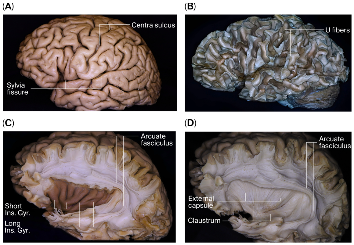
3.1. General Organization and Orientation of Microsurgical Dissection
3.2. Advancement to Deep Structures and Access to the Interhemispheric Region
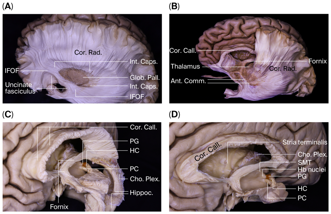
3.3. Stria Medullaris, Fornix, and the Epithalamic Complex
3.4. Microanatomical Characterization of the Posterior and Habenular Commissures
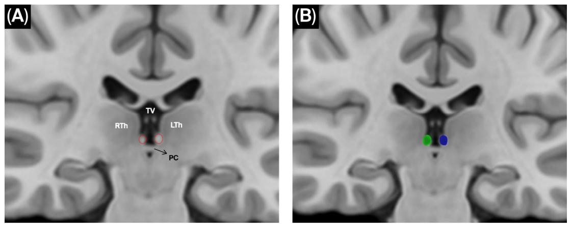
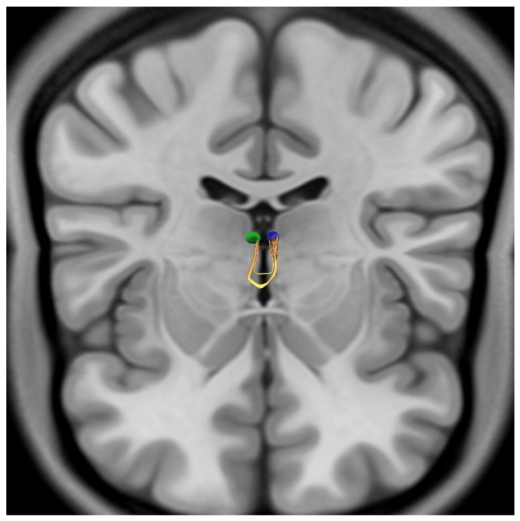
3.5. Tractography Findings
4. Discussion
4.1. Anatomical and Functional Framework of the Posterior Commissure
4.2. Anatomical Characteristics and Clinical Relevance of the Habenular Commissure
4.3. Methodological Considerations: Dissection Versus Tractography
4.4. Microsurgical Relevance and Conceptual Implications of the Habenular and Posterior Commissures
4.5. Limitations
5. Conclusions
Author Contributions
Funding
Institutional Review Board Statement
Informed Consent Statement
Data Availability Statement
Acknowledgments
Conflicts of Interest
Abbreviations
| PC | Posterior commissure |
| HC | Habenular commissure |
References
- Ozdemir, N.G. The Anatomy of the Posterior Commissure. Turk. Neurosurg. 2015, 25, 837–843. [Google Scholar] [CrossRef] [Scilit] [PubMed]
- Barut, O.; Erbaş, M.A.; Baydın, Ş.S. Posterior Komissür Mikrocerrahi Anatomisi. Sinir Sist. Cerrahisi Derg. 2025, 10, 10–18. [Google Scholar] [CrossRef] [Scilit]
- Bianco, I.H.; Wilson, S.W. The Habenular Nuclei: A Conserved Asymmetric Relay Station in the Vertebrate Brain. Philos. Trans. R. Soc. B Biol. Sci. 2009, 364, 1005–1020. [Google Scholar] [CrossRef] [Scilit]
- Piper, J.A.; Musumeci, G.; Castorina, A. The Neuroanatomy of the Habenular Complex and Its Role in the Regulation of Affective Behaviors. J. Funct. Morphol. Kinesiol. 2024, 9, 14. [Google Scholar] [CrossRef] [Scilit]
- Roman, E.; Weininger, J.; Lim, B.; Roman, M.; Barry, D.; Tierney, P.; O’Hanlon, E.; Levins, K.; O’Keane, V.; Roddy, D. Untangling the Dorsal Diencephalic Conduction System: A Review of Structure and Function of the Stria Medullaris, Habenula and Fasciculus Retroflexus. Brain Struct. Funct. 2020, 225, 1437–1458. [Google Scholar] [CrossRef] [Scilit]
- Strotmann, B.; Kögler, C.; Bazin, P.L.; Weiss, M.; Villringer, A.; Turner, R. Mapping of the Internal Structure of Human Habenula with Ex Vivo MRI at 7T. Front. Hum. Neurosci. 2013, 7, 878. [Google Scholar] [CrossRef] [Scilit]
- Choi, S.H.; Chi, J.G.; Kim, Y.B.; Cho, Z.H. Anterior Commissure-Posterior Commissure Revisited. Korean J. Radiol. 2013, 14, 653–661. [Google Scholar] [CrossRef] [Scilit] [PubMed]
- Yeh, F.C. DSI Studio: An Integrated Tractography Platform and Fiber Data Hub for Accelerating Brain Research. Nat. Methods 2025, 22, 1617–1619. [Google Scholar] [CrossRef] [Scilit] [PubMed]
- Essen, D.C.V.; Ugurbil, K.; Auerbach, E.; Barch, D.; Behrens, T.E.J.; Bucholz, R.; Chang, A.; Chen, L.; Corbetta, M.; Curtiss, S.W.; et al. The Human Connectome Project: A Data Acquisition Perspective. NeuroImage 2012, 62, 2222–2231. [Google Scholar] [CrossRef] [Scilit]
- Yeh, F.C.; Wedeen, V.J.; Tseng, W.Y.I. Generalized Q-Sampling Imaging. IEEE Trans. Med. Imaging 2010, 29, 1626–1635. [Google Scholar] [CrossRef] [Scilit]
- Yeh, F.C.; Tseng, W.Y.I. NTU-90: A High Angular Resolution Brain Atlas Constructed by q-Space Diffeomorphic Reconstruction. NeuroImage 2011, 58, 91–99. [Google Scholar] [CrossRef] [Scilit]
- Huang, H.; Xue, R.; Zhang, J.; Ren, T.; Richards, L.J.; Yarowsky, P.; Miller, M.I.; Mori, S. Anatomical Characterization of Human Fetal Brain Development with Diffusion Tensor Magnetic Resonance Imaging. J. Neurosci. Off. J. Soc. Neurosci. 2009, 29, 4263–4273. [Google Scholar] [CrossRef] [Scilit]
- Keene, M.F.L. The Connexions of the Posterior Commissure: A Study of Its Development and Myelination in the Human Foetus and Young Infant, of Its Phylogenetic Development, and of Degenerative Changes Resulting from Certain Experimental Lesions. J. Anat. 1938, 72, 488. [Google Scholar] [PubMed]
- Kucukyuruk, B.; Yagmurlu, K.; Tanriover, N.; Uzan, M.; Rhoton, A.L. Microsurgical Anatomy of the White Matter Tracts in Hemispherotomy. Neurosurgery 2014, 10, 305–324. [Google Scholar] [CrossRef] [Scilit]
- Gülsuna, B.; Güngör, A.; Yazgan, P.; Erol, G.; Middlebrooks, E.H.; Börcek, A.Ö.; Weninger, W.J.; Türe, U. Revisiting the Microsurgical Anatomy of the Sagittal Stratum and Surgical Implications: Fiber Microdissection and Tractography Study. J. Neurosurg. 2024, 141, 1433–1446. [Google Scholar] [CrossRef] [Scilit]
- González-López, P.; Cossu, G.; Thomas, C.M.; Marston, J.S.; Gómez, C.; Pralong, E.; Messerer, M.; Daniel, R.T. Cadaveric White Matter Dissection Study of the Telencephalic Flexure: Surgical Implications. Front. Neurol. 2022, 13, 757757. [Google Scholar] [CrossRef] [Scilit]
- Wakana, S.; Jiang, H.; Nagae-Poetscher, L.M.; Zijl, P.C.M.V.; Mori, S. Fiber Tract-Based Atlas of Human White Matter Anatomy. Radiology 2004, 230, 77–87. [Google Scholar] [CrossRef] [Scilit] [PubMed]
- Fernández-Miranda, J.C.; Rhoton, A.L.; Álvarez-Linera, J.; Kakizawa, Y.; Choi, C.; de Oliveira, E.P. Three-Dimensional Microsurgical and Tractographic Anatomy of the White Matter of the Human Brain. Neurosurgery 2008, 62, SHC989–SHC1028. [Google Scholar] [CrossRef] [Scilit]
- Ranft, K.; Dobrowolny, H.; Krell, D.; Bielau, H.; Bogerts, B.; Bernstein, H.G. Evidence for Structural Abnormalities of the Human Habenular Complex in Affective Disorders but Not in Schizophrenia. Psychol. Med. 2010, 40, 557–567. [Google Scholar] [CrossRef] [Scilit]
- Herkenham, M.; Nauta, W.J.H. Afferent Connections of the Habenular Nuclei in the Rat. A Horseradish Peroxidase Study, with a Note on the Fiber-of-Passage Problem. J. Comp. Neurol. 1977, 173, 123–145. [Google Scholar] [CrossRef] [Scilit] [PubMed]
- Kochanski, R.B.; Dawe, R.; Eddelman, D.B.; Kocak, M.; Sani, S. Identification of the Stria Medullaris Thalami Using Diffusion Tensor Imaging. NeuroImage Clin. 2016, 12, 852–857. [Google Scholar] [CrossRef] [Scilit]
- Roddy, D.W.; Roman, E.; Rooney, S.; Andrews, S.; Farrell, C.; Doolin, K.; Levins, K.J.; Tozzi, L.; Tierney, P.; Barry, D.; et al. Awakening Neuropsychiatric Research Into the Stria Medullaris: Development of a Diffusion-Weighted Imaging Tractography Protocol of This Key Limbic Structure. Front. Neuroanat. 2018, 12, 39. [Google Scholar] [CrossRef] [Scilit]
- Asghar, A.; Narayan, R.; Kumar, P.; Ravi, K.; Tubbs, R.; Patra, A.; Naaz, S. Absence of the Interthalamic Adhesion (ITA) as a Neuroanatomical Association or Risk Factor for Neuropsychiatric Disorders: A Systemic Review and Meta-Analysis. Indian J. Psychiatry 2023, 65, 985–994. [Google Scholar] [CrossRef] [Scilit] [PubMed]
- Akagi, K.; Powell, E.W. Differential Projections of Habenular Nuclei. J. Comp. Neurol. 1968, 132, 263–273. [Google Scholar] [CrossRef] [Scilit] [PubMed]
- Hikosaka, O. The Habenula: From Stress Evasion to Value-Based Decision-Making. Nat. Rev. Neurosci. 2010, 11, 503–513. [Google Scholar] [CrossRef] [Scilit] [PubMed]
- Jones, D.K.; Cercignani, M. Twenty-Five Pitfalls in the Analysis of Diffusion MRI Data. NMR Biomed. 2010, 23, 803–820. [Google Scholar] [CrossRef] [Scilit]
- Vos, S.B.; Jones, D.K.; Jeurissen, B.; Viergever, M.A.; Leemans, A. The Influence of Complex White Matter Architecture on the Mean Diffusivity in Diffusion Tensor MRI of the Human Brain. NeuroImage 2012, 59, 2208–2216. [Google Scholar] [CrossRef] [Scilit]
- Alexander, A.L.; Hasan, K.M.; Lazar, M.; Tsuruda, J.S.; Parker, D.L. Analysis of Partial Volume Effects in Diffusion-Tensor MRI. Magn. Reson. Med. 2001, 45, 770–780. [Google Scholar] [CrossRef] [Scilit]
- Maier-Hein, K.H.; Neher, P.F.; Houde, J.C.; Côté, M.A.; Garyfallidis, E.; Zhong, J.; Chamberland, M.; Yeh, F.C.; Lin, Y.C.; Ji, Q.; et al. The Challenge of Mapping the Human Connectome Based on Diffusion Tractography. Nat. Commun. 2017, 8, 1349. [Google Scholar] [CrossRef] [Scilit]
- Essen, D.C.V.; Smith, S.M.; Barch, D.M.; Behrens, T.E.J.; Yacoub, E.; Ugurbil, K. The WU-Minn Human Connectome Project: An Overview. NeuroImage 2013, 80, 62–79. [Google Scholar] [CrossRef] [Scilit]
- Zemmoura, I.; Serres, B.; Andersson, F.; Barantin, L.; Tauber, C.; Filipiak, I.; Cottier, J.P.; Venturini, G.; Destrieux, C. FIBRASCAN: A Novel Method for 3D White Matter Tract Reconstruction in MR Space from Cadaveric Dissection. NeuroImage 2014, 103, 106–118. [Google Scholar] [CrossRef] [Scilit] [PubMed]
- Xie, T.; Liu, S.; Zhang, X.; Yang, L.; Liu, T.; Chen, P.; Li, Z. Endoscopic Supracerebellar Infratentorial Transpineal Approach for Posterior-Medial Thalamic Lesions: Surgical Technique and Clinical Experience. Oper. Neurosurg. 2024, 27, 187–193. [Google Scholar] [CrossRef] [Scilit]
- García-Cabezas, M.Á.; Pérez-Santos, I.; Cavada, C. Stereotaxic Cutting of Post-Mortem Human Brains for Neuroanatomical Studies. Front. Neuroanat. 2023, 17, 1176351. [Google Scholar] [CrossRef] [Scilit] [PubMed]
- Tovar-Moll, F.; Monteiro, M.; Andrade, J.; Bramati, I.E.; Vianna-Barbosa, R.; Marins, T.; Rodrigues, E.; Dantas, N.; Behrens, T.E.J.; Oliveira-Souza, R.D.; et al. Structural and Functional Brain Rewiring Clarifies Preserved Interhemispheric Transfer in Humans Born without the Corpus Callosum. Proc. Natl. Acad. Sci. USA 2014, 111, 7843–7848. [Google Scholar] [CrossRef] [Scilit] [PubMed]
- Siffredi, V.; Preti, M.G.; Obertino, S.; Leventer, R.J.; Wood, A.G.; McIlroy, A.; Anderson, V.; Spencer-Smith, M.M.; Ville, D.V.D. Revisiting Brain Rewiring and Plasticity in Children Born Without Corpus Callosum. Dev. Sci. 2021, 24, e13126. [Google Scholar] [CrossRef] [Scilit]



Disclaimer/Publisher’s Note: The statements, opinions and data contained in all publications are solely those of the individual author(s) and contributor(s) and not of MDPI and/or the editor(s). MDPI and/or the editor(s) disclaim responsibility for any injury to people or property resulting from any ideas, methods, instructions or products referred to in the content. |
© 2026 by the authors. Licensee MDPI, Basel, Switzerland. This article is an open access article distributed under the terms and conditions of the Creative Commons Attribution (CC BY) license.
Share and Cite
Esen Aydin, A.; Akdeniz, M.E.; Cevik, O.M. Microsurgical Anatomy of the Posterior Commissure and Habenular Commissure in the Human Cadaveric Brain. Brain Sci. 2026, 16, 490. https://doi.org/10.3390/brainsci16050490
Esen Aydin A, Akdeniz ME, Cevik OM. Microsurgical Anatomy of the Posterior Commissure and Habenular Commissure in the Human Cadaveric Brain. Brain Sciences. 2026; 16(5):490. https://doi.org/10.3390/brainsci16050490
Chicago/Turabian StyleEsen Aydin, Aysegul, Mehmet Emin Akdeniz, and Orhun Mete Cevik. 2026. "Microsurgical Anatomy of the Posterior Commissure and Habenular Commissure in the Human Cadaveric Brain" Brain Sciences 16, no. 5: 490. https://doi.org/10.3390/brainsci16050490
APA StyleEsen Aydin, A., Akdeniz, M. E., & Cevik, O. M. (2026). Microsurgical Anatomy of the Posterior Commissure and Habenular Commissure in the Human Cadaveric Brain. Brain Sciences, 16(5), 490. https://doi.org/10.3390/brainsci16050490