


Abstract
Background/Objectives: Most individuals who have a familial or clinical risk of developing psychosis remain free from psychopathology. Identifying neural markers of resilience in these at-risk individuals may help clarify underlying mechanisms and yield novel targets for early intervention. However, in contrast to studies on risk biomarkers, studies on neural markers of resilience to psychosis are scarce. The current study aimed to identify potential brain markers of resilience to psychosis. Methods: A systematic review of the literature yielded a total of 43 MRI studies that reported resilience-associated brain changes in individuals with an elevated risk for psychosis. Label-based meta-analysis was used to synthesize findings across MRI modalities. Results: Resilience-associated brain changes were significantly overreported in the default mode and language network, and among highly connected and central brain regions. Conclusions: These findings suggest that the DMN and language-associated areas and central brain hubs may be hotspots for resilience-associated brain changes. These neural systems are thus of key interest as targets of inquiry and, possibly, intervention in at-risk populations.
Keywords:
resilience; psychosis; high risk; clinical high risk; familial high risk; neuroimaging; MRI; multimodal; brain markers 1. Introduction
Resilience has been defined as “the human ability to adapt in the face of tragedy, trauma, adversity, hardship, and ongoing significant life stressors” [1] (p. 227). Although various other definitions exist in the literature, the common thread among them is the ability to adapt to adversity in a such a manner that psychological and societal functioning are preserved [2,3,4,5,6,7].
Resilience is an important concept in early-psychosis research, as most individuals (nearly 90%) who have a familial high risk (FHR) for psychosis never develop a psychotic episode [8]. Similarly, the majority of individuals (70–80%) who meet criteria for a clinical high risk (CHR) for psychosis (i.e., subthreshold psychotic symptoms combined with functional decline) do not progress to full psychosis [9,10]. While research efforts in the field tend to focus on establishing psychosis risk markers, identifying markers of resilience, and e.g., incorporating them into psychosis-prediction tools [11] or combining them with AI [12], may promote early recognition [13] and thereby improve prognosis. Moreover, identifying neural systems associated with resilience to psychosis may guide efforts to develop novel interventions to target these systems for therapeutic or preventive benefit [14].
In the social sciences, there is a long history of research on resilience in the face of adversity, including familial predisposition to psychosis [5,15,16,17,18,19,20]. In fact, some of the earliest of these studies were conducted with children from parents diagnosed with schizophrenia [20]. These studies, pioneered by Garmezy in the 1970s [21], suggested that “many of these children were “stress-resistant” or “resilient” and capable of living productive lives and adapting to life stressors, despite having a heightened risk for developing a serious mental illness” [2]. More recent work developed a dynamic model of resilience in the presence of (risk for) mental illness as a multifaceted process with interacting internal and external dimensions, as well as continuously evolving life circumstances (see [2]). Internal factors in this model include stress levels, coping skills, and problem-solving abilities. In addition to (and perhaps partly underlying) these psychological and cognitive factors, there may also be neural characteristics that confer increased resilience to mental ill health. For example, studies in so-called “superagers”, who show excellent memory capacity in advanced age (which may reflect resilience to conventional pathways of aging), have identified specific regions of cortical preservation, alongside preserved cognitive performance and better overall mental health [22,23].
To explore the hypothesis that individuals who are resilient to the development of psychosis despite an at-risk profile may show specific neural characteristics that set them apart from both patients and healthy (i.e., non-at-risk) controls, we performed a systematic review of the literature to identify MRI studies in FHR or CHR individuals that reported markers of resilience to psychosis. Because of the sparsity of such research, we included all imaging studies regardless of the MRI modality, adapting our analytic methods to synthesize findings across modalities. To this end, we used a label-based meta-analysis, an ROI-based type of meta-analysis [24,25,26] that relies on tallying the number of times a brain region is reported in the literature as being associated with a specific finding, here being “not developing psychosis despite being at FHR or CHR to psychosis”. This approach allowed us to pool multimodal MRI findings to determine whether specific brain regions or networks are statistically overrepresented among the reported markers of resilience to psychosis across modalities. In addition, to assess putative underlying mechanisms, we incorporated methods from graph analysis to explore whether topological factors (i.e., organizational properties of the brain network) relate to the spatial distribution of resilience markers across the brain.
By identifying putative brain markers of resilience to psychosis, we aimed to provide a reference for future resilience studies. Our overall goal was to foster new hypotheses on neural factors that may confer increased resilience to psychosis and contribute to the discovery of novel targets for early intervention in at-risk individuals.
2. Materials and Methods
2.1. Systematic Review
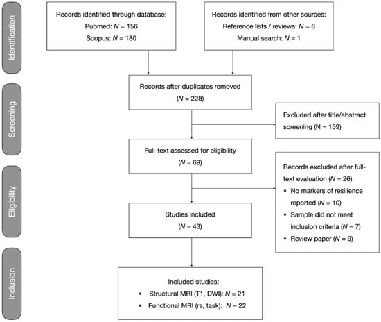
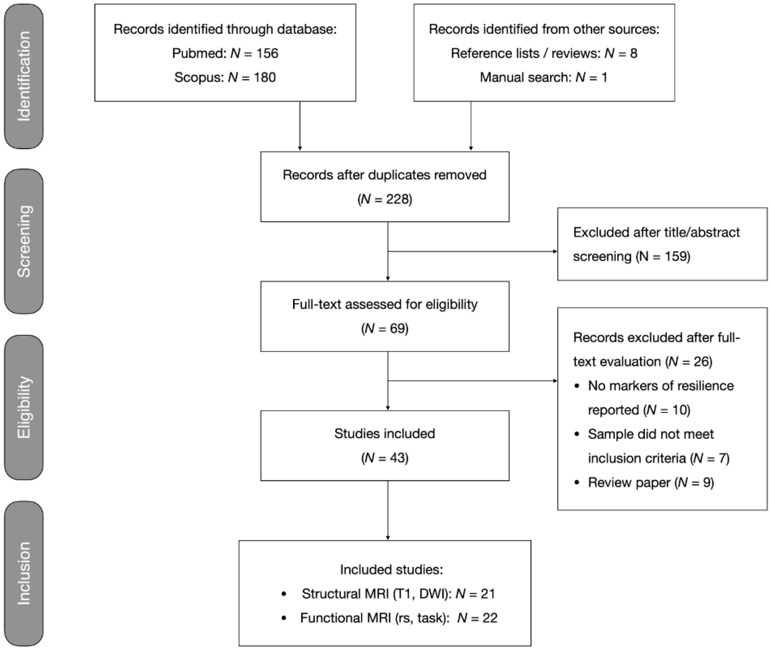
This systematic review was performed according to guidance from the Preferred Reporting Items for Systematic Reviews and Meta-Analysis (PRISMA) [27]. This review was not pre-registered. Two investigators (G.C., J.E.G.) independently performed systematic search, selection, and critical evaluation procedures. Disagreements were discussed and resolved by consensus. Figure 1 shows the flow diagram depicting the search and selection procedures.
Figure 1.
Flowchart of systematic search and selection procedures. Records excluded after full-text evaluation are listed in Supplementary Materials (Table S5).
2.1.1. Search and Selection Procedure
A comprehensive search was conducted in the PubMed and Scopus databases up to January 2020 using the following search terms: [“relatives” OR “siblings” OR “risk”] AND [“psychosis” OR “schizophrenia” OR “bipolar”] AND [“neuroimaging” OR “MRI” OR “imaging”] AND [“resilience” OR “compensatory” OR “protective”]. The retrieved records were supplemented with studies referenced by included studies or relevant review articles or as found by a manual search.
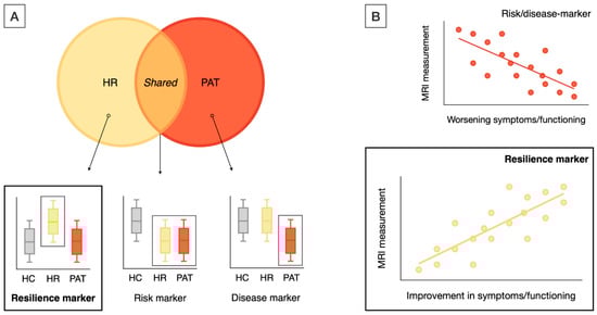
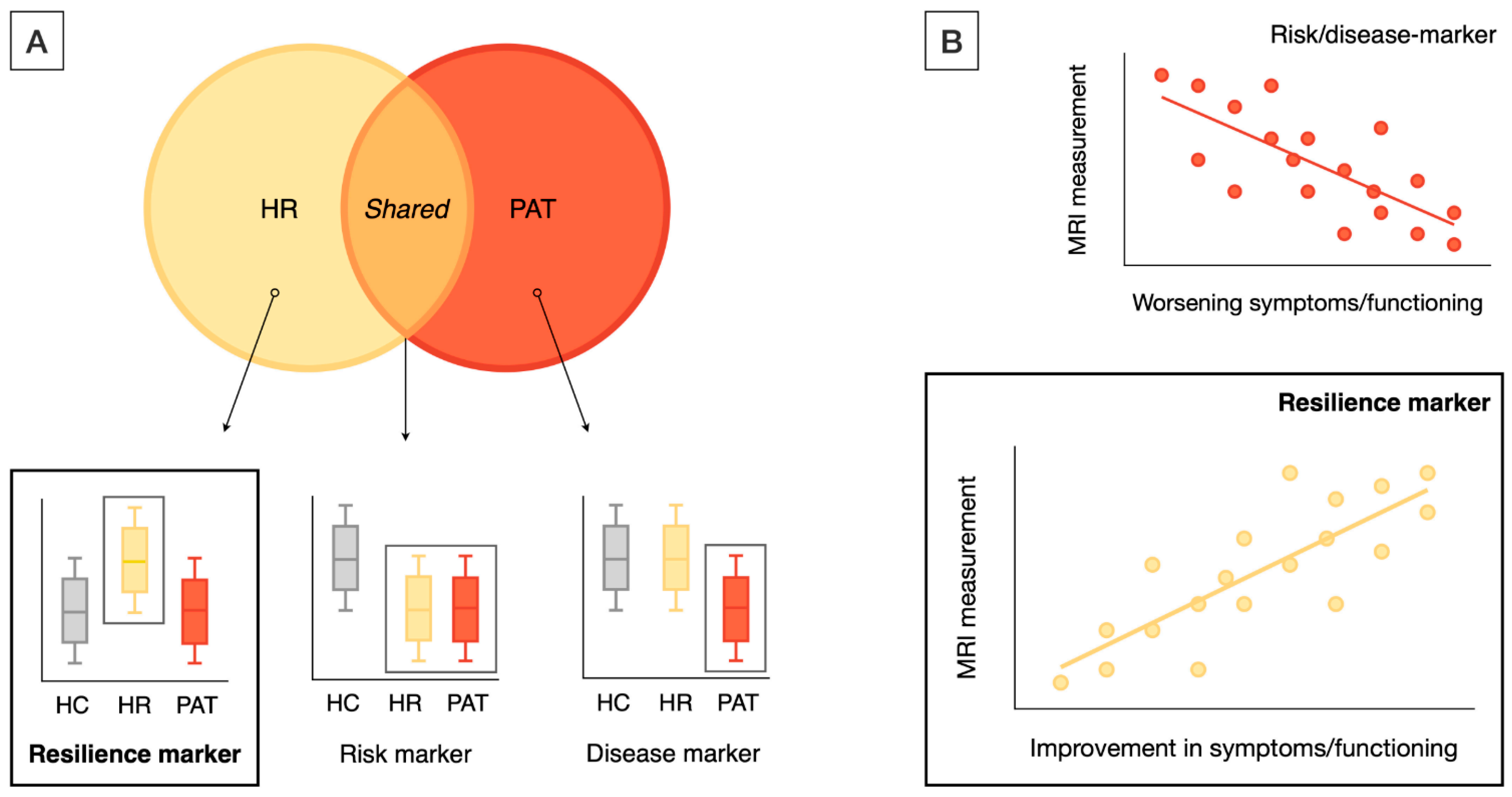
Studies that met the following inclusion criteria were included: (i) original research papers written in English; (ii) neuroimaging studies that used structural MRI (sMRI), diffusion-MRI, and task-related or resting-state fMRI (task-fMRI, rs-fMRI); (iii) studies that included a high-risk (HR) group, including either CHR [28], at-risk mental state [29], or ultra high-risk [30], or FHR individuals, including first-degree relatives (FDRs) of patients with a schizophrenia spectrum or type 1 (i.e., manic-psychotic) bipolar disorder; and (iv) studies that reported neuroimaging markers of resilience to psychosis. Resilience-associated neuroimaging markers were operationalized as MRI markers showing either significant differences in the HR (i.e., either CHR or FHR) group as compared with both the patient and healthy control (HC) groups (Figure 2A) or an association with positive outcomes (e.g., improved function) in the HR samples (Figure 2B). Additional inclusion criteria for label-based meta-analysis included (i) studies that reported ROI-based findings and (ii) studies that reported results in standardized coordinate (e.g., MNI) space.
Figure 2.
Definitions of resilience markers. Resilience markers were operationalized as MRI markers showing unique differences in HR as compared with PAT and HC (A) (as opposed to MRI changes shared between PAT and HR or between HC and HR) or showing correlations with positive outcomes, such as improvements in symptoms and functioning in HR cohorts (B). HR included both CHR and FHR individuals; HC—healthy control group; HR—high risk (i.e., either CHR or FHR) group; PAT—patient group.
2.1.2. Data Extraction
Data were extracted systematically for each publication: sample details, including the sample size, type of high-risk group, and demographics; experimental procedures, including MRI acquisition and analysis; statistical methods, including multiple comparison correction; and main findings, including regional localization based on atlas or MNI coordinates, as applicable. Only results reported as significant were considered.
2.1.3. Critical Evaluation
A critical evaluation was performed on the following reliability criteria: (i) sample size over 20 participants per group (i.e., >60 participants in total), (ii) adequate motion correction [31,32,33], and (iii) appropriate multiple comparison correction. Additional information on the critical evaluation procedure is provided in the Supplementary Materials, and Table S1 provides details on the quality assessment of each included study. Given the paucity of neuroimaging findings on resilience to psychosis, studies with quality concerns were not simply excluded. Rather, their findings were weighted according to the number of putative quality issues, assigning less weight to findings from studies with quality concerns. To this end, the findings from studies with one or two concerns were weighted as 0.67 and 0.33, respectively. The quality-weighted results went into the regional and system-level tallies of resilience effects, as specified below.
2.2. Label-Based Meta-Analysis
2.2.1. Regional Analysis
A region-wise analysis was used to assess whether the multimodal markers of resilience to psychosis converged on specific brain regions. To this end, quality-adjusted findings across imaging modalities were mapped to the Desikan–Killiany (DK) atlas (for details, see Table S4) and tallied per DK region (Supplementary Material, Table S2).
2.2.2. Network-Level Analysis
To assess whether specific brain networks are associated with resilience to psychosis, DK regions were assigned to one of seven networks defined by Yeo et al. (2011), including the default mode, frontoparietal control, somatomotor, visual, limbic, ventral attention, and dorsal attention networks [34], and tallied per network. In a follow-up analysis, the Yeo et al. parcellation was adapted to include a language network, resulting in an eight-network parcellation that was separately analyzed (Supplementary Material, Figure S1).
2.2.3. Graph Theoretical Analysis
To assess whether a brain region’s involvement in resilience relates to its topological role in the overall brain network, we tested regional tallies of resilience-related effects for associations with metrics of brain network organization. To compute these metrics, structural brain networks were reconstructed from an independent sample of healthy controls [35], with connections weighted according to the number of diffusion-MRI-derived tractography streamlines, and used to compute the regional strength, path length, clustering, betweenness centrality, and rich club membership. These metrics provide a measure of a region’s overall connectivity (strength), communication efficiency (path length), local cliquishness (clustering), centrality in the network (betweenness centrality), and whether they pertain to a central core of densely connected brain hubs (rich club membership) [35,36]. No standardization or normalization was applied to these metrics.
2.3. Statistical Analysis
2.3.1. Regional and Network-Level Analyses
Permutation analysis was used to test the statistical significance of regional and network-level findings. For each of 10,000 iterations, empirical findings were randomly redistributed across regions of the DK atlas (as shown in Table S2) and tallied per region in each iteration, creating a regional null distribution of findings under the hypothesis that their localization was driven by chance. For each brain region, the sum of the empirical findings was compared with the regional null distribution and assigned a p-value as the proportion of random iterations that produced a sum equal to, or greater than, the empirical result. No smoothing techniques were applied. The same method was used to assess the statistical significance of the network-level results. An FDR correction was applied to all the results to correct for multiple comparisons.
2.3.2. Graph Theoretical Meta-Analysis
Pearson’s correlation analysis was used to assess the associations between regional tallies of resilience-related effects and regional metrics of brain network organization. The distribution of resilience findings in the rich club versus the non-rich club regions was tested for statistical significance using a permutation analysis following the same procedures specified in Section 2.3.1.
3. Results
3.1. Systematic Review
The literature search yielded a total of 336 records, including 117 duplicates. The remaining 219 records were combined with 9 records from the manual search and cross-checking reference lists of eligible articles and review papers, which yielded a total of 228 records. The screening of the titles and abstracts yielded 69 papers for full-text evaluation. Of these, 26 publications did not meet inclusion criteria (details in Figure 1), leaving 43 papers that were selected for this review.
The 43 selected studies included 16 sMRI, 5 diffusion-MRI, 16 task-fMRI, and 6 rs-fMRI studies (the study details and main findings per imaging modality are given in Appendix A, Table A1, Table A2, Table A3 and Table A4), which comprised a total of 4732 participants, including 1455 HR individuals, 1434 patients with established psychotic illness, and 1843 HC individuals. Out of the 43 studies, 14 reported resilience-associated increases in the regional volume (N = 9) and/or cortical thickness (N = 5) or surface area (N = 1) (Table A1) and 5 reported increases in the structural connectivity (Table A2). No studies reported resilience-associated decreases in the cortical volume, thickness, or surface area or structural connectivity. Resilience-associated changes in the functional activity or connectivity were reported by 14 (Table A3) and 6 (Table A4) studies, respectively, and involved mainly increases in the activation or connectivity (N = 17). Finally, six studies reported other resilience-related effects, including changes in the regional shape (N = 1), structural covariance (N = 1) (Table A1), or global network connectivity/topology (N = 4) (Table A4).
3.2. Label-Based Meta-Analysis
3.2.1. Regional Results
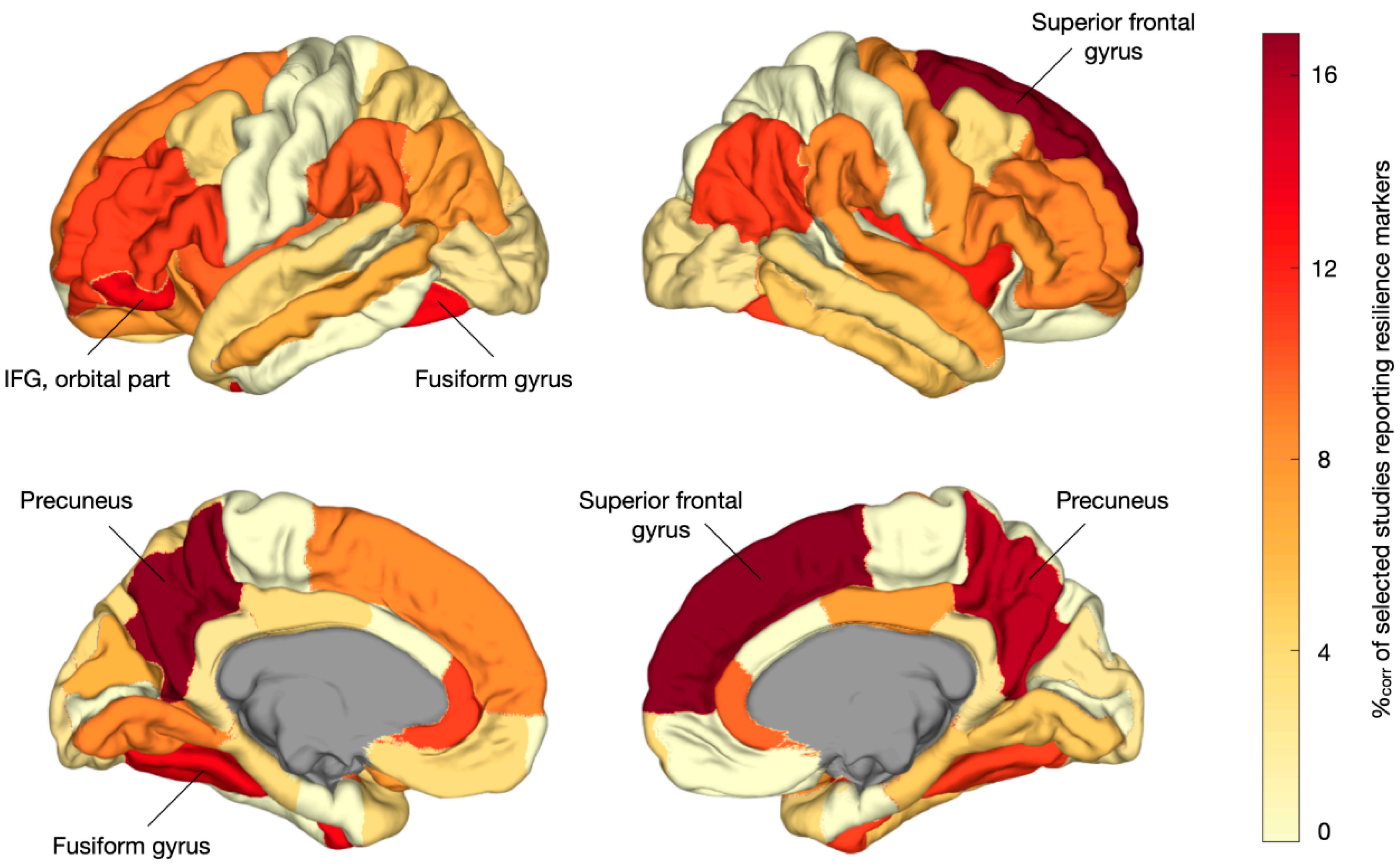
Regionally specific results were reported by 35 studies that comprised a total of 3111 participants (i.e., 1018 in the HR, 881 in the PAT, 1212 in the HC). Figure 3 shows a pooled aggregate of the regional resilience-associated effects across the MRI modalities. In the regional permutation analysis, MRI markers of resilience were overreported among the left and right precuneus (p = 0.008 and p = 0.009, respectively); right superior frontal gyrus (p = 0.007), left fusiform gyrus (FG) (p = 0.028); and left inferior frontal gyrus (IFG), orbital part (p = 0.046). However, these effects did not survive an FDR correction.
Figure 3.
Regional localization of multimodal MRI markers of resilience.
The cortical plots below depict the localization of resilience markers across all the included studies. Darker colors indicate more frequent reporting in the literature as showing resilience-related effects. Regions marked by name were overrepresented among the resilience effects (p < 0.05, permutation testing, non-FDR significant). %corr—corrected percentage of studies reporting region-specific effects; IFG—inferior frontal gyrus.
3.2.2. Network-Level Results
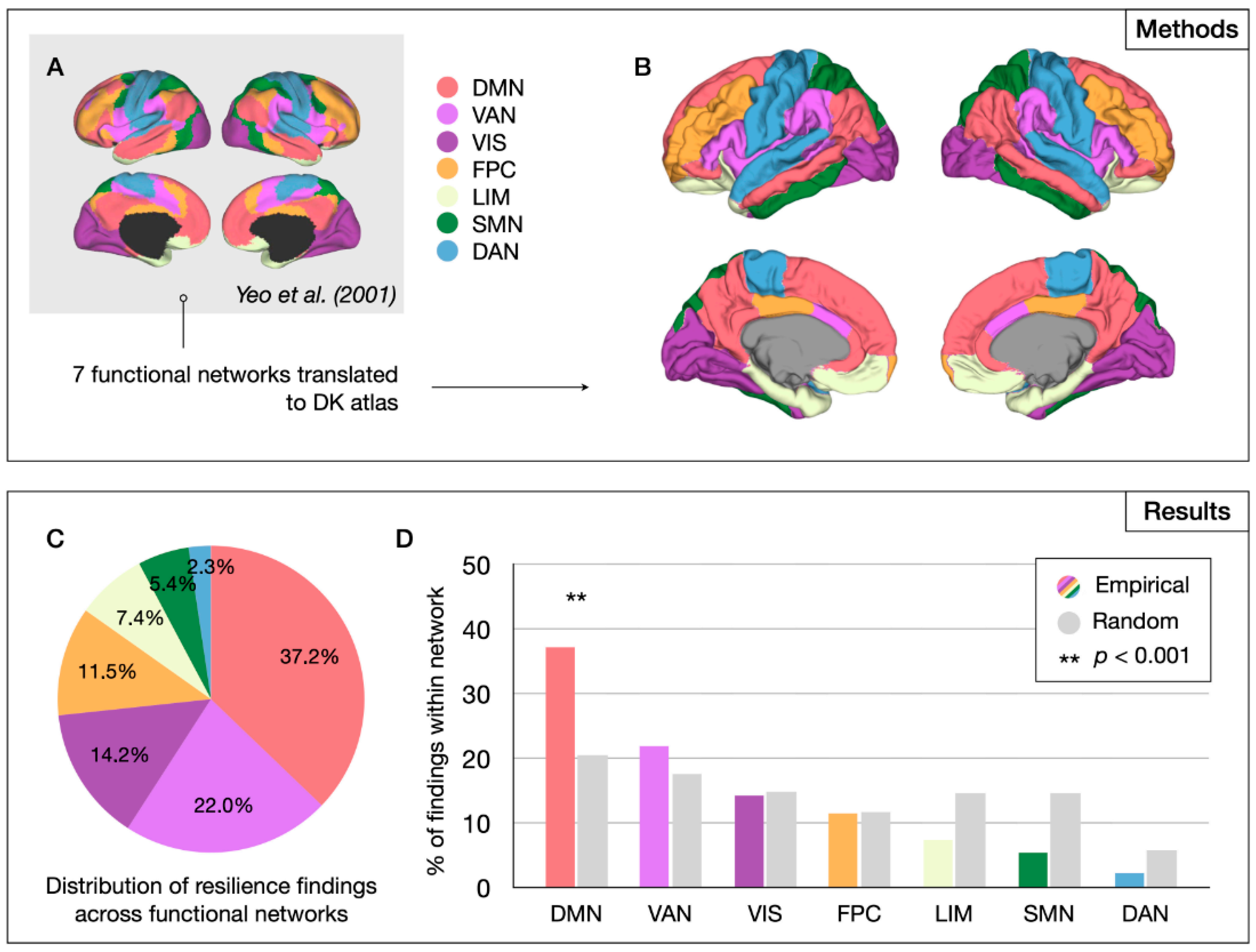
Brain regions were assigned to functional networks as defined by Yeo et al. (Figure 4A,B). Permutation analysis showed that the DMN was significantly overrepresented among reported resilience findings (p < 0.001, permutation testing) (Figure 4C,D). Adapting the Yeo et al. parcellation to include a language network yielded significant results for both the DMN (p < 0.001) and language network (p = 0.006). These findings all survived FDR correction.
Figure 4.
Network-level analysis of resilience markers. Methods (upper panel) and results (lower panel) of the system-level analysis. DK atlas regions were assigned to one of seven functional networks as defined by Yeo et al. (2001) [34] (A), resulting in a seven-network parcellation of the DK atlas (B). Well over a third of all resilience findings were found to be reported among the DMN regions (C) and the overrepresentation of the DMN among the reported resilience findings was statistically significant in permutation analysis (D). DMN—default mode network; FPC—frontoparietal control network; SMN—somatomotor network; VIS—visual network; LIM—limbic network; VAN—ventral attention network; DAN—dorsal attention network.
3.2.3. Graph Theoretical Results
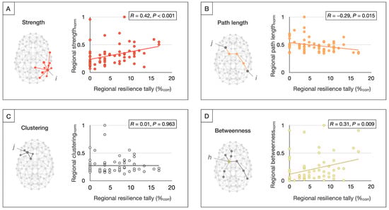
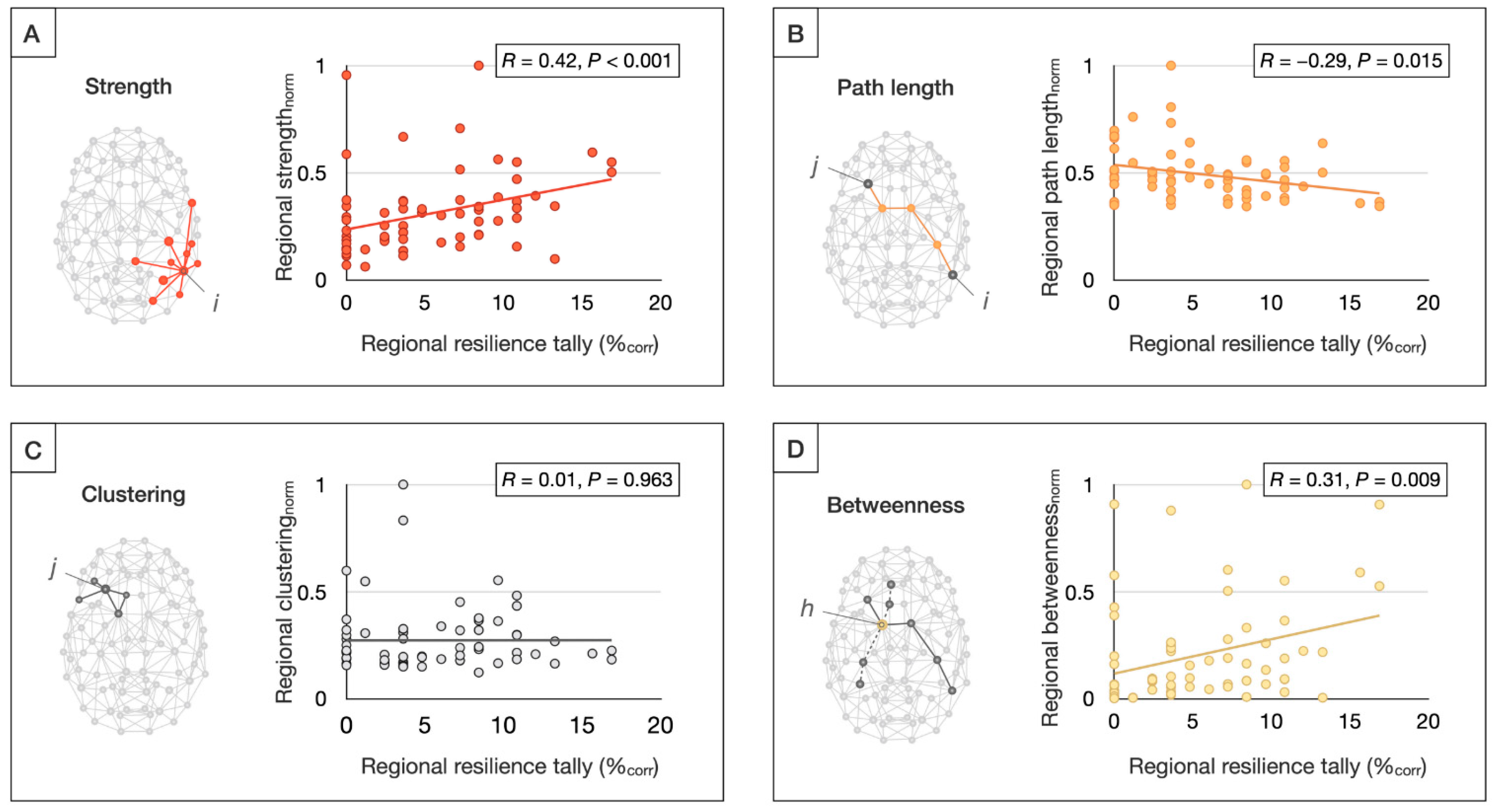
Regional tallies of resilience findings were positively correlated with regional connectivity strength (r = 0.42, p < 0.001) and betweenness centrality (r = 0.31, p = 0.009), and negatively correlated with the path length (r = −0.29, p = 0.015), suggesting that more highly connected, central, and efficient brain regions more commonly show resilience-related effects (Figure 5). Moreover, resilience-related findings were found to be significantly overreported among rich club hubs relative to non-rich club regions (p = 0.018). These findings also survived an FDR correction.
Figure 5.
Graph theoretical meta-regression of resilience markers. Regional tallies of the resilience findings were examined for correlations with the metrics of brain network organization, including strength, reflecting the total sum of connectivity of a given node i (A); path length, computed as the average number of steps from any node i to any node j (B); clustering, signifying the average likelihood that two neighboring nodes of any node j were mutually connected (C); and betweenness centrality, reflecting the fraction of shortest paths in the network that contained a given node h (D). The network metrics were computed from weighted structural connectome reconstructions from a cohort of healthy controls from an independent study [35] and normalized between 0 and 1 for visualization purposes.
4. Discussion
This systematic review and label-based meta-analysis aimed to identify spatially consistent brain markers of resilience to psychosis across structural and functional MRI studies in (clinical and familial) high-risk cohorts and to assess potential underlying mechanisms. To the best of our knowledge, this is the first meta-analytical assessment of the neuroimaging literature on resilience to psychosis.
Our systematic review yielded a total of 43 neuroimaging studies that comprised almost five thousand participants and reported structural and functional brain changes associated with resilience to psychosis in at-risk individuals. Among the 35 studies that reported regionally specific findings, resilience-associated brain changes were found to be significantly overreported among the DMN, language network, and central brain hubs. Although regional findings did not survive multiple comparison corrections, overrepresented areas, including the precuneus and (medial) superior frontal gyrus, fusiform gyrus, and left IFG, converged largely on the same systems. The reported resilience-associated effects in these regions included increases in the volume, cortical thickness, or structural connectivity, and changes in the functional activation and connectivity. It remains to be determined how such brain changes would promote resilience to psychosis.
Two potential mechanisms promoting healthy brain and cognitive functioning include a higher brain reserve and compensatory neuroplasticity. The brain reserve has been defined as a higher quantity of neural resources acting as a buffer to subsequent pathological changes and thereby preserving normal functioning [37]. Often operationalized as a higher brain volume, the brain reserve has been associated with slower clinical deterioration in dementia [38,39], preserved cognition in superagers [40], and gains in cognitive performance after cognitive enhancement therapy for schizophrenia [41]. In the context of high-risk for psychosis, higher premorbid brain volume—globally or in specific regions—or “super-normal” levels of cortical thickness or structural connectivity may buffer an overshoot in synaptic pruning in adolescence, which is thought to contribute to the pathophysiology of psychotic illness [42] and thereby mitigate the disease process. In addition, compensatory neuroplasticity may perhaps underlie some of the resilience-related fMRI results reported in the literature. Changes in the functional activation or functional connectivity may, for example, result from brain regions that actively rewire through synaptic plasticity or inherent neuron excitability as a reciprocal response to changes in other areas [43,44]. Alone or in concert, these processes may play a role in shaping an individual’s capacity for resilience by buffering or offsetting risk-associated brain changes and thereby averting progression to full psychosis in at-risk youth.
The results of our label-based meta-analysis suggest that compensatory or adaptive changes of the DMN may be particularly beneficial to resilience. Our regional analysis yielded significant results for the precuneus and superior frontal gyrus, two important nodes of the DMN, with 10 out of 35 studies with region-specific results (28.6%) implicating either or both regions. Moreover, in our network-level analysis, the DMN was found to be significantly overrepresented among resilience-related results reported in the literature. The DMN’s involvement in psychosis is emphasized by studies showing abnormalities in task-activation and functional connectivity of the DMN in patients with schizophrenia, bipolar disorder, and CHR individuals [45,46,47,48,49,50]. In addition, DMN connectivity has been related to the clinical outcome in the at-risk stage [51,52]. Indeed, a PET study showed that adaptive plasticity of the MPFC, a key part of the DMN, may protect against psychosis development in the context of childhood trauma [53]. Overall, these findings suggest a central role for the DMN in both the risk of and resilience against psychosis. Interestingly, there is evidence that mindfulness-based interventions reduce the connectivity within the DMN [54]. These interventions may thus build resilience by ameliorating the aberrant DMN connectivity associated with psychosis [45]. Indeed, there is recent preliminary evidence that mindfulness-based resiliency training is effective in reducing symptoms among at-risk individuals [55]. Moreover, mindfulness-based real-time fMRI neurofeedback aimed at downregulating DMN showed promise in terms of reducing auditory hallucinations in schizophrenia patients [56]. Finally, preliminary preclinical evidence suggests that targeted early-stage neuromodulation of the medial prefrontal cortex may prevent brain and behavioral abnormalities associated with psychosis development [57], again suggesting that mPFC may be a valuable target for early intervention.
In addition to the DMN, the label-based meta-analysis suggested that the left IFG and larger language network may play a role in promoting resilience to psychosis. The IFG consists of orbital, triangular, and opercular parts and encompasses Broca’s speech area in the dominant (typically left) hemisphere. In regional analysis, a significant effect was found for the left orbital part of the IFG specifically, but 11 out of 35 studies with region-specific results (31.4%) reported that any part of the IFG showed resilience-related effects. In addition, a DWI study (Table A2) reported increased fractional anisotropy (FA) of the arcuate fasciculus in unaffected siblings of schizophrenia patients, while their affected relatives showed an association between arcuate fasciculus FA and symptom severity [58]. Connecting Broca’s area in the IFG to Wernicke’s area in the temporal cortex, the arcuate fasciculus is involved in speech and language processing [59,60,61] and has been implicated in auditory hallucinations [62]. Taken together, these findings suggest that increases in the cortical thickness of the IFG and/or increases in structural connectivity of the arcuate fasciculus connecting IFG to other language areas may attenuate risk for psychotic symptom development. This hypothesis is in line with evidence that language learning and bilingualism can build cognitive reserve and thereby protect against neuropsychiatric disorders [63,64].
Finally, graph-theoretical analysis showed that brain regions with high connectivity and efficiency (i.e., low pathlength) and topological centrality were more likely to be reported in the literature as showing resilience-related effects. In line with this observation, resilience markers were found to be overreported among rich club hubs. These graph theoretical findings are consistent with our regional and network-level results, as rich club hubs, including the precuneus, superior frontal gyrus, and superior parietal gyrus [36], show significant overlap with the DMN [65]. Moreover, the network results extend our regional findings by providing an additional mechanistic hypothesis on why these regions in particular may be beneficial to resilience: given their central role in global brain communication [66,67,68] and the disproportionate impairment of hub-to-hub connectivity observed in schizophrenia [35,69,70], brain hubs may be particularly well positioned to buffer risk-associated brain changes and thereby promote resilience to psychosis.
A number of possible limitations should be considered when interpreting the current results. First, our findings are based on a sparse literature that includes studies with modest sample sizes. We attempted to control for this issue by reducing the relative influence of findings from studies with small sample sizes or quality concerns, but well-powered, methodologically robust studies are needed to confirm our results. In addition, because of the paucity of literature on the neurobiology of resilience, all MRI studies, regardless of the imaging modality and at-risk definition, were included. An advantage of this approach is likely an increased sensitivity to detect putative resilience markers, as different at-risk groups may share risk- and resilience-related characteristics, and such changes may show up in different imaging modalities. A disadvantage is that including such a diverse set of studies precluded a more standard meta-analytical approach that could have yielded more robust findings. Moreover, although they largely matched with the network and graph theoretical results, the regional findings did not survive multiple comparison corrections and should thus be interpreted with caution. Therefore, we suggest that our findings are primarily used to generate novel hypotheses to be confirmed in future studies. In addition, the literature search that formed the basis for the current analysis was performed in January 2020, after which this study was unfortunately interrupted by the pandemic. Because of ensuing clinical obligations, it was not feasible for the research team to update the search to include studies up to 2024. As a result, the current findings may omit important studies that came out after 2020. Another potential limitation is that selected studies included mainly medicated patients, which may obscure the natural biology of the illness. As medication effects in patients can mimic resilience-related effects in high-risk individuals—lithium treatment, for example, has been linked to increased brain volumes, particularly in mood regulatory areas [71,72]—this could hinder the identification of brain markers of resilience. Antipsychotics were shown to mainly influence basal ganglia [73,74]. Given that we focused on cortical effects and on contrasts between high-risk individuals and both patients and healthy controls, it is unlikely that the currently reported results were confounded by the effects of antipsychotic medication. Moreover, given that the label-based analysis relied on tallying results from prior studies in which participant groups were mostly well-matched and scanned on the same magnet, factors such as age, sex, and scanner differences are not expected to drive the current results, although they were not separately assessed in the current study. Finally, our review was based largely on cross-sectional studies. Longitudinal studies are needed to confirm our findings and distinguish between static protective (i.e., brain reserve) and dynamic (i.e., compensatory neuroplastic) processes that promote resilience to psychosis.
5. Conclusions
In conclusion, the current results suggest that protective or adaptive changes in a specific set of neural systems, including DMN-related brain regions, language areas, the fusiform gyrus, and rich club hubs, may have a central role in resilience to psychosis. These observations are of interest as individual differences in these systems may help understand why some at-risk youth develop psychosis while others remain healthy. Moreover, identifying neural systems associated with resilience to psychosis may promote therapeutic innovation in early psychosis and the high-risk state; for example, by identifying novel targets for intervention, such as non-invasive brain stimulation or fMRI-assisted neurofeedback [56,75,76]. If such targeted intervention can induce resilience-associated brain changes, this may slow or prevent progression to psychosis in HR youth “In the hope that, by doing so, they can perhaps be inoculated against disorder” [77].
Supplementary Materials
The following supporting information can be downloaded from https://www.mdpi.com/article/10.3390/brainsci15030314/s1, File S1: PRISMA 2020 Checklist; Figure S1. Language network definition; Supplementary results; List of Abbreviations (for Table A1, Table A2, Table A3 and Table A4); Table S1: Critical evaluation; Table S2. Mapped results—cortical; Table S3. Mapped results—subcortical; Table S4. DK atlas mapping details; Table S5. Studies excluded after full-text assessment, with rationale. References [78,79,80,81,82,83,84,85,86] are cited in the supplementary materials.
Author Contributions
Conceptualization, G.C., W.S.S., M.S.K. and M.E.S.; methodology, G.C. and X.C.; software, G.C. and X.C.; validation, G.C., J.E.G. and X.C., formal analysis, G.C., J.E.G. and X.C.; investigation, G.C. and J.E.G.; resources, G.C., W.C. and M.E.S.; data curation, G.C. and J.E.G.; writing—original draft preparation, G.C.; writing—review and editing, G.C., X.C., Z.Q., S.W.-G., W.C., J.W., W.S.S., M.S.K. and M.E.S.; visualization, G.C. and X.C.; supervision, M.S.K. and M.E.S.; project administration, N/A.; funding acquisition, G.C., S.W.-G., J.W., W.S.S., M.S.K. and M.E.S. All authors have read and agreed to the published version of the manuscript.
Funding
This research was supported by the European Union’s Horizon 2020 research and innovation program under the Marie Sklodowska-Curie grant agreement no. 749201, the Brain and Behavior Research Foundation (BBRF; grant number 29875), and the Netherlands Organization for Health Research and Development (ZonMw; grant number 636320016) (to G.C.); the National Institute of Mental Health (R01MH111448 to S.W.-G., J.W., W.S.S. and M.E.S.; R01MH64023 to M.S.K.; 5U01MH081928 to W.S.S.); and a VA Merit Award (to M.E.S.).
Data Availability Statement
No new data were created. The tables and Supplementary Materials included in this publication contain all the data referenced in this study.
Conflicts of Interest
The authors declare no conflicts of interest.
Abbreviations
The following abbreviations are used in this manuscript:
| CHR | Clinical high risk |
| DK | Desikan–Killiany (atlas) |
| DMN | Default mode network |
| FHR | Familial high risk |
| FG | Fusiform gyrus |
| HC | Healthy control |
| HR | High risk |
| IFG | Inferior frontal gyrus |
| MNI | Montreal Neurological Institute |
| MRI | Magnetic resonance imaging |
| PRISMA | Preferred Reporting Items for Systematic Reviews and Meta-Analysis |
| Rs-fMRI | Resting-state functional MRI |
Appendix A
Table A1.
Anatomical MRI studies.
Table A2.
Diffusion-MRI studies.
Table A3.
Task-based fMRI studies.
Table A4.
Resting-state fMRI studies.
Appendix B
Appendix B.1. Summary of Resilience-Associated Findings in Subcortical Structures
Multiple studies reported putative resilience-related effects of subcortical areas. These results were not further assessed statistically but are summarized below. Details on individual studies can be found in Table A1, Table A2, Table A3 and Table A4.
Appendix B.1.1. Cerebellum
Several papers implicated the cerebellum in relation to resilience to psychosis. Chang et al. (2016) found an increased cerebellar gray matter density, specifically of the anterior and posterior lobes in resilient FHR as compared with HC and schizophrenia patients [99]. In addition, three studies reported larger volumes of the cerebellar vermis in resilient FHR compared with patients and HC [91,93,94].
Appendix B.1.2. Corpus Callosum
In addition, resilience-associated changes of the corpus callosum (CC) were observed in two high-risk cohorts: Kim et al. (2012) found an increased FA of the genu in the first-degree relatives of schizophrenia patients, while patients showed a reduced FA of the splenium [107]. Katagiri et al. (2015) reported that resilient CHR individuals showed an increase in the FA of the anterior CC from the baseline to the one-year follow-up that was correlated with improved subthreshold symptoms [107]. In another study in the same cohort, volume increases in the central CC were found to correlate with improvements in negative symptoms [101].
Appendix B.1.3. Basal Ganglia, Thalamus, Hippocampus, and Amygdala
Finally, five studies reported resilience-associated changes in the basal ganglia, thalamus, amygdala, and hippocampus [95,100,103,110,125]. These results did not converge on any one specific subcortical area.
Appendix C
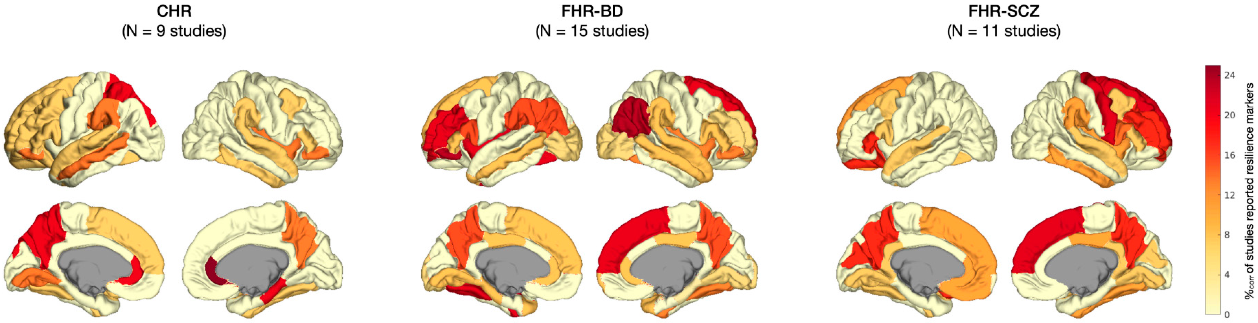
Figure A1.
Patterns of resilience-associated finding per type of high-risk group. Cortical plots depicting localization of resilience markers per type of high-risk group: CHR versus FHR for bipolar disorder (FHR-BD) or FHR for schizophrenia (FHR-SCZ). Darker colors indicate more frequent reporting of resilience-associated effects within studies on this high-risk group. %corr = corrected percentage of studies that reported resilience-associated effects.
References
- Newman, R. APA’s Resilience Initiative. Prof. Psychol. Res. Pract. 2005, 36, 227–229. [Google Scholar] [CrossRef] [Scilit]
- DeLuca, J.S.; Rakhshan Rouhakhtar, P.; Klaunig, M.J.; Akouri-Shan, L.; Jay, S.Y.; Todd, T.L.; Sarac, C.; Andorko, N.D.; Herrera, S.N.; Dobbs, M.F.; et al. Psychosis-like experiences and resilience: A systematic and critical review of the literature. Psychol. Serv. 2022, 19 (Suppl. S1), 120–138. [Google Scholar] [CrossRef] [Scilit]
- Loss, G.A.B. Trauma, and Human Resilience: Have We Underestimated the Human Capacity to Thrive After Extremely Aversive Events? Am. Psychol. 2004, 59, 20–28. [Google Scholar] [CrossRef] [Scilit]
- Brodsky, A.E.; Cattaneo, L.B. A Transconceptual Model of Empowerment and Resilience: Divergence, Convergence and Interactions in Kindred Community Concepts. Am. J. Community Psychol. 2013, 52, 333–346. [Google Scholar] [CrossRef] [Scilit] [PubMed]
- Luthar, S.S.; Cicchetti, D.; Becker, B. The Construct of Resilience: A Critical Evaluation and Guidelines for Future Work. Child. Dev. 2000, 71, 543–562. [Google Scholar] [CrossRef] [Scilit]
- Masten, A.S. Resilience in individual development: Successful adaptation despite risk and adversity. In Educational Resilience in Inner-City America: Challenges and Prospects; Wang, M.C., Gordon, E.W., Eds.; Lawrence Erlbaum Associates, Inc.: Mahwah, NJ, USA, 1994; pp. 3–25. [Google Scholar]
- Ungar, M.; Liebenberg, L. Assessing Resilience Across Cultures Using Mixed Methods: Construction of the Child and Youth Resilience Measure. J. Mix. Methods Res. 2011, 5, 126–149. [Google Scholar] [CrossRef] [Scilit]
- Guimond, S.; Mothi, S.S.; Makowski, C.; Chakravarty, M.M.; Keshavan, M.S. Altered amygdala shape trajectories and emotion recognition in youth at familial high risk of schizophrenia who develop psychosis. Transl. Psychiatry 2022, 12, 202. [Google Scholar] [CrossRef] [Scilit]
- Frangou, S. Neuroimaging Markers of Risk, Disease Expression, and Resilience to Bipolar Disorder. Curr. Psychiatry Rep. 2019, 21, 52. [Google Scholar] [CrossRef] [Scilit]
- Cannon, T.D. How Schizophrenia Develops: Cognitive and Brain Mechanisms Underlying Onset of Psychosis. Trends Cogn. Sci. 2015, 19, 744–756. [Google Scholar] [CrossRef] [Scilit]
- Cannon, T.D.; Yu, C.; Addington, J.; Bearden, C.E.; Cadenhead, K.S.; Cornblatt, B.A.; Heinssen, R.; Jeffries, C.D.; Mathalon, D.H.; McGlashan, T.H.; et al. An Individualized Risk Calculator for Research in Prodromal Psychosis. Am. J. Psychiatry 2016, 173, 980–988. [Google Scholar] [CrossRef] [Scilit]
- Di Stefano, V.; D’Angelo, M.; Monaco, F.; Vignapiano, A.; Martiadis, V.; Barone, E.; Fornaro, M.; Steardo, L.; Solmi, M.; Manchia, M. Decoding Schizophrenia: How AI-Enhanced fMRI Unlocks New Pathways for Precision Psychiatry. Brain Sci. 2024, 14, 1196. [Google Scholar] [CrossRef] [Scilit] [PubMed]
- Caballero, N.; Machiraju, S.; Diomino, A.; Kennedy, L.; Kadivar, A.; Cadenhead, K.S. Recent Updates on Predicting Conversion in Youth at Clinical High Risk for Psychosis. Curr. Psychiatry Rep. 2023, 25, 683–698. [Google Scholar] [CrossRef] [Scilit] [PubMed]
- Vargas, T.; Damme, K.S.; Ered, A.; Capizzi, R.; Frosch, I.; Ellman, L.M.; Mittal, V.A. Neuroimaging Markers of Resiliency in Youth at Clinical High Risk for Psychosis: A Qualitative Review. Biol. Psychiatry Cogn. Neurosci. Neuroimaging 2021, 6, 166–177. [Google Scholar] [CrossRef] [Scilit] [PubMed]
- Masten, A.; Best, K.; Garmezy, N. Resilience and development: Contributions from the study of children who overcome adversity. Dev. Psychopathol. 1990, 2, 425–444. [Google Scholar] [CrossRef] [Scilit]
- Rutter, M. Resilience as a dynamic concept. Dev. Psychopathol. 2012, 24, 335–344. [Google Scholar] [CrossRef] [Scilit]
- Masten, A.S.; Cicchetti, D. Resilience in development: Progress and transformation. In Developmental Psychopathology, 3rd ed.; John Wiley & Sons, Inc.: Hoboken, NJ, USA, 2016; Volume 4, pp. 271–333. [Google Scholar]
- Garmezy, N.; Masten, A.S.; Tellegen, A. The Study of Stress and Competence in Children: A Building Block for Developmental Psychopathology. Child. Dev. 1984, 55, 97–111. [Google Scholar] [CrossRef]
- Rutter, M. Resilience in the Face of Adversity. Protective Factors and Resistance to Psychiatric Disorder. Br. J. Psychiatry 1985, 147, 598–611. [Google Scholar] [CrossRef] [Scilit]
- Luthar, S.S.; Lyman, E.L.; Crossman, E.J. Resilience and Positive Psychology. In Handbook of Developmental Psychopathology; Springer: Boston, MA, USA, 2014; pp. 125–140. [Google Scholar] [CrossRef] [Scilit]
- Stress, N.G. Competence, and Development: Continuities in the Study of Schizophrenic Adults, Children Vulnerable to Psychopathology, and the Search for Stress-Resistant Children. Am. J. Orthopsychiatry 1987, 57, 159–174. [Google Scholar]
- de Godoy, L.L.; Alves, C.A.P.F.; Saavedra, J.S.M.; Studart-Neto, A.; Nitrini, R.; Leite, C.d.C.; Bisdas, S. Understanding brain resilience in superagers: A systematic review. Neuroradiology 2021, 63, 663–683. [Google Scholar] [CrossRef] [Scilit]
- Garo-Pascual, M.; Gaser, C.; Zhang, L.; Tohka, J.; Medina, M.; Strange, B.A. Brain structure and phenotypic profile of superagers compared with age-matched older adults: A longitudinal analysis from the Vallecas Project. Lancet Healthy Longev. 2023, 4, e374–e385. [Google Scholar] [CrossRef] [Scilit]
- Radua, J.; Mataix-Cols, D. Meta-analytic methods for neuroimaging data explained. Biol. Mood Anxiety Disord. 2012, 2, 6. [Google Scholar] [CrossRef] [Scilit] [PubMed]
- Laird, A.R.; McMillan, K.M.; Lancaster, J.L.; Kochunov, P.; Turkeltaub, P.E.; Pardo, J.V.; Fox, P.T. A comparison of label-based review and ALE meta-analysis in the stroop task. Hum. Brain Mapp. 2005, 25, 6–21. [Google Scholar] [CrossRef] [Scilit] [PubMed]
- Scarpazza, C.; Lattanzi, G.M.; Antoniades, M.; Di Fabio, F.; Sartori, G.; Eickhoff, S.B.; McGuire, P.; Tognin, S. Systematic review and multi-modal meta-analysis of magnetic resonance imaging findings in 22q11.2 deletion syndrome: Is more evidence needed? Neurosci. Biobehav. Rev. 2019, 107, 143–153. [Google Scholar] [CrossRef] [Scilit]
- Moher, D.; Liberati, A.; Tetzlaff, J.; Altman, D.G. Preferred reporting items for systematic reviews and meta-analyses: The PRISMA statement. BMJ 2009, 339, b2535. [Google Scholar] [CrossRef]
- McGlashan, T.; Walsh, B.; Woods, S. The Psychosis-Risk Syndrome: Handbook for Diagnosis and Follow-Up; Oxford University Press: New York, NY, USA, 2010. [Google Scholar]
- Yung, A.R.; Yuen, H.P.; McGorry, P.D.; Phillips, L.J.; Kelly, D.; Dell’Olio, M.; Francey, S.M.; Cosgrave, E.M.; Killackey, E.; Stanford, C.; et al. Mapping the onset of psychosis: The Comprehensive Assessment of At-Risk Mental States. Aust. N. Z. J. Psychiatry 2005, 39, 964–971. [Google Scholar] [CrossRef] [Scilit]
- Yung, A.R.; McGorry, P.D.; McFarlane, C.A.; Jackson, H.J.; Patton, G.C.; Rakkar, A. Monitoring and Care of Young People at Incipient Risk of Psychosis. Schizophr. Bull. 1996, 22, 283–303. [Google Scholar] [CrossRef] [Scilit]
- Power, J.D.; Barnes, K.A.; Snyder, A.Z.; Schlaggar, B.L.; Petersen, S.E. Spurious but systematic correlations in functional connectivity MRI networks arise from subject motion. Neuroimage 2012, 59, 2142–2154. [Google Scholar] [CrossRef] [Scilit] [PubMed]
- Satterthwaite, T.D.; Wolf, D.H.; Loughead, J.; Ruparel, K.; Elliott, M.A.; Hakonarson, H.; Gur, R.C.; Gur, R.E. Impact of in-scanner head motion on multiple measures of functional connectivity: Relevance for studies of neurodevelopment in youth. Neuroimage 2012, 60, 623–632. [Google Scholar] [CrossRef] [Scilit]
- Power, J.D.; Schlaggar, B.L.; Petersen, S.E. Recent progress and outstanding issues in motion correction in resting state fMRI. Neuroimage 2015, 15, 536–551. [Google Scholar] [CrossRef] [Scilit]
- Yeo, B.T.; Krienen, F.M.; Sepulcre, J.; Sabuncu, M.R.; Lashkari, D.; Hollinshead, M.; Roffman, J.L.; Smoller, J.W.; Zöllei, L.; Polimeni, J.R.; et al. The organization of the human cerebral cortex estimated by intrinsic functional connectivity. J. Neurophysiol. 2011, 106, 1125–1165. [Google Scholar]
- Collin, G.; Kahn, R.; de Reus, M.; Cahn, W.; van den Heuvel, M. Impaired rich club connectivity in unaffected siblings of schizophrenia patients. Schizophr. Bull. 2014, 40, 438–448. [Google Scholar] [CrossRef] [Scilit] [PubMed]
- Van den Heuvel, M.P.; Sporns, O. Rich-club organization of the human connectome. J. Neurosci. 2011, 31, 15775–15786. [Google Scholar] [CrossRef] [Scilit]
- Groot, C.; Van Loenhoud, A.C.; Barkhof, F.; Van Berckel, B.N.; Koene, T.; Teunissen, C.C.; Scheltens, P.; van der Flier, W.M.; Ossenkoppele, R. Differential effects of cognitive reserve and brain reserve on cognition in Alzheimer disease. Neurology 2018, 90, e149–e156. [Google Scholar] [CrossRef] [Scilit]
- Guo, L.-H.; Alexopoulos, P.; Wagenpfeil, S.; Kurz, A.; Pernecky, R. Brain size and the compensation of Alzheimer’s disease symptoms: A longitudinal cohort study. Alzheimer’s Dement. 2013, 9, 580–586. [Google Scholar] [CrossRef] [Scilit]
- Negash, S.; Xie, S.; Davatzikos, C.; Clark, C.M.; Trojanowski, J.Q.; Shaw, L.M.; Wolk, D.A.; Arnold, S.E. Cognitive and functional resilience despite molecular evidence of Alzheimer’s disease pathology. Alzheimer’s Dement. 2013, 9, e89–e95. [Google Scholar] [CrossRef] [Scilit] [PubMed]
- Rogalski, E.J.; Gefen, T.; Shi, J.; Samimi, M.; Bigio, E.; Weintraub, S.; Geula, C.; Mesulam, M.-M. Youthful Memory Capacity in Old Brains: Anatomic and Genetic Clues from the Northwestern SuperAging Project. J. Cogn. Neurosci. 2013, 25, 29–36. [Google Scholar] [CrossRef] [Scilit] [PubMed]
- Keshavan, M.S.; Eack, S.M.; Wojtalik, J.A.; Prasad, K.M.; Francis, A.N.; Bhojraj, T.S.; Greenwald, D.P.; Hogarty, S.S. A broad cortical reserve accelerates response to cognitive enhancement therapy in early course schizophrenia. Schizophr. Res. 2011, 130, 123–129. [Google Scholar] [CrossRef] [Scilit]
- Selemon, L.D.; Zecevic, N. Schizophrenia: A tale of two critical periods for prefrontal cortical development. Transl. Psychiatry 2015, 5, e623-11. [Google Scholar] [CrossRef] [Scilit]
- Keshavan, M.S.; Mehta, U.M.; Padmanabhan, J.L.; Shah, J. Dysplasticity, metaplasticity, and schizophrenia: Implications for risk, illness, and novel interventions. Dev. Psychopathol. 2015, 27, 615–635. [Google Scholar] [CrossRef] [Scilit]
- Grafman, J. Conceptualizing functional neuroplasticity. J. Commun. Disord. 2000, 33, 345–356. [Google Scholar] [CrossRef] [Scilit]
- Whitfield-Gabrieli, S.; Thermenos, H.W.; Milanovic, S.; Tsuang, M.T.; Faraone, S.V.; McCarley, R.W.; Shenton, M.E.; Green, A.I.; Nieto-Castanon, A.; LaViolette, P.; et al. Hyperactivity and hyperconnectivity of the default network in schizophrenia and in first-degree relatives of persons with schizophrenia. Proc. Natl. Acad. Sci. USA 2009, 106, 1279–1284. [Google Scholar] [CrossRef] [Scilit] [PubMed]
- Whitfield-Gabrieli, S.; Ford, J.M. Default mode network activity and connectivity in psychopathology. Annu. Rev. Clin. Psychol. 2012, 8, 49–76. [Google Scholar] [CrossRef] [Scilit] [PubMed]
- Fryer, S.L.; Woods, S.W.; Kiehl, K.A.; Calhoun, V.D.; Pearlson, G.D.; Roach, B.J.; Ford, J.M.; Srihari, V.H.; McGlashan, T.H.; Mathalon, D.H. Deficient suppression of default mode regions during working memory in individuals with early psychosis and at clinical high-risk for psychosis. Front. Psychiatry 2013, 4, 92. [Google Scholar] [CrossRef] [Scilit] [PubMed]
- Wang, H.; Zeng, L.L.; Chen, Y.; Yin, H.; Tan, Q.; Hu, D. Evidence of a dissociation pattern in default mode subnetwork functional connectivity in schizophrenia. Sci. Rep. 2015, 5, 14655. [Google Scholar] [CrossRef] [Scilit]
- Hu, M.-L.; Zong, X.-F.; Mann, J.J.; Zheng, J.-J.; Liao, Y.-H.; Li, Z.-C.; He, Y.; Chen, X.-G.; Tang, J.-S. A Review of the Functional and Anatomical Default Mode Network in Schizophrenia. Neurosci. Bull. 2017, 33, 73–84. [Google Scholar] [CrossRef] [Scilit]
- Rodríguez-Cano, E.; Alonso-Lana, S.; Sarró, S.; Fernández-Corcuera, P.; Goikolea, J.M.; Vieta, E.; Maristany, T.; Salvador, R.; McKenna, P.J.; Pomarol-Clotet, E. Differential failure to deactivate the default mode network in unipolar and bipolar depression. Bipolar Disord. 2017, 19, 386–395. [Google Scholar] [CrossRef] [Scilit]
- Lee, H.; Lee, D.; Park, K.; Kim, C.; Ryu, S. Default mode network connectivity is associated with long-term clinical outcome in patients with schizophrenia. NeuroImage Clin. 2019, 22, 101805. [Google Scholar] [CrossRef] [Scilit] [PubMed]
- Collin, G.; Nieto-Castanon, A.; Shenton, M.E.; Pasternak, O.; Kelly, S.; Keshavan, M.S.; Seidman, L.J.; McCarley, R.W.; Niznikiewicz, M.A.; Li, H.; et al. Brain Functional Connectivity Data Enhance Prediction of Clinical Outcome in Youth at Risk for Psychosis. Neuroimage Clin. 2020, 26, 102108. [Google Scholar] [CrossRef] [Scilit]
- Kasanova, Z.; Hernaus, D.; Vaessen, T.; van Amelsvoort, T.; Winz, O.; Heinzel, A.; Pruessner, J.; Mottaghy, F.M.; Collip, D.; Myin-Germeys, I. Early-Life Stress Affects Stress-Related Prefrontal Dopamine Activity in Healthy Adults, but Not in Individuals with Psychotic Disorder. PLoS ONE 2016, 11, e0150746. [Google Scholar] [CrossRef] [Scilit]
- Prakash, R.S.; Shankar, A.; Tripathi, V.; Yang, W.F.; Fisher, M.; Bauer, C.C.; Betzel, R.; Sacchet, M.D. Mindfulness Meditation and Network Neuroscience: Review, Synthesis, and Future Directions. Biol. Psychiatry Cogn. Neurosci. Neuroimaging, 2024; in press. [CrossRef] [Scilit]
- DeTore, N.R.; Luther, L.; Deng, W.; Zimmerman, J.; Leathem, L.; Burke, A.S.; Nyer, M.B.; Holt, D.J. Efficacy of a transdiagnostic, prevention-focused program for at-risk young adults: A waitlist-controlled trial. Psychol. Med. 2023, 53, 3490–3499. [Google Scholar] [CrossRef] [Scilit]
- Bauer, C.C.; Okano, K.; Ghosh, S.S.; Lee, Y.J.; Melero, H.; Angeles, C.d.L.; Nestor, P.G.; del Re, E.C.; Northoff, G.; Niznikiewicz, M.A.; et al. Real-time fMRI neurofeedback reduces auditory hallucinations and modulates resting state connectivity of involved brain regions: Part 2: Default mode network -preliminary evidence. Psychiatry Res. 2020, 284, 112770. [Google Scholar] [CrossRef] [Scilit]
- Hadar, R.; Bikovski, L.; Soto-Montenegro, M.L.; Schimke, J.; Maier, P.; Ewing, S.; Voget, M.; Wieske, F.; Götz, T.; Desco, M.; et al. Early neuromodulation prevents the development of brain and behavioral abnormalities in a rodent model of schizophrenia. Mol. Psychiatry 2018, 23, 943–951. [Google Scholar] [CrossRef] [Scilit] [PubMed]
- Boos, H.B.; Mandl, R.C.; van Haren, N.E.; Cahn, W.; van Baal, G.C.M.; Kahn, R.S.; Pol, H.E.H. Tract-based diffusion tensor imaging in patients with schizophrenia and their non-psychotic siblings. Eur. Neuropsychopharmacol. 2013, 23, 295–304. [Google Scholar] [CrossRef] [Scilit]
- Catani, M.; Mesulam, M. The arcuate fasciculus and the disconnection theme in language and aphasia: History and current state. Cortex 2009, 44, 953–961. [Google Scholar] [CrossRef] [Scilit] [PubMed]
- Skeide, M.A.; Brauer, J.; Friederici, A.D. Brain Functional and Structural Predictors of Language Performance. Cereb. Cortex 2016, 26, 2127–2139. [Google Scholar] [CrossRef] [Scilit]
- Glasser, M.F.; Rilling, J.K. DTI Tractography of the Human Brain’s Language Pathways. Cereb. Cortex 2008, 18, 2471–2482. [Google Scholar] [CrossRef] [Scilit] [PubMed]
- Hubl, D.; Koenig, T.; Strik, W.; Federspiel, A.; Kreis, R.; Boesch, C.; Maier, S.E.; Schroth, G.; Lovblad, K.; Dierks, T. Pathways that make voices: White matter changes in auditory hallucinations. Arch. Gen. Psychiatry 2004, 61, 658–668. [Google Scholar] [CrossRef] [Scilit]
- Uddin, L.Q. Cognitive and behavioural flexibility: Neural mechanisms and clinical considerations. Nat. Rev. Neurosci. 2021, 22, 167–179. [Google Scholar] [CrossRef] [Scilit]
- Calvo, N.; Anderson, J.A.E.; Berkes, M.; Freedman, M.; Craik, F.I.M.; Bialystok, E. Gray Matter Volume as Evidence for Cognitive Reserve in Bilinguals With Mild Cognitive Impairment. Alzheimer Dis. Assoc. Disord. 2023, 37, 7–12. [Google Scholar] [CrossRef] [Scilit]
- Collin, G.; Keshavan, M.S. Connectome development and a novel extension to the neurodevelopmental model of schizophrenia. Dialogues Clin. Neurosci. 2018, 20, 101–110. [Google Scholar] [CrossRef] [Scilit]
- van den Heuvel, M.P.; Kahn, R.S.; Goñi, J.; High-cost, O.S. high-capacity backbone for global brain communication. Proc. Natl. Acad. Sci. USA 2012, 109, 11372–11377. [Google Scholar] [CrossRef] [Scilit] [PubMed]
- van den Heuvel, M.P.; Sporns, O. An Anatomical Substrate for Integration among Functional Networks in Human Cortex. J. Neurosci. 2013, 33, 14489–14500. [Google Scholar] [CrossRef] [Scilit] [PubMed]
- de Reus, M.A.; van den Heuvel, M.P. Simulated rich club lesioning in brain networks: A scaffold for communication and integration? Front. Hum. Neurosci. 2014, 8, 647. [Google Scholar] [CrossRef] [Scilit]
- Heuvel, M.P.v.D.; Sporns, O.; Collin, G.; Scheewe, T.; Mandl, R.C.W.; Cahn, W.; Goñi, J.; Pol, H.E.H.; Kahn, R.S. Abnormal rich club organization and functional brain dynamics in schizophrenia. JAMA Psychiatry 2013, 70, 783–792. [Google Scholar] [CrossRef] [Scilit] [PubMed]
- Collin, G.; Scholtens, L.H.; Kahn, R.S.; Hillegers, M.H.J.; van den Heuvel, M.P. Affected Anatomical Rich Club and Structural-Functional Coupling in Young Offspring of Schizophrenia and Bipolar Disorder Patients. Biol. Psychiatry 2017, 82, 746–755. [Google Scholar] [CrossRef] [Scilit]
- Hafeman, D.M.; Chang, K.D.; Garrett, A.S.; Sanders, E.M.; MPhillips, L. Effects of medication on neuroimaging findings in bipolar disorder: An updated review. Bipolar Disord. 2012, 14, 375–410. [Google Scholar] [CrossRef] [Scilit]
- Sassi, R.B.; Nicoletti, M.; Brambilla, P.; Mallinger, A.G.; Frank, E.; Kupfer, D.J.; Keshavan, M.S.; Soares, J.C. Increased gray matter volume in lithium-treated bipolar disorder patients. Neurosci. Lett. 2002, 329, 243–245. [Google Scholar] [CrossRef] [Scilit]
- Keshavan, M.; Anderson, S.; Pettegrew, J. Changes in caudate volume with neuroleptic treatment. Lancet 1994, 344, 1434. [Google Scholar] [CrossRef] [Scilit]
- Chopra, S.; Fornito, A.; Francey, S.M.; O’donoghue, B.; Cropley, V.; Nelson, B.; Graham, J.; Baldwin, L.; Tahtalian, S.; Yuen, H.P.; et al. Differentiating the effect of antipsychotic medication and illness on brain volume reductions in first-episode psychosis: A Longitudinal, Randomised, Triple-blind, Placebo-controlled MRI Study. Neuropsychopharmacology 2021, 46, 1494–1501. [Google Scholar] [CrossRef] [Scilit]
- Orlov, N.D.; Giampietro, V.; O’Daly, O.; Lam, S.L.; Barker, G.J.; Rubia, K.; McGuire, P.; Shergill, S.S.; Allen, P. Real-time fMRI neurofeedback to down- regulate superior temporal gyrus activity in patients with schizophrenia and auditory hallucinations: A proof-of-concept study. Transl. Psychiatry 2018, 4, 46. [Google Scholar] [CrossRef] [Scilit]
- Gupta, T.; Kelley, N.J.; Pelletier-baldelli, A.; Mittal, V.A. Transcranial Direct Current Stimulation, Symptomatology, and Cognition in Psychosis: A Qualitative Review. Front. Behav. Neurosci. 2018, 12, 94. [Google Scholar] [CrossRef] [Scilit] [PubMed]
- Garmezy, N. Children at risk: The search for the antecedents of schizophrenia, part II: Ongoing research programs, issues, and intervention. Schizophr. Bull. 1974, 1, 55–125. [Google Scholar] [CrossRef] [Scilit]
- Ćurčić-Blake, B.; Ford, J.M.; Hubl, D.; Orlov, N.D.; Sommer, I.E.; Waters, F.; Allen, P.; Jardri, R.; Woodruff, P.W.; David, O.; et al. Interaction of language, auditory and memory brain networks in auditory verbal hallucinations. Prog. Neurobiol. 2017, 148, 1–20. [Google Scholar] [CrossRef] [Scilit]
- DeVylder, J.E.; Muchomba, F.M.; Gill, K.E.; Ben-David, S.; Walder, D.J.; Malaspina, D.; Corcoran, C.M. Symptom trajectories and psychosis onset in a clinical high-risk cohort: The relevance of subthreshold thought disorder. Schizophr. Res. 2014, 159, 278–283. [Google Scholar] [CrossRef] [Scilit]
- Katagiri, N.; Pantelis, C.; Nemoto, T.; Tsujino, N.; Saito, J.; Hori, M.; Yamaguchi, T.; Funatogawa, T.; Mizuno, M. Longitudinal changes in striatum and sub-threshold positive symptoms in individuals with an ‘at risk mental state’ (ARMS). Psychiatry Res. Neuroimaging 2019, 285, 25–30. [Google Scholar] [CrossRef] [Scilit]
- Levy, D.L.; Coleman, M.J.; Sung, H.; Ji, F.; Matthysse, S.; Mendell, N.R.; Titone, D. The genetic basis of thought disorder and language and communication disturbances in schizophrenia. J. Neurolinguist. 2010, 23, 176–192. [Google Scholar] [CrossRef] [Scilit] [PubMed]
- Nieto-Castanon, A. Handbook of Functional Connectivity Magnetic Resonance Imaging Methods in CONN; Hilbert Press: Boston, MA, USA, 2020. [Google Scholar]
- Strik, W.; Dierks, T.; Hubl, D.; Horn, H. HaIIucinations, Thought Disorders, and the Language Domain in Schizophrenia. Clin. EEG Neurosci. 2008, 39, 91–94. [Google Scholar] [CrossRef] [Scilit]
- Vita, A.; Dieci, M.; Giobbio, G.M.; Caputo, A.; Ghiringhelli, L.; Comazzi, M.; Garbarini, M.; Mendini, A.P.; Morganti, C.; Tenconi, F.; et al. Language and thought disorder in schizophrenia: Brain morphological correlates. Schizophr. Res. 1995, 15, 243–251. [Google Scholar] [CrossRef] [Scilit] [PubMed]
- Whitfield-Gabrieli, S.; Nieto-Castanon, A. Conn: A functional connectivity toolbox for correlated and anticorrelated brain networks. Brain 2012, 2, 125–141. [Google Scholar] [CrossRef] [Scilit]
- Page, M.J.; McKenzie, J.E.; Bossuyt, P.M.; Boutron, I.; Hoffmann, T.C.; Mulrow, C.D.; Shamseer, L.; Tetzlaff, J.M.; Akl, E.A.; Brennan, S.E.; et al. The PRISMA 2020 statement: An updated guideline for reporting systematic reviews. BMJ 2021, 372, n71. [Google Scholar] [CrossRef] [Scilit]
- Fornito, A.; Yung, A.R.; Wood, S.J.; Phillips, L.J.; Nelson, B.; Cotton, S.; Velakoulis, D.; McGorry, P.D.; Pantelis, C.; Yücel, M. Anatomic Abnormalities of the Anterior Cingulate Cortex Before Psychosis Onset: An MRI Study of Ultra-High-Risk Individuals. Biol. Psychiatry 2008, 64, 758–765. [Google Scholar] [CrossRef] [Scilit] [PubMed]
- Habets, P.; Krabbendam, L.; Hofman, P.; Suckling, J.; Oderwald, F.; Bullmore, E.; Woodruff, P.; Van Os, J.; Marcelis, M. Cognitive Performance and Grey Matter Density in Psychosis: Functional Relevance of a Structural Endophenotype. Neuropsychobiology 2008, 58, 128–137. [Google Scholar] [CrossRef] [Scilit] [PubMed]
- Kempton, M.J.; Haldane, M.; Jogia, J.; Grasby, P.M.; Collier, D.; Frangou, S. Dissociable Brain Structural Changes Associated with Predisposition, Resilience, and Disease Expression in Bipolar Disorder. J. Neurosci. 2009, 29, 10863–10868. [Google Scholar] [CrossRef] [Scilit]
- Greenstein, D.; Lenroot, R.; Clausen, L.; Chavez, A.; Vaituzis, A.; Tran, L.; Gogtay, N.; Rapoport, J. Cerebellar development in childhood onset schizophrenia and non-psychotic siblings. Psychiatry Res. Neuroimaging 2011, 193, 131–137. [Google Scholar] [CrossRef] [Scilit]
- Frangou, S. Brain structural and functional correlates of resilience to Bipolar Disorder. Front. Hum. Neurosci. 2012, 5, 184. [Google Scholar] [CrossRef] [Scilit]
- Pompei, F.; Dima, D.; Rubia, K.; Kumari, V.; Frangou, S. Dissociable functional connectivity changes during the Stroop task relating to risk, resilience and disease expression in bipolar disorder. Neuroimage 2011, 57, 576–582. [Google Scholar] [CrossRef] [Scilit]
- van Erp, T.G.; Thompson, P.M.; Kieseppä, T.; Bearden, C.E.; Marino, A.C.; Hoftman, G.D.; Haukka, J.; Partonen, T.; Huttunen, M.; Kaprio, J.; et al. Hippocampal morphology in lithium and non-lithium-treated bipolar I disorder patients, non-bipolar co-twins, and control twins. Hum. Brain Mapp. 2012, 33, 501–510. [Google Scholar] [CrossRef] [Scilit] [PubMed]
- Eker, C.; Simsek, F.; Yılmazer, E.E.; Kitis, O.; Cinar, C.; Eker, O.D.; Coburn, K.; Gonul, A.S. Brain regions associated with risk and resistance for bipolar I disorder: A voxel-based MRI study of patients with bipolar disorder and their healthy siblings. Bipolar Disord. 2014, 16, 249–261. [Google Scholar] [CrossRef] [Scilit]
- Chakravarty, M.M.; Rapoport, J.L.; Giedd, J.N.; Raznahan, A.; Shaw, P.; Collins, D.L.; Lerch, J.P.; Gogtay, N. Striatal Shape Abnormalities as Novel Neurodevelopmental Endophenotypes in Schizophrenia: A Longitudinal Study. Hum. Brain Mapp. 2015, 1469, 1458–1469. [Google Scholar] [CrossRef] [Scilit]
- Goghari, V.M.; Truong, W.; Spilka, M.J. A magnetic resonance imaging family study of cortical thickness in schizophrenia. Am. J. Med. Genet. Part B Neuropsychiatr. Genet. 2015, 168, 660–668. [Google Scholar] [CrossRef] [Scilit]
- Sarıçiçek, A.; Yalın, N.; Hıdıroğlu, C.; Çavuşoğlu, B.; Taş, C.; Ceylan, D.; Zorlu, N.; Ada, E.; Tunca, Z.; Özerdem, A. Neuroanatomical correlates of genetic risk for bipolar disorder: A voxel-based morphometry study in bipolar type I patients and healthy first degree relatives. J. Affect. Disord. 2015, 186, 110–118. [Google Scholar] [CrossRef] [Scilit] [PubMed]
- Zalesky, A.; Pantelis, C.; Cropley, V.; Fornito, A.; Cocchi, L.; McAdams, H.; Clasen, L.; Greenstein, D.; Rapoport, J.L.; Gogtay, N. Delayed Development of Brain Connectivity in Adolescents With Schizophrenia and Their Unaffected Siblings. JAMA Psychiatry 2015, 72, 900–908. [Google Scholar] [CrossRef] [Scilit] [PubMed]
- Chang, M.; Womer, F.Y.; Bai, C.; Zhou, Q.; Wei, S.; Jiang, X.; Geng, H.; Zhou, Y.; Tang, Y.; Wang, F. Voxel-based morphometry in individuals at genetic high risk for schizophrenia and patients with schizophrenia during their first episode of psychosis. PLoS ONE 2016, 11, e0163749. [Google Scholar] [CrossRef] [Scilit]
- de Wit, S.; Wierenga, L.M.; Oranje, B.; Ziermans, T.B.; Schothorst, P.F.; van Engeland, H.; Kahn, R.S.; Durston, S. Brain development in adolescents at ultra-high risk for psychosis: Longitudinal changes related to resilience. Neuroimage Clin. 2016, 12, 542–549. [Google Scholar] [CrossRef] [Scilit] [PubMed]
- Katagiri, N.; Pantelis, C.; Nemoto, T.; Tsujino, N.; Saito, J.; Hori, M.; Yamaguchi, T.; Funatogawa, T.; Mizuno, M. Symptom recovery and relationship to structure of corpus callosum in individuals with an ‘at risk mental state’. Psychiatry Res. Neuroimaging 2018, 272, 1–6. [Google Scholar] [CrossRef] [Scilit]
- Yalin, N.; Saricicek, A.; Hidiroglu, C.; Zugman, A.; Direk, N.; Ada, E.; Cavusoglu, B.; Er, A.; Isik, G.; Ceylan, D.; et al. Clinical Cortical thickness and surface area as an endophenotype in bipolar disorder type I patients and their first-degree relatives. NeuroImage Clin. 2019, 22, 101695. [Google Scholar] [CrossRef] [Scilit]
- McGlashan, S.W.; Miller, T.H.; Woods, T.J. Structured Interview for Prodromal Syndromes (SIPS)—Version 3.0; PRIME Research Clinic, Yale School of Medicine: New Haven, CT, USA, 2001. [Google Scholar]
- Hoptman, M.; Nierenberg, J.; Bertisch, H.; Catalano, D.; Ardekani, B.; Branch, C.; Delisi, L. A DTI study of white matter microstructure in individuals at high genetic risk for schizophrenia. Schizophr. Res. 2008, 106, 115–124. [Google Scholar] [CrossRef] [Scilit]
- Kim, S.N.; Park, J.S.; Jang, J.H.; Jung, W.H.; Shim, G.; Park, H.Y.; Hwang, J.Y.; Choi, C.-H.; Kang, D.-H.; Lee, J.-M.; et al. Increased white matter integrity in the corpus callosum in subjects with high genetic loading for schizophrenia. Prog. Neuropsychopharmacol. Biol. Psychiatry 2012, 37, 50–55. [Google Scholar] [CrossRef] [Scilit]
- Goghari, V.M.; Billiet, T.; Sunaert, S.; Emsell, L. A diffusion tensor imaging family study of the fornix in schizophrenia. Schizophr. Res. 2014, 159, 435–440. [Google Scholar] [CrossRef] [Scilit]
- Katagiri, N.; Pantelis, C.; Nemoto, T.; Zalesky, A.; Hori, M.; Shimoji, K.; Saito, J.; Ito, S.; Dwyer, D.B.; Fukunaga, I.; et al. A longitudinal study investigating sub-threshold symptoms and white matter changes in individuals with an ‘at risk mental state’ (ARMS). Schizophr. Res. 2015, 162, 7–13. [Google Scholar] [CrossRef] [Scilit]
- Fusar-Poli, P.; Broome, M.; Matthiasson, P.; Woolley, J.; Johns, L.; Tabraham, P.; Bramon, E.; Valmaggia, L.; Williams, S.; McGuire, P. Spatial working memory in individuals at high risk for psychosis: Longitudinal fMRI study. Schizophr. Res. 2010, 123, 45–52. [Google Scholar] [CrossRef] [Scilit] [PubMed]
- Fusar-Poli, P.; Broome, M.; Matthiasson, P.; Woolley, J.; Johns, L.; Tabraham, P.; Bramon, E.; Valmaggia, L.; Williams, S.; McGuire, P. Altered brain function directly related to structural abnormalities in people at ultra high risk of psychosis: Longitudinal VBM-fMRI study. J. Psychiatr. Res. 2011, 45, 190–198. [Google Scholar] [CrossRef] [Scilit] [PubMed]
- Choi, J.-S.; Park, J.-Y.; Jung, M.H.; Jang, J.H.; Kang, D.-H.; Jung, W.H.; Han, J.Y.; Choi, C.-H.; Hong, K.S.; Kwon, J.S. Phase-specific brain change of spatial working memory processing in genetic and ultra-high risk groups of schizophrenia. Schizophr. Bull. 2012, 38, 1189–1199. [Google Scholar] [CrossRef] [Scilit]
- Smieskova, R.; Allen, P.; Simon, A.; Aston, J.; Bendfeldt, K.; Drewe, J.; Gruber, K.; Gschwandtner, U.; Klarhoefer, M.; Lenz, C.; et al. Different duration of at-risk mental state associated with neurofunctional abnormalities. A multimodal imaging study. Hum. Brain Mapp. 2012, 33, 2281–2294. [Google Scholar] [CrossRef] [Scilit] [PubMed]
- Stäblein, M.; Storchak, H.; Ghinea, D.; Kraft, D.; Knöchel, C.; Prvulovic, D.; Bittner, R.A.; Reif, A.; Oertel-Knöchel, V. Visual working memory encoding in schizophrenia and first-degree relatives: Neurofunctional abnormalities and impaired consolidation. Psychol. Med. 2018, 49, 75–83. [Google Scholar] [CrossRef] [Scilit]
- Spilka, M.J.; Arnold, A.E.; Goghari, V.M. Functional activation abnormalities during facial emotion perception in schizophrenia patients and nonpsychotic relatives. Schizophr. Res. 2015, 168, 330–337. [Google Scholar] [CrossRef] [Scilit]
- Sepede, G.; De Berardis, D.; Campanella, D.; Perrucci, M.G.; Ferretti, A.; Salerno, R.M.; Di Giannantonio, M.; Romani, G.L.; Gambi, F. Neural correlates of negative emotion processing in bipolar disorder. Prog. Neuro-Psychopharmacol. Biol. Psychiatry 2015, 60, 1–10. [Google Scholar] [CrossRef] [Scilit]
- Tseng, W.-L.; Bones, B.; Kayser, R.; Olsavsky, A.; Fromm, S.; Pine, D.; Leibenluft, E.; Brotman, M. An fMRI study of emotional face encoding in youth at risk for bipolar disorder. Eur. Psychiatry 2015, 30, 94–98. [Google Scholar] [CrossRef] [Scilit]
- Dima, D.; Roberts, R.E.; Frangou, S. Connectomic markers of disease expression, genetic risk and resilience in bipolar disorder. Transl. Psychiatry 2016, 6, e706–e707. [Google Scholar] [CrossRef] [Scilit]
- Welge, J.A.; Saliba, L.J.; Strawn, J.R.; Eliassen, J.C.; Patino, L.R.; Adler, C.M.; Weber, W.; Schneider, M.R.; Barzman, D.H.; Strakowski, S.M.; et al. Neurofunctional Differences Among Youth With and at Varying Risk for Developing Mania. J. Am. Acad. Child. Adolesc. Psychiatry 2016, 55, 980–989. [Google Scholar] [CrossRef] [Scilit]
- Spilka, M.J.; Goghari, V.M. Similar patterns of brain activation abnormalities during emotional and non-emotional judgments of faces in a schizophrenia family study. Neuropsychologia 2017, 96, 164–174. [Google Scholar] [CrossRef] [Scilit]
- Wiggins, J.L.; Brotman, M.A.; Adleman, N.E.; Kim, P.; Wambach, C.G.; Reynolds, R.C.; Chen, G.; Towbin, K.; Pine, D.S.; Leibenluft, E. Neural Markers in Pediatric Bipolar Disorder and Familial Risk for Bipolar Disorder. J. Am. Acad. Child. Adolesc. Psychiatry 2017, 56, 67–78. [Google Scholar] [CrossRef] [Scilit]
- Nimarko, A.F.; Garrett, A.S.; Carlson, G.A.; Singh, M.K. Neural correlates of emotion processing predict resilience in youth at familial risk for mood disorders. Dev. Psychopathol. 2019, 31, 1037–1052. [Google Scholar] [CrossRef] [Scilit] [PubMed]
- Brüne, M.; Özgürdal, S.; Ansorge, N.; von Reventlow, H.G.; Peters, S.; Nicolas, V.; Tegenthoff, M.; Juckel, G.; Lissek, S. An fMRI study of ‘theory of mind’ in at-risk states of psychosis: Comparison with manifest schizophrenia and healthy controls. Neuroimage 2011, 55, 329–337. [Google Scholar] [CrossRef] [Scilit] [PubMed]
- Willert, A.; Mohnke, S.; Erk, S.; Schnell, K.; Romanczuk-Seiferth, N.; Quinlivan, E.; Schreiter, S.; Spengler, S.; Herold, D.; Wackerhagen, C.; et al. Alterations in neural Theory of Mind processing in euthymic patients with bipolar disorder and unaffected relatives. Bipolar Disord. 2015, 17, 880–891. [Google Scholar] [CrossRef] [Scilit]
- Anticevic, A.; Tang, Y.; Cho, Y.T.; Repovs, G.; Cole, M.W.; Savic, A.; Wang, F.; Krystal, J.H.; Xu, K. Amygdala connectivity differs among chronic, early course, and individuals at risk for developing schizophrenia. Schizophr. Bull. 2014, 40, 1105–1116. [Google Scholar] [CrossRef] [Scilit] [PubMed]
- Guo, S.; Palaniyappan, L.; Yang, B.; Liu, Z.; Xue, Z.; Feng, J. Anatomical distance affects functional connectivity in patients with schizophrenia and their siblings. Schizophr. Bull. 2014, 40, 449–459. [Google Scholar] [CrossRef] [Scilit]
- Doucet, G.E.; Bassett, D.S.; Yao, N.; Glahn, D.C.; Frangou, S. The Role of Intrinsic Brain Functional Connectivity in Vulnerability and Resilience to Bipolar Disorder. Am. J. Psychiatry 2017, 174, 1214–1222. [Google Scholar] [CrossRef] [Scilit]
- Duan, J.; Xia, M.; Womer, F.Y.; Chang, M.; Yin, Z.; Zhou, Q.; Zhu, Y.; Liu, Z.; Jiang, X.; Wei, S.; et al. Dynamic changes of functional segregation and integration in vulnerability and resilience to schizophrenia. Hum. Brain Mapp. 2019, 40, 2200–2211. [Google Scholar] [CrossRef] [Scilit]
- Ganella, E.P.; Seguin, C.; Bartholomeusz, C.F.; Whittle, S.; Bousman, C.; Wannan, C.M.; Di Biase, M.A.; Phassouliotis, C.; Everall, I.; Pantelis, C.; et al. Risk and resilience brain networks in treatment-resistant schizophrenia. Schizophr. Res. 2018, 193, 284–292. [Google Scholar] [CrossRef] [Scilit]
- Guo, S.; He, N.; Liu, Z.; Linli, Z.; Tao, H.; Palaniyappan, L. Brain-Wide Functional Dysconnectivity in Schizophrenia: Parsing Diathesis, Resilience, and the Effects of Clinical Expression. Can. J. Psychiatry 2020, 65, 21–29. [Google Scholar] [CrossRef] [Scilit] [PubMed]
Disclaimer/Publisher’s Note: The statements, opinions and data contained in all publications are solely those of the individual author(s) and contributor(s) and not of MDPI and/or the editor(s). MDPI and/or the editor(s) disclaim responsibility for any injury to people or property resulting from any ideas, methods, instructions or products referred to in the content. |
© 2025 by the authors. Licensee MDPI, Basel, Switzerland. This article is an open access article distributed under the terms and conditions of the Creative Commons Attribution (CC BY) license (https://creativecommons.org/licenses/by/4.0/).