


Featured Application
This review provides a practical framework for selecting cardiac PET radiopharmaceuticals according to the underlying clinical question, including myocardial perfusion, viability, inflammation, sympathetic dysfunction, and cardiac amyloidosis. It may support clinicians and imaging specialists in choosing the most appropriate tracer and may also help guide future research on quantitative workflows and emerging molecular imaging applications in cardiology.
Abstract
Cardiac positron emission tomography (PET) has undergone substantial development in recent years, moving beyond conventional perfusion imaging toward a multiparametric and increasingly quantitative assessment of cardiovascular disease. This article provides a critical narrative overview of the recent cardiac PET literature, with particular emphasis on studies published over the last five years, and discusses both established tracers and emerging radiopharmaceuticals in contemporary cardiology. Among established applications, 18F-FDG remains relevant for myocardial viability assessment and selected inflammatory indications, although its prognostic and therapeutic implications are less uniform than earlier narratives suggested. For myocardial perfusion imaging, 13N-ammonia and 82Rb PET provide robust assessment of myocardial blood flow and myocardial flow reserve, but their clinical interpretation remains strongly influenced by acquisition protocols, software reproducibility, and methodological standardization. The review also addresses newer tracers, including 68Ga-FAPI for fibroblast activation, 18F-flurpiridaz for high-performance perfusion imaging, 18F-FDOPA for cardiac sympathetic dysfunction, and amyloid-binding PET radiopharmaceuticals for cardiac amyloidosis. Overall, recent evidence supports cardiac PET as a powerful platform for physiologic and molecular imaging, but not as a uniform or methodologically neutral technology. Its current value lies in selective, question-driven clinical use, whereas broader implementation will depend on tracer-specific validation, harmonized quantitative workflows, and clear demonstration of incremental benefit over existing imaging strategies.
1. Introduction: Clinical Utility of Positron Emission Tomography in Cardiology
Cardiovascular disease remains the leading cause of mortality worldwide, and ischemic heart disease continues to represent a major diagnostic and therapeutic burden. In this setting, positron emission tomography (PET) has progressively moved from a niche nuclear cardiology technique to a clinically relevant platform for functional, metabolic, and increasingly quantitative cardiovascular assessment [1,2,3]. Its current value does not lie solely in detecting perfusion abnormalities, but in integrating tracer kinetics, absolute flow quantification, and multimodal image analysis into a more comprehensive characterization of myocardial pathophysiology [1,2,3,4].
Among cardiac PET applications, myocardial perfusion imaging has undergone the most substantial transformation. Unlike relative perfusion techniques, PET enables absolute measurement of myocardial blood flow (MBF) and myocardial flow reserve (MFR), thereby providing information on both epicardial coronary stenosis and diffuse or microvascular dysfunction [1,2,4,5,6]. This is particularly relevant in clinical scenarios where relative perfusion imaging may underestimate disease severity, such as balanced multivessel ischemia, early diffuse atherosclerosis, or non-obstructive ischemic syndromes [1,2,4,5,6]. Moreover, PET-derived flow parameters should not be regarded as simple non-invasive replicas of invasive fractional flow reserve (FFR) because they interrogate different physiological domains and may offer complementary information regarding the severity, extent, and hemodynamic consequences of coronary disease [4].
The contemporary clinical utility of cardiac PET is therefore inseparable from the technological evolution that has made quantitative imaging feasible in daily practice. Advances in three-dimensional scanner design, reconstruction methods, radiotracers, pharmacological stress protocols, and automated post-processing software have all contributed to improving the accuracy, reproducibility, and translational usability of PET-based perfusion assessment [2,4,5,6,7,8]. Once myocardial perfusion imaging becomes a quantitative workflow rather than a purely visual test, clinical interpretation increasingly depends on technical factors such as segmentation algorithms, tracer extraction properties, software-specific outputs, and protocol harmonization [6,7,8]. In other words, the clinical strength of cardiac PET is also its methodological vulnerability: its added value is highest when acquisition and analysis are robust, standardized, and reproducible.
A second major domain is myocardial viability imaging. 18F-FDG PET remains the best-established metabolic technique for identifying dysfunctional but viable myocardium in ischemic left ventricular dysfunction and for differentiating scarred tissue from hibernating myocardium [9,10]. However, the literature no longer supports an uncritical view of viability imaging as an automatic gatekeeper for revascularization. More recent reviews have highlighted that the outcome benefit of viability-guided decision-making is less consistent than earlier observational work had suggested, and that the role of 18F-FDG PET should be interpreted within the broader context of contemporary heart failure therapy and patient selection [9,10]. For this reason, viability imaging should be framed as a targeted clinical decision-support tool rather than as a universally decisive test.
Hybrid imaging has further expanded the scope of cardiac PET. PET/CT remains the dominant clinical platform for combining anatomical and functional data, whereas PET/MRI has emerged as a technically attractive approach for simultaneous assessment of perfusion, metabolism, scar burden, tissue characterization, and inflammatory activity [11,12,13]. This multimodal integration is particularly appealing because it may reduce the fragmentation of cardiovascular imaging pathways and enable more biologically coherent interpretation of ischemic and non-ischemic myocardial disease [11,12,13]. At the same time, the clinical adoption of PET/MRI remains constrained by availability, attenuation-correction challenges, workflow complexity, and uncertain incremental impact on management in many real-world settings [12,13].
More recently, artificial intelligence and computational image processing have started to influence cardiac PET beyond experimental settings. AI-based applications have been explored for image reconstruction, segmentation, attenuation correction, denoising, and prognostic modeling, with the aim of improving image quality, reducing noise-related limitations, and extracting clinically relevant information from regional or high-dimensional perfusion data [14,15,16]. These developments are relevant because they align cardiac PET with a broader biomedical engineering trajectory in which diagnostic performance is increasingly shaped not only by the radiotracer and scanner, but also by the digital pipeline used to process and interpret the data [14,15,16]. Nonetheless, the field is still in a transitional phase: technical feasibility is advancing faster than external validation, and algorithmic performance should not be confused with immediate clinical generalizability [14,15,16].
Taken together, the clinical utility of PET in cardiology now extends beyond conventional perfusion imaging and includes quantitative coronary physiology, metabolic characterization of dysfunctional myocardium, hybrid multimodal assessment, and emerging computational enhancement of image interpretation. The real challenge is no longer to demonstrate that cardiac PET can generate clinically meaningful data, but to define under which technical, methodological, and organizational conditions those data are sufficiently reproducible and actionable to justify broader implementation. This translational tension between biological relevance and technical standardization is central to the current role of cardiac PET and provides the rationale for the present review. Accordingly, this article is intended as a critical narrative overview of recent cardiac PET literature rather than as a systematic review, and the following sections discuss established and emerging tracers not only in terms of clinical applications, but also in relation to standardization, reproducibility, software dependence, and translational feasibility.
2. Scope of the Review and Literature Selection
This article was designed as a critical narrative review rather than as a systematic review. The literature review was based on a structured search performed in Scopus and PubMed, with a primary focus on studies published during the last five years, in order to provide an updated overview of established and emerging applications of cardiac PET.
The search was focused on publications related to cardiac PET, myocardial perfusion imaging, myocardial viability, inflammatory cardiac imaging, sympathetic dysfunction, cardiac amyloidosis, and emerging cardiac PET radiopharmaceuticals. Search terms included combinations of general and tracer-specific keywords such as cardiac PET, myocardial blood flow, myocardial flow reserve, myocardial viability, coronary artery disease, heart failure, sarcoidosis, cardiac amyloidosis, 18F-FDG, 13N-ammonia, 82Rb, 68Ga-FAPI, 18F-flurpiridaz, 18F-FDOPA, and amyloid-binding PET tracers.
Priority was given to meta-analyses, systematic reviews, guidelines, prospective clinical studies, and methodologically relevant technical investigations. Observational studies, pilot studies, and case reports were included more selectively, particularly in sections dedicated to emerging tracers, where higher-level evidence is still limited. Publications outside the scope of cardiac PET or not directly relevant to the aims of the review were excluded.
Although the review primarily focused on the most recent literature from the past five years, older references were retained selectively when considered seminal, particularly for first-in-human studies, early proof-of-concept reports, or foundational tracer-development papers. No formal evidence grading system was applied, as this was outside the scope of the manuscript; however, the discussion was consistently guided by the relative maturity of the evidence and by cross-cutting methodological issues such as standardization, reproducibility, software dependence, and translational feasibility. Taken together, the heterogeneity across tracers and clinical applications highlights the need for a structured overview of their biological targets, clinical roles, and limitations, as summarized in Table 1.
Table 1.
Main tracers, targets, applications, strengths, limitations, and maturity of cardiac PET approaches discussed in this review.
3. 18F-FDG PET
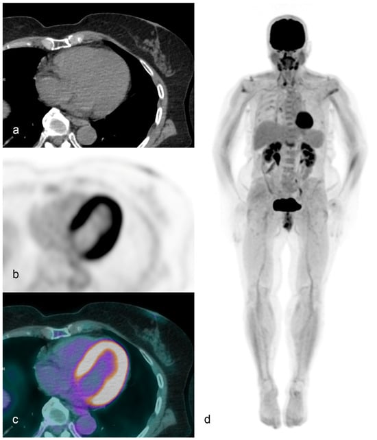
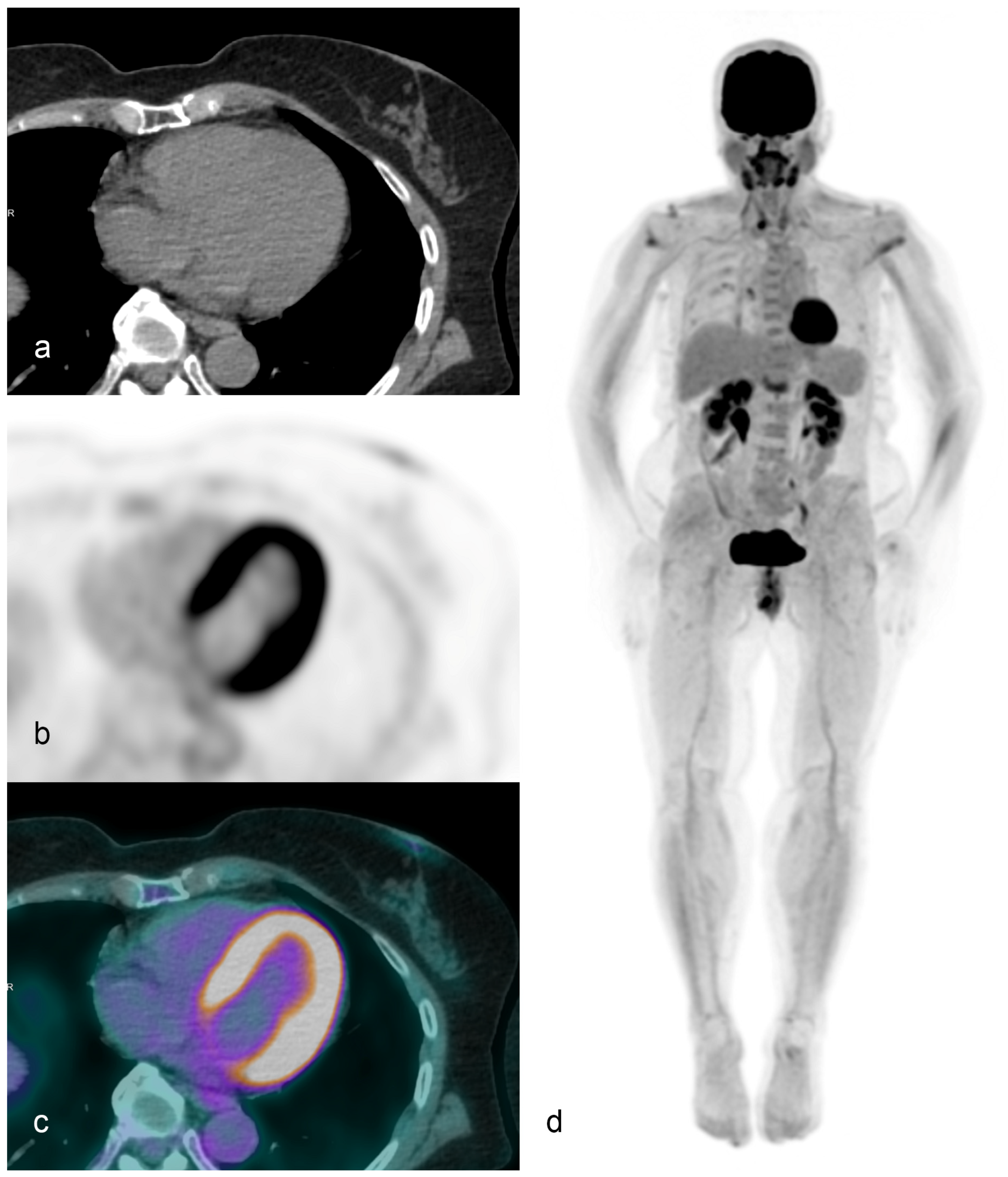
18F-FDG PET remains the main metabolic PET approach in cardiology, but its usefulness is strongly indication-dependent. In ischemic left ventricular dysfunction, its traditional role is the identification of perfusion–metabolism mismatch and residual viable myocardium; in inflammatory disease, especially cardiac sarcoidosis, it is primarily a marker of active inflammatory cell infiltration; in heart transplantation, its role is still preliminary and should not be overstated. Therefore, a single uniformly favorable narrative for FDG PET is no longer justified. The current literature supports a selective rather than indiscriminate use, with stronger evidence in myocardial viability problem-solving and cardiac sarcoidosis than in transplant rejection or generalized heart failure phenotyping [9,17,18,19,20,21]. A representative example of cardiac FDG PET obtained with the total-body uEXPLORER PET/CT system (United Imaging Healthcare, Shanghai, China) is shown in Figure 1. The images demonstrate a regular and homogeneous distribution of myocardial tracer uptake in the left ventricular walls. The use of the uEXPLORER scanner, characterized by a 194 cm axial field of view, allows true total-body acquisition in a single bed position and offers the practical possibility of very short whole-body protocols while maintaining high image quality. In selected patients, this technical configuration may support rapid total-body FDG PET/CT acquisitions, with potential advantages in workflow efficiency and patient comfort [71].
Figure 1.
Representative cardiac 18F-FDG PET/CT acquired on a total-body uEXPLORER scanner. Axial CT image (a), axial PET image (b), fused PET/CT image (c), and whole-body maximum intensity projection (d) are shown. A regular and homogeneous distribution of myocardial 18F-FDG uptake is evident in the left ventricular walls. Owing to the 194 cm axial field of view of the uEXPLORER system, whole-body acquisition can be performed in a single bed position, enabling rapid total-body imaging protocols in selected clinical settings.
3.1. Myocardial Viability and Ischemia
In ischemic cardiomyopathy, FDG PET is conceptually attractive because it identifies dysfunctional myocardium with preserved glucose metabolism, i.e., tissue that may retain the potential for functional recovery after revascularization. However, the interpretation of the contemporary literature is complicated by marked heterogeneity across studies. Available evidence differs substantially in terms of patient selection, referral for revascularization, imaging comparators, and endpoints, ranging from prediction of left ventricular functional recovery to survival, structural reversibility, or multimodality agreement. Therefore, apparently discordant results should not be interpreted as simple contradiction; rather, they reflect the fact that FDG PET may perform differently depending on whether it is used as a metabolic viability marker, a revascularization-selection tool, or one component of an integrated imaging strategy [9,17,18,72].
At a higher level of evidence, the recent review by Kjærulff et al. concluded that preoperative viability PET has not shown consistent predictive value for outcomes after revascularization in chronic ischemic heart failure, and explicitly suggested that alternative PET-derived markers such as myocardial flow reserve may ultimately prove more clinically useful than FDG-based viability imaging alone [9]. This more critical view is supported by the retrospective cohort reported by Luong et al., in which 131 patients were referred for dynamic 82Rb/18F-FDG PET viability testing and 44 subsequently underwent revascularization. In that study, neither static hibernation measures nor dynamic metabolic variables predicted improvement in LVEF, and hibernation also failed to predict survival. Agreement between the PET-defined area of hibernation and the area selected for intervention was also poor, indicating that metabolic viability imaging did not function as a robust standalone predictor of revascularization benefit in routine practice [17].
By contrast, more favorable results have been reported in selected observational cohorts. Park et al. evaluated 53 patients with myocardial scar on SPECT who subsequently underwent PET viability imaging and found that revascularized patients showed improvement in LVEF, GLS, wall-motion score index, and LVEDV, while mortality was lower in the revascularized group than in medically treated patients [18]. However, these findings should be interpreted cautiously, since the study was observational and does not demonstrate that PET-defined viability alone drove outcome differences. Rather, it suggests that FDG PET may still be clinically useful in selected patients undergoing multimodality assessment and therapeutic adjudication [18].
A different perspective is provided by the prospective multimodality study of Kazakauskaite et al., in which 31 patients with previous myocardial infarction and symptomatic heart failure underwent SPECT, FDG PET, and CMR, with 527 segments analyzed. In that study, CMR classified a substantially larger proportion of segments as non-viable than either SPECT or FDG PET, indicating that FDG PET was more permissive in labeling dysfunctional myocardium as metabolically preserved. Patients with greater reversibility also had better exercise capacity, smaller ventricular size, more favorable strain parameters, and smaller infarct burden. These findings do not establish FDG PET as an outcome-defining test, but they support its value within an integrated structural–functional–metabolic framework [72].
Taken together, current FDG PET evidence in ischemic left ventricular dysfunction should be interpreted with caution. Higher-level reviews and recent retrospective cohorts do not support a uniform prognostic or treatment-directing role for FDG viability imaging across all patients considered for revascularization. More favorable findings tend to emerge in selected cohorts, particularly when FDG PET is used in conjunction with other imaging modalities or in clinically unresolved cases. Accordingly, the more positive observational results should be regarded as context-specific or hypothesis-generating rather than as definitive evidence for routine viability-guided decision-making. At present, the literature supports FDG PET primarily as a problem-solving and multimodality tool rather than as a standalone determinant of revascularization benefit [9,17,18,72].
3.2. Cardiac Sarcoidosis
In cardiac sarcoidosis, FDG PET addresses a different biological question: not myocardial viability, but active inflammatory myocardial involvement. The available literature suggests that FDG PET contributes to risk stratification, particularly when abnormal uptake is combined with perfusion abnormalities or involves the right ventricle. However, this evidence remains heterogeneous and is influenced by differences in patient preparation, image interpretation, and comparator modalities [19,21]. Meta-analytic data support a prognostic role for abnormal FDG PET patterns, but they also indicate that the incremental value of PET should be interpreted in relation to cardiac MRI rather than as a universally superior alternative [19,21]. Accordingly, FDG PET in cardiac sarcoidosis is best regarded as a complementary inflammatory imaging tool whose interpretation remains strongly protocol-dependent.
3.3. Metabolic Imaging in Heart Failure
Beyond revascularization-oriented viability assessment, FDG PET may also capture broader aspects of metabolic reserve and ventricular remodeling in ischemic heart failure. However, the evidence base remains largely observational and multimodality-driven, with endpoints ranging from reversibility to mortality prediction rather than from standardized therapeutic algorithms [9,17,72,73]. Therefore, FDG PET in heart failure should currently be interpreted as a selective adjunct for unresolved clinical questions, not as a uniform phenotyping platform across all heart failure populations.
4. 13N-Ammonia PET
13N-ammonia PET remains one of the reference tracers for myocardial perfusion imaging because it combines high image quality with robust kinetic properties for dynamic quantification of myocardial blood flow and myocardial flow reserve. Its main clinical strength is not merely visual detection of perfusion defects, but the possibility of moving from relative perfusion assessment to absolute physiological quantification. This is particularly relevant in multivessel coronary artery disease, balanced ischemia, and microvascular dysfunction, where conventional relative imaging may underestimate disease burden. Recent reviews consistently frame 13N-ammonia PET as a quantitative platform rather than a simple perfusion scan, and this distinction is central to its present clinical value [2,22,23].
4.1. Myocardial Perfusion and Coronary Artery Disease
In patients with suspected or known coronary artery disease, 13N-ammonia PET is primarily used to identify ischemia and to quantify the functional consequences of epicardial and microvascular disease. Contemporary reviews emphasize that both absolute stress MBF and MFR can identify myocardial territories subtended by hemodynamically significant stenoses and improve risk stratification beyond visual perfusion assessment alone [2,22,23]. In this regard, the tracer is best viewed as an established quantitative perfusion platform rather than as a purely visual test.
4.2. Quantification of Myocardial Blood Flow
The real added value of 13N-ammonia PET lies in dynamic acquisition and kinetic modeling. Absolute MBF quantification requires dynamic multiframes, estimation of tracer extraction from blood to myocardium, and conversion of kinetic parameters into physiological flow values. Modern PET systems, list-mode acquisition, and dedicated software have made this workflow increasingly feasible in clinical practice, but the method remains technically demanding and strongly dependent on acquisition and post-processing quality [2,23,24].
A practical step toward wider adoption is simplification of quantification protocols. In the comparative study by Chang et al., including 14 healthy volunteers and 23 clinical patients, global and regional MBF and MFR derived from a simple retention model showed strong correlations with those obtained from the standard two-compartment model. The authors also proposed normal reference values for a short 13N-ammonia retention protocol. This is methodologically important because it addresses one of the main barriers to routine use of quantitative PET, namely the complexity and duration of standard kinetic analysis [24].
4.3. Use in Heart Failure
The role of 13N-ammonia PET in heart failure is more nuanced than in coronary artery disease. The evidence base is thinner and often centers on ischemic cardiomyopathy or left ventricular dysfunction rather than on heart failure as a broad syndrome. In this context, the tracer is useful less for a generic heart failure assessment and more for characterizing coronary flow capacity, residual ischemic burden, and physiological reserve in dysfunctional ventricles. Bober et al. explicitly framed PET stress testing with coronary flow capacity as a way to rethink evaluation of patients with coronary artery disease and left ventricular dysfunction, rather than as a standalone heart failure tool [25].
A critical study here is the PAREPET sub-analysis by Wang et al., which included 174 paired dynamic 11C-meta-hydroxyephedrine and 13N-ammonia PET scans in patients with ischemic cardiomyopathy and LVEF ≤ 35%. During follow-up, 13N-ammonia-derived MBF did not improve sudden cardiac arrest risk discrimination compared with HED uptake defect scores alone [26]. This is an important negative result because it argues against overinterpreting quantitative perfusion metrics as universally informative prognostic markers in advanced ventricular dysfunction.
4.4. Additional Clinical Applications
Beyond its established role in perfusion quantification, several additional applications of 13N-ammonia PET have been explored. However, these should be interpreted as extensions of the technique rather than as equally validated clinical indications, since most available evidence remains based on modest single-center cohorts.
In the study by Kiko et al., 52 patients with suspected coronary artery disease underwent simultaneous 13N-ammonia PET/MRI during vasodilator stress. Although global left ventricular function remained stable in most patients, the few individuals who showed stress-related LVEF decline also had lower CFR and more extensive vessel disease [27]. Likewise, Maurer et al. demonstrated good agreement between gated 13N-ammonia PET and CMR for LVEF, but less robust agreement for absolute ventricular volumes [28]. These studies support technical feasibility, yet their sample size limits broader clinical inference.
Feature-tracking and strain-based approaches further widen the spectrum of potential applications. Kawakubo et al. showed that strain parameters derived from a single 13N-ammonia PET scan differed according to abnormal MFR status [29], while Endo et al. and Yamamoto et al. reported associations between MFR, myocardial strain parameters, and adverse outcomes in hybrid PET/MRI cohorts [30,31]. These data are conceptually interesting, but they remain largely hypothesis-generating and have not yet established a standardized clinical role.
4.5. Challenges and Future Directions
Despite its strengths, 13N-ammonia PET remains constrained by logistics and standardization issues. Its short half-life requires on-site cyclotron production and radiochemistry support, which limits dissemination compared with generator-based or longer-lived tracers. In addition, image quality and quantitative reliability remain sensitive to motion, attenuation artifacts, misregistration, extracardiac activity, and protocol heterogeneity [32].
The next phase of development is likely to be driven by workflow simplification and computational correction. Shorter protocols, simplified retention models, mini-cyclotron solutions, and stronger harmonization of acquisition and post-processing are obvious practical priorities. Artificial intelligence is also beginning to enter this space. Sun et al. demonstrated the feasibility of generating attenuation-corrected rest and stress static or dynamic 13N-ammonia PET images from non-attenuation-corrected data using a generative adversarial network, suggesting that some current technical bottlenecks may eventually be addressed algorithmically [24,33].
Taken together, current evidence supports 13N-ammonia PET primarily as a robust quantitative platform for myocardial perfusion assessment and coronary flow evaluation. By contrast, its use for functional multiparametric modeling, strain analysis, and broader heart failure phenotyping remains promising but still depends on relatively small and center-specific studies, which currently limit external validity and routine clinical generalization.
5. Rubidium-82 PET
Rubidium-82 PET is one of the most widely used tracers for myocardial perfusion imaging because it is generator-produced and therefore more accessible than cyclotron-dependent tracers. Its main clinical advantage is the possibility of combining rapid perfusion imaging with quantitative assessment of myocardial blood flow and myocardial flow reserve in routine practice. However, its strengths should not be overstated. Although 82Rb PET is highly practical and clinically mature, its lower extraction characteristics compared with high-extraction tracers mean that interpretation of diffuse or global disease may sometimes be less straightforward, especially when subtle reductions in flow need to be distinguished from technical or biological variability [35].
The evidence base for 82Rb PET is broader than for most other cardiac tracers and includes registry-level outcome data as well as multiple methodologic studies. Nevertheless, not all endpoints are equally robust. The strongest clinical message relates to its role in quantitative perfusion assessment in coronary artery disease and ischemia with non-obstructive coronary arteries, whereas software-dependent quantification thresholds and dose-related technical variability still limit full standardization.
5.1. Myocardial Perfusion and Coronary Artery Disease
In coronary artery disease, the main strength of 82Rb PET lies in the combined assessment of relative perfusion abnormalities and coronary vasodilator function. This is clinically important because perfusion defects alone may underestimate balanced multivessel ischemia or diffuse non-obstructive disease, whereas MFR and MBF provide a more physiological description of coronary circulatory impairment [35].
The diagnostic literature suggests that 82Rb PET can extract clinically relevant information beyond conventional perfusion scoring. In a retrospective angiographic study of 105 patients, Van Tosh et al. showed that regional asynchrony derived from 82Rb PET/CT performed better than MFR in identifying both multivessel disease and individual stenosed territories [36]. In non-obstructive disease, Zampella et al. reported that both total perfusion defect and MFR independently predicted events in patients with ischemia and non-obstructive coronary arteries, with the highest risk observed when increased defect burden and reduced MFR coexisted [37].
The same principle extends to revascularization-oriented assessment. Assante et al. showed that worsening of either ischemic perfusion defect or MFR after coronary revascularization was associated with a higher event rate, emphasizing that technically successful intervention does not necessarily normalize physiology [38]. In the Danish multicenter registry, revascularization was associated with better outcomes only in patients with impaired MFR, suggesting that 82Rb PET may help identify a subgroup more likely to derive prognostic benefit from revascularization [39]. These studies support a real clinical role for 82Rb PET, but they also remain observational and should be interpreted as decision-support evidence rather than as definitive treatment algorithms.
Among prospective quantitative studies, Dietz et al. showed that reduced stress MBF, reduced MFR, and impaired myocardial flow capacity were all associated with adverse outcomes, but only reduced stress MBF remained independently predictive after mutual adjustment [40]. This is one of the more clinically useful studies in the current 82Rb literature because it addresses which quantitative metric may carry the greatest independent prognostic information.
5.2. Quantification of Myocardial Blood Flow
The differentiating feature of 82Rb PET is quantitative MBF and MFR assessment. This allows a transition from relative perfusion imaging to physiological characterization of the coronary circulation, which is especially important in diffuse coronary disease, microvascular dysfunction, and borderline or discordant cases. However, this is also the most methodologically fragile part of the technique, because quantitative outputs are highly dependent on software, motion correction, dose administration, and low-end calibration [35,41,42].
Liu et al. supported the feasibility of highly automated quantification using the Yale-MQ software, but Byrne et al. demonstrated that repeatability and absolute MFR values varied across commercial platforms [41,43]. Bober et al. further showed that low-end resting MBF values in regions of transmural scar varied substantially by software package, indicating that biologically plausible quantification is not guaranteed across platforms [44].
Dose prescription also matters. Arida-Moody et al. showed that BMI-adjusted dosing reduced scanner saturation while maintaining image quality and yielding only modest biases in MBF and MFR [42]. Tavoosi et al. showed that exponential dosing improved consistency of image quality across a broad range of body weights [45]. Together, these data indicate that 82Rb PET quantification is clinically powerful but methodologically conditional.
5.3. Clinical Strengths and Methodological Constraints
Taken together, 82Rb PET is currently one of the most clinically mature and pragmatically deployable quantitative perfusion tracers in cardiology. Its strongest evidence base concerns coronary artery disease, INOCA, and physiologic risk stratification. However, the interpretation of quantitative thresholds remains center-sensitive, and the apparent robustness of the technique should not obscure the substantial influence of software, protocol design, and tracer administration on the final measurements.
6. Novel Radiopharmaceuticals
6.1. 68Ga-FAPI PET
68Ga-FAPI PET targets fibroblast activation protein, which is expressed in activated fibroblasts rather than in mature fibrosis. In cardiovascular disease, FAPI uptake should therefore be interpreted mainly as a marker of active fibrotic remodeling, not as a direct surrogate of established scar burden [46,47,48].
The most coherent current application is heart failure and myocardial remodeling. Song et al. combined experimental work with preliminary human imaging and showed that myocardial 68Ga-FAPI uptake peaked early after injury, whereas histologic fibrosis became more evident later [47]. Similarly, Wang et al. demonstrated in a pressure-overload heart failure model that myocardial 68Ga-FAPI-04 signal correlated with FAP-positive tissue and predicted later functional deterioration [49]. These studies support biological plausibility but also illustrate that much of the evidence remains preclinical or early translational.
Cardiac amyloidosis is an additional but still exploratory application. In biopsy-proven AL amyloidosis, Wang et al. reported visible myocardial 68Ga-FAPI-04 uptake in most patients and significant correlations with markers of disease severity [50]. By contrast, evidence in ATTR remains limited to isolated reports such as the case described by Kibar et al. [51]. Accordingly, 68Ga-FAPI PET should currently be regarded as promising but exploratory, with clear biologic interest but insufficient clinical validation.
6.2. 18F-Flurpiridaz PET
18F-flurpiridaz is a mitochondrial complex I-targeting perfusion tracer developed to improve PET myocardial perfusion imaging. Its main practical advantages are the physical properties of fluorine-18, which support high-resolution imaging, longer distribution time, and exercise-capable protocols, together with favorable myocardial retention and a strong basis for absolute flow quantification [52,53,54,55].
6.2.1. Clinical Applications
The clearest clinical application of 18F-flurpiridaz is myocardial perfusion imaging for suspected coronary artery disease. In first-in-human evaluation, flurpiridaz showed high and sustained myocardial retention after injection and no major safety concerns [53]. Phase II and phase III studies then showed that flurpiridaz PET could effectively detect coronary artery disease and compare favorably with conventional myocardial perfusion imaging techniques [52,54]. The flurpiridaz 301 trial subset further suggested that stress MBF and MFR derived from dynamic flurpiridaz imaging add diagnostic information beyond patient characteristics and relative perfusion analysis alone [55].
6.2.2. Challenges and Future Directions
Despite this favorable development pathway, 18F-flurpiridaz is not yet a fully settled clinical standard. The second phase III study was designed specifically to strengthen evidence in suspected coronary artery disease and clinically relevant subgroups such as women, obese patients, and diabetic patients [56]. This in itself indicates that broader validation was still considered necessary. Accordingly, flurpiridaz should currently be regarded as the most mature among emerging perfusion tracers, but its ultimate routine role will depend on further real-world implementation and on demonstration of incremental value over already established PET perfusion workflows.
6.3. 18F-FDOPA
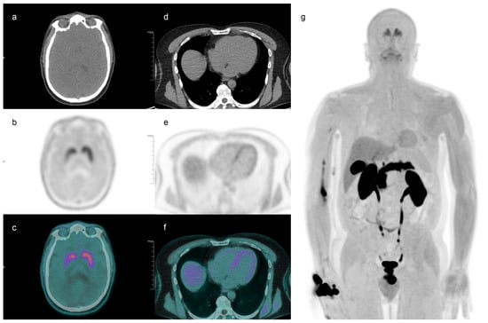
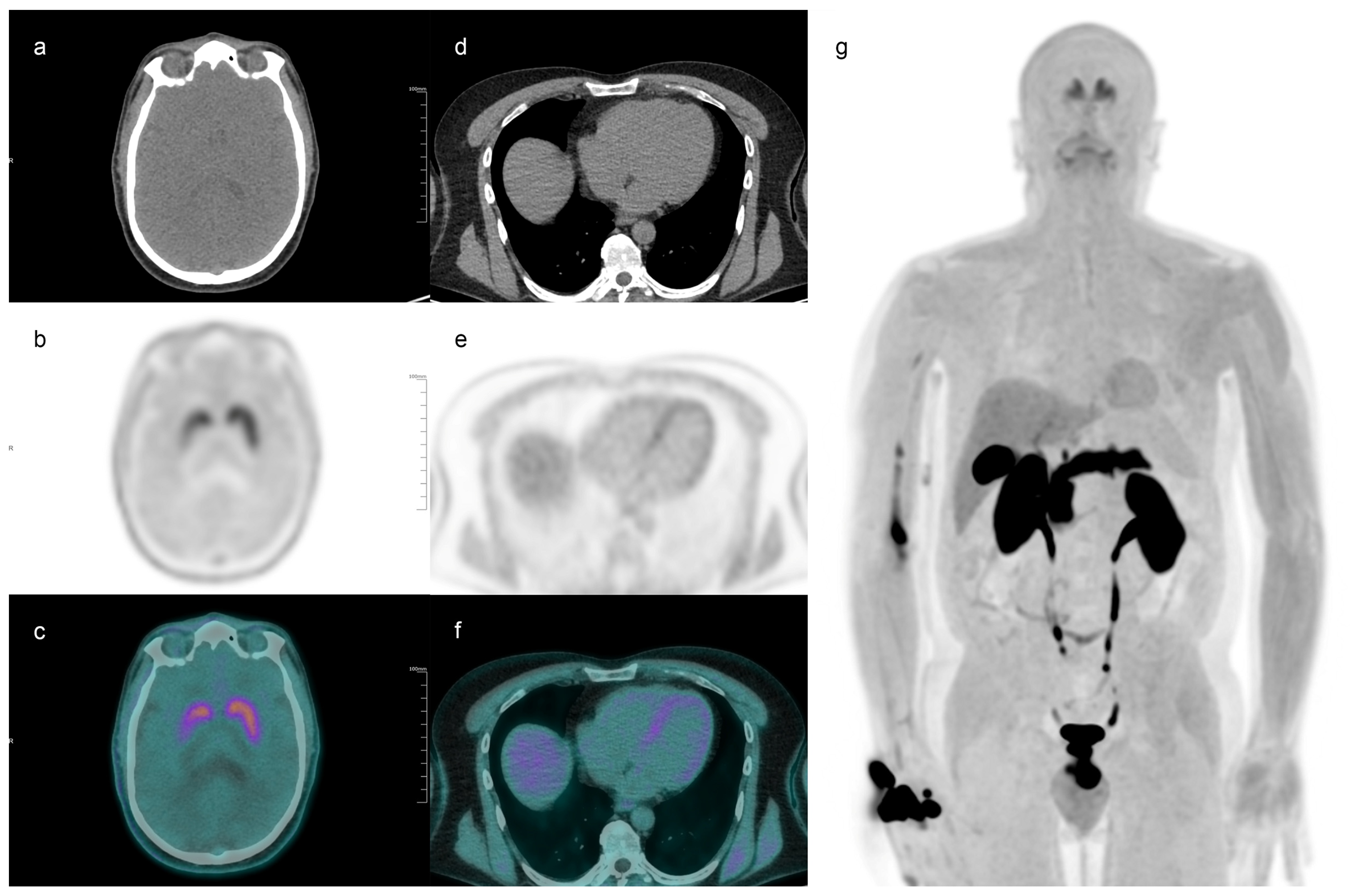
18F-FDOPA occupies a much more niche position in cardiac PET. It is not a perfusion tracer and should not be presented as such. Its emerging cardiac role is linked to imaging of myocardial sympathetic innervation and autonomic dysfunction, which makes it mechanistically closer to innervation tracers than to conventional PET perfusion agents [57,58]. A representative 18F-FDOPA PET/CT study acquired on the uEXPLORER system in a patient with Parkinson’s disease is shown in Figure 2. In addition to the characteristic striatal uptake pattern, the images demonstrate reduced ventricular myocardial uptake of the radiotracer, consistent with impaired cardiac sympathetic innervation [71].
Figure 2.
Representative 18F-FDOPA PET/CT acquired on the uEXPLORER system in a patient with Parkinson’s disease. Axial brain CT, PET, and fused PET/CT images are shown in panels (a–c), axial thoracic CT, PET, and fused PET/CT images in panels (d–f), and whole-body maximum intensity projection in panel (g). The study demonstrates reduced radiotracer uptake in the ventricular myocardium, in keeping with cardiac sympathetic dysfunction in Parkinson’s disease.
The currently available evidence is limited but consistent. In patients with autonomic dysfunction and dopaminergic impairment, Goyal et al. showed that myocardial 18F-FDOPA uptake was significantly lower than in controls, with the myocardium-to-mediastinum ratio yielding an AUC of 0.941 for discrimination [57]. In heart failure, Thanigainathan et al. reported reduced myocardial uptake compared with controls and suggested that regional heterogeneity might be associated with worse clinical status [58]. These findings are interesting, but the evidence base remains small and centered on feasibility rather than routine clinical applicability. Accordingly, 18F-FDOPA should currently be regarded as an exploratory tracer for myocardial sympathetic dysfunction rather than as a broadly applicable cardiac PET agent.
6.4. Radiopharmaceuticals for Cardiac Amyloidosis Imaging
Cardiac amyloidosis is one of the few areas in which truly target-specific PET radiopharmaceuticals may offer information different from traditional perfusion or fibrosis tracers. The main agents studied so far are amyloid-binding tracers such as 11C-PIB, 18F-florbetapir, 18F-florbetaben, and 18F-flutemetamol. In contrast to bone-seeking tracers, these compounds aim to visualize amyloid burden more directly [59,60,61,62,63,64,65,66,67,68,69,70].
The aggregate diagnostic literature is encouraging, although still based on relatively small studies and heterogeneous cohorts. In the meta-analysis by Kim et al., amyloid PET showed high pooled sensitivity and specificity for cardiac amyloidosis [59]. A second meta-analysis by Kim et al. reported similarly high performance for amyloid PET but substantially weaker sensitivity for 18F-NaF [60]. These studies support the diagnostic validity of amyloid PET, but they also reflect the limitations of a small evidence base composed of heterogeneous tracers and modest sample sizes.
Among individual tracers, 18F-florbetapir is the most established in the early cardiac PET literature, with pilot imaging data from Dorbala et al. and autoradiographic support from Park et al. [61,62]. 18F-florbetaben has also shown promising pilot and comparative results, including work by Law et al., Kircher et al., and Genovesi et al. [63,64,65]. The 11C-PIB literature is biologically compelling, but its broader dissemination is limited by the short half-life of carbon-11 [66,69,70]. More recently, 18F-flutemetamol has also entered the field, although experience remains comparatively limited [67,68].
From a clinical perspective, the main limitation of amyloid PET is not biological plausibility but positioning within existing diagnostic pathways. Bone scintigraphy retains a major practical role, especially in non-biopsy ATTR diagnosis, and CMR remains highly informative for tissue characterization. Therefore, PET has not displaced these modalities. The most realistic current interpretation is that amyloid PET may become useful in selected scenarios such as equivocal conventional imaging, direct amyloid burden assessment, early disease characterization, subtype clarification, or longitudinal treatment monitoring. However, the available evidence is still not strong enough to support routine first-line use in clinical cardiology.
7. Integrative Synthesis and Clinical Positioning
The preceding sections suggest that cardiac PET cannot be treated as a single homogeneous technology. Rather, the evidence base differs markedly according to tracer, biological target, and clinical scenario. Established perfusion tracers such as 13N-ammonia and 82Rb are supported by the most robust data for coronary physiology and quantitative myocardial perfusion assessment. FDG PET remains highly relevant, but mainly in selected settings such as viability problem-solving and inflammatory cardiac disease, with more limited evidence for routine treatment-direction across broad populations. Novel tracers such as FAPI, FDOPA, and amyloid PET compounds are biologically interesting and potentially transformative, but they remain uneven in maturity and clinical readiness.
A practical implication is that tracer selection should be driven by the underlying clinical question rather than by generalized enthusiasm for PET. In suspected or known coronary artery disease, quantitative perfusion PET currently provides the clearest clinically actionable information. In ischemic left ventricular dysfunction, FDG PET should be interpreted within a multimodality framework rather than as a standalone arbiter of revascularization benefit. In cardiac amyloidosis and emerging remodeling or autonomic applications, PET should presently be positioned as a complementary or selective tool rather than as a universal replacement for established pathways.
Across tracers, several methodological constraints recur: software dependence, protocol variability, acquisition-related bias, limited inter-center standardization, and uneven external validity of small or single-center studies. These issues explain why apparently impressive tracer-specific findings do not automatically translate into routine clinical adoption. In this sense, the central challenge of cardiac PET is not only biologic innovation, but also methodological stabilization.
To facilitate clinical integration, Table 2 summarizes a pragmatic tracer-selection framework according to the main clinical scenarios discussed in this review.
Table 2.
Suggested clinical positioning of cardiac PET tracers according to the principal clinical scenario.
8. Conclusions
Cardiac PET has evolved from a niche imaging technique into a broad quantitative and molecular platform with relevance across ischemic, inflammatory, autonomic, and infiltrative cardiovascular disease. The field is strongest where PET adds physiologic or biologic information that competing modalities cannot easily provide, particularly in quantitative myocardial perfusion imaging and selected problem-solving indications.
At the same time, the current literature does not support a uniform view of PET as a methodologically neutral or universally superior technology. The maturity of evidence differs substantially across tracers, and broader implementation remains constrained by issues of standardization, reproducibility, software dependence, and incomplete external validation.
As a critical narrative review based on a structured search of recent literature, this article cannot provide a formal evidence-based ranking of all tracers or indications. Nevertheless, the available data support a selective, clinically question-driven use of cardiac PET and suggest that future progress will depend as much on harmonized workflows and robust validation as on the development of new radiopharmaceuticals.
Author Contributions
Conceptualization, A.C. (Agostino Chiaravalloti) and L.V.; methodology, A.C. (Agostino Chiaravalloti), L.V. and M.A.P.; investigation, A.C. (Agostino Chiaravalloti), L.V., M.A.P., A.C. (Antonio Chiaravalloti) and D.D.B.; data curation, L.V. and M.A.P.; writing—original draft preparation, A.C. (Agostino Chiaravalloti) and L.V.; writing—review and editing, A.C. (Agostino Chiaravalloti), L.V., M.A.P., A.C. (Antonio Chiaravalloti) and D.D.B.; visualization, M.A.P. and D.D.B.; supervision, A.C. (Agostino Chiaravalloti); project administration, A.C. (Agostino Chiaravalloti). All authors have read and agreed to the published version of the manuscript.
Funding
This research received no external funding.
Institutional Review Board Statement
Not applicable.
Informed Consent Statement
Not applicable.
Data Availability Statement
No new data were created or analyzed in this study. Data sharing is not applicable to this article.
Acknowledgments
The authors acknowledge the support of SCITER—ETS (www.sciter.it, accessed on 6 April 2026), a non-profit association based in Rome and engaged in promoting research and innovation in the diagnosis and treatment of chronic and debilitating diseases.
Conflicts of Interest
The authors declare no conflicts of interest.
Abbreviations
The following abbreviations are used in this manuscript:
| AI | Artificial intelligence |
| AL | Amyloid light-chain |
| AUC | Area under the curve |
| ATTR | Transthyretin amyloidosis |
| CAD | Coronary artery disease |
| CFR | Coronary flow reserve |
| CMR | Cardiovascular magnetic resonance |
| CNN | Convolutional neural network |
| CT | Computed tomography |
| DPD | 3,3-diphosphono-1,2-propanodicarboxylic acid |
| EF | Ejection fraction |
| FAP | Fibroblast activation protein |
| FAPI | Fibroblast activation protein inhibitor |
| FDG | Fluorodeoxyglucose |
| FDOPA | Fluorodopa |
| FFR | Fractional flow reserve |
| GLS | Global longitudinal strain |
| HED | meta-Hydroxyephedrine |
| INOCA | Ischemia with non-obstructive coronary arteries |
| ITPD | Ischemic total perfusion defect |
| LGE-CMR | Late gadolinium enhancement cardiovascular magnetic resonance |
| LV | Left ventricle/left ventricular |
| LVEDV | Left ventricular end-diastolic volume |
| LVEF | Left ventricular ejection fraction |
| MACE | Major adverse cardiac events |
| MBF | Myocardial blood flow |
| MFR | Myocardial flow reserve |
| MRI | Magnetic resonance imaging |
| PET | Positron emission tomography |
| PIB | Pittsburgh compound B |
| ROC | Receiver operating characteristic |
| RV | Right ventricle/right ventricular |
| SiPM | Silicon photomultiplier |
| SPECT | Single-photon emission computed tomography |
| TPD | Total perfusion defect |
| NT-proBNP | N-terminal pro-B-type natriuretic peptide |
References
- Fernandes, J.; Ferreira, M.J.; Leite, L. Quantificação do fluxo sanguíneo miocárdico por tomografia por emissão de positrões—Atualização. Rev. Port. De. Cardiol. 2020, 39, 37–46. [Google Scholar] [CrossRef] [Scilit]
- Nammas, W.; Maaniitty, T.; Knuuti, J.; Saraste, A. Cardiac perfusion by positron emission tomography. Clin. Physiol. Funct. Imaging 2021, 41, 385–400. [Google Scholar] [CrossRef] [Scilit]
- Tarkin, J.M.; Ćorović, A.; Wall, C.; Gopalan, D.; Rudd, J.H. Positron emission tomography imaging in cardiovascular disease. Heart 2020, 106, 1712–1718. [Google Scholar] [CrossRef] [Scilit] [PubMed]
- Johnson, N.P.; Gould, K.L. How Do PET Myocardial Blood Flow Reserve and FFR Differ? Curr. Cardiol. Rep. 2020, 22, 20. [Google Scholar] [CrossRef] [Scilit]
- Cho, S.G.; Lee, S.J.; Na, M.H.; Choi, Y.Y.; Bom, H.H. Comparison of diagnostic accuracy of PET-derived myocardial blood flow parameters: A meta-analysis. J. Nucl. Cardiol. 2020, 27, 1955–1966. [Google Scholar] [CrossRef] [Scilit] [PubMed]
- Giubbini, R.; Milan, E. Current Role of Myocardial Blood Flow Quantification with PET/CT in the Management of Coronary Artery Disease. Int. J. Cardiovasc. Sci. 2024, 37, e20240115. [Google Scholar] [CrossRef] [Scilit]
- Gould, K.L.; Bui, L.; Kitkungvan, D.; Patel, M.B. Reliability and Reproducibility of Absolute Myocardial Blood Flow: Does It Depend on the PET/CT Technology, the Vasodilator, and/or the Software? Curr. Cardiol. Rep. 2021, 23, 12. [Google Scholar] [CrossRef] [Scilit] [PubMed]
- Klein, R.; Celiker-Guler, E.; Rotstein, B.H.; deKemp, R.A. PET and SPECT Tracers for Myocardial Perfusion Imaging. Semin. Nucl. Med. 2020, 50, 208–218. [Google Scholar] [CrossRef] [Scilit]
- Kjærulff, M.L.G.; Madsen, S.; Luong, T.V.; Pryds, K.; Nielsen, R.R.; Gormsen, L.C. Current Role of Myocardial Viability Imaging Using Positron Emission Tomography in Patients with Chronic Ischemic Heart Failure and Left Ventricular Dysfunction. Curr. Cardiovasc. Imaging Rep. 2023, 16, 83–92. [Google Scholar] [CrossRef] [Scilit]
- Madsen, S.; Dias, A.H.; Lauritsen, K.M.; Bouchelouche, K.; Tolbod, L.P.; Gormsen, L.C. Myocardial Viability Testing by Positron Emission Tomography: Basic Concepts, Mini-Review of the Literature and Experience from a Tertiary PET Center. Semin. Nucl. Med. 2020, 50, 248–259. [Google Scholar] [CrossRef] [Scilit]
- Beitzke, D.; Rasul, S.; Lassen, M.L.; Pichler, V.; Senn, D.; Stelzmüller, M.E.; Nolz, R.; Loewe, C.; Hacker, M. Assessment of Myocardial Viability in Ischemic Heart Disease by PET/MRI: Comparison of Left Ventricular Perfusion, Hibernation, and Scar Burden. Acad. Radiol. 2020, 27, 188–197. [Google Scholar] [CrossRef] [Scilit]
- Kessler, L.; Rischpler, C. Single Tracer Combined Imaging: The Role of PET/MRI from Research Domain to Clinical Arena. Curr. Cardiovasc. Imaging Rep. 2020, 13, 22. [Google Scholar] [CrossRef] [Scilit]
- Manabe, O.; Oyama-Manabe, N.; Tamaki, N. Positron emission tomography/MRI for cardiac diseases assessment. Br. J. Radiol. 2020, 93, 20190836. [Google Scholar] [CrossRef] [Scilit] [PubMed]
- Bors, S.; Abler, D.; Dietz, M.; Andrearczyk, V.; Fageot, J.; Nicod-Lalonde, M.; Schaefer, N.; DeKemp, R.; Kamani, C.H.; Prior, J.O.; et al. Comparing various AI approaches to traditional quantitative assessment of the myocardial perfusion in [(82)Rb] PET for MACE prediction. Sci. Rep. 2024, 14, 9644. [Google Scholar] [CrossRef] [Scilit] [PubMed]
- Jafaritadi, M.; Teuho, J.; Lehtonen, E.; Klén, R.; Saraste, A.; Levin, C.S. Deep generative denoising networks enhance quality and accuracy of gated cardiac PET data. Ann. Nucl. Med. 2024, 38, 775–788. [Google Scholar] [CrossRef] [Scilit] [PubMed]
- Popescu, C.; Laudicella, R.; Baldari, S.; Alongi, P.; Burger, I.; Comelli, A.; Caobelli, F. PET-based artificial intelligence applications in cardiac nuclear medicine. Swiss Med. Wkly. 2022, 152, w30123. [Google Scholar] [CrossRef] [Scilit]
- Luong, T.V.; Ebbehoj, A.; Kjaerulff, M.L.G.; Nielsen, R.; Nielsen, P.H.; Christiansen, E.H.; Tolbod, L.P.; Søndergaard, E.; Gormsen, L.C. Clinical use of cardiac 18 F-FDG viability PET: A retrospective study of 44 patients undergoing post-test revascularization. Int. J. Cardiovasc. Imaging 2022, 38, 2447–2458. [Google Scholar] [CrossRef] [Scilit]
- Park, J.S.; Lee, J.H.; Hong, C.M.; Park, B.E.; Park, Y.J.; Kim, H.N.; Kim, N.; Jang, S.Y.; Bae, M.H.; Yang, D.H.; et al. Impact of Positron Emission Tomography Viability Imaging-Guided Revascularizations on Clinical Outcomes in Patients with Myocardial Scar on Single-Photon Emission Computed Tomography Scans. J. Korean Med. Sci. 2023, 38, e399. [Google Scholar] [CrossRef] [Scilit]
- Aitken, M.; Davidson, M.; Chan, M.V.; Urzua Fresno, C.; Vasquez, L.I.; Huo, Y.R.; McAllister, B.J.; Broncano, J.; Thavendiranathan, P.; McInnes, M.D.F.; et al. Prognostic Value of Cardiac MRI and FDG PET in Cardiac Sarcoidosis: A Systematic Review and Meta-Analysis. Radiology 2023, 307, e222483. [Google Scholar] [CrossRef] [Scilit]
- Dar, O.; Dulay, M.S.; Riesgo-Gil, F.; Morley-Smith, A.; Brookes, P.; Lyster, H.; Rice, A.; Underwood, S.R.; Dunning, J.; Wechalekar, K. Cardiac transplant rejection assessment with 18F-FDG PET-CT: Initial single-centre experience for diagnosis and management. EJNMMI Rep. 2024, 8, 9. [Google Scholar] [CrossRef] [Scilit]
- Kafil, T.S.; Shaikh, O.M.; Fanous, Y.; Benjamen, J.; Hashmi, M.M.; Jawad, A.; Dahrouj, T.; Abazid, R.M.; Swiha, M.; Romsa, J.; et al. Risk Stratification in Cardiac Sarcoidosis with Cardiac Positron Emission Tomography: A Systematic Review and Meta-Analysis. JACC Cardiovasc. Imaging 2024, 17, 1079–1097. [Google Scholar] [CrossRef] [Scilit]
- Miura, S.; Naya, M.; Kumamaru, H.; Ando, A.; Miyazaki, C.; Yamashita, T. Prognostic Value of Modified Coronary Flow Capacity by 13N-Ammonia Myocardial Perfusion Positron Emission Tomography in Patients without Obstructive Coronary Arteries. J. Cardiol. 2022, 79, 247–256. [Google Scholar] [CrossRef] [Scilit]
- Sohn, J.H.; Behr, S.C.; Hernandez Pampaloni, M.; Seo, Y. Quantitative Assessment of Myocardial Ischemia with Positron Emission Tomography. J. Thorac. Imaging 2023, 38, 247–259. [Google Scholar] [CrossRef] [Scilit]
- Chang, C.Y.; Hung, G.U.; Hsu, B.; Yang, B.H.; Chang, C.W.; Hu, L.H.; Huang, W.S.; Wang, H.E.; Wu, T.C.; Liu, R.S. Simplified quantification of (13)N-ammonia PET myocardial blood flow: A comparative study with the standard compartment model to facilitate clinical use. J. Nucl. Cardiol. 2020, 27, 819–828. [Google Scholar] [CrossRef] [Scilit]
- Bober, R.M.; Milani, R.V.; Krim, S.R.; Morin, D.P. PET Stress Testing with Coronary Flow Capacity in the Evaluation of Patients with Coronary Artery Disease and Left Ventricular Dysfunction: Rethinking the Current Paradigm. Curr. Cardiol. Rep. 2021, 23, 50. [Google Scholar] [CrossRef] [Scilit] [PubMed]
- Wang, J.Z.; Zelt, J.G.E.; Kaps, N.; Lavallee, A.; Renaud, J.M.; Rotstein, B.; Beanlands, R.S.B.; Fallavollita, J.A.; Canty, J.M., Jr.; deKemp, R.A. Does quantification of [(11)C]meta-hydroxyephedrine and [(13)N]ammonia kinetics improve risk stratification in ischemic cardiomyopathy. J. Nucl. Cardiol. 2022, 29, 413–425. [Google Scholar] [CrossRef] [Scilit] [PubMed]
- Kiko, T.; Yokokawa, T.; Masuda, A.; Misaka, T.; Yamada, S.; Kaneshiro, T.; Oikawa, M.; Yoshihisa, A.; Nakazato, K.; Takeishi, Y. Simultaneous assessment of coronary flow reserve and left ventricular function during vasodilator stress evaluated by (13)N-ammonia hybrid PET/MRI. Clin. Radiol. 2021, 76, 472.e471–472.e479. [Google Scholar] [CrossRef] [Scilit]
- Maurer, A.; Sustar, A.; Giannopoulos, A.A.; Grünig, H.; Bakula, A.; Patriki, D.; von Felten, E.; Messerli, M.; Pazhenkottil, A.P.; Gebhard, C.; et al. Left ventricular function and volumes from gated [(13)N]-ammonia positron emission tomography myocardial perfusion imaging: A prospective head-to-head comparison against CMR using a hybrid PET/MR device. J. Nucl. Cardiol. 2023, 30, 616–625. [Google Scholar] [CrossRef] [Scilit] [PubMed]
- Kawakubo, M.; Nagao, M.; Yamamoto, A.; Nakao, R.; Matsuo, Y.; Fukushim, K.; Watanabe, E.; Sakai, A.; Sasaki, M.; Sakai, S. (13)N-ammonia positron emission tomography-derived endocardial strain for the assessment of ischemia using feature-tracking in high-resolution cine imaging. J. Nucl. Cardiol. 2022, 29, 2103–2114. [Google Scholar] [CrossRef] [Scilit]
- Endo, K.; Kiko, T.; Yamakuni, R.; Misaka, T.; Yamaki, T.; Nakazato, K.; Fukushima, K.; Takeishi, Y. Prognostic Value of Simultaneous Analysis with Myocardial Flow Reserve and Right Ventricular Strain by Hybrid (13)N-Ammonia Positron Emission Tomography/Magnetic Resonance Imaging in Coronary Artery Disease. Int. Heart J. 2022, 63, 1063–1069. [Google Scholar] [CrossRef] [Scilit]
- Yamamoto, A.; Nagao, M.; Kawakubo, M.; Ando, K.; Nakao, R.; Matsuo, Y.; Sakai, A.; Kaneko, K.; Momose, M.; Sakai, S.; et al. Prediction of cardiovascular events using myocardial strain ratio derived from (13)N-ammonia positron emission tomography. Eur. Radiol. 2023, 33, 3889–3896. [Google Scholar] [CrossRef] [Scilit]
- Alnabelsi, T.; Thakkar, A.; Ahmed, A.I.; Han, Y.; Al-Mallah, M.H. PET/CT Myocardial Perfusion Imaging Acquisition and Processing: Ten Tips and Tricks to Help You Succeed. Curr. Cardiol. Rep. 2021, 23, 39. [Google Scholar] [CrossRef] [Scilit] [PubMed]
- Sun, H.; Wang, F.; Yang, Y.; Hong, X.; Xu, W.; Wang, S.; Mok, G.S.P.; Lu, L. Transfer learning-based attenuation correction for static and dynamic cardiac PET using a generative adversarial network. Eur. J. Nucl. Med. Mol. Imaging 2023, 50, 3630–3646. [Google Scholar] [CrossRef] [Scilit] [PubMed]
- von Felten, E.; Benz, D.C.; Benetos, G.; Baehler, J.; Patriki, D.; Rampidis, G.P.; Giannopoulos, A.A.; Bakula, A.; Gräni, C.; Pazhenkottil, A.P.; et al. Prognostic value of regional myocardial flow reserve derived from (13)N-ammonia positron emission tomography in patients with suspected coronary artery disease. Eur. J. Nucl. Med. Mol. Imaging 2021, 49, 311–320. [Google Scholar] [CrossRef] [Scilit] [PubMed]
- Sciagrà, R.; Lubberink, M.; Hyafil, F.; Saraste, A.; Slart, R.; Agostini, D.; Nappi, C.; Georgoulias, P.; Bucerius, J.; Rischpler, C.; et al. EANM procedural guidelines for PET/CT quantitative myocardial perfusion imaging. Eur. J. Nucl. Med. Mol. Imaging 2021, 48, 1040–1069. [Google Scholar] [CrossRef] [Scilit]
- Van Tosh, A.; Votaw, J.R.; David Cooke, C.; Cao, J.J.; Palestro, C.J.; Nichols, K.J. Relationship of 82Rb PET territorial myocardial asynchrony to arterial stenosis. J. Nucl. Cardiol. 2020, 27, 575–588. [Google Scholar] [CrossRef] [Scilit]
- Zampella, E.; Mannarino, T.; D’Antonio, A.; Assante, R.; Gaudieri, V.; Buongiorno, P.; Panico, M.; Cantoni, V.; Green, R.; Nappi, C.; et al. Prediction of outcome by (82)Rb PET/CT in patients with ischemia and nonobstructive coronary arteries. J. Nucl. Cardiol. 2023, 30, 1110–1117. [Google Scholar] [CrossRef] [Scilit]
- Assante, R.; Zampella, E.; D’Antonio, A.; Mannarino, T.; Gaudieri, V.; Nappi, C.; Arumugam, P.; Panico, M.; Buongiorno, P.; Petretta, M.; et al. Impact on cardiovascular outcome of coronary revascularization-induced changes in ischemic perfusion defect and myocardial flow reserve. Eur. J. Nucl. Med. Mol. Imaging 2024, 51, 1612–1621. [Google Scholar] [CrossRef] [Scilit]
- Toftholm, M.H.; Højstrup, S.; Talleruphuus, U.; Marner, L.; Bjerking, L.; Jakobsen, L.; Christiansen, E.H.; Bouchelouche, K.; Galatius, S.; Prescott, E.; et al. 82-rubidium positron emission tomography determined myocardial flow reserve and outcomes following cardiac revascularisation—A multicentre registry study. Int. J. Cardiol. 2024, 405, 131865. [Google Scholar] [CrossRef] [Scilit]
- Dietz, M.; Kamani, C.H.; Allenbach, G.; Rubimbura, V.; Fournier, S.; Dunet, V.; Treglia, G.; Nicod Lalonde, M.; Schaefer, N.; Eeckhout, E.; et al. Comparison of the prognostic value of impaired stress myocardial blood flow, myocardial flow reserve, and myocardial flow capacity on low-dose Rubidium-82 SiPM PET/CT. J. Nucl. Cardiol. 2023, 30, 1385–1395. [Google Scholar] [CrossRef] [Scilit]
- Liu, H.; Thorn, S.; Wu, J.; Fazzone-Chettiar, R.; Sandoval, V.; Miller, E.J.; Sinusas, A.J.; Liu, Y.H. Quantification of myocardial blood flow (MBF) and reserve (MFR) incorporated with a novel segmentation approach: Assessments of quantitative precision and the lower limit of normal MBF and MFR in patients. J. Nucl. Cardiol. 2021, 28, 1236–1248. [Google Scholar] [CrossRef] [Scilit] [PubMed]
- Arida-Moody, L.; Moody, J.B.; Renaud, J.M.; Poitrasson-Rivière, A.; Hagio, T.; Smith, A.M.; Ficaro, E.P.; Murthy, V.L. Effects of two patient-specific dosing protocols on measurement of myocardial blood flow with 3D (82)Rb cardiac PET. Eur. J. Nucl. Med. Mol. Imaging 2021, 48, 3835–3846. [Google Scholar] [CrossRef] [Scilit] [PubMed]
- Byrne, C.; Kjaer, A.; Olsen, N.E.; Forman, J.L.; Hasbak, P. Test-retest repeatability and software reproducibility of myocardial flow measurements using rest/adenosine stress Rubidium-82 PET/CT with and without motion correction in healthy young volunteers. J. Nucl. Cardiol. 2021, 28, 2860–2871. [Google Scholar] [CrossRef] [Scilit] [PubMed]
- Bober, R.M.; Milani, R.V.; Kachur, S.M.; Morin, D.P. Assessment of resting myocardial blood flow in regions of known transmural scar to confirm accuracy and precision of 3D cardiac positron emission tomography. EJNMMI Res. 2023, 13, 87. [Google Scholar] [CrossRef] [Scilit] [PubMed]
- Tavoosi, A.; Khetarpal, R.; Wells, R.G.; Beanlands, R.S.B.; deKemp, R.A. Exponential dosing to standardize myocardial perfusion image quality with rubidium-82 PET. J. Nucl. Cardiol. 2023, 30, 2477–2489. [Google Scholar] [CrossRef] [Scilit]
- Cui, Y.; Wang, Y.; Wang, S.; Du, B.; Li, X.; Li, Y. Highlighting Fibroblasts Activation in Fibrosis: The State-of-the-Art Fibroblast Activation Protein Inhibitor PET Imaging in Cardiovascular Diseases. J. Clin. Med. 2023, 12, 6033. [Google Scholar] [CrossRef] [Scilit]
- Song, W.; Zhang, X.; He, S.; Gai, Y.; Qin, C.; Hu, F.; Wang, Y.; Wang, Z.; Bai, P.; Wang, J.; et al. (68)Ga-FAPI PET visualize heart failure: From mechanism to clinic. Eur. J. Nucl. Med. Mol. Imaging 2023, 50, 475–485. [Google Scholar] [CrossRef] [Scilit]
- Wang, L.; Huang, S.; Shan, F.; Lou, J.; Liu, X. Recent implications of FAPI PET in cardiac diseases. View 2025, 6, 20250110. [Google Scholar] [CrossRef] [Scilit]
- Wang, G.; Yang, Q.; Wu, S.; Xu, X.; Li, X.; Liang, S.; Pan, G.; Zuo, C.; Zhao, X.; Cheng, C.; et al. Molecular imaging of fibroblast activity in pressure overload heart failure using [(68) Ga]Ga-FAPI-04 PET/CT. Eur. J. Nucl. Med. Mol. Imaging 2023, 50, 465–474. [Google Scholar] [CrossRef] [Scilit]
- Wang, X.; Guo, Y.; Gao, Y.; Ren, C.; Huang, Z.; Liu, B.; Li, X.; Chang, L.; Shen, K.; Ding, H.; et al. Feasibility of (68)Ga-Labeled Fibroblast Activation Protein Inhibitor PET/CT in Light-Chain Cardiac Amyloidosis. JACC Cardiovasc. Imaging 2022, 15, 1960–1970. [Google Scholar] [CrossRef] [Scilit]
- Kibar, A.; Sahin, K.; Ikitimur, B.; Sager, S.; Sayman, H.B. 99mTc-PYP and 68Ga-FAPI PET/CT Images of Hereditary Transthyretin Amyloidosis with Cardiac Involvement. Clin. Nucl. Med. 2024, 49, e476–e477. [Google Scholar] [CrossRef] [Scilit]
- Berman, D.S.; Maddahi, J.; Tamarappoo, B.K.; Czernin, J.; Taillefer, R.; Udelson, J.E.; Gibson, C.M.; Devine, M.; Lazewatsky, J.; Bhat, G.; et al. Phase II Safety and Clinical Comparison with Single-Photon Emission Computed Tomography Myocardial Perfusion Imaging for Detection of Coronary Artery Disease: Flurpiridaz F 18 Positron Emission Tomography. J. Am. Coll. Cardiol. 2013, 61, 469–477. [Google Scholar] [CrossRef] [Scilit]
- Maddahi, J.; Czernin, J.; Lazewatsky, J.; Huang, S.C.; Dahlbom, M.; Schelbert, H.; Sparks, R.; Ehlgen, A.; Crane, P.; Zhu, Q.; et al. Phase I, first-in-human study of BMS747158, a novel 18F-labeled tracer for myocardial perfusion PET: Dosimetry, biodistribution, safety, and imaging characteristics after a single injection at rest. J. Nucl. Med. 2011, 52, 1490–1498. [Google Scholar] [CrossRef] [Scilit] [PubMed]
- Maddahi, J.; Lazewatsky, J.; Udelson, J.E.; Berman, D.S.; Beanlands, R.S.B.; Heller, G.V.; Bateman, T.M.; Knuuti, J.; Orlandi, C. Phase-III Clinical Trial of Fluorine-18 Flurpiridaz Positron Emission Tomography for Evaluation of Coronary Artery Disease. J. Am. Coll. Cardiol. 2020, 76, 391–401. [Google Scholar] [CrossRef] [Scilit] [PubMed]
- Moody, J.B.; Poitrasson-Rivière, A.; Hagio, T.; Buckley, C.; Weinberg, R.L.; Corbett, J.R.; Murthy, V.L.; Ficaro, E.P. Added value of myocardial blood flow using (18)F-flurpiridaz PET to diagnose coronary artery disease: The flurpiridaz 301 trial. J. Nucl. Cardiol. 2021, 28, 2313–2329. [Google Scholar] [CrossRef] [Scilit]
- Bourque, J.M.; Hanson, C.A.; Agostini, D.; Bateman, T.M.; Bax, J.J.; Beanlands, R.S.B.; Berman, D.S.; Garcia, E.V.; Heller, G.V.; Knuuti, J.; et al. Assessing myocardial perfusion in suspected coronary artery disease: Rationale and design of the second phase 3, open-label multi-center study of flurpiridaz (F-18) injection for positron emission tomography (PET) imaging. J. Nucl. Cardiol. 2021, 28, 1105–1116. [Google Scholar] [CrossRef] [Scilit] [PubMed]
- Goyal, H.; Sharma, A.; Patel, C.; Deepak, K.K.; Tripathi, M.; Gupta, P.; Kumar, R.; Bal, C.S.; Goyal, V. Assessment of myocardial sympathetic innervation with 18F-FDOPA-PET/CT in patients with autonomic dysfunction: Feasibility study in IPD patients. J. Nucl. Cardiol. 2022, 29, 1280–1290. [Google Scholar] [CrossRef] [Scilit]
- Thanigainathan, T.; Sharma, A.; Patel, C.; Seth, S.; Roy, A.; Pandey, A.K.; Gupta, P.; Kumar, R.; Kumar, P.; Bal, C.S. Imaging of cardiac sympathetic dysfunction with 18F-FDOPA PET/CT in patients with heart failure: A pilot study. J. Nucl. Cardiol. 2023, 30, 1147–1157. [Google Scholar] [CrossRef] [Scilit]
- Kim, Y.J.; Ha, S.; Kim, Y.-I. Cardiac amyloidosis imaging with amyloid positron emission tomography: A systematic review and meta-analysis. J. Nucl. Cardiol. 2020, 27, 123–132. [Google Scholar] [CrossRef] [Scilit]
- Kim, S.H.; Kim, Y.S.; Kim, S.J. Diagnostic performance of PET for detection of cardiac amyloidosis: A systematic review and meta-analysis. J. Cardiol. 2020, 76, 618–625. [Google Scholar] [CrossRef] [Scilit]
- Dorbala, S.; Vangala, D.; Semer, J.; Strader, C.; Bruyere, J.R., Jr.; Di Carli, M.F.; Moore, S.C.; Falk, R.H. Imaging cardiac amyloidosis: A pilot study using 18F-florbetapir positron emission tomography. Eur. J. Nucl. Med. Mol. Imaging 2014, 41, 1652–1662. [Google Scholar] [CrossRef] [Scilit]
- Park, M.A.; Padera, R.F.; Belanger, A.; Dubey, S.; Hwang, D.H.; Veeranna, V.; Falk, R.H.; Di Carli, M.F.; Dorbala, S. 18F-Florbetapir Binds Specifically to Myocardial Light Chain and Transthyretin Amyloid Deposits: Autoradiography Study. Circ. Cardiovasc. Imaging 2015, 8, e002954. [Google Scholar] [CrossRef] [Scilit]
- Law, W.P.; Wang, W.Y.; Moore, P.T.; Mollee, P.N.; Ng, A.C. Cardiac Amyloid Imaging with 18F-Florbetaben PET: A Pilot Study. J. Nucl. Med. 2016, 57, 1733–1739. [Google Scholar] [CrossRef] [Scilit]
- Kircher, M.; Ihne, S.; Brumberg, J.; Morbach, C.; Knop, S.; Kortüm, K.M.; Störk, S.; Buck, A.K.; Reiter, T.; Bauer, W.R.; et al. Detection of cardiac amyloidosis with (18)F-Florbetaben-PET/CT in comparison to echocardiography, cardiac MRI and DPD-scintigraphy. Eur. J. Nucl. Med. Mol. Imaging 2019, 46, 1407–1416. [Google Scholar] [CrossRef] [Scilit] [PubMed]
- Genovesi, D.; Vergaro, G.; Giorgetti, A.; Marzullo, P.; Scipioni, M.; Santarelli, M.F.; Pucci, A.; Buda, G.; Volpi, E.; Emdin, M. [18F]-Florbetaben PET/CT for Differential Diagnosis Among Cardiac Immunoglobulin Light Chain, Transthyretin Amyloidosis, and Mimicking Conditions. JACC Cardiovasc. Imaging 2021, 14, 246–255. [Google Scholar] [CrossRef] [Scilit] [PubMed]
- Antoni, G.; Lubberink, M.; Estrada, S.; Axelsson, J.; Carlson, K.; Lindsjö, L.; Kero, T.; Långström, B.; Granstam, S.O.; Rosengren, S.; et al. In vivo visualization of amyloid deposits in the heart with 11C-PIB and PET. J. Nucl. Med. 2013, 54, 213–220. [Google Scholar] [CrossRef] [Scilit] [PubMed]
- Dietemann, S.; Nkoulou, R. Amyloid PET imaging in cardiac amyloidosis: A pilot study using (18)F-flutemetamol positron emission tomography. Ann. Nucl. Med. 2019, 33, 624–628. [Google Scholar] [CrossRef] [Scilit]
- Papathanasiou, M.; Kessler, L.; Carpinteiro, A.; Hagenacker, T.; Nensa, F.; Umutlu, L.; Forsting, M.; Brainman, A.; Kleinschnitz, C.; Antoch, G.; et al. (18)F-flutemetamol positron emission tomography in cardiac amyloidosis. J. Nucl. Cardiol. 2022, 29, 779–789. [Google Scholar] [CrossRef] [Scilit]
- Lee, S.P.; Lee, E.S.; Choi, H.; Im, H.J.; Koh, Y.; Lee, M.H.; Kwon, J.H.; Paeng, J.C.; Kim, H.K.; Cheon, G.J.; et al. 11C-Pittsburgh B PET imaging in cardiac amyloidosis. JACC Cardiovasc. Imaging 2015, 8, 50–59. [Google Scholar] [CrossRef] [Scilit]
- Pilebro, B.; Arvidsson, S.; Lindqvist, P.; Sundström, T.; Westermark, P.; Antoni, G.; Suhr, O.; Sörensen, J. Positron emission tomography (PET) utilizing Pittsburgh compound B (PIB) for detection of amyloid heart deposits in hereditary transthyretin amyloidosis (ATTR). J. Nucl. Cardiol. 2018, 25, 240–248. [Google Scholar] [CrossRef] [Scilit]
- Spencer, B.A.; Berg, E.; Schmall, J.P.; Omidvari, N.; Leung, E.K.; Abdelhafez, Y.G.; Tang, S.; Deng, Z.; Dong, Y.; Lv, Y.; et al. Performance Evaluation of the uEXPLORER Total-Body PET/CT Scanner Based on NEMA NU 2-2018 with Additional Tests to Characterize PET Scanners with a Long Axial Field of View. J. Nucl. Med. 2021, 62, 861–870. [Google Scholar] [CrossRef] [Scilit]
- Kazakauskaite, E.; Vajauskas, D.; Unikaite, R.; Jonauskiene, I.; Virbickiene, A.; Zaliaduonyte, D.; Lapinskas, T.; Jurkevicius, R. Comparative Analysis of Myocardial Viability Multimodality Imaging in Patients with Previous Myocardial Infarction and Symptomatic Heart Failure. Medicina 2022, 58, 368. [Google Scholar] [CrossRef] [Scilit]
- Kazakauskaite, E.; Vajauskas, D.; Bardauskiene, L.; Ordiene, R.; Zabiela, V.; Zaliaduonyte, D.; Gustiene, O.; Lapinskas, T.; Jurkevicius, R. The incremental value of myocardial viability, evaluated by 18F-fluorodeoxyglucose positron emission tomography, and cardiovascular magnetic resonance for mortality prediction in patients with previous myocardial infarction and symptomatic heart failure. Perfusion 2023, 38, 1288–1297. [Google Scholar] [CrossRef] [Scilit]

Disclaimer/Publisher’s Note: The statements, opinions and data contained in all publications are solely those of the individual author(s) and contributor(s) and not of MDPI and/or the editor(s). MDPI and/or the editor(s) disclaim responsibility for any injury to people or property resulting from any ideas, methods, instructions or products referred to in the content. |
© 2026 by the authors. Licensee MDPI, Basel, Switzerland. This article is an open access article distributed under the terms and conditions of the Creative Commons Attribution (CC BY) license.