


Abstract
Background: Accurate knowledge of the pterygopalatine fossa (PPF) and the greater palatine canal/foramen (GPC/GPF) is clinically important for administering anesthesia through the canals and for planning surgical access to the posterior maxilla. Cone-beam computed tomography (CBCT) enables evaluation of anatomic structures of interest in individual patients when two-dimensional imaging methods are inadequate. Methods: This cross-sectional study retrospectively analyzed 150 CBCT datasets from residents of Abha city in the Asir region of Saudi Arabia who visited a dental hospital from November 2025 to February 2026; of these, 65 datasets were included. Bilateral measurements of the PPF-GPC complex and the anteroposterior (AP) position of the GPF were taken; subject-level means were used to avoid non-independence. Multivariate models adjusted for age and sex were fitted. Curvature was assessed using a cluster logistic model. Results: The mean length of the PPF-GPC complex was 33.12 ± 3.07 mm (P5-P95 interval, 28.91–39.50). After adjusting for age and sex, males had a longer pathway (+2.25 mm; 95% confidence interval [CI], 0.35–4.15; p = 0.020); pathway length decreased slightly with increasing age (−0.38 mm per decade; 95% CI, −0.73 to −0.03; p = 0.034). The mean distance from the ANS to the GPF was 47.28 ± 3.58 mm and increased with age by 0.67 mm per decade (95% CI, 0.23–1.11; p = 0.003). Curved canals accounted for 56.2% of the sides examined. No significant association was observed between sex and either curvature status or side. Conclusions: In this population, the length of the pathway from the palate to the PPF and the AP location of the GPF showed clinically relevant variations among individuals. Adjusted estimates indicated that sex and age can predictably influence the length of the pathway and the distance of the GPF from the ANS, supporting the use of patient-specific CBCT evaluations to identify landmarks for anesthesia through the canals or when planning posterior maxillary surgical procedures.
1. Introduction
The pterygopalatine fossa (PPF) is a small, fat-filled depression located deep on the side of the skull base. Despite its relatively concealed position, it functions as an important anatomical crossroads connecting multiple adjacent regions. It provides potential pathways for the transfer of disease from the infratemporal fossa to surrounding structures, including the nasal cavity or nasopharynx, the middle cranial fossa, and the orbit [1]. As a result of these communications, the PPF can facilitate the spread of disease processes, including the dissemination of malignant tumors. The complexity of the PPF’s anatomy contributes to difficulties in diagnosing and treating conditions associated with this area [1]. Three-dimensional imaging technologies have become essential in the daily activities of oral and maxillofacial surgeons, as most surgical and therapeutic interventions require precision at the millimeter level and are performed very close to critical anatomical structures, including neurovascular bundles. In the posterior maxillary region, this relationship is particularly relevant for preoperative evaluation of dental implants due to the proximity of the alveolar ridge to the palatal bone (the site of the PPF), the neurovascular bundle descending through the greater palatine foramen, and the surrounding related anatomic structures. Therefore, in this context, preoperative imaging is more than simply a formality [2,3]. Cone-beam computed tomography (CBCT) is a reliable imaging method for selected implant cases as it enables cross-sectional evaluation of bone volume and anatomic boundaries while supporting a risk-based approach to imaging selection and dose optimization [4,5]. Similarly, CBCT dose can vary substantially with field of view and acquisition settings; therefore, evidence-based justification and optimization remain essential [6,7,8]. Several CBCT-based morphometric studies have emphasized the clinical importance of evaluating the length, diameter, and curvature of the greater palatine canal (GPC) prior to surgical and anesthetic procedures. For example, a recent study demonstrated that variations in these parameters can influence the success of palatal nerve blocks and increase the risk of neurovascular injury, while preoperative CBCT assessment enables accurate anesthesia delivery and safer surgical planning [9]. Similarly, another study found that anatomical variations in the canal may affect anesthetic access and overall surgical safety, supporting the need for individualized imaging-based evaluation [10]. Earlier studies have also shown that consideration of GPC and PPF morphometry improves procedural success and reduces complications such as inadequate anesthesia or inadvertent spread into adjacent anatomical spaces [11,12,13]. Together, these findings reinforce the role of CBCT as an essential tool in preoperative assessment of the posterior maxilla. PPF anesthesia may be considered in selected posterior maxillary procedures where broader maxillary nerve anesthesia is required. The GPC is clinically significant as a transpalatal route to the PPF for maxillary nerve block. Although described as a potentially effective alternative when traditional approaches are challenging, its safety depends on accurate knowledge of canal length, width, curvature, and the spatial relationship between the greater palatine foramen and the PPF [14,15]. Importantly, evidence-based morphometric values vary across studies and populations, with most reports focusing on either the GPF region or the GPC alone, often using different measurement definitions or imaging modalities [16,17,18,19]. Clinical decision-making regarding needle direction (depth and angle) is limited in direct clinical application due to these discrepancies. This study aimed to describe the PPF, GPC, and greater palatine foramen (GPF), as well as the distances between landmarks, using a coordinated, bilaterally comparative CBCT morphometric approach. It also aimed to characterize a potential “route” to support anatomical understanding and anesthetic landmarking in the posterior maxilla.
2. Methods
2.1. Study Design and Setting
Archived CBCT scans of patients who attended the dental hospital at King Khalid University (KKU-COD) from November 2025 to February 2026—representing a cohort of residents from Abha city in the Asir region of Saudi Arabia—were retrospectively analyzed in this cross-sectional study. The research was conducted in accordance with the Declaration of Helsinki and approved by the Internal Review Board of King Khalid University (HAPO-06-B-001; approval number: KKU-73-2025-26) on 2 October 2025.
2.2. Eligibility Criteria
CBCT scans with adequate bilateral visualization of the PPF and the GPC/GPF region, without evidence of pathology, fractures, destructive lesions, or previous surgeries that could affect anatomical measurements, were included. Scans containing extensive movement artifacts or where a significant portion of the area of interest was not visible were excluded. A total of 150 CBCT scans were screened; 65 met the eligibility criteria and were included in the final analysis.
2.3. Sample Size Considerations
As this was a retrospective morphometric study, the final sample size (n = 65) was based on the number of eligible CBCT scans available in the database. Although the sample size achieved provides a reasonable degree of precision for estimating key means, some degree of error remains inevitable. For example, assuming a standard deviation (SD) of approximately 3 mm for the canal route length, a sample size of 65 would yield an estimated margin of error of roughly ±0.75 mm at the 95% confidence level for the mean. This level of precision is considered sufficient for reporting clinically relevant reference ranges.
2.4. CBCT Acquisition and Reconstruction
The CBCT images were acquired using the KaVo OP 3D Pro CBCT machine (Planmeca, Biberach, Germany), and measurements were taken using OnDemand3D® software version 1.0 (Build 1.0.10.7462; x64 Edition). The specified field of view was 8 × 15 cm, with acquisition settings of 90 kVp, 6.3 mA, and 4.5 s, and a voxel size of 0.2 mm. The images were evaluated using multiplanar reconstruction with adjustable contrast, brightness, and slice orientation to ensure optimal visualization of anatomical landmarks, and a standardized measurement protocol was applied to all scans. For calibration, 20% of the scans were initially evaluated to verify measurement consistency by two independent examiners. The scans were remeasured after one month, and the intraclass correlation coefficient (ICC) was calculated. The inter-examiner reliability was 0.83, which reveals good agreement. Pilot scans were excluded from the final analysis to minimize bias.
2.5. Measurements
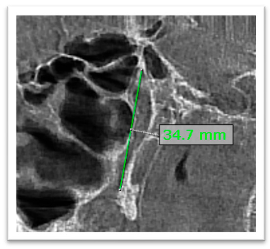
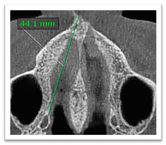
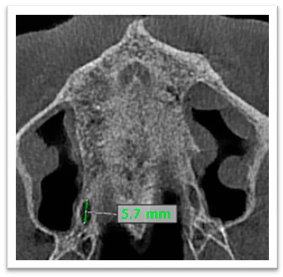
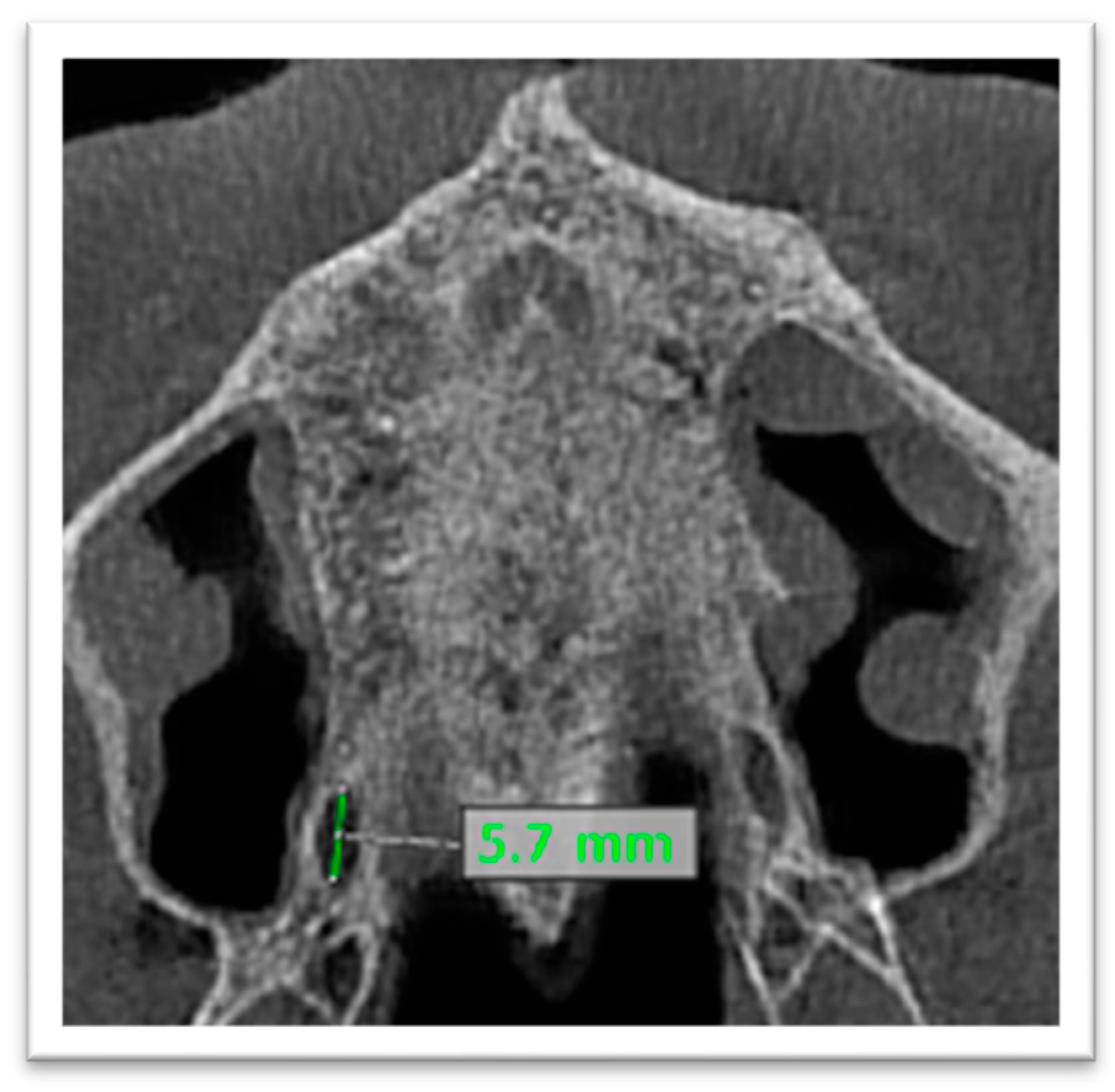
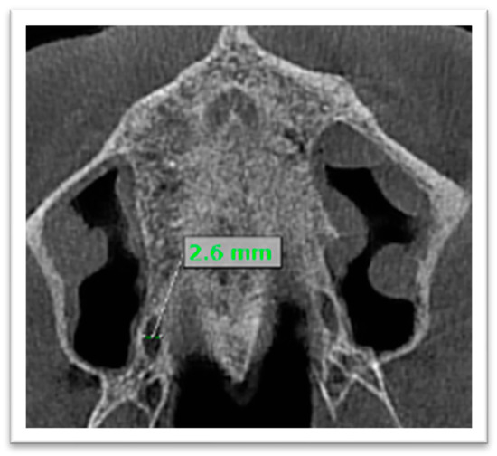
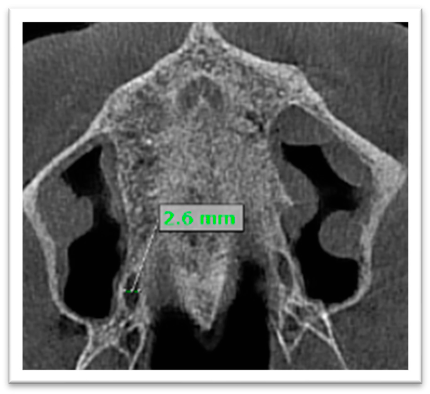
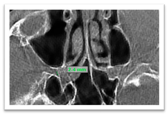
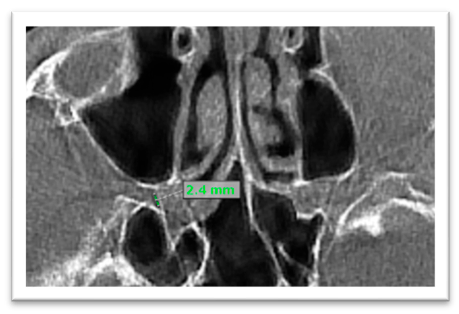
The total length of the PPF-GPC complex was defined as the straight, extending line from the inferior opening of the GPC in the GPF to the superior bony margin of the PPF (Figure 1). The anteroposterior distance of the GPF was measured as the distance between the anterior nasal spine (ANS) and the GPF (GPF-ANS distance) (Figure 2).
Figure 1.
Length of the pterygopalatine fossa—greater palatine canal (PPF-GPC) complex. The total length was defined as a straight extending line from the inferior opening of the GPC in the greater palatine foramen (GPF) to the superior bony margin of the PPF.
Figure 2.
The anteroposterior GPF-ANS distance.
Other measurements included:
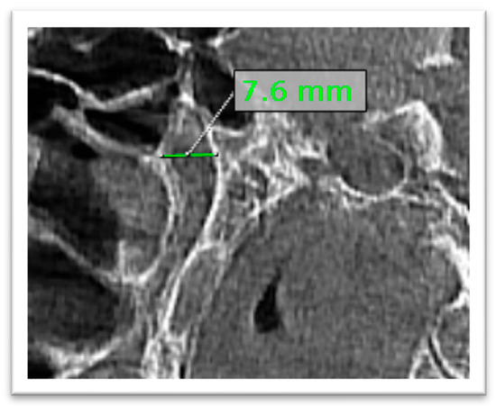
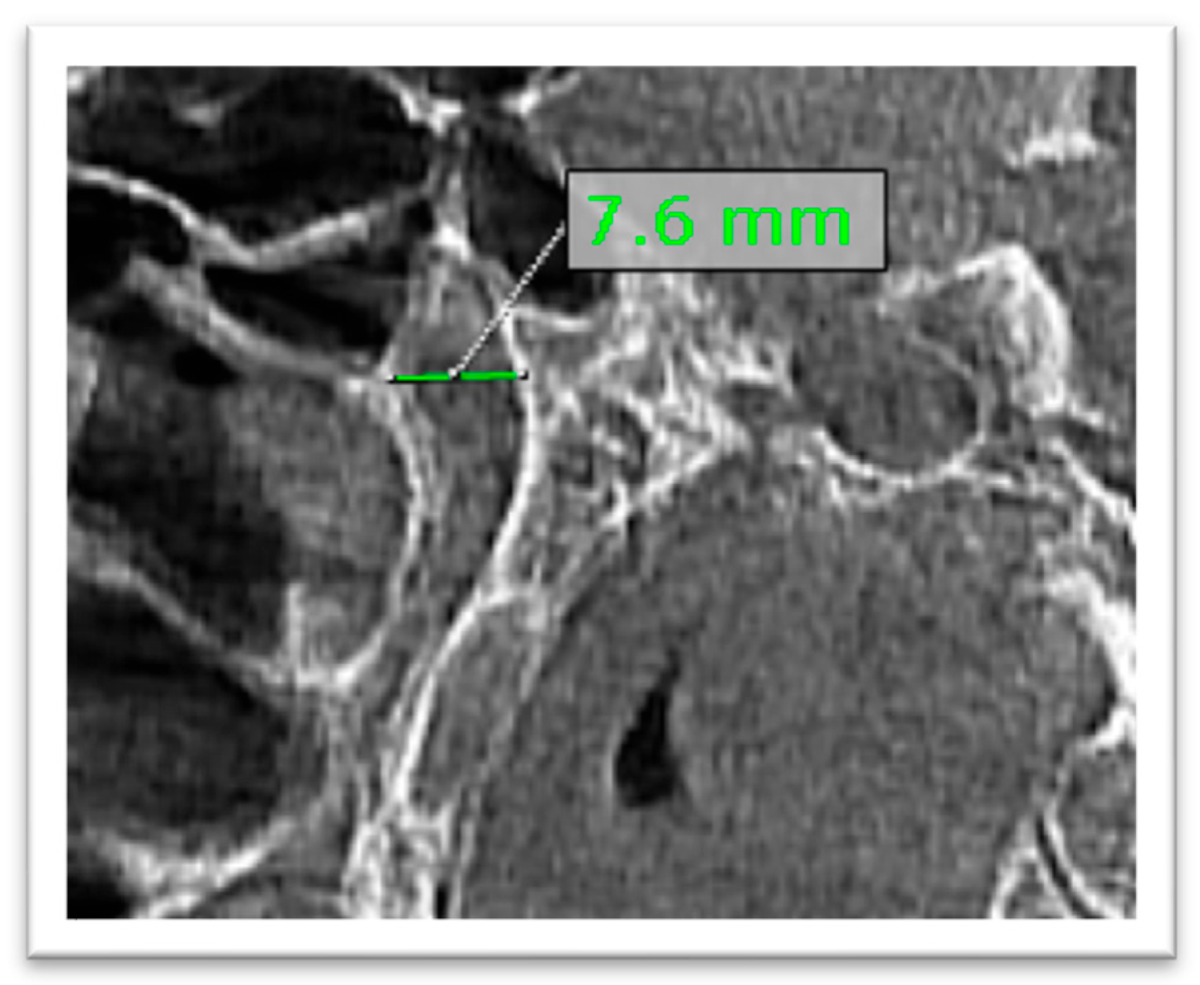
- Diameter of the PPF—defined as the maximum width of the fossa measured on the sagittal CBCT plane (Figure 3).
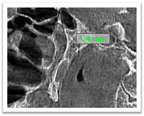
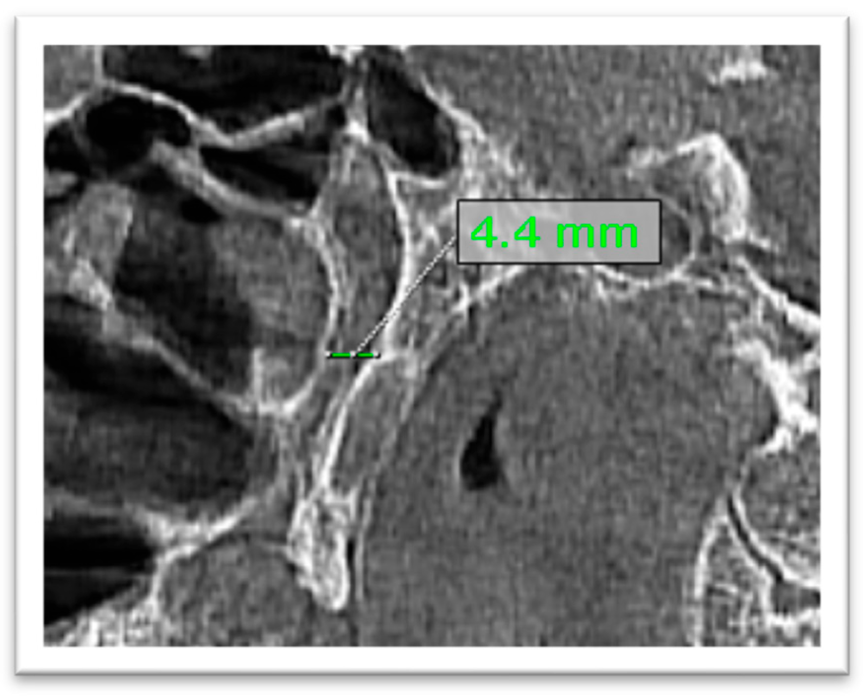
- Diameter (width) of the GPC—measured as the greatest anteroposterior diameter of the canal on the sagittal plane (Figure 4).
- The diameter of the sphenopalatine foramen was measured on the axial plane at its greatest dimension (Figure 7).
- Curvature of the canal is classified according to the observed course of the canal as straight, mild, moderate, severe, or double S, using a visually assessed, non-quantitative morphological classification.
Figure 3.
Diameter of the PPF.
Figure 4.
Diameters of the GPC measured anteroposteriorly at the mid-distance of the canal.
Figure 5.
Anteroposterior diameter of the GPF.
Figure 6.
Mediolateral diameter of the GPF.
Figure 7.
Diameter of sphenopalatine foramen on CBCT axial plane.
2.6. Statistical Analysis
All analyses were conducted using Stata 17 (StataCorp, College Station, TX, USA). The data for each patient were analyzed separately for each side. When conducting multivariate analysis of continuous variables, we used the average of the right and left values for each variable to avoid treating both sides of the same patient as independent observations. Paired t-tests were used to examine differences between the right and left sides. An unequal variance t-test was used to compare males and females for each measure without adjustment. To assess the association between age (per 10-year increase) and gender with the six key morphometric measures, we fitted multivariate linear regression models with robust standard errors. Emphasis was placed on regression coefficients and their corresponding confidence intervals, and model fit indices such as R2 were not reported. Curvature was initially classified into multiple categories; however, for regression analysis, it was dichotomized (straight vs. non-straight) to enable the use of a clustered logistic model. This simplification facilitated more robust estimation using Generalized Estimating Equations (GEE), allowing us to account for within-subject correlations between bilateral observations while avoiding sparse data across multiple categories. Although this approach reduces morphological detail, it can improve model stability and interpretability within the available sample. An exchangeable working correlation structure was specified a priori, as each subject contributed only two correlated observations (bilateral measurements), making this an appropriate assumption of equal correlation between sides; formal model comparison was not performed.
3. Results
The analytical sample included 65 CBCTs spanning a broad adult age range, with a predominantly female distribution (Table 1). Sex was recorded as documented in clinical records (male/female) and reflects biological classification. The age range allowed age-adjusted modeling.
Table 1.
Cohort characteristics (n = 65).
A summary of overall morphometric characteristics and reference ranges for all landmarks is provided in Table 2. The mean length of the palatal-to-PPF path (from the GPF to the top of the bony border of the PPF) was about 33 mm, and most patients were within the range of 29 to 40 mm (Table 2). The average distance from the GPF to the ANS was approximately 47 mm, with most measurements ranging between 41 and 53 mm. There was considerable variability among individuals in the length of the PPF-GPC complex, with a 10 mm difference between the 5th and 95th percentile values. Conversely, the PPF coronal diameter showed relatively narrow variability.
Table 2.
Practical reference ranges for morphometrics (patient-level averages; n = 65).
Right-left comparisons showed high bilateral symmetry in the primary measurements, providing evidence for the use of patient-specific averages in subsequent modeling (Table 3). The near signal for canal width should be interpreted cautiously and does not indicate a consistent clinically meaningful asymmetry.
Table 3.
Right-left symmetry (paired comparisons; n = 65).
In the unadjusted comparisons, males exhibited a longer PPF-GPC path and larger GPF dimensions in both mediolateral and anteroposterior axes (Table 4). In contrast, the anteroposterior position of the GPF relative to the ANS (GPF-ANS distance) was small (mean difference, 0.05 mm; 95% CI, −0.16–0.44), indicating no clear sex-related difference.
Table 4.
Sex comparisons (unadjusted; patient-level averages).
After adjusting simultaneously for age and sex, route length remained approximately 2.3 mm longer in males (95% CI, 0.35 to 4.15), while age showed a slight inverse association with route length (Table 5). Conversely, the GPF-ANS distance increased with age but showed no significant association with sex. These adjusted estimates help translate anatomy into patient-factor-informed expectations rather than relying solely on unadjusted means.
Table 5.
Multivariable associations with key outcomes (n = 65).
Panel A of Table 6 presents the complete distribution of instances for each category of GPC curvature by side. Approximately 43.8% of the examined sides (57 out of 130) exhibited linear trajectories. Non-linear curves were identified on the remaining half of the sides (73 out of 130, or 56.2%). No significant difference was seen in the comparison of right- versus left-sided frequency distributions throughout the five categories of curvature (p = 0.177, Fisher-Freeman-Halton exact test). This observation clinically indicates that the neurovascular route and/or GPC diverges from its typical position, rather than being an anomaly. The age, gender, and side-adjusted GEE logistic regression model predicting a straight versus non-straight curve, as illustrated in panel B, indicates that age exhibits a borderline association with an increased likelihood of a straight curve, while gender and side show no such associations. Panel C illustrates predicted probability, indicating that as patient age grows, the likelihood of possessing a straight canal likewise rises; yet, even at the highest age levels, uncertainty regarding the existence or absence of a straight canal persists.
Table 6.
Greater palatine canal curvature patterns and age/sex-adjusted straightness model.
These findings provide evidence for a practical approach in which clinicians will view the palatal-to-PPF route length and GPF position as a range of possible values influenced by patient factors, rather than fixed anatomical values.
4. Discussion
This cross-sectional study evaluated the morphometric characteristics of the pterygopalatine fossa and the GPC using CBCT. The palatal-to-PPF route length from the GPF to the upper bony border of the PPF averaged 33.1 mm in the CBCT group; however, individual variation was sufficient to render a single “standard” insertion depth inappropriate for all patients. The GPF-ANS distance averaged 47 mm and increased with age, providing a simple A–P reference point for identifying or predicting the location of palatal landmarks. Curved canals were present in many cases, indicating that, in addition to the appropriate depth required for a canal-based approach, knowledge of the canal direction is vital. The GPC is often used as a pathway for maxillary nerve block via the greater palatine foramen. If the canal length varies substantially, over-insertion may enter the pterygopalatine fossa or injure vascular structures. In the current study, the measured GPC approximated the mean of values reported in studies that measured the canal alone and those that combined canal and fossa measurements. For example, a major CBCT-based study comprising 500 patients (1000 canals) reported a mean GPC length of 29 ± 3 mm (range, 22–40 mm), indicating that the measurement represented the canal length alone [20]. Conversely, Aoun et al. defined the GPC–PPF component using the higher radiologic bony aspect of the PPF as the superior limit and reported a mean length of 35.015 ± 3.85 mm (range 24.22–45.30 mm) [15]. In the present study, the endpoint was the upper bony border of the PPF; therefore, our mean (33.1 ± 3.1 mm) aligns more closely with a GPC–PPF component than with canal-only measurements, while still differing slightly due to population and measurement conventions. CT-based anatomical studies indicate that there is no single universal measurement of the GPC length. For instance, the study by Douglas and Wormald, based on CT scans of cadaver heads, reported a mean GPC length of 18.5 mm and a mean PPF height of 21.6 mm; thus, the combined length would be approximately 40 mm [21]. Hwang et al. used three-dimensional reconstruction from CT scans and reported a mean canal length of 13.8 ± 2.0 mm and a mean PPF height of 21.0 ± 3.4 mm (combined, ≈34.8 mm), which is closer to our CBCT-based corridor estimate [12]. More broadly, systematic syntheses and CT/CBCT studies emphasize that the measured ‘length’ depends on how the superior landmark within the PPF is defined and whether the canal is measured alone or as part of a larger anatomical corridor [11].
In the current study, we reported adjusted estimates for both age and sex, rather than unadjusted means for each subgroup. After mutual adjustment, males had a palatal-to-PPF corridor that was 2.3 mm longer, along with larger GPF dimensions compared to females. Similar trends have been reported in earlier CBCT-based studies, with a mean canal length of approximately 32.63 mm overall, and higher values observed in males (33.23 mm) than in females [9]. Similarly, another study demonstrated mean canal lengths of 32.49 mm in males and 30.55 mm in females [22]. These observations suggest the presence of sex-related anatomical variations in the GPC. Clinically, such differences may have important implications, as they can influence the appropriate depth of needle insertion during posterior maxillary anesthesia, as well as surgical access in the posterior maxilla. Interestingly, Aoun et al. found no significant gender differences in canal-fossa length; however, they did find gender-related differences in opening diameters [15]. These findings suggest that sex effects may be parameter-specific (e.g., present for aperture dimensions in some populations but inconsistent for corridor length) and underscore the importance of population context and measurement definitions when translating published morphometrics into clinical expectations [16,19]. Notably, the relatively small number of male participants limits the precision of sex-based estimates, as reflected by the wide confidence intervals. Therefore, the observed sex differences should be interpreted cautiously and considered preliminary, pending validation in larger and more balanced cohorts.
Furthermore, the observed inverse relationship between age and canal length in the current study suggests that anatomical changes over time, possibly related to bone remodeling or maxillary sinus pneumatization, may influence the morphology of this region. In the current study, straight canals accounted for 43.8% of sides, indicating that curvature is common but not consistently predominant. This distribution contrasts with the findings of Aoun et al., who reported that most sides were curved when curvature was classified as binary (straight vs. curved) [15]. Differences in classification strategy, population anatomy, and the choice of the superior PPF landmark can contribute to these variations. In the present study, curvature classification was based on subjective visual assessment without predefined angular or geometric thresholds, which may limit reproducibility. Nonetheless, this simplified approach allowed for practical classification of canal morphology. However, this classification should be considered descriptive and exploratory rather than a reproducible or standardized morphologic framework. Given the high prevalence of curved canals, the straight-line measurement used in the current study may not accurately reflect the functional canal pathway and should not be directly interpreted as a procedural depth or trajectory. Accordingly, these measurements should be considered descriptive rather than prescriptive for clinical application. In addition, intra- and inter-observer agreement for curvature assessment was not formally evaluated. Furthermore, the categorization was simplified into a binary outcome for regression analysis to address sparse data, which may have reduced the granularity of morphological information, although it improved model stability and enabled more reliable statistical estimation within the available sample size. Additionally, intra-observer reproducibility for linear measurements was not formally quantified, which may further limit measurement reliability.
CBCT studies also reveal substantial geometric variability in canal direction and patterns, supporting the practical point that trajectory can be the feature that most strongly determines technical difficulty even when average lengths are similar [13,20]. Clinically, this is important because canal-based deep palatal approaches have been revisited for maxillary nerve block, and technique papers emphasize the need for careful control of depth and direction [12,21,23]. In addition, complications have historically been described with canal-based maxillary nerve blocks, supporting a conservative, risk-aware approach and the selective use of CBCT when anatomy is uncertain, or the consequences of misdirection are high [24]. Although our measurements are not direct implant site metrics, they enhance patient-specific understanding of the posterior palatal anatomy and the neurovascular gateway at the GPF. Previous studies emphasize the importance of the greater palatine artery and foramen in defining a surgical ‘safety zone’. with known inter-individual variation in GPF position and palatal tissue characteristics [25,26]. In this context, reporting the GPF-ANS distance and GPF dimensions can provide a practical bony anchor to support communication, planning, and risk discussion in procedures involving the posterior palate and adjacent neurovascular bundle.
This study has several limitations that should be acknowledged. First, its retrospective, single-center design and use of archived CBCT scans from a convenience-based cohort introduce potential selection bias and limit generalizability. In addition, the relatively high exclusion rate and the lack of consistent demographic data for excluded cases limited the ability to assess potential systematic differences between included and excluded populations, which may further affect external validity. Furthermore, relevant clinical and anatomical variables, including CBCT indications, dental status, skeletal pattern, and other craniofacial factors, were not consistently available and could not be controlled for. No a priori power calculation was performed, and the reported margin of error represents a post hoc estimate; therefore, confidence intervals should be considered the primary measure of precision. Although bilateral measurements were averaged to avoid non-independence, this approach may mask clinically relevant asymmetry; alternative methods such as mixed-effects models could better preserve within-subject variability. Furthermore, curvature was assessed visually without quantitative thresholds, and observer agreement was not evaluated, which may limit reproducibility. Linear measurements were used for their simplicity and reproducibility; however, they may underestimate true canal length in curved trajectories. Finally, the sex imbalance in the cohort further limits the precision and generalizability of sex-based comparisons.
However, this study adds population-specific CBCT morphometrics for a Saudi cohort using (i) a corridor definition that directly reflects the clinically interpreted palatal-to-PPF route (GPF to upper PPF border), (ii) an anterior positional reference anchored to the ANS, and (iii) age- and sex-adjusted estimates presented in an interpretable form. By modeling curvature and accounting for within-person right/left clustering, the study provides an applied framework for anticipating anatomical variability rather than relying on single ‘average’ values. The current findings do not suggest that canal-based maxillary nerve blocks should become routine practice; rather, they provide morphometric information to support informed risk-aware decision-making based on individual patient anatomy [27]. The GEE study is designed to predict the extent of canal straightness rather than evaluate the severity of curvature. While a descriptive classification into five categories has been preserved to illustrate the complete spectrum of anatomical configurations, the regression models intentionally restricted their focus to the comparison of straight and non-straight canals. This conclusion was taken because the distribution of persons classified as having “mild,” “moderate,” “severe” curvature patterns, and “double-S” was uneven, with just one subject exhibiting the “double-S” configuration on either side.
5. Conclusions
Within the limitations of this observational CT-based study, the average length of the PPF-GPC path in this Saudi CBCT cohort was approximately 33 mm, with considerable variation among individuals. After adjusting for demographic characteristics, male sex was significantly associated with increased route length; additionally, increasing age was significantly associated with both shorter path length and increasing distance between the GPF-ANS. Given the frequency of curved canals in the posterior maxilla, both the length and trajectory of the canal should be carefully evaluated when considering this region for anesthesia and surgical planning. However, these findings are descriptive and do not directly assess clinical outcomes or validate specific insertion depths or thresholds. Therefore, individualized anatomical assessment, including preoperative CBCT, may aid in clinical decision-making, but the results should be interpreted with caution. Further studies correlating anatomical findings with clinical outcomes in larger and more diverse populations are warranted.
Author Contributions
Conceptualization, A.H.A.A.; methodology and supervision, H.A.A.; investigation and data curation, A.A.A.; formal analysis and project administration, H.S.A.-s.; methodology and funding, M.Z.; writing—original draft, A.A.D.; writing—review and editing, M.S.H. All authors have read and agreed to the published version of the manuscript.
Funding
The authors extend their appreciation to the Deanship of Research and Graduate Studies at King Khalid University, KSA, for funding this work through the General Research Project (grant number GRP/77/46).
Institutional Review Board Statement
Ethical approval was obtained from the Research Ethics Committee at King Khalid University (HAPO-06-B-001) with the approval number (KKU-73-2025-26) on 2 October 2025. Informed consent from the patients was obtained prior to the study’s conduct.
Informed Consent Statement
The patient consent form was obtained before the study commenced at the King Khalid University, Dental Hospital.
Data Availability Statement
The original contributions presented in this study are included in the article. Further inquiries can be directed to the corresponding author.
Conflicts of Interest
The authors declare no conflicts of interest.
References
- Calcano, G.A.; Johns, H.L.; Diamond-Ames, T.M.; Hernandez-Herrera, G.A.; Vander Wert, C.J.; Frohn, E.P.; Van Abel, K.M.; Morris, J.M.; Pinheiro-Neto, C.D. The Pterygopalatine Fossa in Three Dimensions: A Digital Anatomical Model Derived from Radiographic Imaging, Refined with Peer-Reviewed Literature, and Optimized with Medical Illustration. Oper. Tech. Otolaryngol.-Head Neck Surg. 2025, 36, 2–13. [Google Scholar] [CrossRef] [Scilit]
- Jacobs, R.; Salmon, B.; Codari, M.; Hassan, B.; Bornstein, M.M. Cone Beam Computed Tomography in Implant Dentistry: Recommendations for Clinical Use. BMC Oral Health 2018, 18, 88. [Google Scholar] [CrossRef] [Scilit] [PubMed]
- Horner, K.; Islam, M.; Flygare, L.; Tsiklakis, K.; Whaites, E. Basic Principles for Use of Dental Cone Beam Computed Tomography: Consensus Guidelines of the European Academy of Dental and Maxillofacial Radiology. Dentomaxillofac. Radiol. 2009, 38, 187–195. [Google Scholar] [CrossRef] [Scilit]
- Fokas, G.; Vaughn, V.M.; Scarfe, W.C.; Bornstein, M.M. Accuracy of Linear Measurements on CBCT Images Related to Presurgical Implant Treatment Planning: A Systematic Review. Clin. Oral Implant. Res. 2018, 29, 393–415. [Google Scholar] [CrossRef] [Scilit]
- Tyndall, D.A.; Price, J.B.; Tetradis, S.; Ganz, S.D.; Hildebolt, C.; Scarfe, W.C. Position Statement of the American Academy of Oral and Maxillofacial Radiology on Selection Criteria for the Use of Radiology in Dental Implantology with Emphasis on Cone Beam Computed Tomography. Oral Surg. Oral Med. Oral Pathol. Oral Radiol. 2012, 113, 817–826. [Google Scholar] [CrossRef] [Scilit]
- Schulze, R.K.W.; Drage, N.A. Cone-Beam Computed Tomography and Its Applications in Dental and Maxillofacial Radiology. Clin. Radiol. 2020, 75, 647–657. [Google Scholar] [CrossRef] [Scilit]
- Ludlow, J.B.; Ivanovic, M. Comparative Dosimetry of Dental CBCT Devices and 64-Slice CT for Oral and Maxillofacial Radiology. Oral Surg. Oral Med. Oral Pathol. Oral Radiol. Endod. 2008, 106, 106–114. [Google Scholar] [CrossRef] [Scilit]
- Pauwels, R. Cone Beam CT for Dental and Maxillofacial Imaging: Dose Matters: Table 1. Radiat. Prot. Dosim. 2015, 165, 156–161. [Google Scholar] [CrossRef] [Scilit]
- Pawar, S.; Chhaparwal, Y.; Patil, V.; Pentapati, K.C.; Chhaparwal, S.; Singhal, D.K.; Prabhu, N.; Prabhu, D. Radiomorphometric Analysis of the Greater Palatine Canal and the Pterygopalatine Fossa Using Cone Beam Computed Tomography: A Retrospective Study. Clin. Cosmet. Investig. Dent. 2025, 17, 445–454. [Google Scholar] [CrossRef] [Scilit]
- Duman Tepe, R.; Guray, B.; Senel, S.N.; Cakir Karabas, H. Morphometric Assessment of Greater Palatine Canal and Foramen Variations in Cleft Lip and Palate Patients Using CBCT. J. Stomatol. Oral Maxillofac. Surg. 2025, 126, 102487. [Google Scholar] [CrossRef] [Scilit] [PubMed]
- Tomaszewska, I.M.; Kmiotek, E.K.; Pena, I.Z.; Średniawa, M.; Czyżowska, K.; Chrzan, R.; Nowakowski, M.; Walocha, J.A. Computed Tomography Morphometric Analysis of the Greater Palatine Canal: A Study of 1500 Head CT Scans and a Systematic Review of Literature. Anat. Sci. Int. 2015, 90, 287–297. [Google Scholar] [CrossRef] [Scilit]
- Hwang, S.H.; Seo, J.H.; Joo, Y.H.; Kim, B.G.; Cho, J.H.; Kang, J.M. An Anatomic Study Using Three-dimensional Reconstruction for Pterygopalatine Fossa Infiltration via the Greater Palatine Canal. Clin. Anat. 2011, 24, 576–582. [Google Scholar] [CrossRef] [Scilit] [PubMed]
- Rapado-González, O.; Suárez-Quintanilla, J.A.; Suárez-Cunqueiro, M.M. Anatomical Variations of the Greater Palatine Canal in Cone-Beam Computed Tomography. Surg. Radiol. Anat. 2017, 39, 717–723. [Google Scholar] [CrossRef] [Scilit]
- McKinney, K.A.; Stadler, M.E.; Wong, Y.T.; Shah, R.N.; Rose, A.S.; Zdanski, C.J.; Ebert, C.S.; Wheless, S.A.; Senior, B.A.; Drake, A.F.; et al. Transpalatal Greater Palatine Canal Injection: Radioanatomic Analysis of Where to Bend the Needle for Pediatric Sinus Surgery. Am. J. Rhinol. Allergy 2010, 24, 385–388. [Google Scholar] [CrossRef] [Scilit]
- Aoun, G.; Nasseh, I.; Sokhn, S. Radio-Anatomical Study of the Greater Palatine Canal and the Pterygopalatine Fossa in a Lebanese Population: A Consideration for Maxillary Nerve Block. J. Clin. Imaging Sci. 2016, 6, 35. [Google Scholar] [CrossRef] [Scilit]
- Kim, D.W.; Tempski, J.; Surma, J.; Ratusznik, J.; Raputa, W.; Świerczek, I.; Pękala, J.R.; Tomaszewska, I.M. Anatomy of the Greater Palatine Foramen and Canal and Their Clinical Significance in Relation to the Greater Palatine Artery: A Systematic Review and Meta-Analysis. Surg. Radiol. Anat. 2023, 45, 101–119. [Google Scholar] [CrossRef] [Scilit] [PubMed]
- Martins, T.; Guedes, V.; Martins, E.; Mesquita, P. Morphometric Analysis of the Greater Palatine Foramen: A CBCT Study in Portugal. Surg. Radiol. Anat. 2025, 47, 64. [Google Scholar] [CrossRef] [Scilit]
- Fonseka, M.C.N.; Hettiarachchi, P.V.K.S.; Jayasinghe, R.M.; Jayasinghe, R.D.; Nanayakkara, C.D. A Cone Beam Computed Tomographic Analysis of the Greater Palatine Foramen in a Cohort of Sri Lankans. J. Oral Biol. Craniofacial Res. 2019, 9, 306–310. [Google Scholar] [CrossRef] [Scilit]
- Ikuta, C.R.S.; Cardoso, C.L.; Ferreira-Júnior, O.; Lauris, J.R.P.; Souza, P.H.C.; Rubira-Bullen, I.R.F. Position of the Greater Palatine Foramen: An Anatomical Study through Cone Beam Computed Tomography Images. Surg. Radiol. Anat. 2013, 35, 837–842. [Google Scholar] [CrossRef] [Scilit]
- Howard-Swirzinski, K.; Edwards, P.C.; Saini, T.S.; Norton, N.S. Length and Geometric Patterns of the Greater Palatine Canal Observed in Cone Beam Computed Tomography. Int. J. Dent. 2010, 2010, 292753. [Google Scholar] [CrossRef] [Scilit] [PubMed]
- Douglas, R.; Wormald, P. Pterygopalatine Fossa Infiltration Through the Greater Palatine Foramen: Where to Bend the Needle. Laryngoscope 2006, 116, 1255–1257. [Google Scholar] [CrossRef] [Scilit] [PubMed]
- Jalalian, F.; Sheikhi, M.; Zamaninaser, A. Length and Anatomic Routes of the Greater Palatine Canal as Observed by Cone Beam Computed Tomography. Dent. Res. J. 2013, 10, 155–161. [Google Scholar] [CrossRef] [Scilit]
- Aoun, G.; Zaarour, I.; Sokhn, S.; Nasseh, I. Maxillary Nerve Block via the Greater Palatine Canal: An Old Technique Revisited. J. Int. Soc. Prev. Community Dent. 2015, 5, 359–364. [Google Scholar] [CrossRef] [Scilit]
- Fotopoulos, I.; Fardi, A.; Zisis, V.; Poulopoulos, A.; Dabarakis, N.; Lillis, T. An Updated Digital Approach to Regional Anesthesia: A Pilot Study on Computer-Guided Maxillary Nerve Block via the Greater Palatine Canal. Dent. J. 2025, 13, 521. [Google Scholar] [CrossRef] [Scilit] [PubMed]
- Tavelli, L.; Barootchi, S.; Ravidà, A.; Oh, T.-J.; Wang, H.-L. What Is the Safety Zone for Palatal Soft Tissue Graft Harvesting Based on the Locations of the Greater Palatine Artery and Foramen? A Systematic Review. J. Oral Maxillofac. Surg. 2019, 77, 271.e1–271.e9. [Google Scholar] [CrossRef] [Scilit] [PubMed]
- Yilmaz, H.G.; Boke, F.; Ayali, A. Cone-beam Computed Tomography Evaluation of the Soft Tissue Thickness and Greater Palatine Foramen Location in the Palate. J. Clin. Periodontol. 2015, 42, 458–461. [Google Scholar] [CrossRef] [Scilit]
- Monnet-Corti, V.; Santini, A.; Glise, J.; Fouque-Deruelle, C.; Dillier, F.; Liébart, M.; Borghetti, A. Connective Tissue Graft for Gingival Recession Treatment: Assessment of the Maximum Graft Dimensions at the Palatal Vault as a Donor Site. J. Periodontol. 2006, 77, 899–902. [Google Scholar] [CrossRef] [Scilit]

Disclaimer/Publisher’s Note: The statements, opinions and data contained in all publications are solely those of the individual author(s) and contributor(s) and not of MDPI and/or the editor(s). MDPI and/or the editor(s) disclaim responsibility for any injury to people or property resulting from any ideas, methods, instructions or products referred to in the content. |
© 2026 by the authors. Licensee MDPI, Basel, Switzerland. This article is an open access article distributed under the terms and conditions of the Creative Commons Attribution (CC BY) license.