


Featured Application
Soft back-support exoskeletons such as the CORFOR® may be effective ergonomic solutions to reduce lumbar mechanical load and muscle activity during repetitive manual handling tasks. Their use could help prevent work-related low back disorders in occupational settings where task redesign or automation is not feasible.
Abstract
Back-support exoskeletons have emerged as promising ergonomic interventions to assist workers during repetitive manual handling tasks, particularly when task redesign or automation is not feasible. However, limited research has examined the evolution of lumbar biomechanical parameters throughout the task. This study aimed to evaluate the effects of the CORFOR® soft-back exoskeleton on lumbar loads at L5-S1, muscle activity, and perceived exertion during a repetitive sagittal-plane manual handling task in eleven young shealthy male participants over a 4-min protocol. Results showed a significant increase in perceived lower back exertion over time. However, no significant difference in perceived exertion was found between EXO and FREE conditions despite reductions of 11–13%. Lumbar loads at L5-S1 were significantly reduced when using the soft-back exoskeleton in both lowering and lifting phases (−14% for peak flexion–extension moment, −6% for compression force, and −10% for shear force). In addition, peak flexion–extension moment and peak compression force significantly decreased over time. Significant interactions between exoskeleton use and time were observed for peak L5-S1 flexion–extension moment and right ES muscle activity. Overall, these findings partially support our hypothesis, as the CORFOR® reduced lumbar mechanical demands ES muscle activity, with some additional time-dependent effects observed.
1. Introduction
Manual handling activities are common in industrial occupations and have frequently been investigated as potential contributors to low back disorders (LBDs), the most prevalent and economically impactful musculoskeletal condition in occupational settings [1]. Studies conducted among manual material handlers and industrial workers report high prevalence rates of low back pain, depending on occupational exposure and task characteristics [1,2,3]. Research examining the relationship between handling activities and LBD risk has used heterogeneous exposure assessment methods, ranging from self-reported task frequency to objective biomechanical measurements of trunk motion and loading [1,4,5]. Several studies have identified biomechanical factors such as lifting frequency, load magnitude, trunk flexion, and trunk movement velocity as key determinants of increased LBD risk [2,5,6,7]. For instance, it has been shown that a threshold exceeding 25 lifting actions per day may increase the incidence of LBD [6,7]. Also, repetitive manual handling activities, combined with high task frequency, forceful actions, rapid execution, and limited recovery opportunities, may further raise the risk of developing LBDs [8,9].
Task repetition has been examined in the literature when investigating the risk of LBDs [10,11,12,13,14,15,16]. In these studies, repetitive manual handling tasks were most commonly considered using protocols ranging from 10 cycles per minute over 4 min to 15 cycles per minute over 5 min [11,12,14,15]. Measurements over a longer period, i.e., 15 and 30 min, have been conducted, but with a significant lower movement frequency (3.5 and 5 cycles per minute, respectively) [13,16]. Accordingly, any investigation about task repetition should last at least 4 min, with a frequency of 10 cycles per minute. It is also worth noting that most of these studies simultaneously investigated the influence of wearing an industrial exoskeleton [11,12,14,15,16].
Indeed, back-support exoskeletons have emerged as a promising ergonomic intervention to assist workers [17] during prolonged or repetitive tasks, particularly when task redesign or automation is not feasible [18]. Several studies have shown that back-support exoskeletons can reduce lumbar muscle activity [19,20,21,22], or the perceived exertion in the lower back [19]. For instance, reductions in lumbar muscle activity ranging from 6 to 35% have been reported, along with an average 36% decrease in perceived exertion in the lower back [19]. However, to better characterize the effects of these devices on low back pain (LBP), other parameters such as flexion–extension moments, compression [23] and shear forces [24] appear to be more relevant. During non-repetitive manual handling tasks, the use of back-support exoskeletons has been shown to significantly reduce erector spinae (ES) muscle activity [25,26] and perceived exertion in the lower back [27,28], as well as peak compression [26,29] and shear forces [29].
During repetitive manual handling tasks, the use of back-support exoskeletons also led to significant reductions in (i) ES muscle activity of about 18% [12,14], (ii) compression and shear forces at the L5–S1 level by nearly 10% [11], (iii) perceived exertion in the lower back by roughly 35% [12], and oxygen uptake ranging from 9% [14] to 28% [15]. However, in the aforementioned studies, biomechanical parameters were averaged over the entire duration of the repetitive task, thereby providing no insight into their temporal evolution throughout the task. Although physiological parameters (oxygen uptake) would indeed need participants to reach a steady state prior to averaging data over a larger duration, the literature would gain further knowledge related to the influence of task repetition on LBDs by (i) describing biomechanical parameters over time and (ii) teasing out the possible further influence of back-support exoskeletons over time. In this context, it has been reported that perceived lower-back exertion increased significantly by 75% over time (15 min), regardless of exoskeleton use [16]. Also, exoskeleton use resulted in a significant reduction in ES muscle activity of approximately 27% over the duration of the task. However, these authors did not investigate the temporal evolution of ES muscle activity throughout the task. Finally, none of these studies considered lumbar loads, although such parameters are relevant when focusing on LBDs [24,30].
The aim of this study was to assess the effects of a back-support exoskeleton on lumbar spine biomechanics and subjective responses during a repetitive manual handling task performed in the sagittal plane. We hypothesized that the back-support exoskeleton will significantly reduce peak lumbar loads, lumbar muscle activity, and perceived exertion in the lower back, with these reductions becoming more pronounced as task repetition progresses.
2. Materials and Methods
2.1. Participants
Eleven healthy male participants (age: 22.9 ± 2.6 years; height: 1.77 ± 0.03 m; weight: 74.4 ± 5.2 kg) without back pathologies were included in the present study. Informed written consent was obtained prior to the data collection. The experimental protocol received approval from the ethical committee (no. IRB00012476-2023-17-01-221).
2.2. Exoskeleton
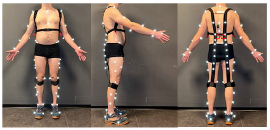
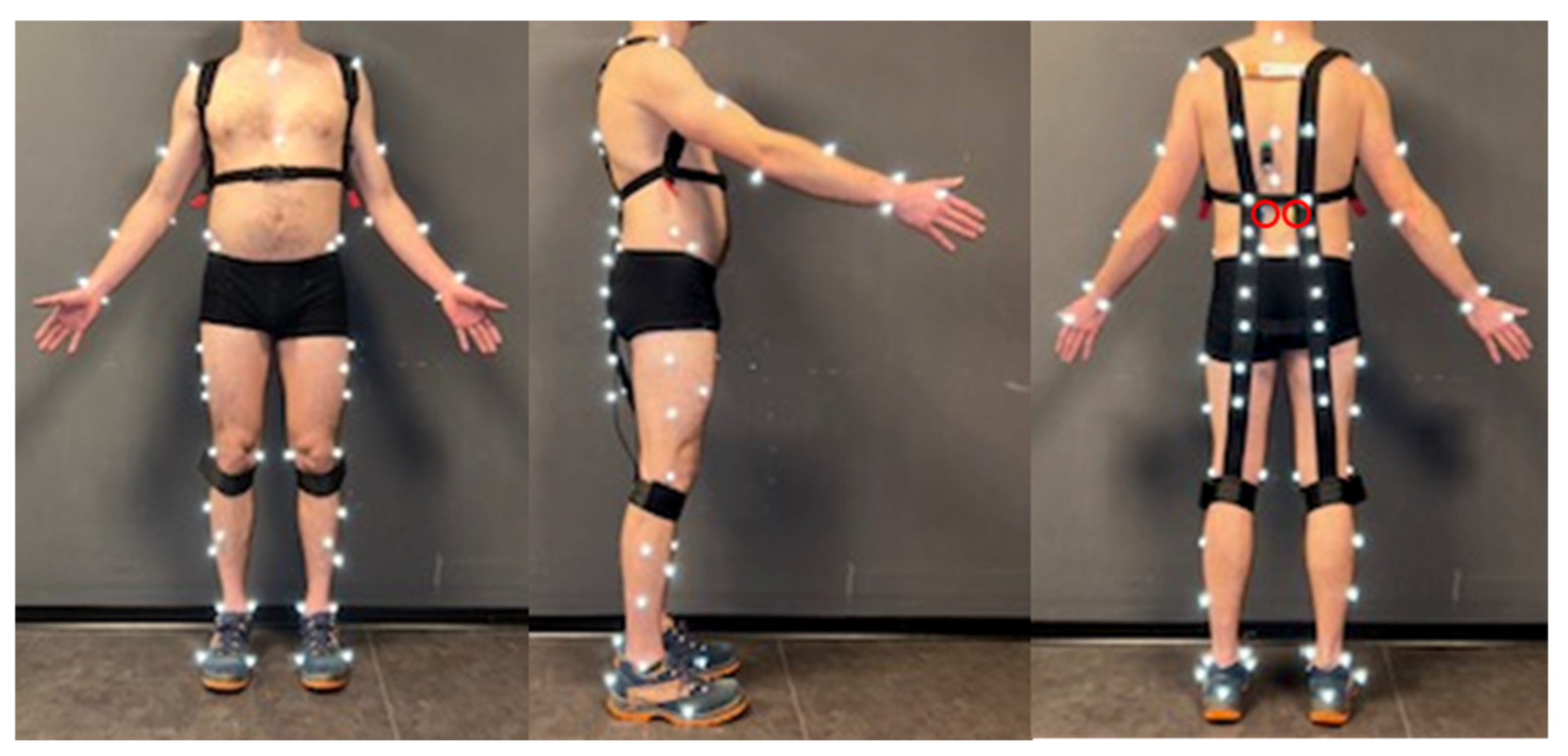
An occupational soft-back exoskeleton (CORFOR®-V2, Villemus, France) was used in this study (Figure 1). This exoskeleton consists of two elastic bands connected to shoulder straps and anchored below the knees (weight: 0.35 kg). It provides passive assistance to back and hip extension by storing and releasing elastic energy (stiffness: 500 N/m). The rationale for this exoskeleton choice is that several previous studies have demonstrated its biomechanical benefits during occupational tasks [21,22,29]. Furthermore, a musculoskeletal model of this device is available in OpenSim, allowing its effects to be investigated using musculoskeletal modeling [29]. From a structural perspective, soft exoskeletons are generally lighter and impose fewer movement constraints than rigid systems [31], making them particularly well-suited to high-frequency repetitive tasks.
Figure 1.
Experimental setup with the soft-back exoskeleton. A Delsys Wireless Duo electrode was placed on the right and left erector spinae (circled in red), and reflective markers were placed on the body (n = 51) and on the assistive device (n = 16) [29].
2.3. Experimental Procedures
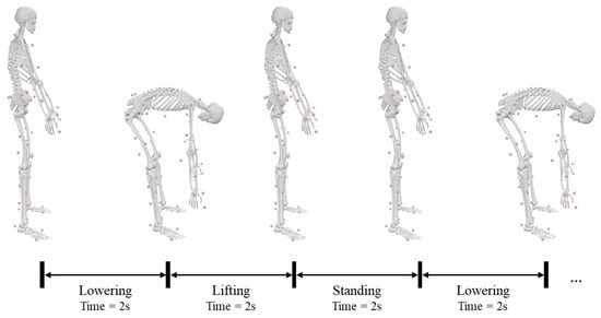
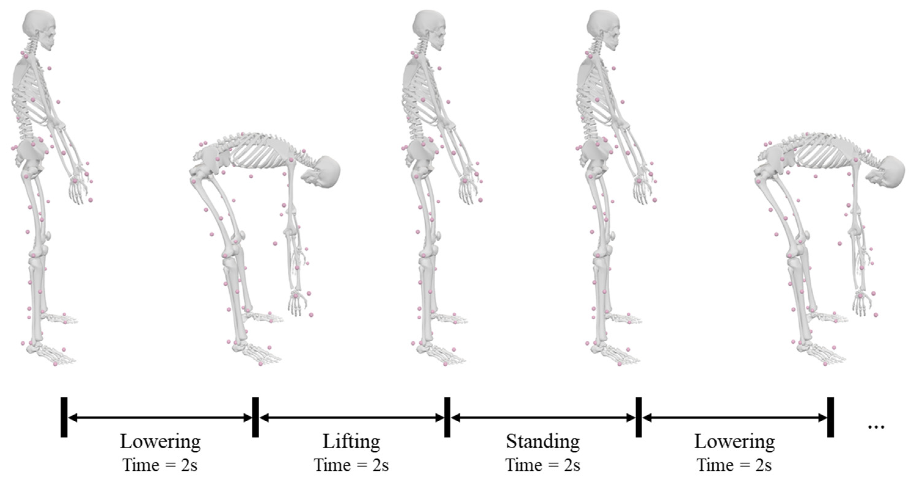
All participants completed a standardized familiarization protocol consisting of four 1-h sessions, including functional and handling tasks performed with a soft-back exoskeleton and a 7 kg load [32]. The CORFOR® soft-back exoskeleton was fitted following the manufacturer’s recommendations. Further preparations were anthropometric measurements, EMG electrode and reflective marker placement, and static trial recording in anatomical reference position. Participants performed a 4-min handling task during which they completed 10 cycles per minute [11,12] of lowering and lifting a 7 kg load using a stoop technique, with (EXO) and without (FREE) the exoskeleton, in a randomized order and separated by at least 10 repeated min of rest. Each handling cycle consisted of a 2-s lowering phase and a 2-s lifting phase. Handling cycles were separated by a 2-s standing period, during which participants held the weight in their hands (Figure 2). The lowering phase began from a standing position and ended when the weight was placed on a support located 28 cm above the ground, without releasing the load. The lifting phase began immediately as participants started to rise with the weight and ended when they returned to a standing position. The beginning and end of each lowering, lifting, and standing phase were paced using a metronome set at 30 bpm.
Figure 2.
Phases of movement during the 4-min handling task: lowering, lifting and standing phases.
2.4. Instrumentation and Data Collection
A 14-camera motion capture system (Arqus A12, Qualisys Track Manager, Göteborg, Sweden) was used to record 3D kinematics data at 200 Hz using 67 reflective markers for body and exoskeleton tracking [29]. Among these markers, 51 anatomical markers were fixed to the skin at the following landmarks: xiphoid process, manubrium, C7, T8, T12, acromions, olecranon processes, radial and ulnar styloid processes, heads of the second metacarpal bones, anterior and posterior superior iliac spines, femoral epicondyles, malleoli, calcanei, and the heads of the first and fifth metatarsals (Figure 1). In addition, 18 technical markers were placed on the arms, forearms, thighs, shanks, and feet to improve segment tracking. 16 markers were dedicated to track the kinematics of the CORFOR® exoskeleton. Occlusions of the anterior and posterior iliac spine markers due to the task, soft-back exoskeleton movement, or the handled weight were filled by the relational method. Surface electromyography (sEMG) was used to continuously record bilateral erector spinae muscle activity using a wireless system (2000 Hz, Trigno™, Wireless Duo, Delsys Inc., Natick, MA, USA) synchronously with kinematics. Electrode placement and skin preparation followed SENIAM recommendations [33]. Sensors positioned over the erector spinae were secured and protected with tape to minimize pressure and movement induced by the exoskeleton’s elastic bands. For both conditions, three-dimensional kinematics and sEMG data were synchronously recorded during three 1-min measurement periods within the 4-min handling task (T1 = 0:00–1:00; T2 = 1:30–2:30; T3 = 3:00–4:00), corresponding to 10 handling cycles per period. Perceived exertion in the lower back was assessed using the Rating Perceived Exertion (RPE) scale [34] at the end of each measurement period.
2.5. Data Analysis
For each measurement period, the lowering and lifting cycles were segmented using the mean position of the hand markers. Marker trajectories were low-pass filtered with a 6 Hz zero-lag 4th order Butterworth filter. Musculoskeletal analysis data and ES muscle activity were analyzed separately for the lowering and lifting phases of the task. For each phase, all variables were averaged over 10 consecutive cycles for each measurement period and condition. Peak lumbar loads were normalized to each subject’s body mass. ES muscle activity was filtered using a 30–450 Hz bandpass bidirectional 4th order Butterworth filter. A root mean square (RMS) value was then calculated for both the right and left ES muscles. Given to the task evaluated, i.e., 4 min of manual handling with a 7 kg load, no normalization of EMG signal amplitude was performed [35].
2.6. Musculoskeletal Modeling
An adapted Lifting Full-Body (LFB) OpenSim musculoskeletal model [36] integrating the exoskeleton [29] was used in this study. The generic LFB model, validated for estimating lumbar loads during manual material handling comprises 30 segments, 29 degree-of-freedom, and 238 Hill-type musculotendon actuators representing the trunk musculature. Musculoskeletal analyses were performed in OpenSim 4.3 [37]. The model was scaled to each participant’s anthropometry using inter-marker distances while preserving body mass and mass distribution. Inverse kinematics and inverse dynamics were then applied to compute joint kinematics and moments, and muscle forces were estimated using static optimization. Compression and shear joint reaction forces at the L5-S1 level were subsequently calculated using the joint reaction analysis tool. For the EXO condition, the soft-back exoskeleton was integrated into the scaled LFB model [29]. The device was modeled as a pair of linear springs following paths defined by 16 markers attached to the elastic bands (eight per spring). Spring resting length and stiffness (500 N/m), measured prior to the experiment, were used to set the model and were assumed to remain constant over time. For both FREE and EXO conditions, the package weight was incorporated into the model by assigning half of the mass to each hand center of mass after the scaling step [38].
As marker kinematics are a major input in the musculoskeletal model alongside the mass of the manipulated load, joint kinematics were also analyzed to establish if a change in joint reaction forces or moment could be the result of a specific kinematic parameter change. Thus, trunk–pelvis (sum of the lumbar intervertebral joints) and hip angle, hip angular velocity and acceleration, and box vertical velocity and acceleration were computed during both conditions and phases.
2.7. Statistics
Data were presented as mean ± standard deviation (SD) calculated over the 10 consecutive cycles for each measurement period and condition, with a significance level of 5% (p < 0.05). Data normality was verified using the Shapiro–Wilk test (p < 0.05). A non-parametric repeated-measures two-way ANOVA (Scheirer–Ray–Hare test) based on ranked data was then conducted to evaluate the effect of exoskeleton use (EXO vs. FREE), time (T1 vs. T2 vs. T3), and their interaction [39]. Because the exoskeleton factor comprised two levels, post-hoc analyses were not required. For the time factor (which included three levels) and interactions, post-hoc pairwise comparisons were performed using Wilcoxon signed-rank tests, with p-values adjusted using the Holm correction for multiple comparisons. Statistical analyses and figures were performed using RStudio (version 4.1.3, 2022, RStudio, Vienna, Austria) [40]. Effect sizes were quantified using partial eta squared (ηp2) for the ANOVA, as commonly recommended for ANOVA [41], and r for the Wilcoxon post-hoc tests [42]. Effect sizes were interpreted according to conventional thresholds: for ηp2, 0.01–0.06 (small), 0.06–0.14 (medium), ≥0.14 (large); for r, 0.1–0.3 (small), 0.3–0.5 (moderate), ≥0.5 (large).
3. Results
3.1. Rating Perceived Exertion
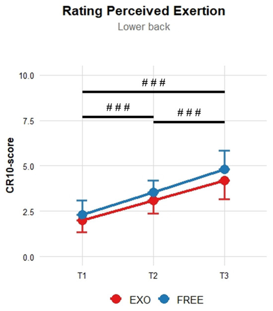
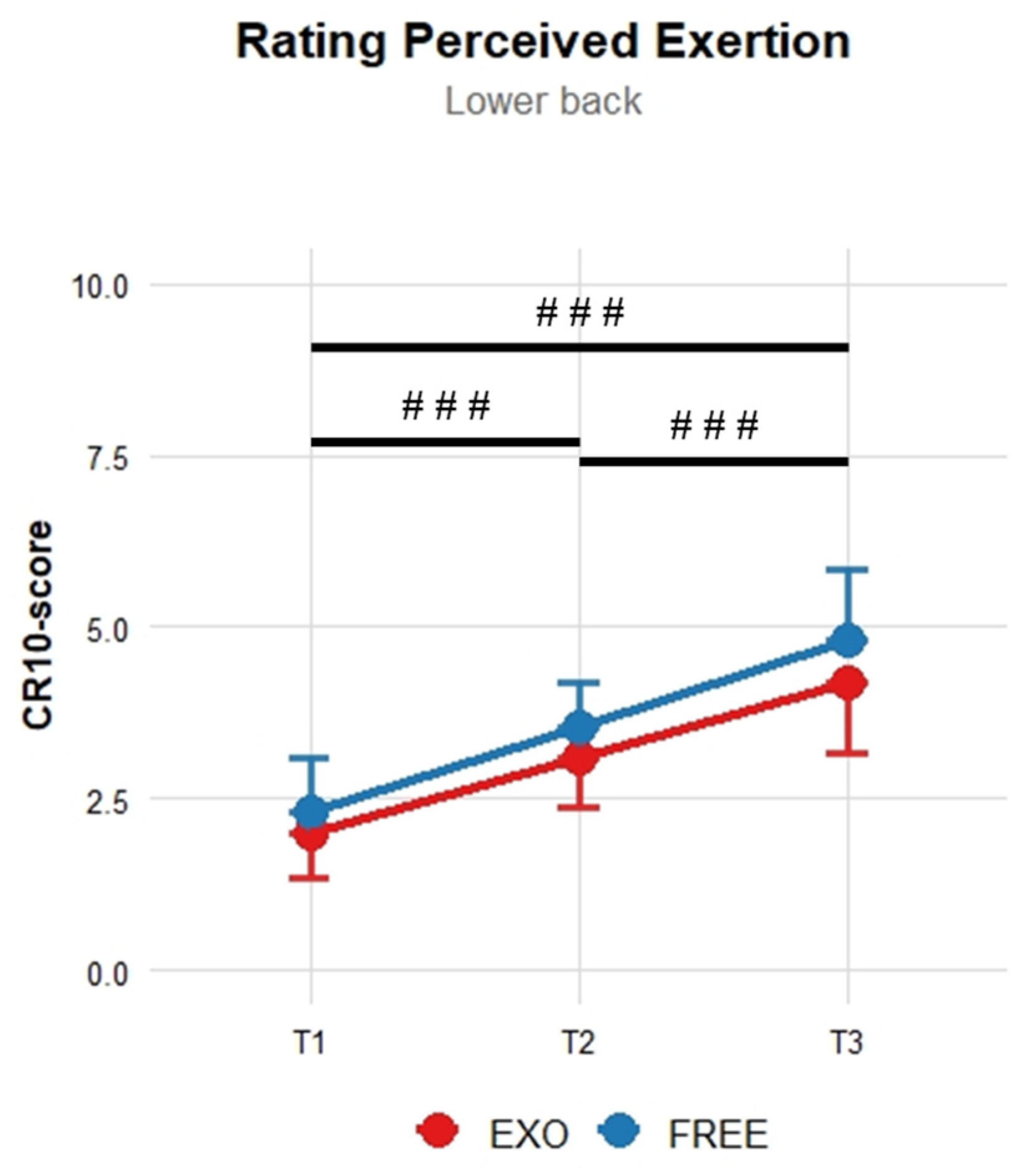
The use of the soft-back exoskeleton did not significantly reduce the RPE in the lower back (p = 0.07; ηp2 = 0.30, large effect size) despite reductions of 11% at T2 and 13% at T1 and T3 (Table 1, Figure 3). However, the RPE in the lower back was significantly increased over time during the handling task (p < 0.001; ηp2 = 0.82, large effect size), with pairwise comparisons showing significant differences between all measurement times (p < 0.001; large r between 0.85 and 0.88; Table 2). No significant interaction between exoskeleton use and time was reported.
Table 1.
Mean ± SD RPE in the lower back (CR10-score), peak L5-S1 flexion–extension moment (N*m/kg), peak L5-S1 compression force (N/kg), and peak L5-S1 shear force (N/kg) during the repetitive sagittal handling task for each measurement period and condition.
Figure 3.
Comparison of mean ± SD RPE values in the lower back. ### Significant time effect (p < 0.001).
Table 2.
Post-hoc pairwise comparisons for the time effect using Wilcoxon signed-rank tests with Holm-adjusted p-values and corresponding effect sizes (r). Pairwise post-hoc comparisons are reported when a significant main time effect was observed. Significant differences are indicated in bold type.
3.2. Lumbar Loads
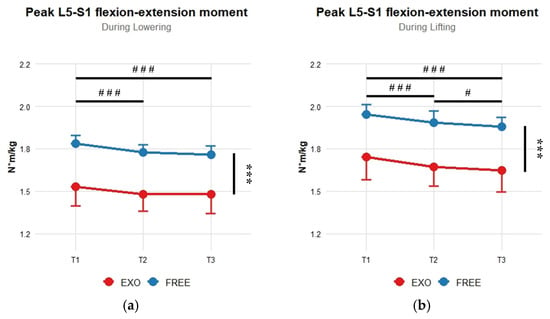
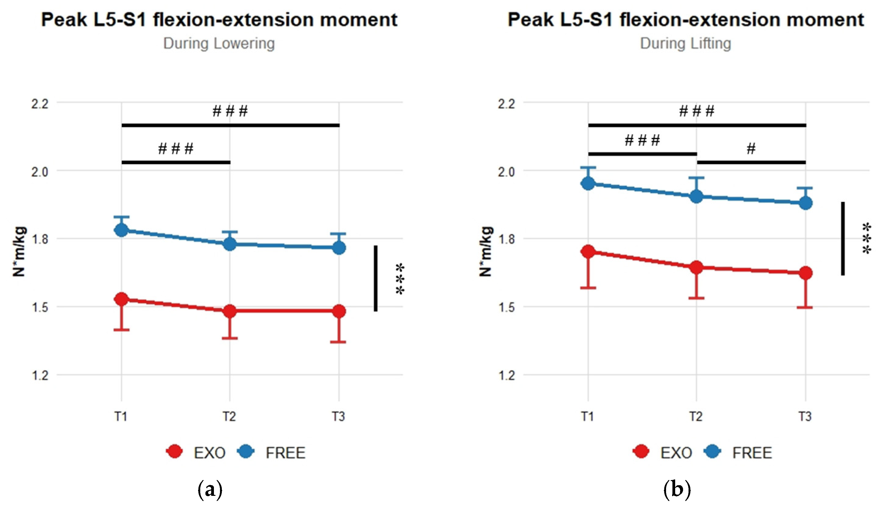
Peak L5-S1 flexion–extension moment (Table 1, Figure 4) was significantly reduced with the use of the soft-back exoskeleton (p < 0.001; ηp2 > 0.91, large effect size) and over time (p < 0.001; ηp2 > 0.67, large effect size) during lowering and lifting phases. Across measurement times, peak L5-S1 flexion–extension moment significantly decreased between T1 and T2 and between T1 and T3, during both lowering and lifting phases (p < 0.001). Between T2 and T3, a significant reduction was observed during the lifting phase only (p < 0.05). The different post-hoc effects for pairwise comparisons when a significant main time effect was obtained are presented in Table 2. Moreover, a significant interaction between exoskeleton use and time was reported for peak L5-S1 flexion–extension moment during the lowering phase (p < 0.01; ηp2 = 0.39). Pairwise comparisons showed a significant reduction with the use of the exoskeleton at each measurement time (p < 0.001).
Figure 4.
Comparison of mean ± SD peak L5-S1 flexion–extension moment normalized values during the lowering (a) and lifting (b) phases. *** Significant exoskeleton main effect (p < 0.001). ### Significant time effect (p < 0.001). # Significant time effect (p < 0.05).
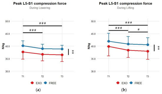
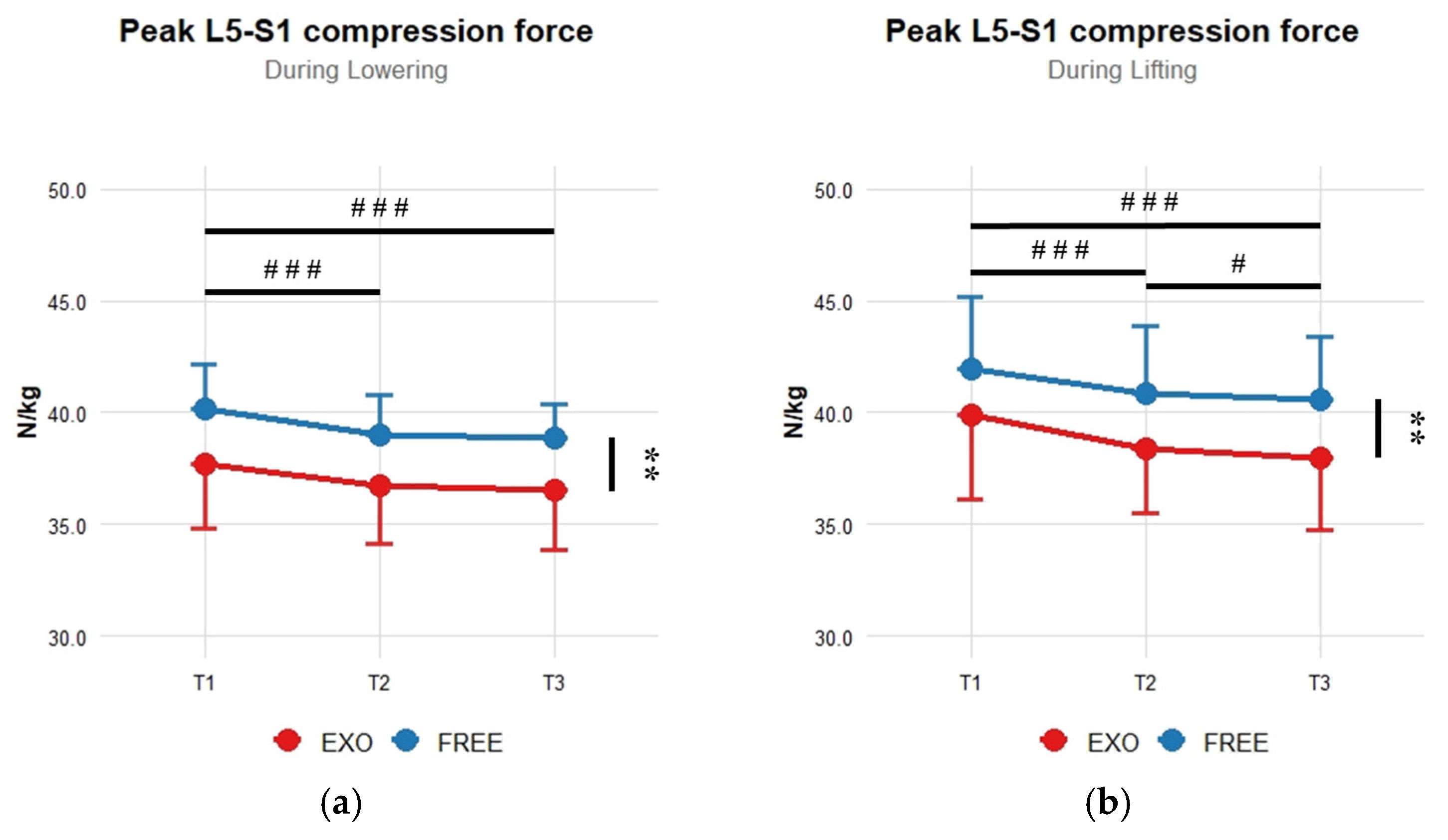
Peak L5-S1 compression force (Table 1, Figure 5) was significantly decreased with the use of the soft-back exoskeleton during both the lowering and lifting phases (p < 0.01; ηp2 > 0.62, large effect size). A significant effect of time was observed during both the lowering and lifting phases (p < 0.001; ηp2 > 0.72, large effect size). Post-hoc pairwise comparisons indicated lower peak L5-S1 compression forces at T2 and T3 compared with T1 during both the lowering and lifting phases (p < 0.001; Table 2), and at T3 compared with T2 during the lifting phase (p < 0.05; Table 2). No significant interaction between exoskeleton use and time was reported for peak L5-S1 compression force.
Figure 5.
Comparison of mean ± SD peak L5-S1 compression force normalized values during the lowering (a) and lifting (b) phases. ** Significant exoskeleton main effect (p < 0.01). ### Significant time effect (p < 0.001). # Significant time effect (p < 0.05).
Peak L5-S1 shear forces (Table 1) was significantly decreased with the use of the soft-back exoskeleton during both the lowering and lifting phases (p < 0.01; ηp2 > 0.62). Across both lowering and lifting, the use of the soft-back exoskeleton corresponded to roughly 14%, 12%, and 8% reductions at T1, T2, and T3 respectively. No significant time effect or interaction between exoskeleton use and time was reported for peak L5-S1 shear force.
3.3. Kinematics
No significant differences were observed in trunk–pelvis and hip kinematics, nor in the kinematics of the handled load, as a function of exoskeleton use or over time. Peak trunk–pelvis flexion angles ranged from 45.3 and 48.8° across all conditions and time points, regardless the lowering or lifting phases. Peak hip flexion angles ranged from 73.7 to 77.7° in the EXO condition and from 73.2 to 73.7° in the FREE condition, with similar values observed throughout the measurement period and for both movement phases. Peak velocities across time varied between 1.6 and 1.8 × 10⁻³ °/s for hip angular velocity and between 1.2 and 1.4 × 10⁻² mm/s for box vertical velocity, independently of condition and movement phase.
3.4. Erector Spinae Muscle Activity
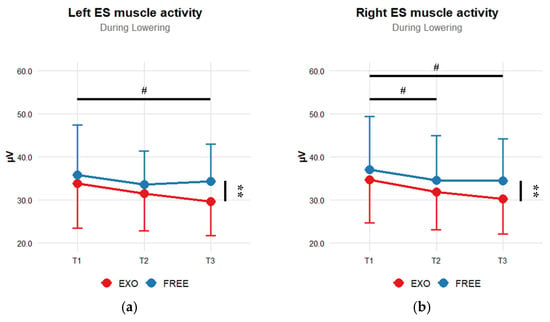
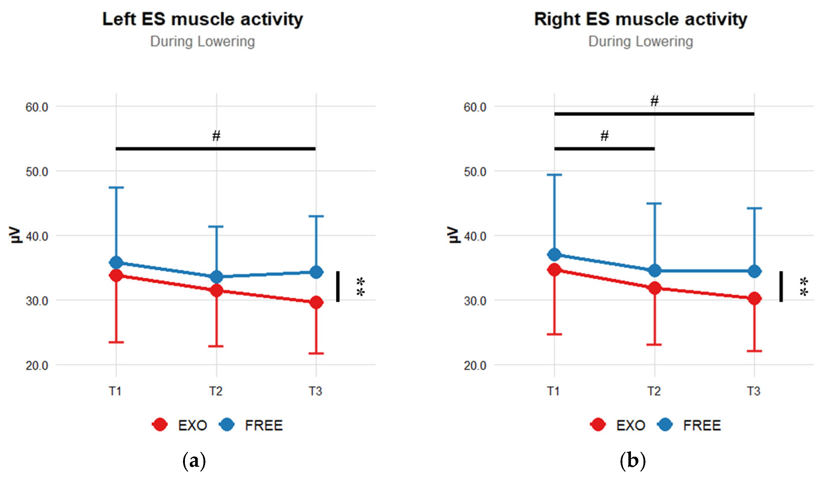
During the lowering phase, RMS values of both left and right ES muscle activity (Figure 6) were significantly reduced with the use of the soft-back exoskeleton (p < 0.01; ηp2 > 0.44, large effect size). Moreover, a significant time effect was observed for both left and right ES muscle activity (p < 0.05; ηp2 > 0.26, large effect size). Pairwise comparisons showed lower RMS values at T3 compared with T1 for both sides, and at T2 compared to T1 for the right ES muscle activity (p < 0.05; Table 2). Finally, significant interaction between exoskeleton use and time was reported for the right ES muscle activity. No significant effect was observed during the lifting phase.
Figure 6.
Comparison of mean ± SD RMS values of left (a) and right (b) erector spinae muscle activity during lowering phase. ** Significant exoskeleton main effect (p < 0.01). # Significant time effect (p < 0.05).
4. Discussion
The main purpose of this study was to assess the effects of the CORFOR® soft-back exoskeleton on lumbar spine biomechanics (loads and muscle activity) and perceived exertion in the lower back during a repetitive manual handling task performed in the sagittal plane.
The primary findings were that the lower back RPE increased significantly over the course of the task (main effect of time). However, RPE values were not significantly lower when using the exoskeleton than in the FREE condition. During both lowering and lifting phases, lumbar loads at L5-S1 were significantly reduced when using the soft-back exoskeleton. Also, peak flexion–extension moment and peak compression force significantly decreased over time in both lowering and lifting phases. Finally, significant interactions between exoskeleton use and time were observed for the peak L5-S1 flexion–extension moment and for the right ES muscle activity. Our findings partially support our hypothesis. Indeed, the soft-back exoskeleton reduced lumbar loads and ES muscle activity, and significant interactions were found for peak L5-S1 flexion–extension moment and right ES muscle activity.
4.1. Rating Perceived Exertion
The increase in lower-back RPE over time in both conditions is consistent with the cumulative demands of repetitive load handling. This rise in perceived exertion highlights that the task remained physically challenging for participants throughout its duration. However, RPE captures the subjective perception of effort and should therefore be interpreted with caution, as it integrates not only muscular demand but also sensory, cognitive, and perceptual factors, whereas biomechanical indicators (e.g., joint moments and EMG activity) provide more localized mechanical and neuromuscular information. Therefore, an increase in RPE can occur even in the absence of increased, or even in the presence of reduced, local biomechanical demands. In the present study, this divergence may reflect a growing subjective awareness of the task over time, despite reduced biomechanical loading in specific muscle groups and joints. When longer repetitive tasks are considered in the literature [13,16], a reduction in repetition frequency appears necessary to maintain a sustainable load level. Overall, this rapid increase in the rating of perceived exertion over the repetitive task supports the consideration of a threshold during such tasks. This is also in agreement with previous studies reporting that around 25 lifting actions per day increase the incidence of LBD [6,7].
The use of the soft-back exoskeleton showed a non-significant tendency to decrease the perceived physical demand at the lumbar level during the handling task (p = 0.07). This reduction ranged from approximately 11 to 13%, which is lower than the average reductions of around 36% reported [19]. This discrepancy may be explained by differences in exoskeleton design. Indeed, the studies primarily involved rigid passive back-support exoskeletons [19], whereas the device evaluated in the present study was a soft-back exoskeleton, which could provide a lower level of mechanical assistance [31].
4.2. Lumbar Loads
Musculoskeletal analyses showed significant reductions in peak flexion–extension moment, peak compression force, and peak shear force at the L5-S1 intervertebral joint when using the soft-back exoskeleton. In the present study, reductions at the L5-S1 intervertebral joint when using the exoskeleton were 14% for the peak flexion–extension moment, roughly 6% for the peak compression force, and approximately 10% for the peak shear force during both lowering and lifting phases.
These results are comparable, with reductions in lumbar mechanical demands observed when wearing the CORFOR® soft-back exoskeleton during a sagittal-plane manual material handling task performed with a stoop technique, including decreases of approximately 14% in peak lumbar flexion moment, 6% in peak compressive force, and 11% in peak shear force at the L5–S1 joint [29]. Similar trends have been reported for other passive exoskeletons during sagittal-plane lifting tasks, with observed decreases in lumbar loading ranging from 14% to 34% for peak flexion–extension moments and from 12% to 28% for peak compressive forces [26,31].
As the musculoskeletal model used in this study uses marker kinematics and a constant handled weight (68.7 N) as inputs, changes in joint loads over time (reaction force and moment) would only be attributed to changes in kinematics. However, statistical analyses revealed no significant differences across the three measurement times. Specifically, the angular amplitudes of the intervertebral joints, as well as the peak angular (hips) and vertical (box) velocities and accelerations, remained unchanged. This absence of significant differences in lumbar spine and hip kinematics, and in the kinematics of the handled load, indicates that neither the use of the soft-back exoskeleton nor the measurement period had a substantial effect on overall movement patterns during the lifting and lowering phases. These findings are inconsistent with previous studies reporting that passive or soft-back exoskeletons influenced lumbar spine and hip joint kinematics [29,31]. However, these studies did not involve a repetitive task. It is therefore possible that motor strategies would only slightly differ when using the exoskeleton and repetitions accumulate. Therefore, even if the classical kinematic parameters did not change during the protocol, subtle changes still led to alterations in lumbar loads with the exoskeleton and across the repetitive task.
Also, lumbar loads were estimated using a static optimization-based approach available in OpenSim in the present study. Consequently, muscle activation patterns were inferred from cost-function minimization rather than experimentally measured neuromuscular activity. Previous studies have shown that subject-specific thoracolumbar musculoskeletal models implemented in OpenSim can reproduce experimentally measured back extensor EMG during dynamic lifting tasks when using EMG-driven approaches [43]. Therefore, the use of an EMG-driven framework in the present study could have resulted in different muscle force distributions and, in turn, different estimates of lumbar loading. However, these approaches are constrained by inherent EMG limitations, such as restricted access to superficial muscles, susceptibility to noise, and MVC-based normalization artifacts [44].
4.3. Erector Spinae Muscle Activity
Together with the reduced lumbar loads, ES muscle activity decreased with the use of the soft-back exoskeleton, but only during the lowering phase. Moreover, time-related effects on ES muscle activity appear less pronounced than those observed for lumbar loads parameters. This limited temporal modulation of ES muscle activity may be explained by the lack of changes in kinematics, as joint angles and velocities strongly influence spinal muscle recruitment [45], which likely contributed to less pronounced effects in ES activity over time. Although these effects are less pronounced, the reductions observed over time may partly reflect a progressive neuromuscular adaptation developing during task execution.
Indeed, EMG-driven and optimization-based modeling studies have shown that when movement patterns remain similar, changes in muscle activity may be subtle, especially when only a single muscle group such as the erector spinae is monitored [46]. Moreover, the task involved moderate load handling (7 kg) over a relatively short duration (4 min), which may not have been sufficient to induce fatigue-related changes in ES recruitment, explaining the stability of EMG signals across measurement periods. Furthermore, coordination among trunk muscles may have compensated for the mechanical assistance provided by the exoskeleton, without changing the measured ES activity [29]. Overall, these results indicate that under the tested conditions, the CORFOR® soft-back exoskeleton does not significantly reduce ES activation, contrary to existing literature, which reported roughly 7% reduction in lower-back muscle activity with exoskeleton use [21,22]. This further suggests that the assistive device may reduce lumbar load through a whole redistribution of muscular effort rather than a specific support of the erector spinae muscles. Future studies incorporating additional trunk muscles and longer-duration tasks could provide further insight into the neuromuscular adaptations induced by exoskeleton use.
4.4. Limitations
Several methodological considerations should be acknowledged. First, the participants in this study were young and healthy males, which limits the applicability of the findings to more diverse working populations. In particular, experienced workers, older adults, or individuals with a history of low-back pain may respond differently to the exoskeleton assistance due to age-related changes in muscle mass and strength, altered motor strategies, or pain-related adaptations. In addition, sex-related differences in anthropometry and pelvic morphology may influence movement patterns and user–exoskeleton interactions, potentially affecting the observed biomechanical and neuromuscular responses. Second, the task was highly controlled and standardized to improve reproducibility, but this design limits the field validity of the results, as real-word work situations typically involve greater variability in movements and postures. Moreover, although the task duration does not fully replicate the longer durations typically observed in occupational settings, the present protocol was not designed to induce substantial fatigue but rather to ensure exposure to repetitive lifting. Nevertheless, a longer protocol duration would imply a lower movement frequency and may not provide more relevant biomechanical evidence regarding repetition and exoskeleton effects.
Third, regarding the variability of EMG signals and the lack of consensus regarding normalization procedure, the results should be interpreted with caution. In this study, EMG data were not normalized. However, such comparisons can be meaningful within the same participant, muscle, and session [35].
Then, the measured kinematic and EMG variables may not have been sensitive enough to detect subtle changes induced by the soft-back exoskeleton, whose level of mechanical assistance is likely lower than that provided by rigid passive devices [31].
5. Conclusions
In summary, we evaluated the effects of a soft-back exoskeleton on lumbar spine biomechanics and perceived exertion during a repetitive sagittal handling task. Using the CORFOR® soft-back exoskeleton progressively decreased flexion–extension moment and compression force over the course of the task regardless of the movement phase. Despite significant reductions in lumbar loads and ES muscle activity using the exoskeleton, RPE did not significantly decreased. However, while RPE at the lower back increased over time throughout the task, flexion–extension moment and compression force decreased progressively over the course of the task regardless of the movement phase. These results occurred without any changes in overall movement patterns.
Taken together, these findings suggest that the CORFOR® soft-back exoskeleton may reduce lumbar mechanical demands and muscle activity primarily through biomechanical or neuromuscular adjustments rather than through obvious modifications of movement patterns. The observed effects are consistent with the design intent of soft passive exoskeletons, which aim to assist the user while preserving natural movement strategies. Future research should focus on longer-duration and higher-intensity tasks (and consider fatigue parameters), include more diverse worker populations, and integrate complementary modeling approaches and measurements to further better understand the mechanisms underlying the interaction between exoskeletons, spinal loading, and user perception in occupational settings.
Author Contributions
Conceptualization, C.T., A.F. and G.M.; methodology, C.T., A.F. and G.M.; software, C.T. and A.F.; validation, C.T., A.F., X.P. and G.M.; formal analysis, C.T. and A.F.; investigation, C.T. and A.F.; resources, G.M.; data curation, C.T.; writing—original draft preparation, C.T. and A.F.; writing—review and editing, C.T., A.F. and G.M.; visualization, C.T., A.F., X.P. and G.M.; supervision, X.P. and G.M.; project administration, X.P. and G.M.; funding acquisition, X.P. and G.M. All authors have read and agreed to the published version of the manuscript.
Funding
Association Nationale de la Recherche et de la Technologie: 2021/0922.
Institutional Review Board Statement
The study was conducted in accordance with the Declaration of Helsinki, and approved by the Ethics Committee of CERSTAPS (no. IRB00012476-2023-17-01-221, 17 January 2023).
Informed Consent Statement
Informed consent was obtained from all subjects involved in the study.
Data Availability Statement
The data presented in this study are available on request from the corresponding author. The data are not publicly available due to ethical regulations.
Conflicts of Interest
The authors declare no conflicts of interest.
Abbreviations
The following abbreviations are used in this manuscript:
| LBD | Low back disorders |
| LBP | Low back pain |
| ES | Erector spinae |
| EXO | Condition with the exoskeleton |
| FREE | Condition without the exoskeleton |
| sEMG | Surface electromyography |
| RPE | Rating perceived exertion |
| RMS | Root mean square |
| LFB | Lifting full-body |
| SD | Standard deviation |
| MVC | Maximal voluntary contraction |
References
- Marras, W.S. Occupational low back disorder causation and control. Ergonomics 2000, 43, 880–902. [Google Scholar] [CrossRef] [Scilit]
- Goswami, S.; Dasgupta, S.; Samanta, A.; Talukdar, G.; Chanda, A.; Ray Karmakar, P.; Chakrabarti, A. Load handling and repetitive movements are associated with chronic low back pain among jute mill workers in India. Pain Res. Treat. 2016, 2016, 7843216. [Google Scholar] [CrossRef] [Scilit]
- Sambeko, B.E.M.; Susanto, N.; Alfanan, A. Manual handling as contributor of low back pain for workers: A case study at PT Sumber Mandiri Jaya, Kabupaten Merauke. Indones. J. Occup. Saf. Health 2024, 13, 29–36. [Google Scholar] [CrossRef] [Scilit]
- Holtermann, A.; Clausen, T.; Jørgensen, M.B.; Burdorf, A.; Andersen, L.L. Patient handling and risk for developing persistent low-back pain among female healthcare workers. Scand. J. Work Environ. Health 2013, 39, 164–169. [Google Scholar] [CrossRef] [Scilit]
- Marras, W.S.; Lavender, S.A.; Leurgans, S.E.; Rajulu, S.L.; Allread, S.G.; Fathallah, F.A.; Ferguson, S.A. The role of dynamic three-dimensional trunk motion in occupationally-related low back disorders. Spine 1993, 18, 617–628. [Google Scholar] [CrossRef] [Scilit] [PubMed]
- Coenen, P.; Gouttebarge, V.; van der Burght, A.S.; van Dieën, J.H.; Frings-Dresen, M.H.; van der Beek, A.J.; Burdorf, A. The effect of lifting during work on low back pain: A health impact assessment based on a meta-analysis. Occup. Environ. Med. 2014, 71, 871–877. [Google Scholar] [CrossRef] [Scilit]
- Kelsey, J.L.; Githens, P.B.; White, A.A.; Holford, T.R.; Walter, S.D.; O’Connor, T.; Calogero, J.A. An epidemiologic study of lifting and twisting on the job and risk for acute prolapsed lumbar intervertebral disc. J. Orthop. Res. 1984, 2, 61–66. [Google Scholar] [CrossRef] [Scilit] [PubMed]
- Albers, J.T.; Hudock, S.D. Biomechanical assessment of three rebar tying techniques. Int. J. Occup. Saf. Ergon. 2007, 13, 279–289. [Google Scholar] [CrossRef] [Scilit] [PubMed]
- Norman, R.; Wells, R.; Neumann, P.; Frank, J.; Shannon, H.; Kerr, M.; The Ontario Universities Back Pain Study (OUBPS) Group. A comparison of peak vs cumulative physical work exposure risk factors for the reporting of low back pain in the automotive industry. Clin. Biomech. 1998, 13, 561–573. [Google Scholar] [CrossRef] [Scilit]
- Li, X.; Komeili, A.; Gül, M.; El-Rich, M. A framework for evaluating muscle activity during repetitive manual material handling in construction manufacturing. Autom. Constr. 2017, 79, 39–48. [Google Scholar] [CrossRef] [Scilit]
- Madinei, S.; Nussbaum, M.A. Estimating lumbar spine loading when using back-support exoskeletons in lifting tasks. J. Biomech. 2023, 147, 111439. [Google Scholar] [CrossRef] [Scilit]
- Madinei, S.; Alemi, M.M.; Kim, S.; Srinivasan, D.; Nussbaum, M.A. Biomechanical assessment of two back-support exoskeletons in symmetric and asymmetric repetitive lifting with moderate postural demands. Appl. Ergon. 2020, 88, 103156. [Google Scholar] [CrossRef] [Scilit] [PubMed]
- Oomen, N.M.; Graham, R.B.; Fischer, S.L. Exploring the role of task on kinematic variability and assessing consistency in individual responses across repetitive manual tasks. Ergonomics 2023, 66, 749–761. [Google Scholar] [CrossRef] [Scilit] [PubMed]
- Schmalz, T.; Colienne, A.; Bywater, E.; Fritzsche, L.; Gärtner, C.; Bellmann, M.; Ernst, M. A passive back-support exoskeleton for manual materials handling: Reduction of low back loading and metabolic effort during repetitive lifting. IISE Trans. Occup. Ergon. Hum. Factors 2022, 10, 7–20. [Google Scholar] [CrossRef] [Scilit]
- Schwartz, M.; Desbrosses, K.; Theurel, J.; Mornieux, G. Using passive or active back-support exoskeletons during a repetitive lifting task: Influence on cardiorespiratory parameters. Eur. J. Appl. Physiol. 2022, 122, 2575–2583. [Google Scholar] [CrossRef] [Scilit]
- Zeng, D.; Qu, S.; Ma, T.; Yin, P.; Gao, H.; Zhao, N.; Xia, Y. The assist performance test of industrial passive waist-assistant exoskeleton on fatigue during a repetitive lifting task. J. Phys. Conf. Ser. 2021, 1748, 062039. [Google Scholar] [CrossRef] [Scilit]
- De Looze, M.P.; Bosch, T.; Krause, F.; Stadler, K.S.; O’Sullivan, L.W. Exoskeletons for industrial application and their potential effects on physical work load. Ergonomics 2016, 59, 671–681. [Google Scholar] [CrossRef] [Scilit]
- Dos Anjos, F.V.; Ghislieri, M.; Cerone, G.L.; Pinto, T.P.; Gazzoni, M. Changes in the distribution of muscle activity when using a passive trunk exoskeleton depend on the type of working task: A high-density surface EMG study. J. Biomech. 2022, 130, 110846. [Google Scholar] [CrossRef] [Scilit]
- Kermavnar, T.; De Vries, A.W.; De Looze, M.P.; O’Sullivan, L.W. Effects of industrial back-support exoskeletons on body loading and user experience: An updated systematic review. Ergonomics 2021, 64, 685–711. [Google Scholar] [CrossRef] [Scilit] [PubMed]
- Theurel, J.; Desbrosses, K. Occupational exoskeletons: Overview of their benefits and limitations in preventing work-related musculoskeletal disorders. IISE Trans. Occup. Ergon. Hum. Factors 2019, 7, 264–280. [Google Scholar] [CrossRef] [Scilit]
- Schwartz, M.; Theurel, J.; Desbrosses, K. Effectiveness of soft versus rigid back-support exoskeletons during a lifting task. Int. J. Environ. Res. Public Health 2021, 18, 8062. [Google Scholar] [CrossRef] [Scilit]
- Thevenot, C.; Pierre, X.; Mornieux, G. Effects of an occupational soft-back exoskeleton during order picking: A field study in logistics. Ergonomics 2025, 68, 1984–1997. [Google Scholar] [CrossRef] [Scilit] [PubMed]
- Pesenti, M.; Antonietti, A.; Gandolla, M.; Pedrocchi, A. Towards a functional performance validation standard for industrial low-back exoskeletons: State of the art review. Sensors 2021, 21, 808. [Google Scholar] [CrossRef] [Scilit]
- Gallagher, S.; Marras, W.S. Tolerance of the lumbar spine to shear: A review and recommended exposure limits. Clin. Biomech. 2012, 27, 973–978. [Google Scholar] [CrossRef] [Scilit] [PubMed]
- Baltrusch, S.J.; Van Dieen, J.H.; Van Bennekom, C.A.; Houdijk, H. Testing an exoskeleton that helps workers with low-back pain: Less discomfort with the passive spexor trunk device. IEEE Robot. Autom. Mag. 2019, 27, 66–76. [Google Scholar] [CrossRef]
- Johns, J.; Schultes, I.; Heinrich, K.; Potthast, W.; Glitsch, U. Biomechanical analysis of different back-supporting exoskeletons regarding musculoskeletal loading during lifting and holding. J. Biomech. 2024, 168, 112125. [Google Scholar] [CrossRef] [Scilit]
- Alemi, M.M.; Madinei, S.; Kim, S.; Srinivasan, D.; Nussbaum, M.A. Effects of two passive back-support exoskeletons on muscle activity, energy expenditure, and subjective assessments during repetitive lifting. Hum. Factors 2020, 62, 458–474. [Google Scholar] [CrossRef] [Scilit] [PubMed]
- Ratke, E.J.; Mcmaster, H.; Vellucci, C.L.; Larson, D.J.; Holmes, M.W.; Beaudette, S.M. Ability of a passive back support exoskeleton to mitigate fatigue related adaptations in a complex repetitive lifting task. J. Biomech. 2025, 181, 112553. [Google Scholar] [CrossRef] [Scilit]
- Favennec, A.; Moissenet, F.; Frère, J.; Mornieux, G. Effects of a soft back exoskeleton on lower lumbar spine loads during manual materials handling: A musculoskeletal modelling study. Comput. Methods Biomech. Biomed. Eng. 2024, 27, 967–975. [Google Scholar] [CrossRef] [Scilit]
- Koopman, A.S.; Näf, M.; Baltrusch, S.J.; Kingma, I.; Rodriguez-Guerrero, C.; Babič, J.; van Dieën, J.H. Biomechanical evaluation of a new passive back support exoskeleton. J. Biomech. 2020, 105, 109795. [Google Scholar] [CrossRef] [Scilit]
- Toxiri, S.; Näf, M.B.; Lazzaroni, M.; Fernández, J.; Sposito, M.; Poliero, T.; Ortiz, J. Back-support exoskeletons for occupational use: An overview of technological advances and trends. IISE Trans. Occup. Ergon. Hum. Factors 2019, 7, 237–249. [Google Scholar] [CrossRef] [Scilit]
- Favennec, A.; Frère, J.; Mornieux, G. Changes in human motor behavior during the familiarization with a soft back-support occupational exoskeleton. Appl. Sci. 2024, 14, 1160. [Google Scholar] [CrossRef] [Scilit]
- Hermens, H.J.; Freriks, B.; Disselhorst-Klug, C.; Rau, G. Development of recommendations for SEMG sensors and sensor placement procedures. J. Electromyogr. Kinesiol. 2000, 10, 361–374. [Google Scholar] [CrossRef] [Scilit]
- Borg, G.A. Psychophysical bases of perceived exertion. Med. Sci. Sports Exerc. 1982, 14, 377–381. [Google Scholar] [CrossRef] [Scilit]
- Besomi, M.; Hodges, P.W.; Clancy, E.A.; Van Dieën, J.; Hug, F.; Lowery, M.; Tucker, K. Consensus for experimental design in electromyography (CEDE) project: Amplitude normalization matrix. J. Electromyogr. Kinesiol. 2020, 53, 102438. [Google Scholar] [CrossRef] [Scilit] [PubMed]
- Beaucage-Gauvreau, E.; Robertson, W.S.; Brandon, S.C.; Fraser, R.; Freeman, B.J.; Graham, R.B.; Jones, C.F. Validation of an OpenSim full-body model with detailed lumbar spine for estimating lower lumbar spine loads during symmetric and asymmetric lifting tasks. Comput. Methods Biomech. Biomed. Eng. 2019, 22, 451–464. [Google Scholar] [CrossRef] [Scilit] [PubMed]
- Delp, S.L.; Anderson, F.C.; Arnold, A.S.; Loan, P.; Habib, A.; John, C.T.; Thelen, D.G. OpenSim: Open-source software to create and analyze dynamic simulations of movement. IEEE Trans. Biomed. Eng. 2007, 54, 1940–1950. [Google Scholar] [CrossRef] [Scilit]
- Akhavanfar, M.; Uchida, T.K.; Clouthier, A.L.; Graham, R.B. Sharing the load: Modeling loads in OpenSim to simulate two-handed lifting. Multibody Syst. Dyn. 2022, 54, 213–234. [Google Scholar] [CrossRef] [Scilit]
- Scheirer, C.J.; Ray, W.S.; Hare, N. The analysis of ranked data derived from completely randomized factorial designs. Biometrics 1976, 32, 429–434. [Google Scholar] [CrossRef] [Scilit]
- R Core Team. R: A Language and Environment for Statistical Computing; R Foundation for Statistical Computing: Vienna, Austria, 2022. [Google Scholar]
- Cohen, J.; Cohen, P. Applied Multiple Regression/Correlation Analysis for the Behavioral Sciences, 1st ed.; Lawrence Erlbaum Associates: Mahwah, NJ, USA, 1983. [Google Scholar]
- Cohen, J. Statistical Power Analysis for the Behavioral Sciences, 1st ed.; Lawrence Erlbaum Associates: Mahwah, NJ, USA, 1988. [Google Scholar]
- Alemi, M.M.; Banks, J.J.; Lynch, A.C.; Allaire, B.T.; Bouxsein, M.L.; Anderson, D.E. EMG validation of a subject-specific thoracolumbar spine musculoskeletal model during dynamic activities in older adults. Ann. Biomed. Eng. 2023, 51, 2313–2322. [Google Scholar] [CrossRef] [Scilit]
- Abdullah, M.; Hulleck, A.A.; Katmah, R. Multibody dynamics-based musculoskeletal modeling for gait analysis: A systematic review. J. Neuroeng. Rehabil. 2024, 21, 178. [Google Scholar] [CrossRef] [Scilit] [PubMed]
- McGill, S.M.; Grenier, S.; Kavcic, N.; Cholewicki, J. Coordination of muscle activity to assure stability of the lumbar spine. J. Electromyogr. Kinesiol. 2003, 13, 353–359. [Google Scholar] [CrossRef] [Scilit] [PubMed]
- Moya-Esteban, A.; van der Kooij, H.; Sartori, M. Robust estimation of lumbar joint forces in symmetric and asymmetric lifting tasks via large-scale electromyography-driven musculoskeletal models. J. Biomech. 2022, 144, 111307. [Google Scholar] [CrossRef] [Scilit] [PubMed]

Disclaimer/Publisher’s Note: The statements, opinions and data contained in all publications are solely those of the individual author(s) and contributor(s) and not of MDPI and/or the editor(s). MDPI and/or the editor(s) disclaim responsibility for any injury to people or property resulting from any ideas, methods, instructions or products referred to in the content. |
© 2026 by the authors. Licensee MDPI, Basel, Switzerland. This article is an open access article distributed under the terms and conditions of the Creative Commons Attribution (CC BY) license.