Article

Computed Tomographic Evaluation of the Superior Mesenteric and Hepatic Arteries and Their Clinical Significance


by
Ali Abduwani
1,
Ilyas Al-Saadi
1,
Mohammed Al-Hajri
1,
Al-Khatab Abdullah Saud Ismaili
2,
Nasser Al Sidairi
2,
Ahmed Al Lawati
3,
Mahmood Salim Nasser Al Riyami
3,
Saleh Baawain
3 and
Srijit Das
1,*
1
Department of Human & Clinical Anatomy, College of Medicine & Health Sciences, Sultan Qaboos University, Muscat 123, Oman
2
Radiology Program, Oman Medical Specialty Board, Muscat 132, Oman
3
Department of Radiology & Molecular Imaging, Sultan Qaboos University Hospital, Muscat 123, Oman
*
Author to whom correspondence should be addressed.
Appl. Sci. 2026, 16(9), 4265; https://doi.org/10.3390/app16094265
Submission received: 29 January 2026 / Revised: 15 April 2026 / Accepted: 22 April 2026 / Published: 27 April 2026

Abstract

The superior mesenteric artery (SMA) is the second unpaired ventral branch of the abdominal aorta. The SMA supplies the abdominal organs that develop from the midgut. This study investigated the morphological characteristics of the SMA by (i) measuring its diameter in different sexes; (ii) assessing the vertical distance between the SMA and inferior mesenteric artery (IMA) origins in males and females, and (iii) observing if the hepatic artery arose from the SMA instead of the celiac trunk. This retrospective cross-sectional study included the contrast-enhanced CT angiograms of 260 patients (n = 205 males and 55 females) who attended the Radiology department at Sultan Qaboos University Hospital from 1 January 2021 to 31 December 2023. All included patients were aged 19–50 years and had no history of vascular pathology that altered the vascular dimensions, nor had any history of major abdominal trauma or abdominal surgeries. The mean diameter of the SMA in the study population was 7.51 ± 1.11 mm. The mean diameter of the SMA was found to be wider in males (7.73 ± 1.05 mm) compared to females (6.71 ± 0.96 mm, p < 0.001). The mean distance between the SMA and IMA was 62.67 ± 10.91 mm. The average distance between SMA and IMA in males and females was found to be 63.36 ± 10.67 mm and 60.09 ± 11.48 mm (p = 0.048), respectively. Incidence of the right hepatic artery originating from SMA, accessory right hepatic artery, and common hepatic artery originating from SMA was 7.7%, 0.38%, and 2.3%, respectively. Prior anatomical knowledge of arteries is important for occlusion, bypass grafting, and endovascular surgeries involving SMA.

1. Introduction

The superior mesenteric artery (SMA) is the main artery that supplies blood to the midgut and the majority of the gastrointestinal tract. It is the second of the three unpaired ventral branches of the abdominal aorta (AA) and supplies the distal portion of the duodenum, jejunum, ileum, cecum, appendix, ascending colon, and the proximal two-thirds of the transverse colon [1]. Owing to its extensive vascular territory, the SMA is essential for maintaining normal gastrointestinal function. Pathology affecting this vessel can lead to significant clinical consequences. A thorough understanding of its anatomy, embryological development, branching patterns, vascular connections, and related pathologies is essential for clinicians, surgeons, and radiologists [1].
Functionally, the SMA plays a critical role in meeting the metabolic demands of the intestine, particularly in the postprandial state when mesenteric blood flow increases substantially. Regulation of SMA blood flow is mediated through a complex interaction of metabolic, hormonal, and neural mechanisms [2]. Advances in imaging technology have significantly improved the evaluation of SMA anatomy and pathology. Computed tomography angiography and magnetic resonance angiography are now widely used non-invasive modalities for detailed assessment of the SMA and its variations [3].
The celiac trunk typically gives rise to three branches: the left gastric artery (LGA), the splenic artery, and the common hepatic artery (CHA). The CHA further divides into proper hepatic artery, gastroduodenal artery, and right gastric artery, with the proper hepatic artery giving rise to the right and left hepatic arteries [4].A recent meta-analysis reported that the classical branching pattern of the celiac trunk was present in 75.15% of individuals, whereas anatomical variations were identified in the remaining 24.85% [5]. Uflacker’s classification is among the most widely used systems for describing anatomical variations in the celiac trunk and has been extensively described in the literature [6,7]. Although hepatic arteries usually originate from the celiac trunk either directly or indirectly, anatomical variations are relatively common. Previous studies have reported hepatic arterial variants in approximately 13% to 48% of individuals [8]. Multiple classification systems have been proposed to describe these variations, which may include ectopic origins or the presence of aberrant or accessory branches [9].
Anatomical variations in the SMA are associated with several clinically significant conditions. SMA diameter is an important diagnostic parameter in spontaneous isolated superior mesenteric artery dissection (SISMAD) and non-occlusive mesenteric ischemia (NOMI) [10,11]. In addition, the vertical distance between the SMA and the IMA is clinically relevant due to the presence of colonic watershed areas, which represent transitional zones between major arterial territories [12]. Because perfusion to these regions is often variable and limited, they are particularly vulnerable to ischemia and may predispose patients to ischemic colitis [13]. Furthermore, variations in the branching patterns of the SMA and hepatic arteries are of particular importance during preoperative planning for abdominal surgeries, including liver transplantation, hepatic tumor resection, pancreatic resection, pancreatoduodenectomy, and biliary procedures, as recognition of these variants can help prevent intraoperative vascular injury and reduce hemorrhagic complications [14].
The common hepatic artery most commonly arises from the celiac trunk [4]. However, less frequently, it may originate from the SMA or directly from the abdominal aorta. Similarly, while the right and left hepatic arteries typically arise from the proper hepatic artery, the right hepatic artery may originate from the SMA as a replaced right hepatic artery, and the left hepatic artery may arise from the LGA as a replaced left hepatic artery. In other cases, the hepatic arteries may have a normal origin, with additional accessory branches. An aberrant right hepatic artery most commonly arises from the SMA, whereas an aberrant left hepatic artery typically originates from the LGA [15].
The SMA serves as a major anatomical landmark that must be preserved during abdominal surgical procedures, particularly pancreatic operations such as pancreaticoduodenectomy. It is also of critical importance in aortic surgeries, especially those involving aneurysms or dissections, where injury to the SMA may result in catastrophic bowel ischemia [16]. Anatomical variations in the SMA have been reported in up to 25% of individuals, and such variations may influence surgical approaches as well as the presentation and severity of ischemic disease [17].
This study aimed to quantify the diameter of the SMA and compare it between sexes, measure the vertical distance between the SMA and IMA, and identify variations in the hepatic artery. Previous studies have primarily evaluated these parameters independently, focusing on arterial diameter, branching patterns, or isolated clinical correlations. In contrast, the present study employs an integrated approach by simultaneously assessing SMA diameter, SMA-IMA distance, and variations in the hepatic arteries. This combined morphometric and anatomical analysis provides a more comprehensive evaluation of SMA-related vascular anatomy and facilitates improved correlation with clinical relevance, addressing a gap in the current literature.

2. Materials and Methods

This retrospective cross-sectional study included contrast-enhanced CT angiograms of 260 patients (n = 205 males, n = 55 females).

2.1. Inclusion Criteria

All patients who attended the Radiology Department at Sultan Qaboos University Hospital between 1 January 2021 and 31 December 2023 were eligible for inclusion if they were aged between 19 and 50 years and underwent contrast-enhanced CT scans of the abdomen. The primary indication for imaging in this cohort was evaluation as part of a trauma protocol (e.g., road traffic accidents or other causes). Only patients with no evidence of abdominal injury or pathology known to affect vessel diameter and vascular compliance were included in the study.

2.2. Exclusion Criteria

All patients who (i) had a history of any pathological condition known to directly impact the dimensions of the vessels, or (ii) had undergone abdominal surgeries, were excluded from the study.
The selected age range (19–50 years) was chosen to assess vascular morphology within a stable adult population while minimizing confounding from age-related changes. Older individuals were excluded to reduce the influence of atherosclerosis, degenerative vascular remodeling, and arterial calcification, which are known to affect vessel diameter and vascular compliance [18,19].

2.3. Radiological Investigations

CT angiography was performed using two multidetector CT scanners available in the Radiology Department at SQUH. The first was a 256-slice GE CT scanner (GE Healthcare, Chicago, IL, USA), and the second was a Siemens SOMATOM Force CT scanner (Siemens Healthinners, Erlangen, Germany) with a 192 × 2 detector configuration. For the SOMATOM Force system, acquisition parameters included dual X-ray tubes (Vectron), a 2 × 192 detector configuration, a gantry scan range of 78 cm, a rotation time of 0.25 s, a temporal resolution of 66 ms, a spatial resolution of 0.24 mm, and tube voltage ranging from 70–150 kV depending on patient characteristics. For the GE system, imaging was performed with a 256-slice configuration, a gantry aperture of 80 cm, a temporal resolution of 140 ms, and a spatial resolution of 0.23 mm.
Two radiologists at our institution participated in image interpretation. The CT angiograms were divided between the two experts, and each examination was independently analyzed by a single radiologist according to a predefined measurement protocol. The data were then collected in a spreadsheet. The SMA diameter was measured on arterial-phase axial images at a point distal to its origin from the abdominal aorta and proximal to the first major branch. Measurements were obtained from outer wall to outer wall. The distance between the SMA and IMA was measured from the inferior border of the origin of the SMA to the superior border of the origin of the IMA in the sagittal view. The axial view was utilized to observe whether any hepatic artery (common hepatic, right hepatic, or left hepatic arteries) originated from the SMA rather than the celiac trunk. The anatomy of the hepatic artery was classified according to Michel’s classification, which describes 10 different variants [20].

2.4. Data Analysis

Statistical analysis was performed using IBM SPSS Statistics (version 26). Continuous variables were expressed as mean ± standard deviation (SD), and categorical variables as frequencies and percentages. Normality of continuous variables was assessed using the Kolmogorov–Smirnov test and showed no significant deviation from normality; therefore, parametric tests were applied. Differences in the diameter of the SMA and the distance between the SMA and IMA between males and females were evaluated using the independent samples t-test. Pearson correlation analysis was used to assess the relationship between age and SMA diameter. Simple linear regression analysis was performed to evaluate the predictive effect of age on SMA diameter. A p-value < 0.05 was considered statistically significant.

3. Results

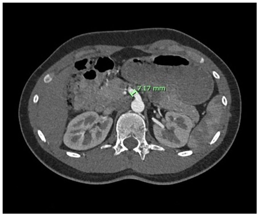
The study sample comprised 260 participants (78.8% were males, and 21.2% were females). The mean ± standard deviation (SD) age was 33.6 ± 8.1 years. The mean SMA diameter in the study population of 260 patients was found to be 7.51 ± 1.11 mm. The mean diameter of the SMA in males was found to be 7.73 ± 1.05 mm, while in females, the mean diameter of the SMA was found to be 6.71 ± 0.96 mm. The difference in the diameter of the SMA between males and females was found to be statistically significant (p < 0.001). Figure 1 shows the diameter of the SMA. The comparison of SMA diameter between sexes is shown in Table 1. Pearson correlation demonstrated no significant association between age and SMA diameter (r = −0.030, p = 0.632). Linear regression analysis similarly showed that age was not a significant predictor of SMA diameter (B = −0.004, R2 = 0.001, p = 0.632).
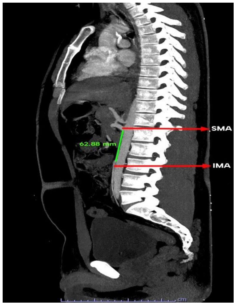
The average distance between the SMA and IMA in 260 patients was 62.67 ± 10.91 mm. The average distance between the SMA and IMA among males was 63.36 ± 10.67 mm. In females, the average distance between the two arteries was 60.09 ± 11.48 mm. The difference in the distance between the two arteries was statistically significant between males and females (p = 0.048). Figure 2 shows the distance between the SMA and IMA. The comparison of the distance between SMA and IMA between males and females is shown in Table 2.
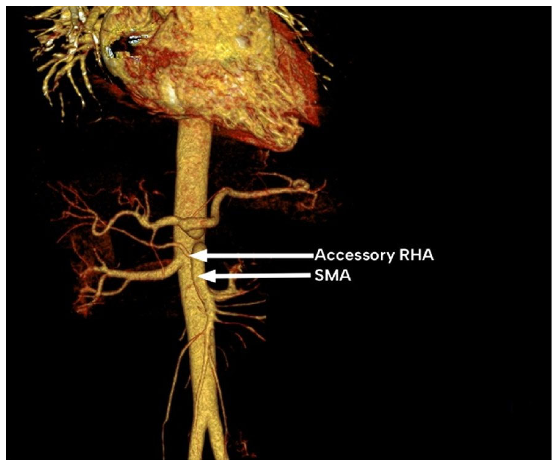
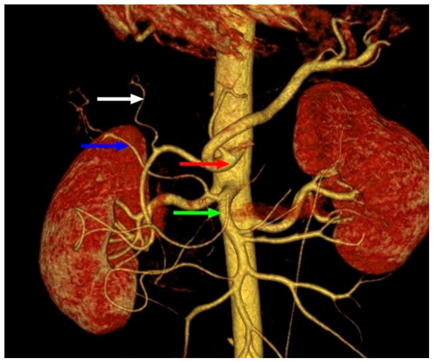
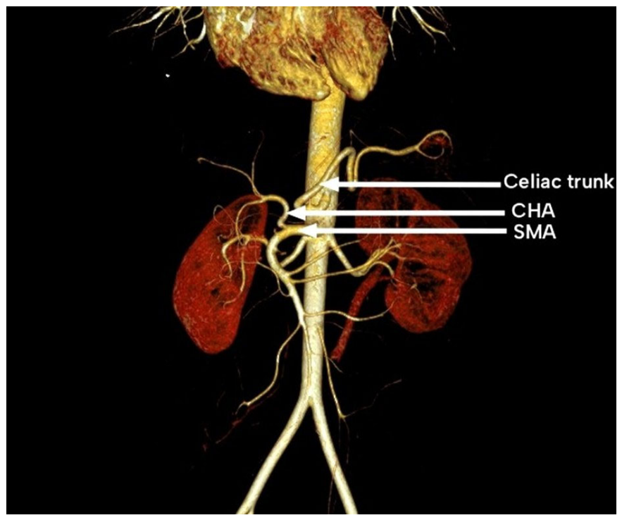
In 228 (87.7%) patients, normal hepatic artery anatomy was observed, with all the hepatic arteries originating from the CT as direct or indirect branches (Michel’s type I). In 20 patients (7.7%), the RHA originated from the SMA, whereas the LHA originated from the CHA (a branch of the celiac trunk). This variation is classified as Michel’s type III. Figure 3 shows the CT scan of a patient in whom Michel’s type III was present. In one patient (0.38%), an accessory RHA was found to originate from the SMA (Michel’s type VI). Figure 4 shows the CT scan of the patient with Michel’s type VI. In 6 patients (2.3%), the entire CHA originated from the SMA rather than the CT. This variation was described as Michel’s type IX. Figure 5 shows a CT scan of a patient in whom Michel’s type IX variation was present.
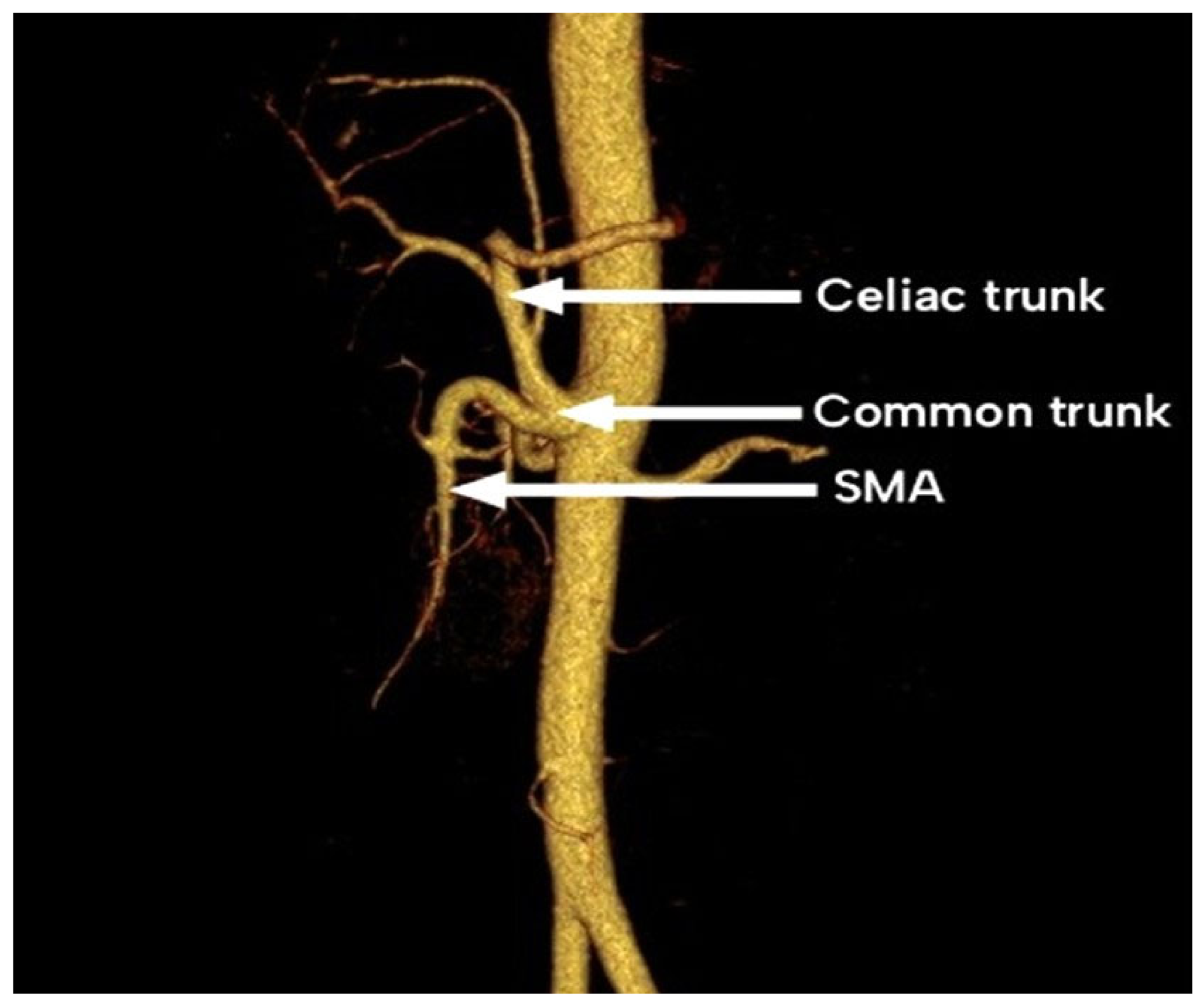
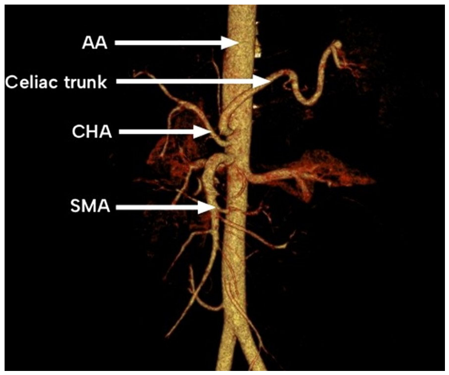
In the other five patients, an anatomical variation was observed that did not fit any of the classes described by Michel. In three patients (1.15%), the celiac trunk and the SMA arose from a single trunk. This variation was not described in Michel’s classification, but was described as Uflacker’s type VI as the celiac-mesenteric trunk. Figure 6 shows a CT scan of a patient demonstrating a common origin of the celiac trunk and the SMA. In another patient (0.38%), the CHA originated as a direct branch from the AA. Figure 7 shows a CT scan of a patient in whom the CHA originated directly from the AA. In the last patient (0.38%), the LHA originated from the SMA, whereas the RHA arose from the celiac trunk.

4. Discussion

Sex-based differences in the diameter of the superior mesenteric artery (SMA) have been previously investigated. A study from Brazil (2022) analyzed CT angiograms of 150 adults (n = 117 males and 33 females) and reported a significantly greater SMA diameter in males compared with females [21]. Table 3 presents a comparison of SMA diameters between sexes as reported by three other studies. In the present study, the observed differences in vascular diameter should be interpreted with caution because of the unequal sex distribution in the cohort (male-to-female ratio 3.73:1), which may limit the generalizability of these findings.
Further supporting the findings of both the previous study and the present study, a Brazilian study conducted in 2020 investigated the influence of sex and age on the diameter and angulation of the abdominal aorta and its branches, using CT data from 157 patients (n = 69 males and 88 females) [23]. The authors reported a statistically significant difference in arterial diameter between males and females [23]. In contrast, some studies have found no significant sex-based differences. For instance, a study from Turkey in 2021, which analyzed data from 210 participants (49% males, 51% females) collected between January and March 2019, reported no statistically significant difference in SMA diameter between sexes [24].
The diameter of the SMA varies considerably among individuals, and such variability may have important implications for endovascular therapy. Accurate knowledge of typical SMA diameters can guide stent or graft selection and influence procedural outcomes. In other vascular beds, smaller vessel diameters have been associated with higher rates of restenosis and repeat revascularization [25]. Furthermore, improved vessel sizing techniques, including imaging-guided assessment, have been shown to enhance procedural success, reduce restenosis, and improve overall patient care [26]. While most previous studies have focused on coronary and peripheral arteries, published data on SMA morphometry are limited. This highlights an opportunity to explore how SMA diameter parameters may inform stent and graft selection and optimize outcomes in mesenteric endovascular interventions.
The vertical distance between the SMA and IMA is an important anatomical parameter that reflects the relative position of the major blood supply to the midgut and hindgut. Although few studies have evaluated this distance, findings suggest considerable variability, which may relate to sex-based differences in vascular dimensions. A recent study from China (2025) involving 316 participants (n = 187 males and 129 females) reported an average SMA–IMA distance of 73.26 mm, with no statistically significant difference between sexes [27]. Another study conducted in India in 2020 [28] examined 50 abdominal aorta specimens obtained from human cadavers (n = 40 males and 10 females) at Guntur General Hospital between 2012 and 2013. This study reported a mean SMA–IMA distance of 49 mm (4.9 cm) but did not perform a sex-based comparison [28].
Understanding anatomical variations in SMA diameter and the vertical distance between the SMA and IMA is critical across multiple specialties, including radiology, gastroenterology, and vascular surgery, as it may reduce surgical time and minimize perioperative vascular complications [29]. The SMA diameter is also a valuable diagnostic parameter. For instance, the SMA-to-superior mesenteric vein (SMV) ratio is used to identify SISMAD; an SMA diameter equal to or exceeding that of the SMV warrants further investigation [11]. Additionally, SMA diameter assessment can aid in diagnosing NOML, a medical emergency. Comparison of recent CT scans with baseline imaging has demonstrated a significant reduction in the SMA diameter distal to the first branch in patients developing NOMI [10]. Incorporating this anatomical knowledge into clinical practice facilitates early diagnosis and timely management.
The detection of the IMA using conventional angiography to locate the vertebral level of the IMA (L3) is unreliable in the elderly due to spinal curvature and osteoporosis, which can alter the vertebral level [30,31]. Therefore, it is preferable to use SMA as a reference point because of its proximity to nearby organs, such as the diaphragm and liver parenchyma. Moreover, it is located anteriorly and centrally in the aorta. Knowing the mean vertical distance between the SMA and IMA in different sexes may assist in identifying the IMA during catheterization [31].
Wilkie’s syndrome, also known as SMA syndrome, is a rare cause of proximal intestinal obstruction resulting from extrinsic compression of the third portion of the duodenum between the SMA and the AA. Although uncommon, delayed diagnosis is associated with substantial morbidity and mortality, underscoring its importance as a differential diagnosis in patients presenting with features of bowel obstruction [32]. The warning signs of Wilkie’s syndrome include early satiety, nausea, vomiting, severe stabbing post-prandial pain, belching, abdominal distension, abdominal soreness, and reflux. In severe cases, malnutrition and significant wasting may be observed [33]. Diagnosis relies on imaging modalities such as computed tomography, abdominal ultrasound, magnetic resonance imaging, endoscopy, and endoscopic ultrasonography. CT is considered the modality of choice, as it allows direct measurement of the angle between the SMA and the AA.
The celiac trunk develops from the tenth segmental vitelline arteries. During development, the LGA originates from the most cranial root, the splenic artery from the middle root, and the CHA from the most caudal root. Normally, the second and third roots regress, resulting in the three arteries arising from a common origin: the celiac trunk. Distal to the CHA root, a fourth root gives rise to the SMA. These four roots are interconnected by a longitudinal anastomosis. The segment of the anastomosis between the third and fourth roots typically regresses, separating the celiac trunk from the SMA. If regression occurs instead between the second and third roots, a gastrosplenic trunk forms, with the CHA arising from the SMA [34]. Similarly, regression of either the first or fourth root can give rise to a celiacomesenteric trunk.
The liver has a dual blood supply. It is supplied by the portal vein and the hepatic artery proper. The liver receives the majority of its blood supply (75%) from the portal vein, while 25% is accounted for by the hepatic artery [35]. To date, no published studies have reported a difference in the proportion of blood flow to the liver with regard to males and females. (The hepatic artery is a branch of the CHA, and it gives rise to the right and left hepatic arteries. Anatomical variations in the hepatic vasculature are common [36]. The variations are mainly of two types: the right or left hepatic artery arises from an ectopic branch (not the hepatic artery proper) and remains as the sole blood supply to that lobe, which is termed a “replaced right hepatic artery” or “replaced left hepatic artery,” abbreviated as rRHA or rLHA, respectively. The other type of anatomical variation is that the normal right and left hepatic arteries are present, originating from the hepatic artery proper, but one or both lobes are supplied by a supernumerary branch. This is termed an “aberrant right hepatic artery” or “aberrant left hepatic artery” and abbreviated as aRHA or aLHA, respectively [34]. Aberrant LHA was reported in 4–18% of cases, with 3% occurring as a replaced vessel that arose from the LGA (rLHA) [37]. Aberrant right hepatic arteries were reported in approximately 8.4–18% of individuals, and 3.7% existed as a replaced RHA (rRHA), which arose from the SMA [37]. Both rLHA and rRHA were reported in 0.8% of individuals [15].
During early embryogenesis, the developing liver is supplied by 3 main branches. The embryonic left hepatic artery, a branch of the LGA that supplies the left lateral segments, the paramedian segments are supplied by the common hepatic artery, and the embryonic right hepatic artery, a branch of the SMA that supplies the lateral right segments. As the fetus develops, some arteries regress to form the typical hepatic vasculature by the end of the 8th week. The variation occurs when the embryonic arteries fail to regress and persist as the blood supply to the liver lobe. For instance, if the embryonic right hepatic artery failed to regress, it would persist as the blood supply to the right lobe of the liver, so the RHA would originate from the SMA (rRHA). Similarly, if the embryonic left hepatic artery failed to regress, the adult LHA would originate from the LGA (rLHA) [38].
Variation in the branching pattern of the hepatic arteries is a common finding, with prevalence ranging from 13–48% [8]. Different classifications have been brought forward to describe the anatomy of the hepatic arteries, including Michel’s classification [20] and the Hiatt classification [39]. The anatomical description of Michel’s classes is shown in Table 4 [40]. The present study found a 12.3% variation in hepatic vascular anatomy. Similar to previous studies, the most common variation was in the right hepatic artery [15,41]. Table 5 compares hepatic arterial variations across studies according to Michel’s classification.
A detailed understanding of hepatic arterial variation is critical for preoperative planning in abdominal surgery, including liver transplantation, hepatic tumor resection, pancreatic resection, pancreatoduodenectomy, and biliary interventions. Accurate delineation of arterial anatomy reduces intraoperative vascular injury, limits hemorrhagic complications, and improves postoperative outcomes [45]. Anomalous hepatic arterial configurations have been associated with a higher incidence of hepatic artery thrombosis following liver transplantation, emphasizing the need for precise vascular assessment in transplant candidates [46]. Recognition of aberrant branches is particularly important in oncological hepatobiliary procedures. In Michel’s type III anatomy, for instance, the rRHA originates from the SMA and courses adjacent to, or traverses, the pancreatic head, rendering it susceptible to iatrogenic injury and potentially compromising oncological clearance during pancreatoduodenectomy [47]. During a laparoscopic cholecystectomy, the RHA is the vessel most commonly injured [48]. Variants in its course further increase this risk; in situations where anatomical landmarks are distorted or ambiguous, an unrecognized RHA variant may be misidentified as the cystic artery and inadvertently ligated [48].

Limitations

In the present study, the diameter of the SMA was compared between sexes. However, we acknowledge that other potentially relevant factors, such as height, weight, body habitus, and comorbidities, were not evaluated due to the retrospective design and incomplete documentation in the Hospital Information System. These variables may influence vascular anatomy and could partly account for interindividual variability. Future studies incorporating comprehensive anthropometric and clinical data could be beneficial to determine these associations.
Moreover, this was a single-center study, and all patients who met the inclusion criteria were included regardless of demographic characteristics. This resulted in a male-to-female ratio of 3.73:1, which may limit the generalizability of the findings to the broader population. Additionally, Interobserver agreement was not assessed, as each CT examination was evaluated by a single reader; this represents a limitation of the study and may affect the reproducibility of the measurements.

5. Conclusions

This study evaluated the SMA in terms of its diameter, the vertical distance between the SMA and IMA, and the variations in the anatomy of the hepatic arteries. A statistically significant difference in SMA diameter and SMA–IMA distance was observed between males and females. In addition, hepatic arterial variations were identified, most commonly involving the right hepatic artery arising from the SMA.
These findings contribute to the existing data on SMA morphometry and anatomical variation. Knowledge of SMA diameter may assist in endovascular procedures, including appropriate device selection and assessment of vascular pathology. The SMA–IMA distance may support radiological localization and intraoperative identification of vascular landmarks, particularly in colorectal and vascular procedures. Furthermore, recognition of hepatic arterial variants is important in preoperative planning for hepatobiliary and transplant surgeries, where it may help reduce the risk of intraoperative vascular injury and associated complications. Overall, the combined evaluation of morphometric parameters and branching patterns provides clinically relevant anatomical information for radiological assessment and surgical planning.

Author Contributions

Conceptualization, S.D.; conducted the study, A.A., I.A.-S., M.A.-H.; data collection, A.A., I.A.-S., M.A.-H., M.S.N.A.R.; interpreted results, A.A., I.A.-S., M.A.-H., A.-K.A.S.I., N.A.S., A.A.L., M.S.N.A.R., S.B.; writing, A.A., I.A.-S., M.A.-H., S.D.; editing, A.A.L.; supervision, S.D. All authors have read and agreed to the published version of the manuscript.

Funding

This research received no external funding.

Institutional Review Board Statement

Ethical approval was obtained (SQU-EC/070\2024 MREC # 3271, approval date 28 April 2024).

Informed Consent Statement

Patient consent was waived as this was a retrospective study.

Data Availability Statement

The data presented in this study are available on request from the corresponding author.

Conflicts of Interest

The authors declare no conflicts of interest.

Abbreviations

The following abbreviations are used in this manuscript:
AAAbdominal aorta
aLHAAccessory Left hepatic artery
aRHAAccessory Right hepatic artery
CHACommon hepatic artery
IMAInferior mesenteric artery
K-S testKolmogorov–Smirnov test
LGALeft gastric artery
LHALeft hepatic artery
NOMINon-occlusive mesenteric ischemia
RHARight hepatic artery
rLHAReplaced Left hepatic artery
rRHAReplaced Right hepatic artery
SISMADSpontaneous Isolated Superior Mesenteric Artery Dissection
SMASuperior mesenteric artery
SQUHSultan Qaboos University Hospital

References

  1. Shaikh, H.; Wehrle, C.J.; Khorasani-Zadeh, A. Anatomy, Abdomen and Pelvis: Superior Mesenteric Artery. In StatPearls; StatPearls Publishing: Treasure Island, FL, USA, 2023. [Google Scholar] [PubMed]
  2. Yu, A.S.L. (Ed.) Brenner & Rector’s the Kidney, 11th ed.; Elsevier: Philadelphia, PA, USA, 2020; ISBN 978-0-323-53265-5. [Google Scholar]
  3. Hagspiel, K.D.; Flors, L.; Hanley, M.; Norton, P.T. Computed Tomography Angiography and Magnetic Resonance Angiography Imaging of the Mesenteric Vasculature. Tech. Vasc. Interv. Radiol. 2015, 18, 2–13. [Google Scholar] [CrossRef]
  4. Ahluwalia, N.; Nassereddin, A.; Arbor, T.C.; Futterman, B. Anatomy, Abdomen and Pelvis: Celiac Trunk. In StatPearls; StatPearls Publishing: Treasure Island, FL, USA, 2026. Available online: https://www.ncbi.nlm.nih.gov/books/NBK459241/?report=printable (accessed on 6 April 2026).
  5. Triantafyllou, G.; Belimezakis, N.; Lyros, O.; Węgiel, A.; Arkadopoulos, N.; Olewnik, Ł.; Tsakotos, G.; Zielinska, N.; Piagkou, M. Prevalence of Coeliac Trunk Variants: A Systematic Review with Meta-Analysis. Ann. Anat. 2025, 259, 152385. [Google Scholar] [CrossRef] [PubMed]
  6. Keskin, N.; Bamac, B.; Cakir, O.; Ciftci, E.; Colak, T.; Barut, C. Evaluation of the Celiac Trunk and Its Branching Pattern by Magnetic Resonance Angiography. Ann. Anat. 2024, 253, 152222. [Google Scholar] [CrossRef] [PubMed]
  7. Arifuzzaman, M.; Nasim Naqvi, S.S.; Adel, H.; Adil, S.O.; Rasool, M.; Hussain, M. Anatomical Variants of Celiac Trunk, Hepatic And Renal Arteries in a Population of Developing Country Using Multidetector Computed Tomography Angiography. J. Ayub Med. Coll. Abbottabad 2017, 29, 450–454. [Google Scholar]
  8. Choi, T.W.; Chung, J.W.; Kim, H.-C.; Lee, M.; Choi, J.W.; Jae, H.J.; Hur, S. Anatomic Variations of the Hepatic Artery in 5625 Patients. Radiol. Cardiothorac Imaging 2021, 3, e210007. [Google Scholar] [CrossRef] [PubMed]
  9. Ugurel, M.S.; Battal, B.; Bozlar, U.; Nural, M.S.; Tasar, M.; Ors, F.; Saglam, M.; Karademir, I. Anatomical Variations of Hepatic Arterial System, Coeliac Trunk and Renal Arteries: An Analysis with Multidetector CT Angiography. Br. J. Radiol. 2010, 83, 661–667. [Google Scholar] [CrossRef]
  10. Pérez-García, C.; De Miguel Campos, E.; Fernández Gonzalo, A.; Malfaz, C.; Martín Pinacho, J.J.; Fernández Álvarez, C.; Herranz Pérez, R. Non-Occlusive Mesenteric Ischaemia: CT Findings, Clinical Outcomes and Assessment of the Diameter of the Superior Mesenteric Artery. Br. J. Radiol. 2018, 91, 20170492. [Google Scholar] [CrossRef]
  11. Lei, Y.; Song, W.; Lin, Y.; Li, H.; Lyu, H.; Chen, J.; Li, Z.; Yin, J.; Xue, J.; Chen, S. The Ratio of Superior Mesenteric Artery Diameter to Superior Mesenteric Vein Diameter Based on Non-Enhanced Computed Tomography in the Early Diagnosis of Spontaneous Isolated Superior Mesenteric Artery Dissection. World J. Emerg. Med. 2022, 13, 202–207. [Google Scholar] [CrossRef]
  12. Amini, A.; Nagalli, S. Bowel Ischemia. In StatPearls; StatPearls Publishing: Treasure Island, FL, USA, 2025. [Google Scholar]
  13. Hung, A.; Calderbank, T.; Samaan, M.A.; Plumb, A.A.; Webster, G. Ischaemic Colitis: Practical Challenges and Evidence-Based Recommendations for Management. Frontline Gastroenterol. 2021, 12, 44–52. [Google Scholar] [CrossRef]
  14. Malviya, K.K.; Verma, A.; Nayak, A.K.; Mishra, A.; More, R.S. Unraveling Variations in Celiac Trunk and Hepatic Artery by CT Angiography to Aid in Surgeries of Upper Abdominal Region. Diagnostics 2021, 11, 2262. [Google Scholar] [CrossRef]
  15. Noussios, G.; Dimitriou, I.; Chatzis, I.; Katsourakis, A. The Main Anatomic Variations of the Hepatic Artery and Their Importance in Surgical Practice: Review of the Literature. J. Clin. Med. Res. 2017, 9, 248–252. [Google Scholar] [CrossRef] [PubMed]
  16. Gray, H.; Standring, S.; Anhand, N. (Eds.) Gray’s Anatomy: The Anatomical Basis of Clinical Practice, 42nd ed.; Elsevier: Amsterdam, The Netherland, 2021; ISBN 978-0-7020-7705-0. [Google Scholar]
  17. Song, S.-Y.; Chung, J.W.; Kwon, J.W.; Joh, J.H.; Shin, S.J.; Kim, H.B.; Park, J.H. Collateral Pathways in Patients with Celiac Axis Stenosis: Angiographic-Spiral CT Correlation. Radiographics 2002, 22, 881–893. [Google Scholar] [CrossRef] [PubMed]
  18. Ahmed, B.; Rahman, A.A.; Lee, S.; Malhotra, R. The Implications of Aging on Vascular Health. Int. J. Mol. Sci. 2024, 25, 11188. [Google Scholar] [CrossRef] [PubMed]
  19. Ohyama, Y.; Redheuil, A.; Kachenoura, N.; Ambale Venkatesh, B.; Lima, J.A.C. Imaging Insights on the Aorta in Aging. Circ. Cardiovasc. Imaging 2018, 11, e005617. [Google Scholar] [CrossRef]
  20. Michels, N.A. Newer Anatomy of the Liver and Its Variant Blood Supply and Collateral Circulation. Am. J. Surg. 1966, 112, 337–347. [Google Scholar] [CrossRef]
  21. De Mello Moura, G.C.; Rezende, L.G.; Navarro, T.P.; Petroianu, A. Angiographic Characteristics of the Intermesenteric Artery. Surg. Radiol. Anat. 2022, 44, 697–701. [Google Scholar] [CrossRef]
  22. Balcerzak, A.; Tubbs, R.S.; Waśniewska-Włodarczyk, A.; Rapacka, E.; Olewnik, Ł. Classification of the Superior Mesenteric Artery. Clin. Anat. 2022, 35, 501–511. [Google Scholar] [CrossRef]
  23. Góes Junior, A.M.D.O.; Albuquerque, F.B.A.D.; Beckmann, F.A.; Centeno, F.V.; Andrade, M.C.D.; Vieira, W.D.B. Sexo e Idade e Sua Influência Sobre a Anatomia Da Aorta Abdominal e Seus Ramos. J. Vasc. Bras. 2020, 19, e20200073. [Google Scholar] [CrossRef]
  24. Keçeli, M. The Superior Mesenteric Artery Angle and Abdominal Main Vessel Diameters in Normovolemic Children: For Practical Sonographic Evaluation. Ultrasound Q. 2021, 37, 343–348. [Google Scholar] [CrossRef]
  25. Plitt, A.; Claessen, B.E.; Sartori, S.; Baber, U.; Chandrasekhar, J.; Aquino, M.; Vijay, P.; Elsayed, S.; Kovacic, J.C.; Sweeny, J.; et al. Impact of Stent Diameter on Outcomes Following Percutaneous Coronary Intervention with Second-generation Drug-eluting Stents: Results from a Large Single-center Registry. Catheter. Cardiovasc. Interv. 2020, 96, 558–564. [Google Scholar] [CrossRef]
  26. Shah, K.; Csore, J.; Roy, T.L. Approaches and Considerations for Optimal Vessel Sizing in Peripheral Vascular Interventions. JVS-Vasc. Insights 2024, 2, 100092. [Google Scholar] [CrossRef]
  27. Chen, J.; Tian, H.; Yu, K.; Tao, S.; Bai, B.; Song, A.; Gu, H. The Clinical Value of Spectral Computed Tomography Reconstruction Technology for the Anatomy of the Superior Mesenteric Artery in Laparoscopic Radical Right Hemicolectomy for Colon Cancer: A Cross-Sectional Study. J. Gastrointest. Oncol. 2025, 16, 1461–1473. [Google Scholar] [CrossRef] [PubMed]
  28. Santhi Priya, K.D.V.; Aruna Kumari, G. Morphometric Study of Abdominal Aorta and Its Branches. Indian J. Clin. Anat. Physiol. 2020, 7, 153–158. [Google Scholar] [CrossRef]
  29. Ekingen, A.; Hatipoğlu, E.S.; Hamidi, C. Distance Measurements and Origin Levels of the Coeliac Trunk, Superior Mesenteric Artery, and Inferior Mesenteric Artery by Multiple-Detector Computed Tomography Angiography. Anat. Sci. Int. 2021, 96, 132–141. [Google Scholar] [CrossRef] [PubMed]
  30. Ke, J.; Cai, J.; Wen, X.; Wu, X.; He, Z.; Zou, Y.; Qiu, J.; He, X.; He, X.; Lian, L.; et al. Anatomic Variations of Inferior Mesenteric Artery and Left Colic Artery Evaluated by 3-Dimensional CT Angiography: Insights into Rectal Cancer Surgery—A Retrospective Observational Study. Int. J. Surg. 2017, 41, 106–111. [Google Scholar] [CrossRef]
  31. Nakayama, Y.; Hayashi, S.; Takeuchi, K.; Kawata, S.; Qu, N.; Itoh, M. Positional Relationships of Abdominal Aortic Branches for Contrast Radiography of the Inferior Mesenteric Artery Using the Coeliac Trunk and Superior Mesenteric Artery as Landmarks. Okajimas Folia Anat. Jpn. 2017, 93, 139–145. [Google Scholar] [CrossRef]
  32. Van Horne, N.; Jackson, J.P. Superior Mesenteric Artery Syndrome. In StatPearls; StatPearls Publishing: Treasure Island, FL, USA, 2025. [Google Scholar]
  33. Navandhar, P.S.; Shinde, R.K.; Gharde, P.; Nagtode, T.; Badwaik, N. Understanding Superior Mesenteric Artery Syndrome: Etiology, Symptoms, Diagnosis, and Management. Cureus 2024, 16, e61532. [Google Scholar] [CrossRef]
  34. Uraoka, M.; Funamizu, N.; Sogabe, K.; Shine, M.; Honjo, M.; Tamura, K.; Sakamoto, K.; Ogawa, K.; Takada, Y. Novel Embryological Classifications of Hepatic Arteries Based on the Relationship between Aberrant Right Hepatic Arteries and the Middle Hepatic Artery: A Retrospective Study of Contrast-Enhanced Computed Tomography Images. PLoS ONE 2024, 19, e0299263. [Google Scholar] [CrossRef]
  35. Eipel, C.; Abshagen, K.; Vollmar, B. Regulation of Hepatic Blood Flow: The Hepatic Arterial Buffer Response Revisited. World J. Gastroenterol. 2010, 16, 6046–6057. [Google Scholar] [CrossRef]
  36. Mugunthan, N.P. Variations in the Origin and Course of Right Hepatic Artery and Its Surgical Significance. J. Clin. Diagn. Res. 2016, 10, AC01–AC04. [Google Scholar] [CrossRef]
  37. Stoudemire, C.E.; Sachsenmeier, C.N.; Link, B.L.; Klein, F.M.; Kulesza, R. A Portal Quadrad with Triple Hepatic Arteries. Anat. Cell Biol. 2023, 56, 276–279. [Google Scholar] [CrossRef]
  38. Alakkam, A.; Hill, R.V.; Saggio, G. Superior Mesenteric Origin of the Proper Hepatic Artery: Embryological and Clinical Implications. Surg. Radiol. Anat. 2016, 38, 747–750. [Google Scholar] [CrossRef]
  39. Hiatt, J.R.; Gabbay, J.; Busuttil, R.W. Surgical Anatomy of the Hepatic Arteries in 1000 Cases. Ann. Surg. 1994, 220, 50–52. [Google Scholar] [CrossRef] [PubMed]
  40. Ramanadham, S.; Toomay, S.M.; Yopp, A.C.; Balch, G.C.; Sharma, R.; Schwarz, R.E.; Mansour, J.C. Rare Hepatic Arterial Anatomic Variants in Patients Requiring Pancreatoduodenectomy and Review of the Literature. Case Rep. Surg. 2012, 2012, 953195. [Google Scholar] [CrossRef] [PubMed]
  41. Balcerzak, A.; Triantafyllou, G.; Demetriou, F.; Piagkou, M.; Olewnik, Ł. The Hepatic Arterial System Variations: A Systematic Review with Meta-Analysis. Ann. Anat. 2026, 263, 152752. [Google Scholar] [CrossRef]
  42. Johnson, P.B.; Cawich, S.O.; Roberts, P.; Shah, S.; Gardner, M.T.; Gordon-Strachan, G.; Pearce, N.W. Variants of Hepatic Arterial Supply in a Caribbean Population: A Computed Tomography Based Study. Clin. Radiol. 2013, 68, 823–827. [Google Scholar] [CrossRef] [PubMed]
  43. Gkaragkounis, A.; Fanariotis, M.; Tepetes, K.; Fezoulidis, I.; Vassiou, K. Celiac Trunk and Hepatic Arteries: Anatomical Variations of Liver Arterial Supply as Detected with Multidetector Computed Tomography in 1520 Patients and Its Clinical Importance. Clin. Anat. 2020, 33, 1091–1101. [Google Scholar] [CrossRef]
  44. Keles, P.; Yuce, I.; Keles, S.; Kantarci, M. Evaluation of Hepatic Arterial Anatomy by Multidetector Computed Tomographic Angiography in Living Donor Liver Transplantation. Biochem. Genet. 2016, 54, 283–290. [Google Scholar] [CrossRef]
  45. Malviya, K.K.; Verma, A. Importance of Anatomical Variation of the Hepatic Artery for Complicated Liver and Pancreatic Surgeries: A Review Emphasizing Origin and Branching. Diagnostics 2023, 13, 1233. [Google Scholar] [CrossRef]
  46. Bekker, J.; Ploem, S.; De Jong, K.P. Early Hepatic Artery Thrombosis after Liver Transplantation: A Systematic Review of the Incidence, Outcome and Risk Factors. Am. J. Transplant. 2009, 9, 746–757. [Google Scholar] [CrossRef]
  47. Samuolyte, A.; Luksaite-Lukste, R.; Kvietkauskas, M. Anatomical Variations of Hepatic Arteries: Implications for Clinical Practice. Front. Surg. 2025, 12, 1593800. [Google Scholar] [CrossRef]
  48. Pesce, A.; Fabbri, N.; Feo, C.V. Vascular Injury during Laparoscopic Cholecystectomy: An Often-Overlooked Complication. World J. Gastrointest. Surg. 2023, 15, 338–345. [Google Scholar] [CrossRef]
Figure 1. Diameter of the superior mesenteric artery.
Applsci 16 04265 g001
Figure 2. Calculation of the distance between the SMA and IMA. SMA: superior mesenteric artery; IMA: inferior mesenteric artery.
Applsci 16 04265 g002
Figure 3. A CT scan showing the right hepatic artery (blue arrow) originating from the superior mesenteric artery (green arrow), while the left hepatic artery (white arrow) originated from the celiac trunk (red arrow). This variation is known as Michel’s type III.
Applsci 16 04265 g003
Figure 4. A CT scan showing an accessory right hepatic artery originating from the superior mesenteric artery (Michel’s type VI). RHA: right hepatic artery; SMA: superior mesenteric artery.
Applsci 16 04265 g004
Figure 5. CT scan showing the common hepatic artery originating from the superior mesenteric artery instead of the celiac trunk (Michel’s type IX). CHA: common hepatic artery; SMA: superior mesenteric artery.
Applsci 16 04265 g005
Figure 6. A CT scan of a patient showing a common trunk that gives rise to the celiac trunk and the superior mesenteric artery. SMA: superior mesenteric artery.
Applsci 16 04265 g006
Figure 7. A CT scan showing a common hepatic artery arising directly from the abdominal aorta. AA: abdominal aorta; CHA: common hepatic artery; SMA: superior mesenteric artery.
Applsci 16 04265 g007
Table 1. Comparison of the diameter of the SMA between males and females (in mm).
Mean SDMinimumMaximump-Value
Males7.73 1.054.5011.00<0.001
Females6.71 0.96 4.10 9.12
Total7.511.114.1011.00
Table 2. Comparison of the distance between SMA and IMA between males and females (in mm).
MeanSDMinimumMaximump-Value
Males63.36 10.67 35.60 96.000.048
Females60.0911.4841.0088.00
Total62.6710.9135.60 96.00
Table 3. Comparison of the diameter of the SMA in the present study with the findings of three other studies, mean ± SD (in mm) [21].
TotalMalesFemalesp-Value
De Mello Moura et al. [21]7.77 ± 1.698.11 ± 1.666.54 ± 1.150.001
Balcerzak et al. [22]7.55 ± 1.247.51 ± 1.127.59 ± 1.37NR
Present study7.51 ± 1.117.73 ± 1.056.71 ± 0.96<0.001
NR: Not reported.
Table 4. Michel’s classification of the hepatic artery with an anatomical description of each class. LHA: left hepatic artery; RHA: right hepatic artery; rLHA: replaced left hepatic artery; rRHA: replaced right hepatic artery; aLHA: accessory left hepatic artery; aRHA: accessory right hepatic artery; CHA: common hepatic artery [40].
Michel’s ClassificationHepatic Artery Anatomical Description
Type INormal anatomy of the hepatic arteries.
Type IILHA branch of LGA (rLHA)
Type IIIRHA branch of SMA (rRHA)
Type IVType I and II in association
Type VAccessory LHA from LGA (aLHA)
Type VIAccessory RHA from SMA (aRHA)
Type VIIaLHA and aRHA
Type VIIIaLHA and rRHA
Type IXCHA from SMA
Type XCHA from LGA
Table 5. Comparison of hepatic arterial variations according to Michel’s classification across different studies.
Michel’s TypePresent Study
n = 260
Johnson et al. [42]
n = 309
Gkaragkounis [43]
n = 1520
Keles et al. [44]
n = 150
I228 (87.7%)196 (63.4%)1108 (72.9%)95 (63.3%)
II-63 (20.4%)137 (9%)10 (6.6%)
III20 (7.7%)16 (5.2%)108 (7.1%)12 (8%)
IV-7 (2.26%)34 (2.2%)3 (2%)
V-17 (5.5%)13 (0.9%)6 (4%)
VI1 (0.4%)2 (0.6%)12 (0.8%)3 (2%)
VII-3 (0.97%)1 (0.1%)-
VIII-4 (1.29%)13 (0.9%)4 (2.6%)
IX6 (2.3%)-20 (1.3%)2 (1.3%)
X----
NC5 (1.9%)1 (0.32%)74 (4.9%)15 (10%)
NC: Not Classified.
Disclaimer/Publisher’s Note: The statements, opinions and data contained in all publications are solely those of the individual author(s) and contributor(s) and not of MDPI and/or the editor(s). MDPI and/or the editor(s) disclaim responsibility for any injury to people or property resulting from any ideas, methods, instructions or products referred to in the content.