


Featured Application
Concurrent/interleaved TMS–fMRI can be used as an MR-guided framework to improve precision, interpretability, and safety in network neuromodulation. By combining individual MRI-based neuronavigation with in-scanner measures of target engagement and network spread, this approach can support more accurate target selection, reduce off-target stimulation, and accelerate the development of clinically meaningful MR-based biomarkers for personalized TMS interventions.
Abstract
Concurrent/interleaved transcranial magnetic stimulation combined with functional MRI (TMS–fMRI) enables causal perturbation of targeted cortical regions while measuring whole-brain MR-based responses during stimulation. This perspective argues that the main translational value of concurrent/interleaved TMS–fMRI lies in operationalizing target engagement and network-level propagation as measurable endpoints, bridging stimulation “dose” to clinically meaningful effects. Rather than proposing a validated gold-standard protocol, we frame concurrent/interleaved TMS–fMRI as a measurement-driven translational approach in which MRI-informed targeting and MR-based readouts can be integrated to quantify target engagement under clearly specified methodological and quality-control conditions. This perspective specifically aims to make explicit an intermediate verification step that remains only partially formalized in current clinical neuromodulation workflows. We propose that MRI-based neuronavigation should move beyond template coordinates toward individualized anatomical and network-informed targeting, with the aim of improving precision, reproducibility, and safety. Building on the field’s evolution from technical feasibility to emerging clinical applications, we outline a staged framework from feasibility to biomarker potential, summarize representative protocol archetypes, and provide pragmatic recommendations for reporting and study design to improve comparability. This framework is intended to guide future concurrent/interleaved TMS–fMRI studies toward biomarker-ready designs and more clinically informative network neuromodulation. We further distinguish offline MRI-informed targeting from potential future real-time or closed-loop implementations, and we emphasize that current biomarker claims should remain proportional to the still heterogeneous evidence base.
1. Introduction
Concurrent/interleaved TMS–fMRI should no longer be treated primarily as a technically demanding experimental setup. We argue that it can be reframed as an MR-guided framework for network neuromodulation with three practical functions. First, it can verify target engagement by measuring whether stimulation evokes a predefined MR-based response at the target and across the intended circuit. Second, it can map network spread—i.e., the distributed propagation of stimulation effects beyond the local site—providing a causal perturbation readout that complements observational fMRI. Third, by linking stimulation parameters and target choice to measurable MR-based endpoints, it can improve target selection and interpretability, thereby supporting safer and potentially more clinically informative neuromodulation. Importantly, we do not propose concurrent/interleaved TMS–fMRI as an already validated gold standard. Rather, we present it as a measurement-driven and hypothesis-generating translational framework intended to reduce uncertainty about what was actually engaged during stimulation and to support more biomarker-ready designs [1,2,3,4,5,6]. More specifically, we argue that the translational value of this framework lies in making explicit a measurement-oriented control phase—based on MR-derived evidence of engagement—that is still not routinely operationalized within clinical neuromodulation workflows.
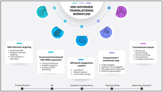
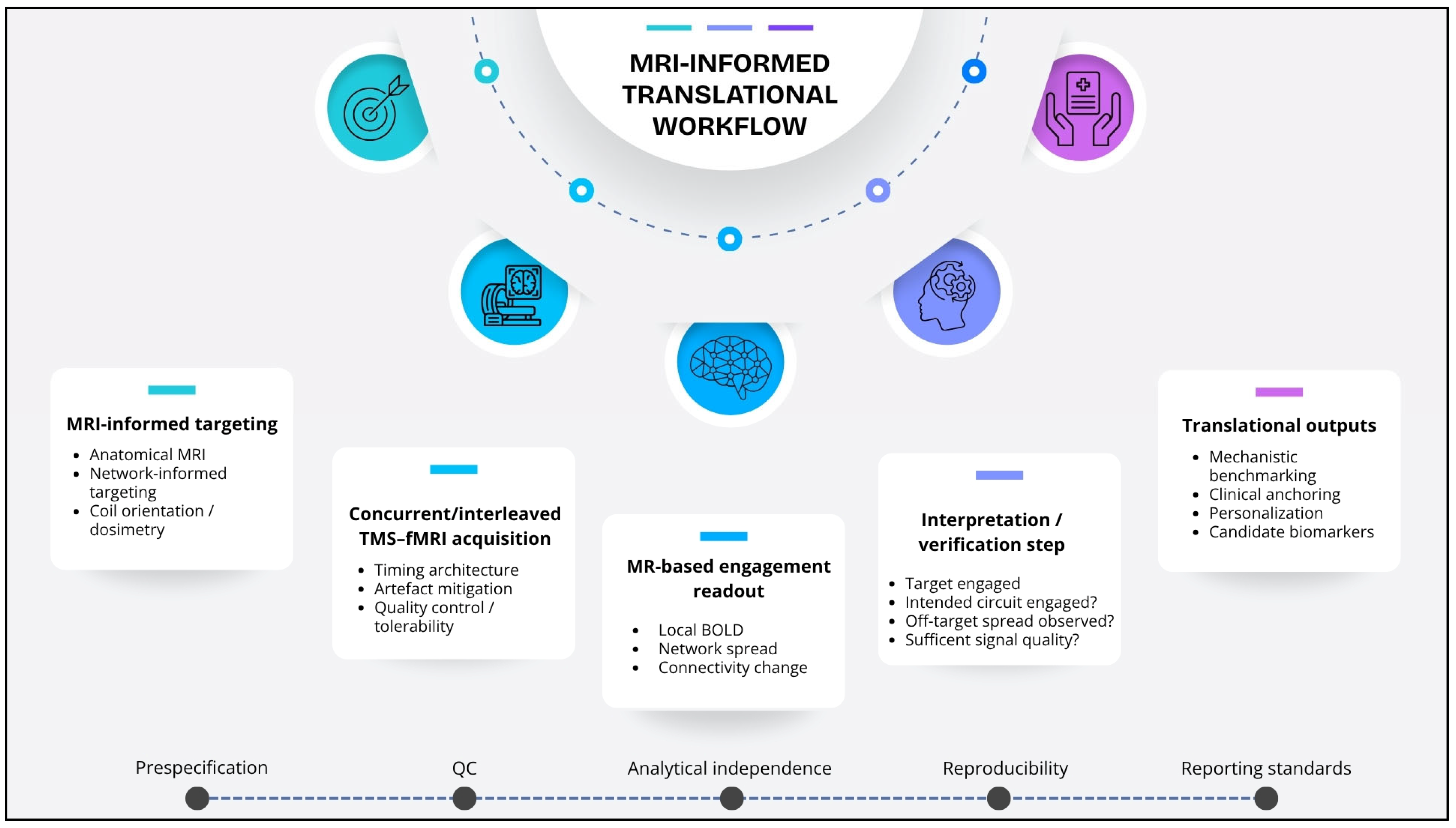
In the present article, the proposed framework refers primarily to an offline MRI-informed approach in which anatomical and/or connectivity-derived MRI information is used before stimulation to define the target, constrain coil placement, and guide interpretation of stimulation effects, while concurrent fMRI provides an in-scanner readout of engagement. This should be distinguished from real-time adaptive or closed-loop neuromodulation systems, in which online neural signals are used to modify stimulation parameters during acquisition. We view the present framework as a realistic intermediate step toward such future systems, rather than as a closed-loop implementation itself [7] (Figure 1).
Figure 1.
Translational workflow of MRI-informed targeting and MR-based target engagement in concurrent/interleaved TMS–fMRI. The scheme summarizes the proposed framework from target definition and acquisition to engagement readouts, interpretation/verification, and translational outputs. Prespecification, quality control, analytical independence, reproducibility, and reporting standards are treated as cross-cutting principles throughout.
1.1. Why TMS Needs MRI-Guided Targeting Beyond Scalp Coordinates
A central obstacle to reproducible TMS effects is that common targeting approaches—particularly scalp heuristics and template-based coordinates—often ignore inter-individual variability that is directly relevant to both efficacy and safety [8]. At the anatomical level, individuals differ in gyral/sulcal geometry, cortical thickness, and scalp-to-cortex distance at nominally equivalent target locations [9]. These differences influence the effective induced field at the cortical surface and can translate into systematic variability in target engagement across participants [10]. At the functional level, the same nominal label (e.g., “DLPFC”) does not necessarily correspond to the same network embedding or downstream circuit configuration in every individual. This is especially consequential for circuit-based indications where clinical response is thought to depend on engaging a distributed network rather than a single cortical coordinate [10,11]. As a result, stimulation can inadvertently engage suboptimal nodes—leading to reduced efficacy—or produce unwanted spread into nearby regions and networks, contributing to discomfort, poor tolerability, or off-target effects.
MRI-guided targeting provides a practical remedy because it supports (i) accurate spatial placement on individual anatomy, (ii) informed coil orientation relative to the local gyral crown, and (iii) an implicit form of dosimetry, since scalp-to-cortex distance and cortical geometry can be considered when setting intensity and interpreting responses [3,4,5,6,12,13,14]. Importantly, MRI guidance is not a “luxury add-on” for concurrent/interleaved TMS–fMRI. In the MR environment—where stimulation timing, artefact mitigation, and tolerability constraints already narrow the experimental design—reducing uncertainty in targeting is one of the most direct ways to improve interpretability and reproducibility.
For clarity, we use MRI-guided to refer to the use of structural and/or functional MRI information for target definition, neuronavigation, coil placement, and stimulation planning. By contrast, we use MR-based to refer to outcome measures derived from magnetic resonance signals, such as local BOLD responses, network propagation patterns, or connectivity changes. The broader term MR-guided framework is used here to indicate the integration of MRI-informed targeting with MR-based readouts of stimulation effects.
1.1.1. Level 1: Anatomical MRI-Guided Targeting
The first level of MRI-guided targeting is individual anatomical neuronavigation using each participant’s structural MRI (typically T1-weighted). The goal is to stimulate a clearly defined cortical surface location on the intended gyrus while standardizing coil placement and orientation [15,16,17]. In practice, this level includes: (a) selecting a target on the individual cortical surface rather than relying on template coordinates; (b) documenting coil orientation relative to local anatomy and cortical geometry (e.g., aligning to the gyral crown and respecting sulcal boundaries); and (c) accounting for scalp-to-cortex distance and related subject-specific anatomical constraints to interpret variability in stimulation effects [18]. Even without sophisticated network modeling, this approach reduces gross targeting error and helps separate true neurophysiological variability from avoidable spatial imprecision. In this sense, anatomical MRI contributes not only to spatial localization, but also to a pragmatic form of dosimetry, because the same nominal stimulation intensity may translate into different effective cortical engagement across participants depending on scalp-to-cortex distance and local cortical geometry.
1.1.2. Level 2: Network-Informed MRI-Guided Targeting
The second level extends anatomical precision to circuit consistency by using individual MRI-derived network information—most commonly resting-state functional connectivity—to choose a sub-target within a broader anatomical region that is maximally connected to the clinically relevant circuit [13,19,20]. For example, rather than stimulating a generic “DLPFC coordinate,” network-informed targeting aims to identify the DLPFC subregion whose connectivity profile best matches the intended downstream network node(s) [10,21,22]. This level is particularly aligned with the logic of concurrent/interleaved TMS–fMRI: if the method’s value lies in mapping causal network effects and measuring target engagement, then targeting should be defined not only by location but also by expected network embedding [19]. In our view, Level 2 targeting provides a principled bridge from MR-based network models to neuromodulation practice, although this approach also requires caution, because target selection and outcome interpretation should be kept as analytically independent as possible to reduce the risk of circular reasoning. Accordingly, network-informed targeting is most informative when targeting criteria, primary engagement endpoints, and confirmatory analyses are specified as explicitly and independently as possible.
2. The Missing Link: “Target Engagement” as a Measurable MR Endpoint
In many neuromodulation settings, stimulation parameters are reported precisely while the resulting brain state change is inferred indirectly from symptoms or behavior—often days or weeks later [23,24]. Concurrent/interleaved TMS–fMRI offers a way to close this gap by providing MR-based outcomes during the stimulation session [25]. This matters because TMS effects are not purely local: they include distributed network responses shaped by ongoing brain state, structural constraints, and baseline functional connectivity [25,26,27]. Without an engagement measure, null or inconsistent clinical effects can reflect (i) inadequate engagement of the intended circuit, (ii) engagement of an unintended circuit, or (iii) engagement that is highly state-dependent and therefore unstable across sessions [25,26]. In the present framework, target engagement is defined as the presence of a predefined and statistically detectable MR-based response within the intended cortical target and/or its hypothesized downstream network. Depending on the study design, this response may be operationalized as a local stimulation-associated BOLD signal change within a prespecified ROI, a predefined network propagation pattern, or a stimulation-associated change in functional connectivity within the intended circuit. In this sense, MR-based target engagement can be considered as an intermediate endpoint between “dose” and downstream outcomes, analogous to a manipulation check for circuit-based interventions. This definition is intentionally operational: target engagement is not treated as a purely conceptual label, but as a measurable intermediate endpoint that should be specified a priori according to the study design, target model, and analytic strategy, rather than assumed as a universal metric across protocols.
A Pragmatic Set of MR-Based Engagement Markers
To keep designs biomarker-ready without overcomplicating analysis, we propose three families of MR-based engagement markers that are feasible across many protocols:
- Local BOLD response: BOLD changes at or near the stimulated cortical site (and immediate proximal regions), interpreted as evidence that the intended cortical node was effectively perturbed [28]. This parameter must be considered with caution as the results in the literature are mixed [29].
- Network spread pattern: The distributed whole-brain response pattern following stimulation, capturing propagation through known networks and revealing whether effects align with the intended circuit architecture [27,30,31,32].
- Connectivity change signature (rest or task): Stimulation-associated changes in functional connectivity (and, when available, effective/causal connectivity) that index modulation of network coupling rather than isolated activation [33,34,35,36].
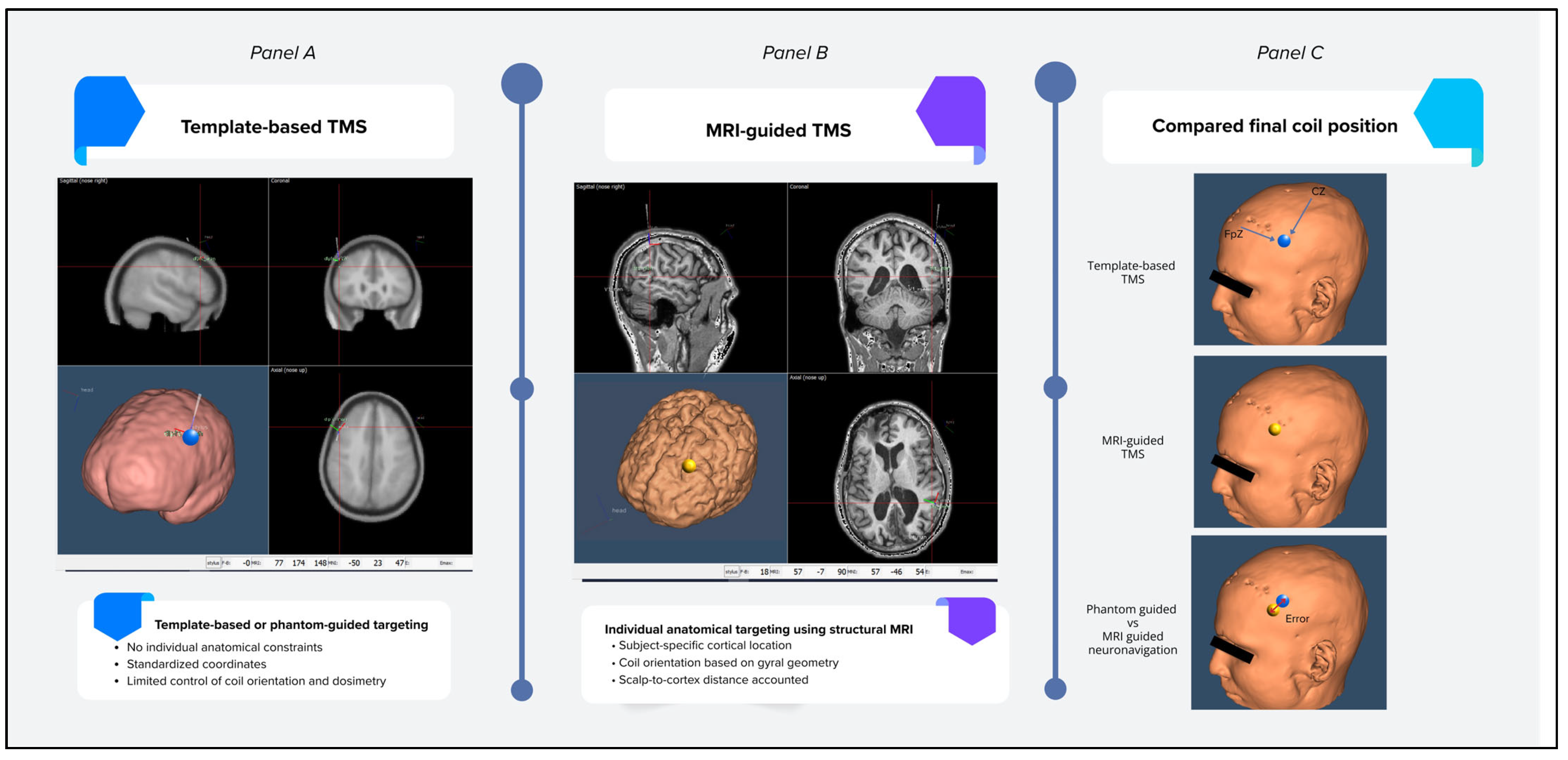
These marker families can be selected a priori (one primary, others secondary) to reduce multiplicity and improve comparability. Crucially, they can be linked to MRI-guided targeting choices and can be evaluated for biomarker potential (association with symptom dimensions; prediction of response) in a staged translational pipeline. Importantly, not all studies need to use all three marker families; rather, one primary engagement readout should ideally be predefined according to the target model, timing architecture, and mechanistic or clinical question (Figure 2).
Figure 2.
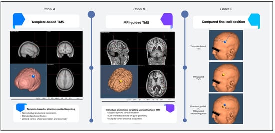
Comparison between MRI-guided neuronavigation and non-MRI-based targeting approaches in TMS. (Panel A) Phantom-based and template-guided targeting rely on standardized coordinates without individual anatomical constraints, potentially increasing spatial variability and uncertainty in effective cortical engagement. (Panel B) MRI-guided neuronavigation allows subject-specific targeting based on individual anatomical MRI, enabling precise localization of the stimulation site, coil orientation aligned with cortical geometry, and consideration of scalp-to-cortex distance. (Panel C) Comparison of final coil position across targeting approaches. The spatial discrepancy between MRI-guided and template-based targeting is illustrated as the Euclidean distance between the two stimulation sites, representing targeting error. This error reflects the extent to which MRI-free approaches may deviate from the subject-specific cortical target identified through MRI-guided neuronavigation, with potential implications for variability in effective cortical engagement. Note: Images were adapted and modified from anatomical templates derived from the AAL3 atlas (Automated Anatomical Labeling v3) and visualized using the Softaxic neuronavigation system (version 3.0.2).
3. From Feasibility to Biomarkers: A Staged Framework
Concurrent/interleaved TMS–fMRI is best conceptualized as progressing through three stages—from feasibility and quality control, to mechanistic benchmarking, to clinical translation. This staged model is intended as a pragmatic translational framework for aligning claims with the maturity of the available evidence, rather than as a validated hierarchy or formal technical standard.
3.1. Stage A—Feasibility and Quality Control in the MR Environment
Stage A work establishes the conditions under which concurrent/interleaved TMS–fMRI can produce interpretable MR signals [25,26]. The central contribution here is not “more findings” but trustworthy measurement: defining stimulation timing architectures (interleaved blocks and/or interslice stimulation), characterizing and mitigating electromagnetic artefacts in EPI acquisitions, and documenting practical constraints related to coil placement, acoustic burden, and participant tolerability inside the scanner [2,25,37]. From a translational standpoint, Stage A sets the minimum quality-control foundation needed for any downstream claim about target engagement or network modulation. Studies that do not clearly report timing, artefact handling, and basic tolerability metrics remain difficult to compare and risky to generalize across sites. Where available, reporting of signal-quality metrics and data loss due to artifact-contaminated volumes or slices may further strengthen interpretability and cross-study comparability [2,24], particularly because the robustness of engagement markers under noise, artifacts, and inter-subject variability cannot be assumed a priori.
3.2. Stage B—Mechanistic Benchmarking and Network Perturbation “Signatures”
Stage B leverages feasible acquisition and QC pipelines to address a different question: what does “expected” engagement look like when stimulation successfully perturbs a known node? Neuroimaging settings characterize local BOLD responses, distributed network spread, and state dependence (rest versus task) as a function of target, intensity, and timing design [38]. The key output of Stage B is a set of benchmark perturbation signatures: reproducible patterns of propagation and connectivity modulation that can serve as reference templates [10,39]. Here, benchmark perturbation signatures are defined as reproducible stimulation-evoked MR response patterns, including local responses, distributed propagation profiles, or connectivity changes, that can serve as reference templates for comparison across individuals, tasks, or clinical populations [9,38]. In our view, these benchmarks are essential for clinical translation because they provide a principled comparator for identifying when patient responses reflect altered network architecture rather than mere methodological noise.
3.3. Stage C—Clinical Translation: From Engagement to Candidate MR-Based Biomarkers
Stage C shifts the emphasis from “can we measure it?” and “what does it look like in principle?” to “does engagement relate to clinically meaningful variation?” Here, concurrent/interleaved TMS–fMRI is used to target engagement within disorder-relevant circuits and to explore whether MR-based signatures—local responses, network spread, or connectivity change patterns—track symptom dimensions, impairment, or (when available) treatment outcome [10,35,40,41]. Importantly, Stage C does not require immediate meta-analytic pooling to be useful; rather, it requires clinically anchored endpoints and reproducible engagement measures that can be compared across protocols [35]. The main barrier at this stage is heterogeneity: differences in targets, stimulation parameters, timing architectures, and analytic pipelines often outpace standardization, limiting generalizable biomarker claims. This is precisely where MRI-guided targeting and a minimal reporting set become critical to accelerate convergence.
Stage C is therefore more precisely defined as the phase in which predefined MR-based engagement measures—such as local stimulation-associated BOLD responses, network spread patterns, or connectivity change signatures—are evaluated in relation to clinically meaningful variation, including symptom severity, cognitive performance, impairment, or treatment outcome [9,34,39,40]. Accordingly, target engagement is not assumed but operationalized through prespecified MR-based variables that can be quantitatively examined in relation to external clinical anchors. At this stage, biomarker claims should remain proportionate to the heterogeneity of the available evidence and should be interpreted as hypothesis-testing rather than confirmatory, pending validation through reproducible designs, out-of-sample prediction, and independent replication.
To make this staged framework operational, Box 1 summarizes representative concurrent/interleaved TMS–fMRI protocol archetypes that map onto Stages A–C and illustrate the feasibility-to-biomarker trajectory.
Box 1. Representative concurrent/interleaved TMS–fMRI protocols illustrating the feasibility-to-biomarker trajectory. This box provides a pragmatic “protocol map” to illustrate how concurrent/interleaved TMS–fMRI has evolved from technical feasibility demonstrations to biomarker-oriented clinical applications. The examples are representative rather than exhaustive and are intended to guide readers in study design and interpretation.
| A. Feasibility and MR-compatibility (technical foundation) |
A1. Interleaved/interslice stimulation for artefact control
|
A2. Safety and tolerability inside the scanner
|
| B. Mechanistic network perturbation (healthy “benchmarking”) |
B1. Single-pulse TMS to map causal network propagation
|
B2. Task-modulated perturbation to test state-dependence
|
| C. Clinical translation (biomarker potential and target engagement) |
C1. Target engagement in mood disorders (DLPFC-centered paradigms)
|
C2. Circuit probing beyond mood disorders (psychosis, stroke, pain as exemplars)
|
D. Practical roadmap (how to design “biomarker-ready” concurrent TMS–fMRI)
|
Across these protocol archetypes, claims of target engagement or biomarker relevance should remain proportional to methodological maturity and should be supported by explicit reporting of timing architecture, artifact mitigation, target definition, and prespecified MR-based endpoints.
In our view, concurrent/interleaved TMS–fMRI protocols can be conceptualized as a continuum from MR-feasibility and quality control, through mechanistic network perturbation benchmarking, to clinically anchored target engagement and biomarker-oriented applications. From a neurocognitive perspective, the value of this continuum is maximized when MR-based engagement readouts are systematically paired with neurophysiological endpoints that operationalize cognition and behavior as emergent properties of distributed neural systems [42]. Concurrent TMS–fMRI is particularly well suited to this aim because it can link a controlled perturbation to immediate network-level signatures while the participant is in a defined cognitive state (rest or task) [40], thereby enabling interpretable brain–behavior mapping rather than post hoc correlation. Moreover, when stimulation is delivered in a temporally structured manner (e.g., repeated pulses or patterned trains) and aligned with task-relevant neural activity, the approach naturally connects to Hebbian principles of plasticity [43]: co-activation of targeted nodes and their downstream partners can strengthen or reshape network coupling, which should be detectable as changes in connectivity signatures and reflected in corresponding behavioral modulation [40,44].
4. Hypothesis: MRI-Guided Targeting + Concurrent/Interleaved TMS–fMRI Will Improve Efficacy and Safety
Building on the staged framework above, we propose that MRI-guided targeting and concurrent/interleaved TMS–fMRI engagement readouts can be combined into a pragmatic translational pipeline. The central hypothesis is that improving spatial and circuit-level targeting (anatomical and network-informed) may yield more consistent target engagement and more interpretable network spread patterns, thereby increasing the probability of clinical benefit while reducing off-target effects and tolerability issues. At the current stage of the literature, however, this proposition should be interpreted as hypothesis-generating rather than as an established clinical standard.
4.1. Hypothesis 1 (Efficacy)
If MRI-guided targeting is individualized at both the anatomical and network-informed levels, the resulting engagement and network spread measured during concurrent/interleaved TMS–fMRI may be more consistently aligned with the intended therapeutic circuit [21,41,45]. We hypothesize that this alignment will increase the probability of a clinically meaningful response, because stimulation will more reliably perturb the network node(s) relevant to symptom generation and treatment mechanisms.
4.2. Hypothesis 2 (Safety and Tolerability)
MRI-guided targeting is hypothesized to reduce suboptimal stimulation (e.g., placement too lateral/medial, unintended proximity to sensitive regions, or mismatched coil orientation) [17]. We hypothesize that improved spatial precision and orientation control will reduce discomfort and off-target spread, thereby lowering dropout rates and improving tolerability—especially in clinical populations where scanner burden and sensory sensitivity can be limiting factors.
4.3. Hypothesis 3 (Personalization)
We further hypothesize that MR-based engagement patterns (local BOLD, network spread, connectivity signatures) can inform pragmatic personalization within feasible clinical workflows [13,46,47]. Rather than requiring complex closed-loop systems, personalization may be achieved via modest adjustments such as micro-shifts in target location within an anatomical region, refined coil orientation, or calibrated intensity based on distance/orientation constraints [13,48], while remaining conceptually distinct from real-time adaptive neuromodulation systems. In this view, concurrent/interleaved TMS–fMRI provides a realistic pathway toward MR-guided personalization by offering a measurable engagement endpoint that can be iteratively optimized.
5. Practical Recommendations for Biomarker-Ready Concurrent/Interleaved TMS–fMRI
To support reproducibility and future quantitative synthesis, we propose the following low-burden recommendations:
- Target definition: Report target in individual MRI space; specify whether targeting was anatomical or network-informed [13,21].
- Coil orientation: Report orientation relative to local anatomy (gyral crown/sulcal boundary) and how it was standardized [15,49].
- Scalp-to-cortex distance: Report distance (or equivalent proxy) and how intensity was calibrated (e.g., %RMT plus distance considerations if used) [15,49].
- Timing architecture: State explicitly whether stimulation was interleaved, interslice, or continuous, and how pulses were synchronized with acquisition [50].
- Artefact mitigation/QC: Describe artifact-handling steps and volume/slice exclusion criteria [49].
- Signal quality: Report signal-quality metrics whenever available (e.g., temporal SNR, motion indices, or data loss due to artifact-contaminated volumes or slices), because interpretability of engagement measures depends on basic acquisition stability.
- Minimal data requirements: Avoid assuming universal thresholds for sample size or signal quality across protocols; instead, report the acquisition, QC, and endpoint-definition features needed to interpret the reliability of engagement detection in the specific study context.
- Primary MR endpoint: Predefine one primary engagement readout (local BOLD vs. network spread vs. connectivity signature).
- Analytical independence: In studies using network-informed targeting, clarify whether the features used for target selection are independent from the primary MR-based outcome measures, in order to reduce the risk of circular interpretation.
- Bias awareness: Explicitly report major potential sources of bias, including stimulation-related artifacts, timing variability relative to image acquisition, anatomical variability affecting effective dosimetry, and inter-subject differences in functional network organization.
- Clinical anchoring: In patient studies, include at least one clinical measure and one tolerability/feasibility measure.
- Verification step: Whenever feasible, specify whether MR-based engagement measures are used as an explicit intermediate control phase to confirm that the intended target and network were measurably engaged before drawing mechanistic or clinical inferences.
- Comparator clarity: Distinguish clearly concurrent/interleaved designs from sequential (pre/post) TMS–fMRI.
- Minimal reporting set: Provide stimulation parameters (target, intensity, pulse count, frequency), MRI parameters (TR, sequence), and preprocessing summary.
- Data transparency: When possible, share derived engagement maps or summary statistics to support cross-study comparability and secondary analyses [39].
- Reproducibility orientation: Whenever feasible, report design features that support reproducibility, such as repeated sessions, within-subject consistency analyses, or prespecified QC thresholds.
- Biomarker validation pathway: When predictive or clinically stratifying claims are made, specify whether validation relies on prespecified models, out-of-sample prediction, cross-validation, or independent replication, in order to distinguish exploratory findings from candidate biomarker evidence.
These recommendations are consistent with recent methodological guidance for concurrent/interleaved TMS–fMRI [2,25] and are intended as low-burden priorities to improve interpretability, comparability, and future biomarker validation rather than to impose full protocol uniformity.
6. Discussion
Concurrent/interleaved TMS–fMRI is often presented as a technically demanding hybrid setup; however, its main value is not only technical but inferential and translational: it links MR acquisition choices (timing, artefact control, interpretability of BOLD under stimulation) to causal circuit probing and to measurable endpoints of target engagement [2,25,26]. Framed in this way, the method becomes an MR-based neuroimaging approach that can reduce uncertainty about whether stimulation actually perturbed the intended node and network. This is particularly relevant in clinical neuromodulation, where variability in targeting and network embedding can drive inconsistent outcomes even when stimulation parameters appear “standardized”.
A key implication of the staged framework proposed here is that biomarker claims should be scaled to the maturity of the evidence [10,35,41]. Stage A feasibility and QC work defines when engagement measures are interpretable; Stage B benchmarking establishes reference perturbation signatures; Stage C begins to test whether engagement signatures relate to clinically meaningful variation (symptoms, impairment, or treatment response) [10,35,41]. Across these stages, the same bottleneck recurs: heterogeneity in targets, timing architectures, and analytic choices often outpaces harmonized reporting, limiting cross-study comparability and slowing convergence toward biomarker-ready evidence. In this sense, the contribution of the present article is not to claim novelty for any single methodological component, but to make explicit the translational role of target engagement as an intermediate MR-based endpoint linking stimulation delivery, circuit-level effects, and clinically relevant outcomes. Any future biomarker use of such engagement measures will require validation beyond descriptive association, including reproducible study designs, predictive testing beyond the original sample, and, ideally, independent replication across cohorts or sites. We suggest that this added value is particularly relevant for intervention-oriented contexts, in which MR-based engagement readouts are still not routinely incorporated as an explicit verification layer between target selection and downstream clinical interpretation.
To address this bottleneck with minimal burden, we emphasize a small set of design and reporting priorities. First, MRI-guided targeting should be reported in individual space, with clear specification of whether targeting was anatomical (Level 1) or network-informed (Level 2), including coil orientation and scalp-to-cortex distance considerations [13,21]. Second, the timing architecture (interleaved or interslice) and QC steps should be stated explicitly [37], because these features directly determine whether observed effects can be interpreted as stimulation-evoked rather than artefactual. Third, studies should predefine a primary MR engagement endpoint—local response, network spread, or a connectivity signature—to reduce multiplicity and improve comparability [10]. In studies adopting network-informed targeting, this also requires sufficient transparency about the independence between the features used for target selection and those used to evaluate engagement, in order to reduce circular interpretation. Finally, clinical studies should anchor engagement metrics to at least one clinical measure and one feasibility/tolerability measure [41], enabling engagement to be evaluated as an intermediate endpoint rather than a purely descriptive neuroimaging finding.
More broadly, concurrent/interleaved TMS–fMRI provides a realistic pathway toward personalization that does not require complex closed-loop systems. Personalization can be operationalized through constrained, clinically feasible adjustments—micro-shifts in target location within an anatomical region, refinement of coil orientation, or intensity calibration informed by anatomy—while using MR-based engagement patterns as a measurable intermediate endpoint [13,15,49]. In this sense, MR-based target engagement can function as a “bridge measure” that links stimulation delivery to circuit-level effects and, ultimately, to patient-relevant outcomes. If adopted with harmonized reporting and reproducible pipelines, this approach can accelerate the development of candidate MR-based biomarkers that are directly actionable for personalized neuromodulation. This perspective should also be understood as complementary to EEG-guided and other multimodal neuromodulation approaches: whereas EEG-based systems may offer higher temporal resolution and greater compatibility with real-time adaptation, concurrent/interleaved TMS–fMRI provides a richer spatial characterization of target engagement and distributed network propagation. We also emphasize that the present framework should not be conflated with real-time adaptive or closed-loop neuromodulation. Although future integration with online physiological feedback is plausible and potentially desirable, current concurrent/interleaved TMS–fMRI applications are more realistically positioned as offline MRI-informed and measurement-guided approaches. Their translational value, therefore, depends not on real-time adaptability per se, but on the rigor with which target definition, signal quality, engagement metrics, and interpretive limits are specified.
Limitations
This article is a Perspective and does not aim to provide an exhaustive systematic review or a quantitative meta-analysis. The concurrent/interleaved TMS–fMRI literature remains heterogeneous with respect to stimulation targets, protocols, timing architectures, and analytic pipelines, and reporting variability limits direct comparability across studies. In addition, the field still lacks universally accepted quantitative benchmarks for engagement detection, reproducibility, and minimal data requirements across protocols, as well as agreed validation pathways for translating MR-based engagement measures into clinically useful biomarkers. Local BOLD responses at the stimulation site are not consistently observed across studies, and network-informed targeting approaches must also consider the possibility of circular interpretation when target selection and outcome evaluation rely on insufficiently independent network features.
A broader protocol was registered in PROSPERO (CRD420261286226), but this manuscript provides a narrative synthesis at a predefined cutoff date; a fully systematic and quantitative synthesis will be reported separately. Accordingly, the present contribution should be interpreted as a forward-looking framework intended to guide study design, highlight practical priorities for MR-based target engagement measures, and accelerate convergence toward biomarker-ready standards, rather than as a finalized technical standard. More specifically, the staged model proposed here should be interpreted as a hypothesis-generating structure intended to guide future empirical testing, simulation-based evaluation, and biomarker validation, rather than as an empirically validated framework in itself.
7. Conclusions
Concurrent/interleaved TMS–fMRI is reaching a stage where methodological choices can be translated into shared design and reporting priorities rather than remaining laboratory-specific solutions. The central opportunity is to treat MR-based target engagement as a decision-relevant endpoint—useful for validating targeting strategies and for determining when network perturbation is sufficiently reliable to support biomarker development. In the near term, progress will depend less on introducing additional protocol variants and more on converging toward a minimal, harmonized reporting set and a small number of reproducible engagement readouts that can be compared across sites and clinical indications. Establishing these shared conventions is the most direct route to making concurrent/interleaved TMS–fMRI not only feasible, but also more methodologically robust and clinically informative.
Author Contributions
Conceptualization, S.P.; methodology, C.D.F., S.P.; validation, S.P.; writing—original draft preparation, C.D.F., S.P.; writing—review and editing, C.D.F., S.P.; visualization, C.D.F.; supervision, S.P.; project administration, S.P. All authors have read and agreed to the published version of the manuscript.
Funding
This research received no external funding.
Institutional Review Board Statement
Not applicable.
Informed Consent Statement
Not applicable.
Data Availability Statement
No new data were created or analyzed in this study. Data sharing is not applicable to this article.
Acknowledgments
During the preparation of this manuscript, the authors used ChatGPT (OpenAI) (version GPT-3.5) to support language editing and to help organize the manuscript structure and draft text. The authors have reviewed and edited the output and take full responsibility for the content of this publication.
Conflicts of Interest
The authors declare no conflicts of interest.
References
- Gugino, L.D.; Romero, J.R.; Aglio, L.S.; Titone, D.; Ramirez, M.; Pascual-Leone, Á.; Grimson, E.; Weisenfeld, N.; Kikinis, R.; Shenton, M.-E. Transcranial Magnetic Stimulation Coregistered with MRI: A Comparison of a Guided versus Blind Stimulation Technique and Its Effect on Evoked Compound Muscle Action Potentials. Clin. Neurophysiol. 2001, 112, 1781–1792. [Google Scholar] [CrossRef] [Scilit]
- Woolgar, A.; Feredoes, E.; Assem, M.; Bassil, Y.; Bergmann, T.O.; Beynel, L.; Burke, M.; Cash, R.F.H.; Comeau, R.M.; Correia, M.M.; et al. Consensus Guidelines for the Use of Concurrent TMS-FMRI in Cognitive and Clinical Neuroscience. Nat. Protoc. 2025, 21, 1–17. [Google Scholar] [CrossRef] [Scilit]
- Sparing, R.; Buelte, D.; Meister, I.G.; Pauš, T.; Fink, G.R. Transcranial Magnetic Stimulation and the Challenge of Coil Placement: A Comparison of Conventional and Stereotaxic Neuronavigational Strategies. Hum. Brain Mapp. 2008, 29, 82–96. [Google Scholar] [CrossRef] [Scilit]
- Di Fazio, C.; Scaliti, E.; Stanziano, M.; Nigri, A.; Demichelis, G.; Tamietto, M.; Palermo, S. Exploring Cortical Excitability Modulation to Promote Cognitive Resilience in Aging: An RTMS Study Protocol. Front. Hum. Neurosci. 2026, 20, 1743236. [Google Scholar] [CrossRef] [Scilit]
- Palermo, S.; Di Fazio, C.; Scaliti, E.; Stanziano, M.; Nigri, A.; Tamietto, M. Cortical Excitability and the Aging Brain: Toward a Biomarker of Cognitive Resilience. Front. Psychol. 2025, 16, 1542880. [Google Scholar] [CrossRef] [Scilit]
- Di Fazio, C.; Scaliti, E.; Stanziano, M.; Nigri, A.; Demichelis, G.; Tamietto, M.; Palermo, S. RTMS for Enhancing Cognitive Reserve: A Case Report. Brain Disord. 2025, 18, 100221. [Google Scholar] [CrossRef] [Scilit]
- Varone, G.; Biabani, M.; Tremblay, S.; Brown, J.C.; Kallioniemi, E.; Rogasch, N.C. The Golden Age of Online Readout: EEG-Informed TMS from Manual Probing to Closed-Loop Neuromodulation. Neuroimage 2025, 322, 121543. [Google Scholar] [CrossRef] [Scilit]
- Liu, B.; Hu, C.; Bao, P. Precision TMS through the Integration of Neuroimaging and Machine Learning: Optimizing Stimulation Targets for Personalized Treatment. Front. Hum. Neurosci. 2025, 19, 1682852. [Google Scholar] [CrossRef] [Scilit]
- Demirci, N.; Holland, M.A. Cortical Thickness Systematically Varies with Curvature and Depth in Healthy Human Brains. Hum. Brain Mapp. 2022, 43, 2064–2084. [Google Scholar] [CrossRef] [Scilit]
- Tik, M.; Vasileiadi, M.; Woletz, M.; Linhardt, D.; Schuler, A.-L.; Williams, N.R.; Windischberger, C. Concurrent TMS/FMRI Reveals Individual DLPFC Dose-Response Pattern. Neuroimage 2023, 282, 120394. [Google Scholar] [CrossRef] [Scilit]
- Klooster, D.; Ferguson, M.A.; Boon, P.; Baeken, C. Personalizing Repetitive Transcranial Magnetic Stimulation Parameters for Depression Treatment Using Multimodal Neuroimaging. Biol. Psychiatry Cogn. Neurosci. Neuroimaging 2021, 7, 536–545. [Google Scholar] [CrossRef] [Scilit]
- Riis, T.; Lunt, S.; Kubanek, J. MRI Free Targeting of Deep Brain Structures Based on Facial Landmarks. Brain Stimul. 2025, 18, 131–137. [Google Scholar] [CrossRef] [Scilit]
- Lynch, C.J.; Elbau, I.; Ng, T.; Wolk, D.; Zhu, S.; Ayaz, A.; Power, J.D.; Zebley, B.; Gunning, F.M.; Liston, C. Automated Optimization of TMS Coil Placement for Personalized Functional Network Engagement. Neuron 2022, 110, 3263–3277.e4. [Google Scholar] [CrossRef] [Scilit]
- Di Fazio, C.; Palermo, S. A Prefrontal Neuromodulation Route for Post-Traumatic Olfactory Dysfunction: A Perspective Supported by Recovery During Left-DLPFC RTMS. Brain Sci. 2026, 16, 99. [Google Scholar] [CrossRef] [Scilit]
- Dannhauer, M.; Huang, Z.; Beynel, L.; Wood, E.; Bukhari-Parlakturk, N.; Peterchev, A.V. TAP: Targeting and Analysis Pipeline for Optimization and Verification of Coil Placement in Transcranial Magnetic Stimulation. J. Neural Eng. 2022, 19. [Google Scholar] [CrossRef] [Scilit]
- Caulfield, K.A.; Fleischmann, H.H.; Cox, C.; Wolf, J.P.; George, M.S.; McTeague, L.M. Neuronavigation Maximizes Accuracy and Precision in TMS Positioning: Evidence from 11,230 Distance, Angle, and Electric Field Modeling Measurements. Brain Stimul. 2022, 15, 1192–1205. [Google Scholar] [CrossRef] [Scilit]
- Demchenko, I.; Al-Shamali, H.F.; Rueda, A.; Tailor, I.; Janssen-Aguilar, R.; Schweizer, T.A.; Dunlop, K.; Graham, S.J.; Bhat, V. Magnetic Resonance Imaging-Guided Neuronavigation for Transcranial Magnetic Stimulation in Mood Disorders: Technical Foundation, Advances, and Emerging Tools. Hum. Brain Mapp. 2025, 46, e70424. [Google Scholar] [CrossRef] [Scilit]
- Janssen, A.M.; Oostendorp, T.F.; Stegeman, D.F. The Coil Orientation Dependency of the Electric Field Induced by TMS for M1 and Other Brain Areas. J. Neuroeng. Rehabil. 2015, 12, 47. [Google Scholar] [CrossRef] [Scilit]
- Gordon, E.M.; Laumann, T.O.; Gilmore, A.W.; Newbold, D.J.; Greene, D.J.; Berg, J.J.; Ortega, M.; Hoyt-Drazen, C.; Gratton, C.; Sun, H.; et al. Precision Functional Mapping of Individual Human Brains. Neuron 2017, 95, 791–807.e7. [Google Scholar] [CrossRef] [Scilit]
- Sadaghiani, S.; Brookes, M.J.; Baillet, S. Connectomics of Human Electrophysiology. Neuroimage 2022, 247, 118788. [Google Scholar] [CrossRef] [Scilit]
- Khosravani, S.; Palm, S.; Drew, W.; Frandsen, S.; Lin, C.; Tirrell, E.; Hindley, L.; Schineller, M.; Garimella, A.; Chiulli, N.; et al. Predictive Value of Subgenual Cingulate Normative Connectivity to TMS Treatment Site for Antidepressant Response in Routine Clinical Practice: A Prospective, Multisite Cohort Study. Mol. Psychiatry 2025, 31, 689–700. [Google Scholar] [CrossRef] [Scilit]
- Tik, M.; Hoffmann, A.; Sladky, R.; Tomova, L.; Hummer, A.; de Lara, L.N.; Bukowski, H.; Pripfl, J.; Biswal, B.B.; Lamm, C.; et al. Towards Understanding RTMS Mechanism of Action: Stimulation of the DLPFC Causes Network-Specific Increase in Functional Connectivity. Neuroimage 2017, 162, 289–296. [Google Scholar] [CrossRef] [Scilit]
- King-Stephens, D. Neuromodulation: The Search Is on for the Most Effective Parameters. Epilepsy Curr. 2020, 20, 359–361. [Google Scholar] [CrossRef] [Scilit]
- Modolo, J.; Legros, A.; Beuter, A. The next Move in Neuromodulation Therapy: A Question of Timing. Front. Comput. Neurosci. 2015, 8, 162. [Google Scholar] [CrossRef] [Scilit]
- Riddle, J.; Scimeca, J.M.; Pagnotta, M.F.; Inglis, B.; Sheltraw, D.; Muse-Fisher, C.; D’Esposito, M. A Guide for Concurrent TMS-FMRI to Investigate Functional Brain Networks. Front. Hum. Neurosci. 2022, 16, 1050605. [Google Scholar] [CrossRef] [Scilit]
- Mizutani-Tiebel, Y.; Tik, M.; Chang, K.-Y.; Padberg, F.; Soldini, A.; Wilkinson, Z.; Voon, C.C.; Bulubas, L.; Windischberger, C.; Keeser, D. Concurrent TMS-FMRI: Technical Challenges, Developments, and Overview of Previous Studies. Front. Psychiatry 2022, 13, 825205. [Google Scholar] [CrossRef] [Scilit]
- Tik, M.; Woletz, M.; Schuler, A.; Vasileiadi, M.; Cash, R.F.H.; Zalesky, A.; Lamm, C.; Windischberger, C. Acute TMS/FMRI Response Explains Offline TMS Network Effects—An Interleaved TMS-FMRI Study. Neuroimage 2022, 267, 119833. [Google Scholar] [CrossRef] [Scilit]
- Bergmann, T.O.; Varatheeswaran, R.; Hanlon, C.A.; Madsen, K.H.; Thielscher, A.; Siebner, H.R. Concurrent TMS-FMRI for Causal Network Perturbation and Proof of Target Engagement. Neuroimage 2021, 237, 118093. [Google Scholar] [CrossRef] [Scilit]
- Rafiei, F.; Rahnev, D. TMS Does Not Increase BOLD Activity at the Site of Stimulation: A Review of All Concurrent TMS-FMRI Studies. eNeuro 2022, 9, 1–14. [Google Scholar] [CrossRef] [Scilit]
- Farzan, F.; Vernet, M.; Shafi, M.M.; Rotenberg, A.; Daskalakis, Z.J.; Pascual-Leone, Á. Characterizing and Modulating Brain Circuitry through Transcranial Magnetic Stimulation Combined with Electroencephalography. Front. Neural Circuits 2016, 10, 73. [Google Scholar] [CrossRef] [Scilit]
- Momi, D.; Wang, Z.; Griffiths, J.D. TMS-Evoked Responses Are Driven by Recurrent Large-Scale Network Dynamics. eLife 2023, 12, e83232. [Google Scholar] [CrossRef] [Scilit]
- Momi, D.; Ozdemir, R.A.; Tadayon, E.; Boucher, P.; Di Domenico, A.; Fasolo, M.; Shafi, M.M.; Pascual-Leone, Á.; Santarnecchi, E. Perturbation of Resting-State Network Nodes Preferentially Propagates to Structurally Rather than Functionally Connected Regions. Sci. Rep. 2021, 11, 12458. [Google Scholar] [CrossRef] [Scilit]
- He, H.; Sun, X.; Doose, J.; Faller, J.; McIntosh, J.R.; Saber, G.T.; Huffman, S.; Hong, L.; Pantazatos, S.P.; Yuan, H.; et al. TMS-Induced Modulation of Brain Networks and Its Associations to RTMS Treatment for Depression: A Concurrent FMRI-EEG-TMS Study. Brain Stimul. 2025, 18, 1955–1965. [Google Scholar] [CrossRef] [Scilit]
- Zhong, G.; Yang, Z.; Jiang, T. Precise Modulation Strategies for Transcranial Magnetic Stimulation: Advances and Future Directions. Neurosci. Bull. 2021, 37, 1718–1734. [Google Scholar] [CrossRef] [Scilit]
- Tan, V.; Jeyachandra, J.; Ge, R.; Dickie, E.W.; Gregory, E.; Vanderwal, T.; Vila-Rodriguez, F.; Hawco, C. Subgenual Cingulate Connectivity as a Treatment Predictor during Low-Frequency Right Dorsolateral Prefrontal RTMS: A Concurrent TMS-FMRI Study. Brain Stimul. 2023, 16, 1165–1172. [Google Scholar] [CrossRef] [Scilit]
- Fernández-Linsenbarth, I.; Mijancos-Martínez, G.; Ruiz-Gómez, S.J.; Bachiller, A.; Osorio-Iriarte, E.; Beño-Ruiz-De-La-Sierra, R.M.; Arjona-Valladares, A.; Roig-Herrero, A.; Molina, V. Transcranial Magnetic Stimulation-Induced Modulation of Functional Connectivity in Healthy Controls: A TMS–EEG Graph Study. Brain Behav. 2025, 15, e70981. [Google Scholar] [CrossRef] [Scilit]
- Jackson, J.; Scrivener, C.L.; Correia, M.M.; Mada, M.; Woolgar, A. Conducting Interslice Stimulation for Concurrent TMS-FMRI. J. Neurosci. Methods 2025, 422, 110513. [Google Scholar] [CrossRef] [Scilit]
- Chang, K.-Y.; Tik, M.; Mizutani-Tiebel, Y.; Schuler, A.-L.; Taylor, P.C.J.; Campana, M.; Vogelmann, U.; Huber, B.; Dechantsreiter, E.; Thielscher, A.; et al. Neural Response during Prefrontal Theta Burst Stimulation: Interleaved TMS-FMRI of Full ITBS Protocols. NeuroImage 2024, 291, 120596. [Google Scholar] [CrossRef] [Scilit]
- Glick, C.; Gajawelli, N.; Sun, Y.; Badami, F.; Saggar, M.; Etkin, A. Concurrent Single-Pulse TMS-FMRI Dataset to Reveal the Causal Connectome in Healthy and Patient Populations. Sci. Data 2025, 12, 1081. [Google Scholar] [CrossRef] [Scilit]
- Schuler, A.; Hartwigsen, G. The Potential of Interleaved TMS-FMRI for Linking Stimulation-Induced Changes in Task-Related Activity with Behavioral Modulations. Brain Stimul. 2024, 18, 37–51. [Google Scholar] [CrossRef] [Scilit]
- Ge, R.; Humaira, A.; Gregory, E.; Alamian, G.; MacMillan, E.L.; Barlow, L.; Todd, R.M.; Nestor, S.M.; Frangou, S.; Vila-Rodriguez, F. Predictive Value of Acute Neuroplastic Response to RTMS in Treatment Outcome in Depression: A Concurrent TMS-FMRI Trial. Am. J. Psychiatry 2022, 179, 500–508. [Google Scholar] [CrossRef] [Scilit]
- Palermo, S. Neurocognition and Placebo Analgesia: Linking in Functional Magnetic Resonance Imaging. In The Neurobiology, Physiology, and Psychology of Pain; Academic Press: Cambridge, MA, USA, 2022; pp. 307–317. [Google Scholar] [CrossRef] [Scilit]
- Fox, K.; Stryker, M.P. Integrating Hebbian and Homeostatic Plasticity: Introduction. Philos. Trans. R. Soc. B Biol. Sci. 2017, 372, 20160413. [Google Scholar] [CrossRef] [Scilit]
- Jannati, A.; Oberman, L.M.; Rotenberg, A.; Pascual-Leone, Á. Assessing the Mechanisms of Brain Plasticity by Transcranial Magnetic Stimulation. Neuropsychopharmacology 2022, 48, 191–208. [Google Scholar] [CrossRef] [Scilit]
- Bossus, L.; Dickson, J.M.; Blaine, C.; Khalilkhani, H.; Khan, A.; Oathes, D.J. Causal Connectivity Maps Derived from Single-Pulse Interleaved TMS/FMRI. Sci. Rep. 2026, 16, 3070. [Google Scholar] [CrossRef] [Scilit]
- Cao, Z.; Xiao, X.; Xie, C.; Wei, L.; Yang, Y.; Zhu, C. Personalized Connectivity-Based Network Targeting Model of Transcranial Magnetic Stimulation for Treatment of Psychiatric Disorders: Computational Feasibility and Reproducibility. Front. Psychiatry 2024, 15, 1341908. [Google Scholar] [CrossRef] [Scilit]
- Rajasekharan, D.; Madore, M.R.; Holtzheimer, P.E.; Lim, K.O.; Williams, L.M.; Philip, N.S. Personalized Models of BeamF3 Targeting in Transcranial Magnetic Stimulation for Depression: Implications for Precision Clinical Translation. Brain Stimul. 2025, 18, 829–837. [Google Scholar] [CrossRef] [Scilit]
- Cash, R.F.H.; Zalesky, A. Personalized and Circuit-Based Transcranial Magnetic Stimulation: Evidence, Controversies, and Opportunities. Biol. Psychiatry 2023, 95, 510–522. [Google Scholar] [CrossRef] [Scilit]
- Dannhauer, M.; Gomez, L.J.; Robins, P.L.; Wang, D.; Hasan, N.I.; Thielscher, A.; Siebner, H.R.; Fan, Y.; Deng, Z. Electric Field Modeling in Personalizing Transcranial Magnetic Stimulation Interventions. Biol. Psychiatry 2023, 95, 494–501. [Google Scholar] [CrossRef] [Scilit]
- Grunze, H.; Born, C. The Impact of Subsyndromal Bipolar Symptoms on Patient’s Functionality and Quality of Life. Front. Psychiatry 2020, 11, 510. [Google Scholar] [CrossRef] [Scilit]
Disclaimer/Publisher’s Note: The statements, opinions and data contained in all publications are solely those of the individual author(s) and contributor(s) and not of MDPI and/or the editor(s). MDPI and/or the editor(s) disclaim responsibility for any injury to people or property resulting from any ideas, methods, instructions or products referred to in the content. |
© 2026 by the authors. Licensee MDPI, Basel, Switzerland. This article is an open access article distributed under the terms and conditions of the Creative Commons Attribution (CC BY) license.