


Abstract
The purpose of this study was to compare knee joint biomechanical characteristics and movement strategies according to the direction of the forward lunge in badminton and to provide practical insights for training and injury prevention. Eighteen female recreational badminton players performed forward lunges in three directions: center (CFL), left (LFL), and right (RFL). Knee joint angles and moments, center of mass (COM) velocity, ground reaction forces (GRF), and knee extensor and flexor muscle forces were analyzed. In addition, continuous biomechanical variables were examined using statistical non-parametric mapping (SnPM). The results showed that LFL demonstrated the fastest approach COM velocity and greater knee flexion moments at initial contact, along with the greatest knee flexor muscle force, which may be indicative of enhanced joint stabilization demand. RFL exhibited a smaller knee flexion angle and lower vertical ground reaction force but showed the greatest posterior braking force and the fastest recovery COM velocity, which may be indicative of greater movement efficiency. CFL showed significantly greater knee adduction angles and internal rotation moments, suggesting elevated rotational loading at the knee that may be associated with increased injury risk. These findings highlight direction-specific knee joint biomechanical characteristics during badminton forward lunges and may provide useful information for developing targeted training and injury prevention approaches.
1. Introduction
Badminton is a popular racket sport played by more than 700 million recreational participants worldwide [1], and it is accessible to individuals of all ages, sexes, and skill levels [2]. In Korea, badminton ranks third in participation rate after golf and soccer and has been reported to be the most popular sport among women [3]. Because badminton requires players to respond to the high speed and rapid directional changes in the shuttlecock, quick and efficient movement together with stable postural control is essential, with footwork representing a key determinant of performance [4,5]. Among various footwork techniques, the lunge is the most frequently performed movement, accounting for approximately 37% of all game-related actions [6], and is closely associated with both performance and injury risk [7].
In particular, the forward lunge is performed not only in the anterior direction but also toward the left and right sides, and its biomechanical characteristics vary according to movement direction [7,8,9]. Previous studies have shown that the left forward lunge may increase the risk of knee injury by producing greater vertical ground reaction force and plantar pressure than other directions [7]. Moreover, lunge direction affects foot loading distribution, knee joint alignment, and muscle activation patterns, resulting in significant differences in the mechanical demands placed on the lower extremities [10].
Badminton is also an asymmetrical sport characterized by frequent use of the dominant side during racket strokes, and lunge landing imposes substantial musculoskeletal demands on the dominant lower limb, requiring a high level of lower-extremity mechanics and muscular activity to maintain knee stability [11]. During landing, ground reaction force can reach approximately 2.5 times body weight [4], and such loading has been associated with musculoskeletal injury and chronic knee pain in prior research [12]. Indeed, although badminton is a non-contact sport, approximately 58% of injuries occur in the lower extremities, with knee injuries being the most frequently reported type [4,13]. Women have been shown to be more susceptible to knee injuries than men because of differences in muscle strength, joint stability, and lower-limb alignment characteristics [14], and recreational players may be at even greater risk than elite athletes because of their relatively lower physical fitness and training levels [15,16].
Nevertheless, most previous studies have focused on elite male players [6,7,17], and research specifically targeting female recreational badminton players remains limited. In addition, earlier studies on badminton lunges have primarily examined discrete variables at specific time points, which limits their ability to fully capture the continuous and dynamic nature of lunge movement [14,18]. Therefore, the present study aimed to continuously compare and analyze knee joint kinetics and lower-limb muscle force-generation patterns during forward lunges performed in different directions (center, left, and right) in female badminton players using statistical parametric mapping (SPM).
Based on prior research, the following hypotheses were formulated. First, LFL was hypothesized to generate greater vertical GRF and knee flexion moments at initial contact compared to CFL and RFL, reflecting the relatively unstable landing pattern associated with the dominant leg crossing the body midline [7,18]. Second, RFL was hypothesized to exhibit a smaller knee flexion angle and greater posterior braking force, consistent with a more upright landing posture facilitating rapid recovery [11,19]. Third, CFL was hypothesized to demonstrate greater knee adduction and internal rotation moments, given the directional mismatch between upper- and lower-limb movement during centrally directed strokes [20,21]. This study may help clarify direction-specific biomechanical characteristics and loading-related factors and provide useful information for badminton footwear design, individualized training, and injury prevention in female recreational players.
2. Materials and Methods
2.1. Participants
Eighteen female recreational badminton players (age: 26.56 ± 4.12 years; height: 165.94 ± 5.76 cm; body mass: 61.17 ± 8.93 kg; playing experience: 5.17 ± 2.71 years) participated in this study. All participants were right-handed (dominant side), had been officially registered as players with the Busan Badminton Association, had at least 3 years of badminton experience, and regularly participated in badminton training two to three times per week or more. This sample size is consistent with those reported in comparable badminton lunge biomechanics studies [6,7,17,18]. The focus on female recreational players was intentional, as this population has been largely underrepresented in prior research, which has predominantly involved male or elite athletes [6,7,17]. Only individuals with no musculoskeletal abnormalities within the 6 months prior to participation were included. Sample size was estimated using G*Power (Version 3.1.9.7; Heinrich Heine University Düsseldorf, Düsseldorf, Germany; one-way repeated-measures ANOVA; α = 0.05; power = 90%). An effect size of f = 0.4 was adopted based on previously reported direction-dependent GRF and knee moment differences in badminton lunge research [7,14], yielding a minimum requirement of 16 participants. Eighteen participants were enrolled to account for potential data loss. This study was approved by the Institutional Review Board of P University, and all participants provided written informed consent prior to participation (IRB No. 2024-10-020).
2.2. Experimental Procedures
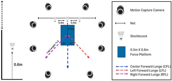
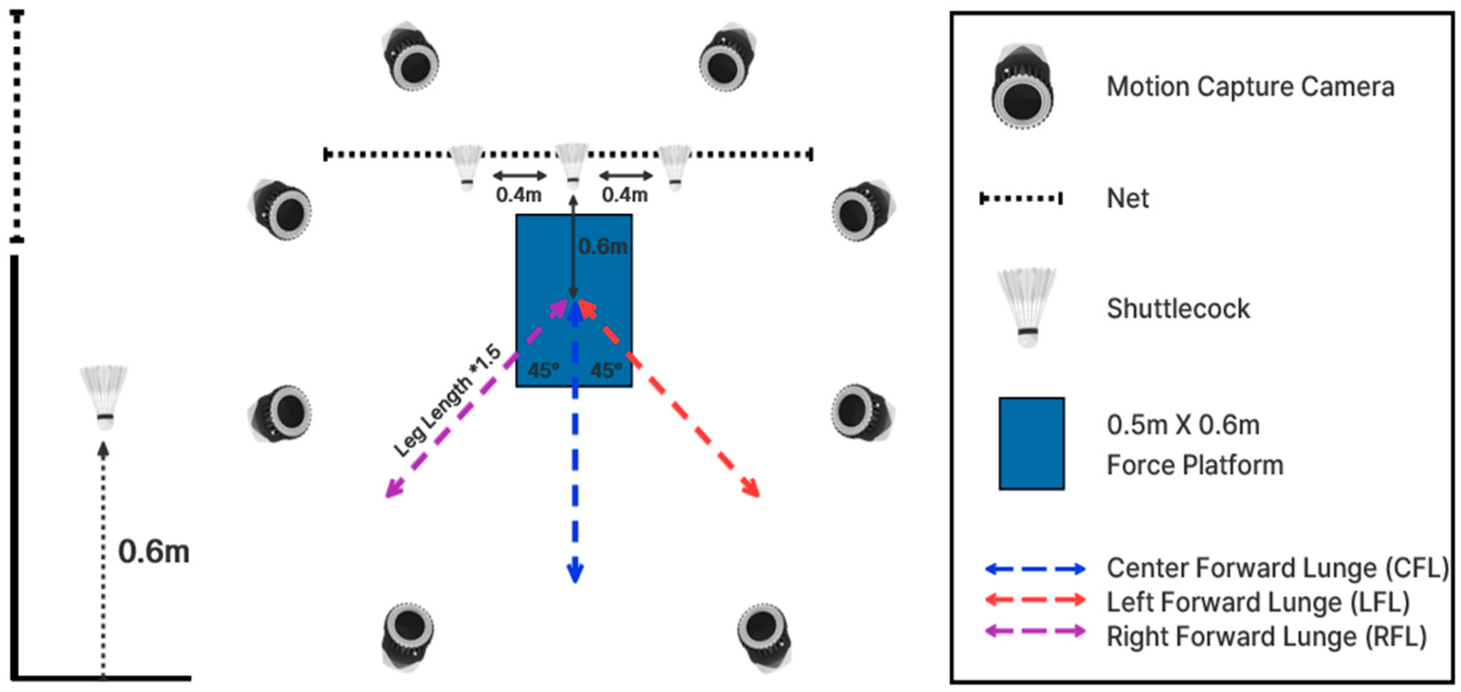
To collect movement data during badminton forward lunges performed in different directions, an eight-camera optical motion capture system (Miqus M3, Qualisys AB, Gothenburg, Sweden; sampling rate: 200 Hz) and one force plate (Kistler 9260AA6, Kistler, Winterthur, Switzerland; sampling rate: 1000 Hz) were used. To replicate conditions similar to actual badminton training and match play, a badminton court mat (N4U-B300, N4U, Republic of Korea, http://www.n4u.co.kr/) was installed in the laboratory. Shuttlecock targets were positioned 0.6 m in front of the force plate and 0.6 m above the ground, with a distance of 0.4 m between adjacent targets. The start and end points of the lunge were marked at a distance corresponding to 1.5 times each participant’s leg length from the center of the force plate, while the left and right target positions were marked at 45° angles. Leg length was defined as the distance from the anterior superior iliac spine to the medial malleolus [6]. More specific details of the lab setup and forward lunge direction are illustrated in Figure 1.
Figure 1.
Illustration of experimental setup (* indicates 1.5 times the participant’s leg length).
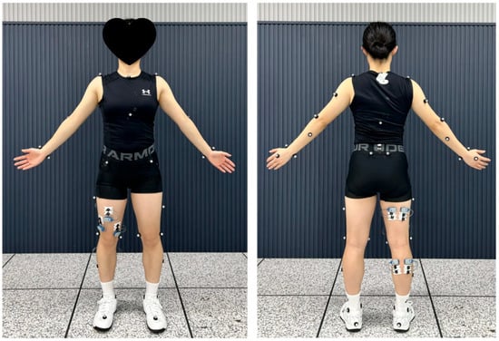
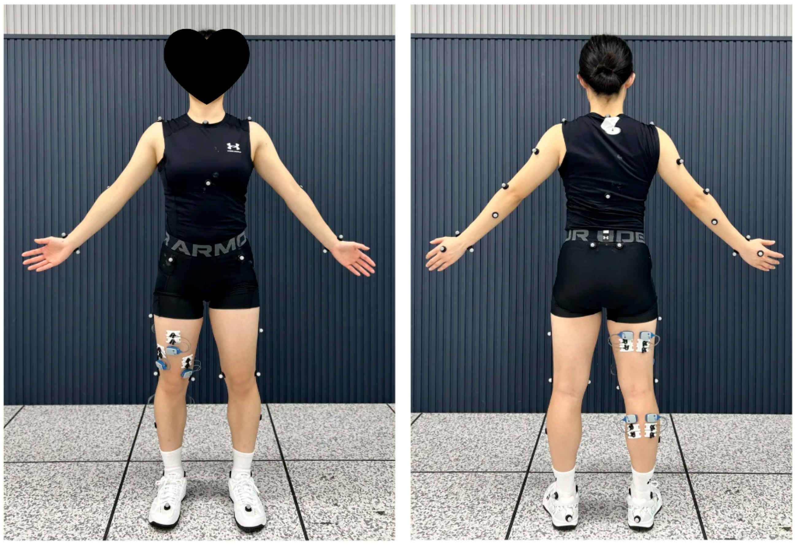
Participants wore tight-fitting tops, tights, and the same socks, along with the provided badminton shoes (Yonex SHB-SR1WEX, Yonex Co., Ltd., Tokyo, Japan), with sizes individually matched between 225 and 250 mm. Based on the OpenSim model [22,23], a total of 38 reflective trajectory markers (12.5 mm) were attached bilaterally to the body (Figure 2). Before data collection, all participants completed a familiarization session to determine an individualized starting position so that, after performing a split step and placing the left foot at the marked lunge initiation point corresponding to 1.5 times leg length, they could land with the right foot (dominant side) on the force plate. The forward lunge task was then performed in three directions: center forward lunge (CFL), left forward lunge (LFL), and right forward lunge (RFL). All participants were instructed to perform each movement with maximal effort from their self-selected starting position. The order of lunge directions was randomized, and each participant performed five trials in each direction, with sufficient rest provided between trials.
Figure 2.
Reflective marker location (left: front; right: back).
2.3. Data Processing and Analysis
In this study, initial contact (IC) was defined as the time point at which the vertical ground reaction force exceeded 20 N, maximum knee flexion (MKF) as the time point of peak knee flexion during stance, and take-off (TO) as the time point at which the vertical ground reaction force decreased below 20 N. The stance phase was divided into two subphases: the braking phase (BP), from IC to MKF, and the propulsion phase (PP), from MKF to TO. For knee joint angles and moments, values at each discrete event were extracted, whereas ground reaction forces and muscle forces were analyzed using phase-averaged values. For continuous-variable analysis, the braking and propulsion phases were each time-normalized to 50%, resulting in a total stance phase normalized to 100%.
Meanwhile, center-of-mass (COM) velocity was analyzed within phases defined using the vertical (Z-axis) position of the right heel marker. The period from the onset of upward movement of this marker to IC was defined as the approach phase, whereas the period from TO to the subsequent downward movement of the same marker was defined as the recovery phase. Raw three-dimensional motion capture data were processed using Visual3D software (Version 2023.01.1, Has-Motion, Kingston, ON, Canada). Marker trajectory and ground reaction force data were filtered using a fourth-order low-pass Butterworth filter, with cutoff frequencies set at 6 Hz and 30 Hz, respectively [24].
Knee flexor and extensor muscle forces were estimated using musculoskeletal modeling and simulation in OpenSim 4.4 (Stanford University, Stanford, CA, USA). The Catelli model was used for musculoskeletal modeling and simulation in the present study. Based on each participant’s anthropometric information, the model was scaled by adjusting the positions of the experimental and virtual markers. Joint angles were then calculated from the marker trajectory data using inverse kinematics (IK), and muscle forces were estimated using static optimization (SO), which minimizes the sum of squared muscle activations across all muscles in the model. All ground reaction force and joint moment data were normalized to body weight (BW), whereas muscle force data were normalized to body mass (kg).
2.4. Statistical Analysis
To examine differences in biomechanical variables among the three forward-lunge directions, discrete variables were analyzed using one-way repeated-measures analysis of variance (ANOVA) in SPSS 29.0 (IBM Corp., Armonk, NY, USA). Continuous variables across the entire movement cycle were analyzed in MATLAB R2023b (The MathWorks, Natick, MA, USA) using the Statistical Parametric Mapping (SPM) package. Because the assumption of normality was not satisfied for all variables, one-way repeated-measures analysis of variance based on one-dimensional statistical non-parametric mapping (SnPM1D; spm1d.stats.nonparam.anova1rm) was performed. The level of statistical significance was set at α = 0.05 for all analyses. Normality of all discrete variables was assessed using the Shapiro–Wilk test. For variables satisfying normality, one-way repeated-measures ANOVA was performed, followed by Bonferroni-corrected paired t-tests for post hoc comparisons. For variables not satisfying normality, the Friedman test was used, with Bonferroni-corrected Wilcoxon signed-rank tests for post hoc comparisons. Effect sizes for parametric ANOVA results are reported as partial eta-squared (partial η2), with values of 0.01, 0.06, and 0.14 interpreted as small, medium, and large, respectively. Additionally, 95% CIs for significant post hoc pairwise comparisons are reported for parametric analyses; non-parametric results are presented without CI, as this is not conventionally applicable. For continuous variables, SnPM analysis was conducted using one-way repeated-measures non-parametric mapping (spm1d.stats.nonparam.anova1rm), as normality was not satisfied for any variable. Post hoc comparisons were performed using Bonferroni-corrected non-parametric paired t-tests (spm1d.stats.nonparam.ttest_paired). For SnPM analyses, standardized effect size reporting has not yet been formally established; results are therefore presented with corresponding critical thresholds and significance intervals.
3. Results
3.1. Center of Mass Velocity
The comparison of center of mass (COM) velocity according to the direction of the badminton forward lunge is presented in Table 1. During the approach phase, which represents the forward movement toward the shuttlecock, COM velocity differed significantly among the three directions (F = 4.069, p = 0.026). Post hoc analysis revealed that LFL (1.33 ± 0.22 m/s) showed a significantly greater COM velocity than CFL (1.25 ± 0.20 m/s) (p < 0.05). A significant difference was also observed during the recovery phase, defined as the phase in which the participant returned to the initial position after performing the lunge (F = 7.469, p = 0.005). Post hoc analysis indicated that RFL (1.18 ± 0.19 m/s) showed a significantly greater COM velocity than CFL (1.08 ± 0.19 m/s) and LFL (1.12 ± 0.16 m/s) (p < 0.05).
Table 1.
Velocity of center of mass according to direction during badminton forward lunge (unit: m/s).
3.2. Knee Joint Kinematics
The comparison of knee joint angles at key events according to the direction of the badminton forward lunge is presented in Table 2. At IC, which represents the moment of landing, a significant difference was observed in the knee flexion angle (X2 = 25.444, p < 0.001). Post hoc analysis revealed that RFL (14.93 ± 5.41°) showed a significantly smaller knee flexion angle than CFL (19.08 ± 6.78°) and LFL (19.63 ± 7.29°) (p < 0.05). At MKF, a significant difference was found in the knee adduction angle (F = 5.081, p = 0.012). Post hoc analysis indicated that CFL (4.03 ± 4.15°) showed a significantly greater knee adduction angle than RFL (2.95 ± 4.39°) (p < 0.05). At TO, a significant difference was observed in the knee external rotation angle (F = 3.707, p = 0.035), However, post hoc analysis did not reveal significant differences among the three directions (p > 0.05).
Table 2.
Knee joint angle according to direction during badminton forward lunge (unit: °).
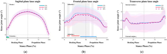
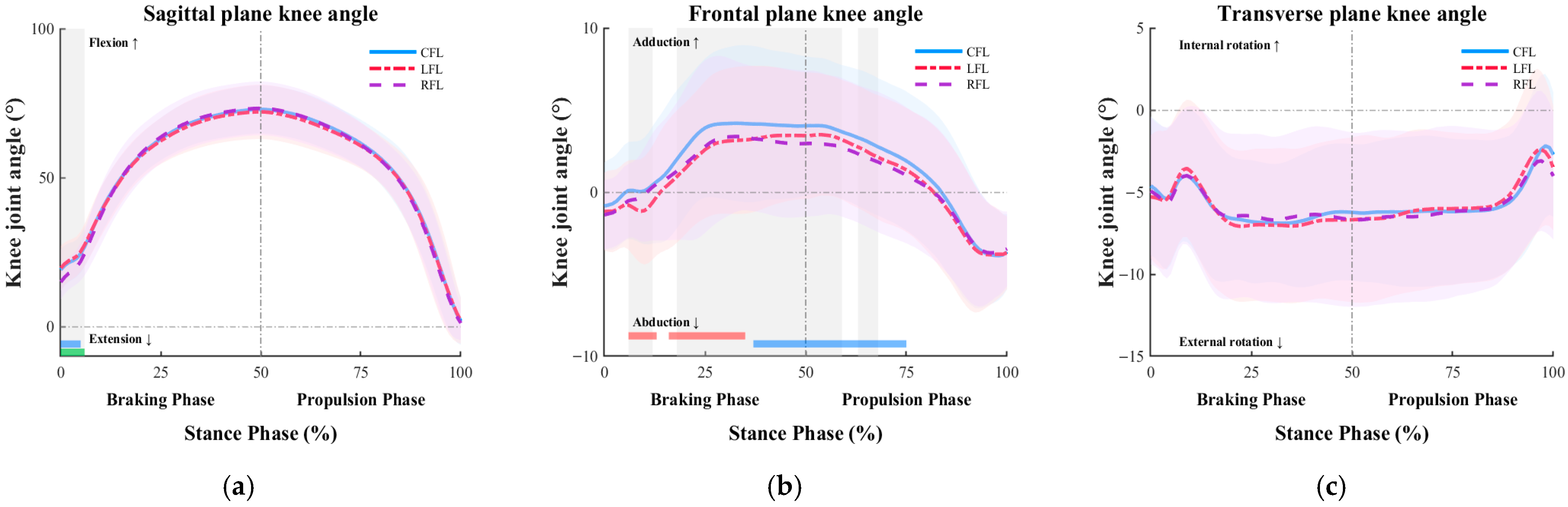
The results of the SnPM analysis examining the continuous changes in knee joint angles according to the direction of the badminton forward lunge are presented in Figure 3. In the sagittal plane, significant differences among the three directions were observed during the early stance phase (p < 0.05). Post hoc analysis revealed significant differences between CFL and RFL (0–5%) and between LFL and RFL (0–6%) (p < 0.05), with RFL showing a smaller knee flexion angle. In the frontal plane, significant differences were observed during 6–12%, 18–59%, and 63–68% of the stance phase (p < 0.05). Post hoc analysis indicated significant differences between CFL and LFL (6–13%, 16–35%) and between CFL and RFL (37–75%) (p < 0.05). Overall, CFL maintained a greater knee adduction angle throughout these phases. In the transverse plane, no significant differences among the three directions were observed throughout the stance phase (p > 0.05).
Figure 3.
The mean (bold line) and standard deviation (shaded area) of the knee joint angle according to direction during badminton forward lunge (gray shading indicates statistically significant differences based on SnPM analysis). The red, blue, and green bars at the bottom indicate significant differences between CFL and LFL, CFL and RFL, and LFL and RFL, respectively. (a) Sagittal plane knee joint angle graph; (b) Frontal plane knee joint angle graph; (c) Transverse plane knee joint angle graph.
3.3. Ground Reaction Forces
The comparison of ground reaction forces according to the direction of the badminton forward lunge is presented in Table 3. For vertical ground reaction force (VGRF), a significant difference was observed during the propulsion phase (F = 8.601, p < 0.001). Post hoc analysis revealed that RFL (0.96 ± 0.06% BW) showed significantly lower VGRF than CFL (0.99 ± 0.08% BW) and LFL (1.01 ± 0.06% BW) (p < 0.05). For anterior–posterior ground reaction force (APGRF), a significant difference was observed during the braking phase (F = 35.885, p < 0.001). Post hoc analysis indicated that LFL (−0.34 ± 0.09% BW) showed the lowest braking force, whereas RFL (−0.45 ± 0.08% BW) showed the greatest braking force (p < 0.05). A significant difference was also observed during the propulsion phase (F = 8.813, p < 0.001). Post hoc analysis revealed that LFL (−0.34 ± 0.09% BW) showed the lowest braking force, whereas RFL (−0.40 ± 0.07% BW) showed the greatest braking force (p < 0.05).
Table 3.
Ground reaction force according to direction during badminton forward lunge (unit: %BW).
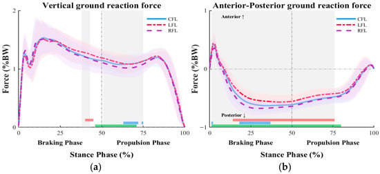
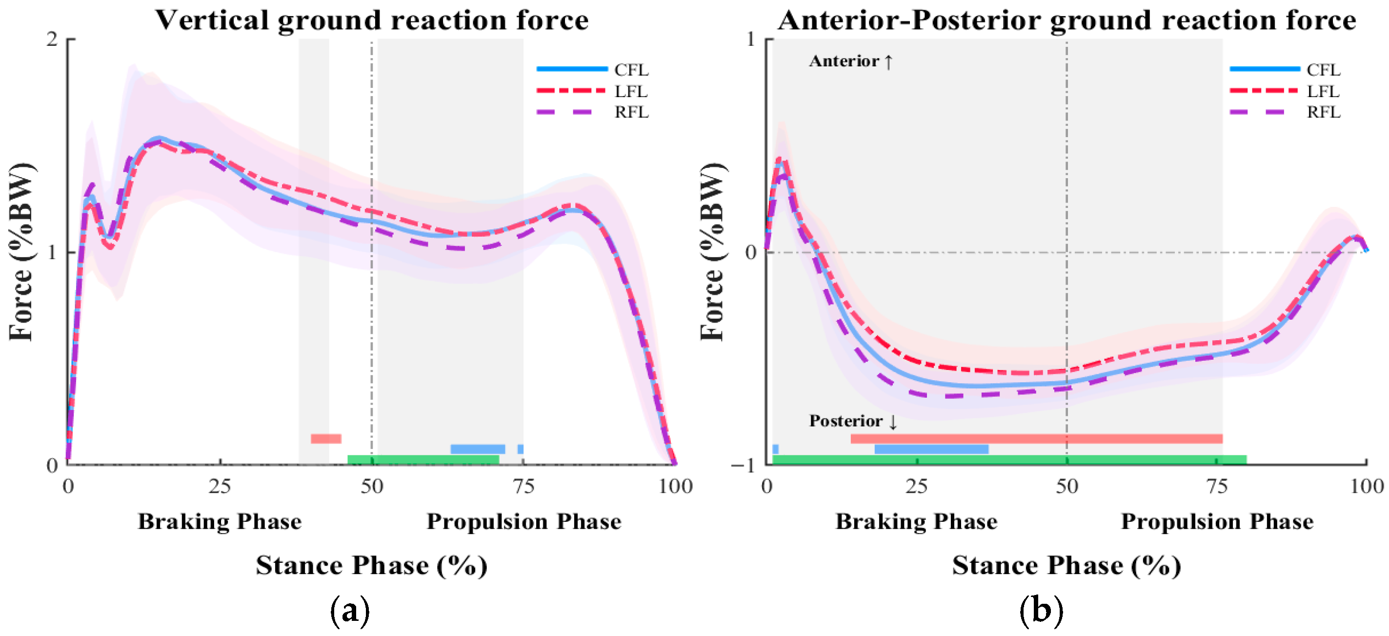
The results of the SnPM analysis examining the continuous changes in ground reaction forces according to the direction of the badminton forward lunge are presented in Figure 4. For vertical ground reaction force (VGRF), a significant difference among the three directions was observed during the late braking phase (38–43%) (p < 0.05). Post hoc analysis revealed a significant difference between CFL and LFL (40–45%) (p < 0.05), with LFL showing greater VGRF. During the propulsion phase, significant differences were observed between 51% and 75% of the stance phase (p < 0.05). Post hoc analysis indicated significant differences between CFL and RFL (63–72%, 74–75%) and between LFL and RFL (46–71%) (p < 0.05), with RFL showing the lowest VGRF. For anterior–posterior ground reaction force (APGRF), significant differences were observed from 1% to 76% of the stance phase across both the braking and propulsion phases (p < 0.05). Post hoc analysis revealed significant differences between CFL and LFL (14–76%), CFL and RFL (1–2%, 18–37%), and LFL and RFL (1–80%) (p < 0.05). In particular, LFL initially showed greater values than RFL during the early braking phase but subsequently exhibited the lowest braking force, whereas RFL maintained the greatest braking force throughout the stance phase.
Figure 4.
The mean (bold line) and standard deviation (shaded area) of the ground reaction force according to direction during badminton forward lunge (gray shading indicates statistically significant differences based on SnPM analysis). The red, blue, and green bars at the bottom indicate significant differences between CFL and LFL, CFL and RFL, and LFL and RFL, respectively. (a) Vertical ground reaction force graph; (b) Anterior–posterior ground reaction force graph.
3.4. Knee Joint Moments
The comparison of knee joint moments at key events according to the direction of the badminton forward lunge is presented in Table 4. At IC, significant differences in knee joint moments were observed in the sagittal (F = 15.599, p < 0.001), frontal (F = 4.794, p = 0.015), and transverse planes (F = 8.843, p < 0.001). Post hoc analysis revealed that, in the sagittal plane, LFL (0.21 ± 0.22 Nm/BW) showed a greater knee flexion moment (p < 0.05). In the frontal plane, CFL (0.09 ± 0.06 Nm/BW) showed a greater knee adduction moment than RFL (0.03 ± 0.09 Nm/BW) (p < 0.05). In the transverse plane, CFL (−0.02 ± 0.03 Nm/BW) exhibited the greatest external rotation moment (p < 0.05). At MKF, significant differences in knee joint moments were also observed in the sagittal (F = 0.862, p < 0.001), frontal (F = 8.480, p = 0.001), and transverse planes (F = 4.920, p = 0.013). Post hoc analysis indicated that LFL (0.68 ± 0.37 Nm/BW) showed the lowest knee flexion moment in the sagittal plane (p < 0.05). In the frontal plane, RFL (−0.14 ± 0.15 Nm/BW) exhibited a smaller knee abduction moment (p < 0.05). In the transverse plane, CFL (0.34 ± 0.17 Nm/BW) showed the greatest internal rotation moment (p < 0.05).
Table 4.
Knee joint moment according to direction during badminton forward lunge (unit: Nm/BW).
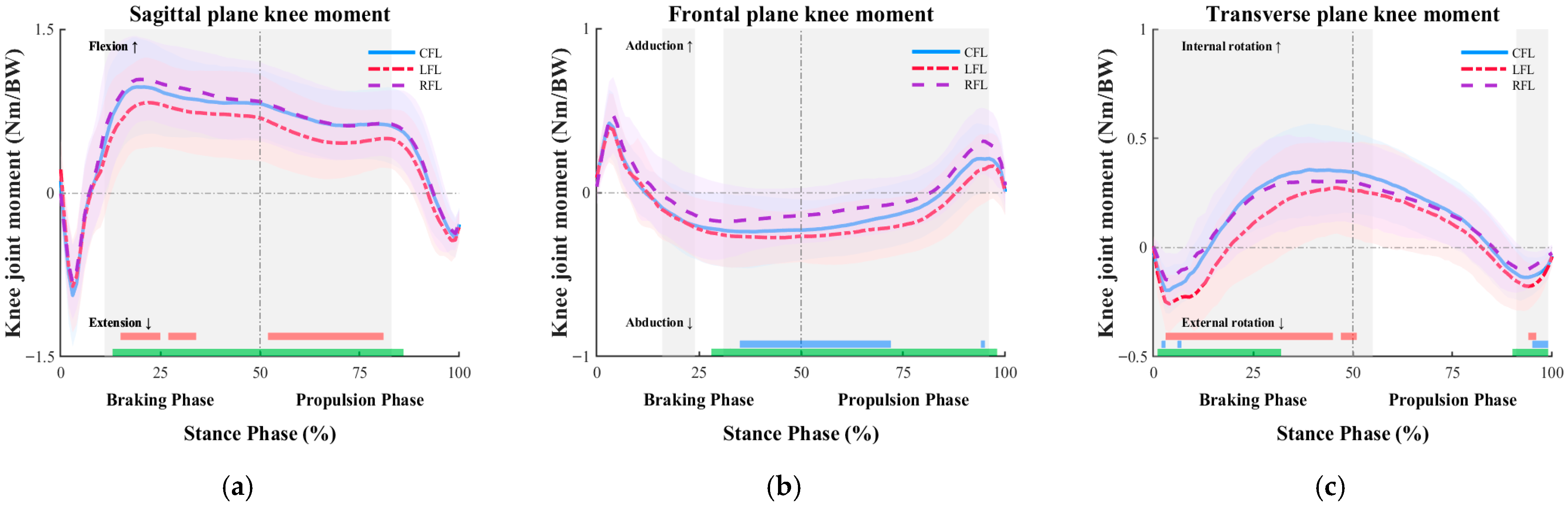
The results of the SnPM analysis examining the continuous changes in knee joint moments according to the direction of the badminton forward lunge are presented in Figure 5. In the sagittal plane, significant differences among the three directions were observed between 11% and 83% of the stance phase (p < 0.05). Post hoc analysis revealed significant differences between CFL and LFL (15–25%, 27–34%, and 52–81%) and between LFL and RFL (13–86%) (p < 0.05). Overall, LFL exhibited lower knee flexion moments throughout most of the stance phase. In the frontal plane, significant differences were observed during 16–24% and 31–96% of the stance phase (p < 0.05). Post hoc analysis indicated significant differences between CFL and RFL (35–72%, 94–96%) and between LFL and RFL (28–98%) (p < 0.05). Overall, RFL showed lower knee abduction moments during most of the stance phase but exhibited greater knee adduction moments during the late propulsion phase. In the transverse plane, significant differences among the three directions were observed during 0–55% and 91–99% of the stance phase (p < 0.05). Post hoc analysis revealed significant differences between CFL and LFL (3–45%, 47–51%, and 94–96%), between CFL and RFL (2–3%, 6–7%, and 95–99%), and between LFL and RFL (1–32% and 90–99%) (p < 0.05). LFL exhibited greater external rotation moments during the early braking phase and the late propulsion phase, whereas it showed the lowest internal rotation moments during the braking phase. In contrast, CFL demonstrated the greatest internal rotation moment during the braking phase.
Figure 5.
The mean (bold line) and standard deviation (shaded area) of the knee joint moment according to direction during badminton forward lunge (gray shading indicates statistically significant differences based on SnPM analysis). The red, blue, and green bars at the bottom indicate significant differences between CFL and LFL, CFL and RFL, and LFL and RFL, respectively. (a) Sagittal plane knee joint moment graph; (b) Frontal plane knee joint moment graph; (c) Transverse plane knee joint moment graph.
3.5. Knee Extensor Muscle Forces
The comparison of knee extensor muscle forces according to the direction of the badminton forward lunge is presented in Table 5. For the vastus medialis, significant differences were observed during both the braking phase (F = 3.304, p = 0.049) and the propulsion phase (F = 1.184, p = 0.318). However, post hoc analysis did not reveal significant differences among the three directions (p > 0.05). For the vastus intermedius, a significant difference was observed during the braking phase (F = 3.309, p = 0.049). However, post hoc analysis did not reveal significant differences among the three directions (p > 0.05). For the vastus lateralis, a significant difference was observed during the braking phase (F = 3.500, p = 0.041). Post hoc analysis indicated that LFL (26.43 ± 10.15 N/kg) showed a significantly lower muscle force than CFL (28.40 ± 9.12 N/kg) (p < 0.05).
Table 5.
Knee extension muscle force according to direction during badminton forward lunge (unit: N/kg).
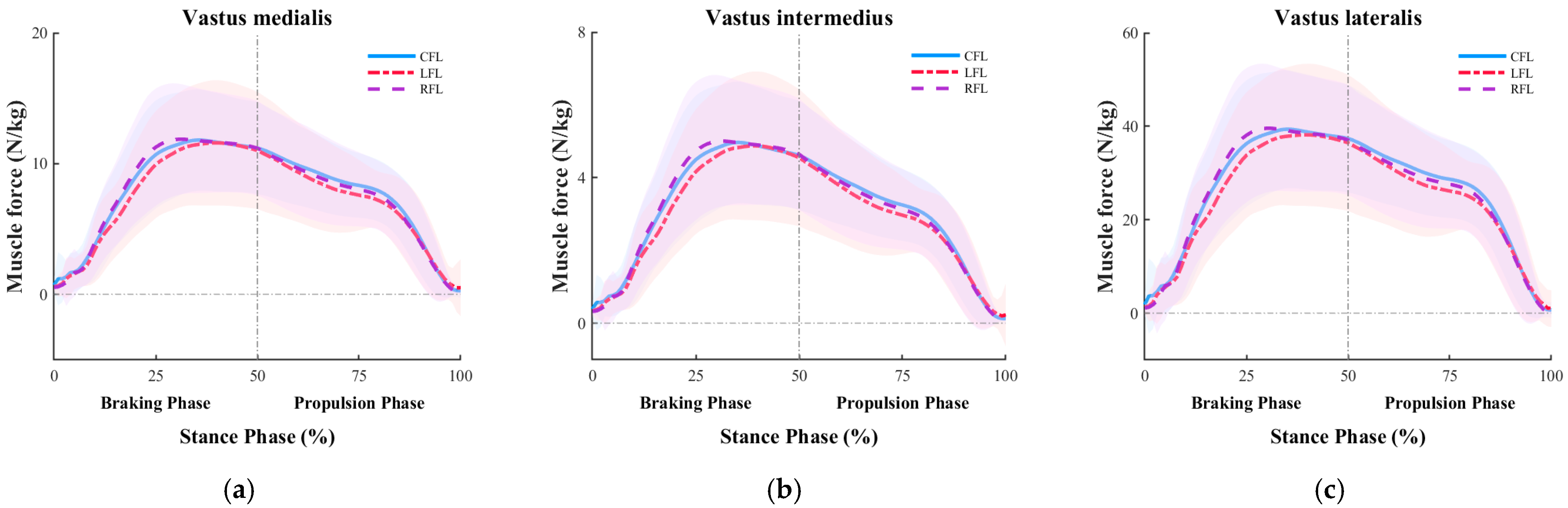
The results of the SnPM analysis examining the continuous changes in knee extensor muscle forces according to the direction of the badminton forward lunge are presented in Figure 6. No significant differences were observed among the three directions for the vastus medialis, vastus intermedius, or vastus lateralis throughout the stance phase (p > 0.05).
Figure 6.
The mean (bold line) and standard deviation (shaded area) of the knee extension muscle force according to direction during badminton forward lunge. (a) Vastus medialis graph; (b) Vastus intermedius graph; (c) Vastus lateralis graph.
3.6. Knee Flexor Muscle Forces
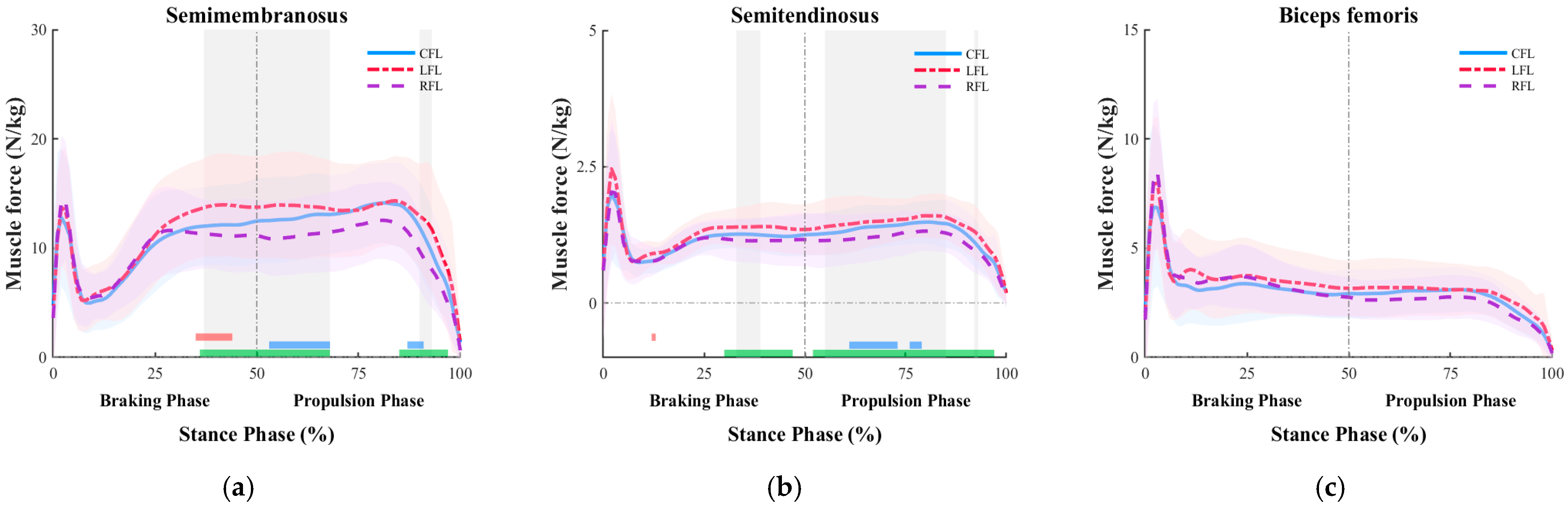
The comparison of knee flexor muscle forces according to the direction of the badminton forward lunge is presented in Table 6. For the semimembranosus, a significant difference was observed during the propulsion phase (F = 12.229, p < 0.001). Post hoc analysis indicated that RFL (10.46 ± 2.22 N/kg) showed significantly lower muscle force (p < 0.05). For the semitendinosus, significant differences were observed during both the braking phase (F = 5.911, p = 0.006) and the propulsion phase (F = 18.689, p < 0.001). Post hoc analysis revealed that during the braking phase, LFL (1.26 ± 0.23 N/kg) showed the greatest muscle force, whereas RFL (1.10 ± 0.25 N/kg) showed the lowest (p < 0.05). Similarly, during the propulsion phase, LFL (1.39 ± 0.27 N/kg) showed the greatest muscle force, whereas RFL (1.11 ± 0.29 N/kg) showed the lowest (p < 0.05). For the biceps femoris, a significant difference was observed during the propulsion phase (F = 8.694, p < 0.001). Post hoc analysis revealed that, LFL (2.88 ± 0.86 N/kg) showed the greatest muscle force, whereas RFL (2.36 ± 0.60 N/kg) showed the lowest (p < 0.05).
Table 6.
Knee flexion muscle force according to direction during badminton forward lunge (unit: N/kg).
The results of the SnPM analysis examining the continuous changes in knee flexor muscle forces according to the direction of the badminton forward lunge are presented in Figure 7. For the semimembranosus, significant differences among the three directions were observed during 37–68% and 90–93% of the stance phase (p < 0.05). Post hoc analysis revealed significant differences between CFL and LFL (35–44%), between CFL and RFL (53–68%, 87–91%), and between LFL and RFL (36–68%, 85–97%) (p < 0.05). Overall, the semimembranosus force was greater in LFL and lower in RFL. For the semitendinosus, significant differences were observed during 33–39%, 55–85%, and 92–93% of the stance phase (p < 0.05). Post hoc analysis indicated significant differences between CFL and LFL (12–13%), between CFL and RFL (61–73%, 76–79%), and between LFL and RFL (30–47%, 52–97%) (p < 0.05). Similarly, the semitendinosus force was greater in LFL and lower in RFL.
Figure 7.
The mean (bold line) and standard deviation (shaded area) of the knee flexion muscle force according to direction during badminton forward lunge (gray shading indicates statistically significant differences based on SnPM analysis). The red, blue, and green bars at the bottom indicate significant differences between CFL and LFL, CFL and RFL, and LFL and RFL, respectively. (a) Semimembranosus graph; (b) Semitendinosus graph; (c) Biceps femoris graph.
4. Discussion
In racket sports such as badminton, the mechanical load imposed on the lower extremities may vary according to movement direction, even when the same movement pattern is performed [17,25]. In particular, movements such as the forward lunge, which are performed rapidly and repeatedly in multiple directions, involve repeated braking and propulsive actions and may therefore increase injury risk in the lower limbs [26]. Kinetic differences have been reported across four lunge directions (left/right forward lunge and left/right backward lunge), with the left forward lunge showing the greatest vertical ground reaction force, loading rate, and plantar pressure [7]. The findings of the present study were consistent with these previous results, as the LFL produced the greatest vertical ground reaction force during both the braking and propulsion phases. This may be explained by the relatively unstable landing pattern that occurs when the dominant (right) leg crosses the body to reach a shuttlecock positioned in the left-front direction during stroke execution, thereby limiting effective impact absorption in the vertical direction [18]. In addition, center-of-mass velocity and anterior ground reaction force were significantly greater during the early braking phase in LFL than in RFL, indicating the use of a stronger braking strategy for rapid deceleration. Accordingly, the knee flexion moment at initial contact was also greater, which may reflect the increased demands for hip and knee flexion associated with a higher approach velocity [27]. Thereafter, LFL showed lower knee flexion moments and posterior ground reaction force throughout both the braking and propulsion phases, whereas elevated hamstring forces particularly in the semimembranosus and semitendinosus may indicate increased demand for dynamic joint stabilization among the three directions. This pattern may reflect a compensatory response consistent with increased demand for anterior tibial translation control and dynamic joint stabilization in a direction associated with potentially greater musculoskeletal loading, as suggested by prior work [28].
In badminton forward lunges, shuttlecock retrieval can generally be performed using either a forehand underhand stroke, in which the palm faces the shuttlecock, or a backhand underhand stroke, in which the dorsum of the hand faces the shuttlecock. Previous studies have suggested that forehand lunges tend to involve harder landings than backhand lunges [29,30]. In the present study, however, RFL showed the lowest vertical ground reaction force and lower external rotation moments during the early braking phase and late propulsion phase. This difference may reflect direction-specific footwork characteristics under the present experimental condition. In previous work using a split-step, smaller rotational moments were also reported during the right forward lunge [24]. In addition, RFL was characterized by a smaller knee flexion angle and the greatest posterior ground reaction force. A lunge posture with less knee flexion, limited forward displacement of the knee beyond the toes, and minimal hip flexion has been described as an efficient technique for facilitating rapid recovery [11,19,31]. Such a posture allows landing with a more upright trunk and provides a stable base for distal limb movement [11,32]. Trunk-mediated joint coordination and core stability may also enhance neuromuscular control, thereby contributing to a movement pattern capable of generating greater deceleration [11]. Notably, although hamstring force and external rotation moment were lower in RFL, center-of-mass velocity was highest in this direction. This pattern may be suggestive of a direction-specific biomechanical tendency in the forehand lunge, in which the dominant leg is actively used while maintaining joint stiffness and utilizing elastic energy stored during strong braking to facilitate rapid recovery [33].
Meanwhile, CFL showed greater knee adduction angle and internal rotation moment than both LFL and RFL. This may be related to the directional mismatch between the upper and lower extremities during movement toward a centrally positioned shuttlecock. While the lower limbs move forward, the upper limb is required to perform a backhand underhand stroke, resulting in a dissociation between upper- and lower-body movement directions. Under this condition, rotational motion generated by the upper limb may be transmitted through the trunk to the lower extremity, increasing rotational demand at the knee. The resulting knee rotational motion may therefore reflect a tendency to redistribute this load while maintaining alignment and balance of the body’s central axis [20,21].
These biomechanical characteristics provide important implications for training strategies. Because LFL appears to involve potentially greater musculoskeletal demand and loading patterns that have been associated with injury risk in prior research [7,14], training should emphasize hamstring strengthening as well as landing and braking techniques based on neuromuscular coordination. In contrast, because RFL appears to rely more on maintaining joint stiffness and utilizing elastic energy, training should focus on active movement and joint-stiffness enhancement to improve rapid recovery and propulsion. For CFL, where upper- and lower-body coordination and maintenance of central alignment appear to be more critical, greater emphasis should be placed on trunk stability and coordination training. In conclusion, the present study identified direction-specific differences in knee joint biomechanics during forward lunges in female recreational badminton players. These findings provide a practical basis not only for establishing more efficient performance strategies but also for developing individualized training programs and targeted injury prevention strategies.
Limitations
Several limitations of this study should be noted. First, the lunge task was performed under standardized laboratory conditions, including a fixed distance of 1.5 times leg length and predetermined shuttle positions. Although this approach ensures reproducibility and enables valid between-direction comparisons consistent with established protocols in badminton biomechanics research [6,7,14], it may not fully capture the natural variability inherent in actual match play. Second, muscle forces were estimated using static optimization in OpenSim without concurrent EMG validation. While this method is widely adopted in sports biomechanics research [18], neuromuscular interpretations, particularly those regarding hamstring-driven loading patterns during LFL, should be regarded as model-based inferences rather than direct measures of muscle activation. Third, injury risk was inferred from biomechanical variables including GRF and joint moments, which serve as established indirect indicators of musculoskeletal loading [7,14]; however, causal relationships with actual injury incidence cannot be confirmed without longitudinal outcome data and validated injury thresholds. Fourth, the absence of formal coordination metrics, such as joint coupling angles or continuous relative phase analysis, means that direction-specific biomechanical differences should be interpreted as outcome-level tendencies rather than direct evidence of underlying movement strategies. Finally, the present findings are specific to female recreational badminton players and may not generalize to male athletes, elite performers, or other age groups. Future studies addressing these constraints incorporating game-realistic conditions, EMG validation, longitudinal injury surveillance, and coordination analyses across diverse populations would further strengthen the practical implications of the present findings.
5. Conclusions
The purpose of this study was to quantitatively compare the biomechanical characteristics of the knee joint across three forward-lunge directions in female recreational badminton players and to analyze direction-specific movement strategies in order to provide practical baseline information for training and injury prevention. The findings showed that each lunge direction presented distinct biomechanical characteristics, suggesting that an integrated and multifaceted approach is needed to improve performance and reduce injury risk.
First, LFL appeared to involve greater knee joint loading and injury risk under relatively unstable landing conditions, with elevated hamstring forces potentially reflecting increased demand for knee joint stabilization. These findings suggest that training for the LFL direction should emphasize lower-limb strength development and neuromuscular coordination to improve shock absorption capacity. In particular, because the hamstrings play an important role in limiting anterior tibial translation and enhancing joint stability, functional strength training targeting this muscle group may be especially important.
Second, RFL appeared to reflect a direction-specific biomechanical tendency toward producing strong braking force despite a relatively limited knee flexion angle and achieving rapid and efficient recovery through the use of stored elastic energy. This suggests that active use of the dominant leg and maintenance of joint stiffness play important roles in efficient performance. Therefore, training for the RFL direction should focus on strengthening the muscles involved in maintaining joint stiffness, as well as on agility-based training designed to maximize the use of elastic energy.
Third, CFL was characterized by increased knee adduction and internal rotation moments, which may be related to the mismatch between upper- and lower-limb movement directions. This pattern appears to represent a rotational loading strategy used to maintain balance of the body’s central axis. Accordingly, training for CFL should emphasize coordination between the trunk and lower limbs, particularly through exercises aimed at improving core stability and trunk–lower extremity linkage.
Future studies should further classify participants according to skill level and playing experience in order to better understand inter-individual differences. In addition, because the present study focused primarily on the knee joint, future research should extend the analysis to multi-joint coordination involving the ankle, hip, and trunk. Such an approach would provide a more comprehensive basis for developing practical training methods and injury prevention strategies.
Author Contributions
Conceptualization, S.J. and S.-j.H.; methodology, S.J., S.-j.H. and J.L.; software, S.J. and J.L.; validation, S.-j.H. and S.C.; formal analysis, S.J. and S.C.; investigation, S.J., S.-j.H. and S.C.; resources, S.J. and S.C.; data curation, S.J. and S.-j.H.; writing—original draft preparation, S.J.; writing—review and editing, S.J. and J.P.; visualization, S.J. and S.-j.H.; supervision, J.P.; project administration, S.C. and J.P.; funding acquisition, S.-j.H. All authors have read and agreed to the published version of the manuscript.
Funding
This research was supported by the Pukyong National University Industry-university Cooperation Foundation’s 2025 Post-Doc support project.
Institutional Review Board Statement
The study was conducted in accordance with the Declaration of Helsinki and approved by the Institutional Review Board of Pukyong National University (IRB No. 2024-10-020 2024.10.29.).
Informed Consent Statement
Informed consent was obtained from all subjects involved in the study after providing detailed explanations of the research purpose, procedures, precautions during the experiment, and the protection of personal information.
Data Availability Statement
The data presented in this study are available upon reasonable request from the corresponding author.
Conflicts of Interest
The authors declare no conflicts of interest.
References
- Badminton World Federation. Badminton Global Fan, Commercial and Broadcast Figures. 2023. Available online: https://corporate.bwfbadminton.com/news-single/2023/01/14/badminton-global-fan-commercial-and-broadcast-figures (accessed on 13 June 2025).
- Valldecabres, R.; Casal, C.A.; Chiminazzo, J.G.C.; De Benito, A.M. Players’ on-court movements and contextual variables in badminton world championship. Front. Psychol. 2020, 11, 1567. [Google Scholar] [CrossRef] [Scilit]
- Ministry of Culture, Sports and Tourism. 2024 National Survey of Sports Participation. 2025. Available online: https://mcst.go.kr/kor/s_policy/dept/deptView.jsp?pSeq=1967&pDataCD=0417000000 (accessed on 22 June 2025).
- Phomsoupha, M.; Laffaye, G. The science of badminton: Game characteristics, anthropometry, physiology, visual fitness and biomechanics. Sports Med. 2015, 45, 473–495. [Google Scholar] [PubMed]
- Yu, L.; Jiang, H.; Mei, Q.; Mohamad, N.I.; Fernandez, J.; Gu, Y. Intelligent prediction of lower extremity loadings during badminton lunge footwork in a lab-simulated court. Front. Bioeng. Biotechnol. 2023, 11, 1229574. [Google Scholar] [CrossRef] [Scilit]
- Kuntze, G.; Mansfield, N.; Sellers, W. A biomechanical analysis of common lunge tasks in badminton. J. Sports Sci. 2010, 28, 183–191. [Google Scholar] [CrossRef] [Scilit]
- Hong, Y.; Wang, S.J.; Lam, W.K.; Cheung, J.T. Kinetics of badminton lunges in four directions. J. Appl. Biomech. 2014, 30, 113–118. [Google Scholar] [CrossRef] [Scilit]
- Hu, X.; Li, J.X.; Hong, Y.; Wang, L. Characteristics of plantar loads in maximum forward lunge tasks in badminton. PLoS ONE 2015, 10, e0137558. [Google Scholar] [CrossRef] [Scilit]
- Yu, L.; Mei, Q.; Mohamad, N.I.; Gu, Y.; Fernandez, J. An exploratory investigation of patellofemoral joint loadings during directional lunges in badminton. Comput. Biol. Med. 2021, 132, 104302. [Google Scholar] [CrossRef] [Scilit] [PubMed]
- Lee, S.; Min, B.; Park, J.; Kim, H.; Shin, D. A comparative study of knee joint instability and muscle activity due to medial or lateral deviation of the knee during lunge exercise. Phys. Ther. Rehabil. Sci. 2024, 13, 163–170. [Google Scholar]
- Huang, M.; Lee, H.; Lin, C.; Tsai, Y.; Liao, J. How does knee pain affect trunk and knee motion during badminton forehand lunges? J. Sports Sci. 2014, 32, 690–700. [Google Scholar] [CrossRef] [Scilit] [PubMed]
- Couppé, C.; Kongsgaard, M.; Aagaard, P.; Vinther, A.; Boesen, M.; Kjaer, M.; Magnusson, S.P. Differences in tendon properties in elite badminton players with or without patellar tendinopathy. Scand. J. Med. Sci. Sports 2013, 23, e89–e95. [Google Scholar]
- Shariff, A.H.; George, J.; Ramlan, A.A. Musculoskeletal injuries among Malaysian badminton players. Singap. Med. J. 2009, 50, 1095. [Google Scholar]
- Lam, W.; Lee, K.; Park, S.; Ryue, J.; Yoon, S.; Ryu, J. Understanding the impact loading characteristics of a badminton lunge among badminton players. PLoS ONE 2018, 13, e0205800. [Google Scholar] [CrossRef] [Scilit]
- Yung, P.S.; Chan, R.H.; Wong, F.C.; Cheuk, P.W.; Fong, D.T. Epidemiology of injuries in Hong Kong elite badminton athletes. Res. Sports Med. 2007, 15, 133–146. [Google Scholar] [CrossRef] [Scilit] [PubMed]
- Jeon, J.; Lim, S.; Cho, J. Designing and conducting an injury study in amateur badminton players. Appl. Sci. 2024, 14, 5194. [Google Scholar] [CrossRef] [Scilit]
- Lam, W.; Ryue, J.; Lee, K.; Park, S.; Cheung, J.T.; Ryu, J. Does shoe heel design influence ground reaction forces and knee moments during maximum lunges in elite and intermediate badminton players? PLoS ONE 2017, 12, e0174604. [Google Scholar] [CrossRef] [Scilit]
- Chen, T.L.; Wang, Y.; Wong, D.W.; Lam, W.; Zhang, M. Joint contact force and movement deceleration among badminton forward lunges: A musculoskeletal modelling study. Sports Biomech. 2022, 21, 1249–1261. [Google Scholar] [CrossRef] [Scilit]
- Mei, Q.; Gu, Y.; Fu, F.; Fernandez, J. A biomechanical investigation of right-forward lunging step among badminton players. J. Sports Sci. 2017, 35, 457–462. [Google Scholar] [CrossRef] [Scilit]
- Kurihara, Y.; Ohsugi, H.; Karasuno, H.; Tagami, M.; Matsuda, T.; Fujikawa, D. Trunk rotation enhances movement of the knee abduction angle while running among female collegiate middle- and long-distance runners. J. Hum. Sport Exerc. 2022, 17, 750–760. [Google Scholar] [CrossRef] [Scilit]
- Walter, J.P.; D’Lima, D.D.; Colwell, C.W., Jr.; Fregly, B.J. Decreased knee adduction moment does not guarantee decreased medial contact force during gait. J. Orthop. Res. 2010, 28, 1348–1354. [Google Scholar] [CrossRef] [Scilit] [PubMed]
- Catelli, D.S.; Wesseling, M.; Jonkers, I.; Lamontagne, M. A musculoskeletal model customized for squatting task. Comput. Methods Biomech. Biomed. Eng. 2019, 22, 21–24. [Google Scholar] [CrossRef] [Scilit] [PubMed]
- Delp, S.L.; Anderson, F.C.; Arnold, A.S.; Loan, P.; Habib, A.; John, C.T.; Thelen, D.G. OpenSim: Open-source software to create and analyze dynamic simulations of movement. IEEE Trans. Biomed. Eng. 2007, 54, 1940–1950. [Google Scholar] [CrossRef] [Scilit]
- Wang, Y.; Xu, L.; Jiang, H.; Yu, L.; Wu, H.; Mei, Q. Biomechanical effects of the badminton split-step on forecourt lunging footwork. Bioengineering 2024, 11, 501. [Google Scholar] [CrossRef] [Scilit] [PubMed]
- Bańkosz, Z.; Winiarski, S. Correlations between angular velocities in selected joints and velocity of table tennis racket during topspin forehand and backhand. J. Sports Sci. Med. 2018, 17, 330. [Google Scholar]
- Robinson, G.; O’Donoghue, P. A movement classification for the investigation of agility demands and injury risk in sport. Int. J. Perform. Anal. Sport 2008, 8, 127–144. [Google Scholar] [CrossRef] [Scilit]
- Chida, K.; Inami, T.; Yamaguchi, S.; Yoshida, Y.; Kohtake, N. Relationship between body center of mass velocity and lower limb joint angles during advance lunge in skilled male university fencers. Biomechanics 2023, 3, 377–388. [Google Scholar] [CrossRef] [Scilit]
- Walsh, M.; Boling, M.C.; McGrath, M.; Blackburn, J.T.; Padua, D.A. Lower extremity muscle activation and knee flexion during a jump-landing task. J. Athl. Train. 2012, 47, 406–413. [Google Scholar] [CrossRef] [Scilit] [PubMed]
- Valldecabres, R.; De Benito, A.M.; Littler, G.; Richards, J. An exploration of the effect of proprioceptive knee bracing on biomechanics during a badminton lunge to the net, and the implications to injury mechanisms. PeerJ 2018, 6, e6033. [Google Scholar] [CrossRef] [Scilit]
- Xie, Z.; Pan, J.; Wu, X.; Liang, H.; Chen, B.; Tan, D.; Huang, Z. Lower limb biomechanical differences between forehand and backhand forward lunges in amateur female badminton players. Front. Bioeng. Biotechnol. 2025, 13, 1558918. [Google Scholar] [CrossRef] [Scilit]
- Lee, J.J.J.; Loh, W.P. A state-of-the-art review on badminton lunge attributes. Comput. Biol. Med. 2019, 108, 213–222. [Google Scholar] [CrossRef] [Scilit]
- Willson, J.D.; Dougherty, C.P.; Ireland, M.L.; Davis, I.M. Core stability and its relationship to lower extremity function and injury. J. Am. Acad. Orthop. Surg. 2005, 13, 316–325. [Google Scholar] [CrossRef] [Scilit]
- Serpell, B.G.; Ball, N.B.; Scarvell, J.M.; Buttfield, A.; Smith, P.N. Muscle pre-activation strategies play a role in modulating kvert for change of direction manoeuvres: An observational study. J. Electromyogr. Kinesiol. 2014, 24, 704–710. [Google Scholar] [CrossRef] [Scilit] [PubMed]

Disclaimer/Publisher’s Note: The statements, opinions and data contained in all publications are solely those of the individual author(s) and contributor(s) and not of MDPI and/or the editor(s). MDPI and/or the editor(s) disclaim responsibility for any injury to people or property resulting from any ideas, methods, instructions or products referred to in the content. |
© 2026 by the authors. Licensee MDPI, Basel, Switzerland. This article is an open access article distributed under the terms and conditions of the Creative Commons Attribution (CC BY) license.