


Abstract
Management of unstable upper lumbar fractures with corpectomy and posterior fixation is technically demanding, and conventional workflows may require intraoperative repositioning, increasing operative complexity. Lateral mini-open upper lumbar corpectomy (LMULC) paired with robotic-assisted (RA) posterior percutaneous pedicle screw fixation (PPPSF) can be performed in a single position to facilitate ventral spinal decompression and stabilization in the anatomically constrained upper lumbar spine. In this study, we describe the operative technique and report four illustrative cases of unstable L1 or L2 fractures treated with single-position LMULC, RA-PPPSF, and short-segment fusion. Clinical, radiological, intraoperative variables and postoperative outcomes were evaluated. The mean age was 52.3 ± 17.7 years. The median operation time was 314 min (range 268–361 min); the median estimated blood loss (EBL) was 225 mL (range 100–400 mL). The median preoperative kyphosis was 10.15° (range 8.4–14.6°), the median postoperative kyphosis measured 6.65° (range 1.7–10.8°) and the median correction achieved was 3.5° (range −2.4–12.9°). The median visual analog scale (VAS) pain score reduced from 7 (range 7–9) preoperatively to 4.5 (range 2–6) postoperatively at discharge. At a median follow-up of 12 months (range 6–15 months), all patients had uncomplicated recoveries, demonstrated solid fusion on imaging, and reported favorable MacNab outcomes. Single-position LMULC with RA-PPPSF was technically feasible in this preliminary illustrative series and resulted in favorable clinical and radiographic outcomes. However, further studies in larger cohorts are warranted to help confirm these findings and better define the potential advantages and limitations of this technique.
1. Introduction
Lumbar corpectomy is a mainstay for the surgical management of fractures, tumors and deformity correction [1]. The procedure can be performed through an anterior [2], posterior [3], or lateral approach, the latter recently gaining popularity [4]. Lateral approaches to the spine provide many advantages over anterior and posterior approaches as they allow access to the retroperitoneal and retropleural corridors, enabling ventral vertebral decompression while minimizing muscle retraction [5,6,7,8,9] and neurovascular injury risk [10,11]. Compared to the posterior approach, the lateral approach facilitates the placement of interbody devices with a larger footprint, providing added stability by increasing the surface area in contact with the vertebral endplates [12,13]. These factors contribute to its association with good outcomes and faster recovery [14]. Management of unstable upper lumbar fractures requiring corpectomy and posterior fixation remains technically demanding because of the complex regional anatomy and the need to achieve efficient decompression and stabilization [8,15,16,17].
Single-position procedures reduce the need for intraoperative repositioning, which may translate into reduced operative times [18,19], lower blood loss [20] and potentially improved outcomes. However, performing pedicle screw placement and precise discectomies in the lateral position without robotic assistance (RA) presents unique ergonomic and navigational challenges [15], especially in the upper lumbar spine due to anatomical constraints posed by the diaphragm and the pleural cavity. RA has been increasingly adopted in spine surgery to help enhance accuracy, efficiency and the ergonomics of single-position lateral workflows [17,21]. Although individual components of this approach have been described previously, reports on their combined application in a single-position, dual-surgeon workflow for the upper lumbar spine remain limited.
Here we present our initial experience with single-position lateral mini-open upper lumbar corpectomy (LMULC) and RA posterior percutaneous pedicle screw fixation (PPPSF) performed in a dual-surgeon workflow through a technical note along with four illustrative cases. The report focuses on the operative workflow, surgical nuances, feasibility, and clinical and radiographic outcomes in the immediate postoperative period and through long-term follow-up.
2. Materials and Methods
Following Institutional Review Board approval (IRB: 18-003951), four patients who had undergone a single-position LMULC with PPPSF in 2024 for traumatic injuries and unstable fractures at L1 or L2 were identified and retrospectively studied. In all cases, the surgery was performed by the operative team with intraoperative computed tomography (iCT)—navigation (NA), fluoroscopy and RA for pedicle screw placement. Patients’ data were accessed under our retrospective Institutional Review Board (IRB)-approved study for spinal pathologies. Direct patient consent was not required by the IRB.
2.1. Surgical Technique
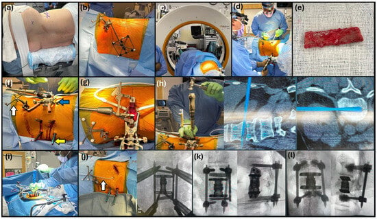
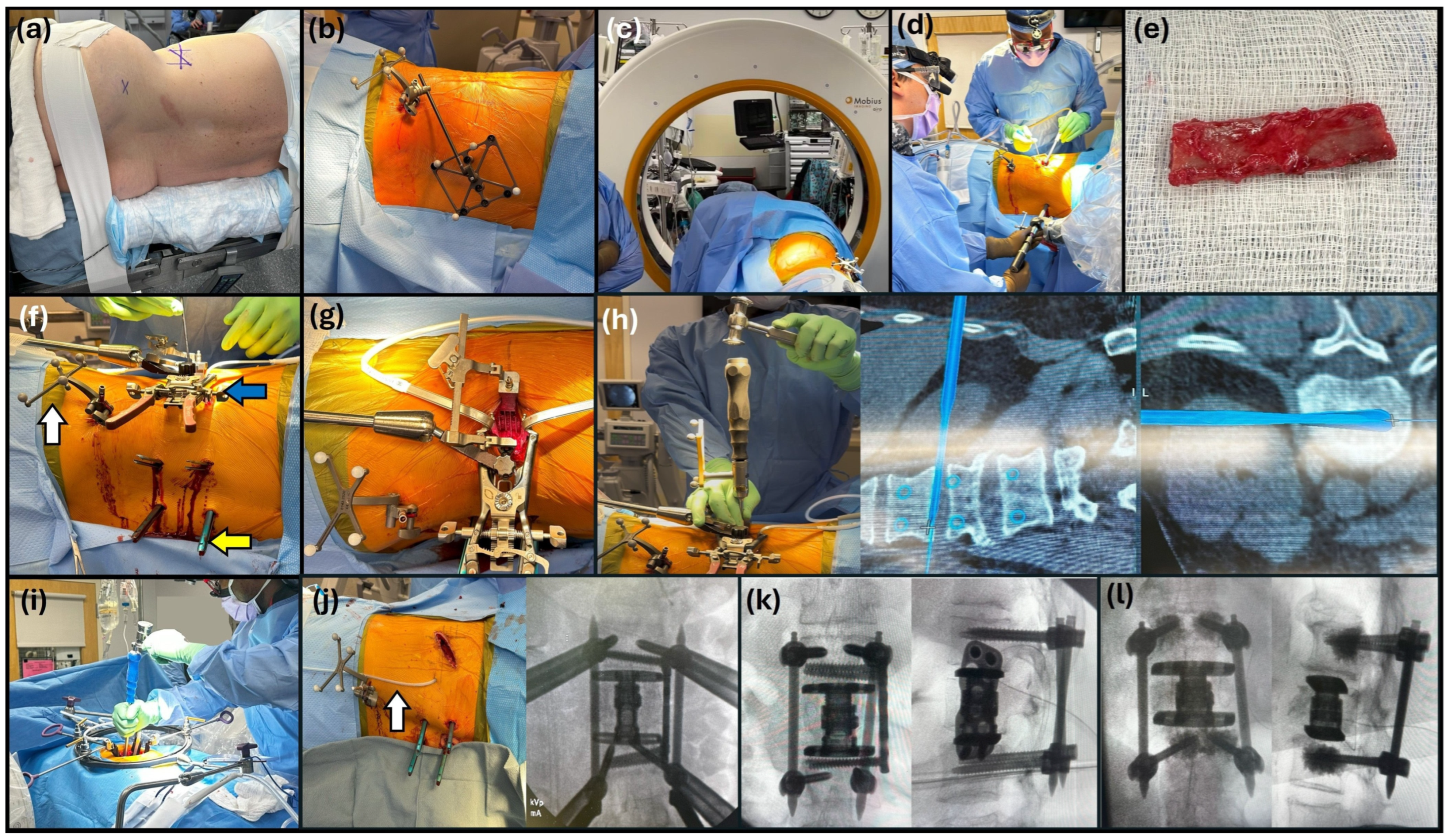
The technique for performing a lateral mini-open lumbar corpectomy has been described by Gandhoke et al. (2015) [16], Uribe et al. (2011) [8] and Yu et al. (2019) [22], and here we outline a detailed perioperative set up, the surgical procedure and a dual-surgeon workflow for a single-position LMULC with RA-PPPSF (Figure 1, Table 1). The patient is positioned on a Jackson table (Mizuho OSI, CA, USA) in the lateral decubitus position with the spine flush with the table edge. Incision planning is guided by fluoroscopy to confirm the true lateral decubitus position followed by an X-ray to localize the correct vertebral body. Following sterile preparation, a digital reference array (DRA) is placed in the posterior superior iliac spine with two Schanz pins. An iCT scan (Airo iCT scanner system, Brainlab, Feldkirchen, Germany) is performed with automatic registration for NA guidance, and the dataset is directly integrated with the spine robot (Globus, Audubon, PA, USA) to plan the trajectory of the pedicle screws. A pilot hole is first drilled and then the pedicle is tapped, followed by screw placement. Screws are placed prior to disc preparation to avoid introducing inaccuracies to the navigation and the screws on the dependent side are placed first. Connecting rods are not inserted until after the LMULC has been performed. While one surgeon places the pedicle screws with RA, a second surgeon performs the lateral exposure for the corpectomy (Figure 1d). For the LMULC, the other surgeon performs the retropleural or retroperitoneal exposure through a partial rib resection. It is important to select the rib that intersects closest to the posterior aspect of the vertebral body, especially if canal decompression is needed. A navigated MIS dilator may be used for the approach and localization. The plane is advanced up to the transverse process (TP). One may be in the retropleural, retroperitoneal or a combination of both depending on patient-specific anatomy. Once a retractor is in place, the segmental artery must be identified, coagulated and then sectioned. Following this, discectomies are performed at the level above and below the corpectomy. A navigated osteotome is used to make the cuts; after that, a high-speed drill with a 6 mm rough diamond and a pituitary rongeur are used to remove the vertebral body. If canal decompression is needed, a 3 mm rough diamond can be used to remove the ipsilateral pedicle for posterior canal exposure. After the corpectomy is completed, an expandable cage, packed with rib autograft material is inserted into the corpectomy defect. At this point a lateral plate can be secured by locking screws. A lateral plate is typically avoided for osteoporotic fractures to avoid placing too many holes in the porous bone. Once these screws are secured, an X-ray is performed to confirm the location of the instrumentation, followed by the closure of the lateral component in layers. A 10-French wound drainage system is left in the retropleural or retroperitoneal cavity. During the closure of the lateral incision, the other surgeon can work on placing and securing the connecting rods between the heads of the pedicle screws. If the pleura has been violated, it can typically be plugged with gelfoam as long as there is no lung injury. It is important to not deflate the lung during a retropleural exposure, as this will increase the likelihood of pleural violation as the tension is helpful. The posterior wound is also closed in layers. A confirmatory post-instrumentation iCT scan is performed before the removal of the MIS towers and DRA in case any screws need to be revised.
Figure 1.
Perioperative set up, surgical procedure steps and dual-surgeon workflow. (a) Right lateral decubitus positioning of patient with left side up and the back flush with the edge of the table; (b) digital reference array (DRA) placement prior to intraoperative computed tomography (iCT) scan; (c) iCT scan; (d) while one surgeon places the posterior percutaneous pedicle screws with the help of the robot the second surgeon performs the lateral exposure for the corpectomy; (e) rib resected to achieve retropleural exposure is saved, to be used later as autograft for the corpectomy cage; (f) surgical field setup: white arrow—iliac-mounted DRA, yellow arrow—minimally invasive (MIS) towers for posterior percutaneous pedicle screw fixation (PPPSF), blue arrow—MIS lateral retractor; (g) retractors placed in the lateral incision to perform the discectomies, the lateral mini-open upper lumbar corpectomy (LMULC) and canal decompression if needed; (h) discectomy and use of the navigated osteotome to make cuts in the vertebral body; (i) placement of expandable corpectomy cage into the defect; (j) confirmatory post-instrumentation scan performed before removal of the MIS towers and DRA, white arrow: 10-French wound drainage system; (k) a lateral plate is placed and secured with locking screws; (l) lateral plate not placed for osteoporotic fractures.
Table 1.
Perioperative set up and key steps in surgery.
2.2. Clinical Outcomes
All patients were discharged with a brace, prescribed for 6 weeks following surgery. Clinical and surgical data including basic demographics, diagnoses, comorbidities, operative time, EBL, postoperative length of stay (LOS), pre- and postoperative pain scores, pre- and postoperative kyphosis measurements, and follow-up length were collected. Vertebral body height at the level of corpectomy was measured on the corresponding preoperative and earliest available postoperative radiographic imaging. Vertebral body height was measured from the anterior portion of the inferior endplate of the vertebral body above the level of the corpectomy to the anterior portion of the superior endplate of the vertebral body below the level of the corpectomy. Total fluoroscopy dose and time were recorded. Functional outcomes were assessed per MacNab criteria [23,24]. Radiographic fusion at the last follow-up was defined as Bridwell Grade I or II [25].
3. Results
3.1. Demographic and Preoperative Characteristics—Table 2
The median age at presentation was 50 (range 26–83) with a 1:1 male to female ratio. Three patients underwent an L1 corpectomy and one underwent an L2 corpectomy. The median BMI was 30.8 kg/m2 (range 28.7–37.8 kg/m2). All patients underwent surgery to achieve stabilization and decompression following a burst fracture; three of them had a grade A4 and one had a grade A3 fracture according to the AO Spine Thoracolumbar Fracture Classification. Functionally, one patient had mild impairment classified as ASIA grade D on preoperative examination. A fall was the most common mechanism of injury occurring in three patients, while one patient had an injury secondary to a motor vehicle collision (MVC). The median preoperative kyphosis was 10.15° (range 8.4–14.6°). The median preoperative pain score on the visual analog scale (VAS) was 7 (range 7–9). The median preoperative vertebral body height at the level of corpectomy was 30.74 mm (range 23.54—31.02 mm).
Table 2.
Preoperative patient characteristics.
3.2. Operative Variables and Immediate Postoperative Course—Table 3
The median operating room (OR) time was 314 min (range 268–361 min). The median EBL was 225 mL (range 100–400 mL). The mean fluoroscopy dose was 2.52 (±1.01) mSv and the median fluoroscopy time was 56.50 s (range 43–80 s). In the three patients in whom the iCT was used, the median preoperative iCT dose was 12.59 mSv (range 11.49–15.42 mSv) and the median postoperative iCT dose was 16.53 mSv (range 7.61–20.03 mSv). No posterior pedicle screw revisions were required in any case following the confirmatory post-instrumentation iCT scan. The median intraoperative kyphosis was 6.1° (range 3.7–8.9°), and in one patient with a preoperative kyphosis of 8.4° an intraoperative lordosis of 7.2° was achieved. The median postoperative VAS score at discharge was 4.5 (range 2–6). No patients received blood transfusions or needed a chest tube placement. There was only one case of pleural violation which was immediately plugged with gelfoam. All patients had an uneventful immediate postoperative period with no complications. The median LOS was 4.5 days (range 4–6 days). The median postoperative kyphosis was 6.65° (range 1.7–10.8°) and the median correction achieved was 3.5° (range −2.4 to 12.9). The median postoperative vertebral body height at the level of corpectomy on postoperative radiographic imaging was 38.48 mm (range 35.27–42.19 mm). The median vertebral body height correction achieved was 7.61 mm (range 4.52–18.65).
Table 3.
Operative variables and postoperative outcomes.
3.3. Patient Outcomes at Last Follow-Up—Table 4
The median follow-up duration was 12 months (range 6–15 months). All patients reported good functional outcomes according to the MacNab criteria and demonstrated radiographic fusion on imaging at the last follow-up. The median postoperative ODI score was 50% (range 24–80%). Except for one patient with pre-existing segmental kyphosis at L5-S1 and associated radicular pain, no long-term complications were observed, with no evidence of cage subsidence, rod fracture, screw loosening, adjacent segment disease, femoral neuropathy or wound complications.
Table 4.
Patient outcomes at last follow-up.
3.4. Illustrative Cases
3.4.1. Case 1
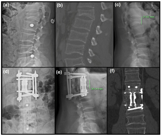
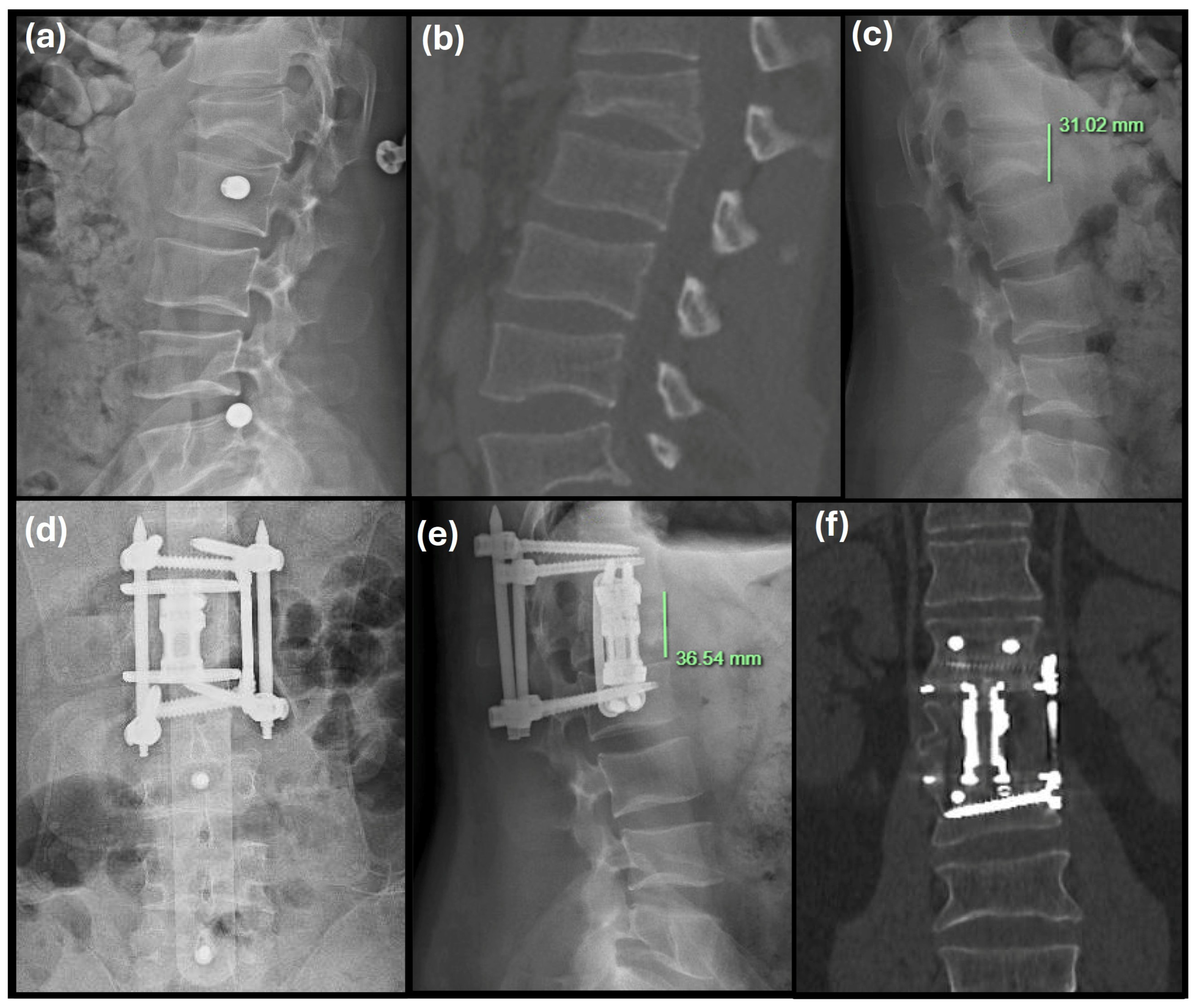
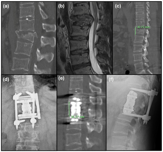
A 26-year-old man presented with a highly comminuted L1 burst fracture after a rollover MVC. Radiographic imaging showed more than 50% vertebral body height loss and retropulsed fragments causing canal narrowing (Figure 2a,b). Despite conservative management with bracing, there was progressive vertebral body collapse and pain intensified to 9 on the VAS. The L1 vertebral body height was 31.02 mm (Figure 2c). Preoperative segmental kyphosis was 14.6°, with bilateral weakness (4/5) on hip flexion and knee extension. Bilateral lower extremity (LE) strength, sensation, and bowel and bladder function were preserved. He underwent an L1 LMULC with RA-PPPSF at T12-L2, intraoperative segmental kyphosis measured 5°, and there was a small pleural breach sealed with gelfoam and a Hemovac drain placed in situ.
Figure 2.
Pre- and postoperative imaging of Case 1: (a) preoperative X-ray lateral view, (b) preoperative CT scan sagittal view, (c) preoperative L1 vertebral body height measurement, (d) postoperative X-ray AP view, (e) postoperative vertebral body height measurement, (f) postoperative CT scan coronal view at 1-year follow-up.
Immediate postoperative imaging demonstrated 1.7° residual kyphosis, representing a 12.9° correction. Hardware was intact without cage subsidence or the need for screw revision (Figure 2d). The patient was discharged on postoperative day (POD) 5 and his pain was a 6 on the VAS at that time. Vertebral body height at the level of corpectomy measured 36.54 mm on the 3-month postoperative radiograph (Figure 2e).
At the 1-year follow-up, CT scan confirmed radiographic fusion (Figure 2f). LE strength improved to 5/5 across all joints with normal bowel and bladder function. The patient did complain of persistent low-back pain with radicular symptoms in the L5-S1 distribution, attributed to his pre-existing segmental kyphosis with prior microdiscectomy at L5-S1. His Oswestry Disability Index (ODI) score was 72% and he had a good MacNab outcome.
3.4.2. Case 2
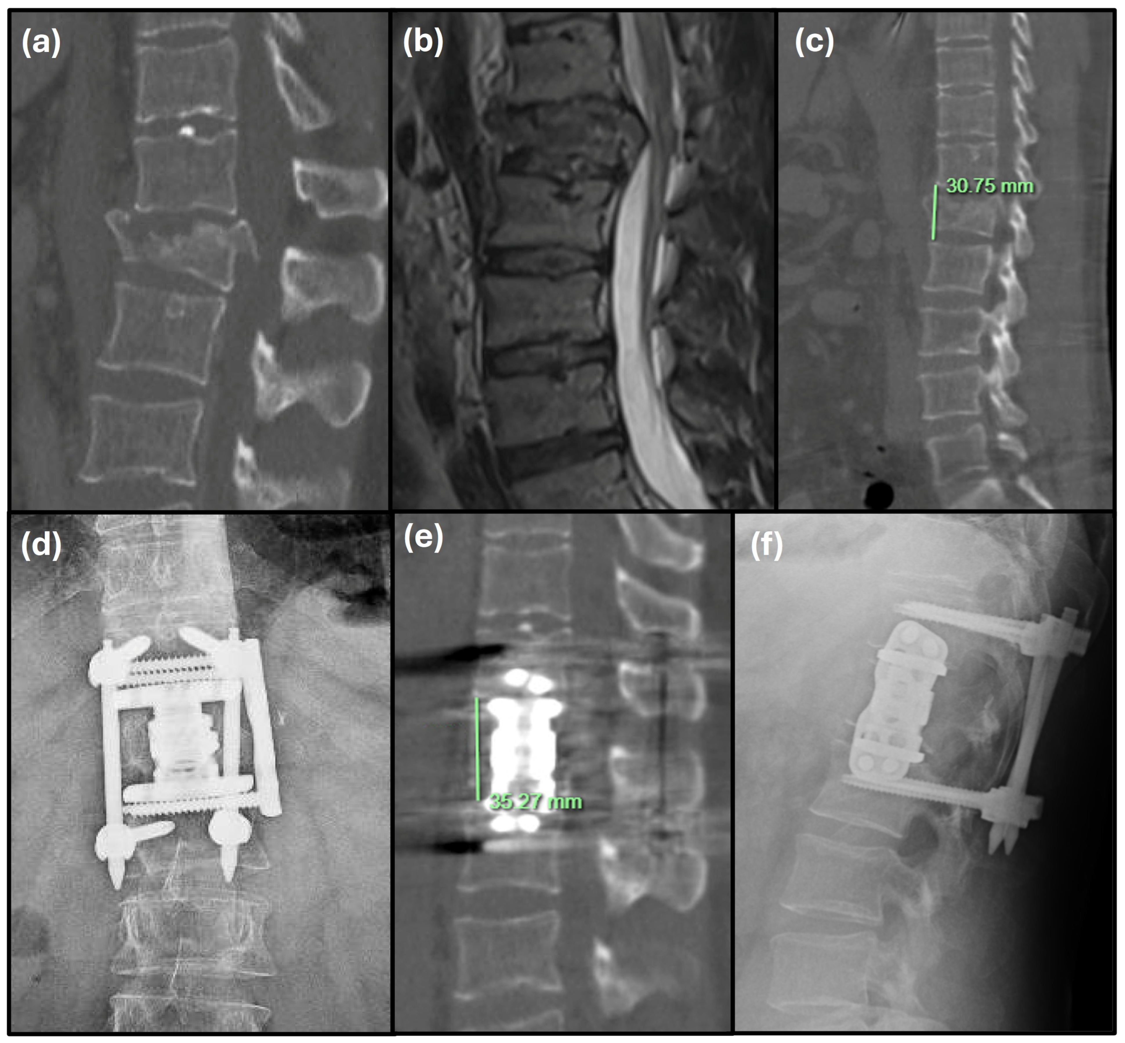
This was a 39-year-old man presenting with severe back pain and acute urinary retention after an 8-foot fall. Imaging revealed a grossly comminuted burst with >50% L1 vertebral-body collapse, posterior wall disruption, and marked retropulsion (Figure 3a,b). There was circumferential osseous encroachment of the thecal sac causing severe canal stenosis and focal segmental kyphosis measured 10.3°. The L1 vertebral body height measured 30.75 mm (Figure 3c). He had bilateral hip and knee weakness (4/5) with preserved distal strength. The patient also demonstrated saddle anesthesia and bladder dysfunction with a post-void residual (PVR) of 700 mL, with intact rectal tone and sensation. He underwent the LMULC at L1 along with canal de-compression followed by RA-PPPSF between T12 and L2. A Globus expandable lateral cage packed with the resected rib along with a lateral plate was used to secure the defect. Intraoperative segmental kyphosis measured 3.7°.
Figure 3.
Pre- and postoperative imaging of Case 2: (a) preoperative CT scan sagittal view, (b) preoperative T2-weighted MRI sagittal view, (c) preoperative L1 vertebral body height measurement, (d) postoperative X-ray AP view, (e) postoperative vertebral body height measurement, (f) postoperative X-ray lateral view at 15-month follow-up.
The immediate postoperative radiographs showed a 5.9° kyphosis, representing a 4.4° correction, with intact instrumentation and no subsidence (Figure 3d). Vertebral body height at the level of corpectomy measured 35.27 mm on immediate postoperative CT imaging (Figure 3e). Pain improved to 5 on VAS, bladder function had normalized, and the patient was discharged on POD 6.
At six months post-surgery LE strength had improved across all joints (5/5) with intermittent right foot numbness. His saddle anesthesia and urinary retention had resolved. At the 15-month follow-up his ODI score was 24%, with a “good” MacNab rating and imaging demonstrated radiographic fusion (Figure 3f).
3.4.3. Case 3
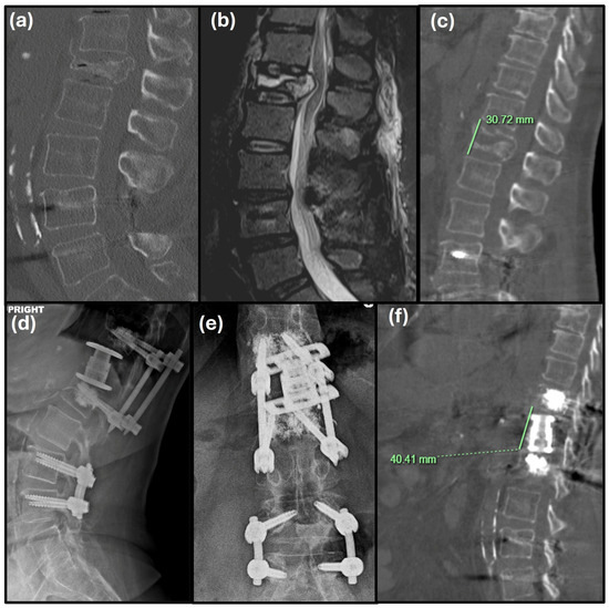
The third case was a 61-year-old woman presenting with escalating back pain (VAS 7) and constipation after a fall. Imaging showed an L1 burst fracture AO-Grade A 4, with posterior wall disruption, and posterior inferior retropulsion narrowing the canal diameter (Figure 4a,b). The L1 vertebral body height was measured at 30.72 mm (Figure 4c) and preoperative focal kyphosis measured 8.4°. Neurologically, she had mild bilateral hip flexor weakness (4+/5), her PVR was 123 mL, and she had not passed any stools in the 7 days since her injury. She underwent a LMULC at L1 with bilateral RA- PPPSF at T12 and L2. A lateral plate was avoided due to the osteopenic bone quality, and the screws were augmented with cement (Figure 4d,e).
Figure 4.
Pre- and postoperative imaging of Case 3: (a) preoperative CT scan sagittal view, (b) preoperative T2-weighted MRI sagittal view, (c) preoperative L1 vertebral body height measurement, (d) postoperative X-ray lateral view, (e) postoperative X-ray AP view, (f) postoperative vertebral body height measurement.
Intraoperative segmental lordosis measured 7.2° and a Hemovac drain was placed during closure. Immediate postoperative imaging showed a 10.8° kyphosis, demonstrating a −2.4° correction, and intact instrumentation without cage subsidence (Figure 4d,e). Vertebral body height at the level of corpectomy measured 40.41 mm on immediate postoperative CT imaging (Figure 4f). Her pain improved to VAS 4 and the patient was discharged on POD 4.
At six months she reported intermittent low-back discomfort; ODI was 37%, and bowel function had normalized. The one-year follow-up CT demonstrated solid fusion. Lower-extremity strength was 5/5 across all joints, bladder and bowel function were fully re-stored, ODI score was 28%, and she had a good MacNab.
3.4.4. Case 4
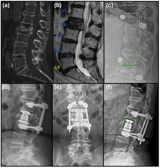
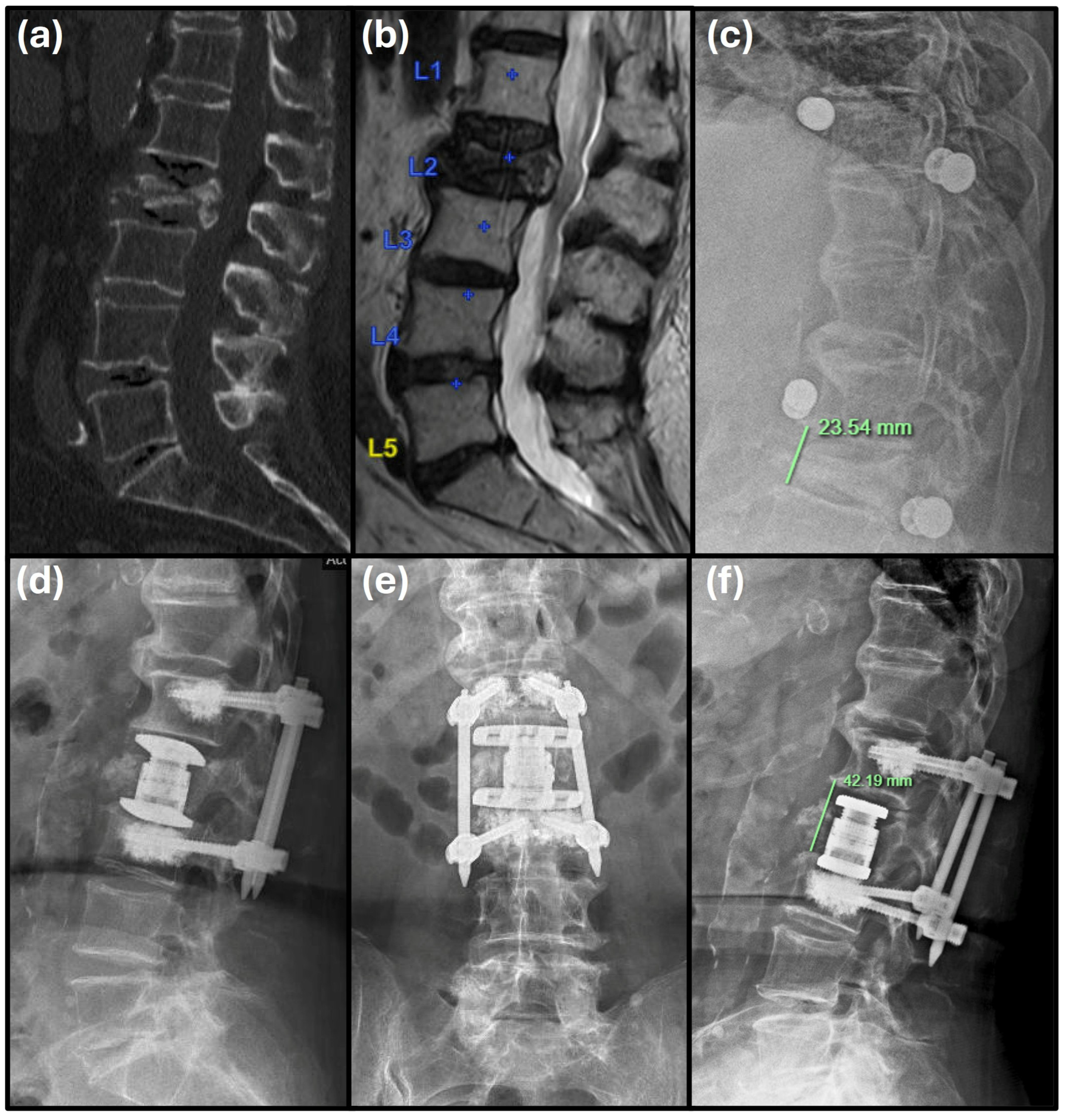
An 83-year-old woman with a history of osteoporosis presented with axial back pain (VAS 7) after a ground-level fall. Imaging identified an L2 burst fracture with mild vertebral body height loss and posterior wall fragment retropulsion causing canal narrowing (Figure 5a,b). She had no neurological deficits and was managed conservatively. However, radiography one week later revealed further vertebral collapse. The L2 vertebral body height measured 23.54 mm (Figure 5c) and segmental kyphosis measured 10°, with associated worsening pain. She underwent an L2 LMULC with bilateral RA-PPPSF at L1 and L3. A preoperative CT was used for intraoperative navigation with the robot. The screws were augmented with cement. Intraoperative segmental kyphosis measured 8.9°, and a 15-French Blake drain was placed in the lateral wound.
Figure 5.
Pre- and postoperative imaging of Case 4: (a) preoperative CT scan sagittal view, (b) preoperative T2-weighted MRI sagittal view, (c) preoperative L2 vertebral body height measurement, (d) postoperative X-ray lateral view, (e) postoperative X-ray AP view, (f) postoperative vertebral body height measurement.
Immediate postoperative imaging showed 7.4° kyphosis, signifying a 2.6° correction, with intact instrumentation, and no cage subsidence (Figure 5d,e). Pain decreased to VAS 2, and the patient was discharged on POD 4. Vertebral body height at the level of corpectomy measured 42.19 mm on the 6-week postoperative radiographic imaging (Figure 5f).
At six months the patient had full LE strength (5/5) in BLEs, normal sensation, and bowel/bladder function. CT confirmed solid fusion and a functional assessment yielded an ODI of 80% and a good MacNab rating.
4. Discussion
In this series, we describe the feasibility and the clinical and radiographic outcomes in the early postoperative period and through the follow-up of patients undergoing a single-stage, single-position, dual-surgeon LMULC with RA-PPPSF, performed in the lateral decubitus position without repositioning. Individual elements of this approach [26,27,28,29] and aspects of the two-surgeon workflow have been reported previously [30]. However, our series highlights their combined application in a streamlined workflow for the upper lumbar region using RA. Early radiographic evidence of osseous union in the upper lumbar spine was observed.
RA was essential in this workflow, particularly for precise pedicle screw placement in the lateral decubitus position, and helped optimize intraoperative ergonomics [28]. Single-position surgeries may offer advantages in surgical workflow, patient safety and efficiency. Avoiding intraoperative repositioning may reduce OR times, radiation and imaging times and has also been associated with shorter duration under anesthesia, decreased infection risk and lower overall cost [31,32]. In contrast, dual-position corpectomies with posterior instrumentation have been associated with longer operative times and greater blood loss [29,31,33,34]. Single-position fusion procedures have also been associated with reduced operative times, lower complication rates and hospital LOS [13,14,18]. Our experience with single-position corpectomies suggests similar practical advantages and highlights the feasibility of this approach. Furthermore, compared to previous reports of single-position lateral minimally invasive corpectomies with posterior instrumentation [26,35], our two-surgeon workflow for LMULC was associated with lower overall blood loss.
Performing a LMULC presents unique challenges due to the proximity of the diaphragm, rib angle, and pleural cavity. Rib resection must be performed carefully to preserve pleural integrity, and diaphragm mobilization may be necessary to expose the L1–L2 disc space.
Compared to the sole anterior or posterior approaches, the lateral approach offers an ideal balance between invasiveness and efficacy. Traditional anterior corpectomy often requires transperitoneal or transpleural access, while posterior-only corpectomy necessitates more extensive muscle dissection and may limit cage placement options [33,34]. Wipplinger et al. found that the anterior approach had a complication rate of over 20% and the posterior approach had a complication rate of over 29% with wound dehiscence/infection and durotomy being the most common complications, respectively. In contrast, the mini-open lateral approach provides direct access with minimized soft tissue disruption and supports anterior column reconstruction with larger, centrally positioned cages. This facilitates improved alignment correction and fusion rates with reduced surgical morbidity [31,33,34].
Considering the potential advantages of the technique described here, we acknowledge the inherent limitations of our study and the technique. This is a retrospective technical series of only four patients, and the goal of this report is therefore to describe the surgical workflow, technical details, and clinical and radiographic outcomes of this approach rather than to draw firm comparisons with other approaches. The absence of a control group prevents direct comparison with other approaches, including dual-position, staged, or non-robotic procedures. The small number of cases also limits the extent of statistical analysis that can be performed. In addition, this approach is resource intensive and relies on surgeon experience, robotic technology, and close coordination between two surgeons in the operating room, which may limit its reproducibility across institutions and practice settings.
Future prospective studies with larger cohorts, longer follow-up, cost effectiveness analyses, and comparison with dual-position or staged procedures will be important to better define the role of this technique.
5. Conclusions
Single-position LMULC combined with RA-PPPSF appears to be a feasible technical option for selected patients with upper lumbar fractures. In this small illustrative series, the technique allowed decompression, stabilization, and fusion within a simultaneous dual-surgeon workflow and was associated with favorable clinical and radiographic outcomes. Larger comparative studies are needed to determine whether this approach offers measurable advantages in operative efficiency, blood loss, complications, or longer-term outcomes.
Author Contributions
Conceptualization, O.O.A., H.G.I. and A.A.O.; methodology, O.O.A., H.G.I., J.P.N.-G.d.L., R.d.L.G.R. and R.A.S.; formal analysis, O.O.A., H.G.I. and A.A.O.; data curation, H.G.I., E.J.W., P.S. and R.d.L.G.R.; writing—original draft preparation, H.G.I., J.P.N.-G.d.L. and W.R.J.IV; writing—review and editing, H.G.I., J.P.N.-G.d.L., W.R.J.IV and O.O.A.; visualization, O.O.A., E.J.W., R.A.S., P.S., Z.L.G. and R.d.L.G.R.; supervision, O.O.A., E.J.W., R.A.S., P.S., Z.L.G., R.d.L.G.R. and A.A.O.; project administration, H.G.I., O.O.A., E.J.W., R.A.S., P.S., Z.L.G., R.d.L.G.R. and A.A.O. All authors have read and agreed to the published version of the manuscript.
Funding
This research received no external funding.
Institutional Review Board Statement
The study was conducted in accordance with the Declaration of Helsinki and approved by the Institutional Review Board (IRB) of Mayo Clinic (18-003951, approved on 29 November 2018).
Informed Consent Statement
Direct patient consent was not required by the IRB.
Data Availability Statement
Due to patient privacy and institutional policy, the underlying data from this retrospective study are not publicly available. De-identified data may be shared upon reasonable request to the corresponding author and with approval from the Institutional Review Board and a data use agreement.
Conflicts of Interest
The authors declare no conflicts of interest.
Abbreviations
The following abbreviations are used in this manuscript:
| LMULC | Lateral mini-open upper lumbar corpectomy |
| PPPSF | Posterior percutaneous pedicle screw fixation |
| iCT | Intraoperative computed tomography |
| MIS | Minimally invasive |
| RA | Robotic assistance |
| NA | Navigation |
| DRA | Digital reference array |
| TP | Transverse process |
| PO | Postoperative |
| LOS | Length of stay |
| BMI | Body mass index |
| EBL | Estimated blood loss |
| AO | Arbeitsgemeinschaft für Osteosynthesefragen |
| ASIA | American spinal injury association |
| mSv | Millisievert |
| PVR | Post-void residual |
| VAS | Visual analog scale |
| ODI | Oswestry disability index |
References
- Piche, J.D.; Butt, B.; Ahmady, A.; Park, P.; Patel, R.; Nassr, A.; Aleem, I. Lateral lumbar corpectomy: Indications and surgical technique with review of the literature. Semin. Spine Surg. 2022, 34, 100949. [Google Scholar] [CrossRef] [Scilit]
- Richardson, B.; Paulzak, A.; Rusyniak, W.G.; Martino, A. Anterior Lumbar Corpectomy with Expandable Titanium Cage Reconstruction: A Case Series of 42 Patients. World Neurosurg. 2017, 108, 317–324. [Google Scholar] [CrossRef] [Scilit] [PubMed]
- Haddadi, K.; Hosseini, S.M.; Khadem, A.; Hashemian, M.B. One-Stage Posterior Only Corpectomy and Fusion in the Treatment of a Unique Acute Low Lumbar L4 Burst Fracture Without Neurologic Deficit: A Case Presentation. Asian J. Neurosurg. 2020, 15, 691–694. [Google Scholar] [CrossRef] [Scilit]
- Stone, L.E.; Diaz-Aguilar, L.D.; Santiago-Dieppa, D.R.; Taylor, W.R.; Nguyen, A.D. Prone-lateral access to the lumbar spine: Single-level corpectomy with approach discussion. Neurosurg. Focus Video 2022, 7, V9. [Google Scholar] [CrossRef] [Scilit] [PubMed]
- Gandhi, S.D.; Liu, D.S.; Sheha, E.D.; Colman, M.W. Prone transpsoas lumbar corpectomy: Simultaneous posterior and lateral lumbar access for difficult clinical scenarios. J. Neurosurg. Spine 2021, 35, 284–291. [Google Scholar] [CrossRef] [Scilit]
- Smith, W.D.; Dakwar, E.; Le, T.V.; Christian, G.; Serrano, S.; Uribe, J.S. Minimally invasive surgery for traumatic spinal pathologies: A mini-open, lateral approach in the thoracic and lumbar spine. Spine 2010, 35, S338–S346. [Google Scholar] [CrossRef] [Scilit]
- Dakwar, E.; Smith, W.D.; Malone, K.T.; Uribe, J.S. Minimally invasive lateral extracavitary resection of foraminal neurofibromas. J. Clin. Neurosci. 2011, 18, 1510–1512. [Google Scholar] [CrossRef] [Scilit] [PubMed]
- Uribe, J.S.; Dakwar, E.; Cardona, R.F.; Vale, F.L. Minimally invasive lateral retropleural thoracolumbar approach: Cadaveric feasibility study and report of 4 clinical cases. Neurosurgery 2011, 68, 32–39; discussion 39. [Google Scholar] [CrossRef] [Scilit]
- Uribe, J.S.; Dakwar, E.; Le, T.V.; Christian, G.; Serrano, S.; Smith, W.D. Minimally invasive surgery treatment for thoracic spine tumor removal: A mini-open, lateral approach. Spine 2010, 35, S347–S354. [Google Scholar] [CrossRef] [Scilit]
- Feeley, A.; Feeley, I.; Clesham, K.; Butler, J. Is there a variance in complication types associated with ALIF approaches? A systematic review. Acta Neurochir. 2021, 163, 2991–3004. [Google Scholar] [CrossRef] [Scilit]
- Milton, R.; Kalanjiyam, G.P.; S, R.; Shetty, A.P.; Kanna, R.M. Dural injury following elective spine surgery—A prospective analysis of risk factors, management and complications. J. Clin. Orthop. Trauma 2023, 41, 102172. [Google Scholar] [CrossRef] [Scilit]
- Pimenta, L.; Turner, A.W.; Dooley, Z.A.; Parikh, R.D.; Peterson, M.D. Biomechanics of lateral interbody spacers: Going wider for going stiffer. Sci. World J. 2012, 2012, 381814. [Google Scholar] [CrossRef] [Scilit]
- Cappuccino, A.; Cornwall, G.B.; Turner, A.W.; Fogel, G.R.; Duong, H.T.; Kim, K.D.; Brodke, D.S. Biomechanical analysis and review of lateral lumbar fusion constructs. Spine 2010, 35, S361–S367. [Google Scholar] [CrossRef] [Scilit]
- Sinkov, V.; Lockey, S.D.; Cunningham, B.W. Single Position Lateral Lumbar Interbody Fusion with Posterior Instrumentation Utilizing Computer Navigation and Robotic Assistance: Retrospective case review and surgical technique considerations. Glob. Spine J. 2022, 12, 75s–81s. [Google Scholar] [CrossRef] [Scilit]
- Hernandez, N.S.; Pennington, Z.; Patel, S.; Brown, N.J.; Broughton, A.; Pham, M.H. Spinal Robotics in Single-Position Lateral Surgery: A Narrative Review of Key Concepts and Considerations. World Neurosurg. 2024, 190, 56–64. [Google Scholar] [CrossRef] [Scilit]
- Gandhoke, G.S.; Tempel, Z.J.; Bonfield, C.M.; Madhok, R.; Okonkwo, D.O.; Kanter, A.S. Technical nuances of the minimally invasive extreme lateral approach to treat thoracolumbar burst fractures. Eur. Spine J. 2015, 24, 353–360. [Google Scholar] [CrossRef] [Scilit] [PubMed]
- Fatima, N.; Massaad, E.; Hadzipasic, M.; Shankar, G.M.; Shin, J.H. Safety and accuracy of robot-assisted placement of pedicle screws compared to conventional free-hand technique: A systematic review and meta-analysis. Spine J. 2021, 21, 181–192. [Google Scholar] [CrossRef] [Scilit]
- Warren, S.I.; Wadhwa, H.; Koltsov, J.C.B.; Michaud, J.B.; Cheng, I. One surgeon’s learning curve with single position lateral lumbar interbody fusion: Perioperative outcomes and complications. J. Spine Surg. 2021, 7, 162–169. [Google Scholar] [CrossRef] [Scilit] [PubMed]
- Rohde, M.; Echevarria, A.; Carrier, R.; Zinner, M.; Ngan, A.; Verma, R. Prone Single Position Approach to Lateral Lumbar Interbody Fusion: Systematic Review and Meta-Analysis. Int. J. Spine Surg. 2024, 18, 408–417. [Google Scholar] [CrossRef] [Scilit]
- Jacome, F.P.; Lee, J.J.; Hiltzik, D.M.; Cho, S.; Pagadala, M.; Hsu, W.K. Single Position Prone Lateral Lumbar Interbody Fusion: A Review of the Current Literature. Curr. Rev. Musculoskelet. Med. 2024, 17, 386–392. [Google Scholar] [CrossRef] [Scilit] [PubMed]
- Yeo, Q.Y.; Pham, M.H.; Oh, J.Y. Single-Position Robotic-Assisted Prone Lateral Fusion: Technical Description and Feasibility. Asian Spine J. 2024, 18, 118–123. [Google Scholar] [CrossRef] [Scilit]
- Yu, J.Y.H.; Fridley, J.; Gokaslan, Z.; Telfeian, A.; Oyelese, A.A. Minimally Invasive Thoracolumbar Corpectomy and Stabilization for Unstable Burst Fractures Using Intraoperative Computed Tomography and Computer-Assisted Spinal Navigation. World Neurosurg. 2019, 122, e1266–e1274. [Google Scholar] [CrossRef] [Scilit]
- Macnab, I. Negative disc exploration. An analysis of the causes of nerve-root involvement in sixty-eight patients. J. Bone Jt. Surg. Am. 1971, 53, 891–903. [Google Scholar] [CrossRef] [Scilit]
- Byvaltsev, V.A.; Kalinin, A.A.; Belykh, E.G.; Aliyev, M.A.; Sanzhin, B.B.; Kukharev, A.V.; Dyussembekov, Y.K.; Shepelev, V.V.; Riew, K.D. Clinical and radiological outcomes of one-level cervical corpectomy with an expandable cage for three-column uncomplicated subaxial type «B» injures: A multicenter retrospective study. Eur. Spine J. 2023, 32, 1644–1654. [Google Scholar] [CrossRef] [Scilit]
- Bridwell, K.H.; Lenke, L.G.; McEnery, K.W.; Baldus, C.; Blanke, K. Anterior fresh frozen structural allografts in the thoracic and lumbar spine. Do they work if combined with posterior fusion and instrumentation in adult patients with kyphosis or anterior column defects? Spine 1995, 20, 1410–1418. [Google Scholar] [CrossRef]
- Tanaka, M.; Singh, M.; Fujiwara, Y.; Uotani, K.; Arataki, S.; Yamauchi, T.; Nakanishi, K. Minimally Invasive Thoracolumbar Corpectomy and Percutaneous Pedicle Screw Fixation with Computer-Assisted Rod-Bending System in Single Lateral Position: Technical Note. World Neurosurg. 2021, 151, 138–144. [Google Scholar] [CrossRef] [Scilit]
- Yamauchi, T.; Jaiswal, A.; Tanaka, M.; Fujiwara, Y.; Oda, Y.; Arataki, S.; Misawa, H. Minimally Invasive L5 Corpectomy with Navigated Expandable Vertebral Cage: A Technical Note. Brain Sci. 2021, 11, 1241. [Google Scholar] [CrossRef] [Scilit] [PubMed]
- Ye, S.; Chen, Y.Z.; Zhong, L.J.; Yu, C.Z.; Zhang, H.K.; Hong, Y. TINAVI robot-assisted one-stage anteroposterior surgery in lateral position for severe thoracolumbar fracture dislocation: A case report. World J. Clin. Cases 2023, 11, 5358–5364. [Google Scholar] [CrossRef] [Scilit] [PubMed]
- Mühlbauer, M.; Pfisterer, W.; Eyb, R.; Knosp, E. Minimally invasive retroperitoneal approach for lumbar corpectomy and anterior reconstruction. Technical note. J. Neurosurg. 2000, 93, 161–167. [Google Scholar] [CrossRef] [Scilit] [PubMed]
- Fayed, I.; Tai, A.; Triano, M.J.; Weitz, D.; Sayah, A.; Voyadzis, J.M.; Sandhu, F.A. Lateral versus prone robot-assisted percutaneous pedicle screw placement: A CT-based comparative assessment of accuracy. J. Neurosurg. Spine 2022, 37, 112–120. [Google Scholar] [CrossRef] [Scilit]
- Ziino, C.; Konopka, J.A.; Ajiboye, R.M.; Ledesma, J.B.; Koltsov, J.C.B.; Cheng, I. Single position versus lateral-then-prone positioning for lateral interbody fusion and pedicle screw fixation. J. Spine Surg. 2018, 4, 717–724. [Google Scholar] [CrossRef] [Scilit] [PubMed]
- Lee, J.J.; Giraldo, J.P.; Morgan, C.D.; Katsevman, G.A.; Koester, S.W.; Dugan, R.K.; Catapano, J.S.; Farber, S.H.; Uribe, J.S. Cost and operating room time savings with single-position prone lateral lumbar interbody circumferential fusion. J. Neurosurg. Spine 2025, 42, 768–774. [Google Scholar] [CrossRef] [Scilit] [PubMed]
- Anania, C.D.; Bono, B.C.; Tropeano, M.; Fornari, M.; Servadei, F.; Costa, F. Single-Stage Posterior Transpedicular Corpectomy and 360-Degree Reconstruction for Thoracic and Lumbar Burst Fractures: Technical Nuances and Outcomes. J. Neurol. Surg. A Cent. Eur. Neurosurg. 2023, 84, 489–497. [Google Scholar] [CrossRef] [Scilit]
- Wipplinger, C.; Lener, S.; Orban, C.; Wipplinger, T.M.; Abramovic, A.; Lang, A.; Hartmann, S.; Thomé, C. Technical nuances and approach-related morbidity of anterolateral and posterolateral lumbar corpectomy approaches-a systematic review of the literature. Acta Neurochir. 2022, 164, 2243–2256. [Google Scholar] [CrossRef] [Scilit] [PubMed]
- Guiroy, A.; Thomas, J.A.; Bodon, G.; Patel, A.; Rogers, M.; Smith, W.; Seale, J.; Camino-Willhuber, G.; Menezes, C.M.; Galgano, M.; et al. Single-Position Transpsoas Corpectomy and Posterior Instrumentation in the Thoracolumbar Spine for Different Clinical Scenarios. Oper. Neurosurg. 2023, 24, 310–317. [Google Scholar] [CrossRef] [Scilit] [PubMed]

Disclaimer/Publisher’s Note: The statements, opinions and data contained in all publications are solely those of the individual author(s) and contributor(s) and not of MDPI and/or the editor(s). MDPI and/or the editor(s) disclaim responsibility for any injury to people or property resulting from any ideas, methods, instructions or products referred to in the content. |
© 2026 by the authors. Licensee MDPI, Basel, Switzerland. This article is an open access article distributed under the terms and conditions of the Creative Commons Attribution (CC BY) license.