


Abstract
Dynamic stretching (DS) comprises repetitive movements throughout the joint range of motion, and DS speed is known to affect athletic performance. However, it is unclear how DS speed affects musculotendinous stiffness (MTS). This study aimed to compare the DS effects at three different speeds on the maximum ankle dorsiflexion angle, maximum passive torque, and MTS. Based on sample size calculation (f = 0.25, α = 0.05, power = 0.80), 12 participants were needed, and 12 healthy university male athletes enrolled. DS to ankle plantar flexors was performed under the following conditions: low-speed (30 reps/min), moderate-speed (60 reps/min), high-speed (120 reps/min), and control (no DS). DS was performed for 15 reps × four sets with a 30 s rest. To assess musculotendinous extensibility, the maximum ankle dorsiflexion angle, maximum passive torque, and MTS were measured before and after DS. The maximum ankle dorsiflexion angle significantly increased after all DS (p = 0.001–0.006, dz = 0.98–1.38) and was significantly larger in the high-speed DS than in the control condition (p = 0.039). MTS significantly increased after high-speed DS (p = 0.038, d = 0.68) but significantly decreased after moderate-speed DS (p = 0.025, dz = −0.75) compared to baseline values. Maximal passive torque significantly increased after low-, moderate-, and high-speed DS (p < 0.001 to p = 0.011, dz = 0.89–1.89) and was significantly higher after high-speed DS than after control (p = 0.004, d = 0.58). These results indicate that DS increases the ankle dorsiflexion angle regardless of speed and is effective in decreasing MTS at moderate speed.
1. Introduction
Dynamic stretching (DS) involves repetitive movements throughout the joint range of motion (ROM) with voluntary contraction of the antagonistic muscles [1]. Static stretching (SS) holds the target muscles at the terminal range of motion (ROM) for a certain time [2,3]. DS and SS are commonly performed as warm-up routines. SS has been reported to increase ROM and decrease muscle strength and power, depending on the stretching protocol [4,5]. In contrast, DS has been reported to increase ROM [6,7] and improve athletic performance, including sprint time, jump height, and agility time [4,8,9]. A survey study on stretching, which included 140 coaches, reported that DS was often performed for injury prevention (73.6%) and performance enhancement (67.8%) [10]. Therefore, in addition to its effects on sports performance, the potential role of DS in injury prevention should also be clarified.
Stretch-induced ROM increases involve not only musculotendinous extensibility but also changes in stretch tolerance [11]. Stretch tolerance is defined as the maximum stretch sensation that an individual can tolerate during stretching [12]. Musculotendinous stiffness (MTS) is an index of muscle-tendon viscoelasticity and is indicated by the slope of the relationship between passive torque and joint angle [13]. Thus, high MTS is considered to increase the risk of musculotendinous injury [14,15], so that stretch-induced changes in MTS are important for preventing injuries. However, the effects of DS on MTS have not been clarified. Herda et al. reported that four sets of 30 s of DS of the hamstrings decreased MTS [6]. In contrast, Mizuno et al. reported that four sets of 30 s of DS to the ankle dorsiflexors did not result in a significant change in MTS [7]. Therefore, the effects of DS on MTS remain unclear.
The DS protocol consists of speed, duration, frequency, and amplitude (range of motion) [16]. These factors can influence MTS after DS. Although long-duration DS (30 s × 10 sets) is considered more effective in decreasing MTS than short-duration DS (30 s × 1 set) [17,18], the adjunct time for warm-ups is limited in sports settings. A survey examining the use of DS in sports settings reported that the duration of one set per muscle group was 22 ± 16 s, and the total time required for a full-body DS routine was approximately 9 min [10]. Therefore, performing DS for a prolonged duration for a single muscle group may not be practical in sports settings. Other factors, such as stretching speed, must also be considered.
Muscle temperature contributes to a decrease in MTS due to DS [16,17]. With high-speed DS, muscle temperature may increase more than with low-speed DS because of the greater intensity of muscle contractions. Additionally, the stretching intensity of the target muscles also increases with high-speed DS; therefore, high-speed DS may decrease MTS more than low-speed DS. However, it is unclear how DS speed affects MTS. Clarifying the effects of DS on MTS at different stretching speeds would be informative for establishing an efficient DS protocol in the future. Therefore, this study aimed to compare the effects of DS on the maximal ankle dorsiflexion angle, maximum passive torque, and MTS of the ankle plantar flexors at three different DS speeds. The hypothesis of this study was that high-speed DS would elicit a greater decrease in MTS than moderate- or low-speed DS.
2. Materials and Methods
2.1. Experimental Approach to the Problem
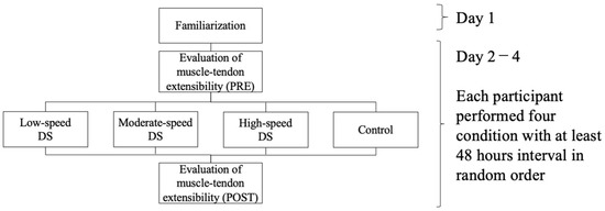
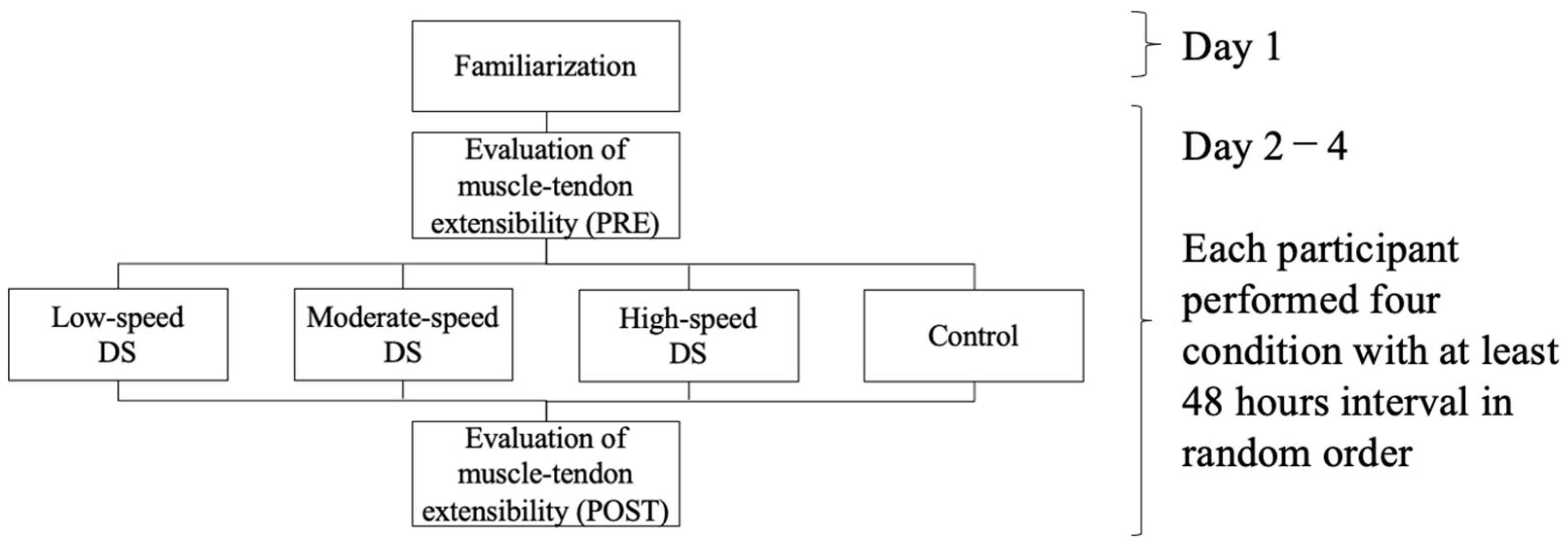
This study used a randomized crossover design to compare the effects of DS on the MTS of the ankle plantar flexors at three different DS speeds (Figure 1). The participants visited the laboratory five times. On the first visit, they were familiarized with the DS procedures, and musculotendinous extensibility was evaluated using DS. The three DS and control conditions were performed in a random order. In this study, DS was controlled without bouncing movements [16]. The DS protocols were as follows: low-speed DS at 30 repetitions/min (rpm); moderate-speed DS at 60 rpm; high-speed DS at 120 rpm; and no stretching (control). Each condition was performed at least 48 h apart, and all measurements were performed simultaneously (±2 h). Before the experiment, the participants sat with rest for 20 min to acclimate to the laboratory environment. The room temperature was set to 25 °C [19,20]. The maximum ankle dorsiflexion angle, passive torque, and MTS were measured before and after DS.
Figure 1.
Experimental design.
2.2. Participants
Twelve healthy competitive university male athletes (age: 22.3 ± 1.4 years; height: 171.8 ± 4.4 cm; weight: 62.3 ± 4.8 kg) participated in this study. The sample size was calculated using G*Power 3.1 software (Heinrich Heine University Düsseldorf, Düsseldorf, Germany), and 12 participants were required to detect a medium effect size f of 0.25 with an alpha level of 0.05, and a statistical power of 0.80 for repeated measures analysis of variance (ANOVA) [18]. Participants with neurological or musculoskeletal disorders were excluded because those conditions may affect musculotendinous function, arthrokinematics, proprioception, and neuromuscular responses to stretching. The participants were asked to refrain from strenuous exercise, strength training, and alcohol consumption for 24 h before the experiment. DS was performed on the plantar flexors of the right ankle for all participants to ensure methodological consistency. The dynamometer used to assess musculotendinous stiffness was configured specifically for right-side measurement, following the protocol of a previous study [21], and standardizing a single limb minimized variability related to repositioning and bilateral differences. Each participant was informed of the details of the study protocol and provided written informed consent to participate. The present research design was approved by the Ethics Committee of the Faculty of Health Sciences, Hokkaido University (approval number: 21-39) and was conducted in accordance with the Declaration of Helsinki.
2.3. Evaluation of Musculotendinous Extensibility
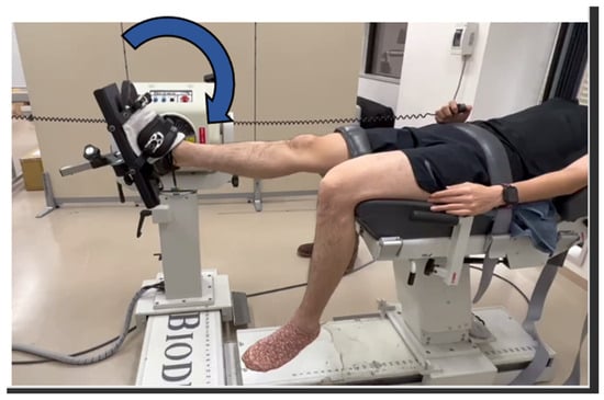
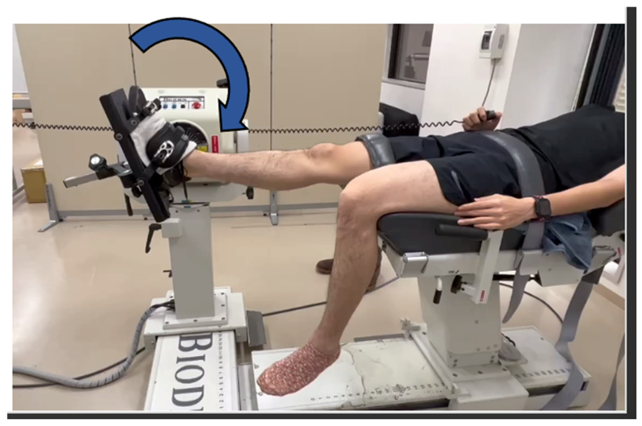
The maximum ankle dorsiflexion angle and passive torque were measured using a Biodex System 3 dynamometer (Biodex Medical Systems, Inc., Shirley, NY, USA) and Myosystem 1200 (Noraxon USA, Inc., Scottsdale, AZ, USA) at a sampling frequency of 1000 Hz. Measurements were performed in the supine position with the knee extended [22] (Figure 2). To prevent compensatory movement during passive ankle dorsiflexion, the pelvic girdle was fixed using a Velcro strap, the lateral malleolus was aligned with the dynamometer axis, and the ankle joint was secured to the footplate in the mid-position of the internal/external rotation. The position in which the footplate was aligned perpendicular to the floor was defined as 0° of ankle dorsiflexion. To minimize changes in passive torque, the ankle dorsiflexion angle was measured twice before each measurement [22]. Before and after DS, the ankle joint was dorsiflexed at an angular speed of 2°/s from a 20° of ankle plantar flexion. The participants were instructed to push the safety button when they felt the maximum stretching of the plantar flexors without pain or with minimal pain. The ankle dorsiflexion angle and torque values at this point were defined as the maximum ankle dorsiflexion angle and maximum passive torque, respectively [19]. The participants were instructed to relax during the measurements to avoid any unnecessary force on their lower limbs.
Figure 2.
Evaluation of the maximum ankle dorsiflexion angle and passive torque.
Passive torque and ankle dorsiflexion angle were processed and analyzed using MATLAB software (R2021b, MathWorks, Natick, MA, USA). All data were filtered using a low-pass filter at 10 Hz, and the torque data were corrected for gravity at the starting position [23]. MTS was defined as the slope of the torque-angle curve obtained from the passive torque and ankle dorsiflexion angle and quantified using a second-order polynomial regression model [24]. It was calculated as the slope of the second-order polynomial curve every 4° in the final 13° of ROM [7]. The average slopes of the four points were used in subsequent analyses. The MTS after stretching was calculated within the same range of dorsiflexion angles as before stretching.
2.4. Dynamic Stretching
DS was performed on the right ankle plantar flexors using a Biodex dynamometer (Biodex Medical Systems, Shirley, NY, USA) in the same position as the musculotendinous extensibility measurement. Only voluntary ankle dorsiflexion was performed without bouncing, and passive ankle plantar flexion was performed by the examiner, who moved the lever arm back to 20° ankle plantar flexion. The stretching speed was set at 30, 60, and 120 rpm using a metronome. Four sets of 15 repetitions with 30 s rest between the sets were performed under each condition [21]. For the control condition, the participants sat on the Biodex System for 5 min and 30 s, which matched the duration of the low-speed condition.
2.5. Statistical Analysis
IBM SPSS Statistics software (ver. 26, IBM Corporation, Armonk, NY, USA) was used for statistical analysis. The Shapiro–Wilk test was used to evaluate the normal distribution of all data. A two-way repeated-measures analysis of variance (condition [low-speed, moderate-speed, high-speed, and control] × time [pre- and post-measurement]) was used to analyze the maximal ankle dorsiflexion angle, MTS, and passive torque. When a significant effect was found, a post hoc test using the Bonferroni correction was conducted. The effect size was determined using partial eta-squared values (ηp2) for the ANOVA. Cohen’s dz was used to assess within-group changes and was calculated by dividing the mean difference by the standard deviation of the differences. Cohen’s d was used to assess between-group differences and was calculated as the difference between group means divided by the pooled standard deviation. The magnitude of the effect sizes was interpreted as small (0.2), moderate (0.5), and large (0.8) [25]. To evaluate the test–retest reliability of the measurements, the intraclass correlation coefficient (ICC (1,4)) among the pre-measurement values for each condition was calculated. The results were as follows: ankle maximum dorsiflexion angle (ICC (1,4) = 0.99, 95% confidence interval (CI) = 0.98–0.99, standard error of the mean [SEM] = 1.21) and MTS (ICC (1,4) = 0.93; 95% CI = 0.95–0.99; SEM = 0.031). The significance level was set at p < 0.05. All data are presented as mean ± standard deviation (SD).
3. Results
3.1. Musculotendinous Stiffness
A significant two-way interaction for condition by time (p = 0.001, ηp2 = 0.374) was found, but there was no main effect of condition (p = 0.535, ηp2 = 0.063) or time (p = 0.426, ηp2 = 0.058). The post hoc test revealed that MTS significantly increased after high-speed DS (p = 0.038, dz = 0.68) and significantly decreased after moderate-speed DS (p = 0.025, dz = −0.75). However, no significant differences were observed before or after low-speed DS (p = 0.614, dz = −0.14) as shown in Table 1.
Table 1.
Changes in musculotendinous stiffness during passive ankle dorsiflexion.
3.2. Maximum Ankle Dorsiflexion Angle
A significant two-way interaction for condition by time (p < 0.001, ηp2 = 0.501) and a main effect of time (p = 0.001, ηp2 = 0.664) were found; however, there was no main effect of condition (p = 0.124, ηp2 = 0.158). The post hoc test revealed that the maximum ankle dorsiflexion angle was significantly increased after low-speed (p = 0.006, dz = 0.98), moderate-speed (p = 0.001, dz = 1.22), and high-speed DS (p = 0.001, dz = 1.38), and it was significantly larger after high-speed DS than that after the control condition (p = 0.039, d = 0.44) as shown in Table 2.
Table 2.
Changes in the maximum ankle dorsiflexion angle during passive ankle dorsiflexion.
3.3. Maximum Passive Torque
A significant two-way interaction between condition and time (p < 0.001, ηp2 = 0.515) and the main effects of condition (p = 0.044, ηp2 = 0.215) and time (p < 0.001, ηp2 = 0.771) were found. The post hoc test revealed that the maximum passive torque significantly increased after low-speed (p = 0.003, dz = 1.07), moderate-speed (p = 0.011, dz = 0.89), and high-speed DS (p < 0.001, dz = 1.89), and it significantly increased after high-speed DS compared to that in the control condition (p = 0.004, d = 0.58) as shown in Table 3.
Table 3.
Changes in the maximum passive torque angle during passive ankle dorsiflexion.
4. Discussion
This study aimed to compare the effects of DS at different speeds on the maximum ankle dorsiflexion angle, maximum passive torque, and MTS of the ankle plantar flexors. The present study found that DS at all three speeds significantly increased the maximum ankle dorsiflexion angle and passive torque at all three speeds. Furthermore, moderate- and high-speed DS significantly decreased and increased the MTS, respectively. These findings partially support our hypothesis.
DS at all three speeds significantly increased the maximum ankle dorsiflexion angle, which is consistent with the results of a previous study [24]. With moderate-speed DS, the maximum ankle dorsiflexion angle increased, and the MTS decreased. In contrast, with high- and low-speed DS, the maximum ankle dorsiflexion angle increased, whereas the MTS increased or remained unchanged. These results may have been influenced by changes in stretch tolerance, which is explained by pain endurance when the muscles and tendons are stretched [12]. Hence, the MTS decreased, and the maximum dorsiflexion angle increased with moderate-speed DS; therefore, moderate-speed DS is recommended to increase musculotendinous extensibility.
Moderate-speed DS decreases MTS, which is consistent with previous reports [6,26]. Herda et al. [6] reported a decrease in MTS with DS, and Oba et al. reported that muscle stiffness decreased after middle-speed DS [26]. Also, Samukawa et al. [27] reported that the myotendinous junction shifted proximally with DS to the ankle plantar flexors, leading to not only the muscle but also the tendon being stretched. These physiological factors are thought to be involved in the decrease in MTS. Repeated “stretch-shortening” of muscles and tendons is considered to change the collagen arrangement and water content in tissues and decrease viscosity [28]. Active movement of muscles and tendons leads to an increase in tissue temperature and a decrease in viscosity [16]. The decrease in MTS observed in the present study could be explained by changes in these physiological responses. Further studies are needed to clarify the mechanisms underlying DS-induced alterations in MTS. The DS protocol (speed and duration) for the ankle plantar flexors used in this investigation was based on a previous study by Mizuno et al. [7], which reported no changes in MTS. Hence, there were changes in MTS in this study. These conflicting results may be explained by differences in the selection of participants. Mizuno et al. included both men and women participants [7]. In contrast, Samukawa et al. and Herda et al. included only male participants [6,27], as in the current study. Sex-related differences in stretching have been reported [29,30], and female hormones are thought to be associated with increased joint flexibility [31]. Therefore, sex differences among the participants may have led to these dissimilarities. However, sex-related differences in the effects of DS have not yet been clarified; therefore, further research is required to address this issue.
A significant increase in MTS was observed after high-speed DS. No previous studies have reported an increase in MTS after the DS. The current findings may be related to the process in which, immediately after high-intensity exercise, the osmotic pressure on the muscle changes in muscle volume due to increased arterial inflow [32], and the accumulation of water in muscle fibers increases intramuscular pressure and muscle hardness [33]. Furthermore, a positive correlation between the change in muscle hardness and MTS has been demonstrated [34]; therefore, high-speed DS may be interpreted as the physiological changes observed in high-intensity exercises.
The MTS was increased with high-speed DS for 120 s (30 s × 4 sets) to the ankle plantar flexors. In a previous study, the MTS did not change after 30 s of high-speed DS (120 rpm) of the hamstring muscles [18]. The targeted muscles and DS durations differed from those in the present study. The hamstring muscles have long muscle fibers and moderate physiological muscle cross-sectional areas throughout the body, whereas the ankle plantar flexors have short muscle fibers and large physiological muscle cross-sectional areas [35]. These anatomical differences may influence changes in the MTS after stretching [36]. Therefore, the optimal DS duration and speed may differ depending on the target muscles. The MTS did not change after 30 s of DS to the hamstring muscles [18] but decreased after 300 s of DS (30 s × 10 sets) [17]. In the present study, the MTS also decreased after 120 s of DS (30 s × 4 sets), suggesting that a certain DS duration is necessary to change the MTS.
Although many studies have reported the effects of DS on athletic performance enhancement [4,8,9], few studies have reported its effects on injury prevention. In this study, the MTS decreased after moderate-speed DS. In a survey on DS use, 73.6% of coaches used it for injury prevention [10]. A high MTS has been reported as a risk factor for musculotendinous strain injuries [15]. In addition, increased tendon compliance increases energy absorption and may reduce muscle damage [37]. Therefore, the present study findings suggest that reduced MTS by moderate-speed DS may prevent musculotendinous-strain injuries.
In the present study, ROM increased with all DS conditions; however, a reduction in musculotendinous stiffness occurred only with moderate-speed DS. This indicates that improvements in muscle and tendon flexibility were specific to the moderate-speed condition, whereas ankle dorsi flexion angles gained at slow and fast speeds were likely attributable to stretch tolerance rather than changes in mechanical properties. Clinically, when the goal is to enhance muscle and tendon flexibility, moderate-speed DS may be the most effective way. In contrast, musculotendinous stiffness was increased with high-speed DS, which may facilitate faster force transmission and potentially augment force production [38]. Taken together, these findings reveal that DS speed should be selected according to the athletic situation, such as prioritizing flexibility enhancement or optimizing force output.
This study had some limitations that should be considered. First, physiological outcomes, such as muscle temperature, blood flow, and muscle hardness, were not determined in this study. Second, the DS protocol was performed with the patient in a supine position using a dynamometer. In clinical and sports settings, DS is primarily performed in a standing position. Stretching posture and position may affect muscle activity, leading to different results. Third, MTS was determined by the slope of the relationship between passive torque and joint angle. However, these outcomes were not analyzed independently. Finally, only male university athletes were included in this study. Therefore, it remains unclear whether similar results can be obtained in non-athletes, women, or the elderly population.
5. Conclusions
This study compared the effects of DS (30 s × 4 sets) on the maximum ankle dorsiflexion angle, maximum passive torque, and MTS of the ankle plantar flexors at three different DS speeds. The maximum ankle dorsiflexion angle increased significantly after DS at all speeds (low, moderate, and high). However, MTS significantly decreased after moderate-speed DS and significantly increased after high-speed DS. The DS speed did not affect the maximum ankle dorsiflexion angle or passive torque. These findings suggest that DS at moderate speed is effective for increasing muscle and tendon flexibility, and DS at high speed is effective for optimizing force output.
Author Contributions
Conceptualization, all authors; methodology, all authors; software, N.K.; validation, N.K.; formal analysis, N.K. and K.O.; investigation, N.K.; resources, M.S.; data curation, N.K. and K.O.; writing—original draft preparation, N.K., K.O. and M.S.; writing—review and editing, N.K., K.O., T.I., Y.K., M.K., M.T., S.K., H.T. and M.S.; visualization, N.K., K.O. and M.S.; supervision, M.S.; project administration, M.S.; funding acquisition, M.S. All authors have read and agreed to the published version of the manuscript.
Funding
This study was partly supported by a Grant-in-Aid JSPS KAKENHI (grant number: 22K1157402).
Institutional Review Board Statement
This study was conducted in accordance with the Declaration of Helsinki and approved by the Ethics Committee of the Faculty of Health Sciences, Hokkaido University (approval number: 21-39; approval date: 17 September 2021).
Informed Consent Statement
Written informed consent was obtained from all participants in this study. Written informed consent was obtained from all participants for the publication of this paper.
Data Availability Statement
The data supporting the conclusions of this article will be made available by the authors upon request.
Conflicts of Interest
The authors declare no conflicts of interest.
References
- Bandy, W.D.; Irion, J.M.; Briggler, M. The Effect of Static Stretch and Dynamic Range of Motion Training on the Flexibility of the Hamstring Muscles. J. Orthop. Sports Phys. Ther. 1998, 27, 295–300. [Google Scholar] [CrossRef] [Scilit]
- Herda, T.J.; Cramer, J.T.; Ryan, E.D.; Mchugh, P.; Stout, J.R. Acute Effects of Static versus Dynamic Stretching on Isometric Peak Torque, Electromyography, and Mechanomyography of the Biceps Femoris Muscle. J. Strength Cond. Res. 2008, 22, 809–817. [Google Scholar] [CrossRef] [Scilit]
- Maeda, N.; Urabe, Y.; Tsutsumi, S.; Sakai, S.; Fujishita, H.; Kobayashi, T.; Asaeda, M.; Hirata, K.; Mikami, Y.; Kimura, H. Acute Effects of Static and Cyclic Stretching on Muscle Stiffness and Hard-Ness of Medial Gastrocnemius Muscle. J. Sports Sci. Med. 2017, 16, 514–520. [Google Scholar]
- Samson, M.; Button, D.C.; Chaouachi, A.; Behm, D.G. Effects of Dynamic and Static Stretching within General and Activity Specific Warm-up Protocols. J. Sports Sci. Med. 2012, 11, 279–285. [Google Scholar]
- Power, K.; Behm, D.; Cahill, F.; Carroll, M.; Young, W. An Acute Bout of Static Stretching: Effects on Force and Jumping Performance. Med. Sci. Sports Exerc. 2004, 36, 1389–1396. [Google Scholar] [CrossRef] [Scilit]
- Herda, T.J.; Herda, N.D.; Costa, P.B.; Walter-Herda, A.A.; Valdez, A.M.; Cramer, J.T. The Effects of Dynamic Stretching on the Passive Properties of the Muscle-Tendon Unit. J. Sports Sci. 2013, 31, 479–487. [Google Scholar] [CrossRef] [Scilit]
- Mizuno, T.; Umemura, Y. Dynamic Stretching Does Not Change the Stiffness of the Muscle-Tendon Unit. Int. J. Sports Med. 2016, 37, 1044–1050. [Google Scholar] [CrossRef] [Scilit] [PubMed]
- Little, T.; Williams, A.G. Effects of Differential Stretching Protocols during Warm-Ups on High-Speed Motor Capacities in Professional Soccer Players. J. Strength Cond. Res. 2006, 20, 203–207. [Google Scholar] [CrossRef] [Scilit]
- Ryan, E.D.; Everett, K.L.; Smith, D.B.; Pollner, C.; Thompson, B.J.; Sobolewski, E.J.; Fiddler, R.E. Acute Effects of Different Volumes of Dynamic Stretching on Vertical Jump Performance, Flexibility and Muscular Endurance. Clin. Physiol. Funct. Imaging 2014, 34, 485–492. [Google Scholar] [CrossRef] [Scilit] [PubMed]
- Takeuchi, K.; Nakamura, M.; Kakihana, H.; Tsukuda, F. A Survey of Static and Dynamic Stretching Protocol. Int. J. Sport Health Sci. 2019, 17, 72–79. [Google Scholar] [CrossRef] [Scilit]
- McHugh, M.P.; Kremenic, I.J.; Fox, M.B.; Gleim, G.W. The Role of Mechanical and Neural Restraints to Joint Range of Motion during Passive Stretch. Med. Sci. Sports Exerc. 1998, 30, 928–932. [Google Scholar] [CrossRef] [Scilit] [PubMed]
- Magnusson, S.; Simonsen, E.B.; Aagaard, P.; Boesen, J.; Johannsen, F.; Kjaer, M. Determinants of musculoskeletal flexibility: Viscoelastic properties, cross-sectional area, EMG and stretch tolerance. Scand. J. Med. Sci. Sports 1997, 7, 195–202. [Google Scholar] [CrossRef] [Scilit]
- Morse, C.I.; Degens, H.; Seynnes, O.R.; Maganaris, C.N.; Jones, D.A. The Acute Effect of Stretching on the Passive Stiffness of the Human Gastrocnemius Muscle Tendon Unit. J. Physiol. 2008, 586, 97–106. [Google Scholar] [CrossRef] [Scilit]
- McHugh, M.P.; Cosgrave, C.H. To Stretch or Not to Stretch: The Role of Stretching in Injury Prevention and Performance. Scand. J. Med. Sci. Sports 2010, 20, 169–181. [Google Scholar] [CrossRef] [Scilit]
- Watsford, M.L.; Murphy, A.J.; McLachlan, K.A.; Bryant, A.L.; Cameron, M.L.; Crossley, K.M.; Makdissi, M. A Prospective Study of the Relationship between Lower Body Stiffness and Hamstring Injury in Professional Australian Rules Footballers. Am. J. Sports Med. 2010, 38, 2058–2064. [Google Scholar] [CrossRef] [Scilit]
- Opplert, J.; Babault, N. Acute Effects of Dynamic Stretching on Muscle Flexibility and Performance: An Analysis of the Current Literature. Sports Med. 2018, 48, 299–325. [Google Scholar] [CrossRef] [Scilit]
- Iwata, M.; Yamamoto, A.; Matsuo, S.; Hatano, G.; Miyazaki, M.; Fukaya, T.; Fujiwara, M.; Asai, Y.; Suzuki, S. Dynamic Stretching Has Sustained Effects on Range of Motion and Passive Stiffness of the Hamstring Muscles. J. Sports Sci. Med. 2019, 18, 13–20. [Google Scholar]
- Takeuchi, K.; Nakamura, M.; Matsuo, S.; Akizuki, K.; Mizuno, T. Effects of Speed and Amplitude of Dynamic Stretching on the Flexibility and Strength of the Hamstrings. J. Sports Sci. Med. 2022, 21, 608–615. [Google Scholar] [CrossRef] [Scilit]
- Oba, K.; Samukawa, M.; Abe, Y.; Suzuki, Y.; Komatsuzaki, M.; Kasahara, S.; Ishida, T.; Tohyama, H. Effects of Intermittent and Continuous Static Stretching on Range of Motion and Musculotendinous Viscoelastic Properties Based on a Duration-Matched Protocol. Int. J. Environ. Res. Public Health 2021, 18, 10632. [Google Scholar] [CrossRef] [Scilit] [PubMed]
- Tanaka, M.; Koshino, Y.; Oba, K.; Sentoku, F.; Komatsuzaki, M.; Kyotani, N.; Ishida, T.; Kasahara, S.; Tohyama, H.; Samukawa, M. Effects of Different Amounts of Dynamic Stretching on Musculotendinous Extensibility and Muscle Strength. Appl. Sci. 2024, 14, 6745. [Google Scholar] [CrossRef] [Scilit]
- Mizuno, T. Effects of Dynamic Stretching Velocity on Joint Range of Motion, Muscle Strength, and Subjective Fatigue. J. Strength Cond. Res. 2022, 36, 2440–2447. [Google Scholar] [CrossRef] [Scilit]
- Oba, K.; Samukawa, M.; Nakamura, K.; Mikami, K.; Suzumori, Y.; Ishida, Y.; Keeler, N.; Saitoh, H.; Yamanaka, M.; Tohyama, H. Influence of Constant Torque Stretching at Different Stretching Intensities on Flexibility and Mechanical Properties of Plantar Flexors. J. Strength Cond. Res. 2021, 35, 709–714. [Google Scholar] [CrossRef] [Scilit]
- Ryan, E.D.; Beck, T.W.; Herda, T.J.; Hull, H.R.; Hartman, M.J.; Costa, P.B.; Defreitas, J.M.; Stout, J.R.; Cramer, J.T. The Time Course of Musculotendinous Stiffness Responses Following Different Durations of Passive Stretching. J. Orthop. Sports Phys. Ther. 2008, 38, 632–639. [Google Scholar] [CrossRef] [Scilit] [PubMed]
- Mizuno, T.; Matsumoto, M.; Umemura, Y. Decrements in Stiffness Are Restored within 10 Min. Int. J. Sports Med. 2013, 34, 484–490. [Google Scholar] [CrossRef] [Scilit]
- Cohen, J. Statistical Power Analysis for the Behavioral Sciences, 2nd ed.; Lawrence Erlbaum Associates: Hillsdale, NJ, USA, 1988. [Google Scholar]
- Oba, K.; Murayama, M.; Kaga, S.; Samukawa, M. Acute Effects of Dynamic Stretching on the Mechanical Properties of the Triceps Surae Muscles Assessed Using Shear Wave Ultrasound Elastography. J. Med. Ultrason. 2025. Epub ahead of print. [Google Scholar] [CrossRef] [Scilit]
- Samukawa, M.; Hattori, M.; Sugama, N.; Takeda, N. The Effects of Dynamic Stretching on Plantar Flexor Muscle-Tendon Tissue Properties. Man. Ther. 2011, 16, 618–622. [Google Scholar] [CrossRef] [Scilit]
- McNair, P.J.; Dombroski, E.W.; Hewson, D.J.; Stanley, S.N. Stretching at the Ankle Joint: Viscoelastic Responses to Holds and Continuous Passive Motion. Med. Sci. Sports Exerc. 2001, 33, 354–358. [Google Scholar] [CrossRef] [Scilit]
- Hoge, K.M.; Ryan, E.D.; Costa, P.B.; Herda, T.J.; Walter, A.A.; Stout, J.R.; Cramer, J.T. Gender Differences in Musculotendinous Stiffness and Range of Motion after an Acute Bout of Stretching. J. Strength Cond. Res. 2010, 24, 2618–2626. [Google Scholar] [CrossRef] [Scilit] [PubMed]
- Komatsuzaki, M.; Samukawa, M.; Oba, K.; Koshino, Y.; Ishida, T.; Kasahara, S.; Tohyama, H. Sex-related differences in stretch-induced musculotendinous stiffness change. Sport Sci. Health 2023, 20, 757–762. [Google Scholar] [CrossRef] [Scilit]
- Burgess, K.E.; Graham-Smith, P.; Pearson, S.J. Effect of Acute Tensile Loading on Gender-Specific Tendon Structural and Mechanical Properties. J. Orthop. Res. 2009, 27, 510–516. [Google Scholar] [CrossRef] [Scilit] [PubMed]
- Howell, J.N.; Chleboun, G.; Conatser, R. Muscle stiffness, strength loss, swelling and soreness following exercise-induced injury in humans. J. Physiol. 1993, 464, 183–196. [Google Scholar] [CrossRef] [Scilit] [PubMed]
- Murayama, M.; Nosaka, K.; Yoneda, T.; Minamitani, K. Changes in Hardness of the Human Elbow Flexor Muscles after Eccentric Exercise. Eur. J. Appl. Physiol. 2000, 82, 361–367. [Google Scholar] [CrossRef] [Scilit] [PubMed]
- Nakamura, M.; Ikezoe, T.; Kobayashi, T.; Umegaki, H.; Takeno, Y.; Nishishita, S.; Ichihashi, N. Acute Effects of Static Stretching on Muscle Hardness of the Medial Gastrocnemius Muscle Belly in Humans: An Ultrasonic Shear-Wave Elastography Study. Ultrasound Med. Biol. 2014, 40, 1991–1997. [Google Scholar] [CrossRef] [Scilit]
- Lieber, R.L.; Fridén, J. Functional and Clinical Significance of Skeletal Muscle Architecture. Muscle Nerve 2000, 23, 1647–1666. [Google Scholar] [CrossRef]
- Opplert, J.; Genty, J.B.; Babault, N. Do Stretch Durations Affect Muscle Mechanical and Neurophysiological Properties? Int. J. Sports Med. 2016, 37, 673–679. [Google Scholar] [CrossRef] [Scilit]
- Witvrouw, E.; Mahieu, N.; Danneels, L.; McNair, P. Stretching and Injury Prevention: An Obscure Relationship. Sports Med. 2004, 34, 443–449. [Google Scholar] [CrossRef] [Scilit]
- Monte, A.; Zignoli, A. Muscle and Tendon Stiffness and Belly Gearing Positively Correlate with Rate of Torque Development during Explosive Fixed End Contractions. J. Biomech. 2021, 114, 110110. [Google Scholar] [CrossRef] [Scilit] [PubMed]

Disclaimer/Publisher’s Note: The statements, opinions and data contained in all publications are solely those of the individual author(s) and contributor(s) and not of MDPI and/or the editor(s). MDPI and/or the editor(s) disclaim responsibility for any injury to people or property resulting from any ideas, methods, instructions or products referred to in the content. |
© 2026 by the authors. Licensee MDPI, Basel, Switzerland. This article is an open access article distributed under the terms and conditions of the Creative Commons Attribution (CC BY) license.