


Abstract
The aim of this study was to evaluate the phytochemical profile and neuroprotective potential of extracts from underground and aerial parts of two Carlina species: Carlina acaulis (CA) and Carlina vulgaris (CV). Metabolic profiling performed via UHPLC-UV-MS/MS revealed a distinct chemotaxonomic differentiation between the taxa. C. acaulis was defined as a “phenolic-acid” chemotype dominated by 5-O-caffeoylquinic acid (up to 32.11 mg/g in leaves), whereas C. vulgaris was characterized as a “flavonoid” chemotype, distinguished by a unique C-glycoside signature, including carlinoside and schaftoside. A significant aspect of scientific novelty is the first-ever identification of flavonolignans (e.g., salcolin A/B and tricin derivatives) in the roots of both species. In biological assays, the extracts demonstrated a multidirectional neuroprotective mechanism. Good antioxidant properties (DPPH, FRAP assays) were confirmed, particularly in CA leaves and CV inflorescences which revealed 61% of DPPH scavenging, along with the capacity to chelate Fe(II) ions (above 60% for CA inflorescence, CV leaves and CV root), a crucial mechanism in ferroptosis prevention. Furthermore, leaf extracts of both species exhibited effective inhibition of acetylcholinesterase (AChE) and butyrylcholinesterase (BChE) exceeding 50%, showing kinetic selectivity towards BChE inhibition. These results position Carlina species as a promising source of metabolites supporting the therapy of neurodegenerative diseases.
1. Introduction
Neurodegenerative diseases, including Alzheimer’s disease (AD), Parkinson’s disease (PD), and amyotrophic lateral sclerosis (ALS), have grown to become one of the key challenges of modern medicine [1]. Due to the progressive aging of the global population, these conditions characterized by irreversible loss of cognitive functions and memory, currently constitute a global health priority [2]. The scale of the problem is reflected in alarming epidemiological forecasts. It is estimated that the number of people affected by dementia, of which Alzheimer’s disease remains the dominant cause (60–80%), will rise from 50 million to as many as 139 million cases by 2050 [3]. This rapid increase in morbidity generates a tremendous economic burden: global costs associated with patient care are projected to rise from $1.3 trillion in 2019 to $2.8 trillion in 2030 [4]. Despite the understanding of the complex pathogenesis of the disease, driven by factors such as oxidative stress, aggregation of toxic proteins (amyloid-β, Tau protein), and cholinergic deficits, therapeutic options remain limited [1,5,6]. Current pharmacotherapy relies mainly on the symptomatic use of acetylcholinesterase inhibitors (donepezil, rivastigmine, or galantamine), which only alleviate symptoms without halting the neurodegeneration process [1]. Furthermore, their long-term use is limited by significant adverse effects, including gastrointestinal disturbances and hepatotoxicity [7,8,9].
The Asteraceae family constitutes a rich reservoir of natural bioactive compounds with documented neuroprotective activity, realized mainly through mechanisms of cholinergic enzyme inhibition based on acetylcholinesterase (AChE) and butyrylcholinesterase (BChE) as well as oxidative stress reduction. Many species belonging to this taxonomic group have undergone detailed pharmacological evaluation, demonstrating promising properties in the context of neurodegenerative disease therapy, including Alzheimer’s disease. A significant avenue of research is the verification of plant extracts’ ability to inhibit AChE, which forms the basis for the symptomatic treatment of cognitive deficits. In in vitro studies, the leaf extract of Blumea lacera distinguished itself with particular activity, demonstrating the strongest inhibitory effect (IC50 = 150 ± 11 µg/mL) among species studied in the Bangladesh region, acting as a non-competitive inhibitor [10]. Equally strong potential was noted in the case of Taraxacum officinale, whose leaf extract achieved an IC50 value of 4.27 µg/mL [9]. Conversely, extracts from the aerial parts of Vernonia oligocephala (especially the ethyl acetate fraction) showed moderate but dual activity against AChE and BChE [11], while the aqueous fraction of Wedelia chinensis confirmed anti-AChE activity with an IC50 of 40.02 µg/mL [9]. It is also worth noting the activity of Mikania micrantha in inhibiting BChE [10]. The therapeutic potential of Asteraceae results largely from the presence of specific secondary metabolites. Sesquiterpenes, such as cornigeraline A isolated from Cynara cornigera or caryophyllene-type compounds from Pulicaria vulgaris, demonstrate the ability to interact with the active sites of cholinergic enzymes [9]. A widely studied species is also Artemisia annua, containing artemisinin with moderate inhibitory potential and strong antioxidant properties [4,9]. Representatives of the genus Achillea, including A. millefolium and the endemic A. cucullata, exhibit similar properties, combining anticholinesterase activity with antioxidant effects [12]. The neuroprotective efficacy of selected species has been confirmed in animal models. The extract from Launaea procumbens significantly inhibited AChE activity in rat brains, which translated into improved associative memory. In turn, the methanolic extract of Sonchus asper not only exhibited inhibitory activity but also reduced oxidative stress levels in brain tissue, improving neurocognitive functions [13].
In light of contemporary phytochemical studies, the genus Carlina (Asteraceae) appears as a promising source of specialized secondary metabolites with targeted neuroprotective activity. The therapeutic uniqueness of the analyzed taxa, specifically Carlina vulgaris and Carlina acaulis, is determined by a specific flavonoid profile, dominated by the presence of C-glycosides. Unlike O-glycosides commonly found in the plant kingdom, these compounds are characterized by a direct carbon–carbon (C-C) bond between the sugar moiety and the flavone core. Such a structural feature confers unique metabolic stability and resistance to enzymatic hydrolysis, significantly increasing their bioavailability and in vivo therapeutic potential [14,15].
The presence of a wide spectrum of apigenin and luteolin derivatives has been confirmed in extracts from C. vulgaris and C. acaulis, including compounds such as vitexin (apigenin 8-C-glucoside), isoshaftoside, orientin, vicenin II, and carlinoside, which is characteristic of the genus [14,15]. These plants constitute a rich reservoir of hydroxycinnamic acids. In the raw material from C. acaulis, the total content of chlorogenic acids can reach levels of 12.6 mg/g dry weight, with a distinct dominance of 5-caffeoylquinic acid and 3-caffeoylquinic acid [14,15].
Such a composed phytochemical matrix determines a multidirectional mechanism of neuroprotection. On one hand, the high content of chlorogenic acid and stable C-glycosides provides strong antioxidant potential, enabling effective neutralization of reactive oxygen species (ROS) and protection of neurons against cytotoxicity induced by factors such as H2O2 [14,15]. On the other hand, the lipophilic fraction of C. acaulis roots, containing polyacetylenic Carlina oxide, exhibits direct inhibitory activity against cholinergic enzymes (AChE and BChE), which is crucial in the symptomatic treatment of cognitive deficits in the course of Alzheimer’s disease [16]. The synergy of action between stable antioxidants and enzymatic inhibitors makes the Carlina genus a significant subject of further research in the context of pharmacotherapy for neurodegenerative disorders.
Although the genus Carlina comprises over 30 species, the selection of Carlina acaulis L. and Carlina vulgaris L. for this study was strictly purposeful. C. acaulis was chosen due to its well-documented traditional medicinal use and the recognized biological potential of its root extracts, which warranted further exploration of its less-studied aerial parts. Concurrently, C. vulgaris was selected for comparative purposes as a widely distributed, yet phytochemically under-investigated species in Central Europe. While both species are morphologically distinct and their botanical identification is well-established, providing detailed comparative chemical mapping is essential from a chemotaxonomic perspective. Such data serve to reinforce their systematic differentiation and, more importantly, to evaluate whether the common and more accessible C. vulgaris can chemically and functionally substitute for the protected and more restricted C. acaulis. Therefore, this study aims to provide a comprehensive phytochemical characterization that supports the taxonomic distinction of these species while assessing their neuroprotective potential.
2. Materials and Methods
2.1. Reagents
LC-MS grade methanol and acetonitrile were purchased from Merck (Darmstadt, Germany). LC-MS grade formic acid was also obtained from Merck (Darmstadt, Germany). Reference standards of 5-caffeoylquinic acid (5-CQA) and shaftoside were purchased from Sigma-Aldrich (St. Louis, MO, USA). n-Hexane was obtained from Fisher Chemical (Schwerte, Germany). Ultrapure water was prepared using a Milli-Q Simplicity 185 purification system (Millipore Corp., Molsheim, France). Enzymes acetylcholinesterase from Electrophorus electricus (electric eel) and butyrylcholinesterase from equine serum, substrates acetyltiocholine and butyryltiocholine iodide, 5,5-dithiobis(2-nitrobenzoic acid) (DTNB), dimethyl sufloxide (DMSO), galantamine, NaH2PO4 (>99%), Na2HPO4 (>99%), TPTZ (2,4,6-Tri(2-pyridyl)-s-triazine)), DPPH (2,2-diphenyl-1-picrylhydrazyl) and ferrozine were obtained from Sigma-Aldrich (St. Louis, MO, USA). Acetic acid (ACS), FeCl3 × 6H2O, FeSO4 × 7H2O, phosphoric acid (ACS), hydrochloric acid (ACS) and metanol (analytic purity grade) were obtained from Polish Reagents (Gliwice, Poland). The standards trolox ((±)-6-hydroxy-2,5,7,8-tetramethylchromane-2-carboxylic acid, 97%), EDTA and gallic acid (>98%) were purchased from Sigma-Aldrich.
The experiment was performed using microplate reader SPECTROstar, BMG Labtech (Ortenberg, Germany).
2.2. Plant Material
The above-ground parts of Carlina acaulis L. and Carlina vulgaris L. were used in this study. We confirm that C. acaulis is a protected species in Poland (Journal of Laws 2014, item 1409). Therefore, the research material was not harvested from the wild but was obtained directly from the collections of the Botanical Garden of Maria Curie-Skłodowska University (UMCS) in Lublin, Poland (Sławinkowska 3 St., 20-810 Lublin). A voucher specimen for this species (voucher specimen no. 2005A) has been deposited at the aforementioned institution. Conversely, C. vulgaris is not a protected species under the same legal act. The aerial parts of this plant were collected from a quarry in Nasiłów, Poland. Although this site is of geological interest, it is not a strictly protected nature reserve, and specific environmental permits were not required for the non-destructive sampling of this non-protected species. The plant materials were provided to our scientific institute by authorized entities. The collection of specimens was conducted in a responsible and minimal manner, ensuring that the local plant population and the surrounding ecosystem remained unaffected. The botanical identification of C. vulgaris was rigorously performed by Dr. Jarosław Mołdoch. Regrettably, a formal voucher specimen for C. vulgaris could not be archived, as the entirety of the collected biomass was processed and consumed during the extraction procedures; however, its taxonomic identity was unambiguously confirmed prior to the study. Finally, the full botanical names of both species were verified with the World Flora Online database (www.worldfloraonline.org, accessed on 25 July 2025).
2.3. Preparation of Extracts
The collected plant material was separated into roots, stems with leaves, and flower heads. The separated plant parts were frozen and lyophilized. The dried material was ground to a particle size of 0.5 mm using an Ultra Centrifugal Mill ZM 200 (Retsch GmbH, Haan, Germany). Subsequently, the powdered material was defatted in a Soxhlet apparatus using n-hexane. The defatted and dried plant material (100 g) was extracted three times with 70% methanol (v/v) at a solid-to-solvent ratio of 1:5 (w/v). The process was conducted in an ultrasonic bath (Bandelin Sonorex Digitec, Bandelin electronic GmbH & Co. KG, Berlin, Germany) operating at a frequency of 35 kHz. The extraction was performed in cycles consisting of 15 min of sonication followed by a 45 min static interval. The entire process was carried out at room temperature. The extracts from the three cycles were combined, and the solvent was evaporated under reduced pressure. The residue was frozen and lyophilized. The obtained extracts were subjected to further chromatographic analyses and neuroprotective activity assays.
2.4. Sample Preparation for UHPLC-MS Analysis and Neuroprotective Activity Assays
For phytochemical standardization, approximately 10 mg of lyophilized crude extracts were accurately weighed and dissolved in 1 mL of 50% methanol (v/v). The resulting solutions were centrifuged using a Sigma 3-16KL centrifuge (Sigma Laborzentrifugen GmbH, Osterode am Harz, Germany) at 12,000× g for 10 min at 4 °C to remove particulates. The clear supernatant was collected, filtered through a 0.22 µm membrane filter, and subjected to quantitative analysis using Ultra-High Performance Liquid Chromatography coupled with Mass Spectrometry (UHPLC-MS). For the assessment of neuroprotective activity, approximately 200 mg of each lyophilized extract was weighed and subjected to appropriate biological assays.
2.5. UHPLC-PDA-MS Analysis
Chromatographic analysis was performed using an ACQUITY UPLC system equipped with a Photodiode Array (PDA) detector and coupled to a Xevo TQ-XS triple quadrupole mass spectrometer (Waters, Milford, MA, USA). The separation of compounds was achieved on a BEH C18 column (100 × 2.1 mm, 1.8 µm; Waters) maintained at 40 °C. The mobile phase consisted of water containing 0.1% formic acid (solvent A) and acetonitrile containing 0.1% formic acid (solvent B). The gradient elution was programmed as follows: a linear increase from 5% to 25% B over 15 min, followed by a rapid increase to 99% B over 0.5 min (washing step), and a return to initial conditions (5% B) over 4 min. The flow rate was set at 0.40 mL/min, and the injection volume was 1 µL. Compound identification was based on the analysis of UV spectra, mass spectra (MS), and available literature data. Data acquisition and processing were performed using MassLynx V4.2 software (Waters Corp., Milford, MA, USA).
2.6. Quantitative Analysis of Phenolic Compounds
Quantitative determination of individual compounds was performed based on UV spectral data acquired at 330 nm for phenolic acids and flavonolignans, and at 270 nm for C-glycosyl flavones. The quantification was carried out using the external standard method, expressing the content as equivalents of reference substances. Phenolic acids and flavonolignans were quantified as 5-caffeoylquinic acid (5-CQA) equivalents, while C-flavonoids were calculated as schaftoside equivalents. Calibration curves were constructed using seven concentration points ranging from 4.8 to 192 µg/mL for 5-CQA (R2 = 0.9993) and from 7 to 280 µg/mL for schaftoside (R2 = 0.9997). The obtained linear regression equations were: y = 0.0125x + 3.4255 for 5-CQA and y = 0.0128x + 2.4559 for schaftoside.
2.7. Determination of Neuroprotective Activity
2.7.1. DPPH Assay
Free radical scavenging activity was evaluated using DPPH method. The studies were performed in accordance with Silva et al. [17] with slight modification. DPPH solution [200 μM] was prepared by dissolving an appropriate amount of the reagent in pure methanol. The analyzed extracts’ samples were linearly diluted in methanol, obtaining the following concentrations in analyzed samples (rounded to 8 decimal) places [mg/mL]: 1; 0.25; 0.125; 0.063; 0.031; 0.016 and 0.008. The DPPH solution was applied onto the microplate including examined samples, blank (methanol), positive and negative controls (gallic acid and ethanol, respectively). Absorbance (517 nm) was recorded every 5 min by 30 min. The biological activity was calculated as percentage free radical scavenging activity. Additionally, IC50 values have been determined.
2.7.2. FRAP Assay
FRAP assay was performed based on [18] with some modifications. The analyzed extracts’ samples (1 mg/mL) were linearly diluted in methanol, obtaining the lowest concentration of 0.078 mg/mL. FRAP solution was prepared freshly each time by mixing (10:1:1 v/v/v) 0.3 M acetate buffer (pH 3.6), 0.01 M TPTZ in 0.04 M HCl and 0.02 M FeCl3 × 6H2O and kept in dark. The analyzed extract samples (50 μL) were mixed with 200 μL of FRAP solution in a microplate. The prepared samples were vortexed and incubated at 37 °C away from light. Thereafter, absorbance was measured at 593 nm using a SPECTROstar Nano microplate reader (BMG LABTECH, Ortenberg, Germany) at regular 5 min intervals. FRAP working solution with methanol instead of sample was used as a blank. Gallic acid was used as the positive control. The results were calculated into µg/mL Fe2+ on the calibration curve which was prepared analogically using an aqueous solution of FeSO4 at eight concentrations, obtaining the calibration curve y = 0.9406x − 0.0115, R2 = 0.9964, and gallic acid equivalents [µg/mL] using the calibration curve y = 1.941x + 0.2067.
2.7.3. Fe(II) Chelation Activity
The assay was based on Singh et al. [19] with small modifications. The analyzed extracts’ samples (1 mg/mL) were linearly diluted in methanol, obtaining the lowest concentration of 0.078 mg/mL. The examined extracts’ samples (50 μL) were mixed with Fe(II) solution [2 mM] and incubated for 10 min in the dark. Afterwards, ferrozine [5 mM] was added and the mixture was incubated for 10 min in the dark. The next step was sample dilution by adding methanol, obtaining 250 μL of mixture in each well. The absorbance of the solution was recorded at 562 nm. As a reference metal chelator, EDTA was used. The final result was the average of three replicates.
2.7.4. AChE and BChE Inhibition
Basis of determination of AChE and BChE inhibitory activity was spectrophotometric Ellman assay with slight modifications [20]. The studied extracts’ samples were prepared in MeOH:H2O (1:1 v/v), and the following concentrations were used [mg/mL]: 1; 0.5; 0.25; 0.125; 0.625; 0.312; 0.0156 and 0.0078, which were obtained by linear dilution. Solutions of enzymes AChE (0.25 U/mL) and BChE (0.25 U/mL) were prepared in phosphate buffer (0.1 M, pH 8). Additional reagents, acetyltiocholine or butyryltiocholine iodide (14 mM) and DTNB (10 mM), were prepared also in phosphate buffer (0.1 M, pH 8). The following procedure was used: 20 µL of solution of studied extracts, 20 µL of AChE or BChE solution were mixed with 140 µL of phosphate buffer pH 8, 0.1 M in a 96-wells plate. After 15 min incubation in 25 °C, substrate solution (10 µL) and DTNB (10 µL) were added to the reaction. Absorbance changes were recorded every 5 min to 30 min. As a standard substance galantamine was used, activity analysis of the model compound was performed for the same concentrations as for the studied compounds to compare their inhibitory activity. The results were expressed as the % inhibition of enzymes in accordance with the equation I [%] = [(A − B)/A] × 100, where A is absorbance of negative control and B is an absorbance of solution with studied compound. Additionally, IC50 values have been determined.
2.8. Statistical Analysis
All obtained results were subjected to statistical analysis. In the case of antioxidant assays and enzyme inhibition, the analysis was performed using statistical tools included in MARS software (ver. 3.42) for microplate reader. Additionally, correlation coefficients were indicated.
3. Results
3.1. Qualitative and Quantitative Characterization of the Polyphenolic Profile
The implementation of an integrated analytical platform based on LC-ESI-MS/MS coupled with UV detection allowed for comprehensive metabolic profiling, resulting in the identification and quantification of 35 polyphenolic compounds. The comparative analysis elucidated significant chemical heterogeneity within the studied matrices, strictly determined by both the taxonomic factor (Carlina acaulis vs. Carlina vulgaris) and the morphological specificity of the individual organs. To facilitate data presentation and systematize the discussion of the observed variability, a unified coding nomenclature reflecting botanical and anatomical origin was established. The acronyms CA and CV were assigned to the species Carlina acaulis L. and Carlina vulgaris L., respectively, while specific plant organs were defined using the suffixes ‘root’ (underground parts), ‘leaves’ (aerial vegetative parts), and ‘inflorescence’ (flower heads). Consequently, the investigated samples were designated as CA-root and CV-root, CA-leaves and CV-leaves, and CA-inflorescence and CV-inflorescence. A detailed characterization of the identified metabolites is provided in Table 1.
Table 1.
Phytochemical profile and quantitative distribution of secondary metabolites in the underground and above-ground organs of Carlina acaulis and Carlina vulgaris. ND: not detected.
Among the group of phenolic acids identified in the analyzed extracts, 5-O-caffeoylquinic acid (5-CQA) emerged as the indisputable predominant compound. However, the comparative analysis highlighted drastic interspecific differences, identifying CA (Carlina acaulis) as a species with a remarkable potential for the biosynthesis and accumulation of this metabolite. The highest concentrations of 5-CQA were recorded in the extracts derived from the aerial organs of C. acaulis. The absolute maximum was observed in the leaf extract (32.1 ± 0.5 mg/g dry weight of the extract), followed closely by the inflorescence extract (21.1 ± 0.3 mg/g dry weight of the extract). These results stand in stark contrast to the profile of the second species. Extracts from the herb of CV (Carlina vulgaris) were characterized by a more than 7-fold lower content of this compound (merely 4.54 mg/g in CV-leaves), suggesting that this quantitative criterion might be used to differentiate both taxa. A significant addition to the characterization of this compound group was the analysis of root systems, which revealed the presence of dicaffeoylquinic acid (di-CQA) isomers—compounds specific to underground parts and absent in extracts from aerial organs. In this aspect as well, quantitative analysis confirmed the advantage of the stemless species, as CA-root samples exhibited a higher total content of di-CQA derivatives compared to the analogous CV-root extracts.
The characterization of the flavonoid group revealed a distinct chemotaxonomic pattern compared to the phenolic acid profile. Although the total content of these compounds in the extracts from vegetative organs (leaves) of both species was quantitatively comparable (CV-leaves: 71.6 mg/g vs. CA-leaves: 84.4 mg/g), the qualitative profile exhibited fundamental distinctness, identifying CV as a taxon specific for flavone C-glycosides. In the extracts from aerial parts of CV, this metabolite class constituted the dominant component, with a marked prevalence of orientin (32.1 mg/g) and schaftoside (24.6 mg/g). Schaftoside possesses particular diagnostic significance, differentiating the investigated taxa at the level of generative organs. In extracts from inflorescences (CV-inflorescence), its concentration reached 18.51 mg/g, whereas in the analogous CA-inflorescence samples, only trace amounts of this compound were recorded (0.56 mg/g). A key chemotaxonomic marker, definitively separating the studied populations, proved to be carlinoside (luteolin-6-C-glucoside-8-C-arabinoside). This compound was exclusively identified in the aerial tissues of CV (both leaves and inflorescences), with a complete absence of detection in any extracts derived from CA. Thus, the presence of carlinoside constitutes a unique specific feature of the chemical fingerprint of C. vulgaris. A key chemotaxonomic marker, definitively separating the studied populations, proved to be carlinoside (luteolin-6-C-glucoside-8-C-arabinoside). This compound was exclusively identified in the aerial tissues of CV, with a complete absence of detection in CA. Thus, the presence of carlinoside constitutes a unique specific feature of the chemical fingerprint of C. vulgaris.
The targeted profiling of the flavonolignan group revealed strict organ specificity, confining the accumulation of these metabolites exclusively to extracts derived from root systems. The complete absence of detection of this compound class in any samples from aerial parts establishes them as specific tissue markers within the studied genus. Comparative analysis of the underground extracts demonstrated a sharp interspecific dichotomy, implying distinct biosynthetic pathways within the studied taxa. The metabolic profile of CA-root samples was defined by a predominance of tricin derivatives, with a total content oscillating around 9 mg/g. In stark contrast, the primary flavonolignan identified in CV-root extracts proved to be salcolin A/B (4.15 mg/g), accompanied by only a marginal contribution of tricin derivatives.
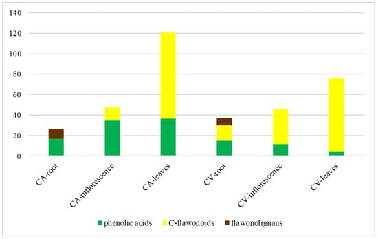
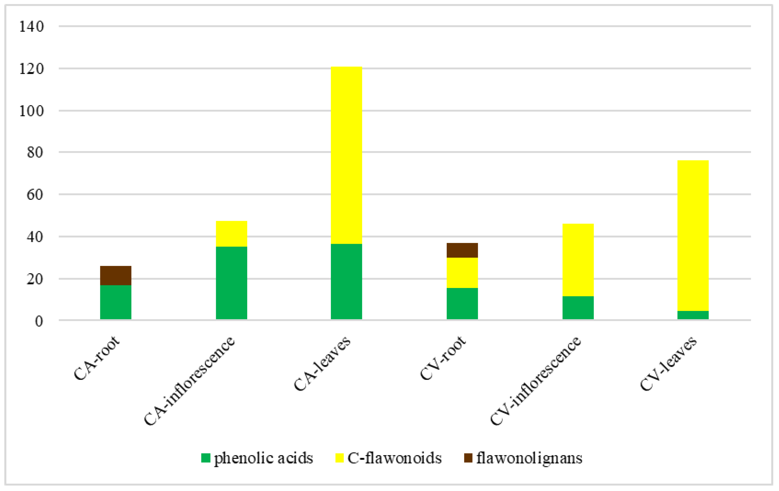
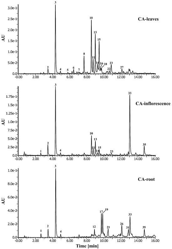
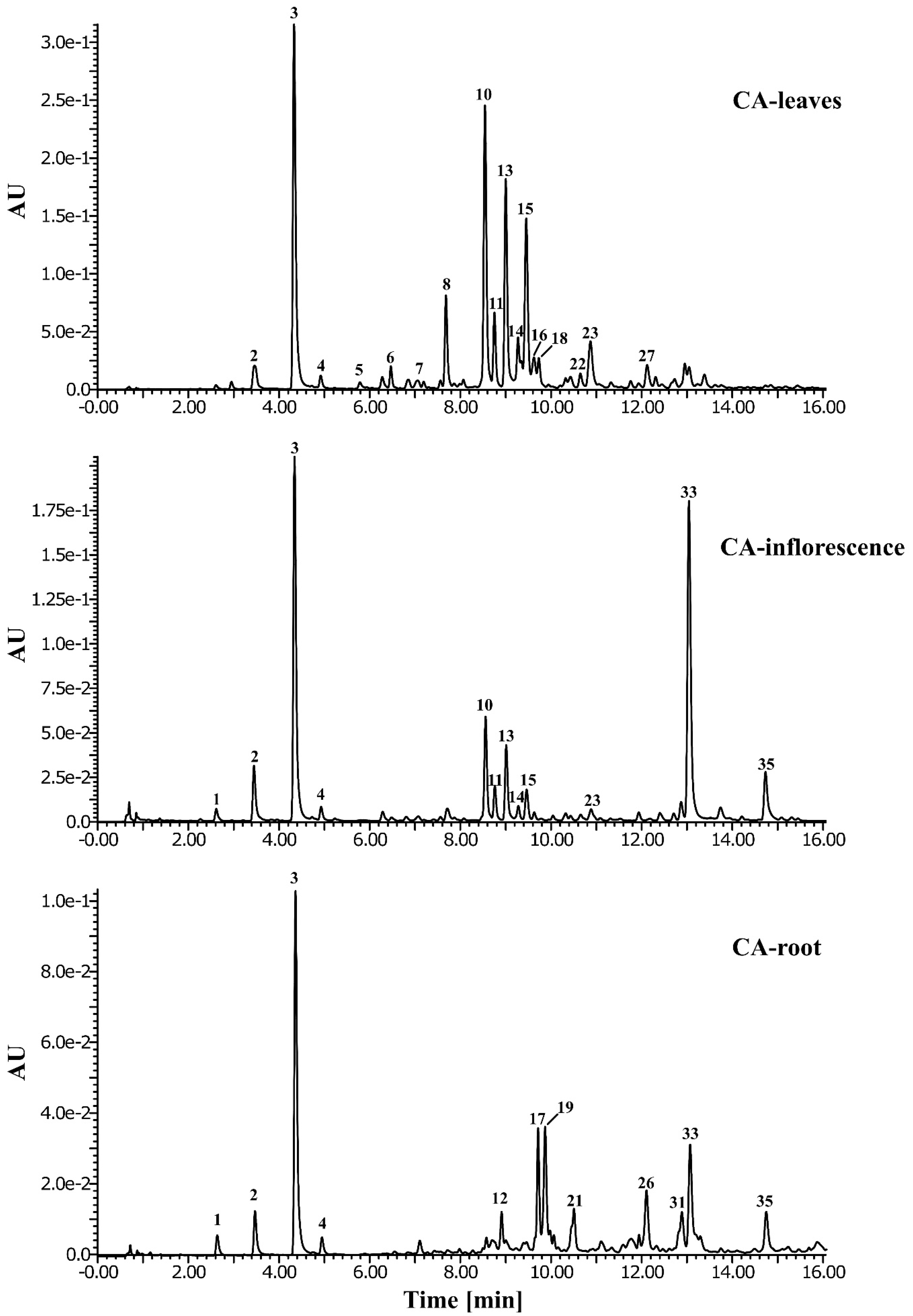
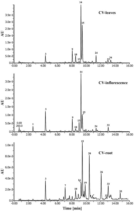
The performed metabolic profiling provides a robust basis for the chemotaxonomic discrimination of the investigated species. While the morphological identification of C. acaulis and C. vulgaris is well-defined, our data demonstrate that the metabolic fingerprint serves as a crucial diagnostic tool to reinforce their systematic separation. The analysis revealed the existence of two distinct chemotypes: C. acaulis manifests as a predominantly ‘acidic’ taxon, characterized by the accumulation of quinic acid esters, whereas C. vulgaris is defined by a unique flavone C-glycosidic signature, with schaftoside and carlinoside serving as stable taxonomic discriminants. In quantitative terms, the leaves of C. acaulis were confirmed as the richest plant matrix, exhibiting a significant polyphenol accumulation exceeding 120 mg/g. A cumulative quantitative analysis, illustrating both the total polyphenol content and the distribution structure of individual compound classes within the analyzed extracts, is visualized in Figure 1. The quantitative characterization is complemented by a qualitative separation pattern: representative chromatographic profiles, documenting the metabolic complexity of individual organs, are compiled in Figure 2 and Figure 3. To ensure data consistency, the peak numbering on the retention profiles is strictly correlated with the identification data provided in Table 1.
Figure 1.
Phytochemical profile and total polyphenol content in the analyzed plants extracts.
Figure 2.
Representative chromatographic profiles (UHPLC-UV) of Carlina acaulis L. extracts. The assembly illustrates the metabolic differentiation of individual organs (roots, inflorescences, leaves), which are labeled directly on the chromatograms. Peak numbering corresponds to the identified compounds listed in Table 1.
Figure 3.
Representative chromatographic profiles (UHPLC-UV) of Carlina vulgaris L. extracts. The assembly illustrates the metabolic differentiation of individual organs (roots, inflorescences, leaves), which are labeled directly on the chromatograms. Peak numbering corresponds to the identified compounds listed in Table 1.
3.2. Determination of Neuroprotective Activity
3.2.1. DPPH Assay
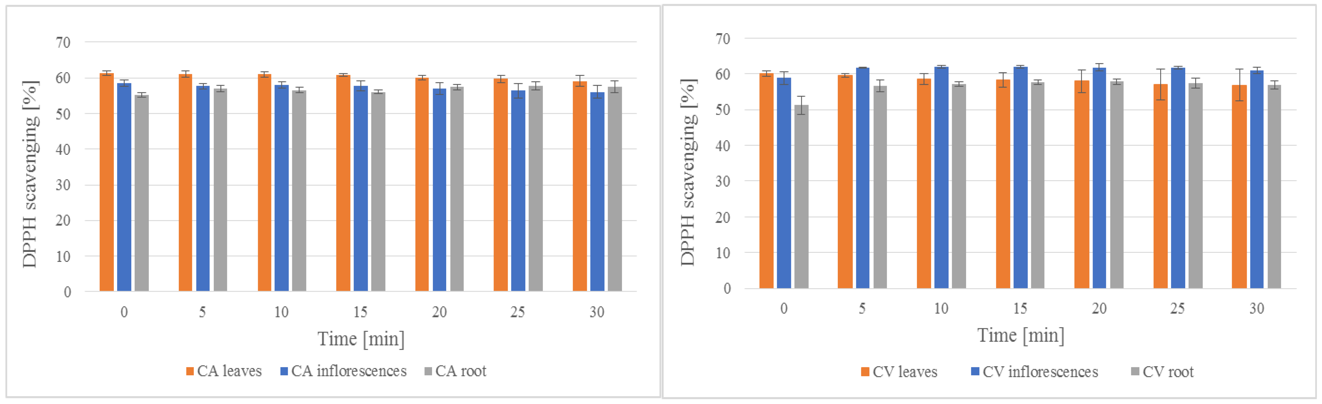
All of the analyzed samples revealed ability to scavenge free radicals. As the concentration of the extracts decreased, the free radical scavenging activity declined significantly in a dose-dependent manner. Nevertheless, the highest concentrations tested (ranging from 0.25 to 1.0 mg/mL) maintained strong and satisfactory antioxidant activity. Figure 4 presents free radical scavenging activity of CA and CV leaves, inflorescence and roots, which can be easily compared.
Figure 4.
Free radical scavenging activity [%] of leaves, inflorescence and roots of CA and CV extracts at concentration 1 mg/mL (n = 4; ±SD).
Analyzing the results, it is easy to notice that both plants reveal good antioxidant activity. In both cases, the analyzed extracts (1 mg/mL) obtained from various parts of plants revealed above 50% of free radical scavenging. In the case of CA, leaves extract reveled higher ability to scavenge free radical, whereas inflorescence and root CV extracts turned out to be better antioxidants than CA. Among them, the most active is CA leaves extract and CV inflorescence extract (Supplementary Materials: Figure S1).
Three of the used concentrations of CA leaves, namely 1, 0.5 and 0.25 mg/mL, revealed activity above 50%, but the activity significantly decreased with sample dilution. A similar trend is observed for CV inflorescence, in which activity was slightly lower than CA leaves extract. Quantitative evaluation and comparison of the activity can be analyzed using IC50 values presented in Table 2. Additionally, statistical analysis revealed strong correlation coefficients between the concentration of the tested samples and their ability to scavenge free radicals. All of the extracts revealed the coefficients >0.75. This result clearly indicates an increase in the activity of the samples as their concentration increases. The highest were observed for root extracts of both plants (>0.98).
Table 2.
IC50 values calculated for all analyzed plant samples and Trolox, a reference compound, in DPPH test. The analyzes were performed for results obtained at 15 min. of reaction (n = 4; ±SD).
To comprehensively evaluate the free radical scavenging potential of the tested extracts, the IC50 values were calculated and compared with Trolox, a well-known standard antioxidant (Table 2). As expected, the pure reference compound exhibited the highest antiradical activity (IC50 = 0.0100 ± 0.0006 mg/mL). Among the analyzed plant samples, the extracts from Carlina acaulis leaves and inflorescences demonstrated the strongest DPPH scavenging ability, with IC50 values of 0.29 ± 0.01 mg/mL and 0.35 ± 0.08 mg/mL, respectively. The extracts from the aerial parts of Carlina vulgaris also showed substantial activity (IC50 ranging from 0.41 to 0.48 mg/mL). In contrast, the root extracts of both species exhibited the lowest antioxidant potential, with IC50 values exceeding 0.8 mg/mL. These findings strongly correlate with the rich phenolic acid and flavonoid profiles identified in the aerial organs of both species.
3.2.2. FRAP Assay
Ferric reducing antioxidant power (FRAP) is a significant assay from an antioxidant point of view. Results obtained for the examined samples (Table 3) revealed good ability of them to allow Fe(III) reduction, which simultaneously impacts antioxidant activity as a whole.
Table 3.
EDTA equivalents in the chelation activity obtained for all analyzed extracts at a concentration of 1 mg/mL at 15 min of reaction (n = 4, ±SD).
The results indicate that all extracts in both the highest and the lowest concentrations are capable of reducing Fe(III) ions. However, we noted a significant decrease in activity when the sample is diluted, so a much better antioxidant effect will be obtained with concentrated extracts. For both plants, extracts from the root showed the lowest activity.
3.2.3. Fe(II) Chelation Activity
Similarly, an important part of the antioxidant assay package is Fe(II) chelation activity. Similarly to previous results, all of the analyzed extracts revealed the examined biological activity (Supplementary Materials: Figure S2).
Based on results presented on Figure 3 and Table 4, most effective Fe(II) chelators are extracts obtained from CA inflorescence, CV roots and CV leaves. A significant difference in activity is observed for root extracts of both plants. Interestingly, opposite results were observed for the activity of extracts from different parts of the plant. We can see that the highest activity of CA extracts was observed in the parts of the plant that showed the lowest activity in CV, and vice versa. Unfortunately, extracts at low concentrations did not reveal Fe(II) chelation activity.
Table 4.
EDTA equivalents obtained for all analyzed extracts at concentration 1 mg/mL at 15 min of reaction (n = 4, ±SD).
Based on statistical analysis, correlation coefficients clearly indicate on a fairly strong and strong correlation between concentration and chelating activity of the extracts. The strongest correlation is observed for leaves extract from CA (coefficient > 0.81), whereas CA inflorescence extract revealed the weaker correlation (coefficient = 0.70).
3.2.4. AChE and BChE Inhibition
Considering biological activity, an important property of analyzed compounds or extracts is their ability to inhibit enzymes, i.e., AChE or BchE, which is an important anti-neurodegenerative aspect of studies.
CA and CV leaves revealed the highest ability to inhibit both used enzymes. The activity was above 50%, whereas activity of galantamine at the same concentration was approx. 90%. The results indicate on weaker inhibitory activity of examined extracts; neveretheless, the high antioxidant activity indicates that the samples are promising sources of multidirectional biological activity.
Analyzing the overall results, the extracts demonstrated comparable inhibitory activity against both AChE and BChE. Extracts at low concentration (below 0.25 mg/mL) showed activity of AChE inhibition at a level below 10%. At the lowest tested concentration (0.0078 mg/mL), the CA and CV leaf extracts exhibited the highest BChE inhibitory activity, reaching 14.3% and 26.83%, respectively. Additional, quantitative evaluation in the form of IC50 is presented in Table 5, and comparison of the activity in the form of a diagram is presented in Supplementary Materials (Figure S3).
Table 5.
IC50 values calculated for all analyzed plant samples and galantamine, a reference compound, in AChE and BChE inhibition tests. The analyzes were performed for results obtained at 15 min of reaction (n = 4; ±SD).
Similarly to antioxidant activity, in the case of enzyme inhibition, positive correlation between concentration of extracts and its activity was observed. In the case of AChE, almost all extracts (only without CA root extract) revealed correlation coefficients above 0.97, whereas in the case of BChE, the highest correlation was observed for CA leaves and CV root extracts (coefficients > 0.96).
4. Discussion
The pronounced compositional distinctness, characterized by the predominance of quinic acid esters (mainly 5-O-caffeoylquinic acid) in C. acaulis and the specific flavone C-glycoside signature in C. vulgaris, enables multidirectional interference with the pathophysiological pathways of neurodegeneration. In vitro results indicate the existence of complementary cytoprotective mechanisms, encompassing the modulation of enzymatic activity and the maintenance of redox homeostasis. The metabolic profiles obtained in this study demonstrate fundamental consistency with recent taxonomic reports. Comparative analysis allowed for the validation of two divergent chemotypes, the characteristics of which are directly reflected in the works of Sowa et al. [14,15].
In the case of Carlina acaulis, our findings unequivocally defined this species as a “phenolic-acid” taxon, characterized by a dominant fraction of quinic acid esters. 5-O-caffeoylquinic acid (5-CQA) was identified as the principal quantitative marker, with the highest accumulation observed in the leaves. This observation is fully consistent with the results published by Sowa et al. [15] who demonstrated that 5-CQA constitutes the dominant metabolite of aerial organs (reaching concentrations of 9.21 mg/g DW), and that the total pool of phenolic acids significantly exceeds the flavonoid content.
In contrast, Carlina vulgaris was identified in our analyses as a representative of the “flavonoid” chemotype, distinguished by a unique signature of C-glycosides. A key element converging with literature data is the identification of carlinoside (luteolin-6-C-glucoside-8-C-arabinoside), alongside high concentrations of vitexin and isoschaftoside. This result directly correlates with the findings of Sowa et al. [15], which define carlinoside as a chemotaxonomic marker of this species that is absent in C. acaulis. These qualitative differences serve as diagnostic fingerprints, reinforcing the morphological classification of the genus Carlina and validating the metabolic divergence between these two taxa
A significant aspect of the scientific novelty of this study is the first-ever reported detection of flavonolignans in Carlina acaulis and Carlina vulgaris. The chromatographic analysis performed (UHPLC-UV-MS/MS) revealed that the accumulation of these rare secondary metabolites exhibits strict organ specificity, being restricted exclusively to the root systems of the investigated plants. Salcolin A or B was identified as the dominant flavonolignan in C. vulgaris root extracts (CV-root). The presence of this compound, a flavonoid–lignin hybrid, has been previously described in the phytochemical literature, including the works of Wenzig et al. [27], as indicated in the identification table. Its detection in the genus Carlina constitutes a new contribution to the chemotaxonomy of the Asteraceae family.
In turn, the metabolic profile of C. acaulis roots (CA-root), and to a lesser extent C. vulgaris, revealed the presence of specific tricin-flavonolignan derivatives. These compounds constitute a unique group of metabolites whose spectral characteristics correspond to the literature data presented by Lee et al. [24] and Bouaziz et al. [25]. This discovery positions the investigated Carlina species as a new, alternative source of these bioactive compounds, the occurrence of which has hitherto been associated mainly with monocotyledons (e.g., Avena sativa) or specific dicotyledonous taxa. The fact that these compounds were not detected in the aerial parts renders them potential chemical markers useful for the identification of root raw material. Full structural elucidation of these metabolites requires their isolation and subsequent advanced identification analyses.
One of the pillars of pharmacological neuroprotection is the preservation of neurotransmitters against enzymatic degradation. The inhibition of acetylcholinesterase (AChE) and butyrylcholinesterase (BChE) is aimed at enhancing acetylcholine levels within synaptic clefts, thereby mitigating cognitive deficits associated with Alzheimer’s disease [16,29]. Our own investigations using enzymatic models demonstrated that leaf extracts from both C. acaulis and C. vulgaris effectively suppress hydrolase activity, exhibiting inhibition rates exceeding 50% at a concentration of 1 mg/mL. From a neurochemical perspective, a significant finding is the demonstrated kinetic selectivity of the extracts towards BChE inhibition over AChE. This selectivity, along with the high overall efficacy, was further corroborated by the quantitative IC50 parameters, which clearly indicated the most potent inhibitory potential for the leaf extracts of both species. This aligns with literature reports indicating the potent inhibitory potential of Carlina constituents, such as Carlina oxide [30]. It should be noted that the absence of Carlina oxide identification in the analyzed root extracts is attributable to the sample preparation protocol employed; this compound, being a highly volatile constituent of the essential oil, likely underwent evaporation during lyophilization and prolonged Soxhlet extraction. Also significant is the fact that the analysis indicates the prevalence of activity towards BChE inhibition, which is important from pharmaceutical point of view. In accordance with numerous data, behavioral and memory changes observed in Alzheimer’s patients are mainly associated with BChE. During disease progression, the importance of BChE increases and becomes the predominant cholinesterase [1,31]. Hence, inhibition of the enzyme is crucial for mental function improvement. The observed inhibition of AChE and BChE by Carlina extracts can be attributed to the synergistic action of their distinct metabolic profiles. In the case of CA, the ‘phenolic-acid’ chemotype, including chlorogenic acid, serves as the primary driver of activity [32]. The literature suggests that the acid inhibits cholinesterases by interacting with the peripheral anionic site (PAS) of the enzyme, often acting through a non-competitive or mixed-type mechanism that prevents the substrate from reaching the catalytic triad. Conversely, the neuroprotective potential of CV appears to be governed by its unique ‘flavonoid’ signature. The C-glycosides that are present are highly resistant to enzymatic hydrolysis. Studies revealed its ability to cross the blood–brain barrier and exert inhibitory effects by forming stable hydrogen bonds within the catalytic active site (CAS) of AChE [33].
Although the IC50 values obtained for CA and CV extracts (ranging from 0.87 to 4.68 mg/mL) are higher than those of the reference galantamine (0.003 mg/mL), these results are consistent with the inhibitory potential reported for other medicinal plant extracts. In the study of complex natural mixtures, IC50 values below 1.0 mg/mL are often considered to represent significant biological activity, as the active metabolites (such as polyphenols or alkaloids) constitute only a fraction of the total extract mass [34,35]. For comparison, several species traditionally used for cognitive disorders exhibit AChE inhibition at small or higher concentration levels, such as extracts from Lamiaceae and Astraceae families, where IC50 values frequently exceed 1–2 mg/mL [36]. Furthermore, the observed dual inhibition of both AChE and BChE at concentrations near 1 mg/mL for leaf extracts, coupled with theirantioxidant capacity, suggests a multi-target mechanism of action. This synergistic approach is increasingly valued in neuroprotective research over high-potency single-target inhibition.
The observed inhibitory activity is likely associated with the rich presence of flavonoids and phenolic acids in the leaf extracts. As indicated by the literature, quercetin forms hydrogen bonds within the enzyme’s active site [1,31], kaempferol acts as an allosteric modulator [31], while other compounds, such as apigenin or luteolin, exhibit dose-dependent inhibition [7,31]. Phenolic acids (binding competitively) may also play a pivotal role [5,9,10,37].
It is also pertinent to highlight a significant gap in the current state of knowledge, which the present results partially bridge. While the neuroprotective activity of free flavonoid aglycones (such as quercetin or apigenin) is widely documented in the literature, the flavonoid C-glycoside fraction identified in our study, constituting the dominant chemotaxonomic marker of C. vulgaris, has not hitherto been the subject of in-depth investigation regarding neuroprotection.
While the in vitro antioxidant properties of the Carlina extracts provide a foundational level of cellular protection, their ability to inhibit cholinesterases, particularly BChE, represents a more targeted and clinically relevant therapeutic mechanism. The observed enzymatic inhibition can be directly correlated with the distinct phytochemical profiles of the investigated species. In C. acaulis, the high concentration of 5-O-caffeoylquinic acid (5-CQA) likely drives the observed anti-cholinesterase activity. Recent comprehensive reviews and in vivo studies confirm that chlorogenic acid and its derivatives possess significant neuroprotective and neuromodulatory functions [38]. Crucially, 5-CQA has been shown to effectively cross the blood–brain barrier (BBB) and restore antioxidative defense mechanisms directly within brain tissues in in vivo models, such as adult zebrafish subjected to chronic stress [39]. This confirms that the predominant phenolic acid in C. acaulis is not merely an in vitro radical scavenger, but a highly bioavailable neuroprotective agent capable of exerting its effects within the central nervous system. Conversely, the enzyme-inhibiting potential of C. vulgaris is strongly associated with its unique flavone C-glycoside signature, including orientin, vitexin, and schaftoside. The therapeutic viability of these compounds relies heavily on their structural stability and systemic bioavailability. The unique carbon–carbon (C-C) bond in C-glycosides prevents their rapid enzymatic hydrolysis in the gastrointestinal tract, significantly enhancing their half-life and systemic circulation compared to standard O-glycosides. In vivo studies robustly support their central nervous system efficacy. For instance, schaftoside has been demonstrated to exert potent neuroprotection by ameliorating cerebral ischemia–reperfusion injury through the enhancement of autophagy and the reduction in neuroinflammation and apoptosis via the AMPK/mTOR signaling pathway [40]. Similarly, orientin and vitexin have been extensively documented to attenuate neuroinflammation, cross the blood–brain barrier (often facilitated by specific transporters or post-microbiome metabolism), and modulate cholinergic pathways in vivo, further validating their role in mitigating cognitive decline [41,42]. Therefore, the synergistic presence of metabolically stable C-glycosides and BBB-permeable phenolic acids positions the investigated Carlina species as highly relevant sources of bioactive compounds for the targeted management of neurodegenerative disorders.
Oxidative stress and dysregulated transition metal homeostasis constitute pivotal factors in the pathogenesis of neurodegenerative processes. Extensive research has demonstrated that free radicals and the resulting oxidative stress are pivotal factors in numerous pathological changes. These include damage to enzymes, DNA, and polyunsaturated fatty acids, as well as the impairment of ATP biosynthesis, all of which are closely associated with neurodegenerative processes [43]. Scavenging free radicals as well as avoiding overload with transition metal ions, which are catalysts for oxidation reactions, are the first steps against pathological changes. Our investigations provided direct evidence of the potent antioxidant potential of the examined extracts (assessed via DPPH and FRAP assays). The highest free radical scavenging activity was observed in C. acaulis leaves and C. vulgaris inflorescences. These observations are strongly supported by the calculated IC50 values, which confirmed that the aerial parts possess a significantly higher capacity to neutralize radicals compared to the root fractions. Furthermore, the FRAP assay results indicate that all extracts are capable of reducing Fe(III) ions, although extracts from the roots showed the lowest activity for both plants.
These results demonstrate a high degree of consistency with literature data for this genus. The dominant activity observed in C. acaulis leaf extracts is primarily attributed to the high abundance of 5-O-caffeoylquinic acid. While the strong antioxidant properties of phenolic acids have been widely known for decades, the specific contribution of this compound to the antiradical potential of C. acaulis is consistent with the findings reported by Sowa et al. [15], who elucidated a strong correlation between antiradical potential and the high abundance of 5-O-caffeoylquinic acid in the aerial parts of this species. Similarly, the high antioxidant potential of C. vulgaris corresponds with earlier reports by Sowa et al. [14], attributing the biological activity of this taxon to a rich flavonoid fraction and the presence of phenolic acids.
As a significant complement to the activity profile described in the aforementioned studies, our research confirmed the capacity of all analyzed extracts to chelate Fe(II) ions. This mechanism is of fundamental importance in the prevention of ferroptosis, effectively inhibiting the metal-catalyzed Fenton reaction and the subsequent generation of highly reactive hydroxyl radicals. According to the literature, this effect is mediated by, among others, phenolic acids, which form stable complexes with metal ions [5,10].
In our experimental model, the most effective chelators proved to be the extracts from C. acaulis inflorescences, C. vulgaris roots and leaves. This indicates a specific organ complementarity in the detoxification of the neuronal environment, significantly expanding the current state of knowledge regarding the chemopreventive potential of the genus Carlina beyond the classical free radical scavenging paradigm described by Sowa in previous publications [14,15].
5. Conclusions
In conclusion, the conducted research confirmed the pronounced metabolic divergence between Carlina acaulis (an ‘acidic’ chemotype) and Carlina vulgaris (a ‘flavonoid’ chemotype). While these species are morphologically distinct, our findings provide a comprehensive chemical mapping that validates their systematic differentiation and establishes stable diagnostic markers, such as carlinoside and specific flavonolignans. A pivotal element of scientific novelty is the first-ever identification of flavonolignans within the root systems of this genus, constituting a significant contribution to the chemotaxonomy of the Asteraceae family. It was demonstrated that the investigated extracts possess multidirectional neuroprotective potential, mediated via the comparable inhibition of both acetylcholinesterase (AChE) and butyrylcholinesterase (BChE)—which was particularly potent for the leaf extracts of both species—alongside potent antioxidant properties. The highest free radical scavenging capacity and reducing power were predominantly observed in the aerial parts, specifically C. acaulis leaves and C. vulgaris inflorescences. These observations were quantitatively validated by the calculated IC50 parameters, which clearly confirmed the superior biological efficacy of the aerial parts over the root fractions in both anticholinesterase and free radical scavenging assays. Complementing the bioactivity profile is the confirmed capacity of all analyzed extracts to chelate ferrous ions, which represents a crucial mechanism in the prevention of ferroptosis. Interestingly, a specific organ complementarity was observed in this regard, where the highest chelating activity in C. acaulis was found in plant parts that exhibited the lowest activity in C. vulgaris, and vice versa. This finding significantly expands the current state of knowledge regarding the chemopreventive action of the genus Carlina.
Supplementary Materials
The following supporting information can be downloaded at https://www.mdpi.com/article/10.3390/app16062907/s1, Figure S1: Free radical scavenging activity obtained for most active extracts—CA leaves and CV inflorescence. The graph shows the change in activity depending on the concentration of the analyzed samples at 15 min of reaction (n = 4, ±SD). Figure S2: Fe(II) chelation activity obtained for all examined extracts at concentration 1 mg/mL (n = 4, ±SD). Figure S3: AChE and BChE inhibitory activity of various part of CA and CV extracts (n = 4, ±SD). All examined extracts and galantamine, a reference compound, were at concentration 1 mg/mL and measured 30 min. after reaction initiation.
Author Contributions
Conceptualization, J.M. and K.W.-K.; methodology, J.M., K.W.-K. and A.S.; software, J.M.; validation, J.M., A.S., K.W.-K., M.R. and M.A.-M.; formal analysis, J.M.; investigation, J.M.; resources, J.M., M.R. and M.A.-M.; data curation, J.M.; writing—original draft preparation, J.M.; writing—review and editing, J.M., A.S., K.W.-K., M.R. and M.A.-M.; visualization, J.M.; supervision, J.M. All authors have read and agreed to the published version of the manuscript.
Funding
This research received no external funding.
Institutional Review Board Statement
Not applicable.
Informed Consent Statement
Not applicable.
Data Availability Statement
The raw data supporting the conclusions of this article will be made available by the authors on request.
Conflicts of Interest
The authors declare no conflicts of interest.
References
- Calderaro, A.; Patanè, G.T.; Tellone, E.; Barreca, D.; Ficarra, S.; Misiti, F.; Laganà, G. The Neuroprotective Potentiality of Flavonoids on Alzheimer’s Disease. Int. J. Mol. Sci. 2022, 23, 14835. [Google Scholar] [CrossRef] [Scilit]
- Diniso, T.; Adeyemi, J.; Oriola, A.; Elufioye, T.; Gondwe, M.; Oyedeji, A. Polyphenolic Contents, Free Radical Scavenging and Cholinesterase Inhibitory Activities of Dalbergiella Welwitschii Leaf Extracts. Plants 2022, 11, 2066. [Google Scholar] [CrossRef] [Scilit]
- Dementia Statistics. Available online: https://www.alzint.org/about/dementia-facts-figures/dementia-statistics/ (accessed on 15 March 2026).
- El Maaiden, E. Phenolic Profiling and Inhibitory Activities of Cholinesterase and β-Amyloid Aggregation in Ziziphus Species Fruit Fractions. J. Food Biochem. 2024, 2024, 4695284. [Google Scholar] [CrossRef] [Scilit]
- Işık, M.; Beydemir, Ş. The Impact of Some Phenolic Compounds on Serum Acetylcholinesterase: Kinetic Analysis of an Enzyme/Inhibitor Interaction and Molecular Docking Study. J. Biomol. Struct. Dyn. 2021, 39, 6515–6523. [Google Scholar] [CrossRef] [Scilit]
- Alotaibi, J.A.M.; Sirwi, A.; El-Halawany, A.M.; Esmat, A.; Mohamed, G.A.; Ibrahim, S.R.M.; Alzain, A.A.; Halawa, T.F.; Safo, M.; Abdallah, H.M. α-Glucosidase, Butyrylcholinesterase and Acetylcholinesterase Inhibitory Activities of Phenolic Compounds from Carthamus tinctorius L. Flowers: In Silico and in Vitro Studies. Saudi Pharm. J. 2024, 32, 102106. [Google Scholar] [CrossRef] [Scilit]
- Durmaz, L.; Karagecili, H.; Gulcin, İ. Evaluation of Carbonic Anhydrase, Acetylcholinesterase, Butyrylcholinesterase, and α-Glycosidase Inhibition Effects and Antioxidant Activity of Baicalin Hydrate. Life 2023, 13, 2136. [Google Scholar] [CrossRef] [Scilit]
- Badawy, S.A.; Hassan, A.R.; Abu Bakr, M.S.; Mohammed, A.E.-S.I. UPLC-qTOF-MS/MS Profiling of Phenolic Compounds in Fagonia arabica L. and Evaluation of Their Cholinesterase Inhibition Potential through in-Vitro and in-Silico Approaches. Sci. Rep. 2025, 15, 5244. [Google Scholar] [CrossRef] [Scilit]
- Deepa, A.V.; Dennis, T. Plant Extracts and Phytochemicals Targeting Alzheimer’s through Acetylcholinesterase Inhibition. Explor. Neurosci. 2025, 4, 100697. [Google Scholar] [CrossRef] [Scilit]
- Uddin, M.J.; Russo, D.; Rahman, M.M.; Uddin, S.B.; Halim, M.A.; Zidorn, C.; Milella, L. Anticholinesterase Activity of Eight Medicinal Plant Species: In Vitro and In Silico Studies in the Search for Therapeutic Agents against Alzheimer’s Disease. Evid.-Based Complement. Altern. Med. ECAM 2021, 2021, 9995614. [Google Scholar] [CrossRef] [Scilit] [PubMed]
- Mahmood, W.; Saleem, H.; Ahmad, I.; Ashraf, M.; Gill, M.S.A.; Ahsan, H.M.; Khan, K.-R.; Chaman, S.; Abbas, S.; Mubashar, A.; et al. In-Vitro Studies on Acetylcholinesterase and Butyrylcholinesterase Inhibitory Potentials of Aerial Parts of Vernonia oligocephala (Asteraceae). Trop. J. Pharm. Res. 2019, 17, 2445. [Google Scholar] [CrossRef] [Scilit]
- Ahmed, S.; Zengin, G.; Fernández-Ochoa, Á.; de la Luz Cádiz-Gurrea, M.; Leyva-Jiménez, F.J.; Elkiran, O.; Cakilcioglu, U.; Akgul, B.H.; Pereira, C.G.; Custódio, L. Exploration of UHPLC-ESI-QTOF-MS Profiles and the Neuroprotective, Antidiabetic, Antioxidant and Cytotoxic Effects of Extracts from Achillea maritima (L.) Ehrend. & Y.P.Guo (Asteraceae) Collected in Türkiye. Plant Foods Hum. Nutr. 2025, 80, 66. [Google Scholar] [CrossRef] [Scilit]
- Patel, S.S.; Raghuwanshi, R.; Masood, M.; Acharya, A.; Jain, S.K. Medicinal Plants with Acetylcholinesterase Inhibitory Activity. Rev. Neurosci. 2018, 29, 491–529. [Google Scholar] [CrossRef] [Scilit]
- Sowa, I.; Mołdoch, J.; Paduch, R.; Strzemski, M.; Szkutnik, J.; Tyszczuk-Rotko, K.; Dresler, S.; Szczepanek, D.; Wójciak, M. Polyphenolic Composition of Carlina acaulis L. Extract and Cytotoxic Potential against Colorectal Adenocarcinoma and Cervical Cancer Cells. Molecules 2023, 28, 6148. [Google Scholar] [CrossRef] [Scilit] [PubMed]
- Sowa, I.; Mołdoch, J.; Dresler, S.; Kubrak, T.; Soluch, A.; Szczepanek, D.; Strzemski, M.; Paduch, R.; Wójciak, M. Phytochemical Profiling, Antioxidant Activity, and Protective Effect against H2O2-Induced Oxidative Stress of Carlina vulgaris Extract. Molecules 2023, 28, 5422. [Google Scholar] [CrossRef] [Scilit]
- Keniche, A.; Kalache, C.; Dib, M.E.A.; El Ouar, I. Assessment of the Inhibition of AChE and BChE by Carthamus Caeruleus Essential Oil and Carline Oxide: Neuroprotective Effects and In Vivo Toxicity Assessment for the Management of Alzheimer’s Disease. Curr. Alzheimer Res. 2025, 22, 368–375. [Google Scholar] [CrossRef] [Scilit]
- Silva, F.; Veiga, F.; Cardoso, C.; Dias, F.; Cerqueira, F.; Medeiros, R.; Cláudia Paiva-Santos, A. A Rapid and Simplified DPPH Assay for Analysis of Antioxidant Interactions in Binary Combinations. Microchem. J. 2024, 202, 110801. [Google Scholar] [CrossRef] [Scilit]
- Spiegel, M.; Kapusta, K.; Kołodziejczyk, W.; Saloni, J.; Żbikowska, B.; Hill, G.A.; Sroka, Z. Antioxidant Activity of Selected Phenolic Acids–Ferric Reducing Antioxidant Power Assay and QSAR Analysis of the Structural Features. Molecules 2020, 25, 3088. [Google Scholar] [CrossRef] [Scilit]
- Singh, H.P.; Kaur, S.; Negi, K.; Kumari, S.; Saini, V.; Batish, D.R.; Kohli, R.K. Assessment of in Vitro Antioxidant Activity of Essential Oil of Eucalyptus citriodora (Lemon-Scented Eucalypt; Myrtaceae) and Its Major Constituents. LWT—Food Sci. Technol. 2012, 48, 237–241. [Google Scholar] [CrossRef] [Scilit]
- Ferreira, J.; Santos, S.; Pereira, H. In Vitro Screening for Acetylcholinesterase Inhibition and Antioxidant Activity of Quercus Suber Cork and Corkback Extracts. Evid. Based Complement. Alternat. Med. 2020, 2020, 3825629. [Google Scholar] [CrossRef] [Scilit] [PubMed]
- Clifford, M.N.; Johnston, K.L.; Knight, S.; Kuhnert, N. Hierarchical Scheme for LC-MSn Identification of Chlorogenic Acids. J. Agric. Food Chem. 2003, 51, 2900–2911. [Google Scholar] [CrossRef] [Scilit]
- Wang, H.; Xiao, B.; Hao, Z.; Sun, Z. Simultaneous Determination of Fraxin and Its Metabolite, Fraxetin, in Rat Plasma by Liquid Chromatography-Tandem Mass Spectrometry and Its Application in a Pharmacokinetic Study. J. Chromatogr. B 2016, 1017–1018, 70–74. [Google Scholar] [CrossRef] [Scilit]
- Kammerer, B.; Kahlich, R.; Biegert, C.; Gleiter, C.H.; Heide, L. HPLC-MS/MS Analysis of Willow Bark Extracts Contained in Pharmaceutical Preparations. Phytochem. Anal. PCA 2005, 16, 470–478. [Google Scholar] [CrossRef] [Scilit] [PubMed]
- Lee, S.-S.; Baek, N.-I.; Baek, Y.-S.; Chung, D.-K.; Song, M.-C.; Bang, M.-H. New Flavonolignan Glycosides from the Aerial Parts of Zizania latifolia. Molecules 2015, 20, 5616–5624. [Google Scholar] [CrossRef] [Scilit]
- Bouaziz, M.; Veitch, N.C.; Grayer, R.J.; Simmonds, M.S.J.; Damak, M. Flavonolignans from Hyparrhenia hirta. Phytochemistry 2002, 60, 515–520. [Google Scholar] [CrossRef] [Scilit] [PubMed]
- Park, J.; Kil, Y.-S.; Ryoo, G.-H.; Jin, C.H.; Hong, M.J.; Kim, J.-B.; Jung, C.-H.; Nam, J.-W.; Han, A.-R. Phytochemical Profile and Anti-Inflammatory Activity of the Hull of γ-Irradiated Wheat Mutant Lines (Triticum aestivum L.). Front. Nutr. 2023, 10, 1334344. [Google Scholar] [CrossRef] [Scilit]
- Wenzig, E.; Kunert, O.; Ferreira, D.; Schmid, M.; Schühly, W.; Bauer, R.; Hiermann, A. Flavonolignans from Avena sativa. J. Nat. Prod. 2005, 68, 289–292. [Google Scholar] [CrossRef] [Scilit] [PubMed]
- Dias, A.L.S.; Rozet, E.; Larondelle, Y.; Hubert, P.; Rogez, H.; Quetin-Leclercq, J. Development and Validation of an UHPLC-LTQ-Orbitrap MS Method for Non-Anthocyanin Flavonoids Quantification in Euterpe oleracea Juice. Anal. Bioanal. Chem. 2013, 405, 9235–9249. [Google Scholar] [CrossRef] [Scilit]
- Barbosa Filho, J.M.; Medeiros, K.C.P.; Diniz, M.d.F.F.M.; Batista, L.M.; Athayde-Filho, P.F.; Silva, M.S.; da Cunha, E.V.L.; Almeida, J.R.G.S.; Quintans-Júnior, L.J. Natural Products Inhibitors of the Enzyme Acetylcholinesterase. Rev. Bras. Farmacogn. 2006, 16, 258–285. [Google Scholar] [CrossRef] [Scilit]
- Prešern, U.; Goličnik, M.; Bavec, A. Genetic and Functional Dynamics of Butyrylcholinesterase in Alzheimer’s Disease: From Mechanisms to Clinical Relevance. Chem. Biol. Interact. 2026, 423, 111809. [Google Scholar] [CrossRef] [Scilit]
- Cichon, N.; Grabowska, W.; Gorniak, L.; Stela, M.; Harmata, P.; Ceremuga, M.; Bijak, M. Mechanistic and Therapeutic Insights into Flavonoid-Based Inhibition of Acetylcholinesterase: Implications for Neurodegenerative Diseases. Nutrients 2024, 17, 78. [Google Scholar] [CrossRef] [Scilit]
- Hung, Y.C.; Kuo, Y.H.; Hsieh, P.W.; Hsieh, T.Y.; Kuo, J.R.; Wang, S.J. Chlorogenic Acid Decreases Glutamate Release from Rat Cortical Nerve Terminals by P/Q-Type Ca2+ Channel Suppression: A Possible Neuroprotective Mechanism. Int. J. Mol. Sci. 2021, 22, 11447. [Google Scholar] [CrossRef] [Scilit]
- Mondal, P.; Gupta, V.; Das, G.; Pradhan, K.; Khan, J.; Gharai, P.K.; Ghosh, S. Peptide-Based Acetylcholinesterase Inhibitor Crosses the Blood-Brain Barrier and Promotes Neuroprotection. ACS Chem. Neurosci. 2018, 9, 2838–2848. [Google Scholar] [CrossRef] [Scilit]
- Mukherjee, P.K.; Kumar, V.; Mal, M.; Houghton, P.J. Acetylcholinesterase inhibitors from plants. Phytomedicine 2007, 14, 289–300. [Google Scholar] [CrossRef] [Scilit]
- Orhan, I.E.; Naz, Q.; Kartal, M.; Tosun, F.; Sener, B.; Choudhary, M.I. In vitro anticholinesterase activity of various alkaloids. Z. Naturforschung C 2004, 59, 839–842. [Google Scholar] [CrossRef] [Scilit]
- Ayaz, M.; Junaid, M.; Ahmed, J.; Ullah, F.; Sadiq, A.; Ahmad, S.; Imran, M. Phenolic contents, antioxidant and anticholinesterase potentials of crude extract, subsequent fractions and crude saponins from Polygonum hydropiper L. BMC Complement. Altern. Med. 2014, 14, 145. [Google Scholar] [CrossRef] [Scilit]
- Katsoulaki, E.-E.; Dimopoulos, D.; Hadjipavlou-Litina, D. Multitarget Compounds Designed for Alzheimer, Parkinson, and Huntington Neurodegeneration Diseases. Pharmaceuticals 2025, 18, 831. [Google Scholar] [CrossRef] [Scilit] [PubMed]
- Nguyen, V.; Taine, E.G.; Meng, D.; Cui, T.; Tan, W. Chlorogenic acid: A systematic review on the biological functions, mechanistic actions, and therapeutic potentials. Nutrients 2024, 16, 924. [Google Scholar] [CrossRef] [Scilit] [PubMed]
- Subba, R.; Fasciolo, G.; Petito, A.; Geremia, E.; Muscari Tomajoli, M.T.; Mondal, A.C.; Napolitano, G.; Venditti, P. Chlorogen-ic Acid Alleviates the Detrimental Effects of Concurrent Hyperglycemia and Chronic Stress on Brain Homeostasis by Modulating Antioxidative Defense in Adult Zebrafish. Antioxidants 2025, 14, 1386. [Google Scholar] [CrossRef] [Scilit]
- Zhang, L.; Wu, M.; Chen, Z. Schaftoside improves cerebral ischemia-reperfusion injury by enhancing autophagy and reducing apoptosis and inflammation through the AMPK/mTOR pathway. Adv. Clin. Exp. Med. 2022, 31, 1343–1354. [Google Scholar] [CrossRef] [Scilit] [PubMed]
- Fahmy, M.I.; Sadek, M.A.; Abdou, K.; El-Dessouki, A.M.; El-Shiekh, R.A.; Khalaf, S.S. Orientin: A com-prehensive review of a promising bioactive flavonoid. Inflammopharmacology 2025, 33, 1713–1728. [Google Scholar] [CrossRef] [Scilit]
- Yan, W.; Cheng, J.; Xu, B. Dietary flavonoids vitexin and isovitexin: New insights into their functional roles in human health and disease prevention. Int. J. Mol. Sci. 2025, 26, 6997. [Google Scholar] [CrossRef] [Scilit] [PubMed]
- Viña, J.; Borrás, C.; Mas-Bargues, C. Free Radicals in Alzheimer’s Disease: From Pathophysiology to Clinical Trial Results. Free Radic. Biol. Med. 2024, 225, 296–301. [Google Scholar] [CrossRef] [Scilit] [PubMed]

Disclaimer/Publisher’s Note: The statements, opinions and data contained in all publications are solely those of the individual author(s) and contributor(s) and not of MDPI and/or the editor(s). MDPI and/or the editor(s) disclaim responsibility for any injury to people or property resulting from any ideas, methods, instructions or products referred to in the content. |
© 2026 by the authors. Licensee MDPI, Basel, Switzerland. This article is an open access article distributed under the terms and conditions of the Creative Commons Attribution (CC BY) license.