


Featured Application
The findings of this study demonstrate the potential of banana flour incorporated with virgin coconut oil for the development of slightly lower digestible, starch-based food products. By promoting amylose–lipid complex formation, banana flour combined with virgin coconut oil can be transformed into a functional ingredient with reduced enzymatic digestibility, making it suitable for bakery products and gluten-free formulations that leverage reduced enzymatic accessibility of starch.
Abstract
This work examined the development of amylose–lipid complexes in green banana flour (Musa × paradisiaca) incorporated with virgin coconut oil (VCO), focusing on their spectral, thermal, and in vitro digestibility characteristics. Firstly, the native banana flour was analyzed for apparent amylose content using a spectrophotometric assay. To facilitate amylose–lipid complexation, both hot-pressed and cold-pressed VCO were incorporated into the banana flour under controlled thermal conditions, after which amylose–lipid interactions were characterized using Fourier-transform infrared and Raman spectroscopy for spectral features and differential scanning calorimetry for thermal behavior. The banana flour exhibited an AAC of 26.40 ± 0.002%. GCMS analysis of FAME derivatized VCO detected medium- to long-chain fatty acids, including octanoic (C8:0), decanoic (C10:0), dodecanoic (C12:0), tetradecanoic (C14:0), and hexadecanoic acids (C16:0) stearic acid (C18:0) and oleic acid (C18:1). FTIR coupled with multivariate analysis and Raman spectra confirmed lipid incorporation/retention in green banana flour through characteristic O–H, C–H, and C=O bands. While DSC revealed distinct endothermic transitions at 89.56 ± 2.17 °C (ΔHₘ = 0.8587 ± 0.1014 J g−1) for hot-pressed VCO and 89.18 ± 0.98 °C (ΔHₘ = 0.6267 ± 0.0777 J g−1) for cold-pressed VCO, consistent with the melting of V-type amylose–lipid complexes. Morphological analysis revealed that thermal treatment transformed native banana flour from irregular granular structures into an amorphous matrix via starch gelatinization, whereas subsequent incorporation of VCO promoted aggregation. In vitro enzymatic digestion showed a slight reduction in starch hydrolysis in VCO-treated samples. The incorporation of an exogenous lipid, such as VCO, into green banana flour promotes the formation of thermally stable amylose–lipid complexes that reduce enzymatic digestibility.
1. Introduction
Bananas rank among the world’s most produced and consumed fruits, with an estimated annual production of ~144 million metric tons, serving as a staple or key energy source for hundreds of millions of people, particularly in tropical countries [1,2]. Green banana flour is increasingly recognized as a promising functional ingredient because it is rich in native starch (often 70–77% total starch) [3]. However, heating and gelatinization disrupt the granular and crystalline structure of banana starch, increasing rapidly digestible fractions. As a result, products formulated with pre-gelatinized native banana starch tend to exhibit higher in vitro starch digestibility and glycemic potential compared with those retaining more granular or retrograded structures [4]. Thus, there is growing interest in identifying green, “clean label” strategies that can alter starch structure and digestibility without relying on chemical derivatization [5].
Among these strategies, the formation of amylose–lipid complexes has emerged as a promising approach to enhance starch resistance to enzymatic hydrolysis [6]. These complexes are of considerable interest because they provide dual benefits: improving nutritional quality while modulating the techno-functional properties of starch-based foods. Specifically, amylose–lipid complexation converts a fraction of readily digestible starch into a more enzyme-resistant form, while simultaneously influencing starch behavior during processing and storage [7]. Classified as resistant starch type 5, these complexes escape digestion in the small intestine, are fermented by gut microbiota, and enhance short-chain fatty acid production, which is linked to improved glycemic control, better lipid profiles, and potential protection against obesity and type 2 diabetes [8].
Mechanistically, this enhanced resistance is driven by the ability of linear amylose chains to form inclusion complexes with lipids. Amylose helices encapsulate the hydrophobic tails of fatty acids or triacylglycerols, generating thermally stable V-type crystalline structures that limit enzyme accessibility [9]. In this context, virgin coconut oil (VCO), an edible oil obtained from the milk of fresh and mature kernels of coconut (Cocos nucifera L.), serves as an effective lipid source due to its high content of medium-chain saturated fatty acids, particularly lauric acid (45–53%) [10,11]. Previous studies have demonstrated that coconut oil can effectively interact with starch from various botanical sources, including cassava [12], rice [13,14], and wheat [15], thereby reducing digestibility and increasing resistant starch content. Moreover, Photinam and Moongngarm [16] reported that, in green banana (Musa sapientum Linn.), the addition of cooking oil to banana starch promotes the formation of functional starch with increased resistant starch and slowly digestible starch fractions.
Unlike isolated banana starch, banana flour represents a more complex food matrix that retains non-starch polysaccharides, proteins, minor lipids, and cellular structures, all of which can influence starch–lipid interactions by modulating starch swelling, amylose leaching, and lipid accessibility. Consequently, the formation and stability of amylose–lipid complexes in banana flour may differ substantially from those observed in purified starch systems. Despite this relevance to real food applications, studies examining amylose–lipid complex formation directly in banana flour remain scarce, and the influence of lipid processing, such as cold-versus hot-pressed virgin coconut oil, on complex formation and digestibility in banana flour has not been systematically investigated. Accordingly, the objective of this study was to investigate the incorporation of cold-pressed and hot-pressed virgin coconut oil into green banana flour (Bungulan variety, Musa × paradisiaca) and to evaluate its effects on amylose–lipid complex formation, thermal behavior, spectroscopic characteristics, and in vitro starch digestibility using kinetic analysis of enzymatic hydrolysis.
2. Materials and Methods
2.1. Banana Flour Preparation
Unripe, physiologically mature Bungulan bananas at nine weeks after inflorescence were obtained from local organic farmers in Kluvi, Lake Sebu, South Cotabato, Philippines (6.1816° N, 124.7337° E), and carefully selected at the full green stage, characterized by firm fruits, a uniformly green peel, and pale cream pulp. The fruits were thoroughly washed under running water, disinfected in a 50-ppm sodium hypochlorite solution, peeled, and sliced before immersion in a 0.1% citric acid solution for 10 min to minimize enzymatic browning. The treated slices were oven-dried at 65 °C until the moisture content was reduced to below 10%, yielding light beige dried banana pieces. These were then milled using a hammer mill to obtain fine green banana flour, which was stored in airtight containers until subsequent use and chemical analyses [17].
2.2. Virgin Coconut Oil Preparation
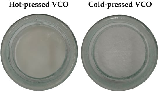
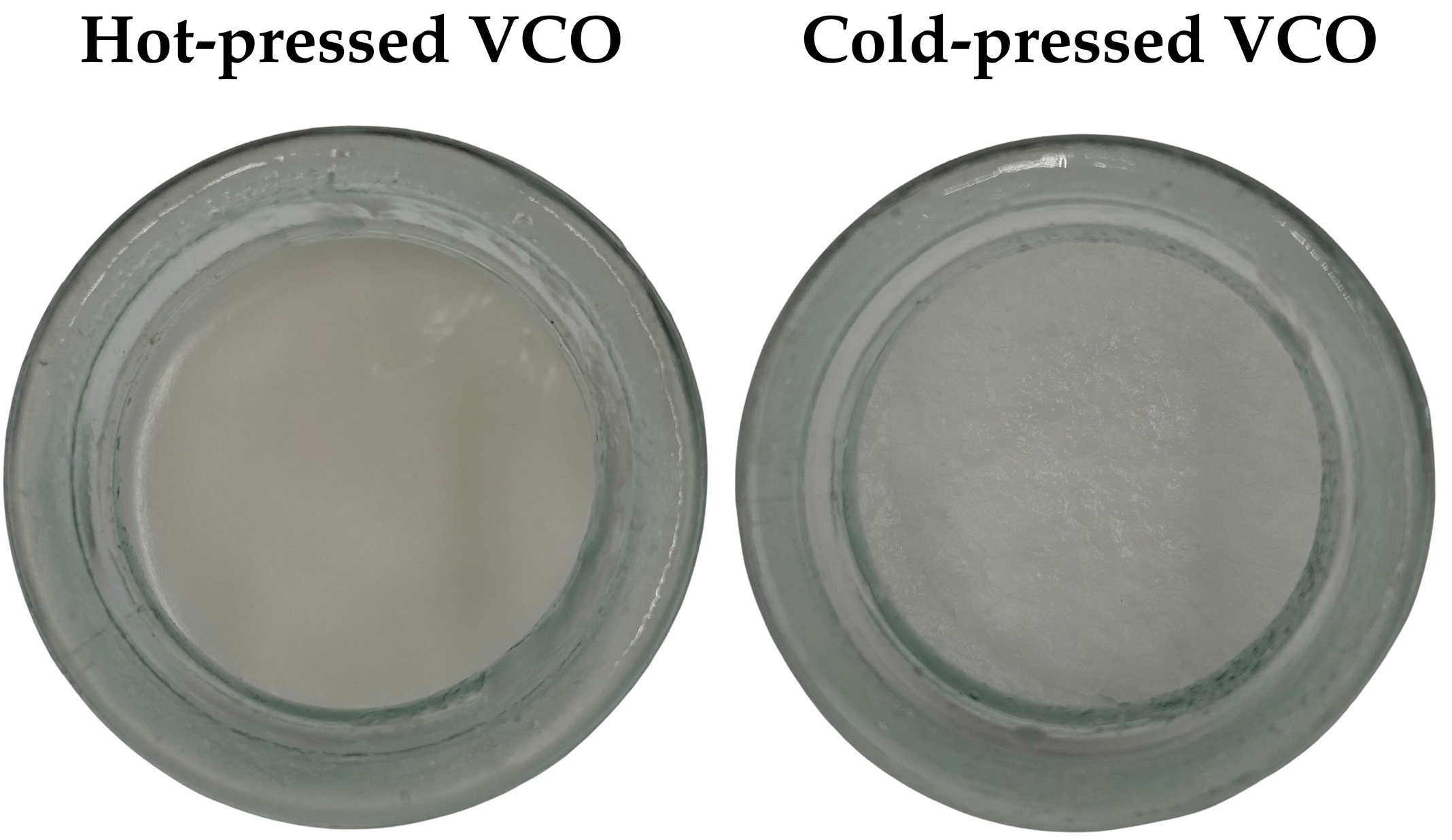
Coconut milk was obtained from 30 mature coconuts (Cocos nucifera). The grated coconut meat was pressed using a hydraulic press, and only the first extraction (pure coconut milk) was collected for VCO production. VCO, as shown in Figure 1, was extracted using both hot (HP) and cold-pressed (CP) methods, as described by Srivastava et al. [18]. For cold extraction, fresh coconut milk was allowed to stand undisturbed for 24 h at 35–40 °C and 75% relative humidity, resulting in four layers. The VCO layer was collected after removing the upper curd and skim layers, while residual oil in the curd was recovered via muslin cloth filtration. For hot extraction, the fermented curd from the cold process was slowly heated until protein coagulation and oil expression occurred. The released oil was separated from the coagulated curd by muslin cloth filtration and collected as hot-extracted coconut oil.
Figure 1.
Photographs of hot-pressed and cold-pressed virgin coconut oil stored at 20 °C.
2.3. Virgin Coconut Oil Derivatization and Gas Chromatography–Mass Spectrometry (GC–MS) with Cold Electron Ionization (GC-EI-MS) Analysis
Fatty acid methyl esters (FAMEs) derived from the triglycerides of virgin coconut oil (VCO) were analyzed using an Agilent 7890A gas chromatograph coupled with a 5977A mass spectrometer (Agilent Technologies, Santa Clara, CA, USA). Separation was performed on an HP-5MS capillary column with the same specifications previously described, consistent with established methods for fatty acid profiling [19]. The oven temperature program was modified for FAME analysis, starting at 80 °C, ramping to 290 °C at 4 °C min−1, and holding at 290 °C for 30 min. The injector parameters remained unchanged except for the injection volume, which was reduced to 1 µL (from 3 µL), and the column head pressure, which was adjusted to 9.3825 psi (previously 8.8085 psi). Sample injection was performed in triplicate. Mass spectrometer settings were maintained as previously described: ion source temperature at 230 °C, quadrupole temperature at 150 °C, transfer line temperature at 280 °C, electron multiplier voltage at 1751 V (gain 1.000), scan speed at 1.562 u s−1, and mass step size at 0.1 m/z. The solvent delay was set to 5.00 min, and the cycle time was slightly adjusted to 355.42 ms per scan to optimize detection.
For peak identification and method validation, the Supelco™ 37 Component FAME Mix (Sigma-Aldrich, St. Louis, MO, USA, 10 mg mL−1 in dichloromethane, CH2Cl2) was used as an external standard. This certified reference mixture provided reliable retention times and mass spectral data, enabling accurate identification and verification of FAMEs present in the coconut oil samples (Merck/Sigma-Aldrich product data sheet).
2.4. Preparation of Banana Flour Incorporated with Virgin Coconut Oil
The BF-VCO composites were prepared following the method of Reddy et al. [20] with modifications. BF samples (500 mg) were placed in centrifuge tubes and mixed with 15 mL of distilled water. The mixture was heated in a water bath at 90 °C for 1 h, with the tube caps loosely fixed to prevent pressure buildup. The oil solution was prepared separately by dissolving 60 mg of VCO in 5 mL of ethanol and then adding it to the BF suspension. This 12% w/w oil-to-flour ratio was chosen because virgin coconut oil is a mixture of medium-chain fatty acids rather than a pure fatty acid. Also, ethanol was used as a carrier solvent to dissolve VCO and facilitate contact between lipid molecules and leached amylose chains during thermal treatment. The mixture was subsequently heated in a water bath at 75 °C for 1 h, cooled to room temperature for 30 min, and washed three times with a 1:1 mixture of distilled water and ethanol (5 mL each), each wash centrifuged at 4000 rpm for 10 min. These ethanol-water washing steps were included to ensure that retained lipids were primarily associated with amylose–lipid inclusion complexes rather than physically adsorbed oil. The resultant pellet was transferred to a beaker, covered with aluminum foil, and dried in an oven at 38 °C for 16 h. A cooked BF without VCO was prepared in parallel using the same procedure and thermal history, except that ethanol and VCO were not added during the complexation step. The cooked BF was subjected to the same heating, cooling, washing, centrifugation, and drying steps as the BF–VCO samples to ensure comparable processing conditions.
2.5. Apparent Amylose Content
The apparent amylose content (AAC) of the samples was determined following the method described by Graham [21], with minor modifications. Briefly, 100 mg of the sample was placed in a test tube and mixed with 1 mL of 95% (v/v) ethanol and 9 mL of 1 M NaOH. The mixture was heated in a boiling water bath to gelatinize the starch, then cooled to room temperature for 1 h. The resulting solution was quantitatively transferred to a 100 mL volumetric flask, diluted to volume with distilled water, and thoroughly mixed. An aliquot (5 mL) of the starch solution was then transferred to a separate 100 mL volumetric flask. Subsequently, 1 mL of 1 M acetic acid and 2 mL of iodine solution (prepared by dissolving 0.2 g iodine and 2.0 g potassium iodide in 100 mL of distilled water) were added, and the flask was filled to volume with distilled water. The solution was shaken vigorously and allowed to stand for approximately 20 min to allow color development. Absorbance was measured at 620 nm using a UV–Vis spectrophotometer (Hitachi U-2900, Hitachi High-Technologies Corporation, Tokyo, Japan). For calibration, an amylose standard solution was prepared by dissolving 40 mg of amylose in 1 mL of 95% ethanol and 9 mL of 1 M NaOH, followed by heating in a boiling water bath for 5–10 min. After cooling to room temperature, the solution was transferred to a 100 mL volumetric flask and diluted to volume with distilled water. Aliquots of 1, 2, 3, 4, and 5 mL were pipetted into separate 100 mL volumetric flasks and treated as described above by acidification with 1 M acetic acid (0.2, 0.4, 0.6, 0.8, and 1.0 mL, respectively) and addition of 2 mL iodine solution. The calibration curve was constructed by plotting absorbance at 620 nm against amylose concentration (8, 16, 24, 32, and 40%). The AAC of the samples was calculated from the standard curve’s linear regression equation.
2.6. Spectroscopic Analysis
To identify functional groups and characteristic vibrational bands associated with starch structure and amylose–lipid complex formation, native BF and BF with VCOs samples were analyzed using a Fourier Transform Infrared spectrophotometer (IRSpirit, Shimadzu Corporation, Kyoto, Japan) coupled to an attenuated total reflectance (ATR) accessory (QATR-S) with a diamond crystal. Mid-infrared spectra over 4000–400 cm−1 were recorded at 4 cm−1 resolution with 64 scans. Using Quasar spectroscopy software Version 1.13.1 [22], mid-FTIR measurements were preprocessed and analyzed. The raw spectra were initially baseline-corrected using a rubber-band algorithm. Subsequently, vector normalization was applied to standardize the spectral intensities. The corrected spectra were then transformed into second-derivative spectra using a 7-point Savitzky–Golay filter. Signal enhancement was further improved by 5-point smoothing, which improved peak resolution and discrimination. The processed FTIR wavenumber data were subsequently subjected to multivariate data analysis, including principal component analysis (PCA) and hierarchical clustering, using MetaboAnalyst 6.0 (https://www.metaboanalyst.ca/ accessed on 28 February 2026).
Complementarily, Raman measurements were performed using a Horiba LabRAM HR Evolution spectrophotometer (Horiba Scientific, Kyoto, Japan) equipped with a 532 nm excitation laser. operated at 10–100% laser power with an acquisition time of 30 s. Raman spectra were acquired with a single accumulation over the spectral ranges of 4000–2400 cm−1 and 1800–400 cm−1, following the method of Aurellano et al. [19] with modifications. All measurements were conducted in triplicate.
2.7. Thermal Analysis
Samples (~5.0 mg) were hydrated with deionized water at a sample-to-water ratio of 1:2 (w/v). The pans were hermetically sealed and allowed to stand for 3 h at room temperature before analysis and heated from 25 to 120 °C at a scanning rate of 10 °C min−1 using a differential scanning calorimeter (TA DSC 25, TA Instruments, New Castle, DE, USA) equipped with a cooling system. Using TRIOSTM software (version 5.9.1, TA Instruments, New Castle, DE, USA), the thermal transition parameters, including the enthalpy of gelatinization (ΔHg), peak temperature (Tp), and the enthalpy of amylose–lipid complexes melting (ΔHm), were analyzed. All measurements were performed in triplicate, and results are reported as mean ± standard deviation.
2.8. Morphology
The morphology of the samples was examined using a confocal laser scanning microscope (CLSM, LEXT OLS5000, Olympus Corporation, Tokyo, Japan). Each sample was mounted onto a glass slide using double-sided adhesive tape to ensure stability during imaging. Observations were conducted at working magnifications of 5× and 20×.
2.9. In Vitro Digestibility
The in vitro enzymatic digestibility of banana flour samples was evaluated using a 3,5-dinitrosalicylic acid (DNS)–based reducing sugar assay, adapted from Minekus et al. [23] with modifications. Briefly, 100 mg of banana flour was weighed into a reaction tube and suspended in 4 mL of 20 mM sodium phosphate buffer containing 6.7 mM NaCl (pH 6.9). The suspension was gelatinized by heating in a water bath at 90 °C for 30 min, followed by cooling to room temperature. An α-amylase solution was prepared fresh by dissolving 10 mg of enzyme powder in 100 mL of cold distilled water, corresponding to an approximate enzyme activity of ~1 U mL−1 under the assay conditions. For enzymatic hydrolysis, a 0.5 mL aliquot of the gelatinized starch suspension was withdrawn and retained as the time-zero (t0) reference. Subsequently, 3.5 mL of the α-amylase solution was added to the remaining suspension, and the reaction mixture was incubated at 37 °C. At predetermined time points during digestion, 1 mL aliquots of the reaction mixture were collected and immediately mixed with 1 mL of DNS reagent to terminate enzymatic activity. The mixtures were heated in a boiling water bath (100 °C) for 15 min to allow color development, rapidly cooled in an ice bath, and diluted with 8 mL of distilled water to obtain a final volume of 10 mL. Absorbance was measured at 540 nm using a UV–Vis spectrophotometer (Hitachi U-2900). Blank assays were prepared using the same procedure, except that the enzyme solution was added only after the DNS treatment to account for background absorbance. Maltose standards were used to construct a calibration curve, and reducing sugar concentration was expressed as maltose equivalents. A maltose standard curve was prepared using a 0.2% (w/v) maltose stock solution. Serial dilutions were prepared by mixing appropriate volumes of the stock solution with distilled water to obtain the following standards: 50, 200, 400, 600, 800, 1000, and 2000 µL (undiluted), each brought to a total volume of 2000 µL with distilled water. A blank was prepared using 2000 µL of distilled water without maltose. To each standard and blank, 1 mL of DNS reagent was added, followed by heating in a boiling water bath (100 °C) for 15 min. The solutions were then rapidly cooled in an ice bath and diluted with 8 mL of distilled water to obtain a final volume of 10 mL. Absorbance was measured at 540 nm using a UV–Vis spectrophotometer (Hitachi U-2900).
Digestion kinetics were analyzed by fitting maltose release data to a first-order exponential model to obtain the apparent rate constant (k) and equilibrium maltose concentration (C∞). A non-linear regression was applied to fit the experimental data to the model equation using scipy.optimize.curve_fit in Python 3.10.
2.10. Statistical Analysis
Data were analyzed using t-test and one-way ANOVA followed by Tukey’s HSD test at a 95% confidence level (p < 0.05) with Microsoft 365 Real Statistics (Release 9.7.6, Microsoft Corp., Redmond, WA, USA).
3. Results and Discussion
3.1. Apparent Amylose Content
Previously, we reported the proximate composition of green Bungulan banana flour, characterized by a moisture of 9.72 ± 0.55%, ash content of 2.43 ± 0.04%, total carbohydrate content of 83.21 ± 0.12%, crude protein of 3.02 ± 0.03%, and crude fat of 1.62 ± 0.02% [17]. Given Bungulan banana flour’s predominantly carbohydrate-based composition, the apparent amylose content (AAC) was further determined using the iodine colorimetric method to better characterize its starch properties. Based on the calibration curve (y = 0.0098x + 0.0149, R2 = 0.9998), the banana flour sample exhibited an AAC of 26.40 ± 0.002% (Figure S1). This value falls well within the range reported in the literature (13.36–42.07% [4]), reflecting the natural variability in starch composition among banana flours. The amylose content of starch plays a crucial role in determining the extent of starch–lipid complex formation [9]. Moreover, bananas, harvested 9 weeks after inflorescence and selected at the full green stage, exhibit high starch accumulation and minimal enzymatic conversion of starch to soluble sugars, as amylolytic activity associated with ripening has not yet commenced [24]. The AAC obtained in this study lies between the values reported for short-grain hard rice (≈22%) [13] and green banana (≈29.64%) [16], indicating a favorable amylose level for effective amylose–lipid complexes formation.
3.2. Lipid Profile of VCO Samples via GC/EI-MS
The fatty acid compositions of derivatized VCO-CP) and VCO-HP were qualitatively characterized using GC/EI-MS, and the identified FAMEs are presented in Table 1 and Table 2. Both oils exhibited highly similar chromatographic profiles, indicating that the extraction method had minimal influence on the primary fatty acid composition. The detected compounds consisted predominantly of saturated fatty acids spanning medium- to long-chain lengths, including octanoic (C8:0), decanoic (C10:0), dodecanoic (C12:0), tetradecanoic (C14:0), and hexadecanoic acids (C16:0), along with smaller contributions from unsaturated oleic acid (C18:1) and saturated stearic acid (C18:0), as shown in the chromatographs (see Figures S2 and S3). This distribution is consistent with the well-established lauric-type classification of coconut oil, which is characteristically rich in medium-chain saturated fatty acids. In this study, lauric acid (C12:0, dodecanoic acid, methyl ester) was 38.02 ± 1.98 and 40.29 ± 4.56% in VCOCP and VCOHP, respectively. This profile aligns with the typical fatty acid composition of coconut oil, in which approximately 91% of fatty acids are saturated, predominantly comprising lauric acid (C12:0, 46.2%), followed by myristic (C14:0, 18.5%), palmitic (C16:0, 9.5%), caprylic (C8:0, 7.5%), and capric acids (C10:0, 6%) [25].
Table 1.
FAME profiles of derivatized VCOCP by means of GC/EI-MS.
Table 2.
FAME profiles of derivatized VCO-HP by means of GC/EI-MS.
Notably, the similarity in FAME composition between VCO-CP and VCO-HP suggests that thermal exposure during hot pressing did not substantially alter the fatty acid backbone of the oil, as was the result of Srivastava et al. [18]. The absence of additional oxidation products or unusual fatty acid derivatives indicates that both VCOs retained their compositional integrity during processing. Similarly, GC-MS analysis of coconut oil extracted at 70 °C identified dodecanoic acid (lauric acid) as the primary component, along with other fatty acids, including myristic and palmitic acids [26].
3.3. Spectroscopic Analysis
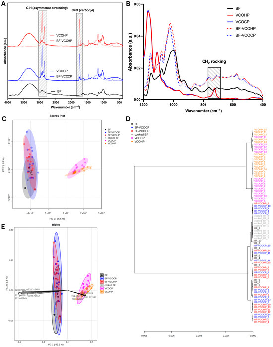
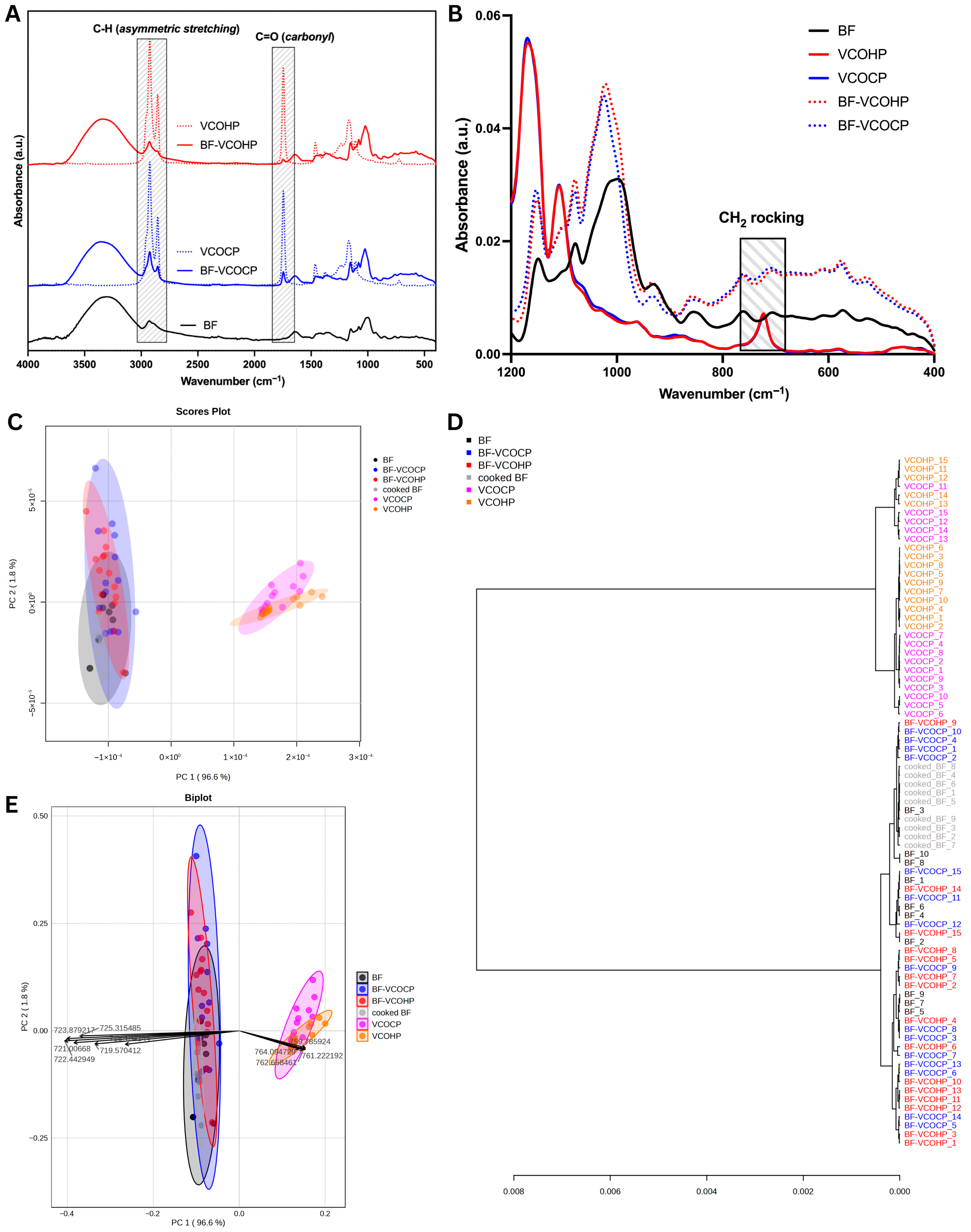
After preparing the BF–VCO composites, mid-FTIR spectroscopy was employed to confirm the incorporation of coconut oil into the banana flour matrix by examining characteristic absorption bands from the 4000–400 cm−1 as listed in Table 3. The FTIR spectra, as shown in Figure 2A, provide clear evidence of the successful retention of VCO, both hot-pressed (HP) and cold-pressed (CP), into BF. The broad O–H stretching band observed in BF between 3600 and 3000 cm−1, which reflects hydrogen bonding among starch hydroxyl groups. In the 3000–2800 cm−1 region, corresponding to asymmetric and symmetric C–H stretching of –CH2 groups, pure VCO exhibits strong absorption bands while BF alone shows only weak signals. Following VCO treatment and subsequent ethanol washing, BF–VCOHP and BF–VCOCP samples exhibited enhanced C–H stretching bands relative to BF, indicating the presence of lipid components retained within the starch matrix. Consistent with our GC-EI-MS results and previous report of Srivastava et al. [18], the FTIR spectra of VCOHP and VCOCP were associated with fatty acid, tocopherol, phytosterol, mono- and di-acylglycerol content.
Table 3.
Assignment of MIR spectral bands in BF, VCO, VCO-BF.
Figure 2.
FTIR spectral characterization and multivariate analysis of BF, VCO, and their composites: (A) full-range mid-FTIR spectra (4000–400 cm−1) highlighting C–H asymmetric stretching and C=O carbonyl regions; (B) fingerprint region (1200–400 cm−1); (C) PCA score plots and (E) PCA biplot showing sample discrimination; and (D) hierarchical cluster analysis (HCA) dendrogram showing sample grouping patterns.
The enhanced C–H bands observed in BF–VCOHP and BF–VCOCP provide evidence of lipid alkyl chains within the starch matrix, with their lower intensity compared to pure VCO indicating restricted chain mobility consistent with amylose inclusion. The appearance of a distinct carbonyl (C=O) stretching band around 1740 cm−1 in the BF–VCO samples, absent in native BF but prominent in pure VCO, further confirms lipid incorporation. This band originates from the ester carbonyl stretching vibration of triacylglycerols present in oils, such as coconut oil [35]. Consistent with the findings of Photinam and Moongngarm for green banana starch–oil systems [16], the emergence of the carbonyl band in the ~1745 cm−1 region is indicative of amylose–lipid complex formation. Moreover, the wheat starch-coconut oil-wheat gluten exhibited a pronounced peak at 1747 cm−1 [36]. Bands at 1459–1451 cm−1 were attributed to CH2 bending, characteristic of lipid alkyl chains [30]. The presence of these bands in VCO–BF further supports lipid integration.
In addition, BF, BF–VCOHP, and BF–VCOCP exhibited a band at ~1150 cm−1, corresponding to coupled C–C and C–O stretching vibrations, while the skeletal vibration associated with the starch α-1,4-glycosidic linkage was evident at ~930 cm−1 [37]. These features indicate that the starch backbone remained structurally intact following amylose–lipid complexation. Moreover, BF–VCOHP and BF–VCOCP did not exhibit the characteristic absorption peak at 725 cm−1 observed in the VCO samples, as shown in Figure 2B. The disappearance of this band suggests that the fatty acid chains were incorporated into the amylose helical cavity. The 725 cm−1 peak is attributed to the CH2 rocking vibration of the lipid chain [38]; its absence indicates restricted molecular mobility, consistent with inclusion complex formation within the amylose helix, as detected by means of FTIR analysis. This finding is consistent with the results of the rice starch–lipid complex reported by Chumsri et al. [13].
Additionally, multivariate analysis focused on the 700–600 cm−1 region reveals a distinct clustering pattern in which raw BF, cooked BF, and BF–VCO composites are clearly separated from the pure VCO samples. This differentiation demonstrates intense spectral discrimination driven by lipid-associated vibrational bands within this specific FTIR range, as illustrated in the PCA score plot (Figure 2C) and biplot (Figure 2E). Also, the loading vectors (see Figure 2E) reveal the spectral features driving the separation; bands around ~764–759 points toward the positive direction, aligning with VCO clusters. At the same time, the hierarchical cluster analysis dendrogram corroborates with PCA findings, separating the dataset into two major clusters (see Figure 2D): (1) pure VCO samples and (2) raw BF, cooked BF, and BF-VCO composites.
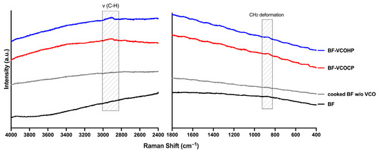
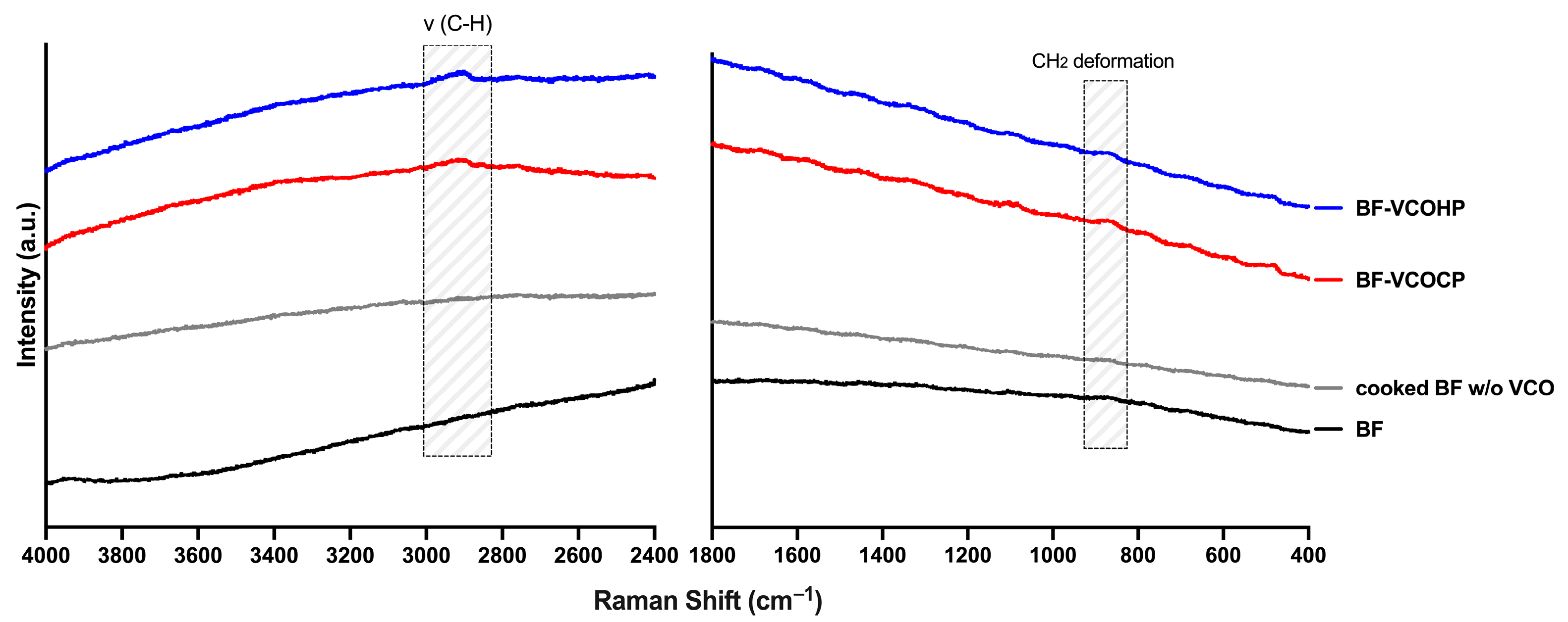
In support of the FTIR results, the Raman spectra (see Figure 3) reveal chemical differences between native banana flour (BF) and BF incorporated with VCOHP and VCOCP. In the ~3000–2800 cm−1 region, the BF–VCO samples exhibited a more pronounced band at ~2920 cm−1, assigned to C–H stretching vibrations, compared with BF and cooked BF. Additionally, within the 1800–400 cm−1 region, a more intense CH2 deformation band around ~880 cm−1 was observed. Similar spectral features have been reported in studies on octenyl succinic anhydride-modified starch [39] and interactions between soybean oil and starch [40]. Collectively, these Raman signatures confirm the incorporation of lipid hydrocarbon chains within the starch matrix.
Figure 3.
Average Raman spectra of BF and BF incorporated with VCO.
3.4. Thermal Analysis
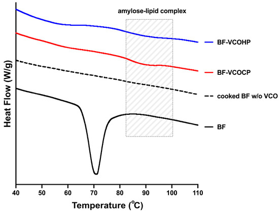
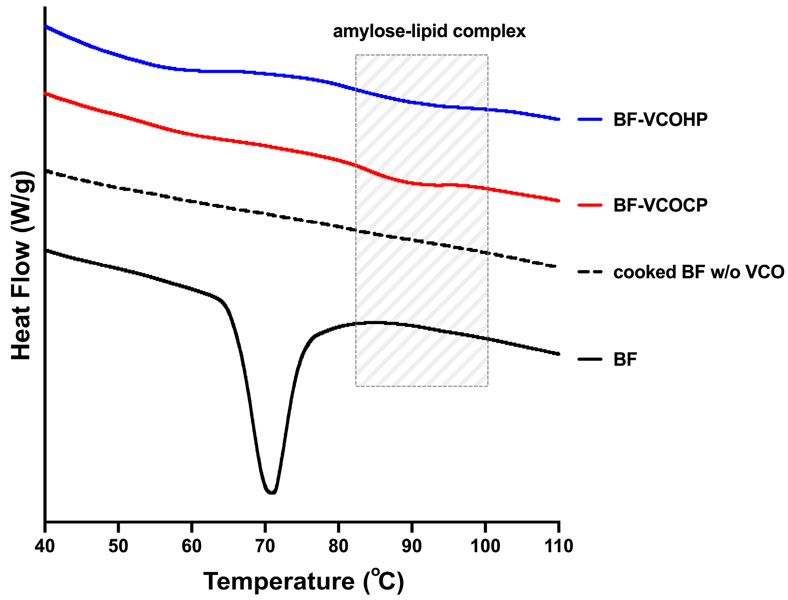
DSC was employed to evaluate the thermal behavior of the BF-VCO systems and to elucidate structural transitions associated with starch gelatinization and amylose–lipid complex formation. The DSC thermograms, as shown in Figure 4, reveal clear thermal distinctions among native BF, cooked BF without oil, and BF incorporated with VCOHP and VCOCP, providing strong evidence for amylose–lipid complex formation. Native banana flour (BF) exhibited a pronounced endothermic gelatinization peak (Tp) at 69.29 ± 3.33 °C with an associated enthalpy (ΔHg) of 9.94 ± 0.30 J g−1, corresponding to the melting of native crystalline regions within the starch granules. These thermal parameters are consistent with the literature, which reports that the Tp of banana starch ranges from 68.03 to 87.6 °C, while the ΔHg ranges from 9.45 to 16.68 J g−1 [4,41]. After cooking, this sharp gelatinization peak disappeared, indicating extensive disruption of starch crystallites during the thermal treatment [42]. Furthermore, no distinct endothermic transitions beyond gelatinization were observed for either native or cooked BF samples.
Figure 4.
DSC thermograms of banana flour (BF), cooked BF, and BF–VCO prepared via hot- and cold-pressing.
After cooking, drying, and storage of the BF–VCO composites, two endothermic peaks were observed in the DSC thermograms. The first peak, followed at approximately 50–60 °C, corresponds to starch retrogradation and is characterized by a broad, shallow endothermic transition [43]. This temperature is lower than the original gelatinization temperature, reflecting the melting of recrystallized amylose and/or amylopectin domains formed during storage. These retrograded structures are less ordered, smaller, and more heterogeneous than native starch crystallites, leading to reduced thermal stability [44]. The second endothermic peak at a higher temperature is attributed to the melting of amylose–lipid complexes formed during processing. BF–VCOHP and BF–VCOCP exhibited additional high-temperature endothermic transitions at 89.56 ± 2.17 °C (ΔHₘ = 0.8587 ± 0.1014 J g−1) and 89.18 ± 0.98 °C (ΔHₘ = 0.6267 ± 0.0777 J g−1), respectively, which were absent in native or oil-free cooked BF. These relatively small but distinct endotherms, located above the native gelatinization range, fall within the temperature window typically assigned to the dissociation or melting of amylose–lipid inclusion complexes of type 1 (amorphous structure), which aligns with the usually observed peak temperatures (~90–110 °C) [45]. Moreover, the relatively low ΔHₘ values also confirm a type 1 complex, suggesting that only a fraction of the starch in banana flour participated in amorphous amylose–lipid complex formation, which is consistent with the use of a flour matrix rather than isolated starch or amylose. Also, our results for the amylose–lipid melting enthalpy are lower than the report by Pinyo et al. on the incorporation of coconut oil (10% w/w) into rice flour (1.22 ± 0.26) [14]. In addition, incorporating 2.5% coconut oil into rice with 21.35% apparent amylose content (AAC) promotes the formation of amylose–lipid complexes with relatively low enthalpy values (0.23 J/g for Peak I and 0.12 J/g for Peak II) during instant rice preparation. This finding suggests that high medium-chain triacylglycerol oils, such as coconut oil, tend to form less thermally stable amylose–lipid complexes [46].
The relatively low melting enthalpy of amylose–lipid can be attributed to both matrix effects and lipid characteristics. In banana flour, starch granules (comprising 70–80%) are embedded within a complex matrix containing 14–17% non-starch polysaccharides and proteins [47,48], which can restrict starch swelling, amylose leaching, and lipid diffusion during thermal treatment. In addition, virgin coconut oil predominantly consists of medium-chain triglycerides [49]. The bulky molecular size of triglycerides may introduce steric hindrance, limiting their ability to enter the interior of the amylose helix and thereby reducing inclusion efficiency compared with free fatty acids or monoacylglycerols [50].
Despite this limited extent of complexation, BF–VCOHP shows a significantly higher ΔHₘ (t-test, p < 0.05) than BF–VCOCP. This implies a greater extent of complexation and/or higher structural order in BF–VCOH, suggesting more V-type crystallites melted during the DSC scan [51]. The higher ΔHₘ may also be associated with the higher mono- and di-acylglycerols content for hot-pressed VCO compared with cold-pressed VCO, which can enhance amylose inclusion efficiency, as reported by Srivastava et al. [18].
The absence of this thermal event in both native BF and cooked BF without VCO confirms that the observed endothermic transition is linked explicitly to lipid incorporation rather than to starch gelatinization alone. While previous studies on green banana starch–oil systems have reported this [16], this study provides the first DSC-based thermal evidence of amylose–exogenous lipid complex formation in the banana flour system. Furthermore, the DSC results corroborate the spectroscopic findings, demonstrating that the incorporation of VCO into banana flour leads to the formation of amylose–lipid complexes, which are absent in oil-free systems. These complexes require higher thermal energy to dissociate, which is consistent with increased structural order and reduced starch accessibility [14].
3.5. Morphology
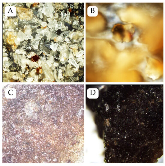
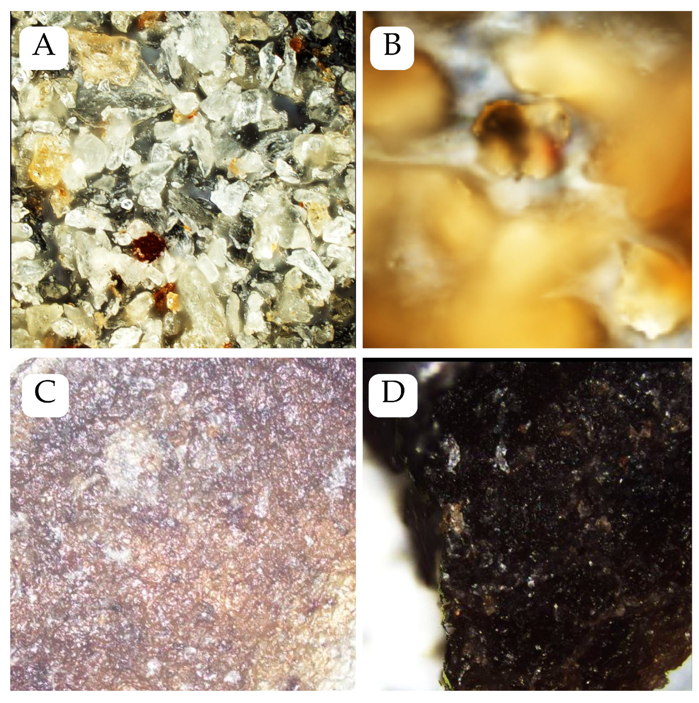
Figure 5A,B show the surface morphology of BF and cooked BF, as observed via confocal laser scanning microscopy. Native BF exhibited irregular, fragmented granules with rough surfaces and an elongated oval shape. Similar observations on extracted green banana starch were also reported by Nimsung et al. [52]. In contrast, the cooked BF sample displayed an amorphous surface structure. This transition is indicative of starch gelatinization, in which thermal treatment disrupts the native semicrystalline arrangement of starch granules, leading to swelling and structural collapse [53]. The smoother morphology observed in the cooked BF, therefore, suggests extensive hydration and molecular reorganization during heating.
Figure 5.
CLSM micrographs of (A) BF and (B) cooked BF captured at 20x magnification and (C) BF–VCOHP and (D) BF–VCOCP captured at 5x magnification.
Both BF-VCOHP (Figure 5C) and BF-VCOCP (Figure 5D) samples exhibited more structural merging than native BF, suggesting enhanced aggregation following thermal processing and lipid incorporation. However, the BF-VCOCP sample showed darker, heterogeneous regions resembling thermally induced browning.
Surface height profiling was performed to characterize microscopic irregularities across the samples further (See Table S1). The height measurements confirm the heterogeneous topography of native BF and a progressive surface smoothing following cooking (cooked BF) and lipid complex formation (BF-VCOHP and -VCOCP). The marked reduction in average surface height—from 3.09 ± 1.00 µm in cooked BF to 1.60 ± 0.45 µm and 1.15 ± 0.16 µm in BF-VCOHP and BF-VCOCP, respectively- suggests decreased surface variability. These structural changes are consistent with starch gelatinization and subsequent amylose–lipid interactions, which facilitate matrix formation and diminish granular definition [54].
3.6. In Vitro Digestibility
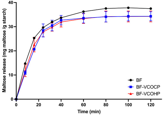
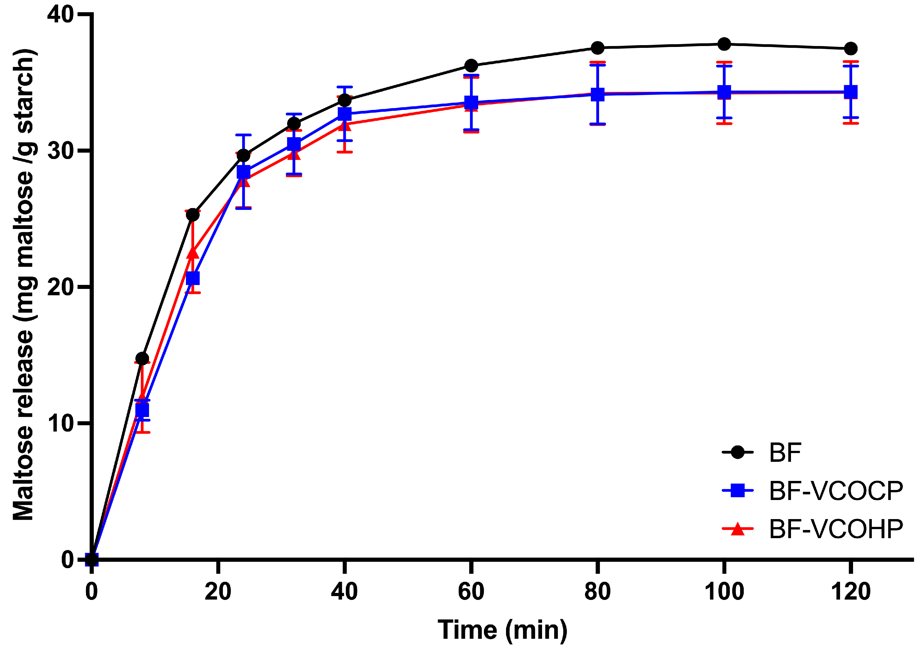
Following thermal evidence of amylose–lipid complex formation, in vitro digestion kinetics were examined to determine how the addition of exogenous VCOs influences the enzymatic digestibility of banana flour. The maltose release profiles clearly demonstrate the effect of VCO incorporation on the enzymatic digestibility of BF. Native BF (Figure 6) exhibits the fastest and highest maltose release throughout the digestion period, characterized by a rapid increase during the initial 30 min, followed by a gradual plateau, indicating high accessibility of starch chains to α-amylase [55]. In contrast, both BF–VCOCP and BF–VCOHP exhibit a noticeably slower initial digestion rate and consistently lower maltose release compared with BF, particularly during the early stages (0–20 min). This reduction suggests that VCO incorporation hinders enzyme access to starch, likely due to the formation of amylose–lipid inclusion complexes that restrict chain mobility and limit the number of hydrolysis sites [56].
Figure 6.
In vitro starch digestion of BF and BF–VCO systems.
As digestion proceeds beyond 30 min, the maltose release curves for all samples approach a plateau; however, the final maltose concentration remains lower for the VCO-containing systems than for BF. This persistent difference suggests that a fraction of the starch in BF–VCO samples remains less digestible even after prolonged enzymatic exposure, consistent with the presence of more enzyme-resistant structures. As shown in Table 4, enzymatic digestion showed reduced starch hydrolysis in VCO-treated samples, with C∞ values decreasing from 37.37 ± 0.0070 mg g−1 in native flour to 34.81 ± 2.10 mg g−1 and 34.43 ± 1.94 mg g−1 for cold- and hot-pressed VCO treatments, respectively. Between the two lipid-incorporated systems, BF–VCOCP (6.85% reduction relative to BF) shows a slightly lower maltose release than BF–VCOHP (7.87% reduction) across most time points, implying a relatively more potent inhibition of starch digestion. Moreover, this VCO-treated sample trend aligns with the DSC result (a higher ΔHₘ), suggesting more stable amylose–lipid complexation in the hot-pressed VCO system.
Table 4.
Kinetic parameters describing the in vitro enzymatic digestibility of banana flour and BF–VCO composites.
Similarly, starch/flour samples were cooked with coconut oil; the proportion of resistant starch increased, and rapidly digestible starch decreased, resulting in a lower extent of in vitro starch hydrolysis compared to oil-free controls [14,16]. Incorporation of coconut oil into other botanical sources of starch also showed a similar reduction in digestibility, attributed to the formation of amylose–lipid complexes. Desai et al. [15] reported that the inclusion of coconut oil with wheat starch (WS) significantly reduced the amount of reducing sugar released during in vitro starch digestion compared to wheat starch alone or wheat starch with wheat gluten (WS-WG). In addition, Faridah et al. [12] reported a reduction in the digestibility of cassava starch-coconut oil. Also, in rice grains, the addition of coconut oil (10% w/w) has been shown to increase SDS content and lower the estimated glycemic index (eGI) compared to a control [57].
Overall, the digestibility data support the conclusion that VCO incorporation effectively reduces starch hydrolysis in banana flour by promoting amylose–lipid interactions that slow enzymatic breakdown. The lowered rate and extent of maltose release are indicative of increased resistant starch formation [58]. These findings reinforce the functional relevance of BF–VCO systems as promising ingredients for developing starch-based foods with a reduced glycemic response.
4. Conclusions
This study successfully demonstrated the formation of amylose–lipid complexes in green banana flour (Musa × paradisiaca) upon incorporation of virgin coconut oil (VCO) extracted via hot-pressing and cold-pressing. The native banana flour exhibited an apparent amylose content of 26.40%. GC/EI-MS characterization showed that both cold-pressed and hot-pressed VCO possessed highly similar fatty acid methyl ester profiles dominated by medium-chain saturated fatty acids, particularly lauric acid. Spectroscopic analyses using FTIR, coupled with multivariate analysis, and Raman spectroscopy confirmed the incorporation of lipids into the starch matrix through characteristic C–H and C=O bands. Thermal analysis by means of DSC provided direct evidence of amylose–lipid complex formation, revealing distinct endothermic transitions (~89 °C) attributable to the melting of V-type amylose–lipid crystallites, which were absent in native and oil-free cooked banana flour. The significantly higher melting enthalpy observed in the hot-pressed VCO system indicates a greater extent and/or higher structural order of amylose–lipid complexes. CLSM analysis revealed that thermal treatment transformed native banana flour from irregular granular structures into an amorphous matrix via starch gelatinization, whereas VCO incorporation promoted aggregation. In vitro digestion results further demonstrated that VCO incorporation slightly reduced starch hydrolysis, as reflected in lower equilibrium digestibility than with native BF. Overall, the findings highlight the potential of banana flour–VCO systems as functional ingredients for formulating starch-rich or gluten-free food products with modulated glycemic responses. Nevertheless, other in vitro/in vivo glycemic response studies are warranted to explore the digestibility of BF-VCO composites.
Supplementary Materials
The following supporting information can be downloaded at: https://www.mdpi.com/article/10.3390/app16062675/s1, Figure S1. Amylose-iodine calibration curve used for the AAC determination of banana flour; Figure S2. Total ion chromatogram of cold-pressed VCO derivatized TGs to FAMEs; Figure S3. Total ion chromatogram of cold-pressed VCO derivatized TGs to FAMEs; Figure S4. Maltose standard curve by DNS method; Table S1. Surface Height Measurements of BF, cooked B, BF-VCOHP, and BF-VCOCP
Author Contributions
Conceptualization, E.S., M.C.T., C.D. and A.B.; methodology, E.S., A.Y.D.M., M.H.E., C.B.B. and K.L.T.; software, E.S., K.L.T. and A.B.; validation, M.C.T. and A.B.; formal analysis, E.S.; investigation, E.S., A.Y.D.M., M.H.E., C.B.B. and K.L.T.; resources, C.J.D. and C.B.B.; writing—original draft preparation, E.S., M.C.T., C.D., K.L.T. and A.B.; writing—review and editing, C.D. and A.B.; visualization, E.S. and A.B.; supervision, M.C.T. and A.B.; project administration, A.B.; funding acquisition, A.B. All authors have read and agreed to the published version of the manuscript.
Funding
This research was funded by De La Salle University Research and Grants Management Office (DLSU-RGMO) with the Project title “Investigating the Untapped Potential of Philippine Green Banana: A Comprehensive Analysis of the Proximate Composition, Phytochemical Constituents, Functional Properties, and Digestibility of Bungulan” with Project No. 42 F R 3TAY24-3TAY25.
Institutional Review Board Statement
Not applicable.
Informed Consent Statement
Not applicable.
Data Availability Statement
The data presented in this study are available in the article.
Acknowledgments
E.S. would like to acknowledge the Bourse de la Région Auvergne-Rhône-Alpes (BRMIE). A.B. would like to acknowledge the Make Our Planet Great Again (MOPGA) Visiting Fellowship Programme for Young Researchers, Southeast Asian Regional Center for Graduate Study and Research in Agriculture Travel Support Grant, and De La Salle University Office of the Provost for the research leave grant.
Conflicts of Interest
The authors declare no conflicts of interest.
Abbreviations
The following abbreviations are used in this manuscript:
| VCO | Virgin coconut oil |
| BF | Banana Flour |
| VCOHP | Hot-pressed VCO |
| VCOCP | Cold-pressed VCO |
| FTIR | Fourier transform infrared spectrophotometer |
| ATR | Attenuated total reflectance |
| DSC | Differential scanning calorimeter |
| Tp | Peak temperature |
| ΔHg | Enthalpy of gelatinization |
| ΔHm | Amylose–lipid complex melting |
References
- Afzal, M.F.; Khalid, W.; Akram, S.; Khalid, M.A.; Zubair, M.; Kauser, S.; Mohamedahmed, K.A.; Aziz, A.; Siddiqui, S.A. Bioactive profile and functional food applications of banana in food sectors and health: A review. Int. J. Food Prop. 2022, 25, 2286–2300. [Google Scholar] [CrossRef] [Scilit]
- Platonovskiy, N.G.; Ibrasheva, L.R.; Obukhova, N.I.; Puchkova, O.S.; Babkina, A.V. International Banana Trade: Volumes, Countries, and Trends. In Sustainable Development of the Agrarian Economy Based on Digital Technologies and Smart Innovations; Springer: Berlin/Heidelberg, Germany, 2024; pp. 25–30. [Google Scholar] [CrossRef] [Scilit]
- Samanros, A.; Lin, J. Physicochemical properties and In Vitro digestibility of starches from different Taiwanese banana cultivars. Int. Food Res. J. 2021, 28, 1257–1267. [Google Scholar] [CrossRef] [Scilit]
- Marta, H.; Cahyana, Y.; Djali, M.; Pramafisi, G. The Properties, Modification, and Application of Banana Starch. Polymers 2022, 14, 3092. [Google Scholar] [CrossRef] [Scilit] [PubMed]
- Compart, J.; Singh, A.; Fettke, J.; Apriyanto, A. Customizing Starch Properties: A Review of Starch Modifications and Their Applications. Polymers 2023, 15, 3491. [Google Scholar] [CrossRef] [Scilit]
- Li, L.; Liu, Z.; Zhang, W.; Xue, B.; Luo, Z. Production and Applications of Amylose-Lipid Complexes as Resistant Starch: Recent Approaches. Starch-Stärke 2021, 73, 2000249. [Google Scholar] [CrossRef] [Scilit]
- Panyoo, A.E.; Emmambux, M.N. Amylose–lipid complex production and potential health benefits: A mini-review. Starch-Stärke 2017, 69, 1600203. [Google Scholar] [CrossRef] [Scilit]
- Ahmmed, R.; Paff, A.; Kong, L.; Li, S.; Cockburn, D.; Tan, L. Effect of resistant starch type 5 on gut health through modulating gut microbiota. Eng. Microbiol. 2025, 6, 100250. [Google Scholar] [CrossRef] [Scilit]
- Guo, Z.; Jia, X.; Miao, S.; Chen, B.; Lu, X.; Zheng, B. Structural and thermal properties of amylose–fatty acid complexes prepared via high hydrostatic pressure. Food Chem. 2018, 264, 172–179. [Google Scholar] [CrossRef] [Scilit] [PubMed]
- Dayrit, F.M. The Properties of Lauric Acid and Their Significance in Coconut Oil. J. Am. Oil Chem. Soc. 2015, 92, 1–15. [Google Scholar] [CrossRef] [Scilit]
- Angeles-Agdeppa, I.; Nacis, J.S.; Capanzana, M.V.; Dayrit, F.M.; Tanda, K.V. Virgin coconut oil is effective in lowering C-reactive protein levels among suspect and probable cases of COVID-19. J. Funct. Foods 2021, 83, 104557. [Google Scholar] [CrossRef] [Scilit]
- Faridah, D.N.; Andriani, I.; Talitha, Z.A.; Budi, F.S. Physicochemical characterization of resistant starch type V (RS5) from manggu cassava starch (Manihot esculenta). Food Res. 2021, 5, 228–234. [Google Scholar] [CrossRef] [Scilit] [PubMed]
- Chumsri, P.; Panpipat, W.; Cheong, L.-Z.; Chaijan, M. Formation of Intermediate Amylose Rice Starch–Lipid Complex Assisted by Ultrasonication. Foods 2022, 11, 2430. [Google Scholar] [CrossRef] [Scilit] [PubMed]
- Pinyo, J.; Wongsagonsup, R.; Panthong, N.; Kantiwong, P.; Huang, Q.; Tangsrianugul, N.; Suphantharika, M. Effects of different edible oils on In Vitro starch digestibility and physical properties of rice starch and rice flour. Int. J. Food Sci. Technol. 2024, 59, 170–180. [Google Scholar] [CrossRef] [Scilit]
- Desai, A.S.; Brennan, M.A.; Zeng, X.; Brennan, C.S. Complex formation, In Vitro digestion, structural, and physicochemical properties of fish oil and wheat starch blend. J. Food Process. Preserv. 2020, 44, e14859. [Google Scholar] [CrossRef] [Scilit]
- Photinam, R.; Moongngarm, A. Effect of adding vegetable oils to starches from different botanical origins on physicochemical and digestive properties and amylose–lipid complex formation. J. Food Sci. Technol. 2023, 60, 393–403. [Google Scholar] [CrossRef] [Scilit] [PubMed]
- De Mesa, A.Y.; Eder, M.H.; Sumayao, R.; Malabed, R.; Ondevilla, J.C.; Tiozon, R.; Sreenivasulu, N.; Dizon, R.M.; Canabuan, C.R.; Torreda, J.A.; et al. Bungulan (Musa x paradisiaca) banana flour: A novel, underutilized ingredient for nutrient-enriched composite bread. Int. J. Gastron. Food Sci. 2025, 42, 101371. [Google Scholar] [CrossRef] [Scilit]
- Srivastava, Y.; Semwal, A.D.; Majumdar, A. Quantitative and qualitative analysis of bioactive components present in virgin coconut oil. Cogent Food Agric. 2016, 2, 1164929. [Google Scholar] [CrossRef] [Scilit]
- Ichihara, K.; Shibahara, A.; Yamamoto, K.; Nakayama, T. An improved method for rapid analysis of the fatty acids of glycerolipids. Lipids 1996, 31, 535–539. [Google Scholar] [CrossRef] [Scilit]
- Reddy, C.K.; Choi, S.M.; Lee, D.-J.; Lim, S.-T. Complex formation between starch and stearic acid: Effect of enzymatic debranching for starch. Food Chem. 2018, 244, 136–142. [Google Scholar] [CrossRef] [Scilit]
- Graham, R. A Proposal for IRRI to Establish a Grain Quality and Nutrition Research Center; International Rice Research Institute (IRRI): Los Baños, Philippines, 2002. [Google Scholar]
- Toplak, M.; Read, S.T.; Sandt, C.; Borondics, F. Quasar: Easy Machine Learning for Biospectroscopy. Cells 2021, 10, 2300. [Google Scholar] [CrossRef] [Scilit] [PubMed]
- Minekus, M.; Alminger, M.; Alvito, P.; Ballance, S.; Bohn, T.; Bourlieu, C.; Carrière, F.; Boutrou, R.; Corredig, M.; Dupont, D.; et al. A standardised static In Vitro digestion method suitable for food—An international consensus. Food Funct. 2014, 5, 1113–1124. [Google Scholar] [CrossRef] [Scilit] [PubMed]
- Cordenunsi-Lysenko, B.R.; Nascimento, J.R.O.; Castro-Alves, V.C.; Purgatto, E.; Fabi, J.P.; Peroni-Okyta, F.H.G. The Starch Is (Not) Just Another Brick in the Wall: The Primary Metabolism of Sugars During Banana Ripening. Front. Plant Sci. 2019, 10, 391. [Google Scholar] [CrossRef] [Scilit] [PubMed]
- Spiazzi, B.F.; Duarte, A.C.; Zingano, C.P.; Teixeira, P.P.; Amazarray, C.R.; Merello, E.N.; Wayerbacher, L.F.; Farenzena, L.P.; Correia, P.E.; Bertoluci, M.C.; et al. Coconut oil: An overview of cardiometabolic effects and the public health burden of misinformation. Arch. Endocrinol. Metab. 2023, 67, e000641. [Google Scholar] [CrossRef] [Scilit] [PubMed]
- Kumalasari, I.D.; Santosa, I.; Sulistiawati, E. GC-MS analysis of chemical compounds of coconut oil extract from dried coconut with low-temperature process. AIP Conf. Proc. 2023, 2702, 070008. [Google Scholar] [CrossRef] [Scilit]
- Amit Kumari, S.; Jamwal, R. Use of FTIR spectroscopy integrated with multivariate chemometrics as a swift, and non-destructive technique to detect various adulterants in virgin coconut oil: A comprehensive review. Food Chem. Adv. 2023, 2, 100203. [Google Scholar] [CrossRef] [Scilit]
- De Luca, M.; Ioele, G.; Grande, F.; Occhiuzzi, M.A.; Chieffallo, M.; Garofalo, A.; Ragno, G. Multivariate Curve Resolution Methodology Applied to the ATR-FTIR Data for Adulteration Assessment of Virgin Coconut Oil. Molecules 2023, 28, 4661. [Google Scholar] [CrossRef] [Scilit] [PubMed]
- Amini Khoozani, A.; Birch, J.; Bekhit, A.E.-D.A. Textural properties and characteristics of whole green banana flour produced by air-oven and freeze-drying processing. J. Food Meas. Charact. 2020, 14, 1533–1542. [Google Scholar] [CrossRef] [Scilit]
- Amit Jamwal, R.; Kumari, S.; Dhaulaniya, A.S.; Balan, B.; Kelly, S.; Cannavan, A.; Singh, D.K. Utilizing ATR-FTIR spectroscopy combined with multivariate chemometric modelling for the swift detection of mustard oil adulteration in virgin coconut oil. Vib. Spectrosc. 2020, 109, 103066. [Google Scholar] [CrossRef] [Scilit]
- Srivastava, Y.; Semwal, A.D.; Sajeevkumar, V.A.; Sharma, G.K. Melting, crystallization and storage stability of virgin coconut oil and its blends by differential scanning calorimetry (DSC) and Fourier transform infrared spectroscopy (FTIR). J. Food Sci. Technol. 2017, 54, 45–54. [Google Scholar] [CrossRef] [Scilit]
- Kumar, P.S.; Saravanan, A.; Sheeba, N.; Uma, S. Structural, functional characterization and physicochemical properties of green banana flour from dessert and plantain bananas (Musa spp.). LWT 2019, 116, 108524. [Google Scholar] [CrossRef] [Scilit]
- Huang, Q.; Liu, C.; Gao, S.; Sun, Y.; Zhang, X.; Yang, L. Effects of green and ripe banana flours on cookie characteristics: A comprehensive study from frozen dough to cookies. LWT-Food Sci. Technol. 2026, 241, 119101. [Google Scholar] [CrossRef] [Scilit]
- Rohman, A.; Che Man, Y.B.; Ismail, A.; Hashim, P. Application of FTIR Spectroscopy for the Determination of Virgin Coconut Oil in Binary Mixtures with Olive Oil and Palm Oil. J. Am. Oil Chem. Soc. 2010, 87, 601–606. [Google Scholar] [CrossRef] [Scilit]
- Sota-Uba, I.; Bamidele, M.; Moulton, J.; Booksh, K.; Lavine, B.K. Authentication of edible oils using Fourier transform infrared spectroscopy and pattern recognition methods. Chemom. Intell. Lab. Syst. 2021, 210, 104251. [Google Scholar] [CrossRef] [Scilit]
- Desai, A.S.; Brennan, M.A.; Zeng, X.-A.; Brennan, C.S. Physiochemical, structural and In Vitro starch digestibility properties of starch blended with fish oil and wheat gluten. J. Food Meas. Charact. 2021, 15, 3005–3014. [Google Scholar] [CrossRef] [Scilit]
- Tan, X.; Wu, F.; Guo, Z.; Xie, F.; Song, C.; Xia, X.; Guo, L.; Feng, Y.; He, Z. Amylose content controls the structures and digestibility of starch-lipid complexes during microwave postprocessing. Carbohydr. Polym. 2025, 358, 123530. [Google Scholar] [CrossRef] [Scilit] [PubMed]
- Mavlanov, U.; Czaja, T.P.; Salimovich, S.S.; Khodjaev, S.; Khakimov, B. Characterization of butter and margarine oil composition using benchtop NMR and FTIR: A comparative study of products from Uzbekistan and Denmark. J. Food Compos. Anal. 2026, 150, 108842. [Google Scholar] [CrossRef] [Scilit]
- Chen, H.; Li, H.; Wu, Y.; Kan, J. Functionality differences between esterified and pregelatinized esterified starches simultaneously prepared by octenyl succinic anhydride modification and its application in dough. Int. J. Biol. Macromol. 2024, 260, 129594. [Google Scholar] [CrossRef] [Scilit] [PubMed]
- Guan, Y.; Zhao, G.; Thaiudom, S. Evaluation of the physico-chemical properties of potato starch-based foods their interactions with milk protein soybean oil. Food Chem. X 2022, 16, 100495. [Google Scholar] [CrossRef] [Scilit] [PubMed]
- Aparicio-Saguilán, A.; Aguirre-Cruz, A.; Méndez-Montealvo, G.; Rodriguez-Ambriz, S.L.; Garcia-Suarez, F.J.; Páramo-Calderón, D.E.; Bello-Pérez, L.A. The effect of the structure of native banana starch from two varieties on its acid hydrolysis. LWT-Food Sci. Technol. 2014, 58, 381–386. [Google Scholar] [CrossRef] [Scilit]
- Tananuwong, K.; Reid, D. DSC and NMR relaxation studies of starch-water interactions during gelatinization. Carbohydr. Polym. 2004, 58, 345–358. [Google Scholar] [CrossRef] [Scilit]
- Wang, S.; Li, C.; Zhang, X.; Copeland, L.; Wang, S. Retrogradation enthalpy does not always reflect the retrogradation behavior of gelatinized starch. Sci. Rep. 2016, 6, 20965. [Google Scholar] [CrossRef] [Scilit] [PubMed]
- Chen, W.; Xu, W.; Junejo, S.A.; Feng, Y.; Gao, T.; Jiang, Y.; Zhang, B.; Jia, X.; Huang, Q. Analytical methods for starch retrogradation assessment. Food Biomacromol. 2024, 1, 107–126. [Google Scholar] [CrossRef] [Scilit]
- Tufvesson, F.; Wahlgren, M.; Eliasson, A. Formation of Amylose-Lipid Complexes and Effects of Temperature Treatment. Part 1. Monoglycerides. Starch-Stärke 2003, 55, 61–71. [Google Scholar] [CrossRef] [Scilit]
- Luangsakul, N.; Ritudomphol, O. Effect of oil addition on In Vitro starch digestibility and physicochemical properties of instant rice. Int. J. Agric. Technol. 2018, 14, 1399–1412. [Google Scholar]
- Cahyana, Y.; Titipanillah, R.; Mardawati, E.; Sukarminah, E.; Rialita, T.; Andoyo, R.; Djali, M.; Hanidah, I.-I.; Setiasih, I.S.; Handarini, K. Non-starch contents affect the susceptibility of banana starch and flour to ozonation. J. Food Sci. Technol. 2018, 55, 1726–1733. [Google Scholar] [CrossRef] [Scilit] [PubMed]
- Thanyapanich, N.; Jimtaisong, A.; Rawdkuen, S. Functional Properties of Banana Starch (Musa spp.) and Its Utilization in Cosmetics. Molecules 2021, 26, 3637. [Google Scholar] [CrossRef] [Scilit] [PubMed]
- Dayrit, F.M. Lauric Acid is a Medium-Chain Fatty Acid, Coconut Oil is a Medium-Chain Triglyceride. Philipp. J. Sci. 2014, 143, 157–166. [Google Scholar]
- Chao, C.; Yu, J.; Wang, S.; Copeland, L.; Wang, S. Mechanisms Underlying the Formation of Complexes between Maize Starch and Lipids. J. Agric. Food Chem. 2018, 66, 272–278. [Google Scholar] [CrossRef] [Scilit]
- Tufvesson, F.; Wahlgren, M.; Eliasson, A. Formation of Amylose-Lipid Complexes and Effects of Temperature Treatment. Part 2. Fatty Acids. Starch-Stärke 2003, 55, 138–149. [Google Scholar] [CrossRef] [Scilit]
- Nimsung, P.; Thongngam, M.; Naivikul, O. Compositions, Morphological and Thermal Properties of Green Banana Flour and Starch. Kasetsart J.-Nat. Sci. 2007, 41, 324–330. [Google Scholar]
- Mohamed, I.O. Effects of processing and additives on starch physicochemical and digestibility properties. Carbohydr. Polym. Technol. Appl. 2021, 2, 100039. [Google Scholar] [CrossRef] [Scilit]
- Liu, Q.; Guan, H.; Guo, Y.; Wang, D.; Yang, Y.; Ji, H.; Jiao, A.; Jin, Z. Structure and In Vitro digestibility of amylose–lipid complexes formed by an extrusion-debranching-complexing strategy. Food Chem. 2024, 437, 137950. [Google Scholar] [CrossRef] [Scilit] [PubMed]
- Reginio, F.C.; Ketnawa, S.; Ogawa, Y. In Vitro examination of starch digestibility of Saba banana [Musa ‘saba’ (Musa acuminata × Musa balbisiana)]: Impact of maturity and physical properties of digesta. Sci. Rep. 2020, 10, 1811. [Google Scholar] [CrossRef] [Scilit] [PubMed]
- Zhou, B.; Chen, N.; Wu, Y.; Peng, X.; Han, K.; Chen, Z.; Xu, M.; Liu, X. Starch-lipid complexes and their application: A review. Int. J. Biol. Macromol. 2025, 310, 142928. [Google Scholar] [CrossRef] [Scilit]
- Tamura, M.; Hoshi, K.; Saito, T.; Sasahara, Y. In Vitro Starch Digestion of Cooked Rice Grain Following the Addition of Various Vegetable Oils. Jpn. Agric. Res. Q. 2022, 56, 261–267. [Google Scholar] [CrossRef] [Scilit]
- Fässler, C.; Arrigoni, E.; Venema, K.; Hafner, V.; Brouns, F.; Amadò, R. Digestibility of resistant starch containing preparations using two in vitro models. Eur. J. Nutr. 2006, 45, 445–453. [Google Scholar] [CrossRef] [Scilit] [PubMed]

Disclaimer/Publisher’s Note: The statements, opinions and data contained in all publications are solely those of the individual author(s) and contributor(s) and not of MDPI and/or the editor(s). MDPI and/or the editor(s) disclaim responsibility for any injury to people or property resulting from any ideas, methods, instructions or products referred to in the content. |
© 2026 by the authors. Licensee MDPI, Basel, Switzerland. This article is an open access article distributed under the terms and conditions of the Creative Commons Attribution (CC BY) license.