


Abstract
Objectives: Horizontal ridge augmentation remains a clinical challenge due to limitations in terms of spatial maintenance, graft stability and predictability of new bone formation. The umbrella-screw tent technique provides mechanical stability for particulate grafts, while adjuvants such as hyaluronic acid (HA) and polynucleotides (PN) may enhance biological remodeling. Evidence for this compound in implant-related bone augmentation is still scarce. Material and methods: In a single patient with a knife-edge alveolar ridge, augmentation was performed in regions 34 to 36 using the umbrella-screw tent technique. The defect was grafted with deproteinized bovine bone mineral (DBBM) mixed with hyaluronic acid (HA) and polynucleotides (PN), supplemented with platelet-rich fibrin (PFR) and covered with a resorbable collagen membrane. After six months, two implants were installed, and a biopsy was obtained by trepanation for histological and histomorphometric analysis. Results: Healing occurred without compromise, with no signs of infection or graft exposure. Horizontal bone gain averaged 4.5 mm, corresponding to a relative Target Performance Index (TPI-h) of 75%. Histomorphometric analysis revealed a total mineralized fraction of 76.4%, consisting of 36.1% newly formed bone and 40.3% residual DBBM particles. The xenogeneic granules were completely integrated into mature bone, with no signs of inflammation or foreign body reaction. Conclusion: The case report illustrates that the combination of DBBM with HA and PN, stabilized by the umbrella-screw tent technique, can lead to significant new bone formation and favorable graft integration. Although limited by its single-case design, the case report provides preliminary insights into the synergistic potential of HA and PN as biological enhancers in bone augmentation, warranting further controlled studies.
1. Introduction
The initial state of both hard and soft tissues determines not only the level of challenge in implant positioning but also whether an implant can succeed or fail. Regarding the ease of implant positioning through hard-tissue modeling, bone augmentation in both vertical and horizontal dimensions is now a routine, safe procedure performed using various operative techniques. Although bone augmentation techniques vary, success and complication rates differ, and consensus on the superiority of any method is difficult to reach [1]. All techniques have in common that the transplantation of bone or bone substitutes creates space and immobility while allowing more or less the ingrowth of vessels to support the transformation in living bone. Massive bone blocks for extensive bone augmentation provide immobile space, but revascularization and turnover may be a challenge due to their compacted shape. Using a shell technique, conversely, it is feasible to utilize particulated bone or bone substitute, facilitating the ingrowth of vessels. Granular bone graft substitutes are also used in the umbrella-screw tent technique, a ridge augmentation method known for its technical simplicity and low complication rates, making it easily manageable [2]. It shares the common basic biological principle of creating the requisite spatial conditions, ensuring graft immobilization so that revascularization can occur, followed by subsequent ossification. Using xenogeneic particulate bone eliminates the need for a donor site surgery [3]. As mentioned above, autogenous massive bone blocks are not infrequently associated with failure of turnover into living tissue and severe infections at the autologous graft site [4]. On the other hand, particulated bone or bone substitutes must be stabilized to prevent graft mobility during healing. Several techniques, such as titanium-reinforced membranes, titanium meshes, and magnesium membranes are used to immobilize those grafts. Conversely, the shielding of the particulate bone substitute material provided by large-diameter screwheads renounces the need for further stabilizing items favoring resorbable membranes, thus bypassing most associated complications such as exposure and infection [4].
Xenogeneic bone substitutes, such as deproteinized bovine bone, have gained acceptance as an alternative to conventional autologous and allogenic bone grafts, due to their volume-stable integration into the recipient region and osteoconductive properties [5]. Due to structural, biochemical and biological differences, the biological cascade for healing and regeneration is delayed, and the absolute volume of new bone formation may be reduced [6]. Aimed at fostering de-novo bone formation for xenogeneic particulates, recent endeavors take the route of adding adjuvants for boosting regeneration. The most recent approach has been the use of hyaluronic acid, a naturally occurring polysaccharide of the glycosaminoglycan family, which is abundant in the extracellular matrix of human tissues, including connective tissue, synovial fluid, skin, periodontal tissues, muscles, and specific organs. Hyaluronic acid is involved in various biological processes at the cellular level, with functional effects depending on its molecular weight [7,8]. Hyaluronic acid has been demonstrated to influence signaling pathways in the context of cell proliferation and differentiation [8] and to participate in the phases of wound healing [9]. Some modified, cross-linked derivatives appear to enhance specific biological responses [10]. Regarding bone formation, the literature suggests that hyaluronic acid improves the handling of particulate graft substitutes, acts as a carrier molecule for growth factors, and fosters favorable environmental conditions for osteogenesis [8]. Yet, the specific mechanisms of interaction remain unresolved; the literature appears unsettled regarding the effectiveness of hyaluronic acid as a regeneration-promoting adjuvant [8], particularly in its applications in dentistry.
Chained derivatives of DNA or RNA, known as polynucleotides, have also been combined with hyaluronic acid additives to aid in regeneration processes. In cells such as fibroblasts [11,12,13], polynucleotides induce cell growth, proliferation and viability [12], resulting in a general effect of enhanced wound healing, accompanied by reduced fibrosis and increased surface epithelialization [11,12,14]. Their use has also been successful in reducing pain perception in knee osteoarthritis [15] and improving the healing of ulcers on the limbs [14]. Specifically regarding osteoblasts [15,16] and bone regeneration overall, polynucleotides seem to enhance bone turnover and the osseous remodeling process when added to biomaterial grafts [16]. Its role is attributed to the effect on the purinergic adenosine receptor, whose activation modulates angiogenesis, triggers osteoblast activity and reduces inflammatory processes with a corresponding reduction in cardinal symptoms [16,17]. The potential of polynucleotides in aiding wound healing has been laid out in these early studies; yet their application in dental periodontology and implantology is recent and evidence is thus scarce.
This case report describes a Class IV knife-edge ridge (Cawood and Howel classification [18]) that was augmented, aiming to showcase the potential remodeling process of the bone after successful augmentation with the umbrella-screw tent technique using deproteinized bovine bone mineral (DBBM) combined with polynucleotides (PN) and hyaluronic acid (HA). This case report aims to serve as motivation for further research efforts in this field.
2. Material and Methods
2.1. Ethic Legals
The study was approved by the Vilnius Region Biomedical Research Ethics Committee based on the Declaration of Helsinki, registration number “2025/7-1684-1133”, issued on 30 June 2025. Written informed consent for publication was obtained from the patient.
2.2. Organization and Surgical Procedure
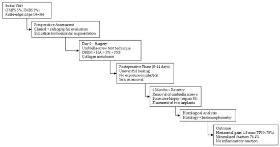
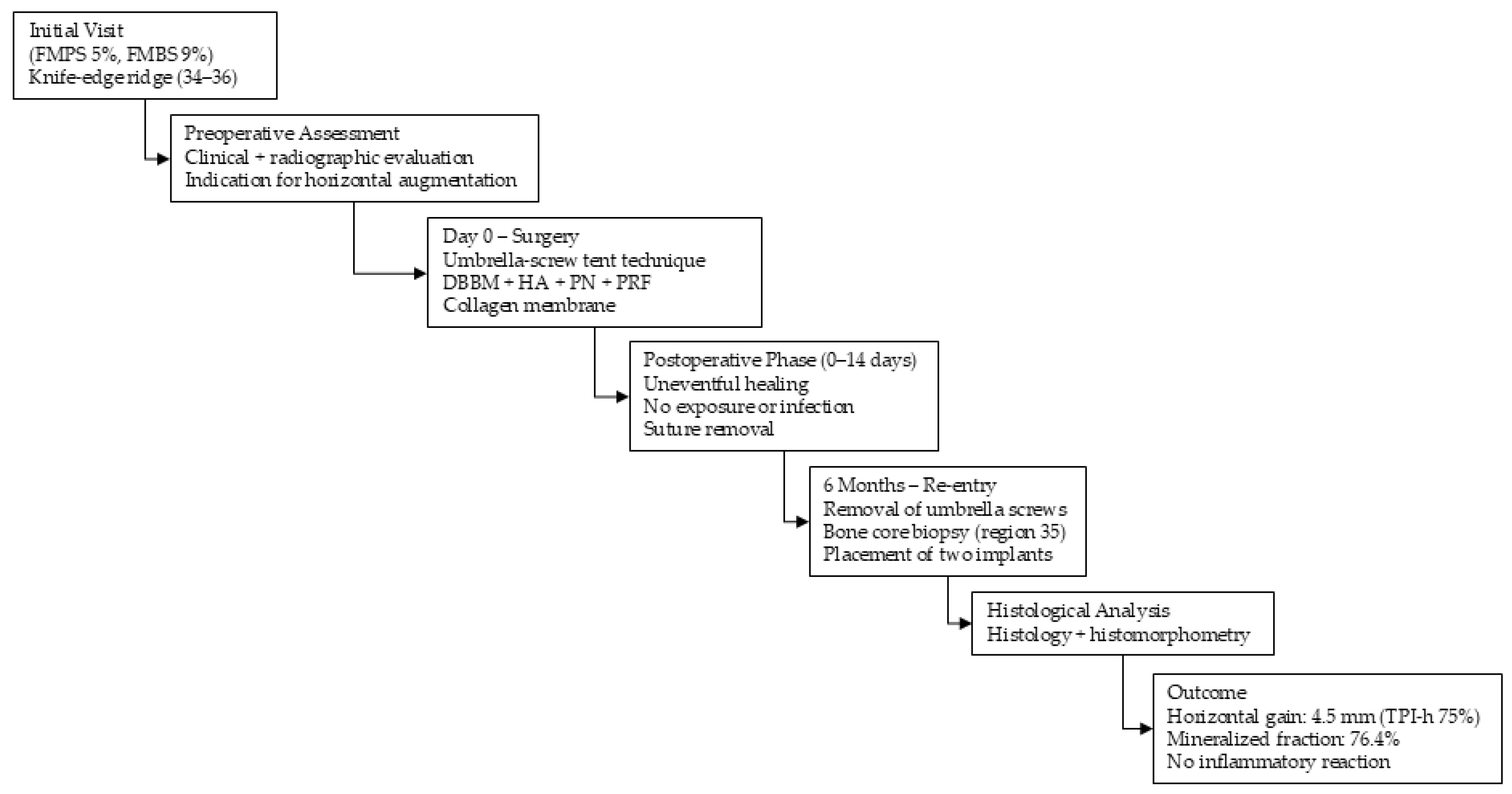
The case report was organized according to the CARE Protocol [19]. The patient has lost teeth in the visible area, has noticed that bone is missing, does not want the adjacent teeth to be prepared, and would like to have fixed teeth again. The timeline of the planned treatment is illustrated by the care timeline (see Figure 1).
Figure 1.
Care Timeline.
An 85-year-old female patient with unremarkable general health who takes 100 mg of aspirin prophylactically and has healthy periodontium (FMPS = 5%, FMBS = 9%) was in need of two implants. Alternative augmentation strategies, such as block grafting or conventional guided bone regeneration, were considered but rejected due to patient preference and defect morphology. The surgical procedure was performed in two stages. Following a preoperative examination of the intraoral conditions based on clinical and radiographic criteria, the need for horizontal bone augmentation for proper implant installation was established for regions 34 to 36. The last author performed the surgeries under aseptic conditions in his private clinic. The patient rinsed preoperatively with 1% chlorhexidine gluconate solution for 60 s to reduce the bacterial load in the oral cavity. Analgesia was ensured by treatment under local anesthesia with Ultracain DS forte® (Sanofi-Aventis, Frankfurt, Germany).
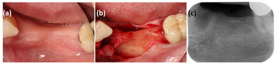
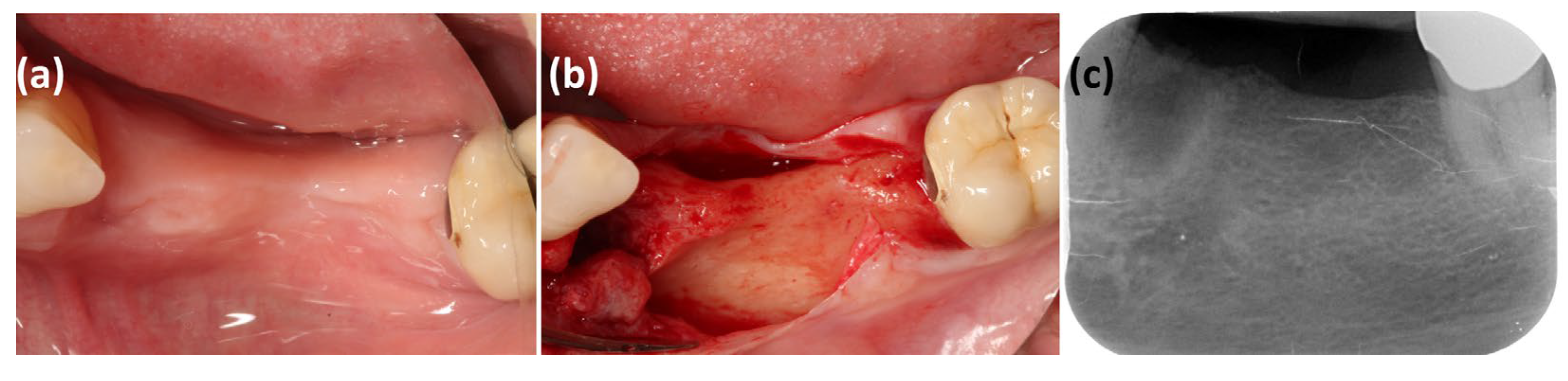
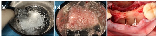
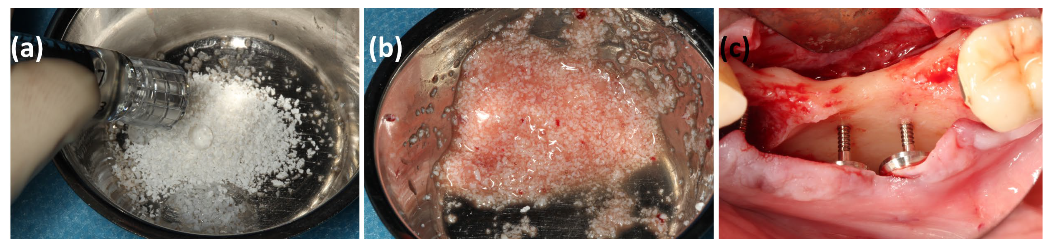
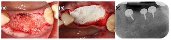
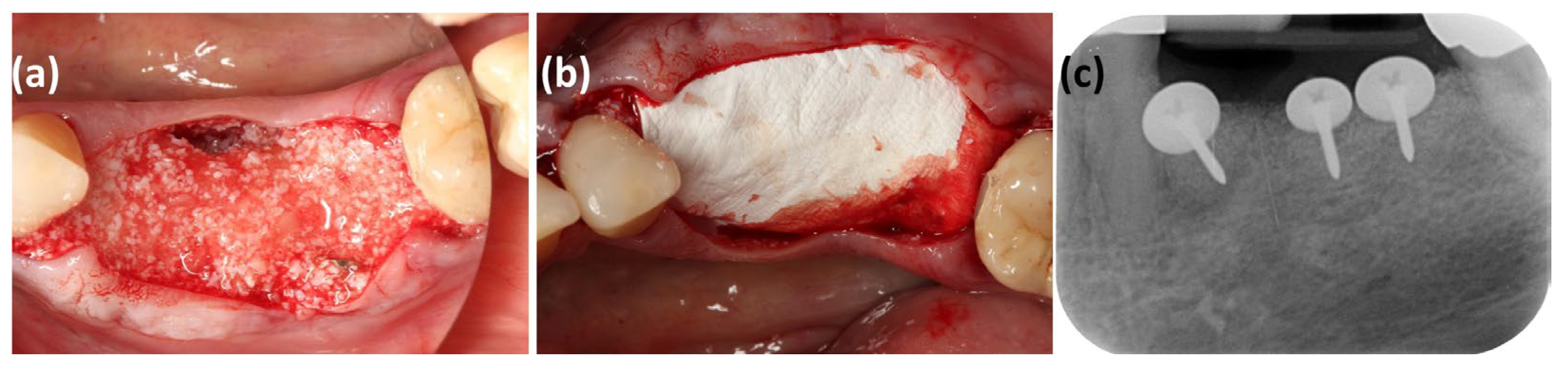
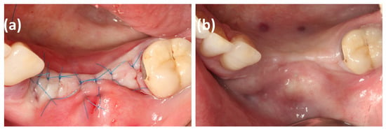
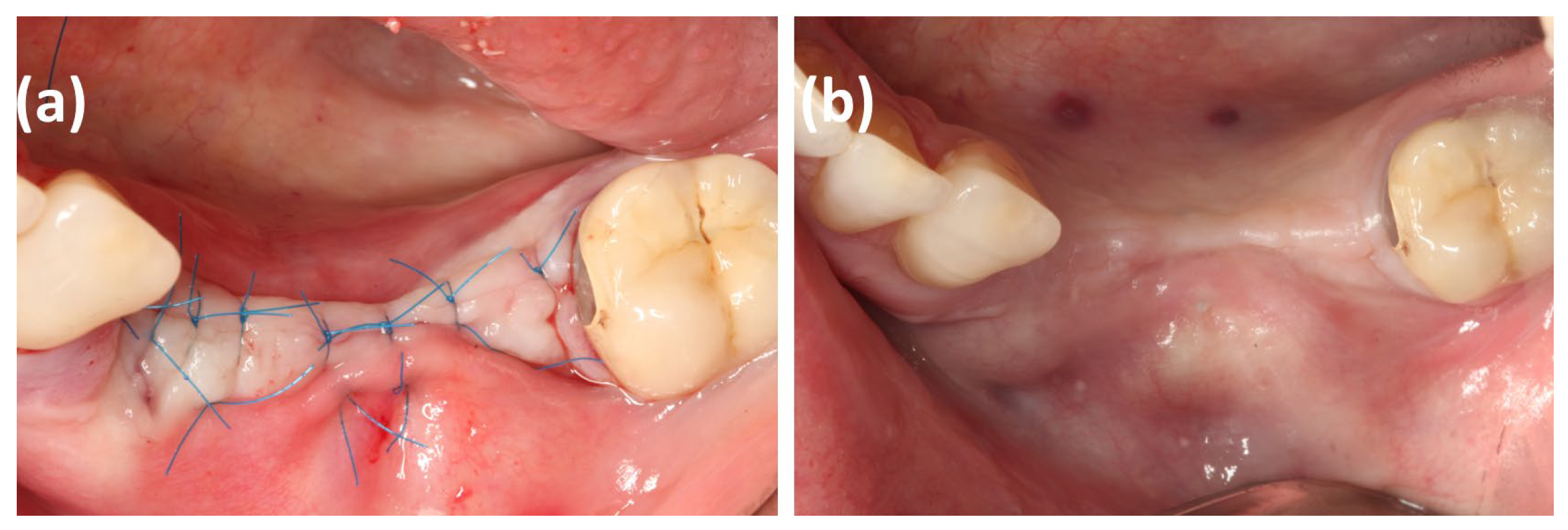
A crestal incision with a releasing incision followed by a full-thickness flap using a microsurgical approach (see Figure 2) was applied. The early periosteal incision and flap mobilization at the beginning minimized subsequent swelling and bleeding due to the existing astringent effect. In the following, a split flap was prepared buccally and lingually at the mucogingival junction to enable mobilization of the flap and later tension-free wound closure. Three umbrella-screws (2 × 8.0 mm and 1 × 10.0 mm, screw head diameter of 10.0 mm, Geistlich Biomaterials, Wohlhusen, Switzerland) were positioned on the buccal side to create the necessary space for the intended augmentation (see Figure 3). To compensate the expected shrinkage of the augmented site of <1 mm in total, the head of the screws were positioned 1 mm more buccally than the targeted bone surface. A horizontal volume gain of 6.0 mm was sought (5.0 mm target volume plus 1.0 mm for shrinkage compensation). The defect was augmented with a mixture of xenogeneic bone mineral (DBBM), the so called BioOss® (Geistlich Biomaterials, Wolhusen, Switzerland), pasted with a gel based on a non-crosslinked hyaluronic acid (HA) and polynucleotides (PN) (Regenfast®, Geistlich Biomaterials, Wolhusen, Switzerland) and platelet-rich fibrin (PRF) (max. 3000 rpm for 10 min, according to the protocol of Choukroun et al. [20]), cut into small pieces as described before (see Figure 3) [2]. The addition of the agents facilitated intraoperative handling of the particulate bone substitute material in order to achieve sticky bone quality. A non-crosslinked resorbable collagen membrane (see Figure 4) was used as a barrier (BioGide®, Geistlich Biomaterials, Wohlhusen, Switzerland) placed on top of the augmented site without any further fixation. The flap was adapted with horizontal mattress sutures placed deep in the vestibule, and the incision was closed with interrupted sutures using 6/0 polypropylene monofilament (Medic, Kilkis, Greece) (see Figure 5). An antibiotic regimen with amoxicillin-clavulanic acid 875/125 mg twice a day (1-0-1), starting 2 h before treatment, was prescribed for one week. After 14 days, the sutures were removed.
Figure 2.
(a,b) The initial clinical situation revealed an insufficient bone supply in the horizontal direction with a knife-edge bone margin. (c) Radiographic assessment showed adequate vertical relation.
Figure 3.
(a) The deproteinized bovine bone mineral (DBBM) was mixed with the pre-packaged mixture of hyaluronic acid (HA) and polynucleotides (PN). (b) Platelet-rich fibrin (PRF) membranes were cut into small particles and added to the mixture. (c) The positioning of three umbrella-screws provided the required space and immobilization for the subsequent graft.
Figure 4.
(a) The generated volume of the augmentation with deproteinized bovine bone mineral (DBBM), platelet-rich fibrin (PRF), and a mixture of hyaluronic acid (HA) and polynucleotides (PN) is visible. (b) The collagen membrane does not need to be tacked, as the umbrella-screws provided the immobile space. (c) Radiographic assessment confirms the positioning of the screws and suggests an increased volume.
Figure 5.
(a) Flap preparation and suturing provided a flap passively mobilized to cover the augmented site. (b) The healed site indicated an uneventful healing.
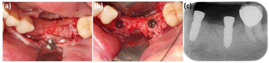
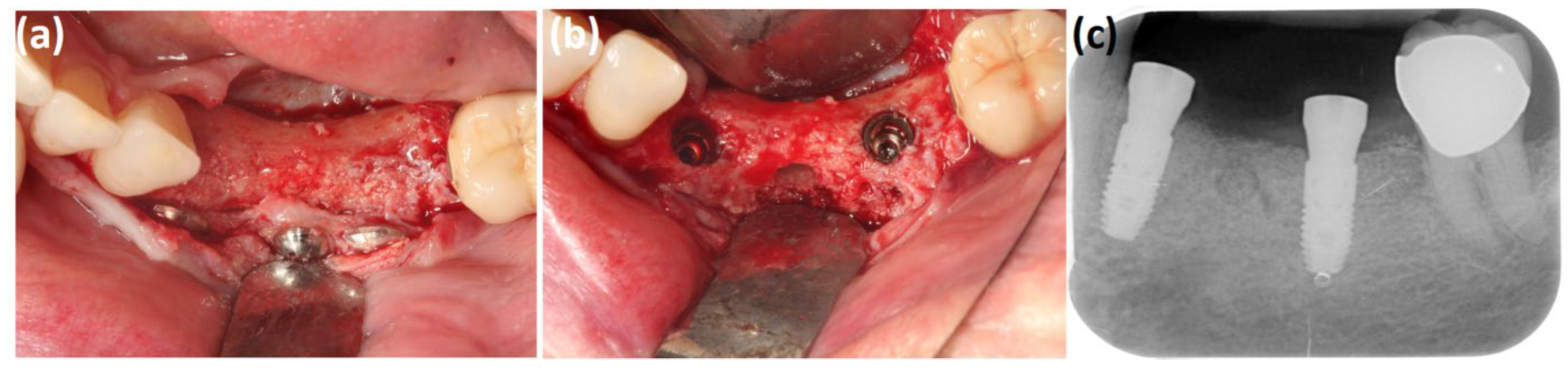
Implant placement was performed six months after the initial surgery. All screws were in place at reentry; no early exposure occurred. The umbrella-screws were removed after raising a full-thickness flap under a single-shot antibiotic regimen (2 g amoxicillin-clavulanic acid 875/125 mg) 2 h before surgery. As the volume of the ridge was regained as planned (see Figure 6), the implants (Astra EV, 9.0 mm, Ø, 4.2 mm; Dentsply-Sirona, Bensheim, Germany) were placed in the desired position with excellent primary stability (20 Ncm and 33 Ncm, respectively) (Implamed, W+H, Bürmosos, Austria). Healing abutments were placed for open healing. A bone core (diameter 2.8 mm and 5.0 mm length) for biopsy was collected horizontally from region 35 using a trephine drill (see Figure 6). The flap was sutured as described above.
Figure 6.
(a) After 6 months of healing, the gained volume is visible after successful augmentation. The umbrella-screws are in situ. (b) After removing the screws, two implants were placed in regions 34 and 36. The location where the bone biopsy was harvested is visible on the buccal bone surface. (c) The radiograph confirmed the prosthetically correct position of the implants two-dimensionally. Slightly higher radiopacity indicates a horizontal dimensional gain in region 34–36. The circular biopsy site is visible in region 35.
2.3. Clinical Analysis
To measure the efficacy of augmentation, the Target Performance Index (TPI) has been introduced to measure the efficacy of augmentation [2]. The distance from the bottom of the screw head to the pre-existing bone level in the horizontal direction (US-hd) was measured at baseline. After the healing period, horizontal bone resorption along the screw was assessed by measuring the distance between the screw head and the newly formed bone (US-hnb). The horizontal bone gain (bg-h) was calculated according to the equation . This allowed us to compute the relative TPI-h according to The absolute TPI-h was determined by subtracting the actual bone gain from the augmentation value.
2.4. Histological and Histomorphometric Analysis
After fixation of the bone biopsy samples in 4% formalin for seven days, dehydration was performed using a graded ethanol series. The ethanol concentration was increased from 70% to 100% every 24 h during storage. The samples were then degreased in xylene (Merck, Darmstadt, Germany) for one day. Subsequently, infiltration, embedding, and polymerization of the specimens were carried out using Technovit 9100 (Heraeus Kulzer, Hanau, Germany) according to the manufacturer’s instructions. Following the procedure, the samples were sectioned into 500 µm thick slices using a low-speed rotary diamond saw (Microslice, Metals Research, Cambridge, UK). Mounted on opaque acrylic slides (Maertin, Freiburg, Germany), the sections were reduced to a final thickness of approximately 60 µm using a rotary grinding plate (Struers, Willich, Germany). Finally, the specimens were stained with Azure II and Pararosaniline (Merck, Darmstadt, Germany). Imaging was conducted using an Axio Imager M1 microscope equipped with a digital AxioCam HRc (Carl Zeiss, Oberkochen, Germany). Histomorphometric analysis was performed using the analySIS FIVEsoftware, version 5.0, Series A5287300. (Soft Imaging System, Münster, Germany).
3. Results
3.1. Clinical Analysis
In this case report, horizontal bone augmentation combining a deproteinized bovine bone material (DBBM) (BioOss®, Geistlich Biomaterials, Wolhusen, Switzerland) mixed with a gel based on hyaluronic acid (HA) and polynucleotides (PN) (Regenfast®, Geistlich Biomaterials, Wolhusen, Switzerland) and platelet-rich fibrin (PRF) was used to augment a knife-edged alveolar ridge with a width of 1.0 mm to 2.0 mm to insert two implants in optimal restorative position. The surgical intervention and healing were uneventful, with no incidents such as infection, soft-tissue dehiscence, or exposure of the screws or the membrane. The desired volume gain was achieved, allowing the implants to be placed in the planned position. At the macroscopic scale, the graft appeared well-embedded in the recipient region and was exposed as living tissue, with adequate vascularization and no signs of inflammation. Bone quality was classified as D3 (according to Misch [21]), and the implants achieved excellent primary stability. The torque values ranged from 20 Ncm in the posterior region to 33 Ncm in the more anterior region. The wound healed properly within a reasonable timeframe.
3.2. Efficacy of Bone Augmentation
Several parameters were evaluated to quantify the efficiency of the augmentation. The target augmentation volume, which is determined by the positioning of the screws, describes the distance between the screw heads and the pre-existing bone level (US-hd). This ranged between 5.5 mm and 7.0 mm and averaged 6.0 mm (Øaug). After 6 months, maturation of the augmentate along the screws resulted in horizontal bone resorption of approximately 1.5 mm (US-hnb).
The horizontal bone gain (bg-h) was 4.5 mm, with a relative TPI-h of 75%. The absolute TPI-h (TPI-h = Øaug − bg-h) was 1.5 mm in total.
3.3. Histological and Histomorphometric Analysis
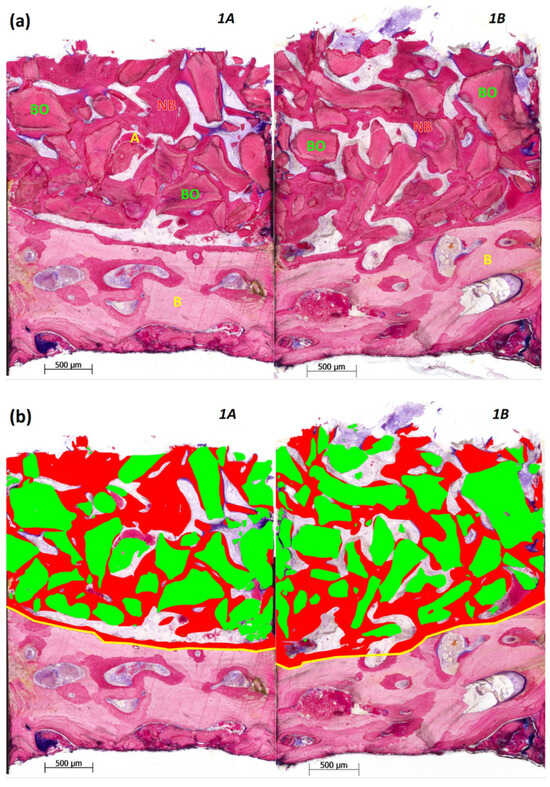
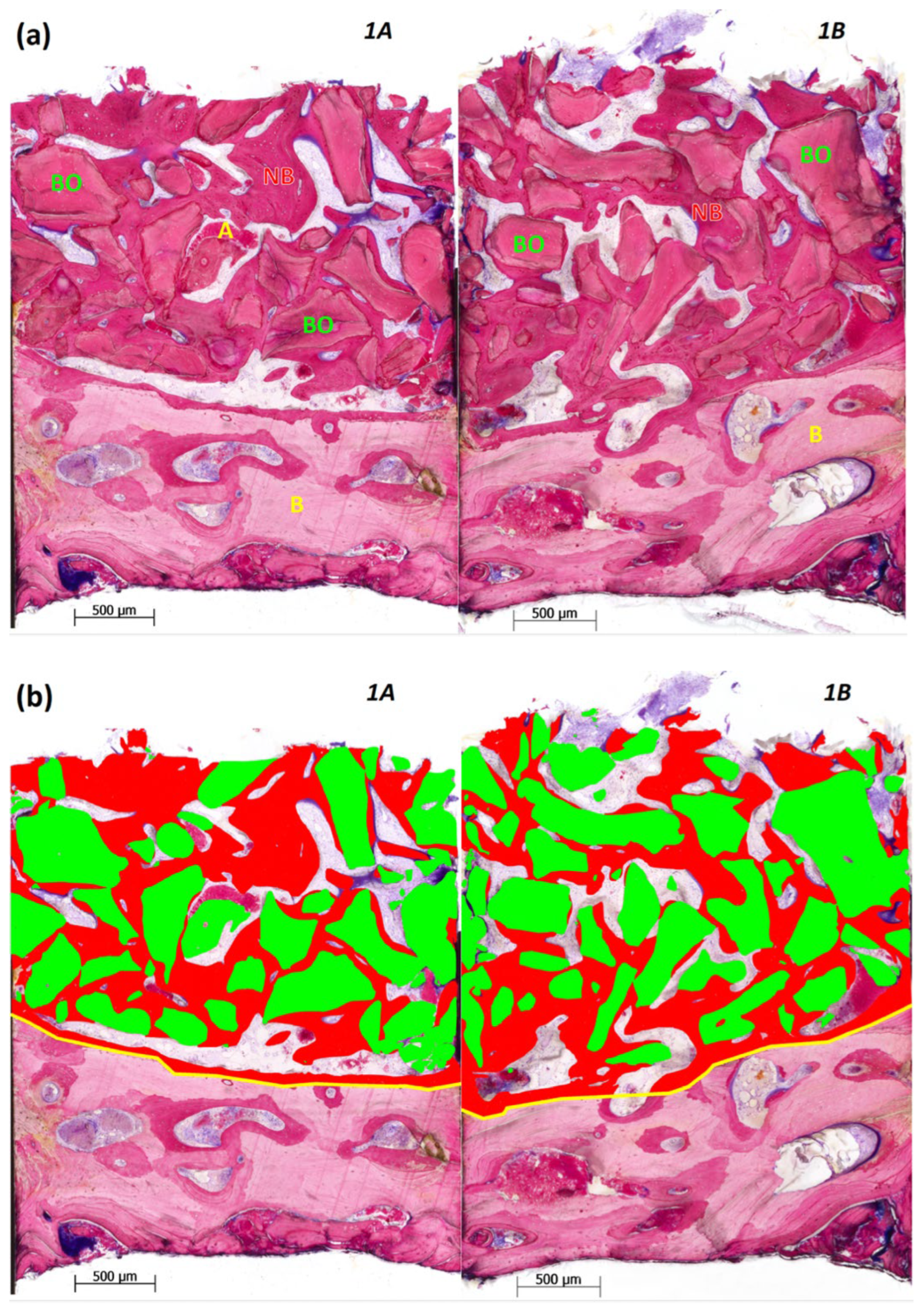
The bone biopsy was histologically processed in two sections (1A, 1B), measuring on average 3.55 × 2.75 mm2, and analyzed concerning its structural components (see Table 1). In the composition, a differentiation was made between new bone mineral and BioOss® particles, compromising the total mineralized fraction, as well as amorphous calcified substance (corresponding to bone dust compacted during trephination into the bone marrow) and connective tissue or bone marrow, respectively. The total mineralized phase content averaged 76.4%, specifically 78.4% in sample 1A and 74.5% in sample 1B. This consisted of almost balanced proportions of new bone mineral at 36.1% (37.6% in 1A and 34.7% in 1B) and BioOss® granules at 40.3% (40.8% in 1A and 39.8% in 1B). The second-highest fraction, representing connective tissue and bone marrow, respectively, accounted for 22.2% and was approximately 20.2% in section 1A and 24.2% in section 1B. Small quantities of an amorphous calcified substance between 1.3% in section 1A and 1.4% in section 1B were detected.
Table 1.
Structural composition of the bone biopsy, obtained for sections 1A and 1B, with indication of the average.
At a microscopic scale, in both sections, the BioOss® granules were embedded entirely in newly formed bone, with no signs of a foreign body reaction or inflammation (see Figure 7). A consistent distribution of the xenogeneic material and newly formed bone was observed. The latter extended entirely through the sections from the top of the residual or older bone to the outer margins, implying remodeling processes over the entire height of the augmentation with no sign of less maturation in the far side of the ridge.
Figure 7.
Overviews of sections 1A and 1B at a magnification of ×50. (a) The lower image segments in section 1A (on the left) and section 1B (on the right) demonstrate older or residual bone (B). The upper image segments depict BioOss® particles (BO) and newly formed bone (NB) with amorphous calcified material (A) in between. BioOss® particles (BO) and newly formed bone mineral (NB) appear in dark magenta, remaining older or residual bone (B) in magenta, and soft tissue in blue. In the surrounding area, well-vascularized loose connective tissue is located. (b) The yellow lines define the areas of interest, scaled to 100%. For histomorphometric purposes, BioOss® particles are marked in green and newly formed bone is marked in red.
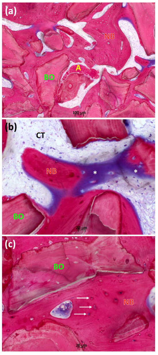
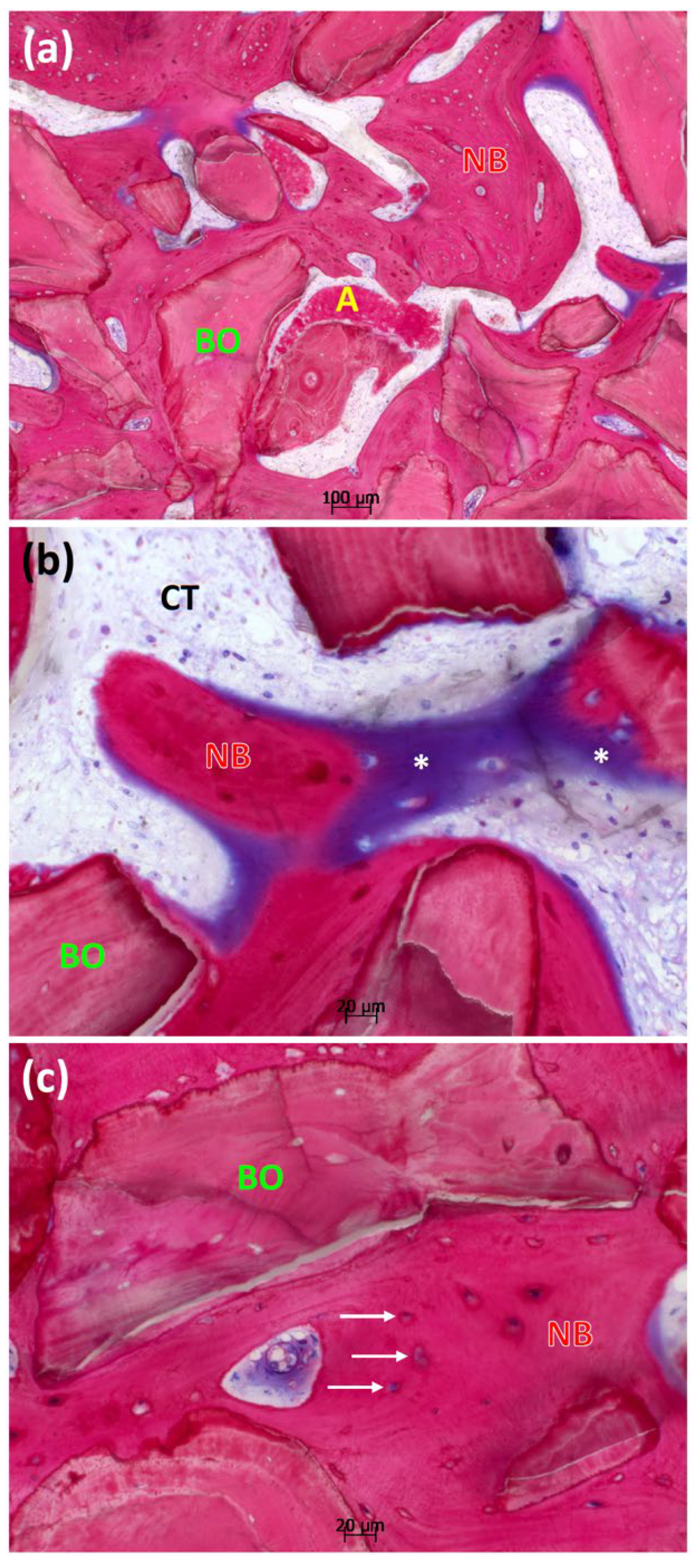
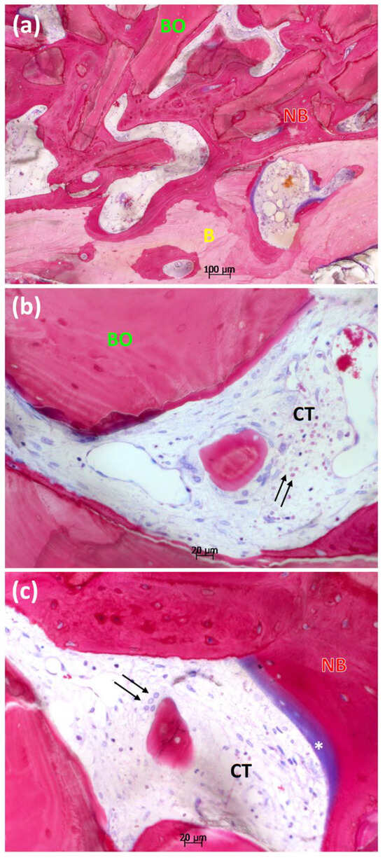
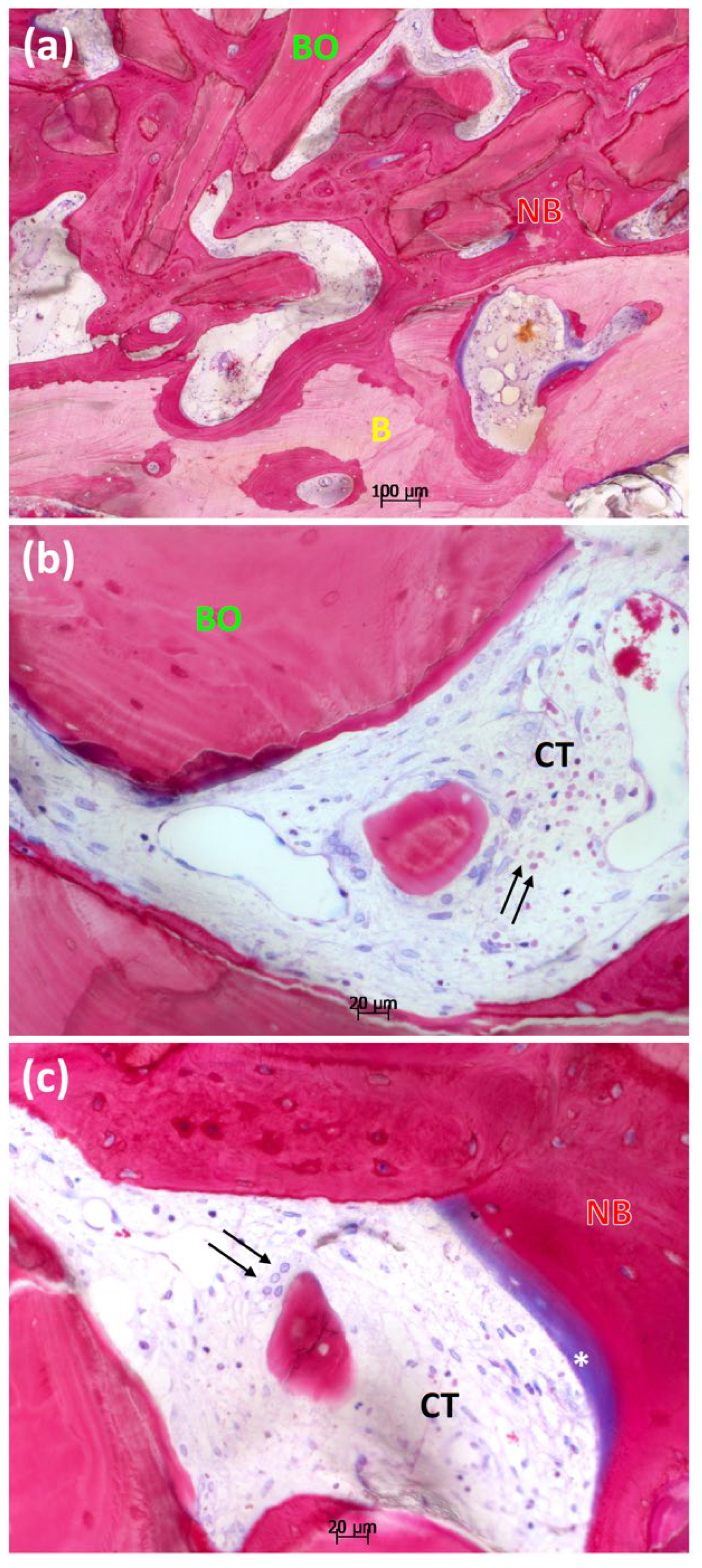
In detail (see Figure 8 and Figure 9), the new bone formed a structural network around the BioOss® particles and primarily enveloped these, completely integrating the xenogeneic material into the adjacent tissue. In section 1A, amorphous calcified material (see Figure 8) was observed in small quantities. BioOss® granules and new bone are surrounded by connective tissue. Younger, not yet mineralized bone spans the area of osteoid formation (see Figure 8) between the mineralized substance. Osteocytes lacunae (see Figure 8) appear within the newly formed, already mineralized bone. Within the caverns, the presence of osteocytes, which developed from osteoblasts, is expected, as they are walled off as star-shaped cells with long cell processes within the mineralized matrix during the formation of new bone. They are considered a hallmark of mature bone. In section 1B, the fundamental structural arrangement is comparable to section 1A. It appears that the newly formed bone has fused with the residual older bone (see Figure 9). Section 1B reveals a few multinucleated giant cells (see Figure 9), not recognizable in section 1A. The connective tissue is well vascularized and shows no signs of inflammation. Vessel formation can be observed as well as osteoid formation.
Figure 8.
Detailed excerpts of the overviews of section 1A at a magnification between ×50 and ×200. (a) BioOss® particles (BO) and newly formed bone (NB) with residuals of amorphous calcified material (A) are visible. (b) BioOss® particles (BO) are surrounded by newly formed bone (NB). The remaining regions are filled with well-vascularized connective tissue (CT). Osteoid formations (asterisk) are stained in purple. (c) Osteocyte lacunae (arrows) are formed in the new, mineralized bone.
Figure 9.
Detailed excerpts of the overviews of section 1B at a magnification between ×50 and ×200. (a) Newly formed bone (NB) appears to be closely interwoven with residual older bone (B), creating a network with incorporated BioOss® particles (BO). (b,c) Multinucleated giant cells (arrow) are visible. (c) Osteoid formation (asterisk) indicates the formation of new bone (NB).
4. Discussion
Implants should be used in a prosthetically optimized manner in order to avoid subsequent peri-implant, aesthetic, and functional problems and to facilitate oral hygiene. Particularly after prolonged tooth loss, bone atrophy can occur in the surgical area, requiring augmentation to enable adequate implant placement. The present case demonstrates that horizontal ridge augmentation with deproteinized bovine bone mineral (DBBM) combined with hyaluronic acid (HA) and polynucleotides (PN), stabilized by the umbrella-screw tent technique in addition to platelet-rich fibrin (PRF) as a vehicle, resulted in substantial bone formation and favorable clinical outcomes. The use of HA and PN in clinical procedures is uncomplicated and simple due to its formulation as a gel, without significantly increasing the effort required by the practitioner. The most striking aspect of this case, though, was the quantity and quality of the gained bone, particularly in terms of the high proportion of the newly formed bone within the augmented volume after six months, especially in more distant areas from the former ridge, and the maturity of the regenerated bone, accompanied by excellent graft integration and absence of inflammatory responses.
Typically, an average bone gain is used in studies to compare the outcomes of different surgical approaches. The success of operative bone augmentation procedures is difficult to quantify and compare across techniques because several factors affect outcomes, including the total augmented volume, the linear amount of augmented material, the materials used, the initial defect morphology, the targeted region, and the patient’s medical status, among others. Alone, the during- and post-operative complications can differ widely among techniques. To quantify the amount of regenerated bone, we favor the TPI classification [2] to enhance comparability across techniques, which here reached a TPI-h of 75%. Compared with previously published data, the observed horizontal bone gain of approximately 4.5 mm, accompanied by linear resorption of about 1.5 mm after 6 months, exceeds the mean outcomes reported for guided bone regeneration or conventional augmentation. The systematic review by Naenni et al. [22] documented a pooled bone gain of 3.30 mm ± 0.49 mm accompanied by an average graft resorption of 1.33 mm ± 0.45 mm over the same period. Similar results were reported in a meta-analysis by Elnayef et al. [23], which found horizontal bone gain of 3.61 mm ± 0.27 mm and resorption of 1.22 mm ± 0.28 mm with guided bone regeneration. While compounding deproteinized bovine bone mineral with hyaluronic acid and polynucleotides similar to the described procedure, Beretta et al. [24] observed a significantly higher bone gain with an improvement in the quality of the regenerated bone. Herein, the horizontal bone gain was 4.91 mm ± 0.88 mm, comparable to the present case. Although the relative bone gain alone cannot be taken as predictive of the success or effectiveness of a technique, it does provide a quantifiable reference for bone augmentation over defined timeframes, helping to track resorption rates and the onset of dimensional stability. Initially, smaller bone dimensions appear to offer better long-term prospects, often exhibiting higher total volume gains; however, this is offset by the excessive augmentation required [2,22]. Given the clinical condition in the present case, augmentation with a xenogeneic graft, combined with a hyaluronic acid and polynucleotide-based gel, did not experience severe resorption and provided sufficient bone for implant placement.
For the characterization of the regenerated bone, histomorphometric assessments are usually performed, such as quantifying the amount of new bone relative to the bone substitute and soft tissue, and counting inflammatory cells at a certain time point. This case report demonstrates successful augmentation with a significant new bone formation. With a total mineralized content of 76.4%, consisting of BioOss® particles at 40.3% and newly formed bone at 36.1%, the active turnover in terms of bone mass is remarkable. The graft’s integration into the newly formed bone extended across the entire sample, covering it like a mesh and fully embedding the xenogeneic particles. In a case report examining the osseointegration of an implant after sinus augmentation with DBBM after 4 years [25], the osteoconductive integration of BioOss® particles through embedding in newly formed bone correlates with the present histology, whereby the particulate bone substitute material is not completely remodeled or replaced by autogenous bone. Rather, it appears to serve as a long-term stable, volume-preserving scaffold due to its low resorption processes [25]. As a secondary finding, small amounts of an amorphous calcified substance are noticeable, also described in Ref. [26]. Although unclear in origin, it was conjectured as bone meal resulting from sample removal by trephination. The highest qualitative healing potential is expected in socket-preservation cases where all four walls are intact. It is recognized that the regenerative healing potential declines as the number of bone walls decreases, while bone augmentation becomes considerably more challenging. Despite the extreme resorption of the ridge in this case report, the rate of new bone formation is comparable to, or even higher than, values reported in studies of lateral ridge augmentation and alveolar ridge preservation using deproteinized bovine bone mineral alone. In this context, Heberer et al. [27] reported new bone formation rates of approximately 25%, while Carderopoli et al. [28] and Schlee et al. [29] reported similar rates of 26.3 ± 16.9% and 26.0 ± 1.35%, respectively. Serrano Méndez et al. [30] determined values of new bone formation of 35.3 ± 16.8% after alveolar ridge preservation using a deproteinized cancellous bovine bone xenograft. In contrast, the study by Kauffmann et al. [26] reveals significantly lower amounts of new bone formation (12.8%) with predominantly remaining BioOss® particles in cases with lateral ridge augmentation and reduced wall area, using deproteinized bovine bone mineral without any additions. Overall, the total mineralized phase content of 41.6% is about one-third below the fraction when using a xenogeneic substitute together with hyaluronic acid (67.5% with 37.2% newly formed bone) in the same study [26] and about one-third below the fraction in our case report when using a hyaluronic acid and polynucleotide-based gel (76.4% with 40.3% newly formed bone) after a 6-month healing period. A direct comparison of the two cases is difficult, simply due to the different augmentation techniques (namely, the umbrella-screw tent technique versus the membrane fixation technique with tacks). Nevertheless, the relative difference in the composition with or without using a hyaluronic acid product is particularly remarkable. Despite differences in operative conditions, we can only conjecture on the observed pronounced new bone formation and enhanced graft integration resulting from the combined application with hyaluronic acid and polynucleotides. The above case report is consistent with the case series by Beretta et al. [24], who used the same hyaluronic acid and polynucleotide-based gel, together with deproteinized bovine bone mineral, for bone augmentation. Their histomorphometric evaluation revealed patient-specific variations in the relative composition of the compositional elements. In a biopsy, the quantities of newly formed bone amounted to 41.2 ± 12.4%, while xenogeneic graft particles remained at 29.9 ± 11.8% [24]—a range consistent with our observations herein. A typical histologic structure with tightly embedded BioOss® granules, surrounded by newly formed bone, strikingly resembles the histologic sections shown above. Both histologically and radiologically, Beretta et al. [24] confirmed higher bone density, with increased new bone formation and graft material integration, and few to no clinical signs of inflammation when hyaluronic acid and polynucleotides were used. They concluded that polynucleotides enhance the remodeling potential of bone, promote wound healing, and reduce resorptive processes [24]—features that were also observed in the clinical case reported here.
When it comes to the regenerative potential of hyaluronic acid, beneficial effects have been demonstrated in cell proliferation, differentiation [8] and angiogenesis [31]. When evaluated in extraction sockets during ridge preservation, hyaluronic acid combined with xenogeneic bone substitutes resulted in a larger mineralized fraction, higher bone density, and reduced dimensional changes compared with the xenogeneic-only control group over 3 months [32]. A histological study showed similar results after 6 months, where the quantitative bone supply could be increased at the expense of xenogeneic residual particles in favor of new bone in lateral ridge augmentation when combining deproteinized bovine bone mineral with hyaluronic acid [26]. As mentioned above, Kauffmann et al. [26] also reported a positive effect on the quality of bone. For hyaluronic acid, Kloss et al. [33] investigated the effect of a non-crosslinked derivate in cases with a missing buccal wall and documented statistically significantly less shrinkage in horizontal and vertical dimensions. In a systematic review, Lorenzi et al. [34] found a statistically nonsignificant advantage for new bone formation if hyaluronic acid was added to the bone substitute. Platelet-rich fibrin was used primarily due to its advantageous handling properties, which result in a sticky consistency of the bovine bone mineral particles. Because it is derived directly from the patient’s blood, platelet-rich fibrin is popular for use in dental augmentation procedures [35], despite limited evidence to date supporting its regenerative benefits. A systematic review summarizes potential effects for platelet-rich fibrin, including its functions in tissue regeneration, reduced infection and less dimensional bone shrinkage [35]. Since only a few heterogeneous randomized controlled trials have been identified that meet the inclusion criteria, the risk of bias is exceptionally high.
Other studies also show promising effects when combining hyaluronic acid and polynucleotides. For gingival fibroblasts, the combination of polynucleotides and hyaluronic acid not only increased cell growth, proliferation, and migration but also induced a change in cell morphology of spheroids, linked to increased cell viability in vitro [12]. When used simultaneously, polynucleotides and hyaluronic acid appear to act synergistically. For instance, a much lower concentration of polynucleotides (64 µg/mL) in combination with hyaluronic acid (1 mg/mL) was required to achieve the same effect on cell growth of human fibroblasts as compared to the sole use of polynucleotides (320 µg/mL) [13], indicating that hyaluronic acid could be used to amplify the activity of polynucleotides in wound healing. At a non-cellular stage, polynucleotides appear to enhance tissue regenerative capacity by increasing the synthesis and expression of collagen types I and III, thereby facilitating and accelerating wound healing and reducing fibrotic scar formation [11]. A beneficial outcome was also observed in ulcerative lesions, where clinical signs of inflammation appeared reduced in association with polynucleotides [14]. In this case report, superficial wound healing may have benefited from the addition of hyaluronic acid and polynucleotides, proceeding regularly without delays or irregularities and showing no signs of atypical inflammation, infection or dehiscence. The remodeling processes of the osseous components were also clearly visible after 6 months and confirmed by histology.
In summary, several limitations must be acknowledged. This is a single-case report without a control group, and conclusions regarding efficacy must be cautious. The observed outcomes may also be influenced by patient-specific factors, surgical technique, and the use of platelet-rich fibrin, which was primarily applied for handling benefits but may also contribute to tissue regeneration. Histological evaluation was limited to a single biopsy site and may not fully represent the entire grafted area. Despite these limitations, the clinical and histological findings provide valuable preliminary evidence. The integration of polynucleotides and hyaluronic acid into xenogeneic grafting procedures appears biologically promising and may help overcome the known limitations of deproteinized bovine bone mineral alone. Future randomized controlled trials with larger patient cohorts and standardized protocols are warranted to evaluate the reproducibility, quantify the regenerative potential, and define the long-term stability of this biomaterial combination. It should be noted that the obtained values fall within the upper range described for DBBM without additives, but drawing direct conclusions about superiority is not possible. Furthermore, it is unclear whether PRF affected the results. So it cannot be concluded that the positive results are due to the addition of hyaluronic acid and polynucleotides. The results have to be interpreted with care. The patient, who herself is interested in science, was impressed and very satisfied with the result.
5. Conclusions
Although only a single case is highlighted here, its clinical and histological outcomes offer promising insights into the substantial potential for ongoing development in regenerative bone augmentation. This case report, although limited in terms of extrapolation power, is primarily intended to motivate further research efforts in this field, especially in view of the expanding range of techniques and available materials. Within the limitations, the present findings highlight three key aspects:
- The umbrella-screw tent technique provided predictable stability and space maintenance, enabling successful horizontal ridge augmentation.
- The combination of deproteinized bovine bone mineral with hyaluronic acid and polynucleotides as well as platelet-rich fibrin resulted in a high proportion of newly formed bone and excellent graft integration, exceeding values commonly reported for DBBM alone.
- These outcomes suggest a synergistic regenerative effect of PN and HA, warranting further controlled clinical studies to validate their role as biological enhancers in bone augmentation.
Author Contributions
Conceptualization, all authors; methodology, J.L., A.P. and M.S.; validation, A.P. and R.S.; formal analysis, J.L., F.R. and M.S.; investigation, J.L. and M.S.; resources, F.R. and M.S.; data curation, J.L. and M.S.; writing—original draft preparation, J.L., F.R. and M.S.; writing—review and editing, A.P. and R.S.; visualization, J.L. and M.S.; supervision, M.S.; project administration, J.L., F.R. and M.S.; funding acquisition, M.S. All authors have read and agreed to the published version of the manuscript.
Funding
The histology was paid by Geistlich Biomaterials, Vertriebsgesellschaft mbH, Baden-Baden, Germany. This research received no further external funding.
Institutional Review Board Statement
The study was approved by the Ethical Committee of Vilnius Regional Biomedical Research with the registration code “2025/7-1684-1133”, issued on 30 June 2025.
Informed Consent Statement
Written informed consent for publication was obtained from the patient.
Data Availability Statement
The data presented in this study are available on request from the corresponding author. The data are not publicly available due to privacy.
Acknowledgments
All authors declare that this publication is original research, not submitted for publication elsewhere. All authors and co-authors were involved in the work leading to the publication of this paper. No artificial intelligence was used in writing the manuscript. Geistlich Pharma supported the histologic preparation of the sample. The authors would like to thank Anette Lindner from the University of Freiburg for the histologic preparations, histomorphometric measurements and analysis.
Conflicts of Interest
The last author indicates an economic conflict of interest. He profits economically from the sale of the umbrella-screws. All other authors declare that they have no known competing financial interests or personal relationships that could have appeared to influence the work reported in this paper.
References
- Esposito, M.; Grusovin, M.G.; Felice, P.; Karatzopoulos, G.; Worthington, H.V.; Coulthard, P. Interventions for replacing missing teeth: Horizontal and vertical bone augmentation techniques for dental implant treatment. Cochrane Database Syst. Rev. 2009, 2009, Cd003607. [Google Scholar] [CrossRef] [Scilit]
- Stumpf, T.; Rathe, F.; Heumann, C.; Sader, R.; Schlee, M. Retrospective analysis of augmentation procedures with umbrella screws, a novel tenting technique: A consecutive case series in 279 patients. Quintessence Int. 2024, 55, 28–40. [Google Scholar] [PubMed]
- Khoury, F.; Hanser, T. Mandibular bone block harvesting from the retromolar region: A 10-year prospective clinical study. Int. J. Oral. Maxillofac. Implant. 2015, 30, 688–697. [Google Scholar] [CrossRef] [Scilit]
- Sanz-Sánchez, I.; Sanz-Martín, I.; Ortiz-Vigón, A.; Molina, A.; Sanz, M. Complications in bone-grafting procedures: Classification and management. Periodontol. 2000 2022, 88, 86–102. [Google Scholar] [CrossRef] [Scilit] [PubMed]
- Moussa, N.T.; Dym, H. Maxillofacial Bone Grafting Materials. Dent. Clin. N. Am. 2020, 64, 473–490. [Google Scholar] [CrossRef] [Scilit] [PubMed]
- De Risi, V.; Clementini, M.; Vittorini, G.; Mannocci, A.; De Sanctis, M. Alveolar ridge preservation techniques: A systematic review and meta-analysis of histological and histomorphometrical data. Clin. Oral. Implant. Res. 2015, 26, 50–68. [Google Scholar] [CrossRef] [Scilit]
- Vigetti, D.; Karousou, E.; Viola, M.; Deleonibus, S.; De Luca, G.; Passi, A. Hyaluronan: Biosynthesis and signaling. Biochim. Biophys. Acta 2014, 1840, 2452–2459. [Google Scholar] [CrossRef] [Scilit]
- Zhao, N.; Wang, X.; Qin, L.; Zhai, M.; Yuan, J.; Chen, J.; Li, D. Effect of hyaluronic acid in bone formation and its applications in dentistry. J. Biomed. Mater. Res. A 2016, 104, 1560–1569. [Google Scholar] [CrossRef] [Scilit]
- Frenkel, J.S. The role of hyaluronan in wound healing. Int. Wound J. 2014, 11, 159–163. [Google Scholar] [CrossRef] [Scilit]
- Chircov, C.; Grumezescu, A.M.; Bejenaru, L.E. Hyaluronic acid-based scaffolds for tissue engineering. Rom. J. Morphol. Embryol. 2018, 59, 71–76. [Google Scholar]
- Colangelo, M.T.; Govoni, P.; Belletti, S.; Squadrito, F.; Guizzardi, S.; Galli, C. Polynucleotide biogel enhances tissue repair, matrix deposition and organization. J. Biol. Regul. Homeost. Agents 2021, 35, 355–362. [Google Scholar] [CrossRef] [Scilit]
- Colangelo, M.T.; Vicedomini, M.; Belletti, S.; Govoni, P.; Guizzardi, S.; Galli, C. A Biomimetic Polynucleotides–Hyaluronic Acid Hydrogel Promotes the Growth of 3D Spheroid Cultures of Gingival Fibroblasts. Appl. Sci. 2023, 13, 743. [Google Scholar] [CrossRef] [Scilit]
- Guizzardi, S.; Uggeri, J.; Belletti, S.; Cattarini, G. Hyaluronate Increases Polynucleotides Effect on Human Cultured Fibroblasts. J. Cosmet. Dermatol. Sci. Appl. 2013, 3, 124–128. [Google Scholar] [CrossRef]
- De Caridi, G.; Massara, M.; Acri, I.; Zavettieri, S.; Grande, R.; Butrico, L.; de Franciscis, S.; Serra, R. Trophic effects of polynucleotides and hyaluronic acid in the healing of venous ulcers of the lower limbs: A clinical study. Int. Wound J. 2016, 13, 754–758. [Google Scholar] [CrossRef] [Scilit]
- Guizzardi, S.; Galli, C.; Govoni, P.; Boratto, R.; Cattarini, G.; Martini, D.; Belletti, S.; Scandroglio, R. Polydeoxyribonucleotide (PDRN) promotes human osteoblast proliferation: A new proposal for bone tissue repair. Life Sci. 2003, 73, 1973–1983. [Google Scholar] [CrossRef] [Scilit]
- Manfredini, M.; Poli, P.P.; Beretta, M.; Pellegrini, M.; Salina, F.E.; Maiorana, C. Polydeoxyribonucleotides Pre-Clinical Findings in Bone Healing: A Scoping Review. Dent. J. 2023, 11, 280. [Google Scholar] [CrossRef] [Scilit]
- Bitto, A.; Oteri, G.; Pisano, M.; Polito, F.; Irrera, N.; Minutoli, L.; Squadrito, F.; Altavilla, D. Adenosine receptor stimulation by polynucleotides (PDRN) reduces inflammation in experimental periodontitis. J. Clin. Periodontol. 2013, 40, 26–32. [Google Scholar] [CrossRef] [Scilit]
- Cawood, J.I.; Howell, R.A. A classification of the edentulous jaws. Int. J. Oral. Maxillofac. Surg. 1988, 17, 232–236. [Google Scholar] [CrossRef] [Scilit]
- Riley, D.S.; Barber, M.S.; Kienle, G.S.; Aronson, J.K.; von Schoen-Angerer, T.; Tugwell, P.; Kiene, H.; Helfand, M.; Altman, D.G.; Sox, H.; et al. CARE guidelines for case reports: Explanation and elaboration document. J. Clin. Epidemiol. 2017, 89, 218–235. [Google Scholar] [CrossRef] [Scilit] [PubMed]
- Choukroun, J.; Adda, F.; Schoeffler, C.; Vervelle, A. Une opportunite’ en paro-implantologie: Le PRF. Implantodontie 2001, 42, 55–62. [Google Scholar]
- Misch, C.E. Contemporary Implant Dentistry, 3rd ed.; Elsevier: Amsterdam, The Netherlands, 2007. [Google Scholar]
- Naenni, N.; Lim, H.C.; Papageorgiou, S.N.; Hämmerle, C.H.F. Efficacy of lateral bone augmentation prior to implant placement: A systematic review and meta-analysis. J. Clin. Periodontol. 2019, 46, 287–306. [Google Scholar] [CrossRef] [Scilit] [PubMed]
- Elnayef, B.; Porta, C.; Del Amo, F.S.-L.; Mordini, L.; Gargallo-Albiol, J.; Hernández-Alfaro, F. The Fate of Lateral Ridge Augmentation: A Systematic Review and Meta-Analysis. Int. J. Oral. Maxillofac. Implants 2018, 33, 622–635. [Google Scholar] [CrossRef] [Scilit] [PubMed]
- Beretta, M.; Manfredini, M.; Dellavia, C.P.B.; Pellegrini, G.; Maiorana, C.; Poli, P.P. Xenograft Combined with a Mixture of Polynucleotides and Hyaluronic Acid (PN-HA) for Horizontal Alveolar Bone Regeneration: Clinical and Histologic Assessments in a Case Series. Int. J. Periodontics Restor. Dent. 2025, 45, 209–219. [Google Scholar] [CrossRef] [Scilit]
- Scarano, A.; Pecora, G.; Piattelli, M.; Piattelli, A. Osseointegration in a sinus augmented with bovine porous bone mineral: Histological results in an implant retrieved 4 years after insertion. A case report. J. Periodontol. 2004, 75, 1161–1166. [Google Scholar] [CrossRef] [Scilit]
- Kauffmann, F.; Fickl, S.; Sculean, A.; Fischer, K.R.; Friedmann, A. Alveolar ridge alterations after lateral guided bone regeneration with and without hyaluronic acid: A prospective randomized trial with morphometric and histomorphometric evaluation. Quintessence Int. 2023, 54, 712–722. [Google Scholar]
- Heberer, S.; Al-Chawaf, B.; Jablonski, C.; Nelson, J.J.; Lage, H.; Nelson, K. Healing of ungrafted and grafted extraction sockets after 12 weeks: A prospective clinical study. Int. J. Oral. Maxillofac. Implant. 2011, 26, 385–392. [Google Scholar]
- Cardaropoli, G.; Araújo, M.; Hayacibara, R.; Sukekava, F.; Lindhe, J. Healing of extraction sockets and surgically produced-augmented and non-augmented-defects in the alveolar ridge. An experimental study in the dog. J. Clin. Periodontol. 2005, 32, 435–440. [Google Scholar] [CrossRef] [Scilit]
- Schlee, M.; Seitz, O.; Sader, R. Histologic Characterization of Human Extraction Sockets 3 Years After Grafting: A Case Report. Int. J. Periodont. Rest. Dent. 2015, 35, 380–386. [Google Scholar] [CrossRef] [Scilit]
- Serrano Méndez, C.A.; Lang, N.P.; Caneva, M.; Ramírez Lemus, G.; Mora Solano, G.; Botticelli, D. Comparison of allografts and xenografts used for alveolar ridge preservation. A clinical and histomorphometric RCT in humans. Clin. Implant. Dent. Relat. Res. 2017, 19, 608–615. [Google Scholar] [CrossRef] [Scilit]
- Kyyak, S.; Blatt, S.; Wiesmann, N.; Smeets, R.; Kaemmerer, P.W. Hyaluronic Acid with Bone Substitutes Enhance Angiogenesis In Vivo. Materials 2022, 15, 3839. [Google Scholar] [CrossRef] [Scilit]
- Lee, J.B.; Chu, S.; Ben Amara, H.; Song, H.Y.; Son, M.J.; Lee, J.; Kim, H.Y.; Koo, K.T.; Rhyu, I.C. Effects of hyaluronic acid and deproteinized bovine bone mineral with 10% collagen for ridge preservation in compromised extraction sockets. J. Periodontol. 2021, 92, 1564–1575. [Google Scholar] [CrossRef] [Scilit] [PubMed]
- Kloss, F.R.; Kau, T.; Heimes, D.; Kämmerer, P.W.; Kloss-Brandstätter, A. Enhanced alveolar ridge preservation with hyaluronic acid-enriched allografts: A comparative study of granular allografts with and without hyaluronic acid addition. Int. J. Implant. Dent. 2024, 10, 42. [Google Scholar] [CrossRef] [Scilit] [PubMed]
- Lorenzi, C.; Leggeri, A.; Cammarota, I.; Carosi, P.; Mazzetti, V.; Arcuri, C. Hyaluronic Acid in Bone Regeneration: Systematic Review and Meta-Analysis. Dent. J. 2024, 12, 263. [Google Scholar] [CrossRef] [Scilit]
- Miron, R.J.; Zucchelli, G.; Pikos, M.A.; Salama, M.; Lee, S.; Guillemette, V.; Fujioka-Kobayashi, M.; Bishara, M.; Zhang, Y.; Wang, H.L.; et al. Use of platelet-rich fibrin in regenerative dentistry: A systematic review. Clin. Oral. Investig. 2017, 21, 1913–1927. [Google Scholar] [CrossRef] [Scilit]

Disclaimer/Publisher’s Note: The statements, opinions and data contained in all publications are solely those of the individual author(s) and contributor(s) and not of MDPI and/or the editor(s). MDPI and/or the editor(s) disclaim responsibility for any injury to people or property resulting from any ideas, methods, instructions or products referred to in the content. |
© 2026 by the authors. Licensee MDPI, Basel, Switzerland. This article is an open access article distributed under the terms and conditions of the Creative Commons Attribution (CC BY) license.