


Featured Application
Understanding the molecular pharmacology of caffeine and direct activators of AMP-Activated Protein Kinase (AMPK) can lead to the development of novel treatment strategies for cancer and diseases associated with ageing.
Abstract
The methylxanthine caffeine is one of the most widely consumed neuroactive substances globally. Caffeine in the context of coffee consumption has been associated with diverse effects on human health and has been widely reviewed. Caffeine has drawn considerable interest due to its effects on cellular signalling, DNA damage sensitivity, Target of Rapamycin (TOR) activity, activation of AMP-activated Protein Kinase (AMPK), and extension of chronological lifespan in fission yeast and other model organisms. The cell cycle effects of caffeine have overshadowed its modulation of DNA damage and repair. Caffeine provides an interesting example of how scientific knowledge evolves and is influenced by trends within epochs. Despite recent advances in our understanding of how caffeine modulates cell cycle progression, these findings raise many new questions. These include its structural similarity to adenine and the mechanisms by which it affects DNA damage and repair, and its impact on TOR Complex 2 (TORC2) activity. Herein, I review our current knowledge on the cellular effects of caffeine and focus primarily on studies in fission yeast. It proposes that caffeine be considered a radiomimetic-like compound, outlining our current state of knowledge and future directions in applying its pharmacology in the prevention and treatment of disease.
1. Introduction
Radiomimetic definition: A substance or effect that produce effects like those of radiation. Specifically, it describes drugs that imitate the effects of radiation, such as certain chemotherapy agents like nitrogen mustards [1].
Caffeine (1,3,7-trimethylxanthine) is a methylxanthine contained in numerous beverages, including coffee, tea, cocoa and energy drinks. As such, caffeine is among the world’s most widely consumed stimulants [2]. While the literature is conflicting, the evidence suggests caffeine consumption is associated with positive health effects. Following ingestion, caffeine is widely distributed throughout the body, where it exerts multiple effects on cellular physiology. It is metabolised to the related methylxanthines theophylline, theobromine and paraxanthine through the liver [3]. Caffeine consumption is believed to impact the development and severity of diseases associated with ageing, such as Alzheimer’s Disease, neurodegeneration, type II diabetes and cancer. Recent studies suggest that coffee does exert beneficial effects, particularly in women and individuals with certain psychiatric disorders [2,4,5]. This is exemplified by its impact on Adenosine Monophosphate (AMP)-activated Protein Kinase (AMPK) and TORC1/2 activity [6,7,8]. It is crucial to note that caffeine, as a drug, differs substantially from coffee, which contains numerous other compounds that can positively affect human health [2,9]. Chemically, caffeine is classified as a methylxanthine, as are theobromine (3,7-dimethylxanthine) and theophylline (1,3-dimethylxanthine) found in chocolate and tea, respectively [10]. Caffeine functions as a plant defence against insect pests and can also suppress the growth of neighboring plants. Caffeine also suppresses microbial growth by activating several cellular signalling pathways [10,11,12,13].
Early studies in the 1960s and 70s identified caffeine as a DNA intercalating agent and inhibitor of DNA repair that exhibits mutagenic activity [14,15,16]. These early findings also suggested a protective effect of caffeine by enhancing apoptosis in cells following ultraviolet (UV)-induced DNA damage [17,18]. These studies identified caffeine as a genotoxin, but this observation appears to have been overshadowed by progress in our understanding of cell cycle regulation [7]. It remains unclear by what mechanisms caffeine induces DNA damage. Later studies showed that caffeine overrides DNA damage checkpoint signalling. When cells incur DNA damage, kinases such as Ataxia Telangiectasia Mutated (ATM) and ataxia telangiectasia and Rad3-related (ATR) activate downstream signalling to delay cell cycle progression to allow time for DNA repair. Compromised DNA damage repair is a major element in cancer cell development and tumorigenesis. It plays a crucial role in cancer treatment as it enhances cell death, particularly via apoptosis. Thus, this effect of caffeine aroused much interest due to its potential clinical relevance. For instance, synergistic effects between caffeine and other radiomimetic agents would widen their therapeutic index and reduce side-effect-related toxicity [19,20]. ATM and ATR are members of the phosphatidylinositol 3-kinase-related kinases (PIKKs) protein family. It was proposed that caffeine inhibits ATM-related kinases to override DNA damage checkpoint signalling [21]. This proved controversial, as caffeine was also shown to enhance ATM signalling in another model. Furthermore, related kinases like Target of Rapamycin Complex 1 (TORC1), DNA Protein Kinase (DNA-PK) and ATR are also potential targets of caffeine, precluding its use as a molecular tool to study ATM signalling [22]. In vitro assays may not reflect cellular conditions where caffeine-mediated interactions can be influenced by constraints such as substrate availability and binding affinity. The fission yeast Schizosaccharomyces pombe and the budding yeast Saccharomyces cerevisiae have proven to be powerful model systems for cell cycle, radiobiological and caffeine-related studies [23,24]. Investigations on ageing in both yeasts revealed that TORC1 is the target of caffeine [6,8,25]. This was later confirmed in related studies, suggesting caffeine advances mitosis by positively regulating Cdc2 activity through TORC1-dependent and -independent mechanisms [7]. Importantly, these studies have identified the AMP-activated Protein Kinase (AMPK) as an indirect target of caffeine. AMPK acts as a sensor of cellular energy states and environmental conditions, directly inhibiting TORC1 to integrate these states with metabolism and cell division. Additionally, AMPK is also involved in DNA damage repair [26]. Presently, caffeine is proposed to enhance DNA damage sensitivity to genotoxic or radiomimetic agents by advancing DNA replication and mitosis. However, caffeine also interacts physically with DNA and activates the DNA Damage Response (DDR) pathway. Furthermore, studies in fission yeast suggest it mediates its cell cycle and DNA damage response effects via AMPK.
2. Methylxanthines as Enhancers of DNA Damage Sensitivity
Caffeine and its metabolites theobromine and theophylline are classified as purine methylxanthines and are structurally related to adenine (Figure 1).
Figure 1.
Chemical structures of adenine, xanthine and methylxanthines. The N3 methyl group is required for the DNA-damage-sensitising effect of caffeine, theobromine, and theophylline. Paraxanthine does not enhance sensitivity to DNA damage. 8-bromocaffeine enhances sensitivity to ionising radiation. 8-bromocaffeine and 8-methoxycaffeine inhibit topoisomerase II activity. Xanthine (69-89-6), deoxyadenosine (958-09-8), hypoxanthine (68-94-0), adenine (73-24-5), caffeine (58-08-2), theophylline (58-55-9), paraxanthine (611-59-6), pentoxifylline (611-59-6), 8-bromocaffeine (10381-82-5). Structures were obtained from freely available sources.
This structural similarity likely underlies their biological effects, such as adenine metabolism and DNA repair. Studies have suggested that all three methylxanthines exert similar effects on the cell cycle, and the N3 methyl group is required for these activities. Thus, paraxanthine, which lacks the N3 methyl group, was reported not to mimic the effects of caffeine [1]. In addition, the other methyl groups clearly influence the physiological effects of the respective methylxanthines. However, theophylline has been shown to suppress the toxic effects of caffeine in fission yeast. Thus, their activities are not strictly identical. Yet most studies in this research field investigate the activity of caffeine [27]. The reported cellular effects of caffeine occurred at physiologically relevant doses of approximately 1 mM (200 µg/mL) [1]. Other studies have used higher doses of caffeine, but these would be toxic in a clinical setting [28,29]. Research in the 1960s and 70s identified caffeine as a DNA intercalating agent. Later studies confirmed that caffeine binds to DNA and alters its physical characteristics but does not intercalate with it [29,30,31,32,33]. Other studies demonstrated that caffeine interferes with UV-induced DNA damage repair and increases apoptosis in cells under these conditions [14]. Work in bacteria revealed that caffeine interferes with the repair of UV-induced DNA damage. Bacteria repair UV-induced DNA damage by a process known as photoreactivation. In this context, caffeine interferes with the removal of pyrimidine dimers and other forms of UV-induced DNA damage by DNA photolyase and the (A)BC excinuclease subunit UvrA. Caffeine induces the non-specific binding of UvrA, sequestering it away from sites of DNA damage. These investigators concluded that caffeine inhibits nucleotide excision repair (NER), albeit at relatively high doses of 10 mM. It thus remained unclear if the effects of caffeine on DNA damage repair could be applied clinically [34,35]. Additional studies revealed that caffeine also enhances the sensitivity of mammalian cell lines to alkylating agents [1,36]. Together, these studies suggested that caffeine interferes with some form of DNA damage repair linked to replication.
Cells defective in UV-induced DNA damage repair maintain viability using a number of pathways, collectively referred to as “post-replication repair” [37]. It was proposed that caffeine interferes with this pathway. Other studies, however, suggested that caffeine drives S phase to induce a synergistic effect with alkylating agents [16,38]. Studies in yeast have also proposed a genotoxic role for caffeine, as evidenced by the requirement for DNA repair enzymes in mediating resistance to caffeine. These studies strongly suggest that caffeine induces DNA damage that requires homologous recombination (HR) repair. Mutants that fail to express Rad3, DNA replication factor A subunit Ssb3, Rad51 and Rad54 all display sensitivity to caffeine [11,39]. The finding that Ssb3 is required for the repair of DNA damage during S phase further points to an effect of caffeine on genome stability during replication [40]. Additional studies in budding yeast propose that caffeine interferes with DNA damage repair by suppressing the expression of the Sae2 and Dna2 nucleases. Caffeine also inhibits Rad51-dependent HR by interfering with its binding to DNA strand breaks. This suggests that caffeine can enhance sensitivity to genotoxins by inhibiting DNA strand-break repair, independently of Rad3 inhibition [7,41,42,43,44]. Caffeine has also been reported to protect cells from reactive oxygen species (ROS)-induced cellular damage [21]. Paradoxically, it can also enhance sensitivity to hydrogen peroxide (H2O2) in mutants that fail to express Rad52, consistent with HR-dependent DNA damage [45]. As discussed below, the precise mechanisms by which caffeine enhances sensitivity to a particular drug must thus be interpreted with caution [7,46].
Derivatives of caffeine exhibiting differential structure–activity relationships have been synthesised. A modified derivative of caffeine, 8-bromocaffeine, has been reported to increase sensitivity to ionising radiation [47]. Caffeine, 8-bromocaffeine, 8-methylcaffeine and an 8-(substituted) aryloxycaffeine also inhibit topoisomerase II activity to varying degrees [48,49,50]. Like caffeine, resistance to camptothecin in fission yeast is mediated by genes involved in HR repair. Inhibition of topoisomerase II activity may partially account for the genotoxic effects of caffeine [51]. Together, these early studies suggested that caffeine could enhance sensitivity to DNA damage through three potential mechanisms. (i) Caffeine could itself induce DNA damage and repair mechanisms that interact synergistically with radiomimetic drugs; (ii) caffeine could inhibit specific forms of DNA damage caused by said drugs; and (iii) caffeine could drive S-phase progression, thus enhancing sensitivity to radiomimetic drugs by inducing lethal levels of DNA damage. The decades spanning the mid-1970s to the early 21st century led to the elucidation of cell cycle regulatory mechanisms. While these studies aided in deciphering the molecular pharmacology of caffeine, they also overshadowed the early work on this enigmatic compound [15,16,52,53,54,55,56,57,58].
3. Cell Cycle Regulation in Fission Yeast
3.1. Regulation of Cdc2 Controls the Timing of Mitosis in Fission Yeast
Fission yeast has proven to be a versatile model system for studying cell cycle regulation, responses to DNA damage and environmental stress, and consequently, deciphering how caffeine modulates these effects [54]. An in-depth understanding of fission yeast cell cycle regulation is required to elucidate the potential effects of caffeine on cell division. The key regulator of cell cycle regulation in fission yeast is the cyclin-dependent kinase (CDK) Cdc2 (Figure 2) [59,60].
Figure 2.
Regulation of cell cycle progression by the environmental stress (ESR) and DNA damage response (DDR) pathways in fission yeast. Caffeine modulates cellular signalling through activation of the Sty1-regulated ESR. Simultaneously, caffeine also activates Ssp1 through an unknown mechanism (top of figure). Ssp1 activates Ssp2 and partly accounts for the effect of caffeine on cell cycle progression via TORC1 (left-hand side of figure). TORC2 integrates cellular states with cell cycle regulation. The effect of caffeine on TORC2 remains to be investigated (right-hand side of figure). The major regulator of cell cycle progression is Cdc2, which the DDR negatively regulates. Caffeine modulates Cdc2 activity by enhancing Cdc25 stability (bottom of figure). Green lines indicate activating pathways. Red lines indicate inhibitory pathways. Progression through the cell cycle and mitosis is regulated by the cyclin-dependent kinase Cdc2. Cdc13 is a G2 cyclin required for Cdc2 activation. Cdc13 and Cdc25 undergo ubiquitin-dependent degradation at anaphase. The Wee1 kinase inhibits Cdc2 activity until it is activated by the Cdc25 phosphatase at mitosis. Cdc2 activity is downregulated at anaphase to permit cytokinesis via the Septation Initiation Network (SIN) (bottom left). The ESR regulates cell cycle progression via Ssp1 and Sty1 (Top left and top right, respectively). Nutrient deprivation and caffeine activate Sty1, leading to Srk1 stabilisation and accumulation. Srk1 phosphorylates Cdc25. Cdc25 accumulates in the cytoplasm in a Rad24 (14-3-3)-dependent manner, preventing Cdc2 activation. Following glucose withdrawal, Wee1 also contributes to Cdc2 inhibition and transient G2 arrest. Ssp1 facilitates the resumption of cell division by inhibiting Srk1, resulting in Cdc25 dephosphorylation and nuclear localisation. Ssp1 and Ppk34 activate Ssp2 (AMPK) in response to environmental and nutrient stress. Ssp2 activation advances mitosis by inhibiting TORC1. TORC1 inhibition leads to PP2A inhibition and activation of Cdc2 via the Greatwall/Ppk18-endosulfine pathway. TORC2 (centre right) also regulates Cdc2 activity and is required for cell cycle re-entry following activation of DNA damage checkpoint signalling. TORC2 may regulate Cdc25 stability by regulating the stability of the E3 ligase Pub1 in a Gsk3-dependent manner. TORC1 and TORC2 coordinate nutrient import with cellular metabolism and cell cycle progression. Direct inhibition of TORC1 by Rapamycin or Torin1 also advances mitosis. Torin1 differs from caffeine and Rapamycin by inhibiting the activity of both TORC1 and TORC2. Various agents directly or indirectly induce DNA damage. DNA damage is detected by the Rad9-Rad1-Hus1 (911) complex. This leads to the recruitment and activation of Rad3 (ATM). Rad3 phosphorylates Cds1 and Chk1 during S phase and G2, respectively. Cds1 and Chk1, in turn, phosphorylate Cdc25 to inhibit its activity and relocation to the cytoplasm. Additionally, non-phosphorylated Cdc25 can be a target for ubiquitin-dependent degradation. Caffeine suppresses DNA damage-induced Cdc25 degradation. Ssp2 has been implicated in the DDR. Dss1 is a proteasome assembly chaperone that recruits Cdc25 to double-strand breaks and mediates DNA repair. Cut8 tethers the proteasome to the nucleus and, together with Rhp6, is involved in DNA double-strand break repair. Cut8 mediates resistance to caffeine, and Rhp6 is required for caffeine-induced mitosis. Rad51, Rad54 and Ssb3 mediate resistance to caffeine and facilitate HR-directed DNA damage repair (IR: ionising radiation). The figure was prepared by the author using MS PowerPoint. See text for further details and references.
In G1 and S-phase, Cdc2 complexes with the cyclins Cig1 and Cig2 and these complexes are negatively regulated by Rum1. In G2 and mitosis, Cdc13 is the major cyclin driving cell cycle progression. The Cdc25 phosphatase and Wee1 kinase regulate the phosphorylation of tyrosine residue 15 (Tyr15), which in turn regulates Cdc2 activity. Wee1 maintains Cdc2 Tyr15 phosphorylation until mitosis. At mitosis, Pom1-mediated inhibition of the Cdr1 and Cdr2 kinases is relieved, facilitating Wee1 inhibition. Wee1 stability is regulated by the Pof1 and Pof3 F-box proteins, which target it for ubiquitin-dependent degradation [60,61,62]. Cdc25 levels rise throughout interphase, but its activity is negatively regulated by phosphorylation of serine and threonine residues. Cdc25 negative regulation induces binding to the 14-3-3-related chaperone Rad24 and stockpiling within the cytoplasm. The cytoplasmic sequestration of Cdc25 appears to be the major regulatory mechanism in fission yeast. Additionally, Cdc25 is degraded within the nucleus by the HECT-type E3 ligase Pub1 [63]. Cdc25 is further activated by Cdc2-mediated phosphorylation to reach full activity at mitosis. Following mitosis, Cdc2 and Cdc25 activity is suppressed to allow activation of the Septation Initiation Network (SIN) and cytokinesis. The Clp1 phosphatase enhances Cdc25 ubiquitin-dependent degradation [54,64].
3.2. Role of TORC1 in the Fission Yeast Cell Cycle
TORC1 regulates the timing of cell division in response to nutrient availability. Additionally, TORC1 and TORC2 integrate environmental conditions with cell division and the execution of cell mating programs [65]. Cells delay cell cycle progression in response to nutrient availability and environmental stress conditions. This delay permits the expression of stress response genes and is required for viability. For instance, glucose limitation induces a G2-phase arrest that is dependent on Wee1, and wee1 mutants lose viability under these conditions [66]. Exposure to potassium chloride (KCl) similarly induces a temporary G2-phase delay. Following exposure to environmental or nutrient stress, several pathways interact to first delay and then advance cell cycle progression in fission yeast [67,68,69]. Firstly, these conditions activate the stress-activated protein kinase (SAPK) Sty1, a homologue of mammalian p38 [70]. Sty1 induces Srk1 activation, which phosphorylates Cdc25. Phosphorylated Cdc25 is excluded from the nucleus and stockpiled within the cytoplasm to induce cell cycle arrest [71]. Exposure to 0.6M KCl is sufficient to compensate for deregulated DNA damage signalling, indicating a robust delay of cell cycle progression (See below) (Figure 2) [72]. Sty1-mediated cell cycle arrest is subsequently suppressed by the calcium/calmodulin (CaMKK) kinase Ssp1, which inhibits Srk1 to reactivate Cdc25 [73]. Simultaneously, Ssp1 activates the AMPK homologue Ssp2. Ssp2 inhibits TORC1 activity, leading to the activation of the Greatwall-related kinases Ppk18 and Cek1, which in turn activate the endosulfine Igo1. Igo1 inhibits PP2A phosphatase activity, which regulates Cdc25 and Wee1, permitting Cdc2 activation and progression into mitosis [60,74] p. 2. The cyclical inhibition of TORC1 activity mediates the downstream inhibition of the PP2A phosphatase, permitting the subsequent activation of Cdc25 at mitosis. Nitrogen withdrawal and changes in amino acid availability similarly inhibit TORC1 activity to advance mitosis in a manner dependent on Cdc25 and Wee1 [75].
3.3. Role of TORC2 in the Fission Yeast Cell Cycle
TORC2 plays important roles in the maintenance of genomic stability, ESR regulation and cell cycle re-entry. TORC2 activity is required for cell cycle re-entry following DNA damage. TORC2 (tor1) mutants activate DNA damage checkpoint signalling normally, but subsequently fail to dephosphorylate Cdc2 [76,77,78]. TORC2 can influence Cdc25 activity by modulating its stability in a Gsk3 and Pub1-dependent manner (Figure 2) [63,64,79]. TORC1 and TORC2 thus integrate the Environmental Stress Response (ESR) with cell cycle progression [80].
3.4. DNA Damage Checkpoint Signalling in Fission Yeast
Cells must also arrest cell cycle progression in response to DNA damage. Cells continuously sustain DNA damage resulting from endogenous metabolic processes or external factors, including ionising and ultraviolet radiation, cigarette smoke or genotoxic drugs, e.g., bleomycin [19]. This is necessary to prevent errors in DNA replication that can introduce mutations in S-phase or chromosome missegregation during mitosis. Delays in cell cycle progression also permit time for the repair of damaged DNA. Cells unable to arrest replication or delay mitosis following DNA damage rapidly lose viability. These delays in cell cycle progression can occur in G1, S or G2 phases and are referred to as DNA damage checkpoints. The activation of these checkpoints occurs via the inhibition of Cdc2. Conversely, cells that activate DNA damage checkpoints but fail to repair DNA damage also lose viability [81]. Together, these pathways, which include cell cycle arrest, DNA damage repair and cell cycle re-entry, are referred to as the DNA Damage Response (DDR). In fission yeast, the ATM and ATR-related PIKKs Rad3 and Tel1 regulate the DNA damage response. Rad3, with other DNA damage-sensing and transducer proteins such as Crb2, Hus1, Mrc1, Nbs1, Rad1, and Rad9, activates the Cds1 and Chk1 kinases in S and G2 phases, respectively. A G1-specific DNA damage checkpoint has also been reported in fission yeast but acts independently of Rad3 [82]. Cds1 is activated in response to stalled replication forks, nucleotide depletion or S-phase-specific DNA damage. Cds1 activity involves more than DNA damage checkpoint activation, as cds1 mutants exposed to hydroxyurea (HU) lose viability despite the presence of Chk1. Thus, Cds1 also regulates gene expression and enables the firing of stalled replication forks [83,84]. Cds1 phosphorylates Cdc25 to inhibit its activity and mediate its nuclear export. Additionally, Cds1 activates Wee1 or the related S-phase kinase Mik1 to phosphorylate and inactivate Cdc2 [85,86,87].
3.5. Redundant Checkpoint Signalling Mechanisms in Fission Yeast
Additional redundant DNA damage checkpoint mechanisms have been reported in fission yeast. Cdc25(9A)-GFP and Cdc25(12A)-GFP mutants lack the 9 or 12 serine and threonine residues usually phosphorylated by Cds1, Chk1 and Srk1 sites and are degraded following exposure to HU but not phleomycin [88,89]. Thus, clearance of nuclear Cdc25 is important for DNA damage checkpoint induction, particularly during S phase [7,54,89]. Later studies identified the Dss1 proteasome assembly chaperone as a further regulator of DNA damage checkpoint signalling in fission yeast. Dss1 likely exerts this activity by regulating nuclear Cdc25 activity levels. Dss1 recruits Cdc25, Rad24, RNA export factor Rae1 and recombination protein Rhp51p to DNA double-strand breaks. Dss1 thus links DNA damage-induced checkpoint activation to DNA repair and 26S proteasomal activity [90,91]. Dss1 may be clinically relevant, as it has been shown to physically interact with breast cancer susceptibility gene product BRCA2 and induce p53 degradation in human cancer cells [92,93]. The Hsp9 heat shock protein negatively regulates Cdc25 to maintain DNA damage checkpoint activity. Hsp9 has been reported to mediate stress-induced inhibition of Cdc25, delaying progression through mitosis [94]. These studies demonstrated that inhibition of phosphatase activity, cytoplasmic sequestration, DNA binding and ubiquitin-dependent degradation collectively inhibit Cdc25 activity during DDR and ESR activation.
4. Effects of Caffeine on Cell Cycle Regulation and DNA Damage Sensitivity in Fission Yeast
4.1. Caffeine Overrides DNA Damage Checkpoint Signalling Independently of Rad3 in Fission Yeast
The molecular characterisation of the cell cycle, DDR, and later ESR pathway interactions permitted further elucidation of caffeine’s molecular pharmacology [54,70]. Early studies in fission yeast, and mammalian cells, demonstrated that caffeine overrides DNA damage checkpoint signalling [19,81,95]. Caffeine was reported to inhibit Rad3 and ATM/ATR activity in vitro [95,96,97]. Additionally, caffeine was shown to activate the Sty1-regulated ESR, which is coupled to cell cycle regulation via TOR signalling [7,39,54,72,89]. While Rad3 and its homologues were initially reported to be targets of caffeine, this view has always been controversial. Firstly, caffeine inhibits several members of the PIKK-like kinase family, making it unsuitable for in vivo studies on their kinase activity (Figure 3) [98].
Figure 3.
Functional domains of PIKK-related kinases and their relative amino acid lengths. mTOR (mechanistic Target of Rapamycin) regulates cellular responses in response to nutrient availability. ATM (Ataxia-telangiectasia mutated), ATR (Ataxia-telangiectasia and Rad3-related), and DNA-PKCS (DNA-dependent protein kinase catalytic subunit) regulate cellular responses to DNA damage. SMG1 (Suppressor of Morphogenesis in Genitalia) regulates nonsense-mediated mRNA decay. TRRAP (Transformation/transcription domain-associated protein) is a transcription factor co-activator involved in DNA double-strand break recognition and repair. The FRB domain is required for rapamycin binding. The figure was prepared by the author using MS PowerPoint.
Furthermore, while caffeine can inhibit Rad3 and ATM in vitro, there is conflicting evidence for this activity in vivo. In fission yeast, in vitro inhibition of Rad3 correlated poorly with its effect on DNA damage checkpoint signalling. In our studies, caffeine failed to inhibit Cds1 and Chk1 phosphorylation in marked contrast to the deletion of rad3 in fission yeast. As reported by Cortez, caffeine-induced Chk1 phosphorylation occurs independently of ATM in an ATR-dependent manner. These findings confirmed that caffeine does not mimic rad3 gene deletion in fission yeast. Although Tel1 is the fission yeast homologue of ATM, the main regulator of both S phase and G2 DNA damage checkpoints is Rad3 (Figure 2) [89,95,98]. Interestingly, we also observed that Cds1 and Rad3 regulate Cdc25 expression at both the mRNA and protein levels. Furthermore, exposure to caffeine, like KCl, induced Cdc25 accumulation in fission yeast. These observations suggested that caffeine might regulate Cdc25 activity via the ESR independently of Rad3 [72,89,99]. The ability of caffeine to induce Cdc2 dephosphorylation, independently of Rad3, could thus be linked to the inhibition of TORC1 and downstream PP2A activation or TORC2 activation [54,74,77].
4.2. Caffeine Overrides DNA Checkpoint Signalling by Inducing Cdc25 Accumulation
We hypothesised that caffeine might override DNA damage checkpoint signalling independently of Rad3 inhibition by inducing Cdc25 accumulation. Activation of the ESR in fission yeast induces transient cell cycle arrest through Srk1-dependent Cdc25 phosphorylation (Figure 2) [67,73]. In our studies, caffeine mimicked the effects of KCl on cell cycle progression in fission yeast. Exposure to 10 mM caffeine induced transient Sty1- and Srk1-dependent cell cycle arrest, followed by rapid progression through mitosis. Thus, both caffeine and KCl can compensate for the loss of checkpoint signalling in fission yeast rad3∆ and rad1∆ mutants respectively [72,89]. Our initial studies demonstrated that caffeine induces the nuclear accumulation of Cdc25. Caffeine was particularly effective at stabilising Cdc25 in mik1∆ mutants. This finding further suggests that negative regulation of Cdc25 stability is particularly important during S phase. Furthermore, caffeine-induced Cdc2 dephosphorylation was dependent on Cdc25 expression. Caffeine suppressed cell cycle progression when cdc2-3w cdc25∆ mutants were exposed to HU [89,99]. Additional studies demonstrated that caffeine suppressed Wee1 expression in the presence of HU (An inhibitor of ribonucleotide synthesis and DNA replication) or phleomycin (An analogue of bleomycin that induces DNA double-strand breaks via production of oxidative moieties). In these studies, Wee1 stability was dependent on the 14-3-3 Rad24 protein [99]. The stability of both Cdc25 and Wee1 is regulated by phosphorylation [59,62,88]. We concluded that caffeine induces mitosis in fission yeast by modulating Cdc25 and Wee1 activity independently of Rad3.
4.3. Caffeine Drives Mitosis by Inhibiting TORC1 Activity
Later investigators identified TORC1 as the target of caffeine in budding and fission yeast [6,8]. We noted that the effect of caffeine on cell cycle progression resembled that of cells exposed to the TOR inhibitors rapamycin and Torin1 (Figure 2) [99]. It remained unclear if caffeine directly inhibits TORC1 or indirectly through its activation of the fission yeast AMPK catalytic subunit Ssp2. In fission yeast, Ssp1 is required for cell cycle re-entry following activation of the ESR, where it suppresses Srk1-mediated Cdc25 inhibition [67,68,73]. Similarly, activation of the ESR induces Ssp1-dependent Ssp2 activation. Ssp2 inhibits TORC1 to advance cells into mitosis via the Ppk18/Cek1-Igo1-PP2A pathway [69,100]. Phosphorylated Ssp2 also migrates to the nucleus, where it regulates gene expression and DNA damage repair [26,101,102,103,104]. The ability of caffeine to advance mitosis was partially abolished in ssp1∆ and ssp2∆ mutants. In the presence of phleomycin, caffeine failed to advance mitosis in these mutants, in contrast to the pan-TOR inhibitor Torin1. The ability of caffeine to advance mitosis was also inhibited in igo1∆ mutants. These studies suggested that caffeine indirectly inhibits TORC1 to advance mitosis [7]. Caffeine induced DNA replication despite its inability to advance mitosis in ssp2∆ mutants. Additionally, caffeine can advance mitosis in ssp2∆ mutants. Exposure to caffeine induced Ssp2 phosphorylation in an Ssp1-dependent manner. Caffeine also suppressed Sck2 expression, which negatively regulates Ppk18 in a TORC1-dependent manner. At present, we propose that caffeine advances mitosis in fission yeast through indirect inhibition of TORC1 by the AMPK homologue Ssp2 and Cdc25 stabilisation. Deletion of ssp2 blocked the effect of caffeine on mitosis but not DNA replication in fission yeast cells previously exposed to phleomycin (Figure 2) [7].
4.4. Potential Roles for TORC2 in Mediating the Cell Cycle Effects of Caffeine
In-depth studies on the effect of caffeine on TORC2 signalling have not been reported. TORC2 plays a key role in mediating the ESR, cell cycle and responses to DNA damage (Figure 2) [77,105,106]. Activation of the ESR activates TORC2, and tor1∆ mutants were sensitive to caffeine in our studies [7,99]. TORC2 activity is required for cell cycle re-entry following activation of the DDR in fission yeast. Caffeine-induced TORC2 activation could thus drive cells into mitosis inappropriately [7,105]. TORC2 can regulate cell cycle progression by regulating the stability of the HECT-type E3 ligase Pub1. In fission yeast, Pub1 targets Cdc25 for ubiquitin-dependent degradation. Active TORC2 inhibits Gsk3 in a Gad8-dependent manner (Figure 2). Activation of TORC2 by caffeine can thus induce Cdc25 accumulation by suppressing Gsk3 and Pub1 activity [7,63,64,79]. TORC2 activation by caffeine may also contribute to its DNA sensitising effects by modulating chromatin organisation and DNA repair pathways regulated by this complex [76,77,105]. Caffeine can advance mitosis in previously untreated ssp2∆ mutants and suppresses Cdc2 phosphorylation in the presence of genotoxins.
4.5. Uncoupling of Cell Cycle and HR Effects of Caffeine on DNA Damage Sensitivity
Caffeine clearly overrides DNA damage checkpoint signalling and has been shown to interfere with DNA damage repair, particularly HR. It has been proposed that caffeine enhances DNA damage sensitivity independently of its cell cycle effects [44,46,95]. Our studies suggest that caffeine can enhance sensitivity to phleomycin in stationary-phase fission yeast cells. Caffeine also enhanced the sensitivity of rhp6∆ mutants to DNA damage, without advancing them through mitosis. We have demonstrated that caffeine activates AMPK in fission yeast in an Ssp1-dependent manner. AMPK has been identified as a regulator of the DDR and has been proposed to regulate DNA damage repair [102,103]. In contrast to caffeine, exposure to phleomycin did not increase AMPK phosphorylation. We noted that the AMPK beta-subunit, Amk2, is required for cell cycle re-entry in the presence of caffeine. Caffeine also enhances the sensitivity of amk2∆ mutants to KCl. As Ssp2 is required for caffeine-induced cell cycle progression, but not resistance, these observations suggest an Amk2-specific role in DNA damage repair [7]. Glucose deprivation similarly induces DNA damage and activates the DDR and AMPK to effect repair [66]. We also noted that AMPK mediates the resistance of fission yeast cells co-exposed to HU and caffeine. Furthermore, AMPK, unlike Ssp1, was required for resistance to phleomycin following prolonged exposure in the presence of caffeine [7].
Caffeine’s effects on DNA repair may mask its effects on DNA damage sensitivity [7,14,20]. For instance, the antioxidative effects of caffeine may counteract phleomycin-generated ROS. Caffeine-induced AMPK activation may also increase DNA repair efficacy when combined with other genotoxic agents [39,107]. Our studies similarly suggest a paradoxical effect of caffeine on DNA damage sensitivity in fission yeast. Firstly, caffeine induces DNA damage and activates AMPK, which in turn regulates DNA damage repair. However, caffeine-induced activation of AMPK advances mitosis, as do its effects on Cdc25 and Wee1 stability in the presence of genotoxins. This activity appears to be particularly important for enhancing sensitivity to HU. Thus, in wild-type fission yeast cells, caffeine overrides DNA damage checkpoint signalling in an AMPK-dependent manner but enhances sensitivity to genotoxins independently of this kinase. Interestingly, ssp2∆ and amk2∆ mutants show enhanced sensitivity to HU in combination with caffeine relative to wild-type cells. The enhanced sensitivity to phleomycin is further enhanced by co-exposure to caffeine. The DDR pathway also regulates caffeine tolerance, suggesting that caffeine is itself genotoxic and HR is required for tolerance in fission yeast. Caffeine-induced AMPK activation thus increases DNA damage sensitivity by advancing mitosis, but “masks” it by activating AMPK-dependent DNA damage repair mechanisms. This paradoxical effect of caffeine on DNA damage sensitivity should be considered when investigating its synergistic effects with other genotoxins, e.g., inhibitors of replication versus DNA intercalating agents [7,39,89,99,108].
It has previously been proposed that the ability of caffeine to enhance DNA damage sensitivity, is uncoupled from its cell cycle effects. Caffeine-mediated inhibition of HR also induces synergy with other genotoxins [44]. In fission yeast, caffeine tolerance clearly requires DDR-regulated HR-mediated DNA damage repair. Exposure to caffeine thus induces DNA damage, which requires HR for repair, as demonstrated by the sensitivity of HR mutants [39]. While HR is required for caffeine tolerance, the methylxanthine also appears to suppress the activity of this DNA damage repair pathway. In budding yeast, caffeine inhibits HR by suppressing Dna2p and Sae2p expression. These nucleases are required for 5′-3′ end resection during HR. In this study, caffeine indirectly suppressed HR by inducing the proteasomal degradation of Dna2p and Sae2p [41] (Table 1).
Table 1.
Fission yeast gene products and their homologues in budding yeast and humans. These genes regulate cell cycle progression, DNA damage and repair, and environmental stress responses. Further details are included in the text. Cell cycle genes are indicated in green, DDR genes in red and ESR genes in black.
Caffeine can thus enhance sensitivity to DNA damage by suppressing the HR-mediated repair of double-strand breaks [41,42,44]. Caffeine can also increase sensitivity to DNA damage by inhibiting topoisomerase activity. However, caffeine has been shown to suppress the effects of topoisomerase inhibitors, likely by direct binding to limit their availability. As mentioned above, caffeine’s ability to enhance DNA damage sensitivity is context-dependent. For instance, caffeine can also inhibit the removal of UV-induced DNA adducts, increasing the level of keratinocyte apoptosis. The ability of caffeine to enhance sensitivity to genotoxins can occur via various mechanisms, which in turn are influenced by the mechanisms that underlie this damage [1,14,21,49,108,109,110].
Together, these studies suggest that caffeine enhances DNA damage sensitivity by cell cycle-dependent and independent mechanisms. Caffeine thus drives mitosis via AMPK, but this may mask its enhancement of DNA damage sensitivity. Caffeine likely interferes with DNA repair of replication-induced damage, leading to double-strand breaks that require HR-mediated repair. However, caffeine suppresses HR, resulting in the accumulation of DNA damage and hyperactivation of the DDR. Caffeine-induced Cdc25 accumulation may also affect DNA fidelity, especially during S-phase. In the presence of other genotoxins, caffeine enhances sensitivity to DNA damage by advancing mitosis and inhibiting DNA damage repair. Caffeine-activated DNA damage repair mechanisms may interfere with pathways that mediate resistance to other genotoxins, producing a synergistic effect. Caffeine can thus enhance DNA damage sensitivity independently of DNA damage checkpoint override. The precise mechanism by which caffeine enhances DNA damage sensitivity may also depend on the molecular characteristics of the interacting genotoxin.
4.6. Caffeine, AMPK, DNA Damage and Chronological Lifespan
Glucose deprivation in fission yeast induces transient Rad3 activation and a Wee1-dependent delay in G2. This cell cycle delay is required for glucose-mediated CLS extension. The DDR also plays a key role in maintaining the viability of stationary-phase and quiescent fission yeast cells [76,111,112]. Additionally, exposure of fission yeast cells to caffeine similarly induces a transient delay in cell cycle progression. Cell cycle progression induced by caffeine is dependent on Cdc25 expression. Caffeine-induced Sty1 activation likely contributes to Cdc25 inhibition. In fission yeast, both caffeine and KCl can suppress the sensitivity to DNA damage of DNA damage checkpoint mutants in a Sty1-dependent manner (Figure 2) [7,72]. The role of Wee1 in mediating the cell cycle effects of caffeine remains unclear. Activation of the DDR is clearly required for resistance to caffeine. This suggests that Wee1 does play some role in fission yeast cells in the presence of caffeine [39]. Co-exposure to caffeine and genotoxins suppresses Wee1 expression. As a result of the simultaneous stabilisation of Cdc25 and Cdc2 dephosphorylation, however, wee1 deletion does not appear to increase sensitivity to caffeine combined with HU relative to wild-type cells. This is partly due to the requirement for Cdc2 activity suppression to initiate cytokinesis and size constraints in G1 [99]. Studies in fission yeast have demonstrated that Ssp1 and Ssp2 (AMPK) are required for ESR-induced mitotic induction [7,69]. Furthermore, AMPK is required for survival under the low glucose conditions present during progression into the stationary phase. Interestingly, Ssp2 appears not to be required for autophagy induction in fission yeast under these conditions [113]. Indirect activation of Ssp2 by caffeine advances mitosis by suppressing TORC1 activity independently of DNA damage. The effect of caffeine on mitosis correlated poorly with its effects on DNA damage activity. We noted that the fission yeast AMPKβ homologue, Amk2, facilitated cell cycle re-entry in the presence of caffeine. Furthermore, Amk2 mediated resistance to HU and phleomycin. We previously reported that caffeine prevents the degradation of nuclear Cdc25, especially in the presence of HU. Caffeine may thus amplify DNA damage by interfering with replication fidelity [7,89]. Amk2 is required for the nuclear localisation of Ssp2, mimicking the role of the AMPKβ subunit in mammalian cells [101,114]. AMPK has also been implicated in DNA damage repair [26]. Our findings suggest that caffeine induces AMPK-sensitive DNA damage. Caffeine can thus interact synergistically with other genotoxins to enhance DNA-damage sensitivity. A similar mechanism has been reported in the tardigrade Hypsibius exemplaris, where compromised replication enhances sensitivity to DNA damage. These investigators suggest that replication fidelity is central to resistance to DNA damage [115].
Crucially, we demonstrated that caffeine extends CLS in fission yeast in a manner dependent on Amk2, Ssp1 and Ssp2 [7]. However, the link between caffeine-induced DNA damage, AMPK and CLS extension remains unclear. Further studies using drugs that directly activate AMPK will be useful in this regard.
5. Purine Metabolism
The literature does not provide evidence of caffeine binding to any specific molecular target other than DNA. A striking feature of methylxanthines is their structural similarity to adenine (Figure 1) [3]. As such, it cannot be ruled out that caffeine might affect cellular metabolism by interfering with purine metabolism or purine-regulated activity. Purine analogues are also used in cancer therapy [116]. Adenine plays vital roles not only in DNA replication but also in DNA damage repair. Adenine is synthesised via the adenine synthesis pathway, with inositol monophosphate (IMP) being the primary xanthine. IMP-derived products such as AMP, ATP and guanine triphosphate (GTP) play central roles in cellular energy homeostasis and metabolism [117,118]. Caffeine could potentially interact with enzymes involved in purine synthesis. Alternatively, caffeine may indirectly interfere with purine synthesis by affecting mitochondrial function, modulating cellular energy states or diverting adenine towards DNA repair [119]. Lastly, caffeine is also structurally related to uric acid, which can also function as an antioxidant [120]. This effect of caffeine cannot be attributed solely to its scavenging effect on hydroxyl radicals. Importantly, hydroxylation and demethylation of caffeine produce products that can regenerate oxidised adenine by the repair effect. Exposure of caffeine to hydroxyl radicals generates compounds such as uric acid and theophylline, which also protect against DNA base damage [121]. Caffeine has also been associated with telomere lengthening in patients with severe psychiatric disorders [4]. As mentioned previously, caffeine-induced TORC2 activation may impact the regulation of telomere length [105]. These observations are thus consistent with a genotoxic effect of caffeine on cellular signalling [7].
Adenine is a precursor of ATP, which reflects a cell’s energetic state [26,103,117,119]. Activation of the ESR has been associated with a transient fall in ATP levels. Furthermore, high external ATP concentrations can suppress ESR-mediated Ssp2 (AMPK) activation in fission yeast [69,122]. The effects of caffeine on mitochondria and ATP metabolism remain unclear. The effects of caffeine on DNA damage and repair (discussed below) further suggest an increase in ATP-dependent cellular signalling in fission yeast [7]. Experimental evidence suggests that caffeine likely impacts ATP metabolism in both fission yeast and higher organisms, and further studies are certainly warranted in this area [7,123].
6. Caffeine as a Radiomimetic
Although predominantly studied for its effects on DNA damage checkpoint signalling, early research suggested that caffeine is a genotoxin, i.e., that it binds to DNA and induces DNA damage. Our most recent findings suggest that caffeine indeed synergises with other genotoxins to increase their toxicity, independently of its cell-cycle effects. In these experiments using fission yeast, co-exposure to 10 mM caffeine strongly enhanced sensitivity to 20 mM HU or 5 µg/ML phleomycin [7]. Early studies demonstrated that caffeine exhibits mutagenic effects in mammalian cells, albeit at high concentrations. Synthetic derivatives also induce mutagenic effects, in particular, chromosomal aberrations. The structure–activity relationships of caffeine and its synthetic derivatives will require further investigation. At present, the effect of caffeine on DNA conformation and structural integrity at physiologically relevant concentrations remains unclear [124].
Caffeine inhibits cell division at higher concentrations and is thus toxic to both unicellular and multicellular organisms [1,6,10,125]. Caffeine has been proposed to act as a natural pesticide. Its activity against DNA damage repair under high-UV conditions could in theory provide potent antimicrobial activity [10]. Caffeine has also been shown to exhibit selective antimicrobial activity and enhance the activity of some antibiotics. In this context, its synergistic activity with DNA gyrase inhibitors is of particular interest [126,127]. In combination, studies on caffeine from the 1960s to date suggest that it acts as a genotoxin in both prokaryotic and eukaryotic cells [7,11,39,58,89]. Early studies suggested that caffeine intercalates with DNA and can modify reduced adenine bases [121,128]. Studies in fission yeast and mammalian cells demonstrate that caffeine does not inhibit Rad3 or ATM. Rather, exposure to caffeine induces their hyperactivation [89,98]. The activation of Rad3 and its requirement for caffeine tolerance suggest its toxicity is linked to DNA damage [39]. Caffeine-induced Cdc25 accumulation may cause DNA damage during replication, which subsequently leads to double-strand breaks. Furthermore, caffeine has been shown to interfere with DNA damage repair pathways involved in repairing UV-induced lesions [41]. Database analyses indicate that similar pathways regulate resistance to both bleomycin and caffeine (Figure 4F, Tables S1 and S2). At present, caffeine should be characterised as potentially inducing “radiomimetic-like” effects requiring further characterisation (Table 2). Caffeine may thus induce low-level cumulative DNA damage that requires repair and recombination pathways. Simultaneously, caffeine inhibits this repair, inducing further accumulation of DNA damage. Exposure to caffeine may activate additional DNA repair mechanisms, enhancing the toxicity of co-administered genotoxic agents (Figure 2, Figure 4 and Figure 5). It should be noted that this overlap in genes involved in resistance to bleomycin and caffeine is not proof that the latter directly induces DNA damage. Further studies using multiple model systems and physiologically relevant doses of caffeine will be required to address this hypothesis.
Figure 4.
Genes that regulate DNA recombinational repair modulate resistance to both caffeine and bleomycin. (A). Enriched ontology clusters for genes involved in DNA recombinational repair (GO:0006310), regulating resistance to caffeine. (B). Enriched ontology clusters for genes involved in DNA recombinational repair (GO:0006310), regulating resistance to bleomycin. (C). Protein–protein interaction networks and gene clusters for DNA recombinational repair pathways regulating resistance to caffeine. (D). Protein–protein interaction networks and gene clusters for DNA recombinational repair pathways regulating resistance to bleomycin. (E). MCODE clusters for pathways regulating resistance to caffeine (left) and bleomycin (right). (F). Genes that regulate DNA recombination, repair and replication confer resistance to bleomycin (FYPO:0009031) and caffeine (FYPO:0000073) accessed on 22 March 2026. The number of genes analysed for DNA repair, caffeine and bleomycin resistance is indicated. Analyses were carried out using Metascape version v3.5.20260701 (https://metascape.org/gp/#/main/step1, accessed on 22 March 2026) [129] and Pombase (https://www.pombase.org/) [130]. Appropriate gene lists were submitted for direct analysis. S. pombe was selected as the input and analysis species and submitted for express analysis. Genes that regulate DNA recombinational repair and mediate resistance to caffeine or bleomycin show considerable overlap. This does not prove that bleomycin and caffeine induce DNA damage by similar mechanisms. Rather, they suggest that both compounds induce DNA damage and that HR plays a vital role in mediating resistance to them.
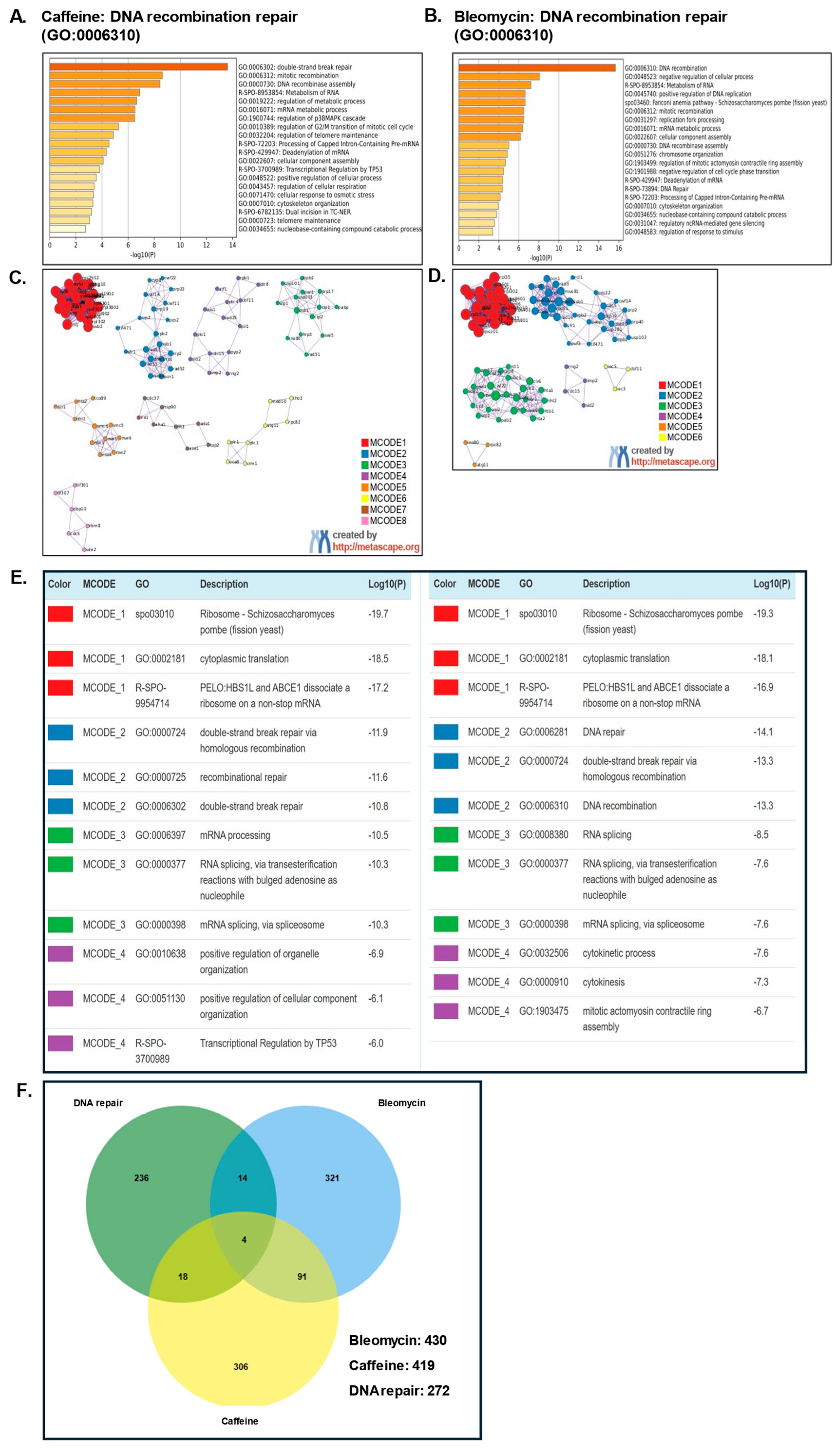
Table 2.
Selected studies on the effect of caffeine on DNA damage sensitivity and repair. Caffeine has been reported to enhance DNA damage sensitivity by targeting checkpoint signalling and DNA damage repair. The relatively high doses of caffeine employed are of questionable clinical relevance; these studies provide mechanistic insights. Similar effects may be achieved using synthetic caffeine derivatives with better clinical profiles (Exp.—experimental evidence).
Figure 5.
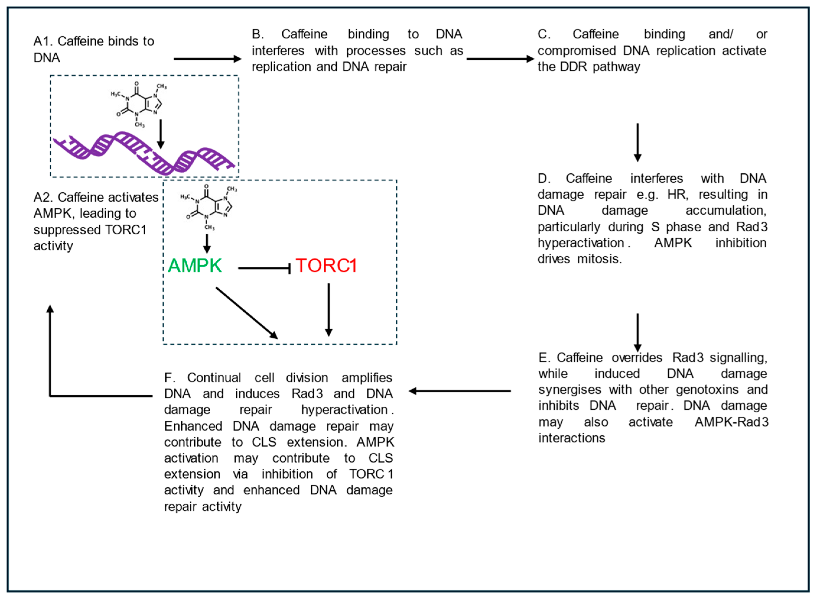
Mechanisms of caffeine-induced DNA damage, AMPK activation, DNA repair inhibition and CLS extension in fission yeast. The figure was prepared by the author using MS PowerPoint.
In fission yeast, the effect of caffeine on mitosis occurs independently of DNA damage checkpoints and correlates poorly with the effect on DNA damage sensitivity. Crucially, caffeine can enhance sensitivity to DNA damage in non-dividing cells. In combination, several studies suggest that caffeine induces DNA damage while simultaneously inhibiting its repair. Caffeine likely enhances sensitivity to other genotoxins via these mechanisms. Caffeine should therefore be regarded as a DNA-damaging agent. Activation of DNA damage repair pathways by caffeine may lead to enhanced genomic stability and CLS extension [7].
7. Future Directions
As already reported by Blasina et al. [132], the clinical relevance of caffeine is limited by the high concentrations of caffeine used in experimental studies. Furthermore, the use of diverse model systems complicates interpretation of the data. Solving this riddle will require novel, carefully designed experiments. Lower concentrations of caffeine may synergise with other genotoxic agents. This review aims to determine if caffeine can be considered a radiomimetic agent. For this assay, the simplest definition, i.e., compounds that mimic the effect of IR or UV on DNA integrity, has been used [1,116,135]. Traditionally, such agents have included some antibiotics such as bleomycin as well as antimetabolites and alkylating agents. The precise effect of caffeine is likely influenced by the nature of the synergising genotoxic agent and experimental conditions. Studies in fission yeast suggest that caffeine enhances DNA damage sensitivity by checkpoint override and radiomimetic-like effects. This was particularly evident when caffeine was combined with HU.
At present, the literature suggests that caffeine induces DNA damage and thus acts as a radiomimetic compound. Evidence for this assertion is provided by findings showing that the DDR pathway is required for resistance to caffeine [39]. Caffeine also enhances sensitivity to other genotoxins by various mechanisms. These include cell cycle checkpoint override via indirect TOR inhibition and the inhibition of DNA damage repair. Recently, caffeine was also shown to modulate DNA damage sensitivity by activating AMPK in fission yeast. Importantly, AMPK activation is also linked to caffeine-induced extension of CLS in fission yeast. These findings raise new questions on how caffeine-induced DNA damage is linked to AMPK activation, DNA repair and CLS.
Caffeine-induced CLS extension has drawn much attention. The ability of caffeine to indirectly inhibit TORC1 activity via AMPK activation mimics the effects of glucose and calorie restriction. Direct and indirect activators of AMPK, such as compound 991 (ex229), AICAR and metformin, can also influence cellular pathways linked to cell cycle regulation and DNA repair [136,137,138].
In humans, caffeine is metabolised into theobromine and theophylline soon after ingestion. As such, model systems such as yeast may lead to misleading results. It will be interesting to determine if other methylxanthines and caffeine derivatives mimic caffeine’s effect on DNA damage sensitivity and CLS extension. Further studies on the effect of caffeine on mitochondrial DNA integrity and function are also warranted [7]. Similarly, studies comparing the effects of caffeine with activators of AMPK will be important. For example, they can help to determine if AMPK activation can extend CLS independently of DNA damage. They can also help to determine the extent to which caffeine-induced DNA damage influences its effect on CLS. Clinically, it will be important to determine if direct AMPK activation is superior to its indirect activation. Understanding how caffeine-mediated AMPK inhibition differs from direct activation by compound 991 in terms of TORC1 inhibition and autophagy is also important [139,140,141,142].
Deciphering Cdc25 regulation has been central to understanding how caffeine overrides DNA damage checkpoint signalling in fission yeast [7,89]. Proper Cdc25 regulation is also important for survival following cell cycle exit [143]. Although extensively characterised, some important questions regarding Cdc25 regulation remain to be addressed. Caffeine stabilises Cdc25, and the phosphatase is required for DNA damage checkpoint override. As such, it has been proposed that caffeine may be a useful tool for studying Cdc25 activity [54]. The role of Dss1 in regulating Cdc25 stability requires further research in the context of caffeine [90]. The role of TORC2 in mediating caffeine’s effects in fission yeast also requires further investigation. In particular, the precise role of Gsk3 in regulating Cdc25 stability remains unclear (Figure 2) [144]. Addressing these questions will provide further insights into the cellular effects of caffeine.
8. Conclusions
The major aim of this review is to determine whether caffeine can be classed as a radiomimetic compound. Historically, radiomimetic compounds have been characterised as compounds that mimic the effect of radiation on DNA, namely their ability to damage DNA [1,117,144,145,146]. I propose that caffeine can indeed be classified as a radiomimetic-like compound, based on the following findings.
- i.
- Caffeine functions as a defence mechanism against plant pathogens, and other bioorganic compounds, such as bleomycin, are also radiomimetic.
- ii.
- Caffeine inhibits the repair of UV-induced DNA damage, a highly effective defence mechanism against microbial pathogens in environments where UV indices are high. Caffeine has also been shown to be mutagenic.
- iii.
- Caffeine binds to DNA and alters its physical characteristics. This may interfere with DNA replication and repair.
- iv.
- Caffeine inhibits HR-mediated DNA damage repair.
- v.
- Rad3 and HR enzymes are required for resistance to caffeine in fission yeast.
- vi.
- Caffeine synergises with other radiomimetic compounds to enhance their lethality even in non-dividing cells.
- vii.
- There is a strong overlap between genes that confer resistance to caffeine and bleomycin alone and in combination.
A simple model (Figure 5) proposes that caffeine induces cumulative low-level DNA damage that induces HR. Current evidence supports a model in which caffeine produces radiomimetic-like effects through combined replication stress, checkpoint modulation, and inhibition of DNA repair.
Hence, cells defective in Rad3-mediated checkpoint signalling or HR exhibit increased sensitivity to caffeine. Interestingly, enhanced DNA repair correlates with reduced cancer incidence and longevity in higher organisms [147]. It can therefore be theorised that exposure to caffeine upregulates DNA repair pathways. Such an effect could extend CLS by improving genomic stability [148]. Caffeine and its derivatives show continued promise as clinical agents, especially in the context of mTOR activity and ageing. The effect of caffeine on AMPK makes for exciting future research.
Supplementary Materials
The following supporting information can be downloaded at: https://www.mdpi.com/article/10.3390/app162110611/s1, Supplementary Table S1. Genes that mediate resistance to caffeine and bleomycin. Gene lists were obtained from PomBase (PomBase—The Schizosaccharomyces pombe genome database) using the ontology terms Resistance to caffeine (FYPO:0000073) and Resistance to bleomycin (FYPO:0009031) [126]. Supplementary Table S2. Genes that are involved in DNA damage repair and mediate resistance to bleomycin and/or caffeine. Data is represented in Figure 4F.
Funding
This research received no external funding.
Data Availability Statement
No new data were created or analyzed in this study. Data sharing is not applicable to this article.
Acknowledgments
During the preparation of this manuscript, the author used Metascape and Pombase for the purposes of identifying gene interactions [125,126]. The author has reviewed and edited the output and takes full responsibility for the content of this publication.
Conflicts of Interest
The author declares no conflicts of interest.
Abbreviations
The following abbreviations are used in this manuscript:
| AMPK | AMP-activated Protein Kinase |
| ATM | Ataxia Telangiectasia Mutated |
| ATR | Ataxia telangiectasia and Rad3-related |
| CaMKK | Calcium/calmodulin-dependent protein Kinase Kinase 2 |
| CLS | Chronological lifespan |
| DDR | DNA Damage Response |
| DNA-PKCS | DNA-dependent protein kinase catalytic subunit |
| ESR | Environmental Stress Response |
| FRB | FKBP-rapamycin binding domain |
| HECT | Homologous to E6AP C-Terminus |
| HR | Homologous recombination |
| HU | Hydroxyurea |
| KCl | Potassium chloride |
| NER | Nucleotide excision repair |
| PIKK | Phosphatidylinositol 3-kinase-related kinases |
| ROS | Reactive oxygen species |
| SAPK | Stress-Activated Protein Kinase |
| SIN | Septation Initiation Network |
| SMG1 | Suppressor of Morphogenesis in Genitalia |
| TOR | Target of Rapamycin Complex |
| TORC1 | Target of Rapamycin Complex 1 |
| TORC2 | Target of Rapamycin Complex 2 |
| TRRAP | Transformation/transcription domain-associated protein |
References
- Byfield, J.E.; Murnane, J.; Ward, J.F.; Calabro-Jones, P.; Lynch, M.; Kulhanian, F. Mice, men, mustard and methylated xanthines: The potential role of caffeine and related drugs in the sensitization of human tumours to alkylating agents. Br. J. Cancer 1981, 43, 669–683. [Google Scholar] [CrossRef] [Scilit] [PubMed]
- Emadi, R.C.; Kamangar, F. Coffee’s Impact on Health and Well-Being. Nutrients 2025, 17, 2558. [Google Scholar] [CrossRef] [Scilit] [PubMed]
- Barcelos, R.P.; Lima, F.D.; Carvalho, N.R.; Bresciani, G.; Royes, L.F. Caffeine effects on systemic metabolism, oxidative-inflammatory pathways, and exercise performance. Nutr. Res. 2020, 80, 1–17. [Google Scholar] [CrossRef] [Scilit] [PubMed]
- Mlakar, V.; Di Forti, M.; Halff, E.F.; Srivastava, D.P.; Akkouh, I.; Djurovic, S.; Martin-Ruiz, C.; Quintana, D.S.; Birkenæs, V.; Steen, N.E.; et al. Coffee intake is associated with telomere length in severe mental disorders. BMJ Ment. Health 2025, 28, e301700. [Google Scholar] [CrossRef] [Scilit] [PubMed]
- James, J.E. Are coffee’s alleged health protective effects real or artifact? The enduring disjunction between relevant experimental and observational evidence. J. Psychopharmacol. 2018, 32, 850–854. [Google Scholar] [CrossRef] [Scilit] [PubMed]
- Rallis, C.; Codlin, S.; Bähler, J. TORC 1 signaling inhibition by rapamycin and caffeine affect lifespan, global gene expression, and cell proliferation of fission yeast. Aging Cell 2013, 12, 563–573. [Google Scholar] [CrossRef] [Scilit] [PubMed]
- Alao, J.-P.; Kumar, J.; Stamataki, D.; Rallis, C. Dissecting the cell cycle regulation, DNA damage sensitivity and lifespan effects of caffeine in fission yeast. Microb. Cell 2025, 12, 141–156. [Google Scholar] [CrossRef] [Scilit] [PubMed]
- Wanke, V.; Cameroni, E.; Uotila, A.; Piccolis, M.; Urban, J.; Loewith, R.; De Virgilio, C. Caffeine extends yeast lifespan by targeting TORC1. Mol. Microbiol. 2008, 69, 277–285. [Google Scholar] [CrossRef] [Scilit] [PubMed]
- Abdel-Sattar, E.; Agarwal, S.; Ahmed, S.; Akaihata, H.; Alam, F.; Alavi, M.; Anand, J.; Anastácio, L.R.; Andrade, S.; Apetrei, C.; et al. List of contributors. In Tea in Health and Disease Prevention; Elsevier: Amsterdam, The Netherlands, 2025; pp. xxix–xxxvii. [Google Scholar] [CrossRef] [Scilit]
- Nathanson, J.A. Caffeine and Related Methylxanthines: Possible Naturally Occurring Pesticides. Science 1984, 226, 184–187. [Google Scholar] [CrossRef] [Scilit] [PubMed]
- Gutiérrez-Sánchez, G.; Roussos, S.; Augur, C. Effect of caffeine concentration on biomass production, caffeine degradation, and morphology of Aspergillus tamarii. Folia Microbiol. 2013, 58, 195–200. [Google Scholar] [CrossRef] [Scilit] [PubMed]
- Kim, Y.-S.; Choi, Y.-E.; Sano, H. Plant vaccination: Stimulation of defense system by caffeine production in planta. Plant Signal. Behav. 2010, 5, 489–493. [Google Scholar] [CrossRef] [Scilit] [PubMed]
- Kim, Y.-S.; Uefuji, H.; Ogita, S.; Sano, H. Transgenic tobacco plants producing caffeine: A potential new strategy for insect pest control. Transgenic Res. 2006, 15, 667–672. [Google Scholar] [CrossRef] [Scilit] [PubMed]
- Selby, C.P.; Sancar, A. Molecular mechanisms of DNA repair inhibition by caffeine. Proc. Natl. Acad. Sci. USA 1990, 87, 3522–3525. [Google Scholar] [CrossRef] [Scilit] [PubMed]
- Parker, V.P.; Lieberman, M.W. Levels of DNA polymerases α,β, and γ control and repair-deficient human diploid fibroblasts. Nucl. Acids Res. 1977, 4, 2029–2038. [Google Scholar] [CrossRef] [Scilit] [PubMed]
- Lehmann, A.R. Postreplication repair of DNA in mammalian cells. Life Sci. 1974, 15, 2005–2016. [Google Scholar] [CrossRef] [Scilit] [PubMed]
- Heffernan, T.P.; Kawasumi, M.; Blasina, A.; Anderes, K.; Conney, A.H.; Nghiem, P. ATR–Chk1 Pathway Inhibition Promotes Apoptosis after UV Treatment in Primary Human Keratinocytes: Potential Basis for the UV Protective Effects of Caffeine. J. Investig. Dermatol. 2009, 129, 1805–1815. [Google Scholar] [CrossRef] [Scilit] [PubMed]
- Kawasumi, M.; Lemos, B.; Bradner, J.E.; Thibodeau, R.; Kim, Y.; Schmidt, M.; Higgins, E.; Koo, S.; Angle-Zahn, A.; Chen, A.; et al. Protection from UV-induced skin carcinogenesis by genetic inhibition of the ataxia telangiectasia and Rad3-related (ATR) kinase. Proc. Natl. Acad. Sci. USA 2011, 108, 13716–13721. [Google Scholar] [CrossRef] [Scilit] [PubMed]
- Bode, A.M.; Dong, Z. The enigmatic effects of caffeine in cell cycle and cancer. Cancer Lett. 2007, 247, 26–39. [Google Scholar] [CrossRef] [Scilit] [PubMed]
- Mashayekhi-Sardoo, H.; Aftabi, K.; Azami, A.; Baradaran Rahimi, V.; Askari, V.R. Radioprotection or radiosensitization? Unraveling caffeine’s bifurcated effects on radiation-induced damage. Toxicol. Mech. Methods 2026, 36, 29–64. [Google Scholar] [CrossRef] [Scilit] [PubMed]
- Sabisz, M.; Skladanowski, A. Modulation of Cellular Response to Anticancer Treatment by Caffeine: Inhibition of Cell Cycle Checkpoints, DNA Repair and More. Curr. Pharm. Biotechnol. 2008, 9, 325–336. [Google Scholar] [CrossRef] [Scilit] [PubMed]
- Blackford, A.N.; Jackson, S.P. ATM, ATR, and DNA-PK: The Trinity at the Heart of the DNA Damage Response. Mol. Cell 2017, 66, 801–817. [Google Scholar] [CrossRef] [Scilit] [PubMed]
- Denoth Lippuner, A.; Julou, T.; Barral, Y. Budding yeast as a model organism to study the effects of age. FEMS Microbiol. Rev. 2014, 38, 300–325. [Google Scholar] [CrossRef] [Scilit] [PubMed]
- Hayles, J.; Nurse, P. Introduction to Fission Yeast as a Model System. Cold Spring Harb. Protoc. 2018, 2018, pdb.top079749. [Google Scholar] [CrossRef] [Scilit] [PubMed]
- Ruta, L.L.; Farcasanu, I.C. Saccharomyces cerevisiae and Caffeine Implications on the Eukaryotic Cell. Nutrients 2020, 12, 2440. [Google Scholar] [CrossRef] [Scilit] [PubMed]
- Steinberg, G.R.; Hardie, D.G. New insights into activation and function of the AMPK. Nat. Rev. Mol. Cell Biol. 2023, 24, 255–272. [Google Scholar] [CrossRef] [Scilit] [PubMed]
- Benkö, Z.; Sipiczki, M. Caffeine tolerance in Schizosaccharomyces pombe: Physiological adaptation and interaction with theophylline. Can. J. Microbiol. 1993, 39, 551–554. [Google Scholar] [CrossRef] [Scilit] [PubMed]
- Crescenzi, E.; Palumbo, G.; De Boer, J.; Brady, H.J.M. Ataxia Telangiectasia Mutated and p21CIP1 Modulate Cell Survival of Drug-Induced Senescent Tumor Cells: Implications for Chemotherapy. Clin. Cancer Res. 2008, 14, 1877–1887. [Google Scholar] [CrossRef] [Scilit] [PubMed]
- Haynes, R.H.; Collins, J.D.B. The Mutagenic Potential of Caffeine. In Caffeine; Dews, P.B., Ed.; Springer: Berlin/Heidelberg, Germany, 1984; pp. 221–238. [Google Scholar] [CrossRef] [Scilit]
- Domon, M.; Barton, B.; Porte, A.; Rauth, A.M. The Interaction of Caffeine with Ultra-violet-light-irradiated DNA. Int. J. Radiat. Biol. Relat. Stud. Phys. Chem. Med. 1970, 17, 395–399. [Google Scholar] [CrossRef] [Scilit] [PubMed]
- Fritzsche, H.; Lang, H.; Sprinz, H.; Pohle, W. On the interaction of caffeine with nucleic acids. Biophys. Chem. 1980, 11, 121–131. [Google Scholar] [CrossRef] [Scilit] [PubMed]
- Husain, I.; Griffith, J.; Sancar, A. Thymine dimers bend DNA. Proc. Natl. Acad. Sci. USA 1988, 85, 2558–2562. [Google Scholar] [CrossRef] [Scilit] [PubMed]
- Sideropoulos, A.S.; Shankel, D.M. Mechanism of caffeine enhancement of mutations induced by sublethal ultraviolet dosages. J. Bacteriol. 1968, 96, 198–204. [Google Scholar] [CrossRef] [Scilit] [PubMed]
- Wu, S.; He, Y.; Qiu, X.; Yang, W.; Liu, W.; Li, X.; Li, Y.; Shen, H.-M.; Wang, R.; Yue, Z.; et al. Targeting the potent Beclin 1–UVRAG coiled-coil interaction with designed peptides enhances autophagy and endolysosomal trafficking. Proc. Natl. Acad. Sci. USA 2018, 115, E5669–E5678. [Google Scholar] [CrossRef] [Scilit] [PubMed]
- Worthy, T.E.; Epler, J.L. Characterization of Excision Repair in Neurospora crassa. J. Bacteriol. 1973, 115, 498–505. [Google Scholar] [CrossRef] [Scilit] [PubMed]
- Palitti, F.; Becchetti, A. Effect of caffeine on sister chromatid exchanges and chromosomal aberrations induced by mutagens in Chinese hamster cells. Mutat. Res. 1977, 45, 157–159. [Google Scholar] [CrossRef] [Scilit] [PubMed]
- Callegari, A.J.; Kelly, T.J. UV irradiation induces a postreplication DNA damage checkpoint. Proc. Natl. Acad. Sci. USA 2006, 103, 15877–15882. [Google Scholar] [CrossRef] [Scilit] [PubMed]
- Murnane, J.P.; Byfield, J.E.; Ward, J.F.; Calabro-Jones, P. Effects of methylated xanthines on mammalian cells treated with bifunctional alkylating agents. Nature 1980, 285, 326–329. [Google Scholar] [CrossRef] [Scilit] [PubMed]
- Calvo, I.A.; Gabrielli, N.; Iglesias-Baena, I.; García-Santamarina, S.; Hoe, K.-L.; Kim, D.U.; Sansó, M.; Zuin, A.; Pérez, P.; Ayté, J.; et al. Genome-wide screen of genes required for caffeine tolerance in fission yeast. PLoS ONE 2009, 4, e6619. [Google Scholar] [CrossRef] [Scilit] [PubMed]
- Cavero, S.; Limbo, O.; Russell, P. Critical Functions of Rpa3/Ssb3 in S-Phase DNA Damage Responses in Fission Yeast. PLoS Genet. 2010, 6, e1001138. [Google Scholar] [CrossRef] [Scilit] [PubMed]
- Tsabar, M.; Eapen, V.V.; Mason, J.M.; Memisoglu, G.; Waterman, D.P.; Long, M.J.; Bishop, D.K.; Haber, J.E. Caffeine impairs resection during DNA break repair by reducing the levels of nucleases Sae2 and Dna2. Nucleic Acids Res. 2015, 43, 6889–6901. [Google Scholar] [CrossRef] [Scilit] [PubMed]
- Zelensky, A.N.; Sanchez, H.; Ristic, D.; Vidic, I.; van Rossum-Fikkert, S.E.; Essers, J.; Wyman, C.; Kanaar, R. Caffeine suppresses homologous recombination through interference with RAD51-mediated joint molecule formation. Nucleic Acids Res. 2013, 41, 6475–6489. [Google Scholar] [CrossRef] [Scilit] [PubMed]
- Gentner, N.E.; Werner, M.M.; Hannan, M.A.; Nasim, A. Contribution of a caffeine-sensitive recombinational repair pathway to survival and mutagenesis in UV-irradiated Schizosaccharomyces pombe. Molec. Gen. Genet. 1978, 167, 43–49. [Google Scholar] [CrossRef] [Scilit] [PubMed]
- Asaad, N.A.; Zeng, Z.-C.; Guan, J.; Thacker, J.; Iliakis, G. Homologous recombination as a potential target for caffeine radiosensitization in mammalian cells: Reduced caffeine radiosensitization in XRCC2 and XRCC3 mutants. Oncogene 2000, 19, 5788–5800. [Google Scholar] [CrossRef] [Scilit] [PubMed]
- Quiñones-González, C.A.; Arredondo-Mendoza, G.I.; Jiménez-Salas, Z.; Larriba-Calle, G.; Ruiz-Herrera, J.; Campos-Góngora, E. Genotoxic effect of caffeine in Yarrowia lipolytica cells deficient in DNA repair mechanisms. Arch. Microbiol. 2019, 201, 991–998. [Google Scholar] [CrossRef] [Scilit] [PubMed]
- Rowley, R.; Zhang, J. Caffeine-mediated override of checkpoint controls. A requirement for rhp6 (Schizosaccharomyces pombe). Genetics 1999, 152, 61–71. [Google Scholar] [CrossRef] [Scilit] [PubMed]
- Smirnova, I.S.; Suslov, A.V.; Noskin, L.A. Modification of DNA damage in rat hepatocytes following whole-body roentgen radiation using cysteamine and 8-bromocaffeine. Radiobiologiia 1983, 23, 653–656. [Google Scholar] [PubMed]
- Tornaletti, S.; Andersen, A.H.; Christiansen, K.; Pedrini, A.M. 8-Methoxycaffeine inhibition of Drosophila DNA topoisomerase II. Biochim. Biophys. Acta (BBA)-Gene Struct. Expr. 1992, 1131, 30–34. [Google Scholar] [CrossRef] [Scilit] [PubMed]
- Shin, C.; Strayer, J.M.; Wani, M.A.; Snapka, R.M. Rapid evaluation of topoisomerase inhibitors: Caffeine inhibition of topoisomerases in vivo. Teratog. Carcinog. Mutagen. 1990, 10, 41–52. [Google Scholar] [CrossRef] [Scilit] [PubMed]
- Kadi, A.A.; El-Tahir, K.E.H.; Jahng, Y.; Rahman, A.F.M.M. Synthesis, biological evaluation and Structure Activity Relationships (SARs) study of 8-(substituted)aryloxycaffeine. Arab. J. Chem. 2019, 12, 2356–2364. [Google Scholar] [CrossRef] [Scilit]
- Malik, M.; Nitiss, J.L. DNA Repair Functions That Control Sensitivity to Topoisomerase-Targeting Drugs. Eukaryot. Cell 2004, 3, 82–90. [Google Scholar] [CrossRef] [Scilit] [PubMed]
- Fornace, A.J.; Kohn, K.W.; Kann, H.E. DNA single-strand breaks during repair of UV damage in human fibroblasts and abnormalities of repair in xeroderma pigmentosum. Proc. Natl. Acad. Sci. USA 1976, 73, 39–43. [Google Scholar] [CrossRef] [Scilit] [PubMed]
- Moseley, J.B. Wee1 and Cdc25: Tools, pathways, mechanisms, questions. Cell Cycle 2017, 16, 599–600. [Google Scholar] [CrossRef] [Scilit] [PubMed]
- Alao, J.P.; Sunnerhagen, P. Caffeine as a tool for investigating the integration of Cdc25 phosphorylation, activity and ubiquitin-dependent degradation in Schizosaccharomyces pombe. Cell Div. 2020, 15, 10. [Google Scholar] [CrossRef] [Scilit] [PubMed]
- Fabre, F. Relation between repair mechanisms and induced mitotic recombination after UV irradiation, in the yeast Schizosaccharomyces pombe. Effects of caffeine. Mol. Gen. Genet. 1972, 117, 153–166. [Google Scholar] [CrossRef] [Scilit] [PubMed]
- Gentner, N.E.; Werner, M.M. Repair in Schizosaccharomyces pombe as measured by recovery from caffeine enhancement of radiation-induced lethality. Mol. Gen. Genet. 1975, 142, 171–183. [Google Scholar] [CrossRef] [Scilit] [PubMed]
- Loprieno, N.; Schüpbach, M. On the effect of caffeine on mutation and recombination in Schizosaccharomyces pombe. Mol. Gen. Genet. 1971, 110, 348–354. [Google Scholar] [CrossRef] [Scilit] [PubMed]
- Grigg, G.W. Caffeine-death in Escherichia coli. Molec. Gen. Genet. 1968, 102, 316–335. [Google Scholar] [CrossRef] [Scilit] [PubMed]
- Raleigh, J.M.; O’Connell, M.J. The G2 DNA damage checkpoint targets both Wee1 and Cdc25. J. Cell Sci. 2000, 113, 1727–1736. [Google Scholar] [CrossRef] [Scilit] [PubMed]
- Lucena, R.; Alcaide-Gavilán, M.; Anastasia, S.D.; Kellogg, D.R. Wee1 and Cdc25 are controlled by conserved PP2A-dependent mechanisms in fission yeast. Cell Cycle 2017, 16, 428–435. [Google Scholar] [CrossRef] [Scilit] [PubMed]
- Qiu, C.; Yi, Y.; Lucena, R.; Wu, M.; Sun, J.; Wang, X.; Jin, Q.; Wang, Y. F-box proteins Pof3 and Pof1 regulate Wee1 degradation and mitotic entry in fission yeast. J. Cell Sci. 2018, 131, jcs202895. [Google Scholar] [CrossRef] [Scilit] [PubMed]
- Margolis, S.S.; Perry, J.A.; Weitzel, D.H.; Freel, C.D.; Yoshida, M.; Haystead, T.A.; Kornbluth, S. A Role for PP1 in the Cdc2/Cyclin B–mediated Positive Feedback Activation of Cdc25. MBoC 2006, 17, 1779–1789. [Google Scholar] [CrossRef] [Scilit] [PubMed]
- Nefsky, B.; Beach, D. Pub1 acts as an E6-AP-like protein ubiquitiin ligase in the degradation of cdc25. EMBO J. 1996, 15, 1301–1312. [Google Scholar] [CrossRef] [Scilit]
- Esteban, V.; Sacristán, M.; Andrés, S.; Bueno, A. The Flp1/Clp1 phosphatase cooperates with HECT-type Pub1/2 protein-ubiquitin ligases in Schizosaccharomyces pombe. Cell Cycle 2008, 7, 1269–1276. [Google Scholar] [CrossRef] [Scilit] [PubMed]
- Alao, J.-P.; Legon, L.; Rallis, C. Crosstalk between the mTOR and DNA Damage Response Pathways in Fission Yeast. Cells 2021, 10, 305. [Google Scholar] [CrossRef] [Scilit] [PubMed]
- Masuda, F.; Ishii, M.; Mori, A.; Uehara, L.; Yanagida, M.; Takeda, K.; Saitoh, S. Glucose restriction induces transient G2 cell cycle arrest extending cellular chronological lifespan. Sci. Rep. 2016, 6, 19629. [Google Scholar] [CrossRef] [Scilit] [PubMed]
- Hanyu, Y.; Imai, K.K.; Kawasaki, Y.; Nakamura, T.; Nakaseko, Y.; Nagao, K.; Kokubu, A.; Ebe, M.; Fujisawa, A.; Hayashi, T.; et al. Schizosaccharomyces pombe cell division cycle under limited glucose requires Ssp1 kinase, the putative CaMKK, and Sds23, a PP2A-related phosphatase inhibitor. Genes Cells 2009, 14, 539–554. [Google Scholar] [CrossRef] [Scilit] [PubMed]
- Deng, L.; Lee, M.E.; Schutt, K.L.; Moseley, J.B. Phosphatases Generate Signal Specificity Downstream of Ssp1 Kinase in Fission Yeast. Mol. Cell. Biol. 2017, 37, e00494-16. [Google Scholar] [CrossRef] [Scilit] [PubMed]
- Schutt, K.L.; Moseley, J.B. Transient activation of fission yeast AMPK is required for cell proliferation during osmotic stress. Mol. Biol. Cell 2017, 28, 1804–1814. [Google Scholar] [CrossRef] [Scilit] [PubMed]
- Alao, J.P.; Sunnerhagen, P. Rad3 and Sty1 function in Schizosaccharomyces pombe: An integrated response to DNA damage and environmental stress? Mol. Microbiol. 2008, 68, 246–254. [Google Scholar] [CrossRef] [Scilit] [PubMed]
- López-Avilés, S.; Grande, M.; González, M.; Helgesen, A.-L.; Alemany, V.; Sanchez-Piris, M.; Bachs, O.; Millar, J.B.A.; Aligue, R. Inactivation of the Cdc25 phosphatase by the stress-activated Srk1 kinase in fission yeast. Mol. Cell 2005, 17, 49–59. [Google Scholar] [CrossRef] [Scilit] [PubMed]
- Alao, J.P.; Huis In ’T Veld, P.J.; Buhse, F.; Sunnerhagen, P. Hyperosmosis enhances radiation and hydroxyurea resistance of Schizosaccharomyces pombe checkpoint mutants through the spindle checkpoint and delayed cytokinesis. Mol. Microbiol. 2010, 77, 143–157. [Google Scholar] [CrossRef] [Scilit] [PubMed]
- Gómez-Hierro, A.; Lambea, E.; Giménez-Zaragoza, D.; López-Avilés, S.; Yance-Chávez, T.; Montserrat, M.; Pujol, M.J.; Bachs, O.; Aligue, R. Ssp1 CaMKK: A Sensor of Actin Polarization That Controls Mitotic Commitment through Srk1 in Schizosaccharomyces pombe. PLoS ONE 2015, 10, e0143037. [Google Scholar] [CrossRef] [Scilit] [PubMed][Green Version]
- Aono, S.; Haruna, Y.; Watanabe, Y.-H.; Mochida, S.; Takeda, K. The fission yeast Greatwall-Endosulfine pathway is required for proper quiescence/G0 phase entry and maintenance. Genes Cells 2019, 24, 172–186. [Google Scholar] [CrossRef] [Scilit] [PubMed]
- Atkin, J.; Halova, L.; Ferguson, J.; Hitchin, J.R.; Lichawska-Cieslar, A.; Jordan, A.M.; Pines, J.; Wellbrock, C.; Petersen, J. Torin1-mediated TOR kinase inhibition reduces Wee1 levels and advances mitotic commitment in fission yeast and HeLa cells. J. Cell Sci. 2014, 127, 1346–1356. [Google Scholar] [CrossRef] [Scilit] [PubMed]
- Cohen, A.; Lubenski, L.; Mouzon, A.; Kupiec, M.; Weisman, R. TORC2 is required for the accumulation of γH2A in response to DNA damage. J. Biol. Chem. 2024, 300, 107531. [Google Scholar] [CrossRef] [Scilit] [PubMed]
- Cohen, A.; Mouzon, A.; Sprecher, U.; Kupiec, M.; Weisman, R. TORC2 coordinates MBF-dependent transcription and restrains oxidative stress responses during DNA replication stress in fission yeast. J. Biol. Chem. 2026, 302, 111242. [Google Scholar] [CrossRef] [Scilit] [PubMed]
- Schonbrun, M.; Laor, D.; López-Maury, L.; Bähler, J.; Kupiec, M.; Weisman, R. TOR Complex 2 Controls Gene Silencing, Telomere Length Maintenance, and Survival under DNA-Damaging Conditions. Mol. Cell. Biol. 2009, 29, 4584–4594. [Google Scholar] [CrossRef] [Scilit] [PubMed]
- Wang, T.; Woodman, P.; Humphrey, S.J.; Petersen, J. Environmental control of Pub1 (NEDD4 family E3 ligase) in Schizosaccharomyces pombe is regulated by TORC2 and Gsk3. Life Sci. Alliance 2022, 5, e202101082. [Google Scholar] [CrossRef] [Scilit] [PubMed]
- Ikai, N.; Nakazawa, N.; Hayashi, T.; Yanagida, M. The reverse, but coordinated, roles of Tor2 (TORC1) and Tor1 (TORC2) kinases for growth, cell cycle and separase-mediated mitosis in Schizosaccharomyces pombe. Open Biol. 2011, 1, 110007. [Google Scholar] [CrossRef] [Scilit] [PubMed]
- Osman, F.; McCready, S. Differential effects of caffeine on DNA damage and replication cell cycle checkpoints in the fission yeast Schizosaccharomyces pombe. Mol. Gen. Genet. 1998, 260, 319–334. [Google Scholar] [CrossRef] [Scilit] [PubMed]
- Bøe, C.A.; Krohn, M.; Rødland, G.E.; Capiaghi, C.; Maillard, O.; Thoma, F.; Boye, E.; Grallert, B. Induction of a G1-S checkpoint in fission yeast. Proc. Natl. Acad. Sci. USA 2012, 109, 9911–9916. [Google Scholar] [CrossRef] [Scilit] [PubMed]
- Tanaka, K.; Russell, P. Cds1 phosphorylation by Rad3-Rad26 kinase is mediated by forkhead-associated domain interaction with Mrc1. J. Biol. Chem. 2004, 279, 32079–32086. [Google Scholar] [CrossRef] [Scilit] [PubMed]
- Brondello, J.M.; Boddy, M.N.; Furnari, B.; Russell, P. Basis for the checkpoint signal specificity that regulates Chk1 and Cds1 protein kinases. Mol. Cell Biol. 1999, 19, 4262–4269. [Google Scholar] [CrossRef] [Scilit] [PubMed]
- Biswas, P.; Kar, P.; Ghosh, S. Nitrosative stress induces a novel intra-S checkpoint pathway in Schizosaccharomyces pombe involving phosphorylation of Cdc2 by Wee1. Free Radic. Biol. Med. 2015, 86, 145–155. [Google Scholar] [CrossRef] [Scilit] [PubMed]
- Boddy, M.N.; Furnari, B.; Mondesert, O.; Russell, P. Replication Checkpoint Enforced by Kinases Cds1 and Chk1. Science 1998, 280, 909–912. [Google Scholar] [CrossRef] [Scilit] [PubMed]
- Christensen, P.U.; Bentley, N.J.; Martinho, R.G.; Nielsen, O.; Carr, A.M. Mik1 levels accumulate in S phase and may mediate an intrinsic link between S phase and mitosis. Proc. Natl. Acad. Sci. USA 2000, 97, 2579–2584. [Google Scholar] [CrossRef] [Scilit] [PubMed]
- Frazer, C.; Young, P.G. Carboxy-terminal phosphorylation sites in Cdc25 contribute to enforcement of the DNA damage and replication checkpoints in fission yeast. Curr. Genet. 2012, 58, 217–234. [Google Scholar] [CrossRef] [Scilit] [PubMed]
- Alao, J.P.; Sjölander, J.J.; Baar, J.; Özbaki-Yagan, N.; Kakoschky, B.; Sunnerhagen, P. Caffeine stabilizes Cdc25 independently of Rad3 in Schizosaccharomyces pombe contributing to checkpoint override. Mol. Microbiol. 2014, 92, 777–796. [Google Scholar] [CrossRef] [Scilit] [PubMed]
- Selvanathan, S.P.; Thakurta, A.G.; Dhakshnamoorthy, J.; Zhou, M.; Veenstra, T.D.; Dhar, R. Schizosaccharomyces pombe Dss1p Is a DNA Damage Checkpoint Protein That Recruits Rad24p, Cdc25p, and Rae1p to DNA Double-strand Breaks. J. Biol. Chem. 2010, 285, 14122–14133. [Google Scholar] [CrossRef] [Scilit] [PubMed]
- Paraskevopoulos, K.; Kriegenburg, F.; Tatham, M.H.; Rösner, H.I.; Medina, B.; Larsen, I.B.; Brandstrup, R.; Hardwick, K.G.; Hay, R.T.; Kragelund, B.B.; et al. Dss1 Is a 26S Proteasome Ubiquitin Receptor. Mol. Cell 2014, 56, 453–461. [Google Scholar] [CrossRef] [Scilit] [PubMed]
- Marston, N.J.; Richards, W.J.; Hughes, D.; Bertwistle, D.; Marshall, C.J.; Ashworth, A. Interaction between the product of the breast cancer susceptibility gene BRCA2 and DSS1, a protein functionally conserved from yeast to mammals. Mol. Cell Biol. 1999, 19, 4633–4642. [Google Scholar] [CrossRef] [Scilit] [PubMed]
- Wei, S.-J.; Williams, J.G.; Dang, H.; Darden, T.A.; Betz, B.L.; Humble, M.M.; Chang, F.-M.; Trempus, C.S.; Johnson, K.; Cannon, R.E.; et al. Identification of a Specific Motif of the DSS1 Protein Required for Proteasome Interaction and p53 Protein Degradation. J. Mol. Biol. 2008, 383, 693–712. [Google Scholar] [CrossRef] [Scilit] [PubMed]
- Ahn, J.; Won, M.; Choi, J.-H.; Kyun, M.-L.; Cho, H.-S.; Park, H.-M.; Kang, C.-M.; Chung, K.-S. Small heat-shock protein Hsp9 has dual functions in stress adaptation and stress-induced G2-M checkpoint regulation via Cdc25 inactivation in Schizosaccharomyces pombe. Biochem. Biophys. Res. Commun. 2012, 417, 613–618. [Google Scholar] [CrossRef] [Scilit] [PubMed]
- Moser, B.A.; Brondello, J.M.; Baber-Furnari, B.; Russell, P. Mechanism of caffeine-induced checkpoint override in fission yeast. Mol. Cell Biol. 2000, 20, 4288–4294. [Google Scholar] [CrossRef] [Scilit] [PubMed][Green Version]
- Sarkaria, J.N.; Busby, E.C.; Tibbetts, R.S.; Roos, P.; Taya, Y.; Karnitz, L.M.; Abraham, R.T. Inhibition of ATM and ATR kinase activities by the radiosensitizing agent, caffeine. Cancer Res. 1999, 59, 4375–4382. [Google Scholar] [PubMed]
- Marechal, A.; Zou, L. DNA Damage Sensing by the ATM and ATR Kinases. Cold Spring Harb. Perspect. Biol. 2013, 5, a012716. [Google Scholar] [CrossRef] [Scilit] [PubMed]
- Cortez, D. Caffeine Inhibits Checkpoint Responses without Inhibiting the Ataxia-Telangiectasia-mutated (ATM) and ATM- and Rad3-related (ATR) Protein Kinases. J. Biol. Chem. 2003, 278, 37139–37145. [Google Scholar] [CrossRef] [Scilit] [PubMed]
- Alao, J.P.; Johansson-Sjölander, J.; Rallis, C.; Sunnerhagen, P. Caffeine Stabilises Fission Yeast Wee1 in a Rad24-Dependent Manner but Attenuates Its Expression in Response to DNA Damage. Microorganisms 2020, 8, 1512. [Google Scholar] [CrossRef] [Scilit] [PubMed]
- Davie, E.; Forte, G.M.A.; Petersen, J. Nitrogen Regulates AMPK to Control TORC1 Signaling. Curr. Biol. 2015, 25, 445–454. [Google Scholar] [CrossRef] [Scilit] [PubMed]
- Valbuena, N.; Moreno, S. AMPK phosphorylation by Ssp1 is required for proper sexual differentiation in fission yeast. J. Cell Sci. 2012, 125, jcs.098533. [Google Scholar] [CrossRef] [Scilit] [PubMed]
- Shimura, T.; Sunaga, K.; Yamazaki, M.; Honoka, N.; Sasatani, M.; Kamiya, K.; Ushiyama, A. Nuclear DNA damage-triggered ATM-dependent AMPK activation regulates the mitochondrial radiation response. Int. J. Radiat. Biol. 2024, 100, 584–594. [Google Scholar] [CrossRef] [Scilit] [PubMed]
- Sanli, T.; Steinberg, G.R.; Singh, G.; Tsakiridis, T. AMP-activated protein kinase (AMPK) beyond metabolism: A novel genomic stress sensor participating in the DNA damage response pathway. Cancer Biol. Ther. 2014, 15, 156–169. [Google Scholar] [CrossRef] [Scilit] [PubMed]
- Lu, M.; Dong, X.; Wu, C.; Wang, G.; Wang, H.; Pan, Y.; Qin, Y.; Song, Y.; Pan, H.; Liu, S.; et al. AMPK phosphorylates WIP1 to promote DNA repair and radioresistance in cancer cells. Cell Death Dis. 2025, 16, 864. [Google Scholar] [CrossRef] [Scilit] [PubMed]
- Schonbrun, M.; Kolesnikov, M.; Kupiec, M.; Weisman, R. TORC2 is required to maintain genome stability during S phase in fission yeast. J. Biol. Chem. 2013, 288, 19649–19660. [Google Scholar] [CrossRef] [Scilit] [PubMed]
- Ikeda, K.; Morigasaki, S.; Tatebe, H.; Tamanoi, F.; Shiozaki, K. Fission yeast TOR complex 2 activates the AGC-family Gad8 kinase essential for stress resistance and cell cycle control. Cell Cycle 2008, 7, 358–364. [Google Scholar] [CrossRef] [Scilit] [PubMed]
- Hecht, S.M. Bleomycin: New Perspectives on the Mechanism of Action. J. Nat. Prod. 2000, 63, 158–168. [Google Scholar] [CrossRef] [Scilit] [PubMed]
- Sage, E.; Harrison, L. Clustered DNA lesion repair in eukaryotes: Relevance to mutagenesis and cell survival. Mutat. Res.-Fundam. Mol. Mech. Mutagen. 2011, 711, 123–133. [Google Scholar] [CrossRef] [Scilit] [PubMed]
- Han, W.; Ming, M.; He, Y.-Y. Caffeine Promotes Ultraviolet B-induced Apoptosis in Human Keratinocytes without Complete DNA Repair. J. Biol. Chem. 2011, 286, 22825–22832. [Google Scholar] [CrossRef] [Scilit] [PubMed]
- Traganos, F.; Kapuscinski, J.; Gong, J.; Ardelt, B.; Darzynkiewicz, R.J.; Darzynkiewicz, Z. Caffeine prevents apoptosis and cell cycle effects induced by camptothecin or topotecan in HL-60 cells. Cancer Res. 1993, 53, 4613–4618. [Google Scholar] [PubMed]
- Fletcher, J.; Griffiths, L.; Caspari, T. Nutrient Limitation Inactivates Mrc1-to-Cds1 Checkpoint Signalling in Schizosaccharomyces pombe. Cells 2018, 7, 15. [Google Scholar] [CrossRef] [Scilit] [PubMed]
- Gangloff, S.; Arcangioli, B. DNA repair and mutations during quiescence in yeast. FEMS Yeast Res. 2017, 17, fox002. [Google Scholar] [CrossRef] [Scilit] [PubMed]
- Matsuzawa, T.; Fujita, Y.; Tohda, H.; Takegawa, K. Snf1-Like Protein Kinase Ssp2 Regulates Glucose Derepression in Schizosaccharomyces pombe. Eukaryot. Cell 2012, 11, 159–167. [Google Scholar] [CrossRef] [Scilit] [PubMed]
- Afinanisa, Q.; Cho, M.K.; Seong, H.-A. AMPK Localization: A Key to Differential Energy Regulation. Int. J. Mol. Sci. 2021, 22, 10921. [Google Scholar] [CrossRef] [Scilit] [PubMed]
- Quiroga-Artigas, G.; Fontanié, P.; Lacroix, B.; Molina, M.D.; Moriel-Carretero, M. Compromised DNA replication in gut cells underlies sensitivity to genotoxic stress in the tardigrade Hypsibius exemplaris. FEBS J. 2026, febs.70542. [Google Scholar] [CrossRef] [Scilit] [PubMed]
- Deli, G. Mechanism of Action and Use of Radiomimetic Compounds. Hadmérnök 2022, 17, 101–115. [Google Scholar] [CrossRef] [Scilit]
- Lu, Z.; Li, J.; Liu, Y.; Li, H.; Sun, Y.; Geng, R.; Song, J.; Liu, J. Purine metabolism in tumorigenesis and its clinical implications. Semin. Oncol. 2025, 52, 152409. [Google Scholar] [CrossRef] [Scilit] [PubMed]
- Tran, D.H.; Kim, D.; Kesavan, R.; Brown, H.; Dey, T.; Soflaee, M.H.; Vu, H.S.; Tasdogan, A.; Guo, J.; Bezwada, D.; et al. De novo and salvage purine synthesis pathways across tissues and tumors. Cell 2024, 187, 3602–3618.e20. [Google Scholar] [CrossRef] [Scilit] [PubMed]
- De Vitto, H.; Arachchige, D.B.; Richardson, B.C.; French, J.B. The Intersection of Purine and Mitochondrial Metabolism in Cancer. Cells 2021, 10, 2603. [Google Scholar] [CrossRef] [Scilit] [PubMed]
- Pinz, M.P.; Medeiros, I.; Carvalho, L.A.D.C.; Meotti, F.C. Is uric acid a true antioxidant? Identification of uric acid oxidation products and their biological effects. Redox Rep. 2025, 30, 2498105. [Google Scholar] [CrossRef] [Scilit] [PubMed]
- Vieira, A.J.S.C.; Gaspar, E.M.; Santos, P.M.P. Mechanisms of potential antioxidant activity of caffeine. Radiat. Phys. Chem. 2020, 174, 108968. [Google Scholar] [CrossRef] [Scilit]
- Forte, G.M.; Davie, E.; Lie, S.; Franz-Wachtel, M.; Ovens, A.J.; Wang, T.; Oakhill, J.S.; Maček, B.; Hagan, I.M.; Petersen, J. Import of extracellular ATP in yeast and man modulates AMPK and TORC1 signalling. J. Cell Sci. 2019, 132, jcs223925. [Google Scholar] [CrossRef] [Scilit] [PubMed]
- Kanlaya, R.; Subkod, C.; Nanthawuttiphan, S.; Thongboonkerd, V. Caffeine causes cell cycle arrest at G0/G1 and increases of ubiquitinated proteins, ATP and mitochondrial membrane potential in renal cells. Comput. Struct. Biotechnol. J. 2023, 21, 4552–4566. [Google Scholar] [CrossRef] [Scilit] [PubMed]
- Ostertag, W.; Duisberg, E.; Stürmann, M. The mutagenic activity of caffeine in man. Mutat. Res. 1965, 2, 293–296. [Google Scholar] [CrossRef] [Scilit] [PubMed]
- Alao, J.P.; Sunnerhagen, P. The ATM and ATR inhibitors CGK733 and caffeine suppress cyclin D1 levels and inhibit cell proliferation. Radiat. Oncol. 2009, 4, 51. [Google Scholar] [CrossRef] [Scilit] [PubMed]
- Woziwodzka, A.; Krychowiak-Maśnicka, M.; Gołuński, G.; Łosiewska, A.; Borowik, A.; Wyrzykowski, D.; Piosik, J. New Life of an Old Drug: Caffeine as a Modulator of Antibacterial Activity of Commonly Used Antibiotics. Pharmaceuticals 2022, 15, 872. [Google Scholar] [CrossRef] [Scilit] [PubMed]
- Castro-Díaz, R.; Silva-Beltrán, N.P.; Gámez-Meza, N.; Calderón, K. The Antimicrobial Effects of Coffee and By-Products and Their Potential Applications in Healthcare and Agricultural Sectors: A State-of-Art Review. Microorganisms 2025, 13, 215. [Google Scholar] [CrossRef] [Scilit] [PubMed]
- Tornaletti, S.; Russo, P.; Parodi, S.; Pedrini, A.M. Studies on DNA binding of caffeine and derivatives: Evidence of intercalation by DNA-unwinding experiments. Biochim. Biophys. Acta (BBA)-Gene Struct. Expr. 1989, 1007, 112–115. [Google Scholar] [CrossRef] [Scilit] [PubMed]
- Zhou, Y.; Zhou, B.; Pache, L.; Chang, M.; Khodabakhshi, A.H.; Tanaseichuk, O.; Benner, C.; Chanda, S.K. Metascape provides a biologist-oriented resource for the analysis of systems-level datasets. Nat. Commun. 2019, 10, 1523. [Google Scholar] [CrossRef] [Scilit] [PubMed]
- Carme, P.; Rutherford, K.; Bähler, J.; Mata, J.; Wood, V. PomBase in 2026: Expanding Knowledge, Modelling Connections. Genetics 2026, 232, iyag001. [Google Scholar] [CrossRef] [Scilit] [PubMed]
- Lau, C.C.; Pardee, A.B. Mechanism by which caffeine potentiates lethality of nitrogen mustard. Proc. Natl. Acad. Sci. USA 1982, 79, 2942–2946. [Google Scholar] [CrossRef] [Scilit] [PubMed]
- Blasina, A.; Price, B.D.; Turenne, G.A.; McGowan, C.H. Caffeine inhibits the checkpoint kinase ATM. Curr. Biol. 1999, 9, 1135–1138. [Google Scholar] [CrossRef] [Scilit] [PubMed]
- Kaufmann, W.K.; Heffernan, T.P.; Beaulieu, L.M.; Doherty, S.; Frank, A.R.; Zhou, Y.; Bryant, M.F.; Zhou, T.; Luche, D.D.; Nikolaishvili-Feinberg, N.; et al. Caffeine and human DNA metabolism: The magic and the mystery. Mutat. Res.-Fundam. Mol. Mech. Mutagen. 2003, 532, 85–102. [Google Scholar] [CrossRef] [Scilit] [PubMed]
- Lu, Y.-P.; Lou, Y.-R.; Peng, Q.-Y.; Xie, J.-G.; Nghiem, P.; Conney, A.H. Effect of Caffeine on the ATR/Chk1 Pathway in the Epidermis of UVB-Irradiated Mice. Cancer Res. 2008, 68, 2523–2529. [Google Scholar] [CrossRef] [Scilit] [PubMed]
- Sax, K.; Sax, H.J. Radiomimetic beverages, drugs, and mutagens. Proc. Natl. Acad. Sci. USA 1966, 55, 1431–1435. [Google Scholar] [CrossRef] [Scilit] [PubMed]
- Szewczuk, M.; Boguszewska, K.; Kaźmierczak-Barańska, J.; Karwowski, B.T. The role of AMPK in metabolism and its influence on DNA damage repair. Mol. Biol. Rep. 2020, 47, 9075–9086. [Google Scholar] [CrossRef] [Scilit] [PubMed]
- Guigas, B.; Viollet, B. Targeting AMPK: From Ancient Drugs to New Small-Molecule Activators. In AMP-activated Protein Kinase; Cordero, M.D., Viollet, B., Eds.; Experientia Supplementum; Springer International Publishing: Cham, Switzerland, 2016; Volume 107, pp. 327–350. [Google Scholar] [CrossRef] [Scilit] [PubMed]
- Lai, Y.-C.; Kviklyte, S.; Vertommen, D.; Lantier, L.; Foretz, M.; Viollet, B.; Hallén, S.; Rider, M.H. A small-molecule benzimidazole derivative that potently activates AMPK to increase glucose transport in skeletal muscle: Comparison with effects of contraction and other AMPK activators. Biochem. J. 2014, 460, 363–375. [Google Scholar] [CrossRef] [Scilit] [PubMed]
- Ladli, M.; Richard, C.; Aguilar, L.C.; Ducamp, S.; Bondu, S.; Sujobert, P.; Tamburini, J.; Lacombe, C.; Azar, N.; Foretz, M.; et al. Finely-tuned regulation of AMP-activated protein kinase is crucial for human adult erythropoiesis. Haematologica 2019, 104, 907–918. [Google Scholar] [CrossRef] [Scilit] [PubMed]
- Johanns, M.; Pyr Dit Ruys, S.; Houddane, A.; Vertommen, D.; Herinckx, G.; Hue, L.; Proud, C.G.; Rider, M.H. Direct and indirect activation of eukaryotic elongation factor 2 kinase by AMP-activated protein kinase. Cell. Signal. 2017, 36, 212–221. [Google Scholar] [CrossRef] [Scilit] [PubMed]
- Kazyken, D.; Dame, S.G.; Wang, C.; Wadley, M.; Fingar, D.C. Unexpected roles for AMPK in the suppression of autophagy and the reactivation of MTORC1 signaling during prolonged amino acid deprivation. Autophagy 2024, 20, 2017–2040. [Google Scholar] [CrossRef] [Scilit] [PubMed]
- Yung, M.M.H.; Ngan, H.Y.S.; Chan, D.W. Targeting AMPK signaling in combating ovarian cancers: Opportunities and challenges. Acta Biochim. Biophys. Sin. 2016, 48, 301–317. [Google Scholar] [CrossRef] [Scilit] [PubMed]
- Sajiki, K.; Hatanaka, M.; Nakamura, T.; Takeda, K.; Shimanuki, M.; Yoshida, T.; Hanyu, Y.; Hayashi, T.; Nakaseko, Y.; Yanagida, M. Genetic control of cellular quiescence in S. pombe. J. Cell Sci. 2009, 122, 1418–1429. [Google Scholar] [CrossRef] [Scilit] [PubMed]
- Jain, C.K.; Majumder, H.K.; Roychoudhury, S. Natural Compounds as Anticancer Agents Targeting DNA Topoisomerases. Curr. Genom. 2017, 18, 75–92. [Google Scholar] [CrossRef] [Scilit] [PubMed]
- Zhang, Z.; Chen, S.; Mei, H.; Xuan, J.; Guo, X.; Couch, L.; Dobrovolsky, V.N.; Guo, L.; Mei, N. Ginkgo biloba leaf extract induces DNA damage by inhibiting topoisomerase II activity in human hepatic cells. Sci. Rep. 2015, 5, 14633. [Google Scholar] [CrossRef] [Scilit] [PubMed]
- Choi, S.; Shin, M.; Kim, W.-Y. Targeting the DNA damage response (DDR) of cancer cells with natural compounds derived from Panax ginseng and other plants. J. Ginseng Res. 2025, 49, 1388–1391. [Google Scholar] [CrossRef] [Scilit] [PubMed]
- Paniagua, I.; Joyce, J.A. Insights into a long life without cancer: The case of the bowhead whale. Mol. Oncol. 2026, 20, 1388–1391. [Google Scholar] [CrossRef] [Scilit] [PubMed]
- Li, L.; Guan, Y.; Chen, X.; Yang, J.; Cheng, Y. DNA Repair Pathways in Cancer Therapy and Resistance. Front. Pharmacol. 2021, 11, 629266. [Google Scholar] [CrossRef] [Scilit] [PubMed]
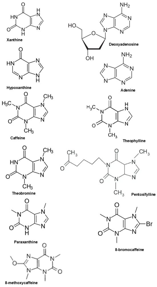
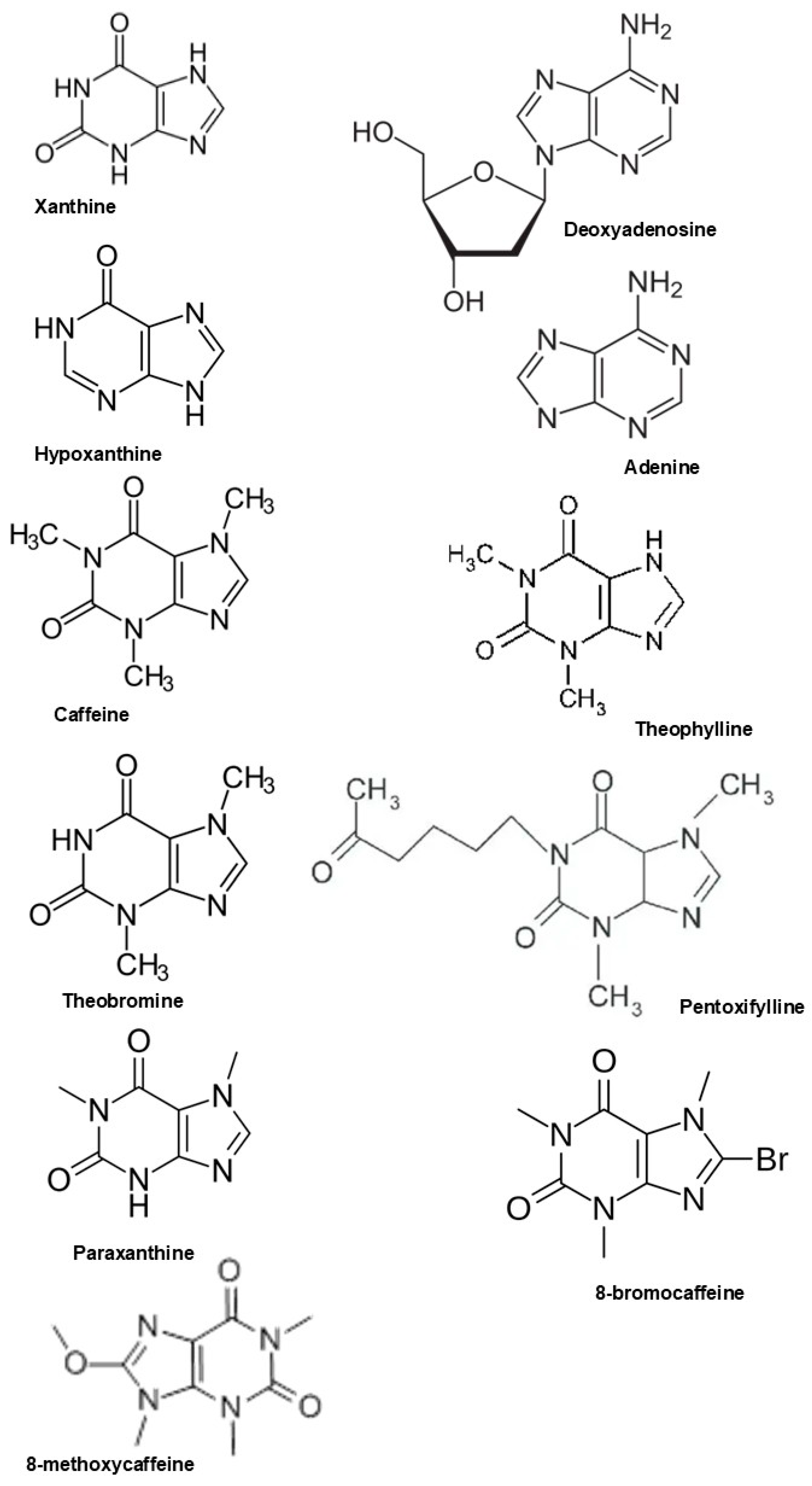
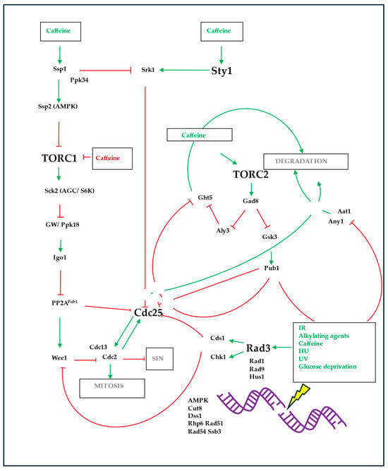
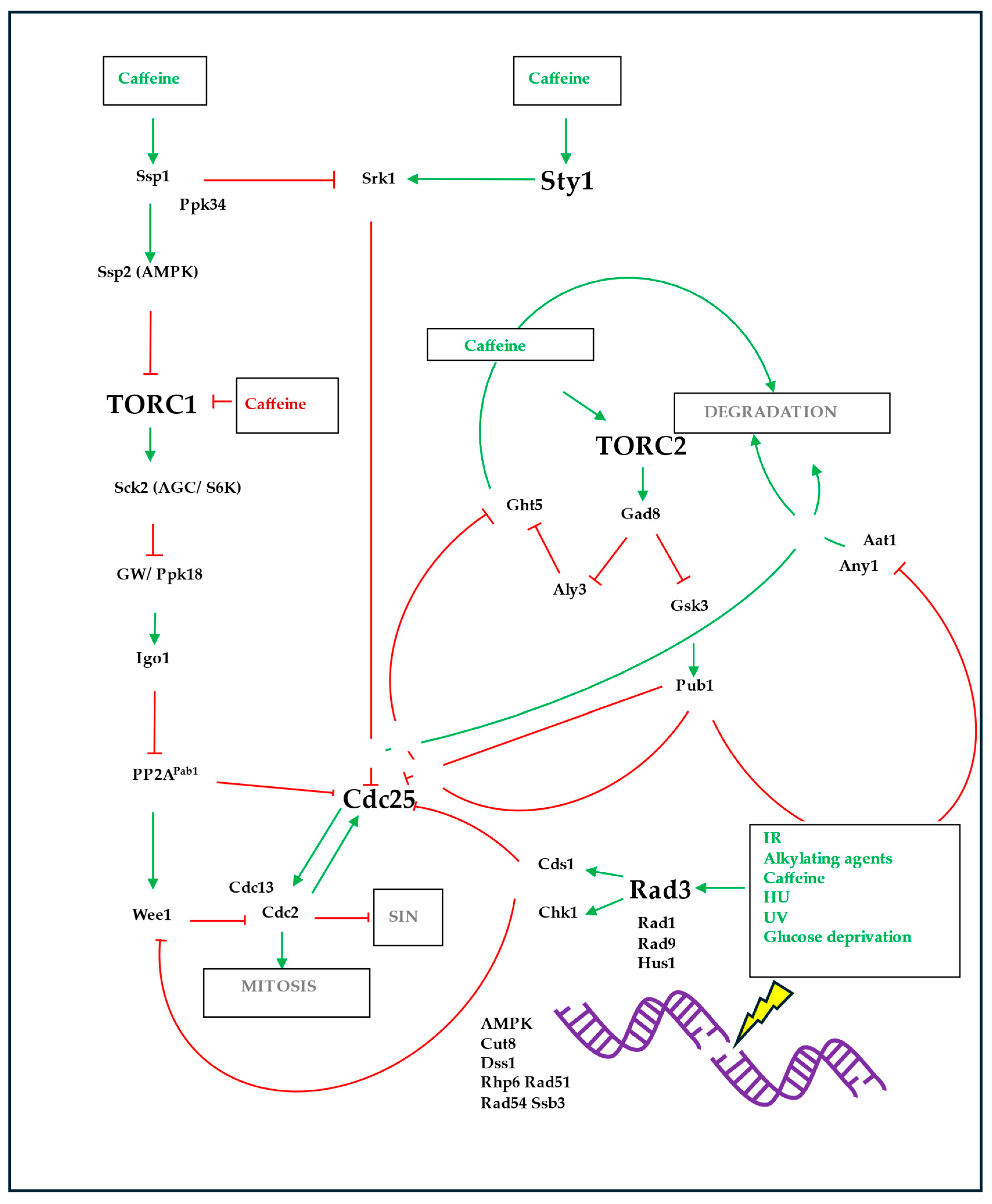
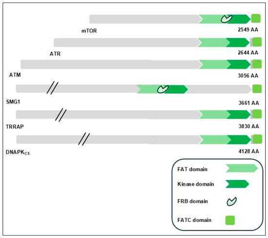
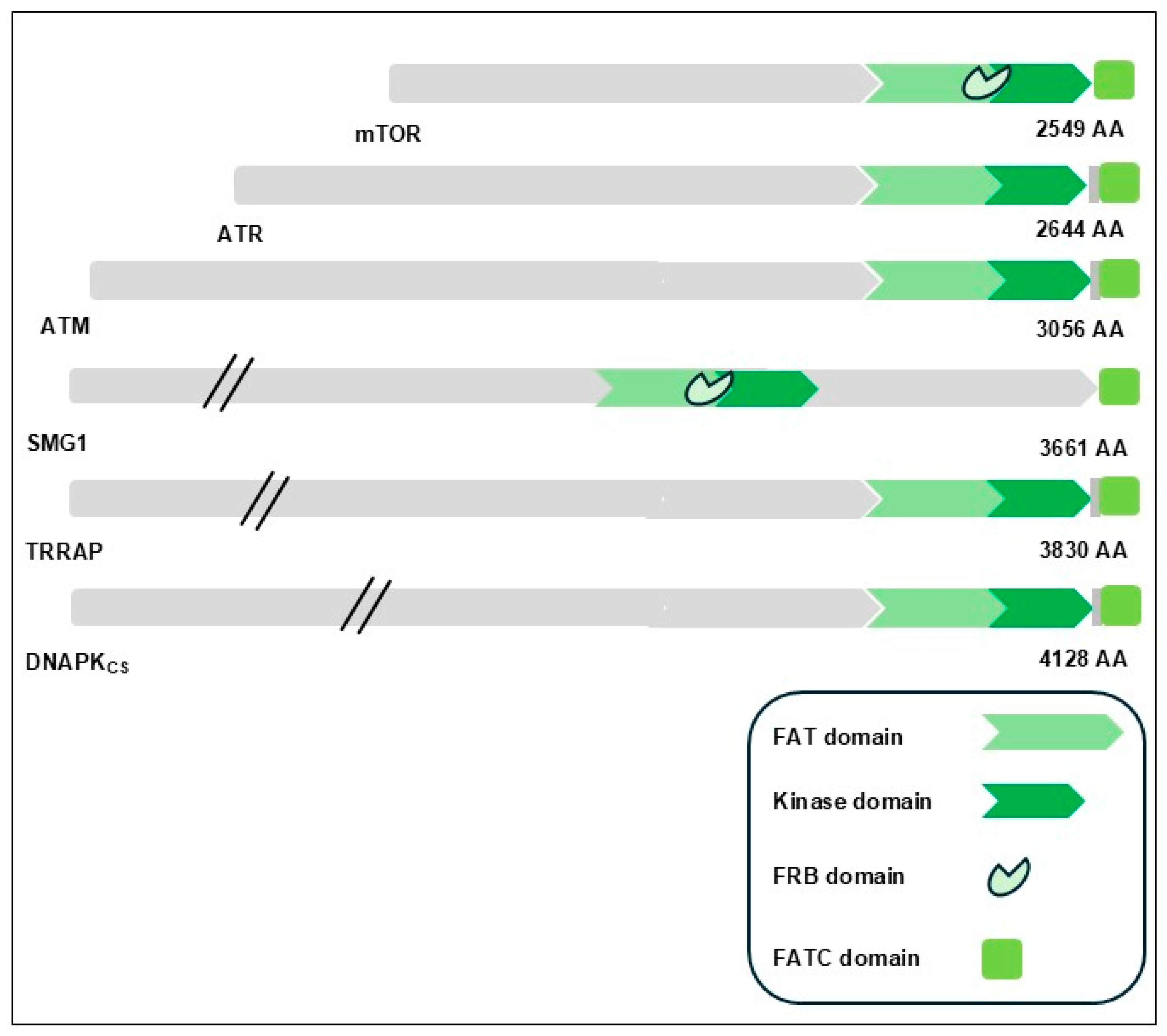

Disclaimer/Publisher’s Note: The statements, opinions and data contained in all publications are solely those of the individual author(s) and contributor(s) and not of MDPI and/or the editor(s). MDPI and/or the editor(s) disclaim responsibility for any injury to people or property resulting from any ideas, methods, instructions or products referred to in the content. |
© 2026 by the author. Licensee MDPI, Basel, Switzerland. This article is an open access article distributed under the terms and conditions of the Creative Commons Attribution (CC BY) license.